User login
VIDEO: Bionic exoskeleton helps paralyzed patients walk
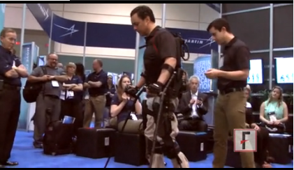
ORLANDO – It’s a bionic suit, a battery-powered exoskeleton, or as Chris Tagatac calls it, a wearable robot.
Mr. Tagatac, who is paralyzed from the lower ribs down, demonstrated the computer- and battery-operated Ekso Bionics suit at the annual meeting of the Healthcare Information and Management Systems Society, and he spoke with us about his experience. Watch the video to learn more about him and the technology.
The video associated with this article is no longer available on this site. Please view all of our videos on the MDedge YouTube channel
On Twitter @aliciaault
On Twitter @naseemsmiller
AT HIMSS14
Vision test has additive effect on concussion detection in athletes
Concussion in athletes was detected with greater accuracy when a simple vision test was added to other established tests in a prospective study of 217 members of the men’s football, women’s soccer, and women’s lacrosse teams at the University of Florida.
All concussive cases were correctly identified when the King-Devick (K-D) test was used in conjunction with the Balance Error Scoring System (BESS), which is part of the Sports Concussion Assessment Tool 3 (SCAT3), Dr. Laura Balcer and her associates reported Feb. 26 in an abstract released in advance of the annual meeting of the American Academy of Neurology in Philadelphia.
Furthermore, differences between baseline assessments and postinjury assessment showed that the K-D test identified a higher percentage of athletes with worsened scores than did the Standardized Assessment of Concussion (SAC) test (79% vs. 52%).
Using the K-D and SAC tests together also was better than using either of the tests alone, with 89% of concussions identified.
In a statement issued by the AAN, Dr. Balcer of the departments of population health and neurology at New York University noted that "the visual pathways are commonly affected in concussion. [And] adding a vision based test to evaluate athletes on the sidelines may allow us to better detect more athletes with concussion more quickly. This is particularly important since not all athletes reliably report their symptoms of concussion."
The findings add to previous work by Dr. Balcer and her colleagues in a similarly sized cohort of athletes from the University of Pennsylvania varsity football, sprint football, and women’s and men’s soccer and basketball teams (J. Neurol. Sci. 2011;309:34-9), and lend further support for the use of the K-D test as a rapid screening tool to assess players’ concussion during sporting events.
Up to 3.8 million sports-related concussive injuries have been estimated to occur in the United States each year, with diagnosis based on a variety of symptoms and signs that may include headache, unsteadiness, confusion, or behavior that is out of character for the individual concerned. It is important to be able to assess head injuries quickly, as concussion may be a result of a more serious neurological injury that needs emergency hospital treatment.
Unlike other tests for concussion that ideally need to be administered by a medical professional, anyone can administer the K-D test. This, together with the fact it takes less than 1 minute to complete, makes it ideal to use on the sidelines as an objective means of whether a concussive injury warrants more urgent attention.
The K-D test was developed in 1976 and assesses saccade, or the quick, simultaneous movement of both eyes in the same direction, as well as subjects’ language and level of concentration. It involves subjects’ quickly reading aloud a series of single-digit numbers shown to them on three test cards of increasing complexity. Subjects read the test cards from left to right and a stopwatch is used to record the time it takes them to complete each one. A time score is then obtained as the sum of all three test card times.
For the present study, the University of Florida sports teams administered several tests for concussion at the beginning of their seasons and again if concussion was suspected during the season. Thirty athletes experienced a first concussion during their athletic season.
The researchers found that worsening symptom severity scores obtained using the Post-Concussion Scale correlated strongly with increasing time to complete the K-D test (P less than .001).
"Among specific symptoms, light and noise sensitivities were particularly well correlated with K-D worsening," Dr. Balcer and her colleagues wrote in their abstract.
Furthermore, athletes who took longer to complete the K-D test at baseline had worse baseline score for visual motor speed assessed using the Immediate Post-Concussion Assessment and Cognitive Testing (ImPACT) scale, which was conducted as part of routine clinical practice for concussion management but not diagnoses.
"The implications are that adding a vision-based test to sideline concussion assessment enables us to have a group of tests – a composite – that, when used together, can potentially help identify athletes with concussion," Dr. Balcer commented in an interview.
"A group of tests also helps add to clinical diagnosis of concussion immediately post injury by testing the brain’s many pathways that can be affected, including vision, cognition, and balance," she said.
"While no one test or group of tests can substitute for the fact that concussion is a clinical diagnosis, based on the judgment of the health care provider, trainer, parent, or athlete, having quick sideline tools that can be easily administered by laypeople adds an important element to how we identify concussions among youth athletes," Dr. Balcer observed. Young athletes are a particular group for which athletic trainers and physicians are not usually available on the sidelines.
The K-D test is relevant to any sporting activity in which there is likely to be physical contact between players or there is a chance of head injury or collision. In addition to football, soccer, and lacrosse, its use has been tested in sports such as boxing (Neurology 2011;76:1456-62), basketball (J. Neurol. Sci. 2011;309:34-9), ice hockey (J. Neurol. Sci. 2013;328:28-31), and rugby (J. Neurol. Sci. 2013;326:59-63).
The study was partly supported by the National Institutes of Health. Dr. Balcer declared no relevant conflicts of interest.
FROM THE 2014 AAN ANNUAL MEETING
Major finding: 89%-100% of concussive injuries were identified when the King-Devick test was combined with standard concussion tests.
Data source: A prospective study of 217 male and female athletes at risk for sporting-related concussion.
Disclosures: The study was partly supported by the National Institutes of Health. Dr. Balcer declared no relevant conflicts of interest.
Vasodilator cocktail beats single-agent infusion for subarachnoid hemorrhage vasospasm
SAN DIEGO – Cerebral vasospasms open up, on average, 34.9% more when patients are infused with an intra-arterial cocktail of nicardipine, verapamil, and nitroglycerin, instead of the usual approach of nicardipine or verapamil alone, according to a retrospective study from the University of Texas, Houston.
Investigators there compared the cocktail to single-agent infusions in patients with vasospasms due to aneurysmal subarachnoid hemorrhages, after the offending aneurysms had been clipped or coiled.
Fifty-four patients with 116 spasmed vessels were infused with verapamil 10 mg or nicardipine 5 mg per vascular territory. Another 50 patients with 106 spasmed vessels were infused with verapamil 10 mg, nicardipine 5 mg, and nitroglycerin 200 mcg per vascular territory. The patients underwent repeat cerebral angiography at least 15 minutes after treatment.
In addition to having greater average dilation, the cocktail group had significantly greater improvement in the ratio of arterial lumen diameter before and after treatment than did the single-agent group (45.8% vs. 10.9%, respectively). The effect was independent of age.
There was a trend toward more modified Rankin Scale (mRS) scores of 0-2 in the multiple-agent group, but it was not statistically significant. Lead investigator Dr. Peng Roc Chen, a cerebrovascular neurosurgeon at the University of Texas, Houston, has launched a prospective randomized trial with his colleagues to investigate the matter further. Nine medical centers have signed up so far, and they are looking for more.
Single-agent infusion is standard practice in the United States, usually with verapamil, but it doesn’t work well, "and there’s no conclusive literature suggesting the best intra-arterial infusion regimen for vasospasm, particularly when balloon angioplasty is not feasible," Dr. Chen said at the International Stroke Conference, sponsored by the American Heart Association.
The team hoped for synergistic effects by combining commonly used intra-arterial vasodilators with different mechanisms of action, while avoiding the cardiovascular instability that comes with high-dose infusion of single agents. Verapamil and nicardipine are both calcium-channel blockers, but they work on different receptors, he said.
At discharge, 24 (44.4%) patients in the single-agent group had an mRS score of 0-2, 28 (51.9%) had an mRS score of 3-5, and 2 (3.7%) had died. At 3 months, 25 of the 34 patients not lost to follow-up (73.5%) had an mRS score of 0-2, seven (20.6%) had an mRS score of 3-5, and 2 (5.9%) had died.
In the multiagent group, 31 (62%) had an mRS score of 0-2 at discharge, 16 (32%) had an mRS score of 3-5, and 3 patients (6%) had died. At 3 months, 29 of the 36 patients not lost to follow-up (80.6%) had an mRS score of 0-2, 4 (11.1%) had an mRS score of 3-5, and 3 (8.3%) had died.
Small numbers and follow-up loss may have contributed to the lack of outcome significance. It’s also possible that subarachnoid hemorrhage drove outcomes, regardless of vasospasm treatment. In any case, multiagent infusion is now the standard approach at Dr. Chen’s medical center; the single-agent patients were historical controls, he said.
Sixteen (29.6%) patients in the single-agent arm needed additional treatment, either repeat infusions or balloon angioplasties. Twenty-two (44%) needed additional treatment in the multiple-agent group (P = .128).
Dr. Chen did not report patient demographics, but said there were no differences in post-treatment blood pressures, heart rate changes, or intracranial pressures between the two treatment groups. Patients who had balloon angioplasties before vasodilation were excluded from the study.
Verapamil and nicardipine were infused at a rate of 1 mg/min and nitroglycerin, at a rate of 100 mcg/min. In the single-agent arm, the investigators found no differences in vessel diameter change between verapamil and nicardipine.
The investigators reported having no relevant financial disclosures.

|
|
We’ve been learning that combining therapies seems to be better than giving a single agent. Whether this will translate into something we’ll be using down the line remains to be seen. We have to look at the safety of giving the three together. There’s a possibility, perhaps, that you could increase the diameter of the vessel too much, to the point where the patient will have edema, or you drop the pressure too much. Those issues may not have been picked up retrospectively.
Dr. Jose Suarez is head of the section of vascular neurology and neurocritical care at Baylor College of Medicine, Houston. He reported having no relevant financial disclosures.

|
|
We’ve been learning that combining therapies seems to be better than giving a single agent. Whether this will translate into something we’ll be using down the line remains to be seen. We have to look at the safety of giving the three together. There’s a possibility, perhaps, that you could increase the diameter of the vessel too much, to the point where the patient will have edema, or you drop the pressure too much. Those issues may not have been picked up retrospectively.
Dr. Jose Suarez is head of the section of vascular neurology and neurocritical care at Baylor College of Medicine, Houston. He reported having no relevant financial disclosures.

|
|
We’ve been learning that combining therapies seems to be better than giving a single agent. Whether this will translate into something we’ll be using down the line remains to be seen. We have to look at the safety of giving the three together. There’s a possibility, perhaps, that you could increase the diameter of the vessel too much, to the point where the patient will have edema, or you drop the pressure too much. Those issues may not have been picked up retrospectively.
Dr. Jose Suarez is head of the section of vascular neurology and neurocritical care at Baylor College of Medicine, Houston. He reported having no relevant financial disclosures.
SAN DIEGO – Cerebral vasospasms open up, on average, 34.9% more when patients are infused with an intra-arterial cocktail of nicardipine, verapamil, and nitroglycerin, instead of the usual approach of nicardipine or verapamil alone, according to a retrospective study from the University of Texas, Houston.
Investigators there compared the cocktail to single-agent infusions in patients with vasospasms due to aneurysmal subarachnoid hemorrhages, after the offending aneurysms had been clipped or coiled.
Fifty-four patients with 116 spasmed vessels were infused with verapamil 10 mg or nicardipine 5 mg per vascular territory. Another 50 patients with 106 spasmed vessels were infused with verapamil 10 mg, nicardipine 5 mg, and nitroglycerin 200 mcg per vascular territory. The patients underwent repeat cerebral angiography at least 15 minutes after treatment.
In addition to having greater average dilation, the cocktail group had significantly greater improvement in the ratio of arterial lumen diameter before and after treatment than did the single-agent group (45.8% vs. 10.9%, respectively). The effect was independent of age.
There was a trend toward more modified Rankin Scale (mRS) scores of 0-2 in the multiple-agent group, but it was not statistically significant. Lead investigator Dr. Peng Roc Chen, a cerebrovascular neurosurgeon at the University of Texas, Houston, has launched a prospective randomized trial with his colleagues to investigate the matter further. Nine medical centers have signed up so far, and they are looking for more.
Single-agent infusion is standard practice in the United States, usually with verapamil, but it doesn’t work well, "and there’s no conclusive literature suggesting the best intra-arterial infusion regimen for vasospasm, particularly when balloon angioplasty is not feasible," Dr. Chen said at the International Stroke Conference, sponsored by the American Heart Association.
The team hoped for synergistic effects by combining commonly used intra-arterial vasodilators with different mechanisms of action, while avoiding the cardiovascular instability that comes with high-dose infusion of single agents. Verapamil and nicardipine are both calcium-channel blockers, but they work on different receptors, he said.
At discharge, 24 (44.4%) patients in the single-agent group had an mRS score of 0-2, 28 (51.9%) had an mRS score of 3-5, and 2 (3.7%) had died. At 3 months, 25 of the 34 patients not lost to follow-up (73.5%) had an mRS score of 0-2, seven (20.6%) had an mRS score of 3-5, and 2 (5.9%) had died.
In the multiagent group, 31 (62%) had an mRS score of 0-2 at discharge, 16 (32%) had an mRS score of 3-5, and 3 patients (6%) had died. At 3 months, 29 of the 36 patients not lost to follow-up (80.6%) had an mRS score of 0-2, 4 (11.1%) had an mRS score of 3-5, and 3 (8.3%) had died.
Small numbers and follow-up loss may have contributed to the lack of outcome significance. It’s also possible that subarachnoid hemorrhage drove outcomes, regardless of vasospasm treatment. In any case, multiagent infusion is now the standard approach at Dr. Chen’s medical center; the single-agent patients were historical controls, he said.
Sixteen (29.6%) patients in the single-agent arm needed additional treatment, either repeat infusions or balloon angioplasties. Twenty-two (44%) needed additional treatment in the multiple-agent group (P = .128).
Dr. Chen did not report patient demographics, but said there were no differences in post-treatment blood pressures, heart rate changes, or intracranial pressures between the two treatment groups. Patients who had balloon angioplasties before vasodilation were excluded from the study.
Verapamil and nicardipine were infused at a rate of 1 mg/min and nitroglycerin, at a rate of 100 mcg/min. In the single-agent arm, the investigators found no differences in vessel diameter change between verapamil and nicardipine.
The investigators reported having no relevant financial disclosures.
AT THE INTERNATIONAL STROKE CONFERENCE
Major finding: Patients who received nicardipine, verapamil, and nitroglycerin had significantly greater improvement in the ratio of arterial lumen diameter before and after treatment than did those who received verapamil or nicardipine alone (45.8% vs. 10.9%, respectively).
Data Source: A retrospective study of 50 patients infused with an intra-arterial vasodilator cocktail and 54 infused with verapamil or nicardipine.
Disclosures: The investigators reported having no relevant financial disclosures.
.
Postconcussion discharge compliance checkered in kids
NAPLES, FLA. – Compliance with discharge instructions is mixed among children diagnosed with a concussion in the emergency department, a study showed.
In the prospective cohort study, 87% of patients, aged 8-17 years, followed the recommended stepwise return to play (RTP) protocol for concussion management.
More than one-third (39%), however, reported returning to play on the day of their injury, Dr. Vivian Hwang said at the annual scientific assembly of the Eastern Association for the Surgery of Trauma (EAST).
"Concussion education remains essential to improve early recognition of concussion symptoms and to discourage premature return to play before symptoms resolve," said Dr. Hwang, an emergency physician at the Inova Fairfax Hospital for Children, Falls Church, Va.
Invited discussant and EAST past president Dr. Michael Pasquale described the data as "troubling," observing that nearly all the states and the District of Columbia have enacted concussion laws, some featuring the RTP protocol.
He questioned whether the 39% of children returning to play on the day of their injury did so before the concussion was identified, as this would suggest a lack of awareness rather than noncompliance.
Dr. Hwang said these children were asked about return to play at their initial ED visit, so this likely does represent lack of awareness, although the high number still speaks to the need for improvement.
Student athletes in Virginia are required to have and sign off on concussion education each year, but she said she was unaware of any penalties for athletes or parents who fail to comply.
Dr. Hwang and her colleagues prospectively followed 150 patients with a diagnosis of concussion from November 2011 to November 2012 who received standardized discharge instructions stating there should be no return to play or normal activities with symptoms or without medical clearance.
The instructions also recommended the stepwise RTP protocol as described in 2009 by the international Concussion in Sport Group (J. Clin. Neurosci. 2009;16:755-63) and again in its most recent consensus statement (J. Am. Coll. Surg. 2013;216:e55-71).
The RTP protocol has six stages: no activity (complete physical and cognitive rest), light aerobic exercise, sport-specific exercise, noncontact training drills, full-contact practice following medical clearance, and return to normal play. Each step generally takes 24 hours, or about 1 week for the full protocol, with patients continuing to the next level only if they’re asymptomatic at the current level, Dr. Hwang explained.
Telephone follow-up of 125 evaluable patients at 2 weeks post ED visit found that 55 (44%) children had returned to play or normal activities. Of these, 19 (35%) returned to play with symptoms, and 32 (58%) did so without receiving medical clearance, Dr. Hwang said.
Among 116 children surveyed at week 4, 74 (64%) had returned to play, 17 (23%) with symptoms and 34 (46%) without medical clearance.
"Proper medical monitoring is necessary to guide care for pediatric patients," Dr. Hwang said.
Patients in the cohort were mostly male (63%), had private insurance (85%), and were an average age of 13 years. Most concussions were sports related (67%), with soccer the most common culprit (30%) followed by football (11%), lacrosse (8%), and basketball (8%).
The study was limited by the inability to enroll 200 additional concussion patients seen during the study period, follow-up was 83% at 2 weeks and 77% at 4 weeks, the potential for nonresponse bias, and self- and parent reporting of symptoms may have been inaccurate, she said. Standardized neurocognitive tests also were not done.
Dr. Pasquale, chair of surgery at Lehigh Valley Hospital, Allentown, Pa., suggested that the hospital’s discharge instructions are "a little soft.
"You really don’t mandate clearance by a medical professional, and I think it’s an opportunity in your community to improve upon that and drive home to parents the importance of clearing the athlete or nonathlete prior to resumption of activities," he said.
Dr. Hwang replied that the hospital recently expanded its concussion services including a multidisciplinary team to help with concussion screening, postinjury evaluation, and treatment. The hospital also has a concussion committee.
Dr. Hwang and Dr. Pasquale reported having no financial disclosures.
AT EAST 2014
Major finding: By week 2 post ED visit, 55 children had returned to play or normal activities, 35% doing so with symptoms and 58% without medical clearance.
Data source: A prospective cohort study of 150 pediatric patients discharged from the ED with a concussion diagnosis.
Disclosures: Dr. Hwang and Dr. Pasquale reported having no financial disclosures.
Elementary, middle school athletes may lack concussion risk awareness
Concussions making headlines are no longer only relegated to NFL players and other professional athletes. Recent deaths from sports-related brain injuries among high school football players have shone the spotlight on another vulnerable set of athletes: teenagers and children.
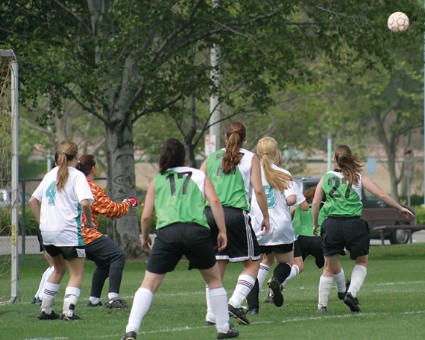
But young football players are not the only ones susceptible to concussions. You can have a concussion in any sport. In fact, studies have shown that female soccer players are especially prone to this injury.
This occurrence was highlighted in a recent study of 351 elite female soccer players aged 11-14 years. Dr. John W. O’Kane of the University of Washington Sports Medicine Clinic in Seattle, and his colleagues found that this group has a higher rate of concussion than has been reported in studies of female soccer players at the high school and college levels. The cumulative concussion incidence in the study was 13% per season or 1.2 concussions per 1,000 athletic exposure hours (JAMA Pediatr. 2014 [doi:10.1001/jamapediatrics.2013.4518]).
Further, the study found that 58.6% of symptomatic players continued play and more than half of them (55.9%) were not evaluated by a qualified health care professional.
These findings underline the lack of awareness among athletes this age about the significance of concussions. It also stresses the importance of educating young athletes about the signs of a concussion and reinforcing that it is in their best interest to tell a coach or trainer when they’ve had a concussion. A lot of times, athletes will not tell anyone because they don’t want to be pulled out of the game, but continuing to play puts them at a greater risk.
Concussions need to be properly managed to prevent them from lasting longer than necessary. In the study, those with concussions had symptoms that lasted a median of 4 days. If athletes continue to play on, they are increasing their risk of sustaining another concussion because recurrence is more likely to happen within 10 days of the first concussion.
Additionally, athletes may be predisposing themselves to "second impact syndrome," which is extraordinarily rare, but when it happens, it’s typically catastrophic and may result in traumatic injury or death. Coaches, health care providers, parents, and especially the athletes need to pay attention to the severity and symptoms of the concussions, how long recovery takes, and the time between concussions.
Right now, based on what studies can tell, the biggest risk factor for chronic brain injuries associated with repetitive concussions is exposure. So the longer you play the sport, the higher the risk you have. For these middle school and high school athletes, that is not a significant problem, but there simply hasn’t been a lot of research done on youth athletes.
This recent study plus the Institute of Medicine’s review of sports-related concussion in athletes from elementary school through young adults that began in late 2013 are great first steps in increasing our understanding of the effects of concussion on young players and helping us to figure out the best way to combat this problem.
Dr. Jordan is the director of the brain injury program and the memory evaluation treatment service at Burke Rehabilitation Hospital in White Plains, N.Y. He currently serves as the chief medical officer of the New York State Athletic Commission, as a team physician for USA Boxing, and as a member of the NFL Players Association Mackey-White Traumatic Brain Injury Committee and the NFL Neuro-Cognitive Disability Committee.
Regionalized trauma care boosts TBI survival
NAPLES, FLA. – A regional trauma system decreased hospital mortality for traumatic brain injury patients by 21% overall and by 26% for severe brain injuries, according to a partially retrospective study.
"Regionalization represents an additional step in attempting to improve outcomes for trauma patients. It can be defined as a tiered, integrated system that attempts to get the right patient to the right place at the right time," Dr. Michael L. Kelly said at the annual meeting of the Eastern Association for the Surgery of Trauma.
A few American studies and several more outside the United States have shown that regionalization decreases mortality in the general trauma population, but similar studies in traumatic brain injury (TBI) patients are scarce.
The Northern Ohio Trauma System (NOTS) was organized in 2010 and includes a transfer line to the Level I trauma center, a nontrauma hospital transfer protocol, a pilot scene triage protocol for emergency medical services, as well as the creation of a trauma-specific ICU in the level I center, explained Dr. Kelly, a sixth-year neurosurgery resident at the Cleveland Clinic, Ohio. The network includes the Level I MetroHealth Medical Center trauma center, two Level II trauma centers, and 12 nontrauma hospitals.
The three-tiered system mandates that TBI patients with a Glasgow Coma Scale (GCS) score of less than 12 and a traumatic mechanism, any penetrating head injury, or any open/depressed skull fracture, be sent to the Level I trauma center if they can be transferred within 15 minutes.
Patients with a GCS of 12-14 and a penetrating mechanism can be transferred to any trauma center, while those with lesser head injuries can remain at their hospital, unless their condition worsens.
For the study, Dr. Kelly and his coauthors analyzed data from 2008 through 2012 for 11,220 patients more than 14 years old with a TBI in the NOTS database, which was populated prospectively beginning in mid-2010.
Level I admissions increased significantly after NOTS by 10% for all TBIs (36% vs. 46%) and by 15% for severe TBIs with a head Abbreviated Injury Scale (AIS) score of 3 or more, he said. The percentage of patients who underwent transfers between NOTS institutions also increased significantly by 7% (7% vs. 14%) and 11% (10% vs. 21%), respectively.
Hospital mortality declined from 6.2% to 4.9% post-NOTS for all TBI patients (P = .005) and from 19% to 14% for the subset with severe TBI (P less than .0001). Mortality for trauma patients in general in Ohio has consistently hovered at 4% to 5% for the last decade, despite efforts to improve outcomes, including a 2002 law requiring the transfer of trauma patients to a validated trauma center, Dr. Kelly said.
In the post-NOTS period, craniotomies increased significantly for all TBIs (2% vs. 3%; P = .003) and for severe TBIs (6% vs. 8%; P = .02). The use of any neurosurgical procedure and hospital length of stay, however, remained constant for both groups in both time periods.
At baseline, the 6,713 post-NOTS patients were significantly older than the 4,507 pre-NOTS patients (55 years vs. 52 years) and less likely to be male (63% vs. 66%) or black (23% vs. 34%). GCS scores were similar (15 for both), as were Injury Severity Scores (14 for both) and the percentage of patients with a head AIS of 3 or more (34% post- vs. 32% pre-).
Multivariate regression analysis showed that the NOTS time period was an independent predictor of survival for all TBIs, with an odds ratio of 0.76, representing a 24% mortality reduction, and odds ratio of 0.72 for severe TBIs, representing a 28% mortality reduction.
"Of some importance, the multivariate model actually strengthened the effect of NOTS on mortality in our patient population," Dr. Kelly said.
Invited discussant Deborah Stein, medical director of neurotrauma critical care at the R Adams Cowley Shock Trauma Center at the University of Maryland Medical Center in Baltimore, described the study as an important contribution to the growing body of literature demonstrating that regionalization of care is associated with improved outcomes.
Dr. Stein went on to congratulate the NOTS members for participating in what was, at times, likely a contentious and difficult process: to bring a diverse group of hospitals to consensus about the best way to care for injured patients.
"Effecting change is difficult enough in a single division, department, or hospital," she remarked.
Following the formal presentation, audience members questioned whether the change in mortality was accomplished by simply shifting patients to nursing homes to die or whether it reflects a more aggressive surgical approach to TBI or improved critical care.
Dr. Kelly said that the creation of a trauma-specific ICU at MetroHealth, the uptick in transfers to the Level I trauma center, and the increased craniotomy rate all likely affected the outcome. Patient disposition data are still being analyzed, but hospice rates remained similar after NOTS was implemented, he said.
Dr. Kelly and his coauthors reported having no financial disclosures.
For nearly 50 years, data has been accumulating regarding the effects of regionalized trauma systems on postinjury outcomes, and the verdict is clear: Having a system in place that is prepared to rapidly transport the severely injured to a trauma center provides the best opportunity to reduce morbidity and mortality in these patients. While this is true in a general sense, a penetrating injury to the torso is not the same as a TBI, with each requiring different resources and expertise for optimal management.
The group at Cleveland MetroHealth in Ohio sought to answer the question of whether or not regionalization of trauma services would demonstrate a benefit in patients with TBI. Reviewing 4 years' worth of data that coincided at its midpoint with the implementation of the Northern Ohio Trauma System (NOTS), the authors demonstrate a significant reduction in mortality in brain-injured patients, including a 28% reduction in patients with severe TBI on multivariate analysis.

|
| Dr. Robert Winfield |
In the end, the commendation from Dr. Stein regarding the ability of the NOTS group, which comprises three hospitals from two health systems, to coalesce into a highly functioning regional trauma system is prescient and reflective of an impressive achievement for the authors. It is a reminder that in a time of scarce resources and competition for health care dollars, maintaining a focus on patient care through a cooperative and collaborative approach will ultimately yield the best results for all involved.
Dr. Robert Winfield is an ACS Fellow and the chair of the ACS Resident and Associate Society, and assistant professor of trauma and acute and critical care surgery at Washington University, St. Louis.
For nearly 50 years, data has been accumulating regarding the effects of regionalized trauma systems on postinjury outcomes, and the verdict is clear: Having a system in place that is prepared to rapidly transport the severely injured to a trauma center provides the best opportunity to reduce morbidity and mortality in these patients. While this is true in a general sense, a penetrating injury to the torso is not the same as a TBI, with each requiring different resources and expertise for optimal management.
The group at Cleveland MetroHealth in Ohio sought to answer the question of whether or not regionalization of trauma services would demonstrate a benefit in patients with TBI. Reviewing 4 years' worth of data that coincided at its midpoint with the implementation of the Northern Ohio Trauma System (NOTS), the authors demonstrate a significant reduction in mortality in brain-injured patients, including a 28% reduction in patients with severe TBI on multivariate analysis.

|
| Dr. Robert Winfield |
In the end, the commendation from Dr. Stein regarding the ability of the NOTS group, which comprises three hospitals from two health systems, to coalesce into a highly functioning regional trauma system is prescient and reflective of an impressive achievement for the authors. It is a reminder that in a time of scarce resources and competition for health care dollars, maintaining a focus on patient care through a cooperative and collaborative approach will ultimately yield the best results for all involved.
Dr. Robert Winfield is an ACS Fellow and the chair of the ACS Resident and Associate Society, and assistant professor of trauma and acute and critical care surgery at Washington University, St. Louis.
For nearly 50 years, data has been accumulating regarding the effects of regionalized trauma systems on postinjury outcomes, and the verdict is clear: Having a system in place that is prepared to rapidly transport the severely injured to a trauma center provides the best opportunity to reduce morbidity and mortality in these patients. While this is true in a general sense, a penetrating injury to the torso is not the same as a TBI, with each requiring different resources and expertise for optimal management.
The group at Cleveland MetroHealth in Ohio sought to answer the question of whether or not regionalization of trauma services would demonstrate a benefit in patients with TBI. Reviewing 4 years' worth of data that coincided at its midpoint with the implementation of the Northern Ohio Trauma System (NOTS), the authors demonstrate a significant reduction in mortality in brain-injured patients, including a 28% reduction in patients with severe TBI on multivariate analysis.

|
| Dr. Robert Winfield |
In the end, the commendation from Dr. Stein regarding the ability of the NOTS group, which comprises three hospitals from two health systems, to coalesce into a highly functioning regional trauma system is prescient and reflective of an impressive achievement for the authors. It is a reminder that in a time of scarce resources and competition for health care dollars, maintaining a focus on patient care through a cooperative and collaborative approach will ultimately yield the best results for all involved.
Dr. Robert Winfield is an ACS Fellow and the chair of the ACS Resident and Associate Society, and assistant professor of trauma and acute and critical care surgery at Washington University, St. Louis.
NAPLES, FLA. – A regional trauma system decreased hospital mortality for traumatic brain injury patients by 21% overall and by 26% for severe brain injuries, according to a partially retrospective study.
"Regionalization represents an additional step in attempting to improve outcomes for trauma patients. It can be defined as a tiered, integrated system that attempts to get the right patient to the right place at the right time," Dr. Michael L. Kelly said at the annual meeting of the Eastern Association for the Surgery of Trauma.
A few American studies and several more outside the United States have shown that regionalization decreases mortality in the general trauma population, but similar studies in traumatic brain injury (TBI) patients are scarce.
The Northern Ohio Trauma System (NOTS) was organized in 2010 and includes a transfer line to the Level I trauma center, a nontrauma hospital transfer protocol, a pilot scene triage protocol for emergency medical services, as well as the creation of a trauma-specific ICU in the level I center, explained Dr. Kelly, a sixth-year neurosurgery resident at the Cleveland Clinic, Ohio. The network includes the Level I MetroHealth Medical Center trauma center, two Level II trauma centers, and 12 nontrauma hospitals.
The three-tiered system mandates that TBI patients with a Glasgow Coma Scale (GCS) score of less than 12 and a traumatic mechanism, any penetrating head injury, or any open/depressed skull fracture, be sent to the Level I trauma center if they can be transferred within 15 minutes.
Patients with a GCS of 12-14 and a penetrating mechanism can be transferred to any trauma center, while those with lesser head injuries can remain at their hospital, unless their condition worsens.
For the study, Dr. Kelly and his coauthors analyzed data from 2008 through 2012 for 11,220 patients more than 14 years old with a TBI in the NOTS database, which was populated prospectively beginning in mid-2010.
Level I admissions increased significantly after NOTS by 10% for all TBIs (36% vs. 46%) and by 15% for severe TBIs with a head Abbreviated Injury Scale (AIS) score of 3 or more, he said. The percentage of patients who underwent transfers between NOTS institutions also increased significantly by 7% (7% vs. 14%) and 11% (10% vs. 21%), respectively.
Hospital mortality declined from 6.2% to 4.9% post-NOTS for all TBI patients (P = .005) and from 19% to 14% for the subset with severe TBI (P less than .0001). Mortality for trauma patients in general in Ohio has consistently hovered at 4% to 5% for the last decade, despite efforts to improve outcomes, including a 2002 law requiring the transfer of trauma patients to a validated trauma center, Dr. Kelly said.
In the post-NOTS period, craniotomies increased significantly for all TBIs (2% vs. 3%; P = .003) and for severe TBIs (6% vs. 8%; P = .02). The use of any neurosurgical procedure and hospital length of stay, however, remained constant for both groups in both time periods.
At baseline, the 6,713 post-NOTS patients were significantly older than the 4,507 pre-NOTS patients (55 years vs. 52 years) and less likely to be male (63% vs. 66%) or black (23% vs. 34%). GCS scores were similar (15 for both), as were Injury Severity Scores (14 for both) and the percentage of patients with a head AIS of 3 or more (34% post- vs. 32% pre-).
Multivariate regression analysis showed that the NOTS time period was an independent predictor of survival for all TBIs, with an odds ratio of 0.76, representing a 24% mortality reduction, and odds ratio of 0.72 for severe TBIs, representing a 28% mortality reduction.
"Of some importance, the multivariate model actually strengthened the effect of NOTS on mortality in our patient population," Dr. Kelly said.
Invited discussant Deborah Stein, medical director of neurotrauma critical care at the R Adams Cowley Shock Trauma Center at the University of Maryland Medical Center in Baltimore, described the study as an important contribution to the growing body of literature demonstrating that regionalization of care is associated with improved outcomes.
Dr. Stein went on to congratulate the NOTS members for participating in what was, at times, likely a contentious and difficult process: to bring a diverse group of hospitals to consensus about the best way to care for injured patients.
"Effecting change is difficult enough in a single division, department, or hospital," she remarked.
Following the formal presentation, audience members questioned whether the change in mortality was accomplished by simply shifting patients to nursing homes to die or whether it reflects a more aggressive surgical approach to TBI or improved critical care.
Dr. Kelly said that the creation of a trauma-specific ICU at MetroHealth, the uptick in transfers to the Level I trauma center, and the increased craniotomy rate all likely affected the outcome. Patient disposition data are still being analyzed, but hospice rates remained similar after NOTS was implemented, he said.
Dr. Kelly and his coauthors reported having no financial disclosures.
AT THE EAST SCIENTIFIC ASSEMBLY
Major finding: Hospital mortality was 6.2% pre-NOTS vs. 4.9% post-NOTS for all TBIs (P = .005) and 19% vs. 14% for severe TBIs (P less than .001).
Data source: A partially retrospective analysis of 11,220 TBI trauma patients.
Disclosures: Dr. Kelly and his coauthors reported having no financial disclosures.
Memory fares better with laser ablation for epilepsy than with surgery
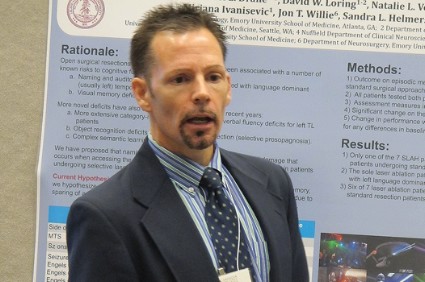
WASHINGTON – Stereotactic laser ablation resulted in better cognitive outcomes in patients with temporal lobe epilepsy than did treatment with standard open surgical approaches, Daniel Drane, Ph.D., reported at the annual meeting of the American Epilepsy Society.
In a study comparing pre- and posttreatment cognitive outcomes in 17 people with temporal lobe epilepsy, those treated with MRI-guided laser ablation had better outcomes on episodic memory measures 6 months after surgery than did the patients treated with standard surgical treatments, said Dr. Drane of the departments of neurology and pediatrics at Emory University, Atlanta. The results suggest that "memory decline requires broader damage to mesial temporal lobe structures or that epilepsy patients are able to reorganize memory more efficiently if structural damage is more selective," he concluded.
The study compared neuropsychological data before surgery and 6 months after surgery in 7 people who were treated with hippocampal laser ablation (5 left and 2 right resections) and 10 people who underwent surgery with standard or selective surgical approaches, including selective amygdalohippocampectomy (5 left and 5 right resections). Patients in both groups were similar in age (mean 36-39 years) and were on a mean of two antiepileptic drugs. Two patients in the surgery group and three in the laser ablation group had had seizures since they were 5 years or younger.
The investigators evaluated episodic memory with measures that included tasks of visual and verbal memory.
After treatment, there was evidence of a significant decline in one or both of the memory tasks in only 1 of the 7 patients treated with laser ablation, compared with 9 of the 10 surgical patients, a statistically significant difference, Dr. Drane said. In addition, six patients who had laser ablation showed a significant improvement in one or more memory measures after treatment, compared with four standard surgery patients.
In a statement issued during the meeting, Dr. Drane said that considering "the presumed importance of the hippocampus in episodic memory, we were surprised by the absence of any decline" in the laser ablation group. The results suggest that the function of the hippocampus may not be completely understood "and that being able to perform such a precise resection may help us learn more about brain regions in a manner that was never before possible in humans," he added.
During a press briefing held during the meeting, Dr. Drane pointed out that laser ablation benefits patients with less pain and a short hospital stay lasting a median of 1 day with no time spent in the intensive care unit.
Another possible benefit of the less invasive procedure is a positive effect on mood, he said. He described a patient in her early 60s with chronic epilepsy and chronic depression who was completely seizure free after laser ablation. But the most striking result of treatment was that she was no longer depressed and was off all antidepressants for the first time since she was a teenager.
This is just one case, but mood may benefit "from not cutting so many regions," he noted.
Dr. Drane said he received funding from the National Institutes of Health to conduct the study.
AT AES 2013
Major finding: After undergoing stereotactic laser ablation, 1 in 7 patients had significant drop in one of two memory tasks, compared with 9 of the 10 who underwent standard surgery, a significant difference.
Data source: A comparison of visual and verbal memory before and after treatment with MRI-guided stereotactic laser ablation or standard surgery in a group of 17 patients with temporal lobe epilepsy.
Disclosures: Dr. Drane received funding from the National Institutes of Health to conduct the study.
TBI survivors see threefold risk of early death
People who survive a traumatic brain injury are three times more likely to die prematurely than are those who have not, according to results from a large population-based study.
The study, which drew from more than 4 decades of data from Swedish national patient registries and death records, also found that nearly half of the TBI patients who died prematurely died from suicide, violence, or injuries.
For their research, published online Jan. 15 (JAMA Psychiatry 2014 [doi:10.1001/jamapsychiatry.2013.3935]), investigators, led by Dr. Seena Fazel of Oxford (England) University, evaluated data from 218,300 men and women who had survived for 6 months or more after TBI with age and sex-matched controls (n = 2,163,190).
Of all the TBI patients, 69.3% were men, 80.5% were not married, and 9.3% had preexisting psychiatric disorders, including 4.4% with substance abuse diagnoses.
Dr. Fazel and his colleagues found that the TBI patients saw a threefold higher mortality before age 56 years, compared with controls, even after researchers adjusted for socioeconomic confounders (adjusted odds ratio, 3.2; 95% confidence interval, 3.0-3.4). Of those who died early, the median age was 18.6 years at the time of the TBI and 40.6 years at death.
The study also looked at unaffected siblings of the TBI patients (n = 150,513) and found significantly greater odds of premature death in the TBI group, compared with their siblings (OR, 2.6; 95% CI, 2.3-2.8). Still, the difference was less than that seen with the general population controls, suggesting that some genetic and environmental factors might be partly responsible for the higher mortality.
The rate of premature deaths among the TBI survivors was low overall, but it was higher among those with psychiatric or substance abuse diagnoses. Mortality from suicide, injuries, and assault was significantly higher in the TBI group than the control group, accounting for 48.6% of premature deaths among TBI patients.
Dr. Fazel and his colleagues wrote in their analysis that in light of these findings, clinical guidelines might need to be revised to focus on preventing mortality beyond the first few months after injury and address high rates of psychiatric comorbidity and substance abuse among TBI patients.
In an editorial accompanying Dr. Fazel and his colleagues’ article, Dr. Robert G. Robinson of the department of psychiatry administration at the University of Iowa in Iowa City, proposed that some of the excess mortality seen among the TBI patients might be attributable to personality – something the study authors also acknowledged as a potential confounder (JAMA Psychiatry 2014 Jan. 15 [doi:10.1001/jamapsychiatry.2013.4241]).
"The preponderance of premature deaths due to external factors suggests that one of the most likely explanations for the findings in the current study is the existence of personality characteristics of impulsiveness, risk-taking behaviors, and proneness to substance abuse. These patients incur a TBI and continue to demonstrate these behaviors after the TBI, which ultimately leads to a fatality," Dr. Robinson wrote.
Dr. Robinson, echoing the recommendations of Dr. Fazel and his colleagues, argued that recognizing such patients is important "because half of these deaths are due to preventable behaviors." Some prevention strategies, he wrote, might include screening TBI patients at discharge for personality characteristics and treating those with the most impulsive risk-taking traits, or administering antidepressants to those seen as vulnerable to depression.
The study authors and Dr. Robinson noted among the study’s strengths of its very large sample size, making it the largest of its kind to date, its long follow-up, and its use of sibling controls. One weakness noted by Dr. Robinson was that the cause of the TBI could not be specified in most cases and that the severity of TBI could only be quantified in some cases by duration of hospitalization.
Because pathologic features, symptoms, and the course of TBI can differ depending on the cause or severity of the injury, Dr. Robinson wrote, "further studies in this area should focus on whether the cause of brain injury, type of brain injury, severity of injury, or premorbid personality characteristics are associated with the highest risk of premature deaths."
Dr. Fazel and his colleagues’ study was funded by grants from the Wellcome Trust, the Swedish Prison and Probation Service, and the Swedish Research Council. None of its authors declared conflicts of interest. Dr. Robinson disclosed no conflicts of interest related to his editorial.
FROM JAMA PSYCHIATRY
Major finding: TBI patients saw higher mortality before age 56 years, compared with controls, even after adjustment for socioeconomic confounders (adjusted OR, 3.2; 95% CI, 3-3.4). Of those who died early, the median age was 18.6 years at the time of the TBI and 40.6 years at death.
Data source: About 2.5 million patient records from Swedish national registries between 1959-2009.
Disclosures: Dr. Fazel and his colleagues’ study was funded by grants from the Wellcome Trust, the Swedish Prison and Probation Service, and the Swedish Research Council. None of its authors declared conflicts of interest. Dr. Robinson disclosed no conflicts of interest related to his editorial.
NIH announces major funding support for concussion, TBI research
The National Institutes of Health announced on Dec. 16 significant financial support for eight research projects focused on traumatic brain injury, which currently ranks as the leading cause of death in young adults.
"We need to be able to predict which patterns of injury are rapidly reversible and which are not," Story Landis, Ph.D., director of the National Institute of Neurological Disorders and Stroke (NINDS), part of the NIH, said in a prepared statement. "This program will help researchers get closer to answering some of the important questions about concussion for our youth who play sports and their parents."
Funding, which totals more than $14 million, comes from the Sports and Health Research Program, a partnership between the NIH, the National Football League, and the Foundation for the National Institutes of Health. The eight projects earmarked to receive support include two cooperative agreements and six pilot studies.
For one of the two cooperative agreements, which will receive $6 million each, researchers led by Dr. Ann C. McKee of Boston University and the U.S. Department of Veterans Affairs will strive to define a set of criteria for the various stages of chronic traumatic encephalopathy (CTE). Then, imaging teams will correlate the findings with brain scans that might be used to diagnose CTE in individuals during their lifetimes. For the other cooperative agreement, researchers led by Wayne Gordon, Ph.D., of Mount Sinai Hospital, New York, have a goal of identifying and describing the chronic effects of mild, moderate, and severe traumatic brain injuries (TBIs) and comparing these with the features of CTE. Then, they will employ various brain imaging techniques in patients with a wide range of head injuries, as well as on postmortem tissue, to identify markers that may eventually be used to diagnose the degenerative effects of TBI.
"Although the two cooperative agreements focus on different aspects of TBI, their combined results promise to answer critical questions about the chronic effects of single versus repetitive injuries on the brain, how repetitive TBI might lead to CTE, how commonly these changes occur in an adult population, and how CTE relates to neurodegenerative disorders like Alzheimer’s disease," Dr. Landis said.
The six pilot studies, which are projected to receive just over $2 million in total funding, focus on ways to improve the diagnosis of concussion and identify potential biomarkers that can be used to track a patient’s recovery. They range from testing of a mobile application designed to track the progress of a young athlete from the time of a concussion injury until they are cleared to return to play, to the development of a portable eye-tracking instrument that can be used to diagnose concussions on the sidelines and to monitor the injury progression in high school and college athletes.
Pediatric epilepsy surgery improved mood, behavior in some patients
WASHINGTON – The vast majority of 100 children who underwent surgical resection for epilepsy at a single center experienced stable or significantly improved mood and anxiety symptoms after surgery.
The findings, which demonstrated good overall outcomes but differential effects based on site and side of surgery, have important implications for counseling parents about the potential effects of surgery, Elizabeth N. Andresen, Ph.D., reported at the annual meeting of the American Epilepsy Society.
The findings are important, because children with epilepsy are at high risk for depression, anxiety, and behavior problems. For example, children with epilepsy are 4.8 times more likely than are children in the general population, and 2.5 times more likely than those with other chronic conditions to develop such disturbances.
Parents are understandably concerned about how surgery might affect their child’s mood, behavior, and personality, and the few prior studies available have provided conflicting results, Dr. Andresen said.
"Now we have some good data that show that kids tend to do well: They tend to be the same, they tend to be pretty happy afterward, they tend to get better," she said.
Presurgically, 36 children with frontal lobe epilepsy reported symptoms of withdrawal and anxiety more often than did 64 children with temporal lobe epilepsy, but these symptoms improved significantly after surgery to levels comparable to or better than those in the temporal lobe epilepsy patients.
This finding was most pronounced among those who underwent left-sided surgery, Dr. Andresen of the Cleveland Clinic said at a press briefing during the meeting.
For example, presurgical T scores for anhedonia, social anxiety, and withdrawn/depression symptoms in frontal lobe vs. temporal lobe epilepsy patients who underwent left-sided surgery were 59 vs. 50, 59 vs. 49, and 59 vs. 58, respectively; after surgery, the T scores were 50 vs. 50, 42 vs. 48, and 55 vs. 59, respectively.
Scores for patients who underwent left-sided surgery significantly improved in evaluations at about 10 months on the social anxiety subscale of the Revised Children’s Manifest Anxiety Scale (RCMAS) and the withdrawal subscale of the Achenbach Child Behavior Checklist (CBCL). Surgical site or type of epilepsy did not appear to influence general improvement on the negative mood subscale of the Children’s Depression Inventory (CDI) and the worry subscale of the RCMAS, Dr. Andresen said.
Similarly, the improvement seen over time across the groups on the withdrawal, social problems, thought problems, and attention problems subscales of the CBCL did not appear to be related to the surgical site.
At the individual level, change scores on the CDI and RCMAS demonstrated clinically significant improvement in overall depression symptoms in 21% of patients overall, including 15% of temporal lobe patients and 33% of frontal lobe epilepsy patients, and in overall anxiety symptoms in 38% of patients overall, including 27% of temporal lobe epilepsy patients and 45% of frontal lobe epilepsy patients.
Particularly pronounced effects were seen with respect to anhedonia and social anxiety in those with frontal lobe epilepsy who underwent left-sided surgery, with 63% and 100% of those patients experiencing improvement on those measures, respectively.
When children with and without depression prior to surgery were analyzed separately, Dr. Andresen found that only 6% of those without depression developed depression after surgery, while 64% of those with depression before surgery were no longer depressed after surgery. Similarly, 11% of those without anxiety before surgery developed anxiety after surgery, while 40% of those with anxiety before surgery were not anxious after surgery.
The proportion of surgery patients developing new depression and anxiety was similar to the proportion of nonsurgical control patients who developed depression or anxiety over 6 months (6% and 15%), Dr. Andresen noted.
"So it looks like surgery isn’t having a big effect on the rate of new problems," she said, noting that while it isn’t having a huge effect "on the rate of making those problems go away," the numbers are moving in the right direction.
Of 64 patients treated for temporal lobe epilepsy, 38 underwent left-sided surgery and 26 underwent right-sided surgery. The other group of 36 patients with frontal lobe epilepsy included 16 who underwent left-sided surgery and 20 who underwent right-sided surgery. Another 53 patients who did not undergo surgery served as controls. Children included in the study were aged 5-16 years (mean of 11 years) with a mean age of epilepsy onset at 5.8 years and a mean duration of 5.3 years. All were taking at least two antiepileptic drugs.
Dr. Andresen said that she had no disclosures.
AT AES 2013
Major finding: Scores on the Children’s Depression Inventory and the Revised Children’s Manifest Anxiety Scale demonstrated clinically significant improvement in overall depression symptoms in 21% of patients overall, including 15% of temporal lobe epilepsy patients and 33% of frontal lobe epilepsy patients.
Data source: A review of 153 surgery patients and nonsurgical controls.
Disclosures: The presenter had no disclosures.
