User login
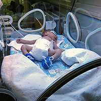
An intervention to track fetal movement attempts to prevent stillbirth
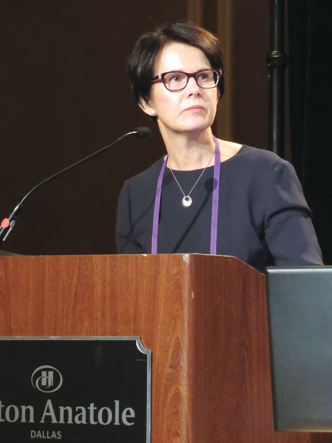
DALLAS – A hospital-wide intervention designed to prevent stillbirth by focusing on reduced fetal movement failed to do so – but did result in significant increases in labor induction and cesarean sections.
After the interventions was implemented, hospitals achieved a stillbirth rate of 4 per 1,000 – not significantly different than the 4.4 per 1,000 rate in the controls, Jane Norman, MD, reported at the meeting sponsored by the Society for Maternal-Fetal Medicine. On the other hand, the risk of a C-section increased by 9% and the risk of a labor induction by 8%, said Prof. Norman of the University of Edinburgh.
The results are disappointing, but consistent with most of the literature on this topic, she said.
“There is very good evidence that, if you ask women to count kicks, it doesn’t prevent stillbirth,” said Dr. Norman, referring to a 2015 Cochrane review. That paper, which examined five studies comprising more than 71,000 women, found that rates of stillbirth were similar between those who employed kick counts and those who did not.
Still, said Dr. Norman, there is evidence that stillbirth often is preceded by a period of reduced fetal movement. And a 2009 quality improvement project conducted in Oslo has driven enthusiasm for the idea of implementing some form of maternal and provider awareness of this area of obstetric care.
Stillbirth rates fell about 2% after the participating hospitals provided written information to women about fetal activity and reduced fetal movement, including an invitation to monitor fetal movements and formalized clinical guidelines for management of reduced fetal movement.
“There is a huge interest in the United Kingdom right now in using reduced fetal movement as an alert to act on to prevent stillbirth,” Dr. Norman said. “The National Health Service has recommended that we talk with women and clinicians about reduced fetal movement,” and try to incorporate it into clinical care.
Dr. Norman and her team wanted to emulate the Oslo experience, with a target of a 30% reduction in stillbirth rate.
The AFFIRM trial employed a clinical management guideline and patient education handout to raise awareness of reduced fetal movement as a trigger to investigate fetal well-being. The study used hospital records from 37 institutions in the United Kingdom and Ireland, which sequentially implemented the package. Hospitals were grouped into eight clusters. The trial began in January 2014. The first cluster began the intervention in mid-March; a new cluster came online every 3 months thereafter, until April 2016. Each cluster used its own baseline data as control.
The leaflet educated women about what fetal movements should feel like in all stages of pregnancy and what to expect from normal fetal movement. It encouraged them to report any lessening or cessation of fetal movement to their health care provider without delay.
The clinical guideline was matched to different gestational stages, but generally advocated cardiotocography and scans to estimate amniotic fluid volume and fetal size in cases of reduced movement with a low threshold for early delivery if there were abnormal findings at 37 weeks or later.
The final sample comprised 385,582 births, with 157,692 in the control period and 227,860 in the intervention period. The mean age of women was 30 years; 50% were white, 50% were overweight or obese, and 15% smoked. About 40% were nulliparous.
In the control arm, there were 691 stillbirths at 24 weeks’ gestational age or older (4.4/1,000). In the intervention arm, there were 921 events (4.06/1,000). The 10% risk reduction was not statistically significant.
The investigators also examined stillbirths occurring at 22 weeks or older, 28 weeks or older, and 37 weeks or older. Again, there were no significant between-group differences. Nor was there a difference in the perinatal mortality rate (0.68% vs. 0.62%).
There were, however, differences in interventions. The risk of a C-section increased by 5% (25.5% vs. 28.5%; adjusted odds ratio, 1.05), and the risk of induction by 9% (33.6% vs. 39.8%; AOR, 1.09). Most of the induction risk was driven by an 11% increase in induction risk at 39 weeks or older. The team also found a 5% increase in the chance of an elective delivery at 39 weeks or older (45.2% vs. 52.4%; AOR, 1.05)
There was a corresponding significant 10% decrease in the chance of spontaneous vaginal delivery (59.8% vs. 57.4%; AOR, 0.90).
Although there was no overall increase in the risk of a neonatal ICU admission, there was a significant 12% increase in the risk of a neonatal ICU admission of at least 48 hours (6.2% vs. 6.7%; AOR, 1.12). There were no other significant differences in neonatal outcomes (gestational age, size at birth, preterm delivery).
“We also looked at the potential impact if we implemented this intervention on a population of 10,000 pregnancies,” Dr. Norman said. “We would have 5 fewer stillbirths – but potentially anywhere from 11 fewer to 3 more. However, we would have 162 more cesarean deliveries and 108 more inductions.”
The AFFIRM trial was sponsored by the University of Edinburgh and the National Health Service. Dr. Norman had no financial disclosures.
SOURCE: Norman JE et al. Am J Obstet Gynecol. 2018;218,S603.
REPORTING FROM THE PREGNANCY MEETING
Key clinical point: Reduced fetal movements as an alert for investigation didn’t reduce stillbirth rates.
Major finding: Hospitals achieved a stillbirth rate of 4/1,000 – not significantly different than the 4.4/1,000 rate before the intervention.
Study details: The randomized, cluster trial comprised 37 hospitals in the United Kingdom and Ireland.
Disclosures: The University of Edinburgh and the National Health Service sponsored the trial. Dr. Norman had no financial disclosures.
Source: Norman JE et al. Am J Obstet Gynecol. 2018;218,S603.
Mother has severe pain — uterine rupture: $9M settlement
Mother has severe pain — uterine rupture: $9M settlement
At 37 weeks’ gestation, a woman went to the hospital with contractions and abdominal pain 2 weeks before her scheduled delivery. She had a previous
She returned the next night after midnight with contractions and a pain score of 9/10, and she was admitted. The nurses spoke with the ObGyn via phone 6 times over the next 8 hours. He prescribed 3 pain medications but the patient’s pain was unresponsive. When the patient called her ObGyn, he told her an ultrasound would be performed and the baby would be delivered in the morning. FHR monitoring became nonreassuring at 8:00
The baby was transferred to the neonatal intensive care unit (NICU) where he received therapeutic hypothermia. The parents were told that there was a 70% chance that the child would have cerebral palsy. The baby required a feeding tube and was hospitalized for 84 days. After discharge, he was moved to a long-term care facility. During his first year of life, he had pneumonia requiring hospitalization. Due to frequent aspiration of food and saliva into his airway, a tracheostomy was placed.
The child has hypoxic ischemic encephalopathy with severe brain damage, including spastic quadriplegic cerebral palsy. He is blind but can hear. He will need 24-hour nursing care for the rest of his life.
PARENT'S CLAIM: The ObGyn did not properly react to the mother’s reported severe pain. A cesarean delivery should have been performed when the mother first reported to the hospital or when she returned the following night.
PHYSICIAN'S DEFENSE: The defense was expected to argue at trial that the mother was not in labor because medication had stopped contractions; therefore it was reasonable to mature longer before delivery. The case settled during the trial.
VERDICT: A $9 million Michigan settlement was reached during mediation.
Baby dies at birth: $1.3M settlement
A 39-year-old woman was admitted to the hospital at 36 weeks’ gestation with regular contractions. Results of an ultrasound showed normal amniotic fluid and an anterior placental location. The patient’s membranes were artificially ruptured, and she received an epidural. The baby remained at plus-one station for more than 7 hours until delivery. Three attempts to rotate the baby over the course of labor failed. Variable decelerations were present on FHR monitoring throughout labor, and deep decelerations were noted within 2 hours of delivery. The ObGyn ordered cesarean delivery due to arrest of labor and fetal distress; delivery began 64 minutes after the decision. The baby’s head was deeply impacted in the pelvis and a Bandl’s ring was encountered. Several attempts were made to dislodge the fetal head. The fourth attempt, in conjunction with enlarging the hysterotomy, was successful. At birth, the baby was pale with no palpable pulse. A 17-minute resuscitation effort failed. An autopsy concluded that the cause of death was a subgaleal hemorrhage in the setting of acute and subacute placental pathology.
PARENT'S CLAIM: The ObGyn was negligent for not performing a cesarean delivery when the baby could not be rotated from a plus-one station and fetal distress was evident. The ObGyn never accessed the patient’s electronic medical records during the 8 hours of labor. An audit trail review revealed the editing and alleged purging of medical records.
PHYSICIAN'S CLAIM: The case was defended on the basis of the autopsy findings, alleging that the baby was compromised before labor and delivery. A pathology expert testified that blood loss from a subgaleal hemorrhage was not necessarily lethal and may occur spontaneously. However, the pathologist conceded that she failed to note complications that occurred during delivery and acknowledged that the autopsy did not document the bruising of the baby’s head, ear, neck, shoulder, and torso that was evident in autopsy photographs.
VERDICT: A $1.3 million Virginia settlement was reached.
Was tachycardic FHR ignored? $3M settlement
A 25-year-old woman with gestational diabetes was scheduled for induction of labor at 39 2/7 weeks’ gestation. When she presented for induction, artificial rupture of the membranes demonstrated clear fluid with no sign of fetal or maternal distress. Labor and delivery was managed by a certified nurse midwife (CNM) employed by an ObGyn group. Just prior to the CNM’s arrival, the FHR monitor tracings showed a series of decelerations; the CNM stopped the oxytocin. As labor progressed, the CNM reintroduced and increased the oxytocin dosage. The infant’s heart rate became tachycardic. The CNM documented a bloody show of vaginal fluid. After the mother began pushing, the FHR signal was lost on the external monitor and only the maternal pulse was detected. As the infant’s head crowned, the FHR monitor was reconnected; an FHR of 210 bpm was detected showing marked tachycardia.
After vaginal delivery, the child was limp and unresponsive. He had hypoxic ischemic encephalopathy and immediately began to have seizures. He was transferred to the NICU at another hospital where he stayed for 34 days. The infant was found to have spastic quadriplegic cerebral palsy, cortical visual impairment, a seizure disorder, right-sided torticollis, plagiocephaly, expressive language disorder, and dysphagia. He will require 24-hour nursing care for the rest of his life.
PARENT'S CLAIM: The CNM failed to consult an ObGyn to determine whether the patient needed an urgent cesarean delivery when FHR monitoring first indicated a worrisome fetal heart rate. The CNM improperly increased the dosage of oxytocin and did not remain at the mother’s bedside until she was fully dilated and began pushing. The CNM failed to rule out placental abruption or to communicate to the ObGyn that there was bloody show in vaginal fluid. The CNM interpreted the maternal heart rate as a reassuring fetal heart rate. When the monitor was reattached, the CNM failed to call for emergency assistance, despite signs of acute fetal distress. Testing ruled out any preexisting neurologic injury or congenital defect.
DEFENDANT'S DEFENSE: The CNM claimed that she delegated the heart monitoring to the labor nurse and relied on the nurse to report any irregularities. The case was settled during the trial.
VERDICT: A $3 million Virginia settlement was reached, which included $2.1M for the infant and $.9M for the mother.
These cases were selected by the editors of OBG Management from Medical Malpractice Verdicts, Settlements & Experts, with permission of the editor, Lewis Laska (www.verdictslaska.com). The information available to the editors about the cases presented here is sometimes incomplete. Moreover, the cases may or may not have merit. Nevertheless, these cases represent the types of clinical situations that typically result in litigation and are meant to illustrate nationwide variation in jury verdicts and awards.
Share your thoughts! Send your Letter to the Editor to [email protected]. Please include your name and the city and state in which you practice.
Get ready for certolizumab for psoriasis
KAUAI, HAWAII – When certolizumab pegol receives marketing approval for moderate to severe psoriasis – which experts say is a virtual lock – it will offer a singular advantage over current anti–tumor necrosis factor (anti-TNF) biologics: strong evidence of safety in pregnancy.
“ ” Kenneth B. Gordon, MD, observed at the Hawaii Dermatology Seminar provided by the Global Academy for Medical Education/Skin Disease Education Foundation.

Lots of women and their families are understandably deeply concerned about using powerful, transformative medications during pregnancy, even though they know from experience how debilitating inadequately treated psoriasis can be.
“Many women of childbearing potential would find [certolizumab] to be a preferential agent if they’re planning to become pregnant,” said Dr. Gordon, professor and chair of the department of dermatology at the Medical College of Wisconsin, Milwaukee.
He cited the CRIB (A Multicenter, Postmarketing Study Evaluating the Transfer of Cimzia From the Mother to the Infant via the Placenta) study results presented by Alexa B. Kimball, MD, at the 2017 annual meeting of the European Academy of Dermatology and Venereology in Geneva as a major step forward in establishing the safety of certolizumab during pregnancy.
CRIB was a prospective postmarketing pharmacokinetic study that evaluated placental transfer of certolizumab from 16 pregnant women on the biologic to their infants. All of the mothers received their last dose of certolizumab for rheumatoid arthritis or other approved indications within 35 days of delivery. Blood samples were collected from mothers, newborns, and umbilical cords within 1 hour of delivery, and again from the infants at weeks 4 and 8 after delivery.
Only one infant had a detectable plasma level of certolizumab at birth, and it was barely measurable at 0.042 mcg/mL, as compared with 49.4 mcg/mL in the mother’s plasma. This is consistent with the fact that certolizumab’s pegylated arm allows only minimal or no placental transfer from mother to infant, so there is essentially no third trimester in utero fetal exposure. In contrast, as Dr. Kimball noted, other anti-TNF biologics lack a pegylated arm and thus preferentially cross the placenta, creating a theoretical increased risk of maternal pregnancy complications and/or congenital malformations.
Dr. Kimball, professor of dermatology at Harvard Medical School and chief executive officer of Harvard Medical Faculty Physicians at Beth Israel Deaconess Medical Center, both in Boston, also has been deeply involved in an ongoing registry (sponsored by certolizumab manufacturer UCB) of several hundred women on certolizumab in pregnancy. The data have reassuringly shown no increased risk of maternal pregnancy complications such as preeclampsia, gestational diabetes, or preterm birth, nor any increase in or pattern of congenital malformations, compared with background rates in the general population.
Dr. Gordon said that while he understands the concerns, he personally doesn’t think the class-wide safety of TNF inhibitors in pregnancy and lactation is a big issue.
“My argument is that anti-TNF agents have been used very frequently in women of childbearing age, and also in women who are pregnant or lactating. And there have not been any side effect signals from that,” he explained.
The prospects of gaining an expanded indication for certolizumab in psoriasis hinge in part on the impressive results of the pivotal phase 3, randomized, double-blind, placebo-controlled CIMPASI-1 and CIMPASI-2 trials. In CIMPASI-1, the week-48 Psoriasis Area and Severity Index (PASI) 75 and PASI 90 response rates were 87.1% and 60.2%, respectively, in patients on the biologic at 400 mg every 2 weeks; among those on certolizumab at 200 mg every 2 weeks, the rates were 67.2% and 42.8%. In CIMPASI-2, the PASI 75 and PASI 90 rates were 81.3% and 62.0% at 400 mg and 78.7% and 59.6% with 200 mg every 2 weeks.
There were no cases of tuberculosis or any other significant safety concerns through 48 weeks, Dr. Gordon said.
“Certolizumab is coming soon for psoriasis,” predicted Craig L. Leonardi, MD, a psoriasis researcher at Saint Louis University. “The data are very impressive. It’s a high-performance drug. There’s no reason why this drug shouldn’t be approved.”
Since Dr. Kimball’s presentation of the CRIB data at the 2017 annual meeting of the European Academy of Dermatology and Venereology, the study has been published (Ann Rheum Dis. 2018 Feb;77[2]:228-33).
Dr. Gordon reported receiving research support from and serving as a paid consultant to numerous pharmaceutical companies developing new psoriasis therapies.
SDEF/Global Academy for Medical Education and this news organization are owned by the same parent company.
KAUAI, HAWAII – When certolizumab pegol receives marketing approval for moderate to severe psoriasis – which experts say is a virtual lock – it will offer a singular advantage over current anti–tumor necrosis factor (anti-TNF) biologics: strong evidence of safety in pregnancy.
“ ” Kenneth B. Gordon, MD, observed at the Hawaii Dermatology Seminar provided by the Global Academy for Medical Education/Skin Disease Education Foundation.
Lots of women and their families are understandably deeply concerned about using powerful, transformative medications during pregnancy, even though they know from experience how debilitating inadequately treated psoriasis can be.
“Many women of childbearing potential would find [certolizumab] to be a preferential agent if they’re planning to become pregnant,” said Dr. Gordon, professor and chair of the department of dermatology at the Medical College of Wisconsin, Milwaukee.
He cited the CRIB (A Multicenter, Postmarketing Study Evaluating the Transfer of Cimzia From the Mother to the Infant via the Placenta) study results presented by Alexa B. Kimball, MD, at the 2017 annual meeting of the European Academy of Dermatology and Venereology in Geneva as a major step forward in establishing the safety of certolizumab during pregnancy.
CRIB was a prospective postmarketing pharmacokinetic study that evaluated placental transfer of certolizumab from 16 pregnant women on the biologic to their infants. All of the mothers received their last dose of certolizumab for rheumatoid arthritis or other approved indications within 35 days of delivery. Blood samples were collected from mothers, newborns, and umbilical cords within 1 hour of delivery, and again from the infants at weeks 4 and 8 after delivery.
Only one infant had a detectable plasma level of certolizumab at birth, and it was barely measurable at 0.042 mcg/mL, as compared with 49.4 mcg/mL in the mother’s plasma. This is consistent with the fact that certolizumab’s pegylated arm allows only minimal or no placental transfer from mother to infant, so there is essentially no third trimester in utero fetal exposure. In contrast, as Dr. Kimball noted, other anti-TNF biologics lack a pegylated arm and thus preferentially cross the placenta, creating a theoretical increased risk of maternal pregnancy complications and/or congenital malformations.
Dr. Kimball, professor of dermatology at Harvard Medical School and chief executive officer of Harvard Medical Faculty Physicians at Beth Israel Deaconess Medical Center, both in Boston, also has been deeply involved in an ongoing registry (sponsored by certolizumab manufacturer UCB) of several hundred women on certolizumab in pregnancy. The data have reassuringly shown no increased risk of maternal pregnancy complications such as preeclampsia, gestational diabetes, or preterm birth, nor any increase in or pattern of congenital malformations, compared with background rates in the general population.
Dr. Gordon said that while he understands the concerns, he personally doesn’t think the class-wide safety of TNF inhibitors in pregnancy and lactation is a big issue.
“My argument is that anti-TNF agents have been used very frequently in women of childbearing age, and also in women who are pregnant or lactating. And there have not been any side effect signals from that,” he explained.
The prospects of gaining an expanded indication for certolizumab in psoriasis hinge in part on the impressive results of the pivotal phase 3, randomized, double-blind, placebo-controlled CIMPASI-1 and CIMPASI-2 trials. In CIMPASI-1, the week-48 Psoriasis Area and Severity Index (PASI) 75 and PASI 90 response rates were 87.1% and 60.2%, respectively, in patients on the biologic at 400 mg every 2 weeks; among those on certolizumab at 200 mg every 2 weeks, the rates were 67.2% and 42.8%. In CIMPASI-2, the PASI 75 and PASI 90 rates were 81.3% and 62.0% at 400 mg and 78.7% and 59.6% with 200 mg every 2 weeks.
There were no cases of tuberculosis or any other significant safety concerns through 48 weeks, Dr. Gordon said.
“Certolizumab is coming soon for psoriasis,” predicted Craig L. Leonardi, MD, a psoriasis researcher at Saint Louis University. “The data are very impressive. It’s a high-performance drug. There’s no reason why this drug shouldn’t be approved.”
Since Dr. Kimball’s presentation of the CRIB data at the 2017 annual meeting of the European Academy of Dermatology and Venereology, the study has been published (Ann Rheum Dis. 2018 Feb;77[2]:228-33).
Dr. Gordon reported receiving research support from and serving as a paid consultant to numerous pharmaceutical companies developing new psoriasis therapies.
SDEF/Global Academy for Medical Education and this news organization are owned by the same parent company.
EXPERT ANALYSIS FROM SDEF HAWAII DERMATOLOGY SEMINAR
Fetal alcohol spectrum disorders incidence exceeds previous estimates
, according to a cross-sectional study.
Philip A. May, PhD, of the University of North Carolina, Gillings School of Global Public Health, and his coauthors assessed 6,639 first-grade children from four communities in the Rocky Mountain, Midwestern, Southeastern, and Pacific Southwestern regions of the United States.
In their report, published Feb. 6 in JAMA, they identified 222 cases of fetal alcohol spectrum disorders, representing conservative prevalence estimates ranging from 11.3 to 50 cases per 1,000 children (JAMA 2018;319[5]:474-82. doi: 10.1001/jama.2017.21896).
Of these children, 27 met the criteria for fetal alcohol syndrome, 104 met the criteria for partial fetal alcohol syndrome, and 91 met the criteria for alcohol-related neurodevelopmental disorder.
“These prevalence estimates are consistent with mounting evidence that harmful fetal alcohol exposure is common in the United States today and highlight the public health burden due to fetal alcohol spectrum disorders,” the authors wrote.
The finding was much higher than previous estimates of the prevalence of fetal alcohol spectrum disorders in the United States. The authors pointed out that routine surveillance methods may have previously underestimated the prevalence because so many children are either misdiagnosed or not diagnosed at all. But even two previous single-site, active-case ascertainment studies conducted in the United States found prevalence rates of 10 and 24 per 1,000 children.
“This consortium study, to our knowledge, was the first to apply active case ascertainment, common methodology, a single classification system, and expert in-person evaluation for a continuum of fetal alcohol spectrum disorders including alcohol-related neurodevelopmental disorder to a large number of children from communities across the United States,” the authors wrote. “These data have highlighted the need for a larger study with broader representation of U.S. communities with general population samples.”
Only 2 of the 222 children had actually been previously diagnosed with a fetal alcohol spectrum disorder, “although many parents and guardians were aware of the learning and behavioral challenges facing their children,” the researchers noted.
“These data confirm that missed diagnoses and misdiagnoses of children are common,” the authors wrote, pointing to a previous study in U.S. schoolchildren that found only one in seven children identified as having fetal alcohol syndrome had been previously diagnosed.
The assessment of fetal alcohol spectrum disorder was based on four domains: growth, dysmorphology, neurodevelopment, and prenatal alcohol exposure – the latter being assessed by interviewing the mother in person or by telephone.
The lowest prevalence – 11.3 per 1,000 children – was seen in the Midwestern community, while the highest – 50 per 1,000 – was in the Rocky Mountain community.
The main weakness of the study was that no individual sample included the entire eligible population because of differing policies on access to children for recruitment and variability in willingness to consent.
“Consent rates for screening ranged from 36.9% to 92.5% in individual samples and overall consent rates for screening averaged only 59.9% of eligible children,” the authors wrote. “If nonconsented children differed from consented, this could have biased prevalence estimates in either direction.”
The researchers acknowledged that the absence of a definitive biomarker for fetal alcohol spectrum disorder meant it was impossible to know for certain that the observed deficits were actually the result of fetal alcohol exposure, and that the prevalence estimates may therefore be overestimated.
The study was funded by grants from the National Institute on Alcohol Abuse and Alcoholism. One author declared grant support from the National Institute on Alcohol Abuse and Alcoholism and personal fees and honorarium from the Alcohol Center. No conflicts of interest were declared.
SOURCE: May, P et al. JAMA 2018;319[5]:474-82. doi: 10.1001/jama.2017.21896.
The finding of higher-than-expected prevalence estimates for fetal alcohol spectrum disorder has significant implications for clinicians and for public health. Many cases are misdiagnosed or not diagnosed, which may be the result of unknown or unconfirmed prenatal alcohol exposure, overlapping diagnostic criteria with other neurodevelopmental disorders, high rates of comorbidity, and the presence of a number of different clinical diagnostic guidelines. There is, therefore, a need for a universal diagnostic approach and the identification of novel and reliable biomarkers for detecting fetal alcohol effects.
In addition, the high prevalence of these disorders points to a need for better education of girls and women of childbearing age about the detrimental effects of alcohol consumption during pregnancy. Primary care clinicians should routinely include appropriate screening for alcohol use in all women of childbearing age in preconceptual health promotion and in contraceptive counseling and refer anyone identified as having an alcohol use disorder to substance abuse programs.
Shannon Lange, MPH, Jurgen Rehm, PhD, and Svetlana Popova, PhD, are with the Institute for Mental Health Policy Research at the Centre for Addiction and Mental Health in Toronto and the University of Toronto. These comments are taken from an accompanying editorial (JAMA 2018, 319[5]:448-9. doi: 10.1001/jama.2017.21895). No conflicts of interest were declared.
, according to a cross-sectional study.
Philip A. May, PhD, of the University of North Carolina, Gillings School of Global Public Health, and his coauthors assessed 6,639 first-grade children from four communities in the Rocky Mountain, Midwestern, Southeastern, and Pacific Southwestern regions of the United States.
In their report, published Feb. 6 in JAMA, they identified 222 cases of fetal alcohol spectrum disorders, representing conservative prevalence estimates ranging from 11.3 to 50 cases per 1,000 children (JAMA 2018;319[5]:474-82. doi: 10.1001/jama.2017.21896).
Of these children, 27 met the criteria for fetal alcohol syndrome, 104 met the criteria for partial fetal alcohol syndrome, and 91 met the criteria for alcohol-related neurodevelopmental disorder.
“These prevalence estimates are consistent with mounting evidence that harmful fetal alcohol exposure is common in the United States today and highlight the public health burden due to fetal alcohol spectrum disorders,” the authors wrote.
The finding was much higher than previous estimates of the prevalence of fetal alcohol spectrum disorders in the United States. The authors pointed out that routine surveillance methods may have previously underestimated the prevalence because so many children are either misdiagnosed or not diagnosed at all. But even two previous single-site, active-case ascertainment studies conducted in the United States found prevalence rates of 10 and 24 per 1,000 children.
“This consortium study, to our knowledge, was the first to apply active case ascertainment, common methodology, a single classification system, and expert in-person evaluation for a continuum of fetal alcohol spectrum disorders including alcohol-related neurodevelopmental disorder to a large number of children from communities across the United States,” the authors wrote. “These data have highlighted the need for a larger study with broader representation of U.S. communities with general population samples.”
Only 2 of the 222 children had actually been previously diagnosed with a fetal alcohol spectrum disorder, “although many parents and guardians were aware of the learning and behavioral challenges facing their children,” the researchers noted.
“These data confirm that missed diagnoses and misdiagnoses of children are common,” the authors wrote, pointing to a previous study in U.S. schoolchildren that found only one in seven children identified as having fetal alcohol syndrome had been previously diagnosed.
The assessment of fetal alcohol spectrum disorder was based on four domains: growth, dysmorphology, neurodevelopment, and prenatal alcohol exposure – the latter being assessed by interviewing the mother in person or by telephone.
The lowest prevalence – 11.3 per 1,000 children – was seen in the Midwestern community, while the highest – 50 per 1,000 – was in the Rocky Mountain community.
The main weakness of the study was that no individual sample included the entire eligible population because of differing policies on access to children for recruitment and variability in willingness to consent.
“Consent rates for screening ranged from 36.9% to 92.5% in individual samples and overall consent rates for screening averaged only 59.9% of eligible children,” the authors wrote. “If nonconsented children differed from consented, this could have biased prevalence estimates in either direction.”
The researchers acknowledged that the absence of a definitive biomarker for fetal alcohol spectrum disorder meant it was impossible to know for certain that the observed deficits were actually the result of fetal alcohol exposure, and that the prevalence estimates may therefore be overestimated.
The study was funded by grants from the National Institute on Alcohol Abuse and Alcoholism. One author declared grant support from the National Institute on Alcohol Abuse and Alcoholism and personal fees and honorarium from the Alcohol Center. No conflicts of interest were declared.
SOURCE: May, P et al. JAMA 2018;319[5]:474-82. doi: 10.1001/jama.2017.21896.
FROM JAMA
Key clinical point: The prevalence of fetal alcohol spectrum disorder in first-grade children in the United States may be as high as 1 in 20.
Major finding: The prevalence of fetal alcohol spectrum disorder ranged from 11.3 to 50 children per 1,000.
Data source: Cross-sectional study of 6,639 first-grade children.
Disclosures: The study was funded by grants from the National Institute on Alcohol Abuse and Alcoholism. One author declared grant support from the National Institute on Alcohol Abuse and Alcoholism and personal fees and honorarium from the Alcohol Center. No conflicts of interest were declared.
Source: May, P et al. JAMA 2018;319[5]:474-82. doi: 10.1001/jama.2017.21896.
Study seeks optimal duration of second stage of labor
, according to researchers.
The researchers performed a retrospective analysis of more than 103,000 pregnancies from the Consortium on Safe Labor, a study of electronic medical records from 12 U.S. sites from 2002 to 2008, according to a study published in Obstetrics & Gynecology.
The duration of the second stage was calculated from the time of 10-cm cervical dilation to the time of birth. The researchers stratified the results into nulliparous women who did or did not receive an epidural, and multiparous women who did or did not receive an epidural.
For nulliparous women, rates of spontaneous vaginal birth (rather than operative vaginal birth or cesarean delivery) without morbidity increased slightly in the second half-hour over the first, then decreased. Rates decreased steadily for multiparous women. Both decreases occurred regardless of epidural status. Deliveries with morbidity varied, to as high as a 12.3% likelihood that a nulliparous woman with an epidural would deliver with maternal or neonatal morbidity or mortality between 3 hours’ and 6 hours’ second-stage duration.
The researchers noted the various society recommendations for when to diagnose second-stage arrest, but concluded: “In our study, we did not observe an inflection at a particular hour mark. ... Ultimately the willingness to accept a certain percentage risk of morbidity to achieve vaginal delivery is up to the woman and clinician.”
The authors reported having no disclosures.
SOURCE: Grantz KL et al. Obstet Gynecol. 2018 Feb;131(2):345-53.
FROM OBSTETRICS & GYNECOLOGY
Barbed sutures shorten cesarean closure time, reduce blood loss
DALLAS – Using knotless barbed sutures to close the uterine incision after cesarean delivery reduced operating time and blood loss, according to a recent randomized controlled study.
“On average, knotless barbed sutures were 1 minute and 43 seconds faster,” said David Peleg, MD, discussing the study results at the meeting sponsored by the Society for Maternal-Fetal Medicine. Average uterine closure time with knotless barbed sutures was 3 minutes and 37 seconds; with smooth sutures, average closure time was 5 minutes and 20 seconds (103-second difference; 95% confidence interval, 67.69-138.47 seconds; P less than .001).

Barbed sutures, in contrast, are usually made of monofilament with barbs created by the addition of tiny diagonal cuts made just partway through the suture material. When the suture is pulled through with the angle of the barbs, it pulls smoothly, but pulling back against the barb angle causes the barbs to protrude and catch against tissue, preventing slippage and eliminating the need for knots.
The fact that the monofilament has been partially cut to create the barbs can also reduce tensile strength, but some of the other disadvantages of smooth sutures are avoided, said Dr. Peleg.
The suture material used for the study was bidirectional, with the barbs running in opposite directions from the midline and a needle swaged onto each end; other barbed suture systems have an integral loop at one end of a unidirectionally barbed length of suture material that is used to anchor the first suture. The brand used was Stratafix.
The prospective study was necessarily unblinded, and compared the knotless barbed sutures with smooth sutures using polyglactin 910 braided material (Vicryl) for use during closure of the uterine incision during cesarean section procedures.
Patients were eligible if they were having an elective cesarean section after at least 38 weeks’ gestation, or if they were having a cesarean for the usual obstetric indications after laboring. Women with previous cesareans who failed a trial of labor were eligible.
The primary outcome was the length of time to close the uterine incision, measured from the start of suturing until hemostasis was achieved; the time included hemostatic suturing.
After a small pilot study that established a baseline suturing time with polyglactin 910 sutures of 6 minutes (standard deviation, 2 minutes and 10 seconds), Dr. Peleg and his collaborators determined that they would need to enroll at least 34 women per study arm to detect a decrease of 25% in suture time – to 4.5 minutes – with barbed sutures.
“The decrease in closure time is not linear,” said Dr. Peleg, so they increased their sample size by 50%, to 51 patients in each group, to ensure statistical significance of the results.
One of the challenges of a surgical randomized controlled trial is ensuring uniform technique; for this study, all patients had epidural or spinal anesthesia and antibiotics before opening. Surgeon clinical judgment was used to determine whether a Pfannenstiel or low transverse incision was made. In either case, there was no closure of the parietal peritoneum or the rectus muscles, and subcutaneous closure was used if tissues were greater than 2 cm in depth. Subcuticular stitches were used if possible.
Looking at secondary outcomes, there was a trend toward shorter total operative time with barbed sutures that didn’t reach statistical significance (20.1 min for barbed sutures vs. 23.1 for conventional, P = .062). However, fewer hemostatic sutures were required when barbed sutures were used: Extra sutures were used in 16 of the barbed group vs. 41 who had conventional sutures (P less than .001). Those receiving barbed sutures also had significantly less blood loss and estimated total blood loss during uterine closure than the conventional suture group (P = .005 and P = .002, respectively).
No study patients experienced serious postoperative complications; there were no infections, hematomas, or other wound complications, said Dr. Peleg of Bar-Ilan University, Zefat, Israel.
On the pro side for wider implementation of the use of barbed sutures for uterine closure stand the quicker closure and better hemostasis, along with the theoretical benefits of having no knots. Additionally, said Dr. Peleg, “there’s a gentle learning curve – it’s relatively easy to get used to the technique” of using barbed sutures. And, he said, “surgeons find them satisfying to use.”
However, he acknowledged the extra expense of barbed suture material – depending on the location and supplier, he estimated the cost could run from 7- to 20-fold for the barbed sutures, which he said cost $23.50 apiece. Also, he said, though the results were statistically significant, “Are they clinically significant? Does a difference in closure time of one minute 43 seconds, and a decrease in blood loss of 47 milliliters matter?”
Other considerations, he said, will require longer-term study. Polydioxanone, used for the barbed sutures, has a longer absorption time – a factor with unknown clinical implications in this application. Other longer-term outcomes, such as vaginal birth after cesarean success rates, rates of uterine rupture, the thickness of the uterine scar, and rates of adhesions and placenta accreta, will need to be tracked for years.
The authors reported no conflicts of interest and specifically reported that they had no relevant consulting or research agreements with suture manufacturers or marketers.
SOURCE: Peleg D et al. The Pregnancy Meeting Abstract 32.
DALLAS – Using knotless barbed sutures to close the uterine incision after cesarean delivery reduced operating time and blood loss, according to a recent randomized controlled study.
“On average, knotless barbed sutures were 1 minute and 43 seconds faster,” said David Peleg, MD, discussing the study results at the meeting sponsored by the Society for Maternal-Fetal Medicine. Average uterine closure time with knotless barbed sutures was 3 minutes and 37 seconds; with smooth sutures, average closure time was 5 minutes and 20 seconds (103-second difference; 95% confidence interval, 67.69-138.47 seconds; P less than .001).
Barbed sutures, in contrast, are usually made of monofilament with barbs created by the addition of tiny diagonal cuts made just partway through the suture material. When the suture is pulled through with the angle of the barbs, it pulls smoothly, but pulling back against the barb angle causes the barbs to protrude and catch against tissue, preventing slippage and eliminating the need for knots.
The fact that the monofilament has been partially cut to create the barbs can also reduce tensile strength, but some of the other disadvantages of smooth sutures are avoided, said Dr. Peleg.
The suture material used for the study was bidirectional, with the barbs running in opposite directions from the midline and a needle swaged onto each end; other barbed suture systems have an integral loop at one end of a unidirectionally barbed length of suture material that is used to anchor the first suture. The brand used was Stratafix.
The prospective study was necessarily unblinded, and compared the knotless barbed sutures with smooth sutures using polyglactin 910 braided material (Vicryl) for use during closure of the uterine incision during cesarean section procedures.
Patients were eligible if they were having an elective cesarean section after at least 38 weeks’ gestation, or if they were having a cesarean for the usual obstetric indications after laboring. Women with previous cesareans who failed a trial of labor were eligible.
The primary outcome was the length of time to close the uterine incision, measured from the start of suturing until hemostasis was achieved; the time included hemostatic suturing.
After a small pilot study that established a baseline suturing time with polyglactin 910 sutures of 6 minutes (standard deviation, 2 minutes and 10 seconds), Dr. Peleg and his collaborators determined that they would need to enroll at least 34 women per study arm to detect a decrease of 25% in suture time – to 4.5 minutes – with barbed sutures.
“The decrease in closure time is not linear,” said Dr. Peleg, so they increased their sample size by 50%, to 51 patients in each group, to ensure statistical significance of the results.
One of the challenges of a surgical randomized controlled trial is ensuring uniform technique; for this study, all patients had epidural or spinal anesthesia and antibiotics before opening. Surgeon clinical judgment was used to determine whether a Pfannenstiel or low transverse incision was made. In either case, there was no closure of the parietal peritoneum or the rectus muscles, and subcutaneous closure was used if tissues were greater than 2 cm in depth. Subcuticular stitches were used if possible.
Looking at secondary outcomes, there was a trend toward shorter total operative time with barbed sutures that didn’t reach statistical significance (20.1 min for barbed sutures vs. 23.1 for conventional, P = .062). However, fewer hemostatic sutures were required when barbed sutures were used: Extra sutures were used in 16 of the barbed group vs. 41 who had conventional sutures (P less than .001). Those receiving barbed sutures also had significantly less blood loss and estimated total blood loss during uterine closure than the conventional suture group (P = .005 and P = .002, respectively).
No study patients experienced serious postoperative complications; there were no infections, hematomas, or other wound complications, said Dr. Peleg of Bar-Ilan University, Zefat, Israel.
On the pro side for wider implementation of the use of barbed sutures for uterine closure stand the quicker closure and better hemostasis, along with the theoretical benefits of having no knots. Additionally, said Dr. Peleg, “there’s a gentle learning curve – it’s relatively easy to get used to the technique” of using barbed sutures. And, he said, “surgeons find them satisfying to use.”
However, he acknowledged the extra expense of barbed suture material – depending on the location and supplier, he estimated the cost could run from 7- to 20-fold for the barbed sutures, which he said cost $23.50 apiece. Also, he said, though the results were statistically significant, “Are they clinically significant? Does a difference in closure time of one minute 43 seconds, and a decrease in blood loss of 47 milliliters matter?”
Other considerations, he said, will require longer-term study. Polydioxanone, used for the barbed sutures, has a longer absorption time – a factor with unknown clinical implications in this application. Other longer-term outcomes, such as vaginal birth after cesarean success rates, rates of uterine rupture, the thickness of the uterine scar, and rates of adhesions and placenta accreta, will need to be tracked for years.
The authors reported no conflicts of interest and specifically reported that they had no relevant consulting or research agreements with suture manufacturers or marketers.
SOURCE: Peleg D et al. The Pregnancy Meeting Abstract 32.
REPORTING FROM THE PREGNANCY MEETING
Key clinical point: Closure time and blood loss were less with barbed sutures closing the uterine incision.
Major finding: Average uterine incision closure time was 3:37 minutes with knotless barbed sutures and 5:20 minutes with conventional smooth sutures.
Study details: Randomized controlled trial of 102 women undergoing cesarean section.
Disclosures: The study had no external sources of funding, and the study authors reported no relevant outside support.
Source: Peleg D et al. The Pregnancy Meeting Abstract 32.
These amniotic biomarkers predicted pPROM in women undergoing fetal surgery for spinal cord defect
DALLAS – Two molecular biomarkers in amniotic fluid seem to predict preterm premature rupture of membranes (pPROM) and subsequent premature delivery in women whose fetuses undergo surgical repair of spinal cord defects.
Matrix metalloproteinase–8 (MMP-8) and lactic acid levels were significantly higher in these women than in a comparator group of women who did not deliver a premature infant after the repair, Aikaterini Athanasiou, MD, said at the meeting sponsored by the Society for Maternal Fetal Medicine.
“Based on this pilot study, it appears that elevated amniotic levels of lactic acid and MMP-8 at time of surgery might identify a subset of women with increased susceptibility for pPROM and shorter time to delivery,” said Dr. Athanasiou, a fellow in obstetrics and gynecology at Cornell University, New York. She presented the work on behalf of primary author Antonio Moron, MD, PhD, of the Federal University of São Paulo.
Dr. Athanasiou was part of the Cornell team that conducted molecular assays on amniotic fluid samples drawn from 26 women carrying fetuses about to have corrective surgery for spinal defects in the fetuses. The women were all patients at the Federal Hospital of São Paulo. Samples were drawn immediately before surgery commenced, frozen, and shipped to Cornell for assay.
After surgical repair, 7 of the women (27%) later experienced pPROM and 19 did not.
At baseline, there were no significant differences between the groups. Women were a mean age of 32 years, with a mean of two prior pregnancies. There were no differences in prior cesarean and vaginal births, smoking status, and fetal gender. The defect was most commonly a myelomeningocele (about 70% of each group). Rachischisis was next most common, occurring in 27% of the pPROM group and 21% of the non-pPROM group. There were two cases of encephalocele, both in the non-pPROM group.
The length of surgery was not significantly different between those who experienced pPROM and those who did not (121 vs. 130 minutes). Wound healing time was 7 days for each group, as was the mean length of hospital stay.
Both groups went a mean of 57 days from surgery to delivery, although the fetal gestational age was almost a week younger in the pPROM group (33.7 vs. 34.4 weeks). These infants were also smaller at birth (2,247 g vs. 2,410 g).
The Cornell team examined five potential biomarkers in each amniotic fluid sample: MMP-8, MMP-9, MMP-2, lactic acid, and interleukin-6 (IL-6). The levels of IL-6, MMP-2, and MMP-9 were similar between groups.
However, lactic acid was significantly higher in the pPROM group (7.1 vs. 5.9 mU/mL). MMP-8 was also significantly elevated in the pPROM group (1.7 vs. 0.6 mcg/mL).
Dr. Athanasiou and her colleagues also observed an inverse relationship between MMP-8 levels and gestational age at delivery, which was statistically significant. There was also an inverse relationship between lactic acid levels and gestational age, but this did not reach statistical significance.
“While further investigations are needed to verify our findings, our data suggest that these differences are present before fetal surgery and that an increase in intra-amniotic anaerobic glycolysis as evidenced by elevated lactic acid may enhance MMP-8 production, which will weaken the maternal-fetal membranes,” Dr. Athanasiou said. “The mechanism may not be related to inflammation, as evidenced by the lack of association between pPROM and IL-6.”
She had no financial disclosures.
SOURCE: Moron A et al. Am J Obstet Gynecol. 2018 Jan:218(1);S64.
REPORTING FROM THE PREGNANCY MEETING
Key clinical point: Lactic acid and MMP-8 in amniotic fluid may have utility as biomarkers predictive of pPROM in women who undergo fetal surgery for spinal cord defects.
Major finding: Women who experienced pPROM had higher amniotic lactic acid (7.1 vs. 5.9 mU/mL) and MMP-8 (1.7 vs. 0.6 mcg/mL) at time of fetal surgery.
Study details: The prospective study comprised 26 women.
Disclosures: Dr. Athanasiou had no financial disclosures.
Source: Moron A et al. Am J Obstet Gynecol. 2018 Jan:218(1);S64.
USPSTF: Screen all pregnant women for syphilis
The U.S. Preventive Services Task Force issued a draft recommendation that all pregnant women be screened for syphilis infection.
The recommendation, released Feb. 6, follows an evidence review of studies conducted since the task force’s most recent recommendation in 2009, which also called for universal screening of pregnant women.
“Despite consistent recommendations and legal mandates, screening for syphilis in pregnancy continues to be suboptimal in certain populations,” the evidence review noted. The rate of congenital syphilis in the United States nearly doubled from 2012 to 2016.
“Because the early stages of syphilis often don’t cause any symptoms, screening helps identify the infection in pregnant women who may not realize they have the disease,” task force member Chien-Wen Tseng, MD, of the University of Hawaii, said in a statement.
Treatment is most effective early in pregnancy, and can reduce the chances of congenital syphilis. The draft recommendation calls for pregnant women to be tested at the first prenatal visit or at delivery, if the woman has not received prenatal care.
Comments can be submitted until March 5 at www.uspreventiveservicestaskforce.org/tfcomment.htm.
[email protected]
Placental transfusion volume unaffected by oxytocin timing
DALLAS – Timing of maternal oxytocin administration did not affect total placental transfusion volume accomplished via delayed umbilical cord clamping, according to a recent randomized, controlled trial.
Of 144 infants born to women who consented to randomization, those whose mothers received intravenous oxytocin within 15 seconds of delivery (n = 70) gained a mean 86 g (standard deviation, 48; 95% confidence interval, 74-97) in the 3 minutes after delivery, before umbilical cord clamping. Infants whose mothers received oxytocin when the umbilical cord was clamped 3 minutes after delivery (n = 74) gained 87 g (SD, 50; 95% CI, 75-98; P for difference between groups = .92). The findings were presented during an abstract session at the meeting sponsored by the Society for Maternal-Fetal Medicine.
It had not previously been known whether immediate oxytocin administration enhanced or diminished placental transfusion volume, said Daniela Satragno, MD, the study’s first author.
The investigators had hypothesized that immediate oxytocin administration would increase the volume of placental transfusion, said Dr. Satragno, of Argentina’s Foundation for Maternal and Child Health. However, their study didn’t bear this out. “When umbilical cord clamping is delayed for 3 minutes, infants receive a clinically significant placental transfusion which is not modified by the administration of IV oxytocin immediately after birth,” wrote Dr. Satragno and her coauthors in the abstract accompanying the presentation.
The study in the American Journal of Obstetrics & Gynecology included healthy term infants born via vaginal delivery; Dr. Satragno said that consent was obtained from mothers in early labor, and that study recruiters did not approach women who were in more advanced stages of labor because of ethical concerns. Women with high-risk pregnancies and those with known fetal anomalies were excluded, as were any who required forceps deliveries, neonatal resuscitation, and those whose deliveries involved a short umbilical cord or a nuchal cord.
In order to estimate the volume of placental transfusion, all infants were weighed on a 1-g precision scale placed at the level of the vagina both immediately after birth and after the umbilical cord was clamped at 3 minutes after delivery. All participating mothers received 10 IU of oxytocin, with the timing varying by intervention arm. A figure of 1.05 g/cc of blood was used to calculate transfusion volume, with change in infant weight used as a proxy for transfusion volume.
Hematocrit levels were also similar between the two groups: infants whose mothers received immediate oxytocin had a mean hematocrit of 57% (SD, 5), while those whose mothers had delayed oxytocin administration had a mean hematocrit of 56.8% (SD, 6).
Dr. Satragno and her collaborators also looked at a number of other secondary outcome measures, including incidence of jaundice and polycythemia in the infant, and maternal postpartum hemorrhage. There were no such adverse events that reached the level of clinical relevance among any of the mothers or infants in the study population, she said.
A larger study had been planned, said Dr. Satragno, but the data safety monitoring board recommended stopping the study for futility after the prespecified interim analysis of data from 144 patients – just 25% of the originally planned sample size.
In response to an audience question, Dr. Satragno acknowledged that blinding was not feasible with their study design, but that she did not think that this affected physician management in the first few minutes postpartum. She noted that delayed cord clamping has been an area of active research at her institution, and protocols are well established.
“Based on the available data, a recalculation of the sample size for a difference of 20% in the volume of placental transfusion would result in a trial of an unrealistically large magnitude,” said Dr. Satragno.
The study was funded by the Foundation for Maternal and Child Health. The authors reported no conflicts of interest.
SOURCE: Satragno D et al. Am J Obstet Gynecol. 2018 Jan;218:S26.
REPORTING FROM THE PREGNANCY MEETING
Key clinical point: The timing of oxytocin administration didn’t affect placental transfusion volume with delayed cord clamping.
Major finding: Placental transfusion volume was 86 g with immediate and 87 g with delayed oxytocin administration (P = .92), with infant weight used as proxy.
Study details: Randomized controlled trial of 144 healthy term infants delivered vaginally after uncomplicated pregnancy.
Disclosures: The study was sponsored by the Foundation for Maternal and Child Health, where Dr. Satragno is employed.
Source: Satragno D et al. Am J Obstet Gynecol. 2018 Jan;218:S26.
Should immediate cord clamping be performed for preterm infants?
WHAT DOES THIS MEAN FOR PRACTICE?
- Evaluate carefully between which babies require immediate resuscitation and which babies can have cord clamping delayed
- Evaluation requires patience on the part of the OB, neonatologist, and nurse resuscitating team
- Close collaboration among all team members is required