User login
False positive on cfDNA screen could mean maternal cancer
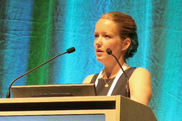
WASHINGTON – When the results of a noninvasive prenatal test are positive for aneuploidy but follow-up fetal karyotype results are normal, an undiagnosed maternal malignancy may be the reason why, Dr. Diana Bianchi reported at the International Conference on Prenatal Diagnosis and Therapy.
The conclusions are based on a study that evaluated several cases of cancers among apparently healthy women who had noninvasive prenatal testing (NIPT) during pregnancy with a cell-free DNA (cfDNA) test, which screens for fetal aneuploidy in chromosomes 13, 18, 21, X, and Y. In these cases, the NIPT results did not match the fetal karyotype results, which were obtained through amniocentesis or chorionic villus sampling.
While uncommon, “occult maternal malignancy is a potential biological explanation for discordant results,” between the fetal karyotype and NIPT, Dr. Bianchi, of the Mother Infant Research Institute at Tufts Medical Center, Boston, and lead author of the study, said at the meeting.

The study findings “underscore the need to perform a diagnostic procedure to ascertain the true fetal karyotype,” she added. The study was published July 13 in JAMA to coincide with Dr. Bianchi’s presentation at the International Conference on Prenatal Diagnosis and Therapy (doi:10.1001/jama.2015.7120).
In the study, she and her associates note that the discordant results are thought to be caused by cfDNA that is “released into the maternal circulation from apoptotic malignant cells.” The first case of a maternal cancer identified as the cause of discordant results between NIPT and fetal karyotype tests was identified in 2013, they said. Other explanations include the death of a twin or having received an organ transplant from a male donor. The researchers examined 125,426 NIPT results from asymptomatic pregnant women, which were processed at one lab between Feb. 15, 2012, and Sept. 30, 2014. Of these tests, 3,757 (3%) were positive for at least one aneuploidy involving chromosomes 13. 18, 21, X, or Y.
Among these cases, 10 women were subsequently diagnosed with cancer after abnormal NIPT results were reported (three cases of B-cell lymphoma and one case each of Hodgkin lymphoma; T-cell leukemia; unspecified adenocarcinoma; leiomyosarcoma; and neuroendocrine, colorectal, and anal carcinomas. Most (seven) had multiple aneuploidies, two had a single trisomy, and one had a single monosomy.
The investigators obtained more information from eight of the women. The other two women were critically ill and did not participate.
The mean age of the eight women was 35 years, and they were diagnosed with cancer while still pregnant or after delivery at a mean of 16 weeks after the first NIPT. Fetal karyotyping had been performed in seven of these cases and showed euploid results. The eight women were asymptomatic when they were screened, and the NIPT results “prompted” the diagnosis in three women, but whether earlier detection of the disease would have made a difference in the outcomes could not be determined, the authors wrote.
The follow-up of these patients was not complete, but the authors estimate that in 20%-44% of cases where multiple aneuploidies are detected and the results are discordant, an occult maternal cancer is the explanation. The study is described by the authors as “preliminary.” Limitations includes the retrospective design and incomplete follow-up information, they wrote.
During her presentation, Dr. Bianchi said that the highest risk group are women with multiple aneuploidies, “or suspiciously, a single autosomal monosomy,” the types of cases that “may warrant more detailed analysis of the whole genome.
“Further studies are needed to determine a recommended medical work-up for women” thought to be at high risk, she added. She referred to a recently published Belgian study, which evaluated a small group of women with the same types of suspicious NIPT results with MRI scans, which picked up evidence of tumors in all three of the women studied.
NIPT, considered an advanced screen not a diagnostic test, involves sequencing of cfDNA in maternal plasma, which contains placental DNA, “used as a proxy for the fetus,” and maternal DNA, the authors explained. NIPT is highly sensitive and specific for detecting trisomies 21, 18, and 13.
The cfDNA tests were manufactured by Illumina and were processed at the company’s clinical laboratory in Redwood City, Calif. Illumina funded the study, and sponsored research funding administered through Tufts Medical Center, which paid for the time that Dr. Bianchi spent working with Illumina employees to design the study, analyze the data, and prepare the manuscript.
This is “the first large-scale study that strengthens the premise that a false-positive NIPT result could be due to the presence of maternal malignancy,” Dr. Roberto Romero and Dr. Maurice Mahoney wrote in an editorial. “Since virtually all cancers have somatic genetic alterations that can be reflected in circulating cfDNA, and many of these derangements include chromosome 21, 18, and 13 (the focus of NIPT), it is plausible that discordant NIPT results could be explained by the presence of a maternal malignancy, particularly if the results of NIPT show multiple aneuploidies,” they wrote.
The results of the study need to be replicated and “the data emphasize the need for performing a diagnostic procedure to determine the fetal karyotype in all situations” where NIPT results are abnormal, especially since it is likely that the use of NIPT will increase in the next few years.
Dr. Romero is with the Eunice Kennedy Shriver National Institute of Child Health and Human Development, Bethesda, Md.; the University of Michigan, Ann Arbor; Michigan State University, East Lansing; and Wayne State University, Detroit. Dr. Mahoney is with the department of genetics, pediatrics, and obstetrics and gynecology at Yale University, New Haven, Conn. Their editorial was published July 13 in JAMA. Dr. Romero reported having no financial disclosures. Dr. Mahoney has been a principal investigator in a study of NIPT that used blood samples submitted to Natera under a contract with Yale University (JAMA 2015 July 13 [doi: 10.1001/jama.2015.7533]).
WASHINGTON – When the results of a noninvasive prenatal test are positive for aneuploidy but follow-up fetal karyotype results are normal, an undiagnosed maternal malignancy may be the reason why, Dr. Diana Bianchi reported at the International Conference on Prenatal Diagnosis and Therapy.
The conclusions are based on a study that evaluated several cases of cancers among apparently healthy women who had noninvasive prenatal testing (NIPT) during pregnancy with a cell-free DNA (cfDNA) test, which screens for fetal aneuploidy in chromosomes 13, 18, 21, X, and Y. In these cases, the NIPT results did not match the fetal karyotype results, which were obtained through amniocentesis or chorionic villus sampling.
While uncommon, “occult maternal malignancy is a potential biological explanation for discordant results,” between the fetal karyotype and NIPT, Dr. Bianchi, of the Mother Infant Research Institute at Tufts Medical Center, Boston, and lead author of the study, said at the meeting.
The study findings “underscore the need to perform a diagnostic procedure to ascertain the true fetal karyotype,” she added. The study was published July 13 in JAMA to coincide with Dr. Bianchi’s presentation at the International Conference on Prenatal Diagnosis and Therapy (doi:10.1001/jama.2015.7120).
In the study, she and her associates note that the discordant results are thought to be caused by cfDNA that is “released into the maternal circulation from apoptotic malignant cells.” The first case of a maternal cancer identified as the cause of discordant results between NIPT and fetal karyotype tests was identified in 2013, they said. Other explanations include the death of a twin or having received an organ transplant from a male donor. The researchers examined 125,426 NIPT results from asymptomatic pregnant women, which were processed at one lab between Feb. 15, 2012, and Sept. 30, 2014. Of these tests, 3,757 (3%) were positive for at least one aneuploidy involving chromosomes 13. 18, 21, X, or Y.
Among these cases, 10 women were subsequently diagnosed with cancer after abnormal NIPT results were reported (three cases of B-cell lymphoma and one case each of Hodgkin lymphoma; T-cell leukemia; unspecified adenocarcinoma; leiomyosarcoma; and neuroendocrine, colorectal, and anal carcinomas. Most (seven) had multiple aneuploidies, two had a single trisomy, and one had a single monosomy.
The investigators obtained more information from eight of the women. The other two women were critically ill and did not participate.
The mean age of the eight women was 35 years, and they were diagnosed with cancer while still pregnant or after delivery at a mean of 16 weeks after the first NIPT. Fetal karyotyping had been performed in seven of these cases and showed euploid results. The eight women were asymptomatic when they were screened, and the NIPT results “prompted” the diagnosis in three women, but whether earlier detection of the disease would have made a difference in the outcomes could not be determined, the authors wrote.
The follow-up of these patients was not complete, but the authors estimate that in 20%-44% of cases where multiple aneuploidies are detected and the results are discordant, an occult maternal cancer is the explanation. The study is described by the authors as “preliminary.” Limitations includes the retrospective design and incomplete follow-up information, they wrote.
During her presentation, Dr. Bianchi said that the highest risk group are women with multiple aneuploidies, “or suspiciously, a single autosomal monosomy,” the types of cases that “may warrant more detailed analysis of the whole genome.
“Further studies are needed to determine a recommended medical work-up for women” thought to be at high risk, she added. She referred to a recently published Belgian study, which evaluated a small group of women with the same types of suspicious NIPT results with MRI scans, which picked up evidence of tumors in all three of the women studied.
NIPT, considered an advanced screen not a diagnostic test, involves sequencing of cfDNA in maternal plasma, which contains placental DNA, “used as a proxy for the fetus,” and maternal DNA, the authors explained. NIPT is highly sensitive and specific for detecting trisomies 21, 18, and 13.
The cfDNA tests were manufactured by Illumina and were processed at the company’s clinical laboratory in Redwood City, Calif. Illumina funded the study, and sponsored research funding administered through Tufts Medical Center, which paid for the time that Dr. Bianchi spent working with Illumina employees to design the study, analyze the data, and prepare the manuscript.
AT ISPD 2015
Key clinical point: Discordant results between noninvasive prenatal testing (NIPT) for aneuploidy and fetal karyotype could be caused by an undiagnosed maternal malignancy.
Major finding: Of the 125,426 NIPT results, 3% (3,757) were positive for at least one aneuploidy involving chromosomes 13, 18, 21, X, or Y. Of these, 10 women were diagnosed with cancer.
Data source: A case series of 10 women diagnosed with cancer after abnormal NIPT results did not match the fetal karyotype, from a cohort of 125,426 NIPT tests processed at a single lab over a 2.5-year period.
Disclosures: The cfDNA tests were manufactured by Illumina and were processed at the company’s clinical laboratory in Redwood City, Calif. Illumina funded the study, and sponsored research funding administered through Tufts Medical Center, which paid for the time that Dr. Bianchi spent working with Illumina employees to design the study, analyze the data, and prepare the manuscript.

Depression, stress don’t predict outcomes in recurrent pregnancy loss
LISBON – Depression and emotional stress did not lead to poorer pregnancy outcomes 1 year after referral for recurrent pregnancy loss in a prospective, longitudinal study.
Among 287 women with recurrent pregnancy loss (RPL), 132 had a live birth or an ongoing pregnancy after 12 weeks gestation 1 year after referral to a tertiary RPL unit.
High scores on the PSS (Cohen Perceived Stress Scale) at referral were not associated with the chance of a subsequent live birth or ongoing pregnancy in unadjusted analysis (odds ratio, 1.012 ; 95% confidence interval 0.98-1.045) or after adjustment for maternal age and number of pregnancy losses prior to referral (OR, 1.015; CI 0.98-1.050).
Moderate to severe depression on the Major Depression Inventory (MDI) also was not associated with the chance of either outcome in unadjusted (OR, 2.12; CI 0.91-4.93) or adjusted (OR, 1.86; CI 0.78-4.42) analyses.

“We did not find an association between emotional distress at referral and the chance for a positive pregnancy outcome,” study author Dr. Astrid Marie Kolte said at the annual meeting of the European Society of Human Reproduction and Embryology.
It is well-known that pregnancy loss is a significant major life event that can invoke grief similar to that after a neonatal death. Studies have also shown that depression and stress are highly prominent among women with RPL.
In a recent study by Dr. Kolte and her associates, moderate to severe depression was identified in 8.6% and high stress levels in 41.2% of 301 women with three or more pregnancy losses before 12 weeks gestation, compared with rates of 2.2% and 23.2%, respectively, among 1,813 women without RPL (Hum. Reprod. 2015;30:777-782).Other groups have found significantly higher prevalence rates of moderate to severe depression in RPL patients, in the range of 15% to 33%, Dr. Kolte of the RPL Unit, Copenhagen University Hospital Rigshospitalet, said.
Data are scarce and results mixed, however, on the impact of depression and stress on subsequent live birth rates, prompting the investigators to examine pregnancy outcomes among the 301 previously studied women.
A total of 185 participants completed a follow-up questionnaire at 1 year containing the same PSS and MDI filled out a referral, along with questions about their pregnancy. Reliable data was available for 102 pregnancies among the 116 nonrespondents, resulting in 287 patients in the current analysis.
Among these women, 82 had given birth to a live child within the first year after referral, 50 had an ongoing pregnancy after 12 weeks’ gestation, and 11 were pregnant at less than 12 weeks’ gestation.
Interestingly, women with a live birth or ongoing pregnancy after 12 weeks’ gestation had significantly lower MDI (13.59 vs. 10.12) and PSS (16.94 vs. 12.47) scores on follow-up. This was not the case for patients without a positive pregnancy outcome for either the MDI (12.65 vs. 12.92) or PSS (16.86 vs. 15.49), Dr. Kolte said.
She noted that the study may have been insufficiently powered because of the low prevalence (8.6%) of major depression at referral and remarkably similar perceived stress among patients with and without live births. However, the study used validated psychometric scales, had good obstetrical follow-up, and is by far the largest study of emotional stress and live birth/ongoing pregnancy to date, she said.
Dr. Kolte expressed frustration at providing the best care for these patients, observing that there is a severe lack of understanding of the pathophysiology of recurrent pregnancy loss and no evidence-based treatment for the majority of RPL patients.
“You can indeed discuss whether a cumulative live birth rate of 70% on average is a good prognosis, but this is still a distressing event for these patients and their partners,” she said.
All newly referred patients and their partners are now being screened for major depression and perceived stress in the Danish RPL Unit, but providers do not have the means to treat those patients with psychological morbidities and must refer them back to their general physician.
“In my opinion, this is an understudied and underprioritized area in early pregnancy management, at least in Denmark,” Dr. Kolte said.
Several attendees echoed this frustration and asked Dr. Kolte for advice. While not all women will need psychological medication, having a psychologist associated with a RPL program is essential, as is a good working relationship with a psychiatrist, she replied.
University Hospital Copenhagen Rigshospitalet funded the study. Dr. Kolte reported having no financial disclosures.
LISBON – Depression and emotional stress did not lead to poorer pregnancy outcomes 1 year after referral for recurrent pregnancy loss in a prospective, longitudinal study.
Among 287 women with recurrent pregnancy loss (RPL), 132 had a live birth or an ongoing pregnancy after 12 weeks gestation 1 year after referral to a tertiary RPL unit.
High scores on the PSS (Cohen Perceived Stress Scale) at referral were not associated with the chance of a subsequent live birth or ongoing pregnancy in unadjusted analysis (odds ratio, 1.012 ; 95% confidence interval 0.98-1.045) or after adjustment for maternal age and number of pregnancy losses prior to referral (OR, 1.015; CI 0.98-1.050).
Moderate to severe depression on the Major Depression Inventory (MDI) also was not associated with the chance of either outcome in unadjusted (OR, 2.12; CI 0.91-4.93) or adjusted (OR, 1.86; CI 0.78-4.42) analyses.
“We did not find an association between emotional distress at referral and the chance for a positive pregnancy outcome,” study author Dr. Astrid Marie Kolte said at the annual meeting of the European Society of Human Reproduction and Embryology.
It is well-known that pregnancy loss is a significant major life event that can invoke grief similar to that after a neonatal death. Studies have also shown that depression and stress are highly prominent among women with RPL.
In a recent study by Dr. Kolte and her associates, moderate to severe depression was identified in 8.6% and high stress levels in 41.2% of 301 women with three or more pregnancy losses before 12 weeks gestation, compared with rates of 2.2% and 23.2%, respectively, among 1,813 women without RPL (Hum. Reprod. 2015;30:777-782).Other groups have found significantly higher prevalence rates of moderate to severe depression in RPL patients, in the range of 15% to 33%, Dr. Kolte of the RPL Unit, Copenhagen University Hospital Rigshospitalet, said.
Data are scarce and results mixed, however, on the impact of depression and stress on subsequent live birth rates, prompting the investigators to examine pregnancy outcomes among the 301 previously studied women.
A total of 185 participants completed a follow-up questionnaire at 1 year containing the same PSS and MDI filled out a referral, along with questions about their pregnancy. Reliable data was available for 102 pregnancies among the 116 nonrespondents, resulting in 287 patients in the current analysis.
Among these women, 82 had given birth to a live child within the first year after referral, 50 had an ongoing pregnancy after 12 weeks’ gestation, and 11 were pregnant at less than 12 weeks’ gestation.
Interestingly, women with a live birth or ongoing pregnancy after 12 weeks’ gestation had significantly lower MDI (13.59 vs. 10.12) and PSS (16.94 vs. 12.47) scores on follow-up. This was not the case for patients without a positive pregnancy outcome for either the MDI (12.65 vs. 12.92) or PSS (16.86 vs. 15.49), Dr. Kolte said.
She noted that the study may have been insufficiently powered because of the low prevalence (8.6%) of major depression at referral and remarkably similar perceived stress among patients with and without live births. However, the study used validated psychometric scales, had good obstetrical follow-up, and is by far the largest study of emotional stress and live birth/ongoing pregnancy to date, she said.
Dr. Kolte expressed frustration at providing the best care for these patients, observing that there is a severe lack of understanding of the pathophysiology of recurrent pregnancy loss and no evidence-based treatment for the majority of RPL patients.
“You can indeed discuss whether a cumulative live birth rate of 70% on average is a good prognosis, but this is still a distressing event for these patients and their partners,” she said.
All newly referred patients and their partners are now being screened for major depression and perceived stress in the Danish RPL Unit, but providers do not have the means to treat those patients with psychological morbidities and must refer them back to their general physician.
“In my opinion, this is an understudied and underprioritized area in early pregnancy management, at least in Denmark,” Dr. Kolte said.
Several attendees echoed this frustration and asked Dr. Kolte for advice. While not all women will need psychological medication, having a psychologist associated with a RPL program is essential, as is a good working relationship with a psychiatrist, she replied.
University Hospital Copenhagen Rigshospitalet funded the study. Dr. Kolte reported having no financial disclosures.
AT ESHRE 2015
Key clinical point: Feelings of stress or depression do not lead to poorer pregnancy outcomes in the first year after referral for recurrent pregnancy loss.
Major finding: Moderate to severe depression was not associated with the chance of a subsequent live birth or ongoing pregnancy in unadjusted (OR, 2.12; 95% CI 0.91-4.93) or adjusted (OR, 1.86; CI 0.78-4.42) analyses.
Data source: Prospective, longitudinal study of 287 patients with recurrent pregnancy loss.
Disclosures: University Hospital Copenhagen Rigshospitalet funded the study. Dr. Kolte reported having no financial disclosures.
Analysis shows link between certain SSRIs and birth defect risk
The periconceptional use of some selective serotonin reuptake inhibitors (SSRIs) appears to have no impact on the development of birth defects, but some birth defects occur more frequently among infants of women treated with paroxetine or fluoxetine early in pregnancy, results from a Bayesian analysis demonstrated.
“Recent meta-analyses and systematic reviews combining data from more than 20 epidemiological studies have reached conflicting conclusions and this uncertainty influences perceptions of the safety of antidepressant use in pregnancy,” researchers led by Jennita Reefhuis, Ph.D., of the National Center on Birth Defects and Developmental Disabilities at the Centers for Disease Control and Prevention, Atlanta, wrote in the British Medical Journal on July 8.

“SSRIs are increasingly used by women of reproductive age and during pregnancy, but the inconsistent reports have limited opportunities for clinicians to carefully evaluate the risk compared with benefit of specific SSRIs for a given patient during pregnancy,” they said.
In an effort to evaluate the association between individual SSRIs and birth defects, the researchers used Bayesian methods to summarize independent findings identified in the literature and to update those findings using data from the U.S. National Birth Defects Prevention Study (NBDPS).
The analysis drew from 10 centers in the United States and included 17,952 mothers of infants with birth defects and 9,857 mothers of infants without birth defects, with estimated dates of delivery between 1997 and 2009. Exposures included the use of citalopram, escitalopram, fluoxetine, paroxetine, or sertraline in the month before through the third month of pregnancy (BMJ 2015;350:h3190 [doi:10.1136/bmj.h3190]).
The main outcome measure was the 14 categories of birth defects that, according to the medical literature, had associations with SSRIs: neural tube defects, anencephaly, all septal defects, ventricular septal defects, right ventricular outflow tract obstructions, cleft palate, cleft lip with or without cleft palate, esophageal atresia, anal atresia, hypospadias, any limb reduction defect, craniosynostosis, gastroschisis, and omphalocele.
The researchers found that sertraline was the most commonly used SSRI (reported use in early pregnancy was about 40%), but none of five previously reported birth defects associated with use of sertraline was confirmed. For nine previously reported associations between maternal SSRI use and birth defects in infants, the researchers found no association.
High posterior odds ratios were observed for five birth defects with paroxetine use: anencephaly (3.2); atrial septal defects (1.8); right ventricular outflow tract obstruction defects (2.4); gastroschisis (2.5); and omphalocele (3.5). Additionally, higher posterior odds ratios were observed for two defects with fluoxetine use: right ventricular outflow tract obstruction cardiac defects (2.0) and craniosynostosis (1.9).
“Although our analysis strongly supports the validity of the associations that were observed, the increase in the absolute risks, if the associations are causal, is small,” the researchers wrote.
They noted that the two strongest posterior odds ratios were seen for maternal paroxetine treatment and anencephaly or right ventricular outflow tract obstruction cardiac defects in the infant. If these associations are causal, the absolute risks in the children of women who are treated with paroxetine early in pregnancy would increase for anencephaly from 2 per 10,000 to 7 per 10,000. For right ventricular outflow tract obstruction cardiac defects, the absolute risk would increase from 10 per 10,000 to 24 per 10,000.
“The absolute risks for these birth defects are still low,” the researchers wrote.
The authors pointed out that their study confirms the need to assess the association between specific SSRIs and specific birth defects, rather than combining an entire drug class or a heterogeneous group of birth defects.
“Although SSRIs are similar pharmacologically, there are chemical differences, and if any of them do have teratogenic activity, it may be completely unrelated to the inhibition of serotonin receptors,” they wrote. ”SSRIs also differ pharmacokinetically, and this could account for differences in teratogenic activity, whether or not the mechanism involved inhibition of serotonin receptors.”
Data collection for the study was funded by the CDC. The authors reported having no financial disclosures.
The periconceptional use of some selective serotonin reuptake inhibitors (SSRIs) appears to have no impact on the development of birth defects, but some birth defects occur more frequently among infants of women treated with paroxetine or fluoxetine early in pregnancy, results from a Bayesian analysis demonstrated.
“Recent meta-analyses and systematic reviews combining data from more than 20 epidemiological studies have reached conflicting conclusions and this uncertainty influences perceptions of the safety of antidepressant use in pregnancy,” researchers led by Jennita Reefhuis, Ph.D., of the National Center on Birth Defects and Developmental Disabilities at the Centers for Disease Control and Prevention, Atlanta, wrote in the British Medical Journal on July 8.
“SSRIs are increasingly used by women of reproductive age and during pregnancy, but the inconsistent reports have limited opportunities for clinicians to carefully evaluate the risk compared with benefit of specific SSRIs for a given patient during pregnancy,” they said.
In an effort to evaluate the association between individual SSRIs and birth defects, the researchers used Bayesian methods to summarize independent findings identified in the literature and to update those findings using data from the U.S. National Birth Defects Prevention Study (NBDPS).
The analysis drew from 10 centers in the United States and included 17,952 mothers of infants with birth defects and 9,857 mothers of infants without birth defects, with estimated dates of delivery between 1997 and 2009. Exposures included the use of citalopram, escitalopram, fluoxetine, paroxetine, or sertraline in the month before through the third month of pregnancy (BMJ 2015;350:h3190 [doi:10.1136/bmj.h3190]).
The main outcome measure was the 14 categories of birth defects that, according to the medical literature, had associations with SSRIs: neural tube defects, anencephaly, all septal defects, ventricular septal defects, right ventricular outflow tract obstructions, cleft palate, cleft lip with or without cleft palate, esophageal atresia, anal atresia, hypospadias, any limb reduction defect, craniosynostosis, gastroschisis, and omphalocele.
The researchers found that sertraline was the most commonly used SSRI (reported use in early pregnancy was about 40%), but none of five previously reported birth defects associated with use of sertraline was confirmed. For nine previously reported associations between maternal SSRI use and birth defects in infants, the researchers found no association.
High posterior odds ratios were observed for five birth defects with paroxetine use: anencephaly (3.2); atrial septal defects (1.8); right ventricular outflow tract obstruction defects (2.4); gastroschisis (2.5); and omphalocele (3.5). Additionally, higher posterior odds ratios were observed for two defects with fluoxetine use: right ventricular outflow tract obstruction cardiac defects (2.0) and craniosynostosis (1.9).
“Although our analysis strongly supports the validity of the associations that were observed, the increase in the absolute risks, if the associations are causal, is small,” the researchers wrote.
They noted that the two strongest posterior odds ratios were seen for maternal paroxetine treatment and anencephaly or right ventricular outflow tract obstruction cardiac defects in the infant. If these associations are causal, the absolute risks in the children of women who are treated with paroxetine early in pregnancy would increase for anencephaly from 2 per 10,000 to 7 per 10,000. For right ventricular outflow tract obstruction cardiac defects, the absolute risk would increase from 10 per 10,000 to 24 per 10,000.
“The absolute risks for these birth defects are still low,” the researchers wrote.
The authors pointed out that their study confirms the need to assess the association between specific SSRIs and specific birth defects, rather than combining an entire drug class or a heterogeneous group of birth defects.
“Although SSRIs are similar pharmacologically, there are chemical differences, and if any of them do have teratogenic activity, it may be completely unrelated to the inhibition of serotonin receptors,” they wrote. ”SSRIs also differ pharmacokinetically, and this could account for differences in teratogenic activity, whether or not the mechanism involved inhibition of serotonin receptors.”
Data collection for the study was funded by the CDC. The authors reported having no financial disclosures.
FROM BMJ
Key clinical point: Some SSRIs may be associated with an increased risk of birth defects.
Major finding: The two strongest posterior odds ratios for the development of birth defects were seen for maternal paroxetine treatment and anencephaly (3.2) or right ventricular outflow tract obstruction cardiac defects (2.4) in infants.
Data source: A Bayesian analysis that drew from 10 centers in the United States and included 17,952 mothers of infants with birth defects and 9,857 mothers of infants without birth defects, with estimated dates of delivery between 1997 and 2009.
Disclosures: Data collection for the study was funded by the Centers for Disease Control and Prevention. The authors reported having no financial disclosures.

Reasonable ovarian stimulation doesn’t increase preterm birth risk
LISBON – A large observational study found no increased risk of preterm birth or low birth weight following in vitro fertilization within acceptable limits of ovarian stimulation compared with unstimulated IVF.
“These findings support that ovarian stimulation is safe for maximizing live birth rates when you use acceptable limits of ovarian stimulation such as less than 20 oocytes,” Dr. Sesh Kamal Sunkara said at the annual meeting of the European Society of Human Reproduction and Embryology.

Epigenetic modifications resulting from ovarian stimulation or embryo culture are under increasing scrutiny as possible contributory factors to adverse perinatal outcomes.
A recent analysis of almost 66,000 singleton births by Dr. Sunkara and her associates showed a significantly higher risk of preterm birth (PTB) and low birth weight (LBW) in women with an excessive response (> 20 oocytes) to ovarian stimulation versus those with a normal response (10-15 oocytes) (Hum. Reprod. 2015; 30:1473-80).
A 2013 systematic review and meta-analysis also identified a higher risk of PTB and early PTB among singletons born after blastocyst- versus cleavage-stage embryo transfer in IVF (Fertil. Steril. 2013; 100: 1615-21.e10).
Both of these confounding factors were taken into account in the current analysis, reported Dr. Sunkara, of Aberdeen Fertility Centre, Aberdeen Maternity Hospital, University of Aberdeen, Scotland.
Using the Human Fertilisation and Embryology Authority database, which includes data for all IVF cycles performed in the United Kingdom from 1991 to 2012, the investigators examined 719,220 fresh IVF stimulated cycles and 135,570 fresh IVF unstimulated cycles, resulting in 105,374 and 10,668 singleton live births, respectively. Surprisingly, most women in either group were aged 18-34 years at the time of treatment, Dr. Sunkara said.
A large proportion of the unstimulated cycles did not have any oocytes retrieved compared with the stimulated group (41.7% vs. ~7%).
The overall birth rate per cycle was significantly higher with stimulation than without stimulation (19.4% vs. 8%), as was the multiple birth rate (24.4% vs. 2.1%), she said.
In the unadjusted analyses, the stimulated versus unstimulated group had significantly higher rates of PTB (9.2% vs. 5.5%; odds ratio, 1.72; 95% confidence interval 1.58-1.88), early PTB (1.7% vs. 0.7%; OR, 2.36; CI, 1.88-2.96), LBW (9.3% vs. 5.1%; OR, 1.91; CI, 1.75-2.09), and very LBW (1.8% vs. 0.8%; OR, 2.23; CI, 1.80-2.77).
No significant differences were observed, however, for each outcome between stimulated and unstimulated cycles following logistic regression and adjustment for maternal age, year of treatment, previous IVF cycles, previous live birth, number of oocytes retrieved (≤ 20 or > 20), and day of embryo transfer (cleavage or blastocyst stage), Dr. Sunkara said.
The adjusted odds ratios were: PTB (aOR, 1.04; C.I. 0.60-1.80), early PTB (aOR, 1.60; C.I. 0.51-5.01), LBW (aOR, 1.93; C.I. 0.95-3.94), and very LBW (aOR, 1.01; C.I. 0.55-4.22).
“The results demonstrated that safe stimulation within acceptable limits does not increase the risk of PTB and LBW,” she said.
Dr. Sunkara reported no having no financial conflicts.
LISBON – A large observational study found no increased risk of preterm birth or low birth weight following in vitro fertilization within acceptable limits of ovarian stimulation compared with unstimulated IVF.
“These findings support that ovarian stimulation is safe for maximizing live birth rates when you use acceptable limits of ovarian stimulation such as less than 20 oocytes,” Dr. Sesh Kamal Sunkara said at the annual meeting of the European Society of Human Reproduction and Embryology.
Epigenetic modifications resulting from ovarian stimulation or embryo culture are under increasing scrutiny as possible contributory factors to adverse perinatal outcomes.
A recent analysis of almost 66,000 singleton births by Dr. Sunkara and her associates showed a significantly higher risk of preterm birth (PTB) and low birth weight (LBW) in women with an excessive response (> 20 oocytes) to ovarian stimulation versus those with a normal response (10-15 oocytes) (Hum. Reprod. 2015; 30:1473-80).
A 2013 systematic review and meta-analysis also identified a higher risk of PTB and early PTB among singletons born after blastocyst- versus cleavage-stage embryo transfer in IVF (Fertil. Steril. 2013; 100: 1615-21.e10).
Both of these confounding factors were taken into account in the current analysis, reported Dr. Sunkara, of Aberdeen Fertility Centre, Aberdeen Maternity Hospital, University of Aberdeen, Scotland.
Using the Human Fertilisation and Embryology Authority database, which includes data for all IVF cycles performed in the United Kingdom from 1991 to 2012, the investigators examined 719,220 fresh IVF stimulated cycles and 135,570 fresh IVF unstimulated cycles, resulting in 105,374 and 10,668 singleton live births, respectively. Surprisingly, most women in either group were aged 18-34 years at the time of treatment, Dr. Sunkara said.
A large proportion of the unstimulated cycles did not have any oocytes retrieved compared with the stimulated group (41.7% vs. ~7%).
The overall birth rate per cycle was significantly higher with stimulation than without stimulation (19.4% vs. 8%), as was the multiple birth rate (24.4% vs. 2.1%), she said.
In the unadjusted analyses, the stimulated versus unstimulated group had significantly higher rates of PTB (9.2% vs. 5.5%; odds ratio, 1.72; 95% confidence interval 1.58-1.88), early PTB (1.7% vs. 0.7%; OR, 2.36; CI, 1.88-2.96), LBW (9.3% vs. 5.1%; OR, 1.91; CI, 1.75-2.09), and very LBW (1.8% vs. 0.8%; OR, 2.23; CI, 1.80-2.77).
No significant differences were observed, however, for each outcome between stimulated and unstimulated cycles following logistic regression and adjustment for maternal age, year of treatment, previous IVF cycles, previous live birth, number of oocytes retrieved (≤ 20 or > 20), and day of embryo transfer (cleavage or blastocyst stage), Dr. Sunkara said.
The adjusted odds ratios were: PTB (aOR, 1.04; C.I. 0.60-1.80), early PTB (aOR, 1.60; C.I. 0.51-5.01), LBW (aOR, 1.93; C.I. 0.95-3.94), and very LBW (aOR, 1.01; C.I. 0.55-4.22).
“The results demonstrated that safe stimulation within acceptable limits does not increase the risk of PTB and LBW,” she said.
Dr. Sunkara reported no having no financial conflicts.
AT ESHRE 2015
Key clinical point: Ovarian stimulation within acceptable limits does not increase the risk for preterm birth or low birth weight.
Major finding: The adjusted odds between stimulated and unstimulated cycles was similar for preterm birth (aOR, 1.04), early preterm birth (aOR 1.60), low birth weight (aOR, 1.93), and very low birth weight (aOR, 1.01).
Data source: An observational analysis of 116,042 singleton live births.
Disclosures: Dr. Sunkara reported no having no financial conflicts.

Hospitals vary widely in low-risk delivery costs
The cost of a low-risk childbirth varies widely across the country, but research is lacking to fully understand why, according to new report in Health Affairs.
Overall, the average hospital facility cost for a maternity stay with a low-risk delivery ranged from $1,189 to $11,986 across 463 hospitals, with a mean of $4,485. There was more than a twofold difference between the 10th and 90th percentiles, Xiao Xu, Ph. D., from Yale University, New Haven, Conn., and her colleagues reported.
“Contrary to the common belief that spending more leads to improved outcomes or at least maintains quality of care, we found a significant positive association between estimated hospital facility cost and serious maternal morbidity rate for low-risk childbirths,” the researchs wrote.
The variation remained regardless of the method of delivery, though the range was wider for cesarean delivery. The average cost of vaginal childbirths ranged from $1,183 to $11,819, and costs associated with a cesarean delivery ranged from $1,249 to $13,688 (Health Aff. 2015; 34: 1212-19 [doi:10.1377/hltaff.2014.1088]).
The findings are based on an analysis of discharge data from the 2011 Nationwide Inpatient Sample, which includes data on nonfederal short-term hospitals in 46 states. The researchers focused on low-risk childbirths and excluded deliveries with identified maternal comorbidities, such as preeclampsia, hypertensive disorders, diabetes, and obesity. They also excluded birth with obstetric risk factors, such as multiple gestation or previous cesarean delivery. The final sample included 463 hospitals and 267,120 births. After weighting the sample, the researchers estimated that the sample represented 1.3 million births nationwide.
Researchers found that hospital characteristics accounted for only 13% of the variation in average costs. For example, research showed that costs were significantly higher at rural hospitals (all were nonteaching facilities) than at urban nonteaching hospitals, but costs between urban teaching and urban nonteaching hospitals were comparable. Additionally, compared with investor-owned private hospitals, nonfederal government hospitals and nonprofit private hospitals had significantly higher facility costs.
After other hospital factors were adjusted for, the researchers found that a 1% increase in the serious maternal morbidity rate was associated with a $296 increase in the average facility cost for a maternity stay across low-risk births.
The rate of cesarean deliveries in the study hospitals ranged from 2% to 39% for low-risk births. In one model, researchers estimated that average facility costs for maternity stays were about $432 higher in hospitals with high cesarean delivery rates than in those with low rates. But after the researchers adjusted for mean length of stay, the association was no longer significant.
The researchers recommended establishing standard definitions and management guidelines for common indications for cesarean delivery, such as labor dystocia and abnormal fetal heart rate tracing to help bring down cesarean rates. “The safe reduction of cesarean deliveries may help reduce facility costs and cost variation for childbirth-related hospitalizations,” Dr. Xu and her colleagues wrote.
The wide variation in maternity stay facility costs presents an opportunity for overall cost containment, the researchers wrote, noting that if hospitals above the 75th percentile could reduce costs to the 75th percentile, it would have generated savings of $290 million in 2011 for costs associated with low-risk childbirths.
The study was supported by an award from the Blue Cross Blue Shield of Michigan Foundation. Dr. Harlan Krumholz, one of the study authors, is chair of the Cardiac Scientific Advisory Board for UnitedHealthcare and has contracts with Medtronic Inc. and Johnson and Johnson.
FROM HEALTH AFFAIRS
Key clinical point: The cost of maternity hospital stays varies widely at U.S. hospitals even among low-risk deliveries.
Major finding: The overall average hospital facility cost for a low-risk delivery ranged from $1,189 to $11,986, with a mean cost of $4,485.
Data source: An analysis of discharge data from the 2011 Nationwide Inpatient Sample, including 463 hospitals and 267,120 births.
Disclosures: The study was supported by an award from the Blue Cross Blue Shield of Michigan Foundation. Dr. Harlan Krumholz, one of the study authors, is chair of the Cardiac Scientific Advisory Board for UnitedHealthcare and has contracts with Medtronic Inc. and Johnson & Johnson.
More Medicaid-insured infants survive with enhanced prenatal care program
Participation in Michigan’s statewide Medicaid enhanced prenatal care program led to significant reductions in infant mortality and newborn deaths among families insured by Medicaid, according to a recent study.
Researchers determined that every 1,000 additional Medicaid-insured births in which the mother participates in the state’s Maternal Infant Health Program (MIHP) would prevent two infant deaths.

“Our study suggests that a state Medicaid-sponsored, population-based, home-visiting enhanced prenatal care program can be a successful approach to reduce mortality risk among Medicaid-insured infants of all races,” wrote Cristian I. Meghea, Ph.D., and his associates at Michigan State University in East Lansing. “The reduced risk of death among infants participating in the enhanced prenatal care program, compared with matched nonparticipants, is consistent with previous findings on the effects of the program on health care utilization and birth outcomes.”
They estimated that program participation could prevent 28% of potential infant deaths among MIHP participants, most likely because of reduced risks of adverse birth outcomes, they reported online (Pediatrics 2015 July 6 [doi:10.1542/peds.2015-0479]).
The researchers analyzed the birth and death records and Medicaid medical claims for all 248,059 Medicaid-insured singleton children born in Michigan between Jan. 1, 2009 and Dec. 31, 2012. The team investigated maternal medical claims running from 3 months before conception through the child’s first birthday. The researchers matched mothers who participated in MIHP by the end of her second trimester with those who had no MIHP involvement, taking into account age, marital status, race/ethnicity, socioeconomic status, county of residence, any smoking in pregnancy, and having a first birth or a birth within the previous 18 months before conception.
Children who participated at all in the MIHP had 27% lower odds of death (odds ratio 0.73) in their first year of life than those with no participation, a number that ranged from 29% reduced odds for black infants to 26% reduced odds for nonblack infants. Infants in the program also had 30% lower odds of neonatal death and 22% lower odds of post neonatal death, compared with those not involved in MIHP.
Further reductions occurred in all these mortality measures when mothers enrolled in MIHP and underwent screening by the end of the second trimester of pregnancy, followed by at least three more prenatal follow-ups in the program. Among this group, odds of infant mortality dropped 30%, odds of neonatal death dropped 33%, and post neonatal death odds dropped 26%.
“Programs targeting Medicaid-insured pregnant women that bundle interventions addressing multiple determinants at multiple levels can be an important mechanism to reach underserved women and their infants at greater risk of infant death,” the authors concluded.
The Michigan Department of Community Health supported the study. The authors reported no relevant financial disclosures.
Participation in Michigan’s statewide Medicaid enhanced prenatal care program led to significant reductions in infant mortality and newborn deaths among families insured by Medicaid, according to a recent study.
Researchers determined that every 1,000 additional Medicaid-insured births in which the mother participates in the state’s Maternal Infant Health Program (MIHP) would prevent two infant deaths.
“Our study suggests that a state Medicaid-sponsored, population-based, home-visiting enhanced prenatal care program can be a successful approach to reduce mortality risk among Medicaid-insured infants of all races,” wrote Cristian I. Meghea, Ph.D., and his associates at Michigan State University in East Lansing. “The reduced risk of death among infants participating in the enhanced prenatal care program, compared with matched nonparticipants, is consistent with previous findings on the effects of the program on health care utilization and birth outcomes.”
They estimated that program participation could prevent 28% of potential infant deaths among MIHP participants, most likely because of reduced risks of adverse birth outcomes, they reported online (Pediatrics 2015 July 6 [doi:10.1542/peds.2015-0479]).
The researchers analyzed the birth and death records and Medicaid medical claims for all 248,059 Medicaid-insured singleton children born in Michigan between Jan. 1, 2009 and Dec. 31, 2012. The team investigated maternal medical claims running from 3 months before conception through the child’s first birthday. The researchers matched mothers who participated in MIHP by the end of her second trimester with those who had no MIHP involvement, taking into account age, marital status, race/ethnicity, socioeconomic status, county of residence, any smoking in pregnancy, and having a first birth or a birth within the previous 18 months before conception.
Children who participated at all in the MIHP had 27% lower odds of death (odds ratio 0.73) in their first year of life than those with no participation, a number that ranged from 29% reduced odds for black infants to 26% reduced odds for nonblack infants. Infants in the program also had 30% lower odds of neonatal death and 22% lower odds of post neonatal death, compared with those not involved in MIHP.
Further reductions occurred in all these mortality measures when mothers enrolled in MIHP and underwent screening by the end of the second trimester of pregnancy, followed by at least three more prenatal follow-ups in the program. Among this group, odds of infant mortality dropped 30%, odds of neonatal death dropped 33%, and post neonatal death odds dropped 26%.
“Programs targeting Medicaid-insured pregnant women that bundle interventions addressing multiple determinants at multiple levels can be an important mechanism to reach underserved women and their infants at greater risk of infant death,” the authors concluded.
The Michigan Department of Community Health supported the study. The authors reported no relevant financial disclosures.
FROM PEDIATRICS
Key clinical point: State Medicaid enhanced prenatal care program participation reduces infant deaths.
Major finding: Infant mortality odds dropped 27% among participants in Michigan’s Maternal Infant Health Program.
Data source: The findings are based on an analysis of birth and death records, Medicaid claims, and Maternal Infant Health Program participation for 248,059 singleton infants born from January 2009 through December 2012 in Michigan.
Disclosures: The Michigan Department of Community Health supported the study. The authors reported no relevant financial disclosures.
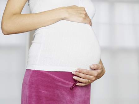
Delivery mortality and complications higher in epileptic women
Pregnant women with a history of epilepsy are at increased risk for death and complications during delivery, according to a new study.
It is estimated that women with a history of epilepsy comprise 0.3%-0.5% of all pregnancies, but there are limited data on the risk of death and complications in the pregnant epileptic patient, Sarah MacDonald of Harvard T.H. Chan School of Public Health, Boston, and her colleagues wrote in their paper published online July 6 in JAMA Neurology.
Ms. MacDonald and her associates sought to determine if patients with a history of epilepsy are at increased risk for morbidity and mortality during delivery by conducting a retrospective cohort study of a weighted sample of 20,449,532 women without and 69,385 women with epilepsy in the Nationwide Inpatient Sample from 2007 to 2011. They obtained epilepsy diagnoses and baseline characteristics from ICD-9-CM codes. To limit the possibility of nonepileptic or eclampsia seizures being recorded as epilepsy, patients in the cohort were restricted to those without a history of hypertensive disorders and the ICD-9-CM code for “epilepsy, unspecified” was removed (JAMA Neurol. 2015 July 6 [doi:10.1001/jamaneurol.2015.1017]).
Patients with a history of epilepsy had a frequency of death of 80 per 100,000 pregnancies, versus 6 deaths per 100,000 pregnancies without a history of epilepsy. The researchers noted a greater than 10-fold higher odds of death for epileptic patients (adjusted odds ratio, 11.46; 95% confidence interval, 8.64-15.19) when accounting for age, income, location, year, and race.
Pregnant women with a history of epilepsy were found to have higher rates of labor induction, cesarean section delivery, longer hospital stays, pregnancy-related hypertension, preeclampsia, and hemorrhage. Likewise, the researchers noted increased rates of gestational diabetes, premature rupture of membranes, chorioamnionitis, and premature delivery in patients with epilepsy. Furthermore, the newborns of epileptic mothers had higher rates of stillbirth, fetal distress, and poor growth. Overall, pregnant women with a history of epilepsy had significantly greater odds of severe morbidity (adjusted OR, 2.53; 95% CI, 2.44-2.63).
“However, despite their heightened risk of complications, we did not observe evidence that women with epilepsy are routinely triaged to high-risk medical centers,” the authors noted. But even though “the risk is substantially heightened on the multiplicative scale,” they said that the absolute rate of maternal death during delivery “is still very rare even among women with epilepsy.”
“Why did WWE [women with epilepsy] die at a higher rate in the MacDonald et al study? The authors suggested a number of possibilities, ranging from obstetrical complications, complications resulting from seizures (aspiration or status epilepticus), and a high rate of SUDEP (sudden unexplained death in epilepsy).”
“The MacDonald et al study provides important new information and demonstrates several risks associated with pregnancy in WWE. However, it raises far more questions than it answers. Most WWE have uncomplicated pregnancies. We need to understand the mechanisms underlying these risks, including death, so that we can identify the specific population at risk and devise interventions to reduce these risks.”
Dr. Jacqueline French is with the Comprehensive Epilepsy Center at New York University, New York. Dr. Kimford Meador is with the Stanford Comprehensive Epilepsy Center at Stanford University in Palo Alto, Calif. Dr. French reported receiving research support from multiple sources, including pharmceutical companies developing epilepsy drugs, the National Institutes of Health, the Epilepsy Therapy Project, the Epilepsy Research Foundation, and the Epilepsy Study Consortium. Dr. Meador reported receiving research support from UCB and other sources, including the National Institutes of Health, the Patient-Centered Outcomes Research Institute, and the Epilepsy Study Consortium. They made these comments in an editorial accompanying the study by Ms. MacDonald and her associates (JAMA Neurol. 2015 July 6 [doi:10.1001/jamaneurol.2015.1356]).
Pregnant women with a history of epilepsy are at increased risk for death and complications during delivery, according to a new study.
It is estimated that women with a history of epilepsy comprise 0.3%-0.5% of all pregnancies, but there are limited data on the risk of death and complications in the pregnant epileptic patient, Sarah MacDonald of Harvard T.H. Chan School of Public Health, Boston, and her colleagues wrote in their paper published online July 6 in JAMA Neurology.
Ms. MacDonald and her associates sought to determine if patients with a history of epilepsy are at increased risk for morbidity and mortality during delivery by conducting a retrospective cohort study of a weighted sample of 20,449,532 women without and 69,385 women with epilepsy in the Nationwide Inpatient Sample from 2007 to 2011. They obtained epilepsy diagnoses and baseline characteristics from ICD-9-CM codes. To limit the possibility of nonepileptic or eclampsia seizures being recorded as epilepsy, patients in the cohort were restricted to those without a history of hypertensive disorders and the ICD-9-CM code for “epilepsy, unspecified” was removed (JAMA Neurol. 2015 July 6 [doi:10.1001/jamaneurol.2015.1017]).
Patients with a history of epilepsy had a frequency of death of 80 per 100,000 pregnancies, versus 6 deaths per 100,000 pregnancies without a history of epilepsy. The researchers noted a greater than 10-fold higher odds of death for epileptic patients (adjusted odds ratio, 11.46; 95% confidence interval, 8.64-15.19) when accounting for age, income, location, year, and race.
Pregnant women with a history of epilepsy were found to have higher rates of labor induction, cesarean section delivery, longer hospital stays, pregnancy-related hypertension, preeclampsia, and hemorrhage. Likewise, the researchers noted increased rates of gestational diabetes, premature rupture of membranes, chorioamnionitis, and premature delivery in patients with epilepsy. Furthermore, the newborns of epileptic mothers had higher rates of stillbirth, fetal distress, and poor growth. Overall, pregnant women with a history of epilepsy had significantly greater odds of severe morbidity (adjusted OR, 2.53; 95% CI, 2.44-2.63).
“However, despite their heightened risk of complications, we did not observe evidence that women with epilepsy are routinely triaged to high-risk medical centers,” the authors noted. But even though “the risk is substantially heightened on the multiplicative scale,” they said that the absolute rate of maternal death during delivery “is still very rare even among women with epilepsy.”
FROM JAMA NEUROLOGY
Key clinical point: Epilepsy is associated with increased risk of maternal death and complications surrounding delivery.
Major finding: Pregnant women with a history of epilepsy had a 10-fold increased likelihood of death (adjusted OR, 11.46; 95% CI, 8.64-15.19) and higher rates of complications.
Data source: A retrospective cohort study of pregnant women using data obtained from the Nationwide Inpatient Sample from 2007 to 2011.
Disclosures: The authors reported no conflicts of interest. The study was supported by a grant from the National Institute of Mental Health and the Eunice Kennedy Shriver National Institute of Child Health and Human Development.
Review shows D&C raises preterm birth risk
LISBON – Dilation and curettage increases the risk for prematurity in subsequent pregnancy, according to a systematic review and meta-analysis of nearly 2 million women.
Overall, D&C for first-trimester miscarriage or pregnancy termination increased the risk for preterm birth (< 37 weeks) by 29% (Odds ratio, 1.29; 95% confidence interval 1.17-1.42).
Subgroup and sensitivity analyses performed to test the robustness of the finding showed D&C upped the risk for very preterm birth (< 32 weeks) by 69% (OR, 1.69; C.I. 1.20-2.38) and critical preterm birth (< 28 weeks) by 68% (OR, 1.68; C.I. 1.47-1.92).

A dose-response relationship was also identified, with multiple D&C procedures raising the risk of preterm birth even higher (OR, 1.74; C.I. 1.10-2.76), Dr. Marike Lemmers reported at the annual meeting of the European Society of Human Reproduction and Embryology.
“This really questions the use of dilation and curettage as first-line treatment for women with a miscarriage or termination of pregnancy since other treatment options are non-invasive, safe, well-tolerated, cheap, and accessible,” Dr. Lemmers, of the Academic Medical Center, Amsterdam, the Netherlands, said.
The increasing popularity of misoprostol as an alternative to D&C or for cervical priming prior to curettage would suggest that D&C use is declining. Still, gynecologists in the Netherlands treat half of all miscarriages with D&C, a figure that likely reflects practice elsewhere, Dr. Willem Ankum, the principal investigator, said during a press briefing.
The authors hypothesize that the mechanism behind the increased risk from D&C is that dilation may damage the cervix and affect cervical tightness or may damage the endometrial lining, leading to a malpresentation.
The systematic review and meta-analysis included 21 studies (7 prospective cohort studies, 11 retrospective cohort studies, and 3 case-control studies) involving 66,003 women with a history of D&C for first trimester miscarriage or termination, and 1,781,786 controls with a medically managed miscarriage or termination or no such events. The quality of the studies varied from 7 to 29 based on the Strobe score.
The primary outcome of risk of preterm birth was significantly increased with D&C regardless of whether the analysis relied only on the prospective studies (OR, 1.28; C.I. 1.01-1.60), retrospective studies (OR, 1.27; C.I. 1.12-1.45), or case-control studies (OR, 1.44; C.I. 1.18-1.77), Dr. Lemmers said.
Several audience members questioned the inclusion of older trials dating back to the late 1970s before prostoglandins had been introduced for cycle priming. One attendee cited a recent Scottish cohort study showing a declining preterm birth rate in women with previous miscarriage, possibly attributable to the increasing use of misoprostol (Cytotec) for miscarriage or termination and cervical priming before D&C (PLoS. Med. 2013; 10: e1001481 [doi:10.1371/journal.pmed.1001481]).
Dr. Lemmers was aware of the Scottish study and said a funnel plot they performed showed “the exact same results” between older and newer studies. Further, a subgroup analysis of 38,905 women from studies dating from 1999 forward also showed an increased risk of preterm birth with D&C versus medical treatment (OR, 1.19; C.I. 1.10-1.28).
She added that it’s unlikely that the use of hysteroscopy with minimal dilation before in-vitro fertilization impacted results, but that outcomes may differ between women with a miscarriage versus termination of an attached pregnancy, although this calculation wasn’t possible based on the available data.
ZonMw, a Dutch organization for health research and development, funded the study. Dr. Lemmers and Dr. Ankum reported having no financial conflicts.
On Twitter @pwendl
LISBON – Dilation and curettage increases the risk for prematurity in subsequent pregnancy, according to a systematic review and meta-analysis of nearly 2 million women.
Overall, D&C for first-trimester miscarriage or pregnancy termination increased the risk for preterm birth (< 37 weeks) by 29% (Odds ratio, 1.29; 95% confidence interval 1.17-1.42).
Subgroup and sensitivity analyses performed to test the robustness of the finding showed D&C upped the risk for very preterm birth (< 32 weeks) by 69% (OR, 1.69; C.I. 1.20-2.38) and critical preterm birth (< 28 weeks) by 68% (OR, 1.68; C.I. 1.47-1.92).
A dose-response relationship was also identified, with multiple D&C procedures raising the risk of preterm birth even higher (OR, 1.74; C.I. 1.10-2.76), Dr. Marike Lemmers reported at the annual meeting of the European Society of Human Reproduction and Embryology.
“This really questions the use of dilation and curettage as first-line treatment for women with a miscarriage or termination of pregnancy since other treatment options are non-invasive, safe, well-tolerated, cheap, and accessible,” Dr. Lemmers, of the Academic Medical Center, Amsterdam, the Netherlands, said.
The increasing popularity of misoprostol as an alternative to D&C or for cervical priming prior to curettage would suggest that D&C use is declining. Still, gynecologists in the Netherlands treat half of all miscarriages with D&C, a figure that likely reflects practice elsewhere, Dr. Willem Ankum, the principal investigator, said during a press briefing.
The authors hypothesize that the mechanism behind the increased risk from D&C is that dilation may damage the cervix and affect cervical tightness or may damage the endometrial lining, leading to a malpresentation.
The systematic review and meta-analysis included 21 studies (7 prospective cohort studies, 11 retrospective cohort studies, and 3 case-control studies) involving 66,003 women with a history of D&C for first trimester miscarriage or termination, and 1,781,786 controls with a medically managed miscarriage or termination or no such events. The quality of the studies varied from 7 to 29 based on the Strobe score.
The primary outcome of risk of preterm birth was significantly increased with D&C regardless of whether the analysis relied only on the prospective studies (OR, 1.28; C.I. 1.01-1.60), retrospective studies (OR, 1.27; C.I. 1.12-1.45), or case-control studies (OR, 1.44; C.I. 1.18-1.77), Dr. Lemmers said.
Several audience members questioned the inclusion of older trials dating back to the late 1970s before prostoglandins had been introduced for cycle priming. One attendee cited a recent Scottish cohort study showing a declining preterm birth rate in women with previous miscarriage, possibly attributable to the increasing use of misoprostol (Cytotec) for miscarriage or termination and cervical priming before D&C (PLoS. Med. 2013; 10: e1001481 [doi:10.1371/journal.pmed.1001481]).
Dr. Lemmers was aware of the Scottish study and said a funnel plot they performed showed “the exact same results” between older and newer studies. Further, a subgroup analysis of 38,905 women from studies dating from 1999 forward also showed an increased risk of preterm birth with D&C versus medical treatment (OR, 1.19; C.I. 1.10-1.28).
She added that it’s unlikely that the use of hysteroscopy with minimal dilation before in-vitro fertilization impacted results, but that outcomes may differ between women with a miscarriage versus termination of an attached pregnancy, although this calculation wasn’t possible based on the available data.
ZonMw, a Dutch organization for health research and development, funded the study. Dr. Lemmers and Dr. Ankum reported having no financial conflicts.
On Twitter @pwendl
AT ESHRE 2015
Key clinical point: Dilation and curettage raises the risk of prematurity in subsequent pregnancy and has a dose-response relationship that ups the risk the more the procedure is performed.
Major finding: D&C increased the risk of preterm birth by 29% (OR, 1.29; 95% C.I. 1.17-1.42).
Data source: Systematic review and meta-analysis of 1.8 million women.
Disclosures: ZonMw, a Dutch organization for health research and development, funded the study. Dr. Lemmers and Dr. Ankum reported having no financial conflicts.
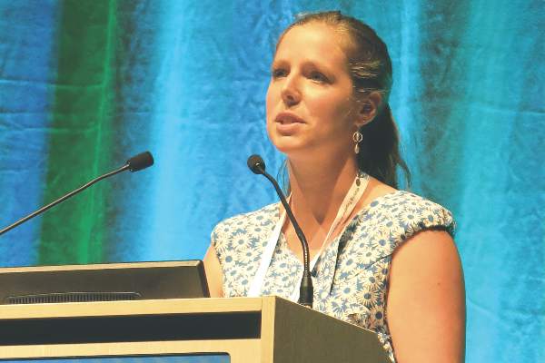
Expectant management holds its own in incomplete misoprostol miscarriage
LISBON – Expectant management is a safe and effective treatment for incomplete miscarriage after misoprostol, results of the MisoREST trial show.
“Because it is non-invasive, we think it should be considered as first-line treatment and to reserve curettage only for specific patients,” Dr. Marianne Verschoor said at the annual meeting of the European Society of Human Reproduction and Embryology.

Misoprostol (Cytotec) is an inexpensive and effective treatment for miscarriage, but about 30% of women are left with an incomplete evacuation of the uterus. Curettage is often performed in these typically asymptomatic women, but carries both short- and long-term risks, said Dr. Verschoor of the Academic Medical Center, Amsterdam.
No study had directly compared the effectiveness of curettage and expectant management, she said. To fill this void, the researchers performed a randomized controlled trial (RCT) and parallel observational cohort study at 27 community and academic hospitals. Women who had sonographic findings of intrauterine remnants and/or a thick endometrium (> 10 mm) 1-2 weeks after misoprostol treatment for first-trimester miscarriage were randomly assigned to curettage within 3 days of diagnosis or expectant management. Women who declined randomization were prospectively followed.
The primary outcome was sonographic evidence of an empty uterine cavity at 6 weeks or an uneventful clinical course at 3 months in the absence of sonography.
Because of strong preferences for expectant management (EM), 59 women were randomized in the RCT (30 curettage, 29 EM) and 197 women were followed prospectively (65 curettage, 132 EM). All groups were comparable at baseline with regard to age, gestational age, and previous miscarriage or curettage.
In the RCT, 93% of women had an empty cavity or uneventful clinical course with curettage vs. 76% with EM. This difference was not statistically significant (relative risk, 1.23; 95% confidence interval, 0.098-1.54), Dr. Verschoor said.
In the observational cohort, 95% of patients choosing curettage had a successful outcome vs. 83% choosing EM. This difference was statistically significant (RR, 1.14; CI, 1.04-1.26).
When the two populations were pooled, 95% of patients (90/95) treated with curettage reached the primary end point vs. 82% of patients (132/161) with EM, which was statistically different (RR, 1.16; CI, 1.06-1.26), she said.
There were no significant differences in secondary outcomes in the RCT between the curettage and EM groups, including intrauterine infection (each 1 patient), excessive blood loss defined as > 500 mL (1 patient vs. 2 patients), or duration of bleeding (mean 5.4 days vs. 5.5 days).
However, one patient had already developed Asherman syndrome in the curettage group within 3 months of follow-up, Dr. Verschoor said. Two patients in each arm underwent reintervention with hysteroscopy and 5 EM patients underwent curettage.
Secondary outcomes in the observational cohort were also comparable between the curettage and EM groups for intrauterine infection (3 vs. 2), excessive blood loss (0 vs. 1), and duration of bleeding (mean 5.7 days vs. 7.1 days). One patient who underwent curettage developed Asherman syndrome vs. none in the EM group, she said. There were 11 reinterventions in the curettage group (7 hysteroscopy, 4 recurettage), compared with 30 in the EM group (12 hysteroscopy, 18 curettage).
A closer look at the EM patients who underwent reintervention revealed that “the interventions were primarily elective,” Dr. Vershcoor said. “Only in 26% of cases was there a medical reason for performing the intervention like bleeding, fever, or excessive abdominal pain.” Histology performed in these patients found no pregnancy tissue in 46%.
A separate quality of life analysis of the MisoREST participants found no statistical differences at baseline, 2, 4, and 12 weeks for the Short Form 36 Health Survey mental and physical score in all four treatment arms. Anxiety and depression measured with the Hospital Anxiety and Depression Scale were also not statistically different at any time, study coauthor Dr. Marike Lemmers, also of the Academic Medical Center, Amsterdam, reported in a poster at the meeting.
ZonMw, a Dutch organization for health research and development, funded the study. Dr. Verschoor reported having no financial conflicts.
On Twitter @pwendl
LISBON – Expectant management is a safe and effective treatment for incomplete miscarriage after misoprostol, results of the MisoREST trial show.
“Because it is non-invasive, we think it should be considered as first-line treatment and to reserve curettage only for specific patients,” Dr. Marianne Verschoor said at the annual meeting of the European Society of Human Reproduction and Embryology.
Misoprostol (Cytotec) is an inexpensive and effective treatment for miscarriage, but about 30% of women are left with an incomplete evacuation of the uterus. Curettage is often performed in these typically asymptomatic women, but carries both short- and long-term risks, said Dr. Verschoor of the Academic Medical Center, Amsterdam.
No study had directly compared the effectiveness of curettage and expectant management, she said. To fill this void, the researchers performed a randomized controlled trial (RCT) and parallel observational cohort study at 27 community and academic hospitals. Women who had sonographic findings of intrauterine remnants and/or a thick endometrium (> 10 mm) 1-2 weeks after misoprostol treatment for first-trimester miscarriage were randomly assigned to curettage within 3 days of diagnosis or expectant management. Women who declined randomization were prospectively followed.
The primary outcome was sonographic evidence of an empty uterine cavity at 6 weeks or an uneventful clinical course at 3 months in the absence of sonography.
Because of strong preferences for expectant management (EM), 59 women were randomized in the RCT (30 curettage, 29 EM) and 197 women were followed prospectively (65 curettage, 132 EM). All groups were comparable at baseline with regard to age, gestational age, and previous miscarriage or curettage.
In the RCT, 93% of women had an empty cavity or uneventful clinical course with curettage vs. 76% with EM. This difference was not statistically significant (relative risk, 1.23; 95% confidence interval, 0.098-1.54), Dr. Verschoor said.
In the observational cohort, 95% of patients choosing curettage had a successful outcome vs. 83% choosing EM. This difference was statistically significant (RR, 1.14; CI, 1.04-1.26).
When the two populations were pooled, 95% of patients (90/95) treated with curettage reached the primary end point vs. 82% of patients (132/161) with EM, which was statistically different (RR, 1.16; CI, 1.06-1.26), she said.
There were no significant differences in secondary outcomes in the RCT between the curettage and EM groups, including intrauterine infection (each 1 patient), excessive blood loss defined as > 500 mL (1 patient vs. 2 patients), or duration of bleeding (mean 5.4 days vs. 5.5 days).
However, one patient had already developed Asherman syndrome in the curettage group within 3 months of follow-up, Dr. Verschoor said. Two patients in each arm underwent reintervention with hysteroscopy and 5 EM patients underwent curettage.
Secondary outcomes in the observational cohort were also comparable between the curettage and EM groups for intrauterine infection (3 vs. 2), excessive blood loss (0 vs. 1), and duration of bleeding (mean 5.7 days vs. 7.1 days). One patient who underwent curettage developed Asherman syndrome vs. none in the EM group, she said. There were 11 reinterventions in the curettage group (7 hysteroscopy, 4 recurettage), compared with 30 in the EM group (12 hysteroscopy, 18 curettage).
A closer look at the EM patients who underwent reintervention revealed that “the interventions were primarily elective,” Dr. Vershcoor said. “Only in 26% of cases was there a medical reason for performing the intervention like bleeding, fever, or excessive abdominal pain.” Histology performed in these patients found no pregnancy tissue in 46%.
A separate quality of life analysis of the MisoREST participants found no statistical differences at baseline, 2, 4, and 12 weeks for the Short Form 36 Health Survey mental and physical score in all four treatment arms. Anxiety and depression measured with the Hospital Anxiety and Depression Scale were also not statistically different at any time, study coauthor Dr. Marike Lemmers, also of the Academic Medical Center, Amsterdam, reported in a poster at the meeting.
ZonMw, a Dutch organization for health research and development, funded the study. Dr. Verschoor reported having no financial conflicts.
On Twitter @pwendl
AT ESHRE 2015
Key clinical point: Expectant management is effective and safe in women with incomplete evacuation of the uterus after misoprostol treatment for miscarriage.
Major finding: Curettage and expectant management resulted in similar rates of empty uterus at 6 weeks (95% vs. 82%).
Data source: A randomized controlled trial and prospective observational cohort.
Disclosures: ZonMw, a Dutch organization for health research and development, funded the study. Dr. Verschoor reported having no financial conflicts.

Age drives miscarriage risk in RA patients
LISBON – Advanced age appears to trump disease activity or antirheumatic drug treatment as the driving force behind miscarriage in women with rheumatoid arthritis.
“The risk for miscarriage in rheumatoid arthritis is age dependent,” Dr. Jenny Brouwer said at the annual meeting of the European Society of Human Reproduction and Embryology.

She reported on 162 evaluable pregnancies in 239 Dutch women enrolled during 2002-2010 in the nationwide Pregnancy-Induced Amelioration of Rheumatoid Arthritis (PARA) study, the world’s largest prospective cohort of pregnant RA patients. They had a mean age of 32.3 years and a mean RA duration of 4.9 years. Overall, 49.4% were nulliparous and 13.6% had suffered a previous miscarriage.
Prednisone (37%), sulfasalazine (33%), and NSAIDs (29%) were most commonly used in the periconceptional period. Women eligible for PARA had to have stopped methotrexate for at least 3 months prior to attempting to conceive, a parameter included because it is an indicator for more severe disease.
Varying reports have shown no increased miscarriage rate in women with RA, while others have reported a significantly higher risk. These studies, however, were cross-sectional or retrospective in nature and lacked detailed information on preconception disease activity and antirheumatic drug use, observed Dr. Brouwer of Erasmus Medical Center, Rotterdam, the Netherlands.
Among the 162 pregnancies, 28 miscarriages occurred (17.3%), roughly half in weeks 5-9.
Reassuringly, the miscarriage rate is comparable with that in the general population, she said. Indeed, a recent study from the Nurses Health Study II also reported a miscarriage rate of 17.3%.
Disease Activity Score based on 28 joints (DAS28) was higher 6 and 12 weeks after miscarriage than before pregnancy, with 33% of women having a disease flare after miscarriage.
“This can be a sign that women who have more active disease have a higher risk for miscarriage and should be monitored more closely when their disease activity in the preconceptional period increases,” Dr. Brouwer said.
Women who miscarried were significantly older than were women with an ongoing pregnancy (mean 33.9 years vs. 32 years; P = .022). They also were more likely to be positive for anti-citrullinated protein antibodies (ACPA) (82% vs. 60%; P = .058), have higher DAS28 scores (3.92 vs. 3.59; P = .166), and have used methotrexate in the past (82% vs. 68%; P = .174).
No significant association was observed between miscarriage and corticosteroids, anti-inflammatories, or biological therapies, she said.
Because of the low frequency of miscarriages in the study, logistic regression showed only a trend toward higher miscarriage risk in association with increasing age (OR, 1.12; P = .065 per year of age increase) and ACPA positivity (OR, 2.47; P = .092), Dr. Brouwer said.
“Since the confidence interval is 0.99-1.25, there clearly seems to be a trend toward a higher risk for occurrence of miscarriage with increasing age, which is only logical since in the general population we also see an increase in miscarriage rate with increasing age,” she said in an interview. “My expectation is that, in a larger RA cohort, with a larger absolute number of miscarriages, the association will be significant.”
Follow-up revealed that, within 1 year of miscarriage, 68% of women became pregnant again, 14% stopped trying to conceive, and 11% were lost to follow-up. The live birth rate in the subsequent pregnancy was 90%. A follow-up study of these children did not find an increased rate of congenital malformations, despite the use of RA medications during pregnancy, Dr. Brouwer said.
The audience questioned whether the investigators are suggesting that rheumatoid arthritis has no role in miscarriage, observing that the study outcomes look very similar to patients without RA. Dr. Brouwer responded that there seems to be a higher sensitivity in patients with ACPA using methotrexate in the past but that this failed to reach significance.
“The results found in this study: some association with more severe disease – ACPA positivity and past methotrexate use – although not significant are a confirmation to us that extra care should be given to suppressing active disease already before actively trying to achieve a pregnancy,” she told this publication.
For more details, the full manuscript has been published (Arthritis Rheumatol. 2015;67:1738-43).
Reumafond, the Dutch Arthritis Foundation, funded the study. Dr. Brouwer reported no financial conflicts.
LISBON – Advanced age appears to trump disease activity or antirheumatic drug treatment as the driving force behind miscarriage in women with rheumatoid arthritis.
“The risk for miscarriage in rheumatoid arthritis is age dependent,” Dr. Jenny Brouwer said at the annual meeting of the European Society of Human Reproduction and Embryology.
She reported on 162 evaluable pregnancies in 239 Dutch women enrolled during 2002-2010 in the nationwide Pregnancy-Induced Amelioration of Rheumatoid Arthritis (PARA) study, the world’s largest prospective cohort of pregnant RA patients. They had a mean age of 32.3 years and a mean RA duration of 4.9 years. Overall, 49.4% were nulliparous and 13.6% had suffered a previous miscarriage.
Prednisone (37%), sulfasalazine (33%), and NSAIDs (29%) were most commonly used in the periconceptional period. Women eligible for PARA had to have stopped methotrexate for at least 3 months prior to attempting to conceive, a parameter included because it is an indicator for more severe disease.
Varying reports have shown no increased miscarriage rate in women with RA, while others have reported a significantly higher risk. These studies, however, were cross-sectional or retrospective in nature and lacked detailed information on preconception disease activity and antirheumatic drug use, observed Dr. Brouwer of Erasmus Medical Center, Rotterdam, the Netherlands.
Among the 162 pregnancies, 28 miscarriages occurred (17.3%), roughly half in weeks 5-9.
Reassuringly, the miscarriage rate is comparable with that in the general population, she said. Indeed, a recent study from the Nurses Health Study II also reported a miscarriage rate of 17.3%.
Disease Activity Score based on 28 joints (DAS28) was higher 6 and 12 weeks after miscarriage than before pregnancy, with 33% of women having a disease flare after miscarriage.
“This can be a sign that women who have more active disease have a higher risk for miscarriage and should be monitored more closely when their disease activity in the preconceptional period increases,” Dr. Brouwer said.
Women who miscarried were significantly older than were women with an ongoing pregnancy (mean 33.9 years vs. 32 years; P = .022). They also were more likely to be positive for anti-citrullinated protein antibodies (ACPA) (82% vs. 60%; P = .058), have higher DAS28 scores (3.92 vs. 3.59; P = .166), and have used methotrexate in the past (82% vs. 68%; P = .174).
No significant association was observed between miscarriage and corticosteroids, anti-inflammatories, or biological therapies, she said.
Because of the low frequency of miscarriages in the study, logistic regression showed only a trend toward higher miscarriage risk in association with increasing age (OR, 1.12; P = .065 per year of age increase) and ACPA positivity (OR, 2.47; P = .092), Dr. Brouwer said.
“Since the confidence interval is 0.99-1.25, there clearly seems to be a trend toward a higher risk for occurrence of miscarriage with increasing age, which is only logical since in the general population we also see an increase in miscarriage rate with increasing age,” she said in an interview. “My expectation is that, in a larger RA cohort, with a larger absolute number of miscarriages, the association will be significant.”
Follow-up revealed that, within 1 year of miscarriage, 68% of women became pregnant again, 14% stopped trying to conceive, and 11% were lost to follow-up. The live birth rate in the subsequent pregnancy was 90%. A follow-up study of these children did not find an increased rate of congenital malformations, despite the use of RA medications during pregnancy, Dr. Brouwer said.
The audience questioned whether the investigators are suggesting that rheumatoid arthritis has no role in miscarriage, observing that the study outcomes look very similar to patients without RA. Dr. Brouwer responded that there seems to be a higher sensitivity in patients with ACPA using methotrexate in the past but that this failed to reach significance.
“The results found in this study: some association with more severe disease – ACPA positivity and past methotrexate use – although not significant are a confirmation to us that extra care should be given to suppressing active disease already before actively trying to achieve a pregnancy,” she told this publication.
For more details, the full manuscript has been published (Arthritis Rheumatol. 2015;67:1738-43).
Reumafond, the Dutch Arthritis Foundation, funded the study. Dr. Brouwer reported no financial conflicts.
AT ESHRE 2015
Key clinical point: Miscarriage rates are comparable in patients with rheumatoid arthritis and the general public and are driven largely by advanced age.
Major finding: Women who miscarried had a mean age of 33.9 years vs. 32 years in those with an ongoing pregnancy (P = .022)
Data source: Analysis of 162 pregnancies in a Dutch prospective cohort study.
Disclosures: Reumafond, the Dutch Arthritis Foundation, funded the study. Dr. Brouwer reported no financial conflicts.