User login
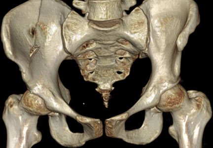
Discovery May Lead to New Drugs for Osteoporosis
Researchers at Washington University School of Medicine in St. Louis have discovered what appears to be a potent stimulator of new bone growth. The finding could lead to new treatments for osteoporosis and other diseases that occur when the body doesn’t make enough bone.
“We have been looking for new ways to stimulate bone formation,” said principal investigator Fanxin Long, PhD. “The tools we already have are very good at slowing the breakdown of bone, but we need better ways to stimulate new bone growth.”
Studying mice, Dr. Long and colleagues focused on a pathway involved in bone formation. The so-called WNT proteins carry messages into cells and regulate embryonic and adult tissue in mammals, including humans. The WNT proteins enter cells from the outside and then can activate multiple pathways inside those cells.
Reporting in the January 30 issue of PLOS Genetics, Dr. Long and colleagues said that a specific member of the WNT family of proteins dramatically enhances bone formation, and it works through a mechanism that has not been well studied in bone before. It is called the mTOR pathway, and it interprets a cell’s surrounding environment, and nutritional and energy status.
“By analyzing that information, mTOR can determine whether a cell should go into a mode to make lots of stuff, like proteins or, in this case, new bone,” explained Dr. Long, a Professor of Orthopedic Surgery at Washington University’s School of Medicine. “Bone formation is an energetically expensive process, so it makes sense that some regulator would tell a cell whether there is sufficient energy and material to manufacture new bone.”
Dr. Long and his colleagues studied mice that made either normal levels or an extra amount of WNT proteins. They found that a particular WNT protein, WNT7B, is a potent stimulator of bone formation in mice. Mice engineered to make additional WNT7B manufactured new bone at much higher rates than normal mice.
The researchers also found that the protein created more bone by greatly increasing the number of bone-manufacturing cells in the mice. Our bones are in a constant state of flux as the number of bone-making osteoblast cells fluctuates, while the number of bone-degrading osteoclast cells also adjusts.
The WNT7B protein had no effect on the total activity of osteoclasts but substantially increased the number of osteoblast cells. And it did so by stimulating the mTOR pathway.
“It’s still early, but our finding seems to point out that activating the mTOR pathway may be a good way to stimulate bone growth,” said Dr. Long, who is also a Professor of Medicine and of Developmental Biology. “This is a new twist because much of the current focus in mTOR-related drug development has been on compounds that inhibit the pathway to shut down cancer cells.”
Drugs that inhibit the mTOR pathway also are used to suppress the immune response in patients undergoing organ transplants.
“Many patients develop bone problems within a few months of receiving transplants because of the heavy doses of immunosuppressors they receive,” Dr. Long explained. “Scientists have not looked carefully at how drugs used to prevent organ rejection can have a detrimental effect on bone, but our study would suggest that if those drugs inhibit mTOR, they could disrupt bone formation.”
Next, Dr. Long plans to look more deeply at the mechanism through which the WNT proteins instruct bone cells to activate mTOR and stimulate bone growth. His goal is to learn what happens farther along in that pathway to create new bone. If more specific targets can be identified in the bone-formation process, drugs potentially could be developed to stimulate bone formation in people with osteoporosis without causing unwanted side effects.
Chen J, Tu X, Esen E, et al. WNT7B promotes bone formation in part through mTORC1. PLOS Genet. 2014;10(1):e1004145.
New Study Provides Guidance on Drug Holidays From Popular Osteoporosis Treatments
Doctors commonly recommend drug holidays from certain osteoporosis drugs because of the risks associated with these treatments. Yet little has been known about the ideal duration of the holidays and how best to manage patients during this time.
Bisphosphonates for osteoporosis have been shown to cause fractures in the thigh bones and tissue decay in the jawbone. The American Association of Clinical Endocrinologists recommends a drug holiday from these treatments after 4 to 5 years of bone density stability if osteoporosis is moderate and after 10 years of stability if fracture risk is high.
However, new research from Loyola University researchers reveals that patients should resume treatment if they develop a fracture, have a decline in bone strength, or an early rise in signs indicative of increased fracture risk. The researchers also found that elderly patients and those with very low bone strength should be closely followed during a break from treatment. Their findings were published in the November/December 2013 issue of Endocrine Practice.
The researchers conducted a retrospective chart review of 209 patients who started a drug holiday from bisphosphonates between 2005 and 2010. Eleven patients (5.2%) developed fractures and all patients had a significant increase in bone-specific alkaline phosphatase at 6 months. This level was more pronounced in patients who developed a fracture. While there was no significant change in the bone mineral density of the lumbar spine, there was a statistically significant decline in the femoral neck bone mineral density.
“The results highlight groups who are at risk for fractures during drug holidays and recommendations on when to resume treatment,” said Pauline Camacho, MD, lead study investigator and Director of the Loyola University Osteoporosis and Metabolic Bone Disease Center. “These findings will help us continue to refine the current practice of drug holidays to better manage patients with osteoporosis.”
Chiha M, Myers LE, Ball CA, Sinacore JM, Camacno PM. Long-term follow-up of patients on drug holiday from bisphosphonates: real-world setting. Endocr Pract. 2013;19(6):989-994.
Osteoporosis Drugs Compared for Side Effects, Efficacy
A study comparing the efficacy and tolerability of 2 popular osteoporosis drugs, denosumab and zoledronic acid, found that denosumab had a significantly greater effect on increasing spine bone mineral density and zoledronic acid caused more flu-like symptoms. These findings were presented recently at the American Society for Bone and Mineral Research’s annual meeting.
Lead author Kellen Sheedy, a medical student at Loyola University’s Strich School of Medicine, and colleagues performed a retrospective chart review and survey of 107 patients to compare the efficacy, patient satisfaction, cost, and known adverse effects of denosumab versus zoledronic acid, including muscle pain, back pain, and flu-like symptoms. At 1 year, the denosumab and zoledronic acid groups were statistically similar in all areas except spine bone mineral density (increased 0.060 g/cm2 versus 0.021 g/cm2, respectively) and flu-like symptoms (none versus 29% of patients).
Regarding costs, all of the zoledronic acid treatments were covered by insurance and only 2 participants had a copayment ($150 and $1,500); 93% of the denosumab treatments were covered by insurance and 3 participants had a copayment ($70, $200, and $1,800 for 2 treatments).
Overall, the denosumab group (51 patients) had a higher mean increase in spine bone mineral density and the zoledronic acid group (56 patients) had a higher incidence of flu-like symptoms but the 2 groups were statistically similar in patient satisfaction, the researchers reported.
The FDA approved denosumab in 2010 for postmenopausal women with osteoporosis. It is injected subcutaneously (60 mg) every 6 months. The treatment works by inhibiting bone loss and fracture risk.
Zoledronic acid was approved by the FDA in 2007 for osteoporosis. This treatment is administered intravenously (5 mg) once every 12 months. It is the most potent of the drugs in its class, and it works by interfering with the bone-breakdown process.
“This study helped us quantify the efficacy and adverse effects of these 2 drugs providing further guidance for physicians who prescribe these treatments,” said Pauline Camacho, MD, lead study investigator and Director of the Osteoporosis and Metabolic Bone Disease Center at Loyola University Health System. “While this was the first head-to-head comparison of these 2 treatments, larger prospective studies will be needed to confirm these findings.”
Sheedy K, Camara I, Camacho P. Comparison of efficacy, adverse effects and cost of zoledronic acid and denosumab in the treatment of osteoporosis. J Bone Miner Res. 2014;28(Suppl 1). Available at www.asbmr.org/education/AbstractDetail?aid=f66c3659-3ede-47df-a321-1d8a2c75f587. Accessed February 14, 2014.
Osteoporosis Screening Recommendations May Miss Two-Thirds of Women Ages 50 to 64 Years
The U.S. Preventive Services Task Force (USPSTF) recommends osteoporosis screening for women younger than 65 years whose 10-year predicted risk of major osteoporotic fracture is 9.3% or greater. For identifying screening candidates among women between the ages of 50 to 65 years, however, it is uncertain how the USPSTF strategy compares with the Osteoporosis Self-Assessment Tool (OST) and the Simple Calculated Osteoporosis Risk Estimate (SCORE).
Carolyn Crandall, MD, Professor of Medicine in the Division of General Internal Medicine and Health Services Research at the David Geffen School of Medicine at UCLA, and colleagues examined data from 1994 to 2012 from 5,165 Women’s Health Initiative participants ages 50 to 64 years. Their findings were published in the January 2014 issue of the Journal of Bone and Mineral Research. According to the researchers, the USPSTF strategy would identify only 34% of women who actually had bone mineral density in the osteoporosis range.
For the USPSTF (FRAX major fracture risk ≥ 9.3% calculated without bone mineral density), the OST (score < 2), and SCORE (score > 7) strategies, the researchers assessed sensitivity, specificity, and area under the receiver operating characteristic curve (AUC) to discriminate between those with and without femoral neck T-scores of -2.5 or less. Sensitivity, specificity, and AUC for identifying femoral neck T-scores of -2.5 or less were 34.1%, 85.8%, and 0.60 for USPSTF (FRAX); 79.8%, 66.3%, and 0.73 for OST; and 74%, 70.8%, and 0.72 for SCORE.
“The USPSTF strategy identified about one-third of women ages 50 to 64 years with femoral neck T-scores of -2.5 or less,” the researchers wrote. “Among women ages 50 to 64 years, the USPSTF strategy was modestly better than chance alone and inferior to conventional SCORE and OST strategies in discriminating between women with and without femoral neck T-scores of -2.5 or less.” As a result, the researchers said, following the USPSTF strategy may lead to missed opportunities to decrease fracture risk in at-risk women.
Crandall CJ, Larson J, Gourlay ML, et al. Osteoporosis screening in postmenopausal women 50–64 years old: comparison of U.S. Preventive Services Task Force strategy and two traditional strategies in the Women’s Health Initiative. J Bone Miner Res. 2014; Jan 16 [epub ahead of print].