User login
Children with single ventricle CHD at risk for behavioral, emotional problems
NEW ORLEANS – Single ventricle congenital heart disease (CHD) and worse social determinants of health are associated with more behavior problems and less total competency in children, and this relationship is mediated by disease-related chronic stress, self-perception, and family environment.

Those are key findings from a large analysis of existing cross-sectional data presented at the annual meeting of the American Academy of Pediatrics. The study set out to assess what factors mediate the relationship between CHD severity, social determinants of health, and behavioral and emotional outcomes.
“We know that worse CHD severity is associated with worse parent-reported and self-reported behavioral and emotional functioning in children and adolescents survivors,” lead author Asad Qadir, MD, said in an interview. “ by taking measures that would decrease their and their caregivers’ disease-related chronic stress, improve family functioning, and improve the self-perception of the child. While social determinants of health are not modifiable, they are important for predicting which children may be at risk for behavior problems.”
Dr. Qadir, a cardiology fellow in the department of pediatrics at Northwestern University, Chicago, and colleagues performed a corollary analysis of the Pediatric Cardiac Quality of Life Inventory Testing study, an international, multicenter, cross-sectional study in which parents and patients with CHD completed questionnaires measuring behavioral and emotional functioning, self-perception, family environment, family coping, posttraumatic stress, and illness-related parenting stress (see Qual Life Res. 2008;17:613-26, Pediatrics. 2010;126[3]:498-508, and Cardiol Young. 2014;[2]:220-8). They assessed the relationships between CHD severity and social determinants of health (predictors), disease-related stress and psychosocial adaptation (mediators), and behavioral and emotional outcomes. They used structural equation modeling to determine the effects of predictors and mediators on outcomes, and created multivariable models for each patient- and parent-reported outcome.
The analysis included 981 patient-parent dyads. Of these, 210 patients had mild biventricular CHD, 620 had moderate biventricular CHD, and 151 had single ventricle CHD. The mean age of patients was 13 years and 55% were male. The researchers found that single ventricle CHD and worse social determinants of health were significant predictors of greater disease-related chronic stress for patients and caregivers and worse psychosocial adaptation in CHD survivors, including self-perception and family functioning constructs of cohesion and expressiveness (P less than .001 for all associations). In addition, single ventricle CHD and worse social determinants of health were associated with worse behavioral and emotional outcomes as reported by patients and parents, including internalizing behaviors, externalizing behaviors, and total competency (P less than .001 for all associations).
In multivariable models for all parent-reported outcomes, significant associations were observed between single ventricle CHD, social determinants of health, disease-related stress, child receiving mental health services, and cohesion/conflict in the family environment (P less than .001). In multivariable models for all patient-reported outcomes, significant associations were seen between single ventricle CHD, self-perception, and cohesion/conflict in the family environment (P less than 0.001).
Patient disease-related stress had the strongest association with externalizing problems, and worse social determinants of health significantly lowered patient-reported total competency.
“Many of the relationships found in the study make intuitive sense,” Dr. Qadir said. “For example, less favorable social determinants of health were associated with more parent disease-related chronic stress, which in turn was associated with parent-reported behavior problems in children. What was surprising was that worse behavioral outcomes were specifically associated with single ventricle disease only. Complex biventricular congenital heart disease patients (CHD that required a surgical- or catheter-based intervention) often have worse behavioral and emotional outcomes, similar to single ventricle patients. However, our model would argue that biventricular congenital heart disease complexity patients have more behavioral and emotional issues not because of their disease complexity, but due to their social determinants of health and the amount of disease-related chronic stress in the child and the parent and the amount of psychosocial adaptation found in the child and parent.”
Parent and patient disease-related chronic stress was not only an important mediator of the effect of CHD severity and social determinants of health on behavioral and emotional outcomes, he added, but it also had indirect effects that were mediated by family cohesion/conflict and patient self-perception.
“These data suggest that for those children with worse social determinants of health and single ventricle congenital heart disease, interventions that mitigate disease-related chronic stress, promote family functioning, and promote self-perception in the child may improve or optimize behavioral and emotional functioning during childhood and adolescence in CHD surgical survivors,” Dr. Qadir concluded.
He acknowledged certain limitations of the analysis, including the fact that it was a corollary cross-sectional analysis of an existing data set. “The results do not reflect possible changes over time,” he added. “There was also selection bias as non-English speakers were excluded, and the study population had a greater percentage of Caucasian and highly educated parents with higher income than the general population, which may affect the generalizability of our results.”
The researchers reported having no relevant financial disclosures.
NEW ORLEANS – Single ventricle congenital heart disease (CHD) and worse social determinants of health are associated with more behavior problems and less total competency in children, and this relationship is mediated by disease-related chronic stress, self-perception, and family environment.
Those are key findings from a large analysis of existing cross-sectional data presented at the annual meeting of the American Academy of Pediatrics. The study set out to assess what factors mediate the relationship between CHD severity, social determinants of health, and behavioral and emotional outcomes.
“We know that worse CHD severity is associated with worse parent-reported and self-reported behavioral and emotional functioning in children and adolescents survivors,” lead author Asad Qadir, MD, said in an interview. “ by taking measures that would decrease their and their caregivers’ disease-related chronic stress, improve family functioning, and improve the self-perception of the child. While social determinants of health are not modifiable, they are important for predicting which children may be at risk for behavior problems.”
Dr. Qadir, a cardiology fellow in the department of pediatrics at Northwestern University, Chicago, and colleagues performed a corollary analysis of the Pediatric Cardiac Quality of Life Inventory Testing study, an international, multicenter, cross-sectional study in which parents and patients with CHD completed questionnaires measuring behavioral and emotional functioning, self-perception, family environment, family coping, posttraumatic stress, and illness-related parenting stress (see Qual Life Res. 2008;17:613-26, Pediatrics. 2010;126[3]:498-508, and Cardiol Young. 2014;[2]:220-8). They assessed the relationships between CHD severity and social determinants of health (predictors), disease-related stress and psychosocial adaptation (mediators), and behavioral and emotional outcomes. They used structural equation modeling to determine the effects of predictors and mediators on outcomes, and created multivariable models for each patient- and parent-reported outcome.
The analysis included 981 patient-parent dyads. Of these, 210 patients had mild biventricular CHD, 620 had moderate biventricular CHD, and 151 had single ventricle CHD. The mean age of patients was 13 years and 55% were male. The researchers found that single ventricle CHD and worse social determinants of health were significant predictors of greater disease-related chronic stress for patients and caregivers and worse psychosocial adaptation in CHD survivors, including self-perception and family functioning constructs of cohesion and expressiveness (P less than .001 for all associations). In addition, single ventricle CHD and worse social determinants of health were associated with worse behavioral and emotional outcomes as reported by patients and parents, including internalizing behaviors, externalizing behaviors, and total competency (P less than .001 for all associations).
In multivariable models for all parent-reported outcomes, significant associations were observed between single ventricle CHD, social determinants of health, disease-related stress, child receiving mental health services, and cohesion/conflict in the family environment (P less than .001). In multivariable models for all patient-reported outcomes, significant associations were seen between single ventricle CHD, self-perception, and cohesion/conflict in the family environment (P less than 0.001).
Patient disease-related stress had the strongest association with externalizing problems, and worse social determinants of health significantly lowered patient-reported total competency.
“Many of the relationships found in the study make intuitive sense,” Dr. Qadir said. “For example, less favorable social determinants of health were associated with more parent disease-related chronic stress, which in turn was associated with parent-reported behavior problems in children. What was surprising was that worse behavioral outcomes were specifically associated with single ventricle disease only. Complex biventricular congenital heart disease patients (CHD that required a surgical- or catheter-based intervention) often have worse behavioral and emotional outcomes, similar to single ventricle patients. However, our model would argue that biventricular congenital heart disease complexity patients have more behavioral and emotional issues not because of their disease complexity, but due to their social determinants of health and the amount of disease-related chronic stress in the child and the parent and the amount of psychosocial adaptation found in the child and parent.”
Parent and patient disease-related chronic stress was not only an important mediator of the effect of CHD severity and social determinants of health on behavioral and emotional outcomes, he added, but it also had indirect effects that were mediated by family cohesion/conflict and patient self-perception.
“These data suggest that for those children with worse social determinants of health and single ventricle congenital heart disease, interventions that mitigate disease-related chronic stress, promote family functioning, and promote self-perception in the child may improve or optimize behavioral and emotional functioning during childhood and adolescence in CHD surgical survivors,” Dr. Qadir concluded.
He acknowledged certain limitations of the analysis, including the fact that it was a corollary cross-sectional analysis of an existing data set. “The results do not reflect possible changes over time,” he added. “There was also selection bias as non-English speakers were excluded, and the study population had a greater percentage of Caucasian and highly educated parents with higher income than the general population, which may affect the generalizability of our results.”
The researchers reported having no relevant financial disclosures.
AT AAP 2019
Psoriasis risk rises with TNF inhibitor use in children with inflammatory disorders
, a retrospective cohort study has determined.
“The incidence rate and risk factors of psoriasis in children with IBD [inflammatory bowel disease], JIA [juvenile idiopathic arthritis], or CNO [chronic nonbacterial osteomyelitis] who are exposed to TNFi [tumor necrosis factor inhibitors] are unknown. Additionally, there is a well-established association between these inflammatory conditions and psoriasis development. Yet, as TNFi can both treat and trigger psoriasis, it is not clear how TNFi exposure affects this relationship,” wrote Lisa H. Buckley, MD, of Children’s Hospital at Vanderbilt, Nashville, Tenn., and colleagues. Their report is in Arthritis Care & Research.
The team examined the relationship in children who were treated for an inflammatory disorder at Children’s Hospital of Philadelphia during 2008-2018. IBD was most common at 74%, followed by JIA at 24% and CNO at 2%.
Among 4,111 children with those inflammatory disorders, the psoriasis incidence was 12.3 per 1,000 person-years in exposed children and 3.8 per 1,000 person-years in unexposed. This significant difference equated to a hazard ratio of 3.84 for developing psoriasis after TNFi exposure.
“These data reflect the established association between inflammatory conditions and psoriasis development and suggest that TNFi exposure further increases the risk of psoriasis,” Dr. Buckley and coauthors wrote.
The median duration of follow-up in this study was about 2.5 years for patients exposed to TNFi and 2 years for those unexposed. Among the entire cohort, 39% had been exposed to a TNFi, with 4,705 person-years of follow-up. Among the unexposed children (61%), there were 6,604 person-years of follow-up.
In all, 83 cases of psoriasis developed: 58 in the exposed group and 25 in the unexposed group. Psoriasis incidence varied by disorder. Exposed children with IBD had a higher incidence than did unexposed children (10.9 vs. 2.6 per 1,000 person-years; HR = 4.52). Exposed children with JIA also had a higher incidence than did unexposed children (14.7 vs. 5.5 per 1,000 person-years; HR = 2.90). Among those with CNO, incidences were similar for exposed and unexposed children (33.5 and 38.9 per 1,000 person-years).
A family history of psoriasis significantly increased the risk of psoriasis with a hazard ratio of 3.11, the authors noted. But none of the other covariates (age, sex, race, obesity, methotrexate exposure, and underlying diagnosis) exerted a significant additional risk.
The study had no outside funding source. The authors had no financial disclosures. Dr. Buckley conducted the research when she was a pediatric rheumatology fellow at Children’s Hospital of Philadelphia.
SOURCE: Buckley LH et al. Arthritis Care Res. 2019 Oct 23. doi: 10.1002/ACR.24100
FROM ARTHRITIS RESEARCH & CARE
Flu vaccine cuts infection severity in kids and adults
WASHINGTON –
During recent U.S. flu seasons, children and adults who contracted influenza despite vaccination had significantly fewer severe infections and infection complications, compared with unimmunized people, according to two separate reports from CDC researchers presented at an annual scientific meeting on infectious diseases.
One of the reports tracked the impact of flu vaccine in children using data that the CDC collected at seven medical centers that participated in the agency’s New Vaccine Surveillance Network, which provided information on children aged 6 months to 17 years who were hospitalized for an acute respiratory illness, including more than 1,700 children during the 2016-2017 flu season and more than 1,900 during the 2017-2018 season. Roughly 10% of these children tested positive for influenza, and the subsequent analysis focused on these cases and compared incidence rates among children who had been vaccinated during the index season and those who had remained unvaccinated.
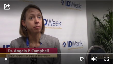
Combined data from both seasons showed that vaccinated children were 50% less likely to have been hospitalized for an acute influenza infection, compared with unvaccinated kids, a pattern consistently seen both in children aged 6 months to 8 years and in those aged 9-17 years. The pattern of vaccine effectiveness also held regardless of which flu strain caused the infections, reported Angela P. Campbell, MD, a CDC medical officer.
“We saw a nice benefit from vaccination, both in previously healthy children and in those with an underlying medical condition,” a finding that adds to existing evidence of vaccine effectiveness, Dr. Campbell said in a video interview. The results confirmed that flu vaccination does not just prevent infections but also cuts the rate of more severe infections that lead to hospitalization, she explained.
Another CDC study looked at data collected by the agency’s Influenza Hospitalization Surveillance Network from adults at least 18 years old who were hospitalized for a laboratory-confirmed influenza infection during five flu seasons, 2013-2014 through 2017-18. The data, which came from more than 250 acute-care hospitals in 13 states, included more than 43,000 people hospitalized for an identified influenza strain and with a known vaccination history who were not institutionalized and had not received any antiviral treatment.

After propensity-weighted adjustment to create better parity between the vaccinated and unvaccinated patients, the results showed that people 18-64 years old with vaccination had statistically significant decreases in mortality of a relative 36%, need for mechanical ventilation of 34%, pneumonia of 20%, and need for ICU admission of a relative 19%, as well as an 18% drop in average ICU length of stay, Shikha Garg, MD, said at the meeting. The propensity-weighted analysis of data from people at least 65 years old showed statistically significant relative reductions linked with vaccination: 46% reduction in the need for mechanical ventilation, 28% reduction in ICU admissions, and 9% reduction in hospitalized length of stay.
Further analysis of these outcomes by the strains that caused these influenza infections showed that the statistically significant benefits from vaccination were seen only in patients infected with an H1N1 strain. Statistically significant effects on these severe outcomes were not apparent among people infected with the H3N2 or B strains, said Dr. Garg, a medical epidemiologist at the CDC.
“All adults should receive an annual flu vaccination as it can improve outcomes among those who develop influenza despite vaccination,” she concluded.
Results from a third CDC study reported at the meeting examined the importance of two vaccine doses (administered at least 4 weeks apart) given to children aged 6 months to 8 years for the first season they receive flu vaccination, which is the immunization approach for flu recommended by the CDC. The findings from a total of more than 7,500 children immunized during the 2014-2018 seasons showed a clear increment in vaccine protection among kids who received two doses during their first season vaccinated, especially in children who were 2 years old or younger. In that age group, administration of two doses produced vaccine effectiveness of 53% versus a 23% vaccine effectiveness after a single vaccine dose, reported Jessie Chung, a CDC epidemiologist.
WASHINGTON –
During recent U.S. flu seasons, children and adults who contracted influenza despite vaccination had significantly fewer severe infections and infection complications, compared with unimmunized people, according to two separate reports from CDC researchers presented at an annual scientific meeting on infectious diseases.
One of the reports tracked the impact of flu vaccine in children using data that the CDC collected at seven medical centers that participated in the agency’s New Vaccine Surveillance Network, which provided information on children aged 6 months to 17 years who were hospitalized for an acute respiratory illness, including more than 1,700 children during the 2016-2017 flu season and more than 1,900 during the 2017-2018 season. Roughly 10% of these children tested positive for influenza, and the subsequent analysis focused on these cases and compared incidence rates among children who had been vaccinated during the index season and those who had remained unvaccinated.
Combined data from both seasons showed that vaccinated children were 50% less likely to have been hospitalized for an acute influenza infection, compared with unvaccinated kids, a pattern consistently seen both in children aged 6 months to 8 years and in those aged 9-17 years. The pattern of vaccine effectiveness also held regardless of which flu strain caused the infections, reported Angela P. Campbell, MD, a CDC medical officer.
“We saw a nice benefit from vaccination, both in previously healthy children and in those with an underlying medical condition,” a finding that adds to existing evidence of vaccine effectiveness, Dr. Campbell said in a video interview. The results confirmed that flu vaccination does not just prevent infections but also cuts the rate of more severe infections that lead to hospitalization, she explained.
Another CDC study looked at data collected by the agency’s Influenza Hospitalization Surveillance Network from adults at least 18 years old who were hospitalized for a laboratory-confirmed influenza infection during five flu seasons, 2013-2014 through 2017-18. The data, which came from more than 250 acute-care hospitals in 13 states, included more than 43,000 people hospitalized for an identified influenza strain and with a known vaccination history who were not institutionalized and had not received any antiviral treatment.
After propensity-weighted adjustment to create better parity between the vaccinated and unvaccinated patients, the results showed that people 18-64 years old with vaccination had statistically significant decreases in mortality of a relative 36%, need for mechanical ventilation of 34%, pneumonia of 20%, and need for ICU admission of a relative 19%, as well as an 18% drop in average ICU length of stay, Shikha Garg, MD, said at the meeting. The propensity-weighted analysis of data from people at least 65 years old showed statistically significant relative reductions linked with vaccination: 46% reduction in the need for mechanical ventilation, 28% reduction in ICU admissions, and 9% reduction in hospitalized length of stay.
Further analysis of these outcomes by the strains that caused these influenza infections showed that the statistically significant benefits from vaccination were seen only in patients infected with an H1N1 strain. Statistically significant effects on these severe outcomes were not apparent among people infected with the H3N2 or B strains, said Dr. Garg, a medical epidemiologist at the CDC.
“All adults should receive an annual flu vaccination as it can improve outcomes among those who develop influenza despite vaccination,” she concluded.
Results from a third CDC study reported at the meeting examined the importance of two vaccine doses (administered at least 4 weeks apart) given to children aged 6 months to 8 years for the first season they receive flu vaccination, which is the immunization approach for flu recommended by the CDC. The findings from a total of more than 7,500 children immunized during the 2014-2018 seasons showed a clear increment in vaccine protection among kids who received two doses during their first season vaccinated, especially in children who were 2 years old or younger. In that age group, administration of two doses produced vaccine effectiveness of 53% versus a 23% vaccine effectiveness after a single vaccine dose, reported Jessie Chung, a CDC epidemiologist.
REPORTING FROM ID WEEK 2019
Fetal MRI may change pregnancy management
CHARLOTTE, N.C. – according to research presented at the annual meeting of the Child Neurology Society. This imaging technique and neurologic consultation complement the information that prenatal ultrasound and obstetric consultations provide and may influence pregnancy management and infant neurologic care significantly.
The fetal diagnosis of posterior fossa abnormalities can be challenging. The prognosis can vary greatly, depending on the diagnosis. Sarah Mulkey, MD, PhD, director of the fetal and neonatal fellowship and a fetal and neonatal neurologist at Children’s National in Washington, and colleagues conducted an analysis to evaluate whether fetal MRI and neurology consultation produce alternative diagnoses for maternal-fetal dyads who are referred to a fetal neurology program because of concern for a fetal posterior fossa anomaly. The researchers also sought to determine how often the postnatal evaluation differed from the fetal diagnosis.
Dr. Mulkey and colleagues retrospectively analyzed cases referred to the Fetal Medicine Institute at Children’s National from January 2012 to June 2018. They included the referral diagnoses of Dandy-Walker continuum, cerebellar hypoplasia, vermis hypoplasia, Blake’s pouch cyst, mega cisterna magna, and other posterior fossa anomalies in their study.
The investigators identified 188 cases that had undergone fetal MRI and neurology consultation. The average gestational age at evaluation was 25 weeks, and the average maternal age was 30 years. Approximately 43% of referrals resulted from a concern regarding Dandy-Walker malformation, and 21% of referrals resulted from a suspicion of mega cisterna magna.
Fetal MRI and neurology consultation resulted in a change from the referral diagnosis or additional information about the fetus in 124 (66%) cases. For example, after imaging and consultation, 15% of referrals were diagnosed with Dandy-Walker malformation, as opposed to the 43% who were suspected of having it. Most referrals with a diagnosis of vermis hypoplasia had a better prognosis after fetal MRI. Fetal MRI and consultation also resulted in new diagnoses of Joubert syndrome and rhombencephalosynapsis. About 19% of referrals were considered normal. “A considerable number of these referrals ended up being for conditions that would have a good outcome,” said Dr. Mulkey.
In addition, the researchers obtained the postnatal diagnosis for 60 of 138 (43%) live-born infants. The fetal diagnosis of Dandy-Walker continuum was confirmed post natally in six of six (100%) cases. Of the 13 cases of fetally diagnosed vermis hypoplasia, 7 (54%) had stable findings, 3 (23%) normalized, and diagnosis changed in 3 (23%). Of the 17 fetally diagnosed Blake’s pouch cysts, 8 (47%) remained stable, 5 (29%) normalized, and diagnosis changed in 4 (24%). Four of nine (44%) cases of fetally diagnosed mega cisterna magna remained stable, two (22%) normalized, and diagnosis changed in three (33%). Overall, prognosis did not change after postnatal imaging.
“There is a high degree of correlation between fetal and postnatal diagnoses for Dandy-Walker continuum, cerebellar hypoplasia, cyst, and ‘other’ diagnoses,” said Dr. Mulkey. “Vermis hypoplasia and Blake’s pouch cyst diagnoses were less consistent.”
The investigators reported no disclosures.
SOURCE: Schlatterer S et al. CNS 2019, Abstract 158.
REPORTING FROM CNS 2019
Edasalonexent may slow progression of Duchenne muscular dystrophy
CHARLOTTE, N.C. – presented at the annual meeting of the Child Neurology Society.

The NF-kB pathway is “fundamental to the pathogenesis and biology of DMD,” said Richard Finkel, MD, chief of neurology at Nemours Children’s Health System in Orlando and principal investigator for the phase 2 study, known as MoveDMD.
A lack of dystrophin, combined with the mechanical stress of muscle contraction, activates the NF-kB pathway and inhibits muscle regeneration. “It is known that there is inflammation and fibrosis and release of cytokines early in life” in patients with DMD, Dr. Finkel said.
Independent of mutation
Edasalonexent is an NF-kB inhibitor that is being developed by Catabasis as a therapy for patients with DMD regardless of the genetic mutation that is causing the disease. It may be used as monotherapy or with other dystrophin-targeted treatments, Dr. Finkel said.
In a mouse model of DMD, an analog of the drug reduced muscle inflammation and increased the force of diaphragm muscle. To assess edasalonexent’s safety, pharmacokinetics, and effects on functional measures and MRI in patients with DMD, Dr. Finkel and colleagues conducted the MoveDMD trial. Investigators enrolled boys aged 4 years to younger than 8 years who were not receiving treatment with corticosteroids.
Researchers first examined drug safety and pharmacokinetics in 17 boys who received the treatment for 1 week. The investigators then followed 16 of these patients off treatment for as long as 6 months. This off-treatment period was followed by a phase 2, placebo-controlled period, during which the 16 patients and another 15 patients received edasalonexent 67 mg/kg/day, edasalonexent 100 mg/kg/day, or placebo for 12 weeks. Patients subsequently entered an open-label extension study.
Dr. Finkel presented a comparison of outcomes during the off-treatment period with outcomes during the open-label extension. “We used these boys as their own internal control, if you wish,” he said.
Creatine kinase levels decreased soon after treatment, as did other markers of muscle disease. The drug “seems to have an early and sustained biomarker response,” Dr. Finkel said.
Annualized rate of change on lower leg muscle MRI-T2 decreased. “There is a relative reduction and stabilization from week 12 all the way out through the open-label extension to 72 weeks,” he said. “It suggests that there is an early and sustained response in stabilization of the MRI as a biomarker.”
Timed function tests
A comparison of the annualized rates of change on timed function tests – including the 10-meter walk/run, time-to-stand, and four-stair-climb, and the North Star Ambulatory Assessment – during the off-treatment and on-treatment periods indicated slowing of disease progression with treatment. “Shortly after starting on drug ... there was a relative stabilization in each of these measures,” Dr. Finkel said.
In addition, the researchers observed an early signal of possible cardiac benefit. Mean heart rate at baseline was 99 bpm. On treatment, it decreased to 92 bpm. “Boys with DMD die typically of cardiomyopathy, so it is important to try to address the cardiac status,” he said.
The drug was safe and well tolerated. Most participants experienced mild gastrointestinal issues, which typically were transient. One serious adverse event during the trial occurred in a patient receiving placebo. Patients tended to have a stable body mass index during treatment, Dr. Finkel said.
During the open-label extension, patients had “clinically meaningful slowing of disease progression on edasalonexent,” relative to the off-treatment period, Dr. Finkel said. Investigators plan to further study edasalonexent for the treatment of DMD in a phase 3 trial. The phase 3 study, PolarisDMD, recently completed enrollment at 40 sites. Results could be available in about a year, Dr. Finkel said.
The study was sponsored by Catabasis. Dr. Finkel disclosed consulting work and grants or research support from Catabasis and other companies.
SOURCE: Finkel R et al. CNS 2019. Abstract PL1-3.
CHARLOTTE, N.C. – presented at the annual meeting of the Child Neurology Society.
The NF-kB pathway is “fundamental to the pathogenesis and biology of DMD,” said Richard Finkel, MD, chief of neurology at Nemours Children’s Health System in Orlando and principal investigator for the phase 2 study, known as MoveDMD.
A lack of dystrophin, combined with the mechanical stress of muscle contraction, activates the NF-kB pathway and inhibits muscle regeneration. “It is known that there is inflammation and fibrosis and release of cytokines early in life” in patients with DMD, Dr. Finkel said.
Independent of mutation
Edasalonexent is an NF-kB inhibitor that is being developed by Catabasis as a therapy for patients with DMD regardless of the genetic mutation that is causing the disease. It may be used as monotherapy or with other dystrophin-targeted treatments, Dr. Finkel said.
In a mouse model of DMD, an analog of the drug reduced muscle inflammation and increased the force of diaphragm muscle. To assess edasalonexent’s safety, pharmacokinetics, and effects on functional measures and MRI in patients with DMD, Dr. Finkel and colleagues conducted the MoveDMD trial. Investigators enrolled boys aged 4 years to younger than 8 years who were not receiving treatment with corticosteroids.
Researchers first examined drug safety and pharmacokinetics in 17 boys who received the treatment for 1 week. The investigators then followed 16 of these patients off treatment for as long as 6 months. This off-treatment period was followed by a phase 2, placebo-controlled period, during which the 16 patients and another 15 patients received edasalonexent 67 mg/kg/day, edasalonexent 100 mg/kg/day, or placebo for 12 weeks. Patients subsequently entered an open-label extension study.
Dr. Finkel presented a comparison of outcomes during the off-treatment period with outcomes during the open-label extension. “We used these boys as their own internal control, if you wish,” he said.
Creatine kinase levels decreased soon after treatment, as did other markers of muscle disease. The drug “seems to have an early and sustained biomarker response,” Dr. Finkel said.
Annualized rate of change on lower leg muscle MRI-T2 decreased. “There is a relative reduction and stabilization from week 12 all the way out through the open-label extension to 72 weeks,” he said. “It suggests that there is an early and sustained response in stabilization of the MRI as a biomarker.”
Timed function tests
A comparison of the annualized rates of change on timed function tests – including the 10-meter walk/run, time-to-stand, and four-stair-climb, and the North Star Ambulatory Assessment – during the off-treatment and on-treatment periods indicated slowing of disease progression with treatment. “Shortly after starting on drug ... there was a relative stabilization in each of these measures,” Dr. Finkel said.
In addition, the researchers observed an early signal of possible cardiac benefit. Mean heart rate at baseline was 99 bpm. On treatment, it decreased to 92 bpm. “Boys with DMD die typically of cardiomyopathy, so it is important to try to address the cardiac status,” he said.
The drug was safe and well tolerated. Most participants experienced mild gastrointestinal issues, which typically were transient. One serious adverse event during the trial occurred in a patient receiving placebo. Patients tended to have a stable body mass index during treatment, Dr. Finkel said.
During the open-label extension, patients had “clinically meaningful slowing of disease progression on edasalonexent,” relative to the off-treatment period, Dr. Finkel said. Investigators plan to further study edasalonexent for the treatment of DMD in a phase 3 trial. The phase 3 study, PolarisDMD, recently completed enrollment at 40 sites. Results could be available in about a year, Dr. Finkel said.
The study was sponsored by Catabasis. Dr. Finkel disclosed consulting work and grants or research support from Catabasis and other companies.
SOURCE: Finkel R et al. CNS 2019. Abstract PL1-3.
REPORTING FROM CNS 2019
Baricitinib may benefit patients with Aicardi-Goutières syndrome
CHARLOTTE, N.C. – Scores on a novel AGS scale improved, and skin and liver complications resolved in children with AGS who received treatment with baricitinib, according to results presented at the annual meeting of the Child Neurology Society.

AGS is caused by various heritable disorders of the innate immunity that result in excessive interferon production. AGS characteristically manifests as an early-onset encephalopathy that causes intellectual and physical disability, but patients may have a wide range of clinical phenotypes. The disease may involve the skin, liver, lungs, heart, and other organs, as well as the brain.
A multisystem disorder
“The neurologic features, while they are the most compelling for us, are really only the tip of the iceberg,” said Adeline Vanderver, MD, program director of the leukodystrophy center, and the Jacob A. Kamens Endowed Chair in Neurologic Disorders and Translational Neurotherapeutics at Children’s Hospital of Philadelphia. “Nearly every single organ system in the body is affected, from either direct interferon injury or from a secondary vasculopathy related to the interferonopathy.”
Dr. Vanderver presented results from the compassionate use study, which assessed whether the JAK inhibitor baricitinib (Olumiant) may decrease interferon signaling in AGS and limit the morbidity of the disease.
The phase 1, open-label trial “included compassionate use of baricitinib in AGS under the argument that these children did not have time to wait for approval of the drug,” said Dr. Vanderver. In 2018, the Food and Drug Administration approved baricitinib for moderate to severe rheumatoid arthritis in adults with an inadequate response to methotrexate.
The phase 1 trial in AGS included 35 patients with mutation-defined AGS and evidence of inflammatory disease that could be targeted by JAK inhibition. The trial population was 36% female. The average age of disease onset was 0.8 years, and patients’ average age at treatment was 6.1 years. The investigators assessed safety and laboratory data every 3 months and conducted clinical assessments every 6 months.
The heterogeneity of AGS phenotypes within families and across genotypes makes treatment trials in this disorder a challenge, Dr. Vanderver said. Outcome measures may have ceiling or floor effects that fail to capture the range of severity of AGS symptoms. Dr. Vanderver and colleagues developed a novel AGS scale to capture the scope of neurologic function in patients with AGS
.
When the researchers applied the AGS scale to a historical cohort of patients, most had stable scores about 6 months after disease onset. “After the first 6 months of the disease, the disease tends to be much more static, as the children have sustained significant neurologic injury,” Dr. Vanderver said.
They applied the novel AGS scale post hoc as an exploratory endpoint in the phase 1 trial. In addition, parents recorded information in a diary about skin involvement, irritability, seizures, and fever. “Over time, we see a reduction, although not always a statistically significant reduction, in symptom burden,” Dr. Vanderver said. The AGS clinical diary scores reflect “what the parents were telling us – that they felt like their children were feeling better during treatment,” she said.
Several patients had skin conditions that improved with treatment. One patient with dermatitis or eczema had the skin abnormality resolve within 3 days. A patient with full-body panniculitis began healing for the first time after about a month of treatment. Seasonal variations and dose adjustments led to fluctuations in some of the skin conditions. Nevertheless, the results suggested significant improvement in skin manifestations in patients with AGS, Dr. Vanderver said.
Patients generally had stable AGS scale scores in the year before treatment, although a couple of patients who were closer to disease onset had precipitous decline in neurologic function, she said. “We had a statistically significant increase in that scale of neurologic function in our patients during the period of the study, even in patients who had sometimes had years of disease duration,” said Dr. Vanderver.
Dr. Vanderver cautioned that she does not want to overstate the changes in function. Patients with AGS may have less potential for recuperation, compared with patients with other conditions. “A child with significant disruptive CNS disease may not recuperate normal functioning,” Dr. Vanderver said, “but it can be clinically meaningful to families if children start having better head control, smile, communicate, even if they might not regain all their motor milestones.”
In addition, a small subset of patients who had potentially life threatening liver complications from the disease experienced rapid normalization and improvement of liver function. “This blockade can be important not just for neurologic function but also to maintain normal physiologic homeostasis of other organs that are affected by the interferonopathy,” Dr. Vanderver said.
Interferon signaling scores decreased in the days after starting treatment and subsequently leveled out.
Serious adverse events that occurred during the trial, such as hospitalizations, were attributable to AGS. One child died from unrecognized pulmonary hypertension, which is now known to be a complication of AGS but was not at the time.
Harnessing a side effect
The most significant and recurrent laboratory abnormality was thrombocytosis. “That is a known complication of this family of drugs that in many cases allowed us to improve previous treatment-resistant thrombocytopenia, so we kind of like that side effect in most cases, but in two cases it did ... result in dose adjustments, although we never had to stop the medication for that.”
The study offers proof of principle that AGS is treatable, Dr. Vanderver said. A phase 2 trial is enrolling patients closer to disease onset. Early treatment of AGS may remain a challenge until there is newborn screening for the disease, she said.
Dr. Vanderver receives grant and in-kind support for translational research without personal compensation from Eli Lilly, Takeda, Illumina, Biogen, Homology, and Ionis. In addition, Dr. Vanderver serves on the scientific advisory boards of the European Leukodystrophy Association and the United Leukodystrophy Foundation, as well as in an unpaid capacity for Takeda, Ionis, Biogen, and Illumina.
Eli Lilly provided support for the phase 1 study. In addition, the study received support from the AGS Association Americas Family Foundation, National Human Genome Research Institute, National Institute of Neurological Disorders and Stroke, and the Children’s Hospital of Philadelphia Research Institute.
SOURCE: Vanderver A et al. CNS 2019. Abstract PL1-6.
CHARLOTTE, N.C. – Scores on a novel AGS scale improved, and skin and liver complications resolved in children with AGS who received treatment with baricitinib, according to results presented at the annual meeting of the Child Neurology Society.
AGS is caused by various heritable disorders of the innate immunity that result in excessive interferon production. AGS characteristically manifests as an early-onset encephalopathy that causes intellectual and physical disability, but patients may have a wide range of clinical phenotypes. The disease may involve the skin, liver, lungs, heart, and other organs, as well as the brain.
A multisystem disorder
“The neurologic features, while they are the most compelling for us, are really only the tip of the iceberg,” said Adeline Vanderver, MD, program director of the leukodystrophy center, and the Jacob A. Kamens Endowed Chair in Neurologic Disorders and Translational Neurotherapeutics at Children’s Hospital of Philadelphia. “Nearly every single organ system in the body is affected, from either direct interferon injury or from a secondary vasculopathy related to the interferonopathy.”
Dr. Vanderver presented results from the compassionate use study, which assessed whether the JAK inhibitor baricitinib (Olumiant) may decrease interferon signaling in AGS and limit the morbidity of the disease.
The phase 1, open-label trial “included compassionate use of baricitinib in AGS under the argument that these children did not have time to wait for approval of the drug,” said Dr. Vanderver. In 2018, the Food and Drug Administration approved baricitinib for moderate to severe rheumatoid arthritis in adults with an inadequate response to methotrexate.
The phase 1 trial in AGS included 35 patients with mutation-defined AGS and evidence of inflammatory disease that could be targeted by JAK inhibition. The trial population was 36% female. The average age of disease onset was 0.8 years, and patients’ average age at treatment was 6.1 years. The investigators assessed safety and laboratory data every 3 months and conducted clinical assessments every 6 months.
The heterogeneity of AGS phenotypes within families and across genotypes makes treatment trials in this disorder a challenge, Dr. Vanderver said. Outcome measures may have ceiling or floor effects that fail to capture the range of severity of AGS symptoms. Dr. Vanderver and colleagues developed a novel AGS scale to capture the scope of neurologic function in patients with AGS
.
When the researchers applied the AGS scale to a historical cohort of patients, most had stable scores about 6 months after disease onset. “After the first 6 months of the disease, the disease tends to be much more static, as the children have sustained significant neurologic injury,” Dr. Vanderver said.
They applied the novel AGS scale post hoc as an exploratory endpoint in the phase 1 trial. In addition, parents recorded information in a diary about skin involvement, irritability, seizures, and fever. “Over time, we see a reduction, although not always a statistically significant reduction, in symptom burden,” Dr. Vanderver said. The AGS clinical diary scores reflect “what the parents were telling us – that they felt like their children were feeling better during treatment,” she said.
Several patients had skin conditions that improved with treatment. One patient with dermatitis or eczema had the skin abnormality resolve within 3 days. A patient with full-body panniculitis began healing for the first time after about a month of treatment. Seasonal variations and dose adjustments led to fluctuations in some of the skin conditions. Nevertheless, the results suggested significant improvement in skin manifestations in patients with AGS, Dr. Vanderver said.
Patients generally had stable AGS scale scores in the year before treatment, although a couple of patients who were closer to disease onset had precipitous decline in neurologic function, she said. “We had a statistically significant increase in that scale of neurologic function in our patients during the period of the study, even in patients who had sometimes had years of disease duration,” said Dr. Vanderver.
Dr. Vanderver cautioned that she does not want to overstate the changes in function. Patients with AGS may have less potential for recuperation, compared with patients with other conditions. “A child with significant disruptive CNS disease may not recuperate normal functioning,” Dr. Vanderver said, “but it can be clinically meaningful to families if children start having better head control, smile, communicate, even if they might not regain all their motor milestones.”
In addition, a small subset of patients who had potentially life threatening liver complications from the disease experienced rapid normalization and improvement of liver function. “This blockade can be important not just for neurologic function but also to maintain normal physiologic homeostasis of other organs that are affected by the interferonopathy,” Dr. Vanderver said.
Interferon signaling scores decreased in the days after starting treatment and subsequently leveled out.
Serious adverse events that occurred during the trial, such as hospitalizations, were attributable to AGS. One child died from unrecognized pulmonary hypertension, which is now known to be a complication of AGS but was not at the time.
Harnessing a side effect
The most significant and recurrent laboratory abnormality was thrombocytosis. “That is a known complication of this family of drugs that in many cases allowed us to improve previous treatment-resistant thrombocytopenia, so we kind of like that side effect in most cases, but in two cases it did ... result in dose adjustments, although we never had to stop the medication for that.”
The study offers proof of principle that AGS is treatable, Dr. Vanderver said. A phase 2 trial is enrolling patients closer to disease onset. Early treatment of AGS may remain a challenge until there is newborn screening for the disease, she said.
Dr. Vanderver receives grant and in-kind support for translational research without personal compensation from Eli Lilly, Takeda, Illumina, Biogen, Homology, and Ionis. In addition, Dr. Vanderver serves on the scientific advisory boards of the European Leukodystrophy Association and the United Leukodystrophy Foundation, as well as in an unpaid capacity for Takeda, Ionis, Biogen, and Illumina.
Eli Lilly provided support for the phase 1 study. In addition, the study received support from the AGS Association Americas Family Foundation, National Human Genome Research Institute, National Institute of Neurological Disorders and Stroke, and the Children’s Hospital of Philadelphia Research Institute.
SOURCE: Vanderver A et al. CNS 2019. Abstract PL1-6.
REPORTING FROM CNS 2019
Celiac disease may underlie seizures
CHARLOTTE, N.C. – , according to a retrospective chart review presented at the annual meeting of the Child Neurology Society. Associations between celiac disease and seizures may have implications for screening and treatment, said study author Shanna Swartwood, MD, a fellow in the department of pediatric neurology at University of Utah in Salt Lake City.

“Screening for [celiac disease] early in patients with epilepsy, specifically with temporal EEG findings and intractable epilepsy, is warranted given the improvement of seizure burden that may result from exclusion of gluten from the diet,” said Dr. Swartwood and colleagues.
About 10% of patients with celiac disease have clinical neurologic manifestations, such as seizures. To characterize features of epilepsy in a pediatric population with celiac disease and to examine the effect of a gluten-free diet on seizure burden, Dr. Swartwood and colleagues reviewed patients treated at Primary Children’s Hospital in Salt Lake City since 2002. They identified patients with ICD-10 codes for seizures or epilepsy and celiac disease and reviewed 187 charts in all.
In all, 40 patients with seizures had biopsy-proven celiac disease, and 22 had a diagnosis of celiac disease based on the presence of antibodies. Among those with biopsy-proven celiac disease, 43% had intractable seizures. Among those with antibody-positive celiac disease, 31% had intractable seizures.
Among patients with intractable epilepsy, seizure onset preceded the diagnosis of celiac disease by an average of 5 years. For patients with nonintractable epilepsy, the first seizure occurred 1 year before the celiac disease diagnosis on average, but some patients received a celiac disease diagnosis first.
Focal seizures with secondary generalization and generalized tonic clonic seizures were the most common seizure types in this cohort. Epileptiform activity most often was seen in the temporal lobe. While other studies in patients with celiac disease have found occipital epileptiform activity to be the most common, only one patient in this cohort had activity in that location, Dr. Swartwood noted.
Patients with intractable seizures who adhered to a gluten-free diet “had a fairly robust response in terms of seizure improvement,” she said. Seizure improvement, including a decrease in seizure frequency or a decrease in antiepileptic medication dosage, occurred in seven of nine patients in the biopsy-proven group and in two of three patients in the antibody-positive group who adhered to a gluten-free diet and had intractable seizures. One patient was able to stop antiepileptic medication, and one patient had a complete resolution of seizure activity.
The researchers plan to further study the relationship between celiac disease and epilepsy, including whether various HLA subtypes of celiac disease correlate with seizures, said coinvestigator Cristina Trandafir, MD, PhD, assistant professor of pediatric neurology at University of Utah.
The chart review included relatively few patients with limited data. Nevertheless, the results suggest that there may be “substantial lag time” from first seizure to celiac disease diagnosis and that “earlier diagnosis and earlier placement on a gluten-free diet may be beneficial,” Dr. Swartwood said. Celiac disease may be asymptomatic, and screening for celiac disease with a blood test may make sense for patients with intractable seizures, she said.
The researchers had no relevant disclosures.
CHARLOTTE, N.C. – , according to a retrospective chart review presented at the annual meeting of the Child Neurology Society. Associations between celiac disease and seizures may have implications for screening and treatment, said study author Shanna Swartwood, MD, a fellow in the department of pediatric neurology at University of Utah in Salt Lake City.
“Screening for [celiac disease] early in patients with epilepsy, specifically with temporal EEG findings and intractable epilepsy, is warranted given the improvement of seizure burden that may result from exclusion of gluten from the diet,” said Dr. Swartwood and colleagues.
About 10% of patients with celiac disease have clinical neurologic manifestations, such as seizures. To characterize features of epilepsy in a pediatric population with celiac disease and to examine the effect of a gluten-free diet on seizure burden, Dr. Swartwood and colleagues reviewed patients treated at Primary Children’s Hospital in Salt Lake City since 2002. They identified patients with ICD-10 codes for seizures or epilepsy and celiac disease and reviewed 187 charts in all.
In all, 40 patients with seizures had biopsy-proven celiac disease, and 22 had a diagnosis of celiac disease based on the presence of antibodies. Among those with biopsy-proven celiac disease, 43% had intractable seizures. Among those with antibody-positive celiac disease, 31% had intractable seizures.
Among patients with intractable epilepsy, seizure onset preceded the diagnosis of celiac disease by an average of 5 years. For patients with nonintractable epilepsy, the first seizure occurred 1 year before the celiac disease diagnosis on average, but some patients received a celiac disease diagnosis first.
Focal seizures with secondary generalization and generalized tonic clonic seizures were the most common seizure types in this cohort. Epileptiform activity most often was seen in the temporal lobe. While other studies in patients with celiac disease have found occipital epileptiform activity to be the most common, only one patient in this cohort had activity in that location, Dr. Swartwood noted.
Patients with intractable seizures who adhered to a gluten-free diet “had a fairly robust response in terms of seizure improvement,” she said. Seizure improvement, including a decrease in seizure frequency or a decrease in antiepileptic medication dosage, occurred in seven of nine patients in the biopsy-proven group and in two of three patients in the antibody-positive group who adhered to a gluten-free diet and had intractable seizures. One patient was able to stop antiepileptic medication, and one patient had a complete resolution of seizure activity.
The researchers plan to further study the relationship between celiac disease and epilepsy, including whether various HLA subtypes of celiac disease correlate with seizures, said coinvestigator Cristina Trandafir, MD, PhD, assistant professor of pediatric neurology at University of Utah.
The chart review included relatively few patients with limited data. Nevertheless, the results suggest that there may be “substantial lag time” from first seizure to celiac disease diagnosis and that “earlier diagnosis and earlier placement on a gluten-free diet may be beneficial,” Dr. Swartwood said. Celiac disease may be asymptomatic, and screening for celiac disease with a blood test may make sense for patients with intractable seizures, she said.
The researchers had no relevant disclosures.
REPORTING FROM CNS 2019
Pediatric epilepsy surgery may improve cognition and behavior
CHARLOTTE, NC – according to a study presented at the annual meeting of the Child Neurology Society. The presence of comorbidities such as mood disorders and autism may influence the likelihood of perceived improvement, whereas the type of surgery may not.

“The parents and the families of the patients perceive that, even if the patients are not completely seizure free, the behavior and cognitive outcomes are better if there is some sort of seizure improvement,” said Trishna Kantamneni, MD, director of pediatric epilepsy at UC Davis in Sacramento.
To assess behavioral and cognitive outcomes following pediatric epilepsy surgery and to identify factors that predict improvement, Dr. Kantamneni and colleagues at the Cleveland Clinic Epilepsy Center retrospectively reviewed 126 patients younger than 18 years who underwent epilepsy surgery for medically refractory epilepsy during 2009-2016.
The primary outcome measure was the Impact of Childhood Neurologic Disability Scale (ICNDS), a parent-reported scale that assesses the behavior, cognition, and physical or neurologic disability of children with epilepsy. Parents completed the ICNDS preoperatively and at 6, 12, and 24 months after surgery. The researchers constructed separate linear mixed effects models to identify predictors of postoperative changes in ICNDS score.
Of the 126 patients, 62.7% were male, the median duration of epilepsy was 4.7 years, and 69.8% were seizure-free at the 2-year follow-up. Postoperative ICNDS scores were available for 103 patients at 6 months and for 54 patients at 24 months.
Before surgery, the average total ICNDS score was 55.7. At 6 months after surgery, the average score was 34.6, and at 24 months, it was 32.1, representing significant improvement from baseline.
In addition, behavior, cognition, and epilepsy subscores also improved post operatively, and the improvement persisted through 24 months. ICNDS scores significantly improved “even in patients who were not seizure-free after surgery,” by an average of about 22 points, the researchers said.
The absence of comorbid autism, cognitive impairment, and global developmental impairment and the absence of anxiety, depression, and ADHD were predictors of improved total ICNDS scores. Tumor pathology and being seizure free at 2 years also predicted improved scores. Duration and type of epilepsy, the number of antiepileptic drugs that patients were taking before surgery, and lobe of surgery were not predictive of improved ICNDS scores.
Dr. Kantamneni had no relevant disclosures.
SOURCE: Kantamneni T et al. CNS 2019, Abstract 51.
CHARLOTTE, NC – according to a study presented at the annual meeting of the Child Neurology Society. The presence of comorbidities such as mood disorders and autism may influence the likelihood of perceived improvement, whereas the type of surgery may not.
“The parents and the families of the patients perceive that, even if the patients are not completely seizure free, the behavior and cognitive outcomes are better if there is some sort of seizure improvement,” said Trishna Kantamneni, MD, director of pediatric epilepsy at UC Davis in Sacramento.
To assess behavioral and cognitive outcomes following pediatric epilepsy surgery and to identify factors that predict improvement, Dr. Kantamneni and colleagues at the Cleveland Clinic Epilepsy Center retrospectively reviewed 126 patients younger than 18 years who underwent epilepsy surgery for medically refractory epilepsy during 2009-2016.
The primary outcome measure was the Impact of Childhood Neurologic Disability Scale (ICNDS), a parent-reported scale that assesses the behavior, cognition, and physical or neurologic disability of children with epilepsy. Parents completed the ICNDS preoperatively and at 6, 12, and 24 months after surgery. The researchers constructed separate linear mixed effects models to identify predictors of postoperative changes in ICNDS score.
Of the 126 patients, 62.7% were male, the median duration of epilepsy was 4.7 years, and 69.8% were seizure-free at the 2-year follow-up. Postoperative ICNDS scores were available for 103 patients at 6 months and for 54 patients at 24 months.
Before surgery, the average total ICNDS score was 55.7. At 6 months after surgery, the average score was 34.6, and at 24 months, it was 32.1, representing significant improvement from baseline.
In addition, behavior, cognition, and epilepsy subscores also improved post operatively, and the improvement persisted through 24 months. ICNDS scores significantly improved “even in patients who were not seizure-free after surgery,” by an average of about 22 points, the researchers said.
The absence of comorbid autism, cognitive impairment, and global developmental impairment and the absence of anxiety, depression, and ADHD were predictors of improved total ICNDS scores. Tumor pathology and being seizure free at 2 years also predicted improved scores. Duration and type of epilepsy, the number of antiepileptic drugs that patients were taking before surgery, and lobe of surgery were not predictive of improved ICNDS scores.
Dr. Kantamneni had no relevant disclosures.
SOURCE: Kantamneni T et al. CNS 2019, Abstract 51.
REPORTING FROM CNS 2019
Delay in EEG monitoring associated with increased seizure duration in pediatric refractory status epilepticus
CHARLOTTE, NC – , according to a multicenter study that was presented at the annual meeting of the Child Neurology Society. Delays in initiating EEG monitoring are associated with longer seizure duration in this patient population.
Neurologists are advised to initiate continuous EEG monitoring rapidly for all cases of pediatric refractory status epilepticus. Little information is available, however, about patterns in the timing of EEG placement. In addition, the relationship between delays in the initiation of continuous EEG and outcomes of refractory status epilepticus are unknown. Dmitry Tchapyjnikov, MD, assistant professor of child neurology at Duke University in Durham, N.C., and colleagues evaluated trends in the time to continuous EEG initiation and examined whether delays are associated with longer seizure duration in children with refractory status epilepticus.
A retrospective analysis of pSERG data
Dr. Tchapyjnikov and colleagues analyzed data from 11 hospitals participating in the Pediatric Status Epilepticus Research Group (pSERG), a prospective, observational cohort. They focused on pediatric patients who were admitted from 2011 to 2017 with refractory status epilepticus, which they defined as a seizure that persisted after treatment with two or more antiseizure medications (ASMs), one of which had to be a nonbenzodiazepine ASM, or a continuous infusion. Eligible patients were between 1 month and 21 years old and had convulsive seizures at onset. Patients who had EEG placement before seizure onset were excluded.
The investigators included in their study 121 patients who had seizure durations of 3 or more hours. Based on an exploratory analysis of various time-point cutoffs, Dr. Tchapyjnikov and colleagues defined delayed continuous EEG placement as placement at more than 5 hours after seizure onset. They used the Kaplan–Meier estimator to assess time to continuous EEG and used covariate-adjusted proportional hazards models to examine the association between delay in continuous EEG placement and seizure duration.
EEG placement overall was delayed
The median time to continuous EEG placement after seizure onset was 9 hours. Approximately 4% of the children had continuous EEG placed within 1 hour, and 74% had it placed within 24 hours.
The investigators found that seizure onset outside the study hospital was associated with a higher likelihood of delayed time to EEG placement. “Females seemed to be more likely to have timely EEG placement,” said Dr. Tchapyjnikov. “I don’t have a physiological explanation for that.” The researchers saw no difference in treatment between patients who had timely EEG placement and those who had delayed EEG placement.
About 68% of children were having seizures at the time of continuous EEG placement. A presumed seizure etiology of CNS infection was associated with a higher likelihood of being in status epilepticus at the time of EEG placement. A history of epilepsy, developmental delay, or home ASM use, however, was associated with a lower likelihood of being in status epilepticus at time of EEG placement.
Dr. Tchapyjnikov’s group found that the 24-hour cumulative probability of seizure resolution was lower among patients who did not have continuous EEG initiation within 5 hours, compared with those who did (56% vs.70%). The association remained significant after the investigators adjusted the data for covariates that were independently associated with 24-hour seizure resolution (hazard ratio, 0.31).
The investigators included in their analysis patients who had seizure resolution before EEG placement, because restricting the analysis to patients who have persistent status epilepticus would have overemphasized the benefits of EEG, according to Dr. Tchapyjnikov. “Looking at the overall hazard ratios is a more conservative way of looking at these data.”
The study was not supported by external funding. Dr. Tchapyjnikov had no relevant disclosures.
REPORTING FROM CNS 2019
Does AED prophylaxis delay seizure onset in children with brain tumors?
CHARLOTTE, N.C. – according to research presented at the annual meeting of the Child Neurology Society. Levetiracetam, oxcarbazepine, and phenytoin are the most common initial prophylactic AEDs administered to children with brain tumors, the researchers said.
The literature indicates that between 20% and 35% of children with brain tumors have seizures, and up to half of these patients have seizure as their presenting symptom. Common practice is to prescribe antiseizure medication after a child has had a first seizure, because the risk for recurrence is high. In 2000, the American Academy of Neurology discouraged prophylactic use of AEDs in children, citing a lack of evidence for efficacy. Most of the data that it reviewed, however, came from adults.
Michelle Yun, a medical student at Weill Cornell Medical College, New York, and colleagues used national Medicaid claims data that had been collected between 2009 and 2012 for children with seizures to conduct a retrospective, observational, case-control study. They included children aged 0-20 years with a diagnosis of brain tumor, a seizure diagnosis within 6 months after brain tumor diagnosis, an AED prescription, and 12 continuous months of Medicaid coverage following brain tumor diagnosis in their analysis. The investigators defined seizure prophylaxis as AED prescription within 30 days after brain tumor diagnosis but before a first seizure diagnosis.
The exposure in the study was AED prescription within 45 days of diagnosis, and the outcome was the time to first seizure. Ms. Yun and colleagues also analyzed the most common initial prophylactic AEDs and the proportion of cases with first seizure diagnosis after prophylactic AED discontinuation, which was defined as a treatment gap longer than 30 days. The study covariates included age, sex, race, ethnicity, and medical comorbidities.
In all, 218 children were included in the study; 40 received AED prophylaxis and 26 received it within 45 days of brain tumor diagnosis. Patients with and without AED prophylaxis were well matched on all covariates.
At 1 year, Ms. Yun and colleagues saw no difference in time to first seizure between the two groups. The median time to first seizure was 75 days in the prophylaxis group and 80 days in the no-prophylaxis group. The researchers observed a transient separation between the two groups, however, in the early months after brain tumor diagnosis. When they examined children who had a seizure during the first 6 months of follow-up, the median time to diagnosis of first seizure was 68 days in children with prophylaxis and 34 days in the no-prophylaxis group. The difference between groups was statistically significant. “When we added all the covariates of interest, we found that there was a protective effect in these children with early seizures,” said Ms. Yun.
Among the study limitations that Ms. Yun acknowledged were its observational, retrospective design and its small sample size. Medicaid data themselves are limited, since states do not report them in a uniform manner, and the data do not include much clinical information. “Something that would be helpful is a prospective clinical study,” Ms. Yun concluded.
The Weill Cornell Clinical and Translational Science Center and the American Academy of Neurology provided funding for the study. The Pediatric Epilepsy Research Foundation provided the Medicaid data. Ms. Yun had no relevant disclosures.
SOURCE: Yun M et al. CNS 2019, Abstract PL2-1.
REPORTING FROM CNS 2019