User login
Sunburns Still Common Despite Protective Efforts
WASHINGTON – While the use of sun protection measures became more common between 2000 and 2010, there was not a corresponding decrease in sunburns, according to an analysis of national data.
Overall among women, staying in the shade, using sunscreen, and wearing clothing to the ankles increased significantly over time by 5%, 6%, and 5%, respectively, between 2000 and 2010. Similarly, among men, staying in the shade, sunscreen use, and wearing clothing to the ankles increased by 7%, 2%, and 5%, respectively. However, the overall prevalence of sunburn did not change significantly over those years. In 2010, 51% of women and 49% of men reported having at least one sunburn in the past year. The results come from a poster presented at the annual meeting of the American Society of Preventive Oncology.

The rates of melanoma and nonmelanoma skin cancers have increased in recent years. Monitoring and reporting sun-protective behaviors and sunburns over time are important ways to measure the impact of skin cancer prevention activity and to track progress toward Healthy People 2020 objectives, noted lead author Dawn M. Holman and her coinvestigators at the Centers for Disease Control and Prevention.
To estimate how commonly people engage in these behaviors that protect against sun exposure, the researchers used data from the National Health Interview Survey – Cancer Control Supplement, for years 2000, 2003, 2005, 2008, and 2010. The survey assessed the following behaviors as related to sun protection and sunburn: use of sunscreen, staying in the shade, wearing a wide-brimmed hat, wearing a long-sleeved shirt, and wearing long clothing to the ankles. Respondents reported their use as being: always, most of the time, sometimes, rarely, or never for each item. Respondents were also asked about the number of sunburns they’ve had in the last year.
Data were weighted to produce nationally representative estimates. Analyses were limited to those aged 18-29 years – age-adjusted to the 2000 U.S. population. The researchers estimated the percentage who reported engaging in each behavior always or most of the time and the percentage reporting one or more sunburns in the past year overall, by gender and by race/ethnicity.
Among women, using sunscreen (37%) and staying in the shade (35%) were the most common reported protective behaviors in 2010. Wearing a long-sleeved shirt (5%) and wearing a wide-brimmed hat (4%) were the least common. Black women were significantly less likely to report sunscreen use than were other racial/ethnic groups.
Among men, wearing long clothing to the ankles (33%) and staying in the shade (26%) were the most commonly reported behaviors in 2010. Fewer men reported using sunscreen (16%), wearing a long-sleeved shirt (8%), and wearing a wide-brimmed hat (7%). Of note, sunburn was significantly more common among non-Hispanic whites, compared with other racial/ethnic groups.
The results point to the "need for continued public health efforts to facilitate sun protection by: creating environments that support protective behaviors and by changing social norms regarding tanning and tanned skin. Facilitating sun protection may prevent sunburns and future increases in the burden of skin cancer," the researchers wrote.
The authors did not report whether they had any relevant financial interests.
WASHINGTON – While the use of sun protection measures became more common between 2000 and 2010, there was not a corresponding decrease in sunburns, according to an analysis of national data.
Overall among women, staying in the shade, using sunscreen, and wearing clothing to the ankles increased significantly over time by 5%, 6%, and 5%, respectively, between 2000 and 2010. Similarly, among men, staying in the shade, sunscreen use, and wearing clothing to the ankles increased by 7%, 2%, and 5%, respectively. However, the overall prevalence of sunburn did not change significantly over those years. In 2010, 51% of women and 49% of men reported having at least one sunburn in the past year. The results come from a poster presented at the annual meeting of the American Society of Preventive Oncology.
The rates of melanoma and nonmelanoma skin cancers have increased in recent years. Monitoring and reporting sun-protective behaviors and sunburns over time are important ways to measure the impact of skin cancer prevention activity and to track progress toward Healthy People 2020 objectives, noted lead author Dawn M. Holman and her coinvestigators at the Centers for Disease Control and Prevention.
To estimate how commonly people engage in these behaviors that protect against sun exposure, the researchers used data from the National Health Interview Survey – Cancer Control Supplement, for years 2000, 2003, 2005, 2008, and 2010. The survey assessed the following behaviors as related to sun protection and sunburn: use of sunscreen, staying in the shade, wearing a wide-brimmed hat, wearing a long-sleeved shirt, and wearing long clothing to the ankles. Respondents reported their use as being: always, most of the time, sometimes, rarely, or never for each item. Respondents were also asked about the number of sunburns they’ve had in the last year.
Data were weighted to produce nationally representative estimates. Analyses were limited to those aged 18-29 years – age-adjusted to the 2000 U.S. population. The researchers estimated the percentage who reported engaging in each behavior always or most of the time and the percentage reporting one or more sunburns in the past year overall, by gender and by race/ethnicity.
Among women, using sunscreen (37%) and staying in the shade (35%) were the most common reported protective behaviors in 2010. Wearing a long-sleeved shirt (5%) and wearing a wide-brimmed hat (4%) were the least common. Black women were significantly less likely to report sunscreen use than were other racial/ethnic groups.
Among men, wearing long clothing to the ankles (33%) and staying in the shade (26%) were the most commonly reported behaviors in 2010. Fewer men reported using sunscreen (16%), wearing a long-sleeved shirt (8%), and wearing a wide-brimmed hat (7%). Of note, sunburn was significantly more common among non-Hispanic whites, compared with other racial/ethnic groups.
The results point to the "need for continued public health efforts to facilitate sun protection by: creating environments that support protective behaviors and by changing social norms regarding tanning and tanned skin. Facilitating sun protection may prevent sunburns and future increases in the burden of skin cancer," the researchers wrote.
The authors did not report whether they had any relevant financial interests.
FROM THE ANNUAL MEETING OF THE AMERICAN SOCIETY OF PREVENTIVE ONCOLOGY
Major Findings: In 2010, 51% of women and 49% of men reported having at least one sunburn in the past year. Sunburn was significantly more common among non-Hispanic whites, compared with other racial/ethnic groups.
Data Source: Researchers used data from the National Health Interview Survey – Cancer Control Supplement for years 2000, 2003, 2005, 2008, and 2010.
Disclosures: The authors did not report whether they had any relevant financial interests.
Skin of Color: Brush Up at the AAD
It's always a good idea to expand your knowledge and expertise in regards to patients with skin of color. Here is a synopsis of the skin of color-themed presentations taking place at the annual American Academy of Dermatology Meeting in San Diego from March 16-20.
On Saturday, March 17, there will be a focus session on Medical & Aesthetic Dermatology in Skin of Color (U041) starting from 7:15 a.m. to 8:45 a.m., followed by a focus session entitled Practical Approach to Medical and Cosmetic Dermatology in Skin of Color Patients (U051) from 12:15 p.m. to 1:45 p.m. There will be a symposium on Disorders of Pigmentation (S019) from 2:00 p.m. to 5 p.m.
On Sunday, March 18, a focus session from 7 a.m. to 8 a.m. will cover Building Beauty: Understanding Facial Proportions, Phi, and Use of Volumizing Soft Tissue Fillers (U073).
Finally, on Monday, March 19, an early focus session from 7:15 a.m. to 8:45 a.m. will cover Hair Disease & the African American Patient (U108), and a second focus session from 2:30 p.m. to 4 p.m. will cover Melasma: Pathogenesis and Treatment (U129). There will also be a forum starting at 12 p.m. to 2 p.m. on Vitiligo (F063).
The online version of the scientific program contains all the information you'll need in regards to the meeting. Hope to see you there!
- Lily Talakoub, M.D.
Do you have questions about treating patients with darker skin? If so, send them to [email protected].
Attack Acne Early in Skin of Color Patients
MIAMI BEACH – Early and aggressive anti-inflammatory therapy – preferably combination – is the key to treating acne and postinflammatory hyperpigmentation in patients with skin of color.
Acne prevalence is about the same in black and white patients, said Dr. Valerie D. Callender. The same mechanisms cause acne and the same treatments, in general, are used regardless of skin type, she said. "What is important is ... there are sequelae of acne that make it a little different, and there are certain, special considerations we have to keep in mind when treating patients with darker skin types."
Prevention of keloids, hypertrophic scars, and postinflammatory hyperpigmentation are among the special considerations in this patient population, Dr. Callender said at the South Beach Symposium.

Keloids and hypertrophic scars usually result from inflammatory acne papules, nodules, and cysts, and can be challenging to treat. The keloids and scarring commonly arise along the jawline and on the chest, shoulder, and back. "It’s important to be very aggressive to resolve the inflammation, to treat them effectively. A lot of these patients do very well with isotretinoin and oral antibiotics," said Dr. Callender of the dermatology department at Howard University in Washington, D.C.
In patients with keloids, consider injection of 20 mg/cc intralesional triamcinolone every 4 weeks, sometimes every 2 weeks, to get these lesions to go down, she said. "Remember that is part of their acne regimen."
Postinflammatory hyperpigmentation (PIH) is a common presenting complaint among skin of color patients with acne or another inflammatory skin condition.
PIH is "psychologically devastating for these patients. We have to treat the PIH just as aggressively as we treat the acne," Dr. Callender said. In some cases, the disfigurement is severe and the hyperpigmented patches and macules can persist for months or even years.
In a study of 2,895 females aged 10-70, prevalence of PIH varied by ethnicity (J. Eur. Acad. Dermatol. Venereol. 2011;25:1054-60). The researchers at Massachusetts General Hospital in Boston found that PIH affected 65% of 384 black study patients and 48% of 258 Hispanic patients. "The other racial groups were less than 20% for PIH; this goes along with what we do in our practices," Dr. Callender said.
There are multiple options for prevention and treatment of PIH. Sunscreen, sun avoidance, and early diagnosis can prevent or minimize adverse effects. Hydroquinone, retinoids, azelaic acid, and/or kojic acid are recommended treatments.
"I love my hydroquinone, I use it a lot," Dr. Callender said. Hydroquinone lightens areas of hyperpigmentation through inhibition of tyrosine conversion to melanin, reduces the number of melanosomes, and inhibits DNA and RNA synthesis of melanocytes.
Topical retinoid agents are useful because they not only treat acne, but also address the hyperpigmentation, she said. Also, once the hyperpigmentation is under control, the topical retinoids help to exfoliate the skin and keep PIH from recurring. "We love to keep these patients on long-term topical retinoid therapy."
Tolerability is very important when prescribing topical retinoids and other agents. Carefully consider each patient’s potential risk for cutaneous irritation, including erythema, peeling, burning, and dryness. Be sure to inform nurses and office staff that when a patient calls about tolerability, "you have to inquire about any changes in pigmentation [as well], especially in skin of color patients," Dr. Callender said. Moisturizers, cleansers, and less irritating vehicles can improve tolerability.
"We also use adjunctive therapies and sunscreen protection [for PIH]. Remember combination therapy is the way to go," she said.
She and her associates conducted a meta-analysis looking at the tolerability of a fixed combination adapalene 0.1% and benzoyl peroxide 2.5% gel product (Epiduo) for acne in patients by Fitzpatrick skin type (J. Clin. Aesthet. Dermatol. 2010;3:15-9). They found erythema, scaling, and dryness scores higher for white patients in all three studies. Burning and stinging scores were not significantly different. "Tolerability is good for your skin of color patients. You don’t need to be overly concerned about a lot of irritation just because their skin is dark."
Chemical peels, lasers, and light-based therapies are additional treatment options for acne. Peels made with glycolic acid, salicylic acid, Jessner’s solution, or a combination is acceptable in skin of color patients. However, "be very, very careful with peels in skin of color patients. Make sure [to] use superficial peeling agents," Dr. Callender said.
More clinical studies of lasers and light-based therapies to treat acne are including the darker skin types, Dr. Callender said. Blue light, diode laser, intense pulse light, and photodynamic therapy are examples. "As we learn how to adjust the settings, they will be safer for skin of color patients," she said.
Dr. Callender disclosed that she is a consultant for Allergan and Galderma, which markets Epiduo; a researcher for Allergan, Galderma, and Intendis; and a member of the speakers’ bureau for Galderma.
MIAMI BEACH – Early and aggressive anti-inflammatory therapy – preferably combination – is the key to treating acne and postinflammatory hyperpigmentation in patients with skin of color.
Acne prevalence is about the same in black and white patients, said Dr. Valerie D. Callender. The same mechanisms cause acne and the same treatments, in general, are used regardless of skin type, she said. "What is important is ... there are sequelae of acne that make it a little different, and there are certain, special considerations we have to keep in mind when treating patients with darker skin types."
Prevention of keloids, hypertrophic scars, and postinflammatory hyperpigmentation are among the special considerations in this patient population, Dr. Callender said at the South Beach Symposium.
Keloids and hypertrophic scars usually result from inflammatory acne papules, nodules, and cysts, and can be challenging to treat. The keloids and scarring commonly arise along the jawline and on the chest, shoulder, and back. "It’s important to be very aggressive to resolve the inflammation, to treat them effectively. A lot of these patients do very well with isotretinoin and oral antibiotics," said Dr. Callender of the dermatology department at Howard University in Washington, D.C.
In patients with keloids, consider injection of 20 mg/cc intralesional triamcinolone every 4 weeks, sometimes every 2 weeks, to get these lesions to go down, she said. "Remember that is part of their acne regimen."
Postinflammatory hyperpigmentation (PIH) is a common presenting complaint among skin of color patients with acne or another inflammatory skin condition.
PIH is "psychologically devastating for these patients. We have to treat the PIH just as aggressively as we treat the acne," Dr. Callender said. In some cases, the disfigurement is severe and the hyperpigmented patches and macules can persist for months or even years.
In a study of 2,895 females aged 10-70, prevalence of PIH varied by ethnicity (J. Eur. Acad. Dermatol. Venereol. 2011;25:1054-60). The researchers at Massachusetts General Hospital in Boston found that PIH affected 65% of 384 black study patients and 48% of 258 Hispanic patients. "The other racial groups were less than 20% for PIH; this goes along with what we do in our practices," Dr. Callender said.
There are multiple options for prevention and treatment of PIH. Sunscreen, sun avoidance, and early diagnosis can prevent or minimize adverse effects. Hydroquinone, retinoids, azelaic acid, and/or kojic acid are recommended treatments.
"I love my hydroquinone, I use it a lot," Dr. Callender said. Hydroquinone lightens areas of hyperpigmentation through inhibition of tyrosine conversion to melanin, reduces the number of melanosomes, and inhibits DNA and RNA synthesis of melanocytes.
Topical retinoid agents are useful because they not only treat acne, but also address the hyperpigmentation, she said. Also, once the hyperpigmentation is under control, the topical retinoids help to exfoliate the skin and keep PIH from recurring. "We love to keep these patients on long-term topical retinoid therapy."
Tolerability is very important when prescribing topical retinoids and other agents. Carefully consider each patient’s potential risk for cutaneous irritation, including erythema, peeling, burning, and dryness. Be sure to inform nurses and office staff that when a patient calls about tolerability, "you have to inquire about any changes in pigmentation [as well], especially in skin of color patients," Dr. Callender said. Moisturizers, cleansers, and less irritating vehicles can improve tolerability.
"We also use adjunctive therapies and sunscreen protection [for PIH]. Remember combination therapy is the way to go," she said.
She and her associates conducted a meta-analysis looking at the tolerability of a fixed combination adapalene 0.1% and benzoyl peroxide 2.5% gel product (Epiduo) for acne in patients by Fitzpatrick skin type (J. Clin. Aesthet. Dermatol. 2010;3:15-9). They found erythema, scaling, and dryness scores higher for white patients in all three studies. Burning and stinging scores were not significantly different. "Tolerability is good for your skin of color patients. You don’t need to be overly concerned about a lot of irritation just because their skin is dark."
Chemical peels, lasers, and light-based therapies are additional treatment options for acne. Peels made with glycolic acid, salicylic acid, Jessner’s solution, or a combination is acceptable in skin of color patients. However, "be very, very careful with peels in skin of color patients. Make sure [to] use superficial peeling agents," Dr. Callender said.
More clinical studies of lasers and light-based therapies to treat acne are including the darker skin types, Dr. Callender said. Blue light, diode laser, intense pulse light, and photodynamic therapy are examples. "As we learn how to adjust the settings, they will be safer for skin of color patients," she said.
Dr. Callender disclosed that she is a consultant for Allergan and Galderma, which markets Epiduo; a researcher for Allergan, Galderma, and Intendis; and a member of the speakers’ bureau for Galderma.
EXPERT ANALYSIS FROM THE SOUTH BEACH SYMPOSIUM
Skin of Color: Microskin Camouflages Imperfections
As a follow-up to our recent blog on using skin camouflage to cover skin disorders, a technology called Microskin is also available to mask skin imperfections.
Microskin, also known as simulated second skin, is a topical skin camouflage that is individually tailored to match a person's skin color and cover conditions such as birthmarks, vitiligo, burns, scars, striae, pigmentary disorders, rosacea, and photodamage.
Unlike traditional make-up, Microskin is an alcohol-based liquid that won't wipe off on clothing. It is waterproof and adheres to the epidermis forming a simulated second skin over the affected area.

One application typically lasts for 1-2 days on the face and several days longer on the body. Small areas are treated with a sponge, whereas larger areas may be airbrushed.
Microskin was developed in Australia, where it was studied at a pediatric burn center to assess the product's impact on burn patients. The U.S. headquarters are located in New York, but consultations are often set up at various cities around the country throughout the year.
Microskin is is not a treatment or medication for skin conditions, but it can be another acceptable option for helping patients achieve cosmetic improvement.
More information may be found at microskincenter.com.
- Naissan Wesley, M.D.
Do you have questions about treating patients with darker skin? If so, send them to [email protected].
As a follow-up to our recent blog on using skin camouflage to cover skin disorders, a technology called Microskin is also available to mask skin imperfections.
Microskin, also known as simulated second skin, is a topical skin camouflage that is individually tailored to match a person's skin color and cover conditions such as birthmarks, vitiligo, burns, scars, striae, pigmentary disorders, rosacea, and photodamage.
Unlike traditional make-up, Microskin is an alcohol-based liquid that won't wipe off on clothing. It is waterproof and adheres to the epidermis forming a simulated second skin over the affected area.
One application typically lasts for 1-2 days on the face and several days longer on the body. Small areas are treated with a sponge, whereas larger areas may be airbrushed.
Microskin was developed in Australia, where it was studied at a pediatric burn center to assess the product's impact on burn patients. The U.S. headquarters are located in New York, but consultations are often set up at various cities around the country throughout the year.
Microskin is is not a treatment or medication for skin conditions, but it can be another acceptable option for helping patients achieve cosmetic improvement.
More information may be found at microskincenter.com.
- Naissan Wesley, M.D.
Do you have questions about treating patients with darker skin? If so, send them to [email protected].
Skin of Color: Hair Practices and Scarring Alopecia
Scarring alopecia is a common and devastating disease in skin of color patients. Although many controversial studies have looked at the effects of styling techniques and chemical treatments on hair loss, no direct relationships have been found.
A recent study published in the Journal of Cosmetic Dermatology found that there was an increased baseline level of the profibrotic cytokine IL-1 alpha and the IL-1 receptor antagonist in the scalp sebum of afro-textured hair. The study also highlighted the increased susceptibility to scarring alopecia in black patients.

Interestingly, the levels of the profibrotic cytokines increased with chemical treatments and decreased with shampooing, which is also a feature unique to black patients.
When evaluating patients with scarring alopecia, I often inquire about shampooing frequency and chemical treatments. The frequency, type of treatment, chemical used, color used, and relaxing techniques are factors I document in order to evaluate hair loss for every patient.
This study sheds light on the increased risk of scarring alopecia in black patients; and it should alert dermatologists to elucidate hair practices in all patients with hair loss.
- Lily Talakoub, M.D.
Do you have questions about treating patients with darker skin? If so, send them to [email protected].
Scarring alopecia is a common and devastating disease in skin of color patients. Although many controversial studies have looked at the effects of styling techniques and chemical treatments on hair loss, no direct relationships have been found.
A recent study published in the Journal of Cosmetic Dermatology found that there was an increased baseline level of the profibrotic cytokine IL-1 alpha and the IL-1 receptor antagonist in the scalp sebum of afro-textured hair. The study also highlighted the increased susceptibility to scarring alopecia in black patients.
Interestingly, the levels of the profibrotic cytokines increased with chemical treatments and decreased with shampooing, which is also a feature unique to black patients.
When evaluating patients with scarring alopecia, I often inquire about shampooing frequency and chemical treatments. The frequency, type of treatment, chemical used, color used, and relaxing techniques are factors I document in order to evaluate hair loss for every patient.
This study sheds light on the increased risk of scarring alopecia in black patients; and it should alert dermatologists to elucidate hair practices in all patients with hair loss.
- Lily Talakoub, M.D.
Do you have questions about treating patients with darker skin? If so, send them to [email protected].
Tips for Spotting Dermatoses in Children With Darker Skin
MIAMI – Some hallmark signs of dermatologic problems in children – especially erythema and hyperpigmentation – often are less obvious in children with skin of color and can require more clinical detective work to diagnose.
Dr. Patricia A. Treadwell narrowed down the most likely dermatoses a pediatrician will encounter in this patient population at a pediatric update sponsored by Miami Children’s Hospital. Atopic and contact dermatitis, phytophotodermatitis, transient neonatal pustular melanosis, and neonatal lupus erythematosus are among the noteworthy clinical challenges, she said at the meeting.

"Children with increased pigmentation in their skin may end up testing your knowledge in terms of looking at their dermatitis and being able to diagnose that. Keep in mind it may be a little bit different in terms of the clinical presentation, but it’s important to identify it and start the proper treatment," said Dr. Treadwell, a pediatric dermatologist at Indiana University Health and Riley Hospital for Children in Indianapolis.
Overcoming this "masking" of a condition by skin pigmentation can be important, Dr. Treadwell said. She cited a patient born with a port-wine stain that went undiagnosed. The infant had subtle erythema and some asymmetry related to the overgrowth of the lesion. "This was not diagnosed based on the fact that the erythema was not apparent. The patient later developed a pyogenic granuloma, which is a complication that can be seen in patients with port-wine stains as they get older."

Erythema can be missed in children of color with atopic dermatitis as well. For this reason, atopic dermatitis may be underdiagnosed in this population overall, she said. Another challenge is the common practice of grading the severity of atopic dermatitis in lighter-skinned patients based on the degree of erythema. "In children with a fair amount of pigmentation in their skin, the erythema may not be noted and the severity will not be recognized."
Similarly, you might need a higher index of clinical suspicion to diagnose a child of color with contact dermatitis. Again, the erythema can be subtle. In contrast, "contact dermatitis can be very clear in a Caucasian patient. But the lesions are the same – linear, asymmetrical, and occurring on exposed areas," Dr. Treadwell said. Pruritus is common, and edema and swelling also occur. Watch for development of vesiculobullous lesions.

Phytophotodermatitis is another dermatologic condition that may require some additional detective work in children of color. Dr. Treadwell described a girl with a unique hyperpigmentation pattern on her legs and arms. She was referred following a vacation in Cancun, Mexico, with her family, and there was a concern about an autoimmune process. "They asked if she needed blood work. I said no, she was eating a mango and went out in the sun." Some of the mango juice splashed on her legs and arms.
Phytophotodermatitis occurs when furocoumarins from tropical fruit, citrus, celery, fennel, or parsnip come into contact with skin subsequently exposed to the sun.
"This condition can have a fairly bizarre pattern of presentation," Dr. Treadwell said. "Again, if there is more pigment in the skin, hyperpigmentation can present in a less common way than might be expected."
Some dermatoses are noted more commonly in children of color, she said. For example, the hyperpigmented macules or pustules that characterize transient neonatal pustular melanosis are reported in 4.4% of African American infants and 0.2% of Caucasian infants. "The percentages here may be related to the pigmentation in the skin. This does occur in Caucasian infants, but it may not be as noticeable."
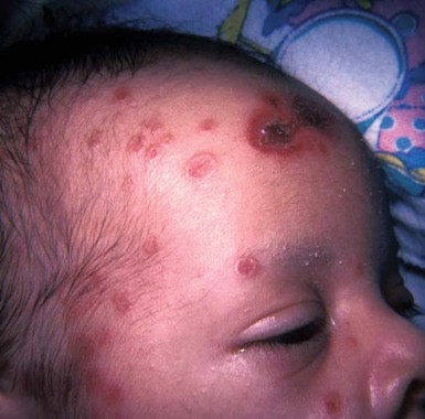
The condition can be present at birth. The macules and pustules can appear anywhere on the body, but most often on the chin, neck, upper chest, and/or lower back. The good news is they fade over time and are benign, so no treatment is necessary. The differential diagnosis from such conditions as herpes simplex and erythema toxicum is important, however, said Dr. Treadwell. One tip is to check for lesions on the palms and soles, which can be diagnostic in transient neonatal pustular melanosis, but not for erythema toxicum. A biopsy can confirm your clinical suspicions.
The cutaneous manifestations of neonatal lupus erythematosus can tip you off to this condition, she said. Skin lesions can be annular, discoid, or atrophic. Some children present with "raccoon eyes." Because this condition is related to maternal antibodies passed through the placenta during gestation, lesions generally clear by 6 months to 1 year of age.
The mother may have a diagnosis of an autoimmune disease, or she may be completely asymptomatic. In one instance, a mother brought her newborn to Dr. Treadwell’s clinic. He had annular lesions on his forehead with some erythema. The lesions were more edematous around the edges. He actually got some sun exposure between his first and second visits, and developed more-discoid lesions in his sun-exposed areas. He also presented with lesions in non–sun exposed areas.
"The mother had a positive ANA [antinuclear antibody] test. I told her she should go to her doctor for urine and blood pressure monitoring," Dr. Treadwell said. "She had no symptoms and thought I didn’t know what I was talking about." Two years later, the woman returned with a newborn daughter who also had neonatal lupus with lesions on her face and some patchy alopecia on her scalp.
"The brother came back in, ... and he had telangiectasias already present at age 2 years."
Your evaluation can be more family centered. Consider testing the parent and affected children for anti-Rho, anti-La, and anti-RNP.
"I usually treat them with sun avoidance, sun protection, and possibly hydrocortisone," Dr. Treadwell said. She also recommends one electrocardiogram to rule out any cardiac consequences.
Dr. Treadwell reported that she had no relevant financial disclosures.
MIAMI – Some hallmark signs of dermatologic problems in children – especially erythema and hyperpigmentation – often are less obvious in children with skin of color and can require more clinical detective work to diagnose.
Dr. Patricia A. Treadwell narrowed down the most likely dermatoses a pediatrician will encounter in this patient population at a pediatric update sponsored by Miami Children’s Hospital. Atopic and contact dermatitis, phytophotodermatitis, transient neonatal pustular melanosis, and neonatal lupus erythematosus are among the noteworthy clinical challenges, she said at the meeting.
"Children with increased pigmentation in their skin may end up testing your knowledge in terms of looking at their dermatitis and being able to diagnose that. Keep in mind it may be a little bit different in terms of the clinical presentation, but it’s important to identify it and start the proper treatment," said Dr. Treadwell, a pediatric dermatologist at Indiana University Health and Riley Hospital for Children in Indianapolis.
Overcoming this "masking" of a condition by skin pigmentation can be important, Dr. Treadwell said. She cited a patient born with a port-wine stain that went undiagnosed. The infant had subtle erythema and some asymmetry related to the overgrowth of the lesion. "This was not diagnosed based on the fact that the erythema was not apparent. The patient later developed a pyogenic granuloma, which is a complication that can be seen in patients with port-wine stains as they get older."
Erythema can be missed in children of color with atopic dermatitis as well. For this reason, atopic dermatitis may be underdiagnosed in this population overall, she said. Another challenge is the common practice of grading the severity of atopic dermatitis in lighter-skinned patients based on the degree of erythema. "In children with a fair amount of pigmentation in their skin, the erythema may not be noted and the severity will not be recognized."
Similarly, you might need a higher index of clinical suspicion to diagnose a child of color with contact dermatitis. Again, the erythema can be subtle. In contrast, "contact dermatitis can be very clear in a Caucasian patient. But the lesions are the same – linear, asymmetrical, and occurring on exposed areas," Dr. Treadwell said. Pruritus is common, and edema and swelling also occur. Watch for development of vesiculobullous lesions.
Phytophotodermatitis is another dermatologic condition that may require some additional detective work in children of color. Dr. Treadwell described a girl with a unique hyperpigmentation pattern on her legs and arms. She was referred following a vacation in Cancun, Mexico, with her family, and there was a concern about an autoimmune process. "They asked if she needed blood work. I said no, she was eating a mango and went out in the sun." Some of the mango juice splashed on her legs and arms.
Phytophotodermatitis occurs when furocoumarins from tropical fruit, citrus, celery, fennel, or parsnip come into contact with skin subsequently exposed to the sun.
"This condition can have a fairly bizarre pattern of presentation," Dr. Treadwell said. "Again, if there is more pigment in the skin, hyperpigmentation can present in a less common way than might be expected."
Some dermatoses are noted more commonly in children of color, she said. For example, the hyperpigmented macules or pustules that characterize transient neonatal pustular melanosis are reported in 4.4% of African American infants and 0.2% of Caucasian infants. "The percentages here may be related to the pigmentation in the skin. This does occur in Caucasian infants, but it may not be as noticeable."
The condition can be present at birth. The macules and pustules can appear anywhere on the body, but most often on the chin, neck, upper chest, and/or lower back. The good news is they fade over time and are benign, so no treatment is necessary. The differential diagnosis from such conditions as herpes simplex and erythema toxicum is important, however, said Dr. Treadwell. One tip is to check for lesions on the palms and soles, which can be diagnostic in transient neonatal pustular melanosis, but not for erythema toxicum. A biopsy can confirm your clinical suspicions.
The cutaneous manifestations of neonatal lupus erythematosus can tip you off to this condition, she said. Skin lesions can be annular, discoid, or atrophic. Some children present with "raccoon eyes." Because this condition is related to maternal antibodies passed through the placenta during gestation, lesions generally clear by 6 months to 1 year of age.
The mother may have a diagnosis of an autoimmune disease, or she may be completely asymptomatic. In one instance, a mother brought her newborn to Dr. Treadwell’s clinic. He had annular lesions on his forehead with some erythema. The lesions were more edematous around the edges. He actually got some sun exposure between his first and second visits, and developed more-discoid lesions in his sun-exposed areas. He also presented with lesions in non–sun exposed areas.
"The mother had a positive ANA [antinuclear antibody] test. I told her she should go to her doctor for urine and blood pressure monitoring," Dr. Treadwell said. "She had no symptoms and thought I didn’t know what I was talking about." Two years later, the woman returned with a newborn daughter who also had neonatal lupus with lesions on her face and some patchy alopecia on her scalp.
"The brother came back in, ... and he had telangiectasias already present at age 2 years."
Your evaluation can be more family centered. Consider testing the parent and affected children for anti-Rho, anti-La, and anti-RNP.
"I usually treat them with sun avoidance, sun protection, and possibly hydrocortisone," Dr. Treadwell said. She also recommends one electrocardiogram to rule out any cardiac consequences.
Dr. Treadwell reported that she had no relevant financial disclosures.
EXPERT ANALYSIS FROM A PEDIATRIC UPDATE SPONSORED BY MIAMI CHILDREN'S HOSPITAL
Skin of Color: Melasma Education for Patients
Melasma can be a distressing condition for our darker skinned patients. When educating them about sun protection, remind them that:
1. SPF only refers to protection against UVB radiation; it has no implication on the amount of protection against UVA. UVA is highly implicated in the progression of melasma. UVA even penetrates window glass so, if your patients drive, sit near a window, and/or are "never in the sun," remind them that they still need UVA and UVB protection every day.
2. Sunscreen needs to be applied 365 days a year. Ultraviolet light is present on cloudy, snowy, and rainy days.

3. "Broad spectrum" does not mean complete coverage. The two sunscreens that offer complete coverage against both UVA and UVB are Anthelios with Mexoryl and Neutrogena with Helioplex technology.
4. Heat can worsen melasma. If your patients work around heat, such as cooking by a hot stove or being around hot air, the heat can contribute to their melasma.
5. Computer monitors emit a small amount of UV. Suggest that melasma patients purchase a UV shield for their screens.
6. UV bracelets or beads help monitor the amount of UV in a given area. With the help of the devices, patients can monitor the amount of UV at home, at work, and in their car. Consider demonstrating the technology to your patients in the office to teach them about UV exposures in their daily environments.
7. Purchasing sunscreens with high SPF and broad spectrum coverage can be difficult. Most sunscreens leave a white or ashy residue on darker skin. Sunscreens with micronized titanium dioxide or zinc oxide can minimize the white residue and are more cosmetically appealing. Similarly, newer foundations and makeup products on the market have been developed that contain high SPF sunscreens in a tinted base. Some of my favorites include Laura Mercier tinted moisturizer and Revision Intellishade. Both have a small amount of tint to counteract the white appearance on darker skin.
8. Practice aggressive sun avoidance and protection before medical management. I don’t treat any patient with melasma unless they are vigilant about sun protection. The lasers, bleaching creams, medications (such as retinoids), and peels we use to treat melasma can make the skin more susceptible to UV radiation which can make melasma worse.
- Lily Talakoub, M.D.
Melasma can be a distressing condition for our darker skinned patients. When educating them about sun protection, remind them that:
1. SPF only refers to protection against UVB radiation; it has no implication on the amount of protection against UVA. UVA is highly implicated in the progression of melasma. UVA even penetrates window glass so, if your patients drive, sit near a window, and/or are "never in the sun," remind them that they still need UVA and UVB protection every day.
2. Sunscreen needs to be applied 365 days a year. Ultraviolet light is present on cloudy, snowy, and rainy days.
3. "Broad spectrum" does not mean complete coverage. The two sunscreens that offer complete coverage against both UVA and UVB are Anthelios with Mexoryl and Neutrogena with Helioplex technology.
4. Heat can worsen melasma. If your patients work around heat, such as cooking by a hot stove or being around hot air, the heat can contribute to their melasma.
5. Computer monitors emit a small amount of UV. Suggest that melasma patients purchase a UV shield for their screens.
6. UV bracelets or beads help monitor the amount of UV in a given area. With the help of the devices, patients can monitor the amount of UV at home, at work, and in their car. Consider demonstrating the technology to your patients in the office to teach them about UV exposures in their daily environments.
7. Purchasing sunscreens with high SPF and broad spectrum coverage can be difficult. Most sunscreens leave a white or ashy residue on darker skin. Sunscreens with micronized titanium dioxide or zinc oxide can minimize the white residue and are more cosmetically appealing. Similarly, newer foundations and makeup products on the market have been developed that contain high SPF sunscreens in a tinted base. Some of my favorites include Laura Mercier tinted moisturizer and Revision Intellishade. Both have a small amount of tint to counteract the white appearance on darker skin.
8. Practice aggressive sun avoidance and protection before medical management. I don’t treat any patient with melasma unless they are vigilant about sun protection. The lasers, bleaching creams, medications (such as retinoids), and peels we use to treat melasma can make the skin more susceptible to UV radiation which can make melasma worse.
- Lily Talakoub, M.D.
Skin of Color: Dermatosis Papulosa Nigra Removal
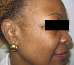
Dermatosis papulosa nigra, also known as DPN, are small, soft brown papules that may occur on the face and neck of patients of African, Latin, Indian, or Asian descent. While they may not reach the size of their histologically similar seborrheic keratosis counterparts, the lesions do represent a sign of aging in darker skinned patients. However, the lesions can be safely, easily, and effectively removed.
Electrodesiccation with a hyfrecator or destruction with the KTP (532 nm) laser are my favorite methods for DPN removal. I prefer not to use curettage or cryotherapy because of the risk for dyspigmentation in darker skinned patients. Case reports of success with fractional photothermolyis (1,550 nm) and Nd:YAG lasers (1,064 nm) have been published.
If electrodesiccation is performed, the application of topical anesthetic prior to the procedure helps make the patient more comfortable. For larger lesions, injection of 1% lidocaine with 1:100,000 epinephrine may be used.
Also, with electrodesiccation, conservative settings (0.6-2.0 W on the low setting) should be used; the lesions are desiccated using a blunt tip for a few seconds until they turn grayish.
Care is taken not to touch the surrounding skin. A sharp tip may be used with very small (less than 1 mm) lesions for more accurate precision. I wipe the tip from time to time with gauze to avoid char accumulation.
Larger or pedunculated lesions may be treated with electrodesiccation or snipped off with gradle scissors.
With the KTP laser, topical anesthesia is usually not required. I use a smaller spot size than the lesion itself to avoid targeting and potentially causing dyspigmentation of the surrounding skin.
A spot size of 1 mm is typically used, with 6-10 ms and 10-15 j/cm2. The laser tip is held approximately 1 cm away from the skin at a 90 degree angle. I start off with the lowest fluence and adjust it higher until the lesions turn grayish and a light popping sound is heard with the laser pulse.
A split-face study published in the American Journal of Dermatologic Surgery in 2009 showed that both electrodesiccation and KTP have comparable efficacy in removal of DPN. Without use of anesthetics, the KTP laser was preferred for patient comfort.
Immediately after treatment, patients can expect the treated lesions to become red and swollen - similar to insect bite reactions - for about an hour. Antibiotic ointment or aquaphor is applied to soothe the skin.
Patients are then told to leave the lesions alone, to avoid picking, and to avoid sun exposure. Patients are also advised to avoid alpha-hydroxy acids and other "anti-aging" products until healed. If the cheeks were treated, make-up (foundation, blush) may be applied in 3 to 4 days. Lesions typically fall off within a week.
If needed, repeat treatment may be performed in 2 to 4 weeks.
If you have any DPN removal tips, please feel free to share!
-Naissan Wesley, M.D.
Do you have questions about treating patients with darker skin? If so, send them to [email protected].