User login
Dark-Skinned Patients Not Getting Skin Cancer Message
NEW YORK – All patients, regardless of skin color, need to be screened for skin cancer and receive sun protection education, according to Dr. Brooke A. Jackson.
"We have done a pretty good job of relaying the skin cancer awareness/risk message to fair skin types, but we still need to work on the message to darker skin types," noted Dr. Jackson. "This includes offering skin cancer screenings to all of our patients regardless of skin color, having a [high] level of suspicion for nonhealing lesions or changing lesions in darker skin types, and discussing skin cancer risks and sun protection with our patients who have darker skin."

Dr. Jackson and her colleagues surveyed 105 dark-skinned adult patients who presented to her private practice in Chicago for a variety of reasons.
Overall, 91 patients identified themselves as black, 9 as Hispanic, 4 as Asian, and 1 as Middle Eastern, noted Dr. Jackson, clinical assistant professor of dermatology at Northwestern University in Chicago.
Of the 105 patients, 9 had a Fitzpatrick skin type of III, 29 had type IV, 64 had type V, and 3 patients had type VI.
Patients read the descriptions for several types of lesions and were asked to identify whether a particular lesion was a risk factor for skin cancer, including "dark spot with irregular border," "new mole," "nonhealing wound," "bleeding lesion," and "shiny pink bump."
Dr. Jackson found that "regardless of ethnic origin or skin type, ‘dark spot with irregular borders’ followed by ‘new mole’ were the most frequent top two choices" selected as being high risk for skin cancer.
"Shiny pink bump" was the least selected choice for recognition of skin cancer and was not selected by any respondents with skin types III and VI, she reported.
Indeed, "15 respondents, most of whom were of African ethnicity and/or had skin type V, were unaware that skin of color was at risk for developing skin cancer," noted Dr. Jackson and her colleagues.
As for skin protective behaviors, 70 of the 91 black patients reported use of sunblock or sunscreen, and 47 used protective clothing. Twenty-nine black patients practiced sun avoidance. Ten of the black patients reported that they took no precaution at all with regard to sun exposure. Similarly, among the 64 Fitzpatrick skin type V patients, 13 reported practicing no sun protection.
Dr. Jackson stated that neither she nor her colleagues had any disclosures relevant to this presentation.
NEW YORK – All patients, regardless of skin color, need to be screened for skin cancer and receive sun protection education, according to Dr. Brooke A. Jackson.
"We have done a pretty good job of relaying the skin cancer awareness/risk message to fair skin types, but we still need to work on the message to darker skin types," noted Dr. Jackson. "This includes offering skin cancer screenings to all of our patients regardless of skin color, having a [high] level of suspicion for nonhealing lesions or changing lesions in darker skin types, and discussing skin cancer risks and sun protection with our patients who have darker skin."
Dr. Jackson and her colleagues surveyed 105 dark-skinned adult patients who presented to her private practice in Chicago for a variety of reasons.
Overall, 91 patients identified themselves as black, 9 as Hispanic, 4 as Asian, and 1 as Middle Eastern, noted Dr. Jackson, clinical assistant professor of dermatology at Northwestern University in Chicago.
Of the 105 patients, 9 had a Fitzpatrick skin type of III, 29 had type IV, 64 had type V, and 3 patients had type VI.
Patients read the descriptions for several types of lesions and were asked to identify whether a particular lesion was a risk factor for skin cancer, including "dark spot with irregular border," "new mole," "nonhealing wound," "bleeding lesion," and "shiny pink bump."
Dr. Jackson found that "regardless of ethnic origin or skin type, ‘dark spot with irregular borders’ followed by ‘new mole’ were the most frequent top two choices" selected as being high risk for skin cancer.
"Shiny pink bump" was the least selected choice for recognition of skin cancer and was not selected by any respondents with skin types III and VI, she reported.
Indeed, "15 respondents, most of whom were of African ethnicity and/or had skin type V, were unaware that skin of color was at risk for developing skin cancer," noted Dr. Jackson and her colleagues.
As for skin protective behaviors, 70 of the 91 black patients reported use of sunblock or sunscreen, and 47 used protective clothing. Twenty-nine black patients practiced sun avoidance. Ten of the black patients reported that they took no precaution at all with regard to sun exposure. Similarly, among the 64 Fitzpatrick skin type V patients, 13 reported practicing no sun protection.
Dr. Jackson stated that neither she nor her colleagues had any disclosures relevant to this presentation.
FROM THE SKIN OF COLOR SEMINAR SERIES
Major Finding: Of the survey respondents, 15 reported being unaware that people with skin of color were at risk for developing skin cancer.
Data Source: A survey of 105 skin of color patients seen at a private dermatology practice in Chicago.
Disclosures: Dr. Jackson stated that neither she nor her colleagues had any disclosures relevant to this presentation.
Eflornithine + Laser 99% Effective for Pseudofolliculitis Barbae
WAIKOLOA, HAWAII – The use of eflornithine cream may increase the effectiveness of laser hair removal for treating pseudofolliculitis barbae, according to Dr. Andrew F. Alexis.
Laser hair removal has proved to be a game changer in the treatment of pseudofolliculitis barbae, a common chronic, inflammatory dermatosis that’s often been a difficult therapeutic challenge, Dr. Alexis said at the seminar sponsored by Skin Disease Education Foundation (SDEF). And the use of adjunctive eflornithine cream makes laser therapy even more effective, based on a recent study, which is one of the few rigorous studies conducted in pseudofolliculitis barbae (PFB) patients, noted Dr. Alexis, director of the skin of color center at St. Luke’s–Roosevelt Hospital and a dermatologist at Columbia University, New York.
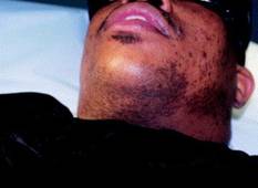
The double-blind placebo controlled study was carried out by U.S. military physicians. PFB, which is predominantly a disorder of black men, has at times been a source of racial tension in the military because the simplest treatment for PFB is to stop shaving and grow a beard, a form of individual expression at odds with regulations.
The study included 27 men with PFB. They received laser therapy once every 4 weeks for 16 weeks. In addition, they applied eflornithine cream to one side of their bearded neck region and placebo to the other side twice daily.
At 16 weeks, the laser plus eflornithine cream side produced a median 99.5% reduction in hair count and inflammatory papules. This was a significantly better result than the median 85% reduction on the laser plus placebo–treated side (J. Am. Acad. Dermatol. 2012 Jan. 8; in press).
The PFB study follows an earlier study by other investigators who demonstrated that eflornithine cream as an adjunct to laser hair removal for facial hirsutism in women was more effective than laser therapy alone (J. Am. Acad. Dermatol. 2007;57:54-9).
Dr. Alexis said performing laser hair removal safely in darker-skinned patients with PFB requires attention to several key principles: longer wavelengths, lower fluences, longer pulse durations, and plenty of epidermal cooling.
"The No. 1 thing is to use longer wavelengths, because the goal is deeper penetration to maximize the ratio of the temperature in the bulb of the follicle to the temperature in the epidermis," he explained.
The long-pulsed 1,064-nm Nd:YAG laser has the lowest rate of associated epidermal burns, hypopigmentation, and other adverse events in darker-skinned patients, as has been shown in a review of a wide assortment of lasers (J. Drugs Dermatol. 2007;6:40-6). It is clearly the safest laser option in patients with skin types IV-VI. The 810-nm diode laser is a reasonable alternative in skin types IV-V, Dr. Alexis said.

In treating patients for PFB with the 1,064-nm Nd:YAG laser, he said that he typically starts with a fluence of 20 J/cm2 and a pulse duration of 20-30 milliseconds. After several sessions, as he makes inroads into the initially dense follicular distribution, he said that he might increase the fluence to a maximum of 50 J/cm2 in the setting of skin type VI, and as high as 100 J/cm2 in skin types IV or V.
Longer pulse durations allow for more efficient epidermal cooling. This minimizes heat injury to melanin-containing epidermal cells. For the 810-nm diode laser, Dr. Alexis said he uses pulse durations of 100 or 400 milliseconds.
Epidermal cooling can be accomplished in several ways. His preferred method is to utilize contact cooling via a sapphire tip or chilled copper plate attached to the laser; the cooling is done before delivering the laser pulse. Alternatively, the epidermal cooling can be done using cold gels, forced air, or spray cooling, although dyschromia can occur in darker skin types if the spray technique isn’t optimal. Another option is to apply an ice pack for 5-10 minutes post treatment.
Laser therapy is expensive, so Dr. Alexis said he likes to give his patients a range of therapeutic options. These include growing a beard, chemical depilation with barium sulfide or calcium thioglycolate every 2-4 days, modification of shaving practices, and salicylic acid chemical peels.
"It’s kind of a long conversation," he said.
Whatever form of therapy the patient decides upon, it’s important that the patient stops tweezing to remove ingrown hairs. This is a common practice that induces trauma and worsens postinflammatory hyperpigmentation.
Dr. Alexis reported that he serves as a consultant to Schick and is on the advisory board for Allergan. SDEF and this news organization are owned by Elsevier.
WAIKOLOA, HAWAII – The use of eflornithine cream may increase the effectiveness of laser hair removal for treating pseudofolliculitis barbae, according to Dr. Andrew F. Alexis.
Laser hair removal has proved to be a game changer in the treatment of pseudofolliculitis barbae, a common chronic, inflammatory dermatosis that’s often been a difficult therapeutic challenge, Dr. Alexis said at the seminar sponsored by Skin Disease Education Foundation (SDEF). And the use of adjunctive eflornithine cream makes laser therapy even more effective, based on a recent study, which is one of the few rigorous studies conducted in pseudofolliculitis barbae (PFB) patients, noted Dr. Alexis, director of the skin of color center at St. Luke’s–Roosevelt Hospital and a dermatologist at Columbia University, New York.
The double-blind placebo controlled study was carried out by U.S. military physicians. PFB, which is predominantly a disorder of black men, has at times been a source of racial tension in the military because the simplest treatment for PFB is to stop shaving and grow a beard, a form of individual expression at odds with regulations.
The study included 27 men with PFB. They received laser therapy once every 4 weeks for 16 weeks. In addition, they applied eflornithine cream to one side of their bearded neck region and placebo to the other side twice daily.
At 16 weeks, the laser plus eflornithine cream side produced a median 99.5% reduction in hair count and inflammatory papules. This was a significantly better result than the median 85% reduction on the laser plus placebo–treated side (J. Am. Acad. Dermatol. 2012 Jan. 8; in press).
The PFB study follows an earlier study by other investigators who demonstrated that eflornithine cream as an adjunct to laser hair removal for facial hirsutism in women was more effective than laser therapy alone (J. Am. Acad. Dermatol. 2007;57:54-9).
Dr. Alexis said performing laser hair removal safely in darker-skinned patients with PFB requires attention to several key principles: longer wavelengths, lower fluences, longer pulse durations, and plenty of epidermal cooling.
"The No. 1 thing is to use longer wavelengths, because the goal is deeper penetration to maximize the ratio of the temperature in the bulb of the follicle to the temperature in the epidermis," he explained.
The long-pulsed 1,064-nm Nd:YAG laser has the lowest rate of associated epidermal burns, hypopigmentation, and other adverse events in darker-skinned patients, as has been shown in a review of a wide assortment of lasers (J. Drugs Dermatol. 2007;6:40-6). It is clearly the safest laser option in patients with skin types IV-VI. The 810-nm diode laser is a reasonable alternative in skin types IV-V, Dr. Alexis said.
In treating patients for PFB with the 1,064-nm Nd:YAG laser, he said that he typically starts with a fluence of 20 J/cm2 and a pulse duration of 20-30 milliseconds. After several sessions, as he makes inroads into the initially dense follicular distribution, he said that he might increase the fluence to a maximum of 50 J/cm2 in the setting of skin type VI, and as high as 100 J/cm2 in skin types IV or V.
Longer pulse durations allow for more efficient epidermal cooling. This minimizes heat injury to melanin-containing epidermal cells. For the 810-nm diode laser, Dr. Alexis said he uses pulse durations of 100 or 400 milliseconds.
Epidermal cooling can be accomplished in several ways. His preferred method is to utilize contact cooling via a sapphire tip or chilled copper plate attached to the laser; the cooling is done before delivering the laser pulse. Alternatively, the epidermal cooling can be done using cold gels, forced air, or spray cooling, although dyschromia can occur in darker skin types if the spray technique isn’t optimal. Another option is to apply an ice pack for 5-10 minutes post treatment.
Laser therapy is expensive, so Dr. Alexis said he likes to give his patients a range of therapeutic options. These include growing a beard, chemical depilation with barium sulfide or calcium thioglycolate every 2-4 days, modification of shaving practices, and salicylic acid chemical peels.
"It’s kind of a long conversation," he said.
Whatever form of therapy the patient decides upon, it’s important that the patient stops tweezing to remove ingrown hairs. This is a common practice that induces trauma and worsens postinflammatory hyperpigmentation.
Dr. Alexis reported that he serves as a consultant to Schick and is on the advisory board for Allergan. SDEF and this news organization are owned by Elsevier.
EXPERT ANALYSIS FROM THE SDEF HAWAII DERMATOLOGY SEMINAR
Skin of Color: Classifying Undereye Circles
How many times a week do you get asked by your patients how to get rid of the "dark circles" under their eyes? The term is a catch-all used by physicians and patients to refer to problems that have a vast range of genetic, environmental, and skin-related causes. It is a common and frustrating problem, with little structure in its definition and few full-proof treatments.
Below is my proposed classification system for the definition of dark circles and clinical pearls for their treatment. Most patients, however, have a combination of each type and multifactorial causes that need to be addressed.

Infraorbital fat pad protrusion. Also known as "eye bags."
Blepharoplasty is the best, and for now the only, solution for severe fat pad prominence. Referral to a board certified plastic surgeon or dermatologic surgeon is recommended.
If the protrusion is mild and tear troughs are prominent, fillers may be injected into the tear trough area to help mask the protrusion. My favorites for this area are hyalauronic acid fillers like Juvéderm Ultra or Restlyane, sometimes double diluted with normal saline or injected with a 32-gauge needle.
For loose skin with "bags," radiofrequency lasers can provide some benefit. The Thermage eyelid tip produces results over 3-6 months, with repeat treatment possible at 6 months. I always advise patients that this treatment is not a replacement for surgery but can provide some benefit in those who are not surgical candidates or who do not want surgery.
Infraorbital edema. Also known as "puffiness."
The infraorbital skin is very thin and highly sensitive to fluid compartmentalization. Seasonal allergies, sinus infections, crying or water retention from high blood pressure or eating high sodium foods are some of the reasons the loose, thin epidermis becomes edematous.
Treat seasonal allergies with over-the-counter allergy medications or prescription medications for resistant allergies or possible sinus infections.
Advise patients to switch their sleep position. Sleep position can be contributing to undereye bags through gravity. Sleeping on the side or stomach can encourage fluids to collect under the eyes. If patients report being a side sleeper, you may notice a heavier bag on the side they report sleeping on. Patients who wake up with puffy eyes can sleep on their back and add an extra pillow under their head.
Also advise patients to avoid rubbing their eyes, going to bed with makeup on, and using harsh cleansers. Anything that irritates the eyes can cause fluids to pool. Sleeping in eye makeup can irritate eyes, causing undereye edema.
Eye bags could be a sign of an underlying medical condition, especially if bags appear suddenly and none of the above conditions apply. Thyroid, cardiovascular, or kidney problems can cause undereye fluid retention and patients will need to see their primary care doctors for further evaluation.
Patients can place an ice pack, slices of cucumbers, chilled tea bags, refrigerated eye gels, or even a package of frozen peas on their eyes. This can constrict leaky blood vessels and lessen the periorbital edema.
Periorbital hyperpigmentation. Also known as "dark circles."
Pigmentation of the periorbital skin is very common in skin of color because of the increased melanin content. Genetics, rubbing, and inflammatory skin diseases such as eczema may play a role in exacerbating the pigmentation of the thin undereye skin.
Again, advise patients to avoid rubbing the area. Chronic rubbing and the development of lichen simplex chronicus can lead to dark, thickened undereye skin.
Retinoic acid creams can help slough the dark pigmented skin. It should, however, be used in very small amounts that increase over a few weeks to avoid severe irritation.
Skin lightening creams with azaleic acid, kojic acid, and glycolic acid can be found in varying strengths. Hydroquinone creams have been successful in lightening undereye hyperpigmentation. Strengths in over-the-counter preparations start at 1-2% and in prescription strength can be compounded to higher than 4%, but caution should be used to avoid further irritation and potential post-inflammatory pigment from these products.
Light chemical peels can assist in lightening dark undereye pigmentation. Peels with hydroquinone or retinoic acid can be used for an added lightening benefit.
Intense pulse light can help minimize undereye pigmentation, particularly UV-induced pigmentation. Q-switched lasers have also been reported to be effective.
Infraorbital tear trough depression.
Most often, dark circles aren't about changes in the color of the skin. Instead, they're created by a loss of volume in the area around the eye, exposing the orbital bone and creating a hollow trough that shows up as a dark circle. These changes are often genetic, but significant weigh loss can also expose undereye tear trough depressions.
- The best way to treat this problem is with a small amount of a hyaluronic acid filler placed by a dermatologist in the trough. Very small aliquots are needed in even the deepest trough but can give outstanding results. Use caution, however; this is a highly technical and injector-dependent procedure.
There are crucial vascular structures around the eye that need to be avoided, and over-filled troughs will give patients a puffy appearance and may pose a worse and more difficult problem to fix. Hyaluronic acid fillers are not FDA approved to treat undereye depressions, so patients should be knowledgeable to the risks and benefits prior to undergoing these procedures.
With age, the skin around the eye becomes thinner, exposing the small capillaries and venules just below the thin epidermal layer. Vascular prominence can leave a bluish undertone to the infraorbital skin, which can cast dark shadows and make the area appear dark or sallow.
Eye creams that contain caffeine can constrict the underlying blood vessels and temporarily diminish small vessel prominence.
For large blue veins, vascular lasers such as a long pulse Nd:YAG laser can be recommended. However, in darker skin types these lasers can cause hyperpigmented scars if not used with adequate skin cooling techniques.
Periorbital static and dynamic rhytids.
Botulinum toxin placed in small aliquots around the orbital rim will reduce the dynamic rhytids in this area. Treatments spaced 3-4 months apart will ensure long lasting benefits and because botulinum toxin wears off, repeat treatments are needed.
Laser resurfacing with CO2 or fractionated CO2 lasers provide excellent benefit for periocular rhytides. A traditional CO2 laser may require repeat treatment in 6-12 months. Fractionated CO2 lasers typically require 4-6 treatments spaced about 4 weeks apart to provide benefit.
Overall tips:
For most of the types of infraorbital issues, makeup can help conceal or mask some imperfections. Patients should choose a concealer that matches or is slightly lighter than their skin tone. If they have mild discoloration, advise or help them pick a liquid formula for more prominent imperfections. A cream, full coverage concealer works best.
Encourage patients to quit smoking, which dehydrates the skin and causes premature aging and collagen degradation.
Always remind patients to apply sunscreen around the eye area. Hyperpigmentation and tear troughs can accentuate with UV-induced skin pigmentation.
- Advise patients to apply a moisturizer to the eye area nightly to keep the skin from becoming dry, irritated, and dehydrated.
- Lily Talakoub, M.D.
Do you have questions about treating patients with darker skin? If so, send them to [email protected].
How many times a week do you get asked by your patients how to get rid of the "dark circles" under their eyes? The term is a catch-all used by physicians and patients to refer to problems that have a vast range of genetic, environmental, and skin-related causes. It is a common and frustrating problem, with little structure in its definition and few full-proof treatments.
Below is my proposed classification system for the definition of dark circles and clinical pearls for their treatment. Most patients, however, have a combination of each type and multifactorial causes that need to be addressed.
Infraorbital fat pad protrusion. Also known as "eye bags."
Blepharoplasty is the best, and for now the only, solution for severe fat pad prominence. Referral to a board certified plastic surgeon or dermatologic surgeon is recommended.
If the protrusion is mild and tear troughs are prominent, fillers may be injected into the tear trough area to help mask the protrusion. My favorites for this area are hyalauronic acid fillers like Juvéderm Ultra or Restlyane, sometimes double diluted with normal saline or injected with a 32-gauge needle.
For loose skin with "bags," radiofrequency lasers can provide some benefit. The Thermage eyelid tip produces results over 3-6 months, with repeat treatment possible at 6 months. I always advise patients that this treatment is not a replacement for surgery but can provide some benefit in those who are not surgical candidates or who do not want surgery.
Infraorbital edema. Also known as "puffiness."
The infraorbital skin is very thin and highly sensitive to fluid compartmentalization. Seasonal allergies, sinus infections, crying or water retention from high blood pressure or eating high sodium foods are some of the reasons the loose, thin epidermis becomes edematous.
Treat seasonal allergies with over-the-counter allergy medications or prescription medications for resistant allergies or possible sinus infections.
Advise patients to switch their sleep position. Sleep position can be contributing to undereye bags through gravity. Sleeping on the side or stomach can encourage fluids to collect under the eyes. If patients report being a side sleeper, you may notice a heavier bag on the side they report sleeping on. Patients who wake up with puffy eyes can sleep on their back and add an extra pillow under their head.
Also advise patients to avoid rubbing their eyes, going to bed with makeup on, and using harsh cleansers. Anything that irritates the eyes can cause fluids to pool. Sleeping in eye makeup can irritate eyes, causing undereye edema.
Eye bags could be a sign of an underlying medical condition, especially if bags appear suddenly and none of the above conditions apply. Thyroid, cardiovascular, or kidney problems can cause undereye fluid retention and patients will need to see their primary care doctors for further evaluation.
Patients can place an ice pack, slices of cucumbers, chilled tea bags, refrigerated eye gels, or even a package of frozen peas on their eyes. This can constrict leaky blood vessels and lessen the periorbital edema.
Periorbital hyperpigmentation. Also known as "dark circles."
Pigmentation of the periorbital skin is very common in skin of color because of the increased melanin content. Genetics, rubbing, and inflammatory skin diseases such as eczema may play a role in exacerbating the pigmentation of the thin undereye skin.
Again, advise patients to avoid rubbing the area. Chronic rubbing and the development of lichen simplex chronicus can lead to dark, thickened undereye skin.
Retinoic acid creams can help slough the dark pigmented skin. It should, however, be used in very small amounts that increase over a few weeks to avoid severe irritation.
Skin lightening creams with azaleic acid, kojic acid, and glycolic acid can be found in varying strengths. Hydroquinone creams have been successful in lightening undereye hyperpigmentation. Strengths in over-the-counter preparations start at 1-2% and in prescription strength can be compounded to higher than 4%, but caution should be used to avoid further irritation and potential post-inflammatory pigment from these products.
Light chemical peels can assist in lightening dark undereye pigmentation. Peels with hydroquinone or retinoic acid can be used for an added lightening benefit.
Intense pulse light can help minimize undereye pigmentation, particularly UV-induced pigmentation. Q-switched lasers have also been reported to be effective.
Infraorbital tear trough depression.
Most often, dark circles aren't about changes in the color of the skin. Instead, they're created by a loss of volume in the area around the eye, exposing the orbital bone and creating a hollow trough that shows up as a dark circle. These changes are often genetic, but significant weigh loss can also expose undereye tear trough depressions.
- The best way to treat this problem is with a small amount of a hyaluronic acid filler placed by a dermatologist in the trough. Very small aliquots are needed in even the deepest trough but can give outstanding results. Use caution, however; this is a highly technical and injector-dependent procedure.
There are crucial vascular structures around the eye that need to be avoided, and over-filled troughs will give patients a puffy appearance and may pose a worse and more difficult problem to fix. Hyaluronic acid fillers are not FDA approved to treat undereye depressions, so patients should be knowledgeable to the risks and benefits prior to undergoing these procedures.
With age, the skin around the eye becomes thinner, exposing the small capillaries and venules just below the thin epidermal layer. Vascular prominence can leave a bluish undertone to the infraorbital skin, which can cast dark shadows and make the area appear dark or sallow.
Eye creams that contain caffeine can constrict the underlying blood vessels and temporarily diminish small vessel prominence.
For large blue veins, vascular lasers such as a long pulse Nd:YAG laser can be recommended. However, in darker skin types these lasers can cause hyperpigmented scars if not used with adequate skin cooling techniques.
Periorbital static and dynamic rhytids.
Botulinum toxin placed in small aliquots around the orbital rim will reduce the dynamic rhytids in this area. Treatments spaced 3-4 months apart will ensure long lasting benefits and because botulinum toxin wears off, repeat treatments are needed.
Laser resurfacing with CO2 or fractionated CO2 lasers provide excellent benefit for periocular rhytides. A traditional CO2 laser may require repeat treatment in 6-12 months. Fractionated CO2 lasers typically require 4-6 treatments spaced about 4 weeks apart to provide benefit.
Overall tips:
For most of the types of infraorbital issues, makeup can help conceal or mask some imperfections. Patients should choose a concealer that matches or is slightly lighter than their skin tone. If they have mild discoloration, advise or help them pick a liquid formula for more prominent imperfections. A cream, full coverage concealer works best.
Encourage patients to quit smoking, which dehydrates the skin and causes premature aging and collagen degradation.
Always remind patients to apply sunscreen around the eye area. Hyperpigmentation and tear troughs can accentuate with UV-induced skin pigmentation.
- Advise patients to apply a moisturizer to the eye area nightly to keep the skin from becoming dry, irritated, and dehydrated.
- Lily Talakoub, M.D.
Do you have questions about treating patients with darker skin? If so, send them to [email protected].
Cultural Practices at Root of Alopecia
NEW YORK – To successfully treat alopecia in darker-skinned patients, it is important to first get to the root of a patient’s hair care regimen, according to Dr. Amy McMichael.
For the past 18 years, Dr. McMichael of Wake Forest University in Winston-Salem, N.C., has run a hair disorders clinic. She said she often finds herself at odds with patients who can’t understand why their hair won’t grow.
"Of course, we know it’s growing; it’s just breaking off. They’re having issues with damaged hair," she said at the Skin of Color Seminar Series. Patients "want me to find some underlying vitamin disorder or disease, or something [in their diet] that they can ‘cut out,’ " she said.

Instead, the key is to specifically ask patients about their hair care regimen. She also uses a "60-second comb test" to assess fragility, whereby she instructs patients to brush their hair over a white pillow for 60 seconds and then count the broken and full-bulb hairs that are seen on the pillow.
More often than not, she said she finds that the number of full telogen hairs do not differ between white and darker-skinned people, but that broken hairs (versus bulb hairs) are found significantly more often in women of African descent.
This standardized approach may help convince women that, indeed, breakage – and not some underlying condition – is at the root of their problem, and that changes in behavior could have big effects.
Traction alopecia is a major issue in this population – even among patients who say they don’t pull their hair – and is likely because of the African American custom of getting tight braids starting at a young age. "They tell me their hair braids were so tight, they couldn’t chew the next day," she said. "That is not normal."
Additionally, many skin of color patients use powerful, lye-based chemical relaxers. The damage inflicted by these products, combined with braids, increases the risk for alopecia.
She pointed to a 2008 study of 574 African school girls and 604 African women that showed that females who both relaxed and braided their hair had a 3.5 times greater risk for traction alopecia, compared with patients who did neither (J. Am. Acad. Dermatol. 2008;59:432-8).
Central centrifugal scarring alopecia is also associated with particular cultural practices. For example, Dr. McMichael cited a 2009 study that looked at 101 black women with the condition and found that there was a strong association between scarring alopecia and patients who reported using sewn-in hair weaves and braided styles with hair extensions (J. Am. Acad. Dermatol. 2009;60:574-8).
A second 2011 study by Dr. McMichael and her colleagues confirmed this, but also found associations with chemical relaxers in 44 surveyed patients (Cosmet. Dermatol. 2011;24:331-7).
She recommends that patients discontinue tight braids, sewn-in weaves, relaxers, and heat treatments. "A lot of women still go under hooded hair dryers," she said. She also advocates serial trimming of the hair every 6-8 weeks, as well as gentle hair conditioning with positively charged silicones and dimethicone coating agents.
"These work very nicely in this population," she said. She also recommends using foams as a vehicle for treatments when available. For patients with more severe issues, however, she has administered intralesional corticosteroids, and followed with an off-label use of topical minoxidil.
Additionally, "a lot of women do well with surgical hair restoration," she said, despite initial patient concerns about it being prohibitively expensive. "It might be much less expensive [than patients think] because they have a small area to treat."
Finally, Dr. McMichael said she refers patients with cicatricial alopecia to the Cicatricial Alopecia Research Foundation.
Dr. McMichael stated that she has been an investigator for Abbott, Allergan, Intendis (now Bayer HealthCare), and Procter and Gamble. She also disclosed serving as a consultant for Allergan, Galderma, Guthy-Ranker, Johnson and Johnson, Procter and Gamble, and Stiefel.
alopecia in black women, Dr. Amy McMichael, skin of color dermatology
NEW YORK – To successfully treat alopecia in darker-skinned patients, it is important to first get to the root of a patient’s hair care regimen, according to Dr. Amy McMichael.
For the past 18 years, Dr. McMichael of Wake Forest University in Winston-Salem, N.C., has run a hair disorders clinic. She said she often finds herself at odds with patients who can’t understand why their hair won’t grow.
"Of course, we know it’s growing; it’s just breaking off. They’re having issues with damaged hair," she said at the Skin of Color Seminar Series. Patients "want me to find some underlying vitamin disorder or disease, or something [in their diet] that they can ‘cut out,’ " she said.
Instead, the key is to specifically ask patients about their hair care regimen. She also uses a "60-second comb test" to assess fragility, whereby she instructs patients to brush their hair over a white pillow for 60 seconds and then count the broken and full-bulb hairs that are seen on the pillow.
More often than not, she said she finds that the number of full telogen hairs do not differ between white and darker-skinned people, but that broken hairs (versus bulb hairs) are found significantly more often in women of African descent.
This standardized approach may help convince women that, indeed, breakage – and not some underlying condition – is at the root of their problem, and that changes in behavior could have big effects.
Traction alopecia is a major issue in this population – even among patients who say they don’t pull their hair – and is likely because of the African American custom of getting tight braids starting at a young age. "They tell me their hair braids were so tight, they couldn’t chew the next day," she said. "That is not normal."
Additionally, many skin of color patients use powerful, lye-based chemical relaxers. The damage inflicted by these products, combined with braids, increases the risk for alopecia.
She pointed to a 2008 study of 574 African school girls and 604 African women that showed that females who both relaxed and braided their hair had a 3.5 times greater risk for traction alopecia, compared with patients who did neither (J. Am. Acad. Dermatol. 2008;59:432-8).
Central centrifugal scarring alopecia is also associated with particular cultural practices. For example, Dr. McMichael cited a 2009 study that looked at 101 black women with the condition and found that there was a strong association between scarring alopecia and patients who reported using sewn-in hair weaves and braided styles with hair extensions (J. Am. Acad. Dermatol. 2009;60:574-8).
A second 2011 study by Dr. McMichael and her colleagues confirmed this, but also found associations with chemical relaxers in 44 surveyed patients (Cosmet. Dermatol. 2011;24:331-7).
She recommends that patients discontinue tight braids, sewn-in weaves, relaxers, and heat treatments. "A lot of women still go under hooded hair dryers," she said. She also advocates serial trimming of the hair every 6-8 weeks, as well as gentle hair conditioning with positively charged silicones and dimethicone coating agents.
"These work very nicely in this population," she said. She also recommends using foams as a vehicle for treatments when available. For patients with more severe issues, however, she has administered intralesional corticosteroids, and followed with an off-label use of topical minoxidil.
Additionally, "a lot of women do well with surgical hair restoration," she said, despite initial patient concerns about it being prohibitively expensive. "It might be much less expensive [than patients think] because they have a small area to treat."
Finally, Dr. McMichael said she refers patients with cicatricial alopecia to the Cicatricial Alopecia Research Foundation.
Dr. McMichael stated that she has been an investigator for Abbott, Allergan, Intendis (now Bayer HealthCare), and Procter and Gamble. She also disclosed serving as a consultant for Allergan, Galderma, Guthy-Ranker, Johnson and Johnson, Procter and Gamble, and Stiefel.
alopecia in black women, Dr. Amy McMichael, skin of color dermatology
alopecia in black women, Dr. Amy McMichael, skin of color dermatology
EXPERT ANALYSIS FROM THE SKIN OF COLOR SEMINAR SERIES
Melanoma in Skin of Color
Skin of Color: Ethnic Differences in Skin Architecture
A reader recently wrote to us with the following question:
A certain ad for a skin care line for darker tones claimed that black skin contains "more" collagen. I try to explain to my clients that skin is skin, with some containing a richer concentration of melanin than others, but this seems too simplistic. Am I wrong? How should I answer?
Answer:
You are right that the answers to these questions are difficult and not completely clear. Differences in melanin content and dispersion, accounting for the difference we see in skin color, are well known and easy to demonstrate.
Other differences among skin in different ethnic groups have been studied but are not as easy to explain. While the data is limited, there are more and more studies showing that some differences in skin architecture and physiology among ethnic groups do exist.
These early studies have suggested that the thickness of the skin is the same in light and dark skin, specifically in the epidermis. Darker skin types, however, may have more cornified cell layers and greater lipid content compared to white stratum corneum.
Another study showed statistically significant differences in ceramide and cholesterol ratios for different ethnicities, with Asians having the highest ratio, white skin intermediate, and black skin the lowest (Br. J. Dermatol. 2010;163:1169-73).
With regards to the dermis, darker skin has been found to have more and larger fibroblasts, smaller collagen fiber bundles, and more macrophages than white skin. This may have implications in the development of keloid formation that we sometimes see an increased incidence of in darker skin.
Other differences are summarized in the following tables:
Objective Differences in Skin Structure and Physiology Based on Race (Semin. Cut. Med. Surg. 2009;28:115-2)
| Evidence supports: | Insufficient evidence* for: | Inconclusive: |
| • Increased melanin content and melanosomal dispersion in persons of color • Multinucleated and larger fibroblasts in black persons compared with white persons • pH black < white skin • Larger mast cell granules, increased parallel-linear striations, and increased tryptase localized to parallel-linear striations in black compared to white skin • Variable racial blood vessel reactivity | Racial differences in: • Skin elastic recovery/extensibility • Skin microflora • Facial pore size** | Racial differences in: • Transepidermal water loss • Water content • Corneocyte desquamation • Lipid content |
* Skin elastic recovery/extensibility, skin microflora, and pore size were labeled as ‘insufficient evidence for’ racial differences rather than 'inconclusive' because only two studies or less examined these variables
** Ethnic differences in the structural properties of facial skin. (J. Dermatol. Sci. 2009;53135-9)
Therapeutic Implications of Key Biologic Differences Between Races
| Biologic factor | Therapeutic implications |
|---|---|
| Epidermis •Increased melanin content, melanosomal dispersion in people with skin of color | •Lower rates of skin cancer in people of color •Less pronounced photoaging •Pigmentation disorders |
| Dermis •Multinucleated and larger fibroblasts in black persons compared with white persons | •Greater incidence of keloid formation in black persons compared with white persons |
| Hair •Curved hair follicle/spiral hairtype in black persons •Fewer elastic fibers anchoring hair follicles to dermis in black persons compared with white persons | •Pseudofolliculitis in black persons who shave compared with white persons •Use of hair products (e.g. relaxers) that may lead to hair and scalp disorders in black persons •Alopecia |
Ethnic Differences in Skin Properties. Textbook of Cosmetic Dermatology, 4th edition. 2010, p. 395-404
The bottom line is that differences in ethnic groups do exist, and research is still being done to elucidate what these differences are.
- Naissan Wesley, M.D.
Do you have questions about treating patients with darker skin? If so, send them to [email protected].
Skin of Color: Cosmeceutical Lightening Agents
Dyspigmentation from acne or inflammatory skin disease is a frustrating problem for both patients and dermatologists.
Postinflammatory hyperpigmentation can last up to 2 years without proper treatment. Dark skin individuals (skin types IV-VI) often have dyschromia, while lighter skin patients (skin types I-III) can have both dyschromia and erythema.
For dyschromia and erythema, cosmeceutical skin care preparations with green tea polyphenols, caffeine, niacinamide, grape seed extract, or coffeeberry may help reduce the inflammation associated with acne scars.
For darker skinned patients, hydroquinone is still the mainstay of therapy. Many dermatologists, given the risks, have shied away from hydroquinone 4% creams. Most of these risks, however, are associated with long-term use.
Short-term, higher dose treatment regimens are more efficacious, have less reported risks, encourage repeated use, and ensure greater compliance as patients see quick results.
Short bursts of compounded hydroquinone has excellent results in skin of color patients. In my practice, I use hydroquinone 8%-10% mixed with retinoic acid 0.025% cream and hydrocortisone 1% cream applied at bedtime for a maximum of 6-8 weeks on acne scars or melasma. This short pulse therapy provides immediate lightening of dark spots and minimizes the risks associated with long-term hydroquinone use.
Patients must be counseled about the risks of ochronosis: They cannot be pregnant, nursing or planning to become pregnant. The skin of some patients may become irritated; for these patients I switch to every-other-day dosing.
After 6-8 weeks of compounded hydroquinone treatment, a step-down treatment regimen – including glycolic acid peels or topical agents like broad spectrum sunscreens and preparations containing arbutin, niacinamide, soy, licorice root extract, or bearberry – provide excellent treatments for continued skin lightening.
- Lily Talakoub, M.D.
Do you have questions about treating patients with darker skin? If so, send them to [email protected].
Skin of Color: The Lack of Insurance Coverage for Melasma
Why is it that office visits for pigmentary disorders, such as vitiligo and hypo- and hyperpigmentation, are often covered by many health insurance plans but a diagnosis code for melasma is not?
Additionally, why are the treatments for vitiligo – including prescriptions such as corticosteroids and phototherapy – covered but treatments for melasma – including topical hydroquinone, chemical peels, and lasers – are not?
I've searched for answers to the these questions, even discussing them with my billing office, but the only answer I've been able to get is that insurance companies consider melasma to be "cosmetic." Even though both conditions may alter a person's appearance and cause cosmetic concerns, vitiligo has been delineated a medical condition because of research demonstrating that the etiology of vitiligo is autoimmune.
While melasma, like photoaging, does harbor sun exposure as a significant factor in the etiology and severity of the condition, melasma is not always due to sun exposure alone.
It has been well-documented that there is often a genetic predisposition and hormonal contribution to melasma. In clinical practice, we often see patients with hormonal shifts, because of either pregnancy or hormonal contraceptives, that develop melasma despite vigorous photoprotection.
Maybe if, in the future, a specific inherited gene is identified that shows a predisposition to melasma in certain individuals, the coverage may change.
Last year, a study was published in the Journal of Investigative Dermatology that identified upregulation of expression of certain genes associated with tyrosinase and Wnt in skin affected by melasma, as well as a down regulation of lipid metabolism associated genes, when compared with non-lesional skin (J. Invest. Dermatol. 2011;131:1692-700).
This type of research is a step in the right direction in identifying the true etiology of melasma. Until we find an answer, does anyone have any other insight as to why insurance coverage is the way it is?
- Naissan Wesley, M.D.
Do you have questions about treating patients with darker skin? If so, send them to [email protected].
Skin of Color: Five Steps to Treating Earlobe Keloids
Earlobe keloids are a common problem in skin of color patients that can be frustrating to both patients and physicians.
An article in the March issue of Dermatologic Surgery (2012;38:406-12) described a new morphologic classification for earlobe keloids and a surgical approach for their management.
Although surgical management is important for resistant, recurrent keloids, I often use a five-step nonsurgical regimen for treating earlobe keloids.
Set expectations and counsel the patient. Keloids can recur.
Avoid triggers. Make sure there is no underlying contact dermatitis from nickel or other metal alloys that can cause inflammation contributing to the scar tissue formation.
Intralesional steroids. Consider starting with Kenalog 20 mg/cc and increase by 10 mg/cc at each 4 week visit for optimal benefit. Often times, I use very gentle subscision in the tough scar tissue with a 26 gauge ½ inch needle to open up channels for the Kenalog to penetrate.
Compression therapy. Clip on compression earrings or a medical compression device called a Zimmer splint, which has two discs that clip on the ear placing pressure on both sides of the lobe.
Use silicone based gels and instruct patients to massage the area several times daily.
Keloids can be a frustrating and aesthetically unacceptable problem. Surgical methods are useful, including keloidectomy, core extirpation, or a combination.
However, in-office and home-based remedies both decrease recurrence rates and improve the size and thickness of keloids so that surgery can be reserved as an adjunct treatment by removing redundant skin rather than thick keloidal scar tissue.
- Lily Talakoub, M.D.
Do you have questions about treating patients with darker skin? If so, send them to [email protected].
Skin of Color: Keloid Scarring Rates Lower Than Expected
A presentation at the Triological Society's 2012 Combined Sections Meeting held in January highlighted that, when compared with white patients, black patients have a sevenfold increased risk for developing keloid scarring following surgery in the head and neck area.
Dr. Lamont Jones and colleagues at Henry Ford Hospital, Detroit, conducted a retrospective chart review of 6,692 patients who had head and neck procedures requiring incisions between 2005 and 2009. Twenty patients developed keloid scarring as a result of their surgery. Keloid rates according to ethnicity were 0.8% in black patients, 0.1% in white patients, and 0.2% in other ethnic groups.
They explain, however, that the keloid rate in black patients is actually significantly lower than previous estimates, which have been as high as 16%.
There were no differences in keloid formation according to gender. Patients who developed keloid scars tended to be younger in age, but the age difference was not statistically significant.
While the decreased rates of keloid formation compared to previous estimates can be reassuring for many black patients who fear surgery, the increased risk over other ethnic groups is still real and should be discussed in preoperative discussions.
- Naissan Wesley, M.D.
Do you have questions about treating patients with darker skin? If so, send them to [email protected].