User login
Climate change: Dermatologists address impact on health, and mobilize to increase awareness
Climate change will increasingly affect the distribution and frequency of insect-borne diseases, cutaneous leishmaniasis, skin cancer, fungal diseases, and a host of other illnesses that have cutaneous manifestations or involve the skin – and dermatologists are being urged to be ready to diagnose clinical findings, counsel patients about risk mitigation, and decrease the carbon footprint of their practices and medical organizations.
“Climate change is not a far-off threat but an urgent health issue,” Misha Rosenbach, MD, associate professor of dermatology at the University of Pennsylvania, Philadelphia, wrote in an editorial with coauthor Mary Sun, a student at Icahn School of Medicine at Mount Sinai, New York. It was first published online in the British Journal of Dermatology last year, titled, “The climate emergency: Why should dermatologists care and how can they act?”.
. Some of the 150-plus members of the ERG have been writing about the dermatologic impacts of climate change – including content that filled the January issue of the International Journal of Women’s Dermatology – and speaking about the issues.
A session at the AAD’s virtual annual meeting in April will address climate change and dermatology – the second such session at an annual meeting – and the first two of three planned virtual symposia led by Dr. Rosenbach and his colleagues, have been hosted by the Association of Professors of Dermatology. The ERG encouraged the AAD’s adoption of a position statement in 2018 about climate change and dermatology and its membership in the Medical Society Consortium on Climate and Health.
“There’s been a lot of conversation in the medical community about the health effects of climate change, but most people leave out the skin,” said Mary L. Williams, MD, clinical professor of dermatology at the University of California, San Francisco, who is a cofounder and coleader with Dr. Rosenbach of the climate change ERG.
“That’s interesting because the skin is the most environmental of all our organs. Of course it will be impacted by all that’s going on,” she said. “We want to bring the dermatologic community and the wider medical community along with us [in appreciating and acting on this knowledge].”
Changing disease patterns
Dr. Rosenbach did not think much about how climate change could affect his patients and his clinical practice until he saw a severe case of hand, foot, and mouth disease in a hospitalized adult in Philadelphia about 10 years ago.

A presentation of the case at an infectious disease conference spurred discussion of how the preceding winters had been warmer and of correlations reported by researchers in China between the incidence of hand, foot, and mouth disease – historically a mild infection in children – and average temperature and other meteorological factors. “I knew about climate change, but I never knew we’d see different diseases in our clinical practice, or old diseases affecting new hosts,” Dr. Rosenbach said in an interview.
He pored over the literature to deepen his understanding of climate change science and the impact of climate change on medicine, and found an “emerging focus” on climate change in some medical journals, but “very little in dermatology.” In collaboration with Benjamin Kaffenberger, MD, a dermatologist at The Ohio State University, and colleagues, including an entomologist, Dr. Rosenbach wrote a review of publications relating to climate change and skin disease in North America.
Published in 2017 in the Journal of the American Academy of Dermatology, the review details how bacteria, viruses, fungi, and parasites are responding to changing weather patterns in North America, and why dermatologists should be able to recognize changing patterns of disease. Globalization plays a role in changing disease and vector patterns, but “climate change allows expansion of the natural range of pathogens, hosts, reservoirs, and vectors that allow diseases to appear in immunologically naive populations,” they wrote.
Patterns of infectious diseases with cutaneous manifestations are already changing. The geographic range of coccidioidomycosis, or valley fever, for instance, “has basically doubled in the Southwest U.S., extending up the entire West Coast,” Dr. Rosenbach said, as the result of longer dry seasons and more frequent wind storms that aerosolize the mycosis-causing, soil-dwelling fungal spores.
Lyme disease and associated tick-borne infections continue to expand northward as Ixodes tick vectors move and breed “exactly in sync with a warming world,” Dr. Rosenbach said. “We’re seeing Lyme in Philadelphia in February, whereas in the past we may not have seen it until May ... There are derms in Maine [whose patients have Lyme disease] who may never have seen a case before, and derms in Canada who are making diagnoses of Lyme [for the first time].”
And locally acquired cases of dengue are being reported in Hawaii, Texas, and Florida – and even North Carolina, according to a review of infectious diseases with cutaneous manifestations in the issue of the International Journal of Women’s Dermatology dedicated to climate change. As with Ixodes ticks, which transmit Lyme disease, rising temperatures lead to longer breeding seasons for Aedes mosquitoes, which transmit dengue. Increased endemicity of dengue is concerning because severe illness is significantly more likely in individuals previously infected with a different serotype.
“Dermatologists should be ready to identify and diagnose these mosquito-borne diseases that we think of as occurring in Central America or tropical regions,” Dr. Rosenbach said. “In my children’s lifetime there will be tropical diseases in New York, Philadelphia, Boston and other such places.”
In his articles and talks, Dr. Rosenbach lays out the science of climate change – for instance, the change in average global temperatures above preindustrial levels (an approximate 1° C rise) , the threshold beyond which the Earth will become less hospitable (1.5° C of warming according to United Nation’s Intergovernmental Panel on Climate Change), the current projections for future warming (an increase of about 3° Celsius by 2100), and the “gold-standard” level of scientific certainty that climate change is human-caused.
Mathematical climate modeling, he emphasized in the interview, can accurately project changes in infection rates. Researchers predicted 10 years ago in a published paper, for instance, that based on global warming patterns, the sand fly vector responsible for cutaneous leishmaniasis would live in the Southern United States and cause endemic infections within 10 years.
And in 2018, Dr. Rosenbach said, a paper in JAMA Dermatology described how more than half – 59% – of the cases of cutaneous leishmaniasis diagnosed in Texas were endemic, all occurring in people with no prior travel outside the United States.
Dr. Williams’ devotion to climate change and dermatology and to the climate change ERG was inspired in large part by Dr. Rosenbach’s 2017 paper in JAAD. She had long been concerned about climate change, she said, but “the review article was really the impetus for me to think, this is really within my specialty.”
Extreme weather events, and the climate-driven migration expected to increasingly occur, have clear relevance to dermatology, Dr. Williams said. “Often, the most vexing problems that people have when they’re forced out of their homes ... are dermatologic,” she said, like infections from contaminated waters after flooding and the spread of scabies and other communicable diseases due to crowding and unsanitary conditions.
But there are other less obvious ramifications of a changing climate that affect dermatology. Dr. Williams has delved into the literature on heat-related illness, for instance, and found that most research has been in the realm of sports medicine and military health. “Most of us don’t treat serious heat-related illnesses, but our skin is responsible for keeping us cool and there’s an important role for dermatologists to play in knowing how the skin does that and who is at risk for heat illness because the skin is unable to do the full job,” she said.
Research is needed to identify which medications can interfere with the skin’s thermoregulatory responses and put patients at risk, she noted. “And a lot of the work on sweat gland physiology is probably 30 years old now. We should bring to bear contemporary research techniques.”
Dermatology is also “in the early stages of understanding the role that air pollution plays in skin disease,” Dr. Williams said. “Most of the medical literature focuses on the effects of pollution on the lungs and in cardiovascular disease.”
There is evidence linking small particulate matter found in wood smoke and other air pollutants to exacerbations of atopic dermatitis and other inflammatory skin conditions, she noted, but mechanisms need to be explored and health disparities examined. “While we know that there are health disparities in terms of [exposure to] pollution and respiratory illness, we have no idea if this is the case with our skin diseases like atopic dermatitis,” said Dr. Williams.
In general, according to the AAD position statement, low-income and minority communities, in addition to the very young and the very old, “are and will continue to be disproportionately affected by climate change.”
Education and the carbon footprint
Viewing climate change as a social determinant of health (SDH ) – and integrating it into medical training as such – is a topic of active discussion. At UCSF, Sarah J. Coates, MD, a fellow in pediatric dermatology, is working with colleagues to integrate climate change into formal resident education. “We know that climate change affects housing, food security, migration ... and certain populations are and will be especially vulnerable,” she said in an interview. “The effects of climate change fit squarely into the social determinant of health curriculum that we’re building here.”

Dr. Coates began to appreciate the link between climate and infectious diseases – a topic she now writes and speaks about – when she saw several patients with coccidioidomycosis as a dermatology resident at UCSF and learned that the cases represented an epidemic in the Central Valley “resulting from several years of drought.”
Her medical school and residency training were otherwise devoid of any discussion of climate change. At UCSF and nearby Stanford (Calif.) University, this is no longer the case, she and Dr. Williams said. “The medical students here have been quite active and are requesting education,” noted Dr. Williams. “The desire to know more is coming from the bottom.”
Mary E. Maloney, MD, professor of medicine and director of dermatologic surgery at the University of Massachusetts, Worcester, sees the same interest from physicians-in-training in the Boston area. They want education about climate science, the impact of climate changes on health and risk mitigation, and ways to reduce medicine’s carbon footprint. “We need to teach them and charge them to lead in their communities,” she said in an interview.

Dr. Maloney joined the AAD’s climate change resource group soon after its inception, having realized the urgency of climate change and feeling that she needed “to get passionate and not just do small things.” As a Mohs surgeon, she expects an “explosion” of skin cancer as temperatures and sun exposure continue to increase.
She urges dermatologists to work to decrease the carbon footprint of their practices and to advocate for local hospitals and other clinical institutions to do so. On the AAD website, members now have free access to tools provided by the nonprofit organization My Green Doctor for outpatient offices to lighten their carbon footprints in a cost-effective – or even cost-saving – manner.
Dr. Maloney’s institution has moved to automated lighting systems and the use of LED lights, she said, and has encouraged ride sharing (prior to the pandemic) and computer switch-offs at night. And in her practice, she and a colleague have been working to reduce the purchasing and use of disposable plastics.
Educating patients about the effects of climate change on the health of their skin is another of the missions listed in the AAD’s position statement, and it’s something that Dr. Coates is currently researching. “It seems similar to talking about other social determinants of health,” she said. “Saying to a patient, for instance, ‘we’ve had some really terrible wildfires lately. They’re getting worse as the seasons go on and we know that’s because of climate change. How do you think your current rash relates to the current air quality? How you think the air quality affects your skin?’ ”
Dr. Rosenbach emphasizes that physicians are a broadly trusted group. “I’d tell a patient, ‘you’re the fourth patient I’ve seen with Lyme – we think that’s because it’s been a warmer year due to climate change,’” he said. “I don’t think that bringing up climate change has ever been a source of friction.”
Climate change will increasingly affect the distribution and frequency of insect-borne diseases, cutaneous leishmaniasis, skin cancer, fungal diseases, and a host of other illnesses that have cutaneous manifestations or involve the skin – and dermatologists are being urged to be ready to diagnose clinical findings, counsel patients about risk mitigation, and decrease the carbon footprint of their practices and medical organizations.
“Climate change is not a far-off threat but an urgent health issue,” Misha Rosenbach, MD, associate professor of dermatology at the University of Pennsylvania, Philadelphia, wrote in an editorial with coauthor Mary Sun, a student at Icahn School of Medicine at Mount Sinai, New York. It was first published online in the British Journal of Dermatology last year, titled, “The climate emergency: Why should dermatologists care and how can they act?”.
. Some of the 150-plus members of the ERG have been writing about the dermatologic impacts of climate change – including content that filled the January issue of the International Journal of Women’s Dermatology – and speaking about the issues.
A session at the AAD’s virtual annual meeting in April will address climate change and dermatology – the second such session at an annual meeting – and the first two of three planned virtual symposia led by Dr. Rosenbach and his colleagues, have been hosted by the Association of Professors of Dermatology. The ERG encouraged the AAD’s adoption of a position statement in 2018 about climate change and dermatology and its membership in the Medical Society Consortium on Climate and Health.
“There’s been a lot of conversation in the medical community about the health effects of climate change, but most people leave out the skin,” said Mary L. Williams, MD, clinical professor of dermatology at the University of California, San Francisco, who is a cofounder and coleader with Dr. Rosenbach of the climate change ERG.
“That’s interesting because the skin is the most environmental of all our organs. Of course it will be impacted by all that’s going on,” she said. “We want to bring the dermatologic community and the wider medical community along with us [in appreciating and acting on this knowledge].”
Changing disease patterns
Dr. Rosenbach did not think much about how climate change could affect his patients and his clinical practice until he saw a severe case of hand, foot, and mouth disease in a hospitalized adult in Philadelphia about 10 years ago.
A presentation of the case at an infectious disease conference spurred discussion of how the preceding winters had been warmer and of correlations reported by researchers in China between the incidence of hand, foot, and mouth disease – historically a mild infection in children – and average temperature and other meteorological factors. “I knew about climate change, but I never knew we’d see different diseases in our clinical practice, or old diseases affecting new hosts,” Dr. Rosenbach said in an interview.
He pored over the literature to deepen his understanding of climate change science and the impact of climate change on medicine, and found an “emerging focus” on climate change in some medical journals, but “very little in dermatology.” In collaboration with Benjamin Kaffenberger, MD, a dermatologist at The Ohio State University, and colleagues, including an entomologist, Dr. Rosenbach wrote a review of publications relating to climate change and skin disease in North America.
Published in 2017 in the Journal of the American Academy of Dermatology, the review details how bacteria, viruses, fungi, and parasites are responding to changing weather patterns in North America, and why dermatologists should be able to recognize changing patterns of disease. Globalization plays a role in changing disease and vector patterns, but “climate change allows expansion of the natural range of pathogens, hosts, reservoirs, and vectors that allow diseases to appear in immunologically naive populations,” they wrote.
Patterns of infectious diseases with cutaneous manifestations are already changing. The geographic range of coccidioidomycosis, or valley fever, for instance, “has basically doubled in the Southwest U.S., extending up the entire West Coast,” Dr. Rosenbach said, as the result of longer dry seasons and more frequent wind storms that aerosolize the mycosis-causing, soil-dwelling fungal spores.
Lyme disease and associated tick-borne infections continue to expand northward as Ixodes tick vectors move and breed “exactly in sync with a warming world,” Dr. Rosenbach said. “We’re seeing Lyme in Philadelphia in February, whereas in the past we may not have seen it until May ... There are derms in Maine [whose patients have Lyme disease] who may never have seen a case before, and derms in Canada who are making diagnoses of Lyme [for the first time].”
And locally acquired cases of dengue are being reported in Hawaii, Texas, and Florida – and even North Carolina, according to a review of infectious diseases with cutaneous manifestations in the issue of the International Journal of Women’s Dermatology dedicated to climate change. As with Ixodes ticks, which transmit Lyme disease, rising temperatures lead to longer breeding seasons for Aedes mosquitoes, which transmit dengue. Increased endemicity of dengue is concerning because severe illness is significantly more likely in individuals previously infected with a different serotype.
“Dermatologists should be ready to identify and diagnose these mosquito-borne diseases that we think of as occurring in Central America or tropical regions,” Dr. Rosenbach said. “In my children’s lifetime there will be tropical diseases in New York, Philadelphia, Boston and other such places.”
In his articles and talks, Dr. Rosenbach lays out the science of climate change – for instance, the change in average global temperatures above preindustrial levels (an approximate 1° C rise) , the threshold beyond which the Earth will become less hospitable (1.5° C of warming according to United Nation’s Intergovernmental Panel on Climate Change), the current projections for future warming (an increase of about 3° Celsius by 2100), and the “gold-standard” level of scientific certainty that climate change is human-caused.
Mathematical climate modeling, he emphasized in the interview, can accurately project changes in infection rates. Researchers predicted 10 years ago in a published paper, for instance, that based on global warming patterns, the sand fly vector responsible for cutaneous leishmaniasis would live in the Southern United States and cause endemic infections within 10 years.
And in 2018, Dr. Rosenbach said, a paper in JAMA Dermatology described how more than half – 59% – of the cases of cutaneous leishmaniasis diagnosed in Texas were endemic, all occurring in people with no prior travel outside the United States.
Dr. Williams’ devotion to climate change and dermatology and to the climate change ERG was inspired in large part by Dr. Rosenbach’s 2017 paper in JAAD. She had long been concerned about climate change, she said, but “the review article was really the impetus for me to think, this is really within my specialty.”
Extreme weather events, and the climate-driven migration expected to increasingly occur, have clear relevance to dermatology, Dr. Williams said. “Often, the most vexing problems that people have when they’re forced out of their homes ... are dermatologic,” she said, like infections from contaminated waters after flooding and the spread of scabies and other communicable diseases due to crowding and unsanitary conditions.
But there are other less obvious ramifications of a changing climate that affect dermatology. Dr. Williams has delved into the literature on heat-related illness, for instance, and found that most research has been in the realm of sports medicine and military health. “Most of us don’t treat serious heat-related illnesses, but our skin is responsible for keeping us cool and there’s an important role for dermatologists to play in knowing how the skin does that and who is at risk for heat illness because the skin is unable to do the full job,” she said.
Research is needed to identify which medications can interfere with the skin’s thermoregulatory responses and put patients at risk, she noted. “And a lot of the work on sweat gland physiology is probably 30 years old now. We should bring to bear contemporary research techniques.”
Dermatology is also “in the early stages of understanding the role that air pollution plays in skin disease,” Dr. Williams said. “Most of the medical literature focuses on the effects of pollution on the lungs and in cardiovascular disease.”
There is evidence linking small particulate matter found in wood smoke and other air pollutants to exacerbations of atopic dermatitis and other inflammatory skin conditions, she noted, but mechanisms need to be explored and health disparities examined. “While we know that there are health disparities in terms of [exposure to] pollution and respiratory illness, we have no idea if this is the case with our skin diseases like atopic dermatitis,” said Dr. Williams.
In general, according to the AAD position statement, low-income and minority communities, in addition to the very young and the very old, “are and will continue to be disproportionately affected by climate change.”
Education and the carbon footprint
Viewing climate change as a social determinant of health (SDH ) – and integrating it into medical training as such – is a topic of active discussion. At UCSF, Sarah J. Coates, MD, a fellow in pediatric dermatology, is working with colleagues to integrate climate change into formal resident education. “We know that climate change affects housing, food security, migration ... and certain populations are and will be especially vulnerable,” she said in an interview. “The effects of climate change fit squarely into the social determinant of health curriculum that we’re building here.”
Dr. Coates began to appreciate the link between climate and infectious diseases – a topic she now writes and speaks about – when she saw several patients with coccidioidomycosis as a dermatology resident at UCSF and learned that the cases represented an epidemic in the Central Valley “resulting from several years of drought.”
Her medical school and residency training were otherwise devoid of any discussion of climate change. At UCSF and nearby Stanford (Calif.) University, this is no longer the case, she and Dr. Williams said. “The medical students here have been quite active and are requesting education,” noted Dr. Williams. “The desire to know more is coming from the bottom.”
Mary E. Maloney, MD, professor of medicine and director of dermatologic surgery at the University of Massachusetts, Worcester, sees the same interest from physicians-in-training in the Boston area. They want education about climate science, the impact of climate changes on health and risk mitigation, and ways to reduce medicine’s carbon footprint. “We need to teach them and charge them to lead in their communities,” she said in an interview.
Dr. Maloney joined the AAD’s climate change resource group soon after its inception, having realized the urgency of climate change and feeling that she needed “to get passionate and not just do small things.” As a Mohs surgeon, she expects an “explosion” of skin cancer as temperatures and sun exposure continue to increase.
She urges dermatologists to work to decrease the carbon footprint of their practices and to advocate for local hospitals and other clinical institutions to do so. On the AAD website, members now have free access to tools provided by the nonprofit organization My Green Doctor for outpatient offices to lighten their carbon footprints in a cost-effective – or even cost-saving – manner.
Dr. Maloney’s institution has moved to automated lighting systems and the use of LED lights, she said, and has encouraged ride sharing (prior to the pandemic) and computer switch-offs at night. And in her practice, she and a colleague have been working to reduce the purchasing and use of disposable plastics.
Educating patients about the effects of climate change on the health of their skin is another of the missions listed in the AAD’s position statement, and it’s something that Dr. Coates is currently researching. “It seems similar to talking about other social determinants of health,” she said. “Saying to a patient, for instance, ‘we’ve had some really terrible wildfires lately. They’re getting worse as the seasons go on and we know that’s because of climate change. How do you think your current rash relates to the current air quality? How you think the air quality affects your skin?’ ”
Dr. Rosenbach emphasizes that physicians are a broadly trusted group. “I’d tell a patient, ‘you’re the fourth patient I’ve seen with Lyme – we think that’s because it’s been a warmer year due to climate change,’” he said. “I don’t think that bringing up climate change has ever been a source of friction.”
Bacteriotherapy passes early test in phase 1 atopic dermatitis study
that also demonstrated “encouraging clinical and mechanistic results,” Richard L. Gallo, MD, PhD, and his coinvestigators have reported in Nature Medicine.
Findings from the 1-week, 54-patient trial of a topical formulation containing Staphylococcus hominis A9 (ShA9) offer evidence that the strain directly kills S. aureus, inhibits the production of S. aureus–generated toxins, and enables expansion of a healthy bacterial community, “allowing the rest of the microbiome to start to recover to normal,” Dr. Gallo, professor and chairman of the department of dermatology at the University of California, San Diego, said in an interview.
“And perhaps most exciting,” Dr. Gallo added, is the finding that the subset of patients with AD who were most responsive to the ShA9 compound – approximately two-thirds of the participants who were randomized to receive it – showed improvement in local EASI (Eczema Area and Severity Index) and SCORAD (Scoring Atopic Dermatitis) scores used to assess inflammation. Plans are underway for a larger and longer trial, he said.
S. aureus commonly colonizes patients with AD and exacerbates disease by causing inflammation. In recent years, Dr. Gallo and other investigators have come to believe that AD is a cyclic disease in which the skin’s microbiome affects the host, and the host affects the microbiome. The goal of bacteriotherapy is to break the cycle of S. aureus colonization and improve the skin immune and barrier dysfunction characteristics of AD, Dr. Gallo said.
ShA9, a bacterium isolated from healthy human skin, was chosen as a potential topical therapy for AD based on its capacity both to selectively kill S. aureus and to inhibit toxin production by S. aureus. Dr. Gallo’s team’s preclinical work involved screening thousands of isolates of coagulase-negative staphylococci for gene products that perform these two functions by expressing both antimicrobial peptides (AMPs) and autoinducing peptides (AIPs), the latter of which inhibit the S. aureus quorum-sending system that leads to toxin production. Most patients with AD lack protective strains of coagulase-negative staphylococci, including S. hominis, prior research has found.
The double-blind phase 1 trial randomized 54 adults with moderate-severe AD affecting the ventral forearms in a 2:1 fashion to receive the proprietary lyophilized preparation of ShA9 or an ShA9-free formulation twice daily for 1 week. All participants were culture positive for S. aureus.
Clinical assessments and skin swabs were obtained before and within an hour after the first application of day 1, and swabs were collected on days 4 and 7 within 4 hours of the first application.
Blinded physician assessments and skin swabs were also obtained at 24, 48, and 96 hours after the final dose on day 7.
Based on structured daily diaries, there were no serious adverse events, and significantly fewer adverse events in those treated with ShA9, compared with the vehicle alone; 55.6% versus 83.3%, respectively, were considered to have adverse events.
The adverse event–reporting system captured the normal fluctuation of eczema and considered any report of fluctuation above baseline to be an adverse event. “Patients treated with the [placebo formulation] had the expected high frequency of itching, burning, and pain that you see with AD but it was encouraging that the frequency of reporting these events was significantly less in those treated with the active [formulation],” Dr. Gallo said in the interview.
Their report describes a decrease in S. aureus in participants treated with ShA9, and increases in ShA9 DNA. Not all S. aureus strains were directly killed by ShA9, but all strains had reduced expression of mRNA for psm-alpha, an important virulence factor. That reduced expression correlated with ShA9 AIPs and improved EASI scores, the latter of which was observed in a post-hoc analysis. “Participants with S. aureus not killed by ShA9 were still sensitive to inhibition of toxin production, a mechanistic outcome that predicted clinical improvement in mice and may require longer therapy to observe clinical improvement in humans,” the investigators wrote.
Local eczema severity was not significantly different between the bacteriotherapy and control groups. But the post-hoc analysis showed that after 7 days of treatment, and up to 4 days after treatment was discontinued, the patients with S. aureus that was sensitive to killing by ShA9 (21 out of 35 total who received the bacteriotherapy) showed improvement in EASI and SCORAD scores, compared with control patients.
Future research will assess the compound in both S. aureus culture-positive and culture-negative patients, and in patients with mild disease, Dr. Gallo said.
The trial was conducted at USCD and the National Jewish Health General Clinical Research Center in Denver, and was sponsored by the National Institute of Allergy and Infectious Diseases. The ShA9 formulation and related technology are licensed to MatriSys Bioscience, of which Dr. Gallo is the cofounder and an advisory board member. Dr. Gallo holds equity interest in the company.
FROM NATURE MEDICINE
Diversification of dermatology workforce takes shape
Stephanie Florez-Pollack, MD, a dermatology resident at the University of Pennsylvania, Philadelphia, began considering the field of dermatology when, as part of the Latino Medical Students Association, she was invited by Amit Pandya, MD, to a pizza party and dermatology discussion during her first year of medical school at the University of Texas Southwestern Medical Center, Dallas.

There, she met three Latinx residents who were part of the University of Texas Southwestern dermatology program. “I was so excited to hear their stories, where they came from, and how they ended up in dermatology,” she said. Dermatology had not been on her radar screen. Now there was a spark.
Volunteering at the free Agape dermatology clinic in Dallas later that year sharpened her interest. “For the first time, I really saw myself working in the field. Hearing patients’ stories and what they went through with what many people might think are very simple skin diseases made me realize there was potential to make a big impact in a person’s life,” said Dr. Florez-Pollack, who immigrated from Colombia with her family when she was 15.
– one that reflects the ethnic and racial make-up of the population. It’s a movement that involves early outreach to medical students, stepped-up mentorship and sponsorship, implicit bias training, and holistic review of residency applicants.

There is no published study of all the changes being made – of how many dermatology programs have new outreach programs, for instance, or new approaches to resident application reviews. However, participation in the American Academy of Dermatology Diversity Champions program increased significantly between 2019 and 2020, and a sizable body of articles and editorials on diversity have been published in the dermatology literature in recent years, including at least several on holistic review of resident applications. Five years ago, there were few publications, sources said.
“The conversation is happening now at multiple levels, including in our peer-reviewed literature, where people are looking objectively at ... how we can make changes for the better,” said Nada Elbuluk, MD, director of the dermatology diversity and inclusion program at the University of Southern California, Los Angeles.

Unpublished findings also indicate that the number of UIM dermatology residents is inching upward. “Residency selection may not be on everyone’s mind, but an understanding of how we select people into our specialty is something every derm should know about and care about,” said Kanade Shinkai, MD, PhD, professor of dermatology at the University of California, San Francisco, and editor in chief of JAMA Dermatology.
The wake-up calls
The impetus for current changes first came in 2015, when Bruce Wintroub, MD, professor and chair of dermatology at UCSF, and interim dean of the medical school at the time, delivered a passionate plenary lecture at the annual AAD meeting about the lack of equity, diversity and inclusion in the specialty and the role of unconscious bias. Moved in part by a “White Coats for Black Lives” die-in by medical students on his campus following the killing of unarmed Black men, Dr. Wintroub called on his colleagues to recruit more UIM physicians into the specialty and to make the field more inclusive.
In 2016, Dr. Wintroub joined Dr. Pandya and two other academic dermatologists in authoring a commentary, published in the Journal of the American Academy of Dermatology, about the need to step up efforts and make diversity a priority. Dermatology was among the least ethnically and racially diverse specialties, second only to orthopedics, they wrote, with black dermatologists making up only 3% of all dermatologists and Hispanics only 4.2% in the United States, compared with 12.8% and 16.3% in the U.S. population, respectively.

For the next year or so, they and others from six academic institutions formed an incubator of sorts, actively tracking and sharing actions they were taking to improve diversity and the practice environment. “We wanted to learn from each other, then disseminate information to other programs,” said Dr. Pandya, who chaired the AAD diversity task force and led the self-branded “diversity champions.” (Dr. Pandya maintains his appointment at UTSW but now practices at the Palo Alto Foundation Medical Group in Sunnyvale, Calif.)
In 2017, at a President’s Conference on Diversity in Dermatology called for by then-AAD president Henry Lim, MD, leaders of the Association of Professors of Dermatology, the Society for Investigative Dermatology, and other dermatology organizations agreed on key action items, which they described in JAAD in 2018: Helping to increase the pipeline of UIM students applying to medical school, increasing UIM medical students’ early exposure to dermatology, and increasing the number of UIM students recruited into dermatology residency programs.
Diversity in the physician workforce has been shown to improve outcomes for all patients, and is important for ameliorating health care disparities and improving satisfaction and care for all patients, the authors of the 2018 paper wrote. “Multiple studies,” they noted, “have shown that UIM physicians are more likely to practice in areas where health care disparities exist.”
For specific proposed actions, the leaders attending the diversity conference drew largely upon the “diversity champions’” experiences, and agreed to fund and officially develop a diversity champions program. They also decided to invest in “bioskills” workshops for undergraduates and medical students at historically black colleges and universities, and other institutions at historically black colleges and universities at medical schools through a partnership with Nth Dimension – an organization founded in 2004 to bring more women and underrepresented minorities into orthopedic surgery.
In the last 2 years, more than 450 UIM students have attended these bioskills workshops, getting a taste of basic dermatology procedures and interacting with dermatologists.
And in September 2020, 157 dermatologists representing 80 programs throughout the country attended the second AAD Diversity Champions conference, up from 84 attendees and 30 programs in 2019. On the agenda: Discussions of holistic application review, mentorship, recruitment of UIM faculty, and a 2-hour session on microaggressions. Similar programs are being led by other dermatology organizations.
(UIM was coined by the American Association of Medical Colleges to describe racial and ethnic populations that are underrepresented in medicine relative to their numbers in the general population.)
Achievement of racial/ethnic diversity “won’t happen unless the field actively encourages people to look at it – which is not what we were doing,” Dr. Wintroub said in an interview. “I think that’s been the major change. We’re opening the door and saying: ‘We want you and we welcome you.’ ”
Rethinking traditional mentorship
For Dr. Florez-Pollack, the door almost shut when she began to hear from fellow medical students that dermatology is “too competitive ... a field for only the top people in the class.” She felt doubt settling in.
“I had peers who were throwing a lot of money toward prep materials ... peers who had siblings in medicine and had started studying from day one [for the step 1 exam],” she said. “I thought, was it really worth the effort? Do I really want to be perfect to get into the field when other fields would be happy to have me as I am right now?”
Her immense enjoyment of an “Art of Observation” elective course helped renew her interest in the field; it reinforced her visual abilities as well as the potential for her to address implicit bias as a dermatologist. She sought Dr. Pandya’s guidance and sponsorship to help her grow connections, polish her resume, and present herself to other faculty.
Doors were opened, she said, for her to secure a 1-year research experience before her final year of medical school with UTSW faculty, and then a 1-month rotation/mentorship with William D. James, MD, professor of dermatology at the University of Pennsylvania, an institution with a history of diversity initiatives and a longstanding skin of color program.
Dr. Florez-Pollack sees her experience reflected in the findings of a recently published study – a thematic content analysis of telephone interviews with applicants to the UTSW dermatology residency program during the 2013-14 and 2014-15 application cycles. Of the 44 applicants who participated in the study, 13 were UIM applicants.
Six of the seven UIM applicants who matched were involved in a pipeline or enrichment program – and were exposed to the field early – compared with one or two of the six UIM applicants who did not match. Underrepresented applicants were more often discouraged from applying (54%) – told, for instance, that they could better serve their communities through other specialties – than were non-UIM applicants (13%). They also were affected more often by a lack of equitable resources, according to comments made by 70% of applicants (UIM and non-UIM).
Also notably, the investigators said, all of the UIM applicants who matched (and the majority of non-UIMs who matched) reported having a mentor during the process of applying, compared with 44% of those who didn’t match.
Rebecca Vasquez, MD, assistant professor of dermatology at UTSW, who led the study, was herself a mentee of Dr. Pandya. “He believed in me and gave me the courage to consider dermatology,” she said. (Dr. Vasquez was one of the Latinx women who inspired Dr. Florez-Pollack, in turn, when they met at Dr. Pandya’s pizza party. Dr. Florez-Pollack assisted with the research and was a coauthor of the study.)

Amy McMichael, MD, professor and chair of dermatology at Wake Forest University, Winston-Salem, N.C., said that dermatology as a field has traditionally been “very good at mentoring.” Many of the dermatology societies have long had mentorship programs, for instance, that guide medical students, and sometimes residents, through defined experiences or through periods of time.
But she advocates going deeper. “When it comes to sponsorship, we fall a little short,” she said. “Sponsorship is about promoting that person to the next level, making sure they achieve what they want to achieve ... putting them up for opportunities they may not have known existed. It’s continuous and focused.”
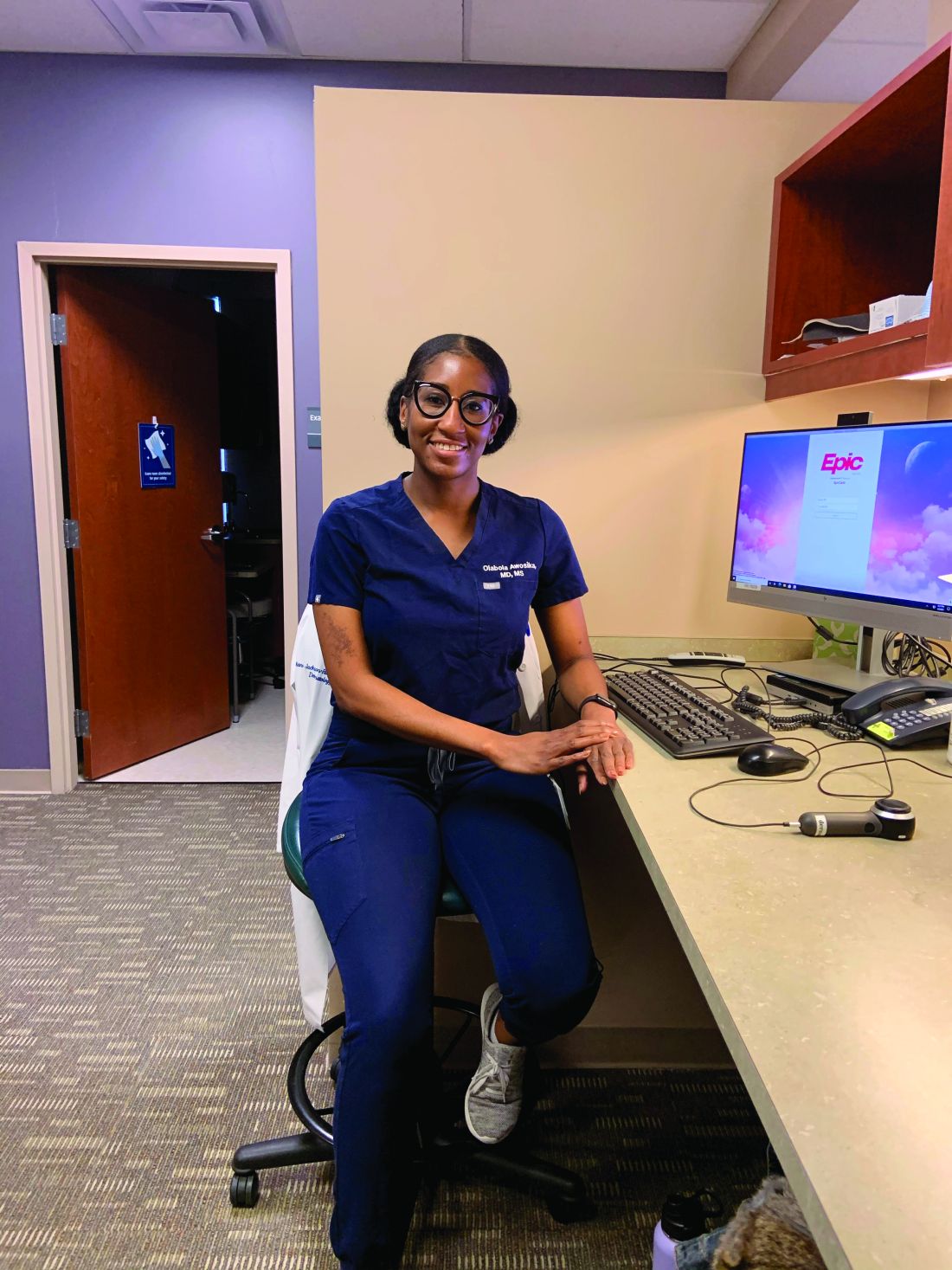
Olabola Awosika, MD, a fourth-year dermatology resident at Henry Ford Health System, Detroit, said her interest in dermatology was solidified during her participation in the AAD’s month-long mentorship program after her second year of medical school at Howard University, Washington. However, it wasn’t until her fourth year, when she did an away rotation at Wake Forest, that she realized that “gaining access to the field” takes years of mentorship, research opportunities, and networking. It was too late.
After initially not matching, she did a rotation at Johns Hopkins in dermatology during an internship year, followed by a 2-year research fellowship at George Washington University, Washington. As does Dr. Florez-Pollack, who now mentors medical students. She also serves on the Women’s Dermatologic Society diversity committee, which is now developing initiatives to help UIM dermatologists “become upwardly mobile in dermatology [after residency], so they have a seat at the table in various settings.”
Holistic review for residency
Dr. Vasquez, who grew up in South Texas in an uninsured family that received most of its medical care across the border in Mexico, believes that the “biggest stride being made today” with respect to diversity in the dermatology workforce – and in the larger physician workforce – is increased understanding of the role of social and cultural capital.
“We’d never really talked about this concept ... about how, if you don’t have the same upbringing, education, and resources, you’re already behind … and you may not do as well on standardized tests,” she said. “Now people are listening to it and understand it. That’s why more programs are looking at how to practice a more holistic approach to selecting applicants to interview.”
Some dermatology programs – at Wake Forest, UCSF, USC, Vanderbilt, and George Washington University for instance – have eliminated the use of U.S. Medical Licensing Examination step 1 scores and Alpha Omega Alpha Medical Honor Society status as filtering/screening metrics. Some also take a “second look” at UIM applicants.
Overall, those making changes are looking “at everything the applicant brings to the table – their story, their experiences,” said Dr. Elbuluk. “We want to understand the full picture of who they are and their journey to becoming a physician.”
Implicit bias training for members of residency review committees is becoming more common, as is such training across the board in dermatology departments. “We have to bring it to the forefront so that we can create more inclusive environments,” said Sharon E. Albers, MD, a dermatologist at Vanderbilt University Medical Center, Nashville, Tenn., who serves as the dermatology department’s diversity liaison to the medical school, and who, as a former faculty member of Meharry Medical College, remains engaged with the historically black institution.
One of her recent efforts, inspired by participation in the diversity champions program, has been developing pipeline programs to speak to middle and high school students about medicine and dermatology. For now, however, she credits a holistic review process for change. Last year, three of the five matched dermatology residents at Vanderbilt were UIM physicians – unprecedented for the institution, Dr. Albers said, and “evidence that holistic review works if you’re very intentional about it.”
Dr. Vasquez has analyzed data from the Accreditation Council of Graduate Medical Education and says that UIM dermatology residents comprised 8.0% of the total in 2019, marking an improvement over the prior 7 years. “We’ve introduced so many changes at the same time,” making it difficult to discern what’s most impactful, she said. “But something is working.”
(The AAMC, which has invested in pipeline programs for more than a decade and has championed holistic medical school admissions, states in its 2019 diversity data report that efforts to improve diversity in medicine have made “only marginal differences” – and that Black males in particular continue to be significantly underrepresented in medical schools. Persistent, structural racism was a common theme in the association’s 2015 report on Black males in medicine.)
Efforts to diversify the workforce – particularly holistic review – haven’t been without detractors. “It brings reaction,” Dr. Wintroub said. “Some people think you’re lowering quality in the field ... but that’s just not true.”
Diversity in the dermatology workforce is important not only for the care of patients from diverse backgrounds, but “perhaps more importantly, it brings new ideas and views and experiences into the field,” he said. “It pushes us to think in new directions ... that can only make our field better and richer.”
“We need to make sure our workforce is representative of the patients we serve – but also that all derms can manage patients of all skin types and demographics,” Dr. Shinkai said. “And we need more diversity in the leadership of our departments and dermatology organizations ... inclusivity needs to extend all the way to the highest reaches of our specialty.”

Adam Friedman, MD, professor and interim chair of dermatology at George Washington University and director of the department’s diversity, equity, and inclusion committee, agreed. “We need to both make sure our workforce mirrors the patients we serve, and said workforce is prepared to manage patients of all skin types and demographics. We need to cover the spectrum, from revamping medical education and mentorship opportunities to advancing diversity in the leadership of our departments, institutions, and societies. Addressing only part of the puzzle will not and can not be enough.”
Correction, 3/3/21: An earlier version of this article misstated Dr. Stephanie Florez-Pollack's name.
Stephanie Florez-Pollack, MD, a dermatology resident at the University of Pennsylvania, Philadelphia, began considering the field of dermatology when, as part of the Latino Medical Students Association, she was invited by Amit Pandya, MD, to a pizza party and dermatology discussion during her first year of medical school at the University of Texas Southwestern Medical Center, Dallas.
There, she met three Latinx residents who were part of the University of Texas Southwestern dermatology program. “I was so excited to hear their stories, where they came from, and how they ended up in dermatology,” she said. Dermatology had not been on her radar screen. Now there was a spark.
Volunteering at the free Agape dermatology clinic in Dallas later that year sharpened her interest. “For the first time, I really saw myself working in the field. Hearing patients’ stories and what they went through with what many people might think are very simple skin diseases made me realize there was potential to make a big impact in a person’s life,” said Dr. Florez-Pollack, who immigrated from Colombia with her family when she was 15.
– one that reflects the ethnic and racial make-up of the population. It’s a movement that involves early outreach to medical students, stepped-up mentorship and sponsorship, implicit bias training, and holistic review of residency applicants.
There is no published study of all the changes being made – of how many dermatology programs have new outreach programs, for instance, or new approaches to resident application reviews. However, participation in the American Academy of Dermatology Diversity Champions program increased significantly between 2019 and 2020, and a sizable body of articles and editorials on diversity have been published in the dermatology literature in recent years, including at least several on holistic review of resident applications. Five years ago, there were few publications, sources said.
“The conversation is happening now at multiple levels, including in our peer-reviewed literature, where people are looking objectively at ... how we can make changes for the better,” said Nada Elbuluk, MD, director of the dermatology diversity and inclusion program at the University of Southern California, Los Angeles.
Unpublished findings also indicate that the number of UIM dermatology residents is inching upward. “Residency selection may not be on everyone’s mind, but an understanding of how we select people into our specialty is something every derm should know about and care about,” said Kanade Shinkai, MD, PhD, professor of dermatology at the University of California, San Francisco, and editor in chief of JAMA Dermatology.
The wake-up calls
The impetus for current changes first came in 2015, when Bruce Wintroub, MD, professor and chair of dermatology at UCSF, and interim dean of the medical school at the time, delivered a passionate plenary lecture at the annual AAD meeting about the lack of equity, diversity and inclusion in the specialty and the role of unconscious bias. Moved in part by a “White Coats for Black Lives” die-in by medical students on his campus following the killing of unarmed Black men, Dr. Wintroub called on his colleagues to recruit more UIM physicians into the specialty and to make the field more inclusive.
In 2016, Dr. Wintroub joined Dr. Pandya and two other academic dermatologists in authoring a commentary, published in the Journal of the American Academy of Dermatology, about the need to step up efforts and make diversity a priority. Dermatology was among the least ethnically and racially diverse specialties, second only to orthopedics, they wrote, with black dermatologists making up only 3% of all dermatologists and Hispanics only 4.2% in the United States, compared with 12.8% and 16.3% in the U.S. population, respectively.
For the next year or so, they and others from six academic institutions formed an incubator of sorts, actively tracking and sharing actions they were taking to improve diversity and the practice environment. “We wanted to learn from each other, then disseminate information to other programs,” said Dr. Pandya, who chaired the AAD diversity task force and led the self-branded “diversity champions.” (Dr. Pandya maintains his appointment at UTSW but now practices at the Palo Alto Foundation Medical Group in Sunnyvale, Calif.)
In 2017, at a President’s Conference on Diversity in Dermatology called for by then-AAD president Henry Lim, MD, leaders of the Association of Professors of Dermatology, the Society for Investigative Dermatology, and other dermatology organizations agreed on key action items, which they described in JAAD in 2018: Helping to increase the pipeline of UIM students applying to medical school, increasing UIM medical students’ early exposure to dermatology, and increasing the number of UIM students recruited into dermatology residency programs.
Diversity in the physician workforce has been shown to improve outcomes for all patients, and is important for ameliorating health care disparities and improving satisfaction and care for all patients, the authors of the 2018 paper wrote. “Multiple studies,” they noted, “have shown that UIM physicians are more likely to practice in areas where health care disparities exist.”
For specific proposed actions, the leaders attending the diversity conference drew largely upon the “diversity champions’” experiences, and agreed to fund and officially develop a diversity champions program. They also decided to invest in “bioskills” workshops for undergraduates and medical students at historically black colleges and universities, and other institutions at historically black colleges and universities at medical schools through a partnership with Nth Dimension – an organization founded in 2004 to bring more women and underrepresented minorities into orthopedic surgery.
In the last 2 years, more than 450 UIM students have attended these bioskills workshops, getting a taste of basic dermatology procedures and interacting with dermatologists.
And in September 2020, 157 dermatologists representing 80 programs throughout the country attended the second AAD Diversity Champions conference, up from 84 attendees and 30 programs in 2019. On the agenda: Discussions of holistic application review, mentorship, recruitment of UIM faculty, and a 2-hour session on microaggressions. Similar programs are being led by other dermatology organizations.
(UIM was coined by the American Association of Medical Colleges to describe racial and ethnic populations that are underrepresented in medicine relative to their numbers in the general population.)
Achievement of racial/ethnic diversity “won’t happen unless the field actively encourages people to look at it – which is not what we were doing,” Dr. Wintroub said in an interview. “I think that’s been the major change. We’re opening the door and saying: ‘We want you and we welcome you.’ ”
Rethinking traditional mentorship
For Dr. Florez-Pollack, the door almost shut when she began to hear from fellow medical students that dermatology is “too competitive ... a field for only the top people in the class.” She felt doubt settling in.
“I had peers who were throwing a lot of money toward prep materials ... peers who had siblings in medicine and had started studying from day one [for the step 1 exam],” she said. “I thought, was it really worth the effort? Do I really want to be perfect to get into the field when other fields would be happy to have me as I am right now?”
Her immense enjoyment of an “Art of Observation” elective course helped renew her interest in the field; it reinforced her visual abilities as well as the potential for her to address implicit bias as a dermatologist. She sought Dr. Pandya’s guidance and sponsorship to help her grow connections, polish her resume, and present herself to other faculty.
Doors were opened, she said, for her to secure a 1-year research experience before her final year of medical school with UTSW faculty, and then a 1-month rotation/mentorship with William D. James, MD, professor of dermatology at the University of Pennsylvania, an institution with a history of diversity initiatives and a longstanding skin of color program.
Dr. Florez-Pollack sees her experience reflected in the findings of a recently published study – a thematic content analysis of telephone interviews with applicants to the UTSW dermatology residency program during the 2013-14 and 2014-15 application cycles. Of the 44 applicants who participated in the study, 13 were UIM applicants.
Six of the seven UIM applicants who matched were involved in a pipeline or enrichment program – and were exposed to the field early – compared with one or two of the six UIM applicants who did not match. Underrepresented applicants were more often discouraged from applying (54%) – told, for instance, that they could better serve their communities through other specialties – than were non-UIM applicants (13%). They also were affected more often by a lack of equitable resources, according to comments made by 70% of applicants (UIM and non-UIM).
Also notably, the investigators said, all of the UIM applicants who matched (and the majority of non-UIMs who matched) reported having a mentor during the process of applying, compared with 44% of those who didn’t match.
Rebecca Vasquez, MD, assistant professor of dermatology at UTSW, who led the study, was herself a mentee of Dr. Pandya. “He believed in me and gave me the courage to consider dermatology,” she said. (Dr. Vasquez was one of the Latinx women who inspired Dr. Florez-Pollack, in turn, when they met at Dr. Pandya’s pizza party. Dr. Florez-Pollack assisted with the research and was a coauthor of the study.)
Amy McMichael, MD, professor and chair of dermatology at Wake Forest University, Winston-Salem, N.C., said that dermatology as a field has traditionally been “very good at mentoring.” Many of the dermatology societies have long had mentorship programs, for instance, that guide medical students, and sometimes residents, through defined experiences or through periods of time.
But she advocates going deeper. “When it comes to sponsorship, we fall a little short,” she said. “Sponsorship is about promoting that person to the next level, making sure they achieve what they want to achieve ... putting them up for opportunities they may not have known existed. It’s continuous and focused.”
Olabola Awosika, MD, a fourth-year dermatology resident at Henry Ford Health System, Detroit, said her interest in dermatology was solidified during her participation in the AAD’s month-long mentorship program after her second year of medical school at Howard University, Washington. However, it wasn’t until her fourth year, when she did an away rotation at Wake Forest, that she realized that “gaining access to the field” takes years of mentorship, research opportunities, and networking. It was too late.
After initially not matching, she did a rotation at Johns Hopkins in dermatology during an internship year, followed by a 2-year research fellowship at George Washington University, Washington. As does Dr. Florez-Pollack, who now mentors medical students. She also serves on the Women’s Dermatologic Society diversity committee, which is now developing initiatives to help UIM dermatologists “become upwardly mobile in dermatology [after residency], so they have a seat at the table in various settings.”
Holistic review for residency
Dr. Vasquez, who grew up in South Texas in an uninsured family that received most of its medical care across the border in Mexico, believes that the “biggest stride being made today” with respect to diversity in the dermatology workforce – and in the larger physician workforce – is increased understanding of the role of social and cultural capital.
“We’d never really talked about this concept ... about how, if you don’t have the same upbringing, education, and resources, you’re already behind … and you may not do as well on standardized tests,” she said. “Now people are listening to it and understand it. That’s why more programs are looking at how to practice a more holistic approach to selecting applicants to interview.”
Some dermatology programs – at Wake Forest, UCSF, USC, Vanderbilt, and George Washington University for instance – have eliminated the use of U.S. Medical Licensing Examination step 1 scores and Alpha Omega Alpha Medical Honor Society status as filtering/screening metrics. Some also take a “second look” at UIM applicants.
Overall, those making changes are looking “at everything the applicant brings to the table – their story, their experiences,” said Dr. Elbuluk. “We want to understand the full picture of who they are and their journey to becoming a physician.”
Implicit bias training for members of residency review committees is becoming more common, as is such training across the board in dermatology departments. “We have to bring it to the forefront so that we can create more inclusive environments,” said Sharon E. Albers, MD, a dermatologist at Vanderbilt University Medical Center, Nashville, Tenn., who serves as the dermatology department’s diversity liaison to the medical school, and who, as a former faculty member of Meharry Medical College, remains engaged with the historically black institution.
One of her recent efforts, inspired by participation in the diversity champions program, has been developing pipeline programs to speak to middle and high school students about medicine and dermatology. For now, however, she credits a holistic review process for change. Last year, three of the five matched dermatology residents at Vanderbilt were UIM physicians – unprecedented for the institution, Dr. Albers said, and “evidence that holistic review works if you’re very intentional about it.”
Dr. Vasquez has analyzed data from the Accreditation Council of Graduate Medical Education and says that UIM dermatology residents comprised 8.0% of the total in 2019, marking an improvement over the prior 7 years. “We’ve introduced so many changes at the same time,” making it difficult to discern what’s most impactful, she said. “But something is working.”
(The AAMC, which has invested in pipeline programs for more than a decade and has championed holistic medical school admissions, states in its 2019 diversity data report that efforts to improve diversity in medicine have made “only marginal differences” – and that Black males in particular continue to be significantly underrepresented in medical schools. Persistent, structural racism was a common theme in the association’s 2015 report on Black males in medicine.)
Efforts to diversify the workforce – particularly holistic review – haven’t been without detractors. “It brings reaction,” Dr. Wintroub said. “Some people think you’re lowering quality in the field ... but that’s just not true.”
Diversity in the dermatology workforce is important not only for the care of patients from diverse backgrounds, but “perhaps more importantly, it brings new ideas and views and experiences into the field,” he said. “It pushes us to think in new directions ... that can only make our field better and richer.”
“We need to make sure our workforce is representative of the patients we serve – but also that all derms can manage patients of all skin types and demographics,” Dr. Shinkai said. “And we need more diversity in the leadership of our departments and dermatology organizations ... inclusivity needs to extend all the way to the highest reaches of our specialty.”
Adam Friedman, MD, professor and interim chair of dermatology at George Washington University and director of the department’s diversity, equity, and inclusion committee, agreed. “We need to both make sure our workforce mirrors the patients we serve, and said workforce is prepared to manage patients of all skin types and demographics. We need to cover the spectrum, from revamping medical education and mentorship opportunities to advancing diversity in the leadership of our departments, institutions, and societies. Addressing only part of the puzzle will not and can not be enough.”
Correction, 3/3/21: An earlier version of this article misstated Dr. Stephanie Florez-Pollack's name.
Analysis characterizes common wound microbes in epidermolysis bullosa
– in a retrospective analysis of over 700 wound cultures from 158 patients across the United States and Canada.
The findings from the EB Clinical Characterization and Outcomes Database speak to the value of surveillance cultures with routine testing for microbial resistance – including mupirocin resistance – and to the importance of antibiotic stewardship not only for oral antibiotics but for topicals as well, according to Laura E. Levin, MD, and Kimberly D. Morel, MD, of the departments of dermatology and pediatrics, Columbia University Irving Medical Center, New York, the lead and senior authors, respectively, of the paper recently published in Pediatric Dermatology.
Almost all of the 158 patients with at least one wound culture recorded in the database from the period of 2001-2018 had one or more positive culture results. Of 152 patients with positive cultures, 131 (86%) were positive for SA and 56 (37%) and 34 (22%) were positive for PA and GAS, respectively. Other bacteria isolated included Corynebacterium spp and Proteus spp. Nearly half (47%) of patients with SA-positive cultures had methicillin-resistant SA, and 68% had methicillin-susceptible SA. (Some patients grew both MSSA and MRSA at different points in time.)
Mupirocin-susceptibility testing was performed at only some of the 13 participating centers. Of 15 patients whose cultures had recorded SA mupirocin-susceptibility testing, 11 had cultures positive for mupirocin-susceptible SA and 6 (40%) had mupirocin-resistant SA isolates (2 patients grew both). Of these six patients, half had isolates that were also methicillin-resistant.
Mupirocin, a topical antibiotic, has been a cornerstone of decolonization regimens for MSSA and MRSA, but resistance has been demonstrated in other research as well and is not specific to EB, wrote Dr. Levin, Dr. Morel, and coauthors.
“Pediatric dermatologists often rely on topical antimicrobials in the treatment of patients’ open wounds to both prevent and treat infection, depending on the clinical scenario,” and surveillance cultures with routine testing for mupirocin resistance can help guide antibiotic choice and management strategies, Dr. Levin said in an interview.
More broadly, she added, “it’s helpful to know what bacteria are routinely colonizing wounds, not causing infection, versus those that are more likely to be associated with infection, chronic wounds, or the risk of developing skin cancer ... [to know] which wounds need to be treated more aggressively.”
A subset of patients with EB have been known to be at risk for squamous cell carcinoma, and research is implicating certain bacteria “as contributing to wound inflammation,” Dr. Morel said in an interview.
SCC was reported in 23 out of 717 patients in the database – but fewer than half of the patients with SCC had recorded wound cultures. The small numbers precluded the identification of microbes that may confer significant risk.
Correlating particular microbes with clinical features also will take more research. About half (57%) of the patients with recorded wound cultures had wounds with purulent exudate or other features of clinical infection. However, the presence or absence of clinical signs of infection was not temporally correlated with culture results in the database.
The 158 patients with recorded wound cultures had a mean age of 12.8 years and represented a range of EB subtypes.
PA was present in the wounds of patients as young as 1 month old, the authors noted. Investigators are “looking to further study PA and characterize clinical features ... to understand more about this microbe and its impact on patients with EB,” Dr. Morel said.
In the meantime, the analysis reaffirms the importance of antibiotic stewardship. Mupirocin is labeled to be used three times a day for a short period of time, but “tends to be prescribed and used less judiciously than intended,” Dr. Morel said. “It’s important [not to overuse it]. We have seen that patients’ culture results become sensitive to mupirocin again in the future when they avoid it for a period of time.”
The work was supported by the EB Research Partnership and EB Medical Research Foundation, as well as an NIH/NCATS grant. No investigator disclosures were listed.
SOURCE: Pediatr Dermatol. 2020 Nov 28. doi: 10.1111/pde.14444.
FROM PEDIATRIC DERMATOLOGY
Systemic sclerosis patients share their perspectives and needs in treatment trials
Patients with systemic sclerosis have variable disease progression but often experience debilitating fatigue, pain, and digestive issues – and they’re extremely concerned about progressive organ damage, according to those who spoke at and provided input at a public meeting on patient-focused drug development for the disease.

The virtual meeting was part of the Food and Drug Administration’s Patient-Focused Drug Development (PFDD) initiative, which began in 2012 and aims to provide a systematic way for patients’ experiences, needs, and priorities to be “captured and meaningfully incorporated” into drug development and evaluation.
Patients rate their most impactful symptoms
Dinesh Khanna, MBBS, MSc, a rheumatologist who directs a scleroderma research program at the University of Michigan, Ann Arbor, attended the meeting after giving an opening presentation on the disease to FDA officials, patients, and other participants. In a later interview, he said that patients’ ratings of their most impactful symptoms was especially striking.

Raynaud’s phenomenon, digestive symptoms, and fatigue were the top three answers to a poll question that asked patients what symptom had the most significant impact on daily life, he noted, “and none of these are being [strongly] addressed right now [in clinical trials] apart from Raynaud’s phenomenon, for which there are some trials ongoing.”
He and other researchers are “struggling with what outcomes measures to use [in their studies],” said Dr. Khanna, the Frederick G.L. Huetwell Professor of Rheumatology at the University. “My takeaway from the meeting as a clinical trialist is that we should be paying close attention to the symptoms that patients tell us are the most important. We should be including these in our trial designs as secondary endpoints, if not primary endpoints. We have not done that [thus far], really.”
Approximately 200,000 patients in the United States have scleroderma, and approximately 75,000-80,000 of these patients have systemic scleroderma, or systemic sclerosis, Dr. Khanna said in his opening presentation. Each year, he estimates, about 6,000 new diagnoses of systemic sclerosis are made.
More than 200 people – patients, FDA officials, and others – participated in the PFDD meeting. Patients participated in one of two panels – one focused on health effects and daily impacts, and the other on treatments – or submitted input electronically. All were invited to answer poll questions.
Raj Nair, MD, one of eight FDA leaders attending the meeting, noted in closing remarks that the pain experienced by patients with systemic sclerosis includes severe pain from Raynaud’s phenomenon and pain caused by digital ulcers and by calcinosis. “We heard about how paralyzing the pain from calcinosis is, and that there are very few options for alleviating this pain,” said Dr. Nair, of the division of rheumatology and transplant medicine.
Another takeaway, he said, is that the “fatigue can be severe and debilitating, leading to days where it is impossible to get out of bed,” and that digestive symptoms can also be severe. “Reflux,” he noted, “requires significant medical intervention.”
Patients describe their experiences
Rosemary Lyons, diagnosed with scleroderma 35 years ago, explained that while her skin is no longer hardened, she is overly sensitive to fabrics and skin care products and has difficulty with sleeping and eating. She moved away from family in the Northeast to live in the South where the climate is warmer, but even on a 90-degree night she needs a blanket and two comforters to curb the cold and attempt to sleep.
Impaired gastrointestinal motility has made food her “biggest problem” for the past 10 years, and because of GI symptoms, she can eat only one meal a day. She also experiences fainting, brain fog, and severe fatigue. On a good day, Ms. Lyons noted, she sometimes opts to do some house chores “knowing that I’ll have 1-3 days of recovery.”
Another patient, Amy Harding, said that 22 years after her scleroderma diagnosis, “the calcinosis I get in my fingers, elbows, toes, and ears tops all the prior symptoms.” The skin tightening and digital ulcers that she experienced in the first 10 years have tapered off, and while Raynaud’s symptoms and heartburn have worsened, they are at least partly manageable with medications, unlike the pain from calcinosis.
Treating symptoms vs. disease may be key in risk-benefit analysis
In questions after patient presentations, FDA officials probed for more perspective on issues such as how fatigue should be assessed, the differences between fatigue and brain fog, the impact of calcinosis on functioning, and how much risk patients would be willing to assume from treatments that have side effects and that may or may not modulate the disease and slow disease progression.
Most patients said in response to an FDA poll question that they definitely (almost 40%) or possibly (almost 50%) would be willing to try a hypothetical new self-injectable medication if it were shown to reduce their most impactful symptoms but had side effects.
“I think what [we’ve been hearing] today is that whether we’re working on the symptoms or the disease itself is [the key]” to patients’ risk-benefit analysis, said meeting moderator Capt. Robyn Bent, RN, MS, of the U.S. Public Health Service, and director of the PFDD.
Anita Devine, diagnosed 13 years ago with systemic sclerosis, was one of several panel members who said she would accept more bothersome treatment side effects and risks “if the gain was control of disease progression and overall quality of life ... and organ preservation.” Ms. Devine, who has needed kidney dialysis and multiple hand surgeries, noted that she previously took anti-neoplastic and anti-inflammatory agents “to try to stem the course of my disease, but unfortunately the disease did not abate.”
Treatments for systemic sclerosis include vasodilators, immunosuppressive medications, antifibrotic therapies, and stem cell transplants, Dr. Khanna said in his opening remarks.
Trials of drugs for scleroderma have focused on early disease that may be amenable to treatment, with the exception of trials for pulmonary arterial hypertension, which affects some patients with systemic sclerosis. There are multiple FDA-approved drugs for pulmonary arterial hypertension and more trials are underway.
Outcomes such as pain and fatigue are included in many of the trials currently underway, but they tend to be lower-level secondary outcomes measures that cannot be incorporated into drug labeling or are more “exploratory in nature,” Dr. Khanna said in the interview.
Dr. Khanna disclosed that he is the chief medical officer (an equity position) for CiVi Biopharma/Eicos Sciences Inc., which is developing a drug for Raynaud’s, and serves as a consultant and grant recipient for numerous companies that make or are developing drugs for systemic sclerosis.
The FDA will accept patient comments until Dec. 15, 2020, at which time comments will be compiled into a summary report, Ms. Bent said.
Patients with systemic sclerosis have variable disease progression but often experience debilitating fatigue, pain, and digestive issues – and they’re extremely concerned about progressive organ damage, according to those who spoke at and provided input at a public meeting on patient-focused drug development for the disease.
The virtual meeting was part of the Food and Drug Administration’s Patient-Focused Drug Development (PFDD) initiative, which began in 2012 and aims to provide a systematic way for patients’ experiences, needs, and priorities to be “captured and meaningfully incorporated” into drug development and evaluation.
Patients rate their most impactful symptoms
Dinesh Khanna, MBBS, MSc, a rheumatologist who directs a scleroderma research program at the University of Michigan, Ann Arbor, attended the meeting after giving an opening presentation on the disease to FDA officials, patients, and other participants. In a later interview, he said that patients’ ratings of their most impactful symptoms was especially striking.
Raynaud’s phenomenon, digestive symptoms, and fatigue were the top three answers to a poll question that asked patients what symptom had the most significant impact on daily life, he noted, “and none of these are being [strongly] addressed right now [in clinical trials] apart from Raynaud’s phenomenon, for which there are some trials ongoing.”
He and other researchers are “struggling with what outcomes measures to use [in their studies],” said Dr. Khanna, the Frederick G.L. Huetwell Professor of Rheumatology at the University. “My takeaway from the meeting as a clinical trialist is that we should be paying close attention to the symptoms that patients tell us are the most important. We should be including these in our trial designs as secondary endpoints, if not primary endpoints. We have not done that [thus far], really.”
Approximately 200,000 patients in the United States have scleroderma, and approximately 75,000-80,000 of these patients have systemic scleroderma, or systemic sclerosis, Dr. Khanna said in his opening presentation. Each year, he estimates, about 6,000 new diagnoses of systemic sclerosis are made.
More than 200 people – patients, FDA officials, and others – participated in the PFDD meeting. Patients participated in one of two panels – one focused on health effects and daily impacts, and the other on treatments – or submitted input electronically. All were invited to answer poll questions.
Raj Nair, MD, one of eight FDA leaders attending the meeting, noted in closing remarks that the pain experienced by patients with systemic sclerosis includes severe pain from Raynaud’s phenomenon and pain caused by digital ulcers and by calcinosis. “We heard about how paralyzing the pain from calcinosis is, and that there are very few options for alleviating this pain,” said Dr. Nair, of the division of rheumatology and transplant medicine.
Another takeaway, he said, is that the “fatigue can be severe and debilitating, leading to days where it is impossible to get out of bed,” and that digestive symptoms can also be severe. “Reflux,” he noted, “requires significant medical intervention.”
Patients describe their experiences
Rosemary Lyons, diagnosed with scleroderma 35 years ago, explained that while her skin is no longer hardened, she is overly sensitive to fabrics and skin care products and has difficulty with sleeping and eating. She moved away from family in the Northeast to live in the South where the climate is warmer, but even on a 90-degree night she needs a blanket and two comforters to curb the cold and attempt to sleep.
Impaired gastrointestinal motility has made food her “biggest problem” for the past 10 years, and because of GI symptoms, she can eat only one meal a day. She also experiences fainting, brain fog, and severe fatigue. On a good day, Ms. Lyons noted, she sometimes opts to do some house chores “knowing that I’ll have 1-3 days of recovery.”
Another patient, Amy Harding, said that 22 years after her scleroderma diagnosis, “the calcinosis I get in my fingers, elbows, toes, and ears tops all the prior symptoms.” The skin tightening and digital ulcers that she experienced in the first 10 years have tapered off, and while Raynaud’s symptoms and heartburn have worsened, they are at least partly manageable with medications, unlike the pain from calcinosis.
Treating symptoms vs. disease may be key in risk-benefit analysis
In questions after patient presentations, FDA officials probed for more perspective on issues such as how fatigue should be assessed, the differences between fatigue and brain fog, the impact of calcinosis on functioning, and how much risk patients would be willing to assume from treatments that have side effects and that may or may not modulate the disease and slow disease progression.
Most patients said in response to an FDA poll question that they definitely (almost 40%) or possibly (almost 50%) would be willing to try a hypothetical new self-injectable medication if it were shown to reduce their most impactful symptoms but had side effects.
“I think what [we’ve been hearing] today is that whether we’re working on the symptoms or the disease itself is [the key]” to patients’ risk-benefit analysis, said meeting moderator Capt. Robyn Bent, RN, MS, of the U.S. Public Health Service, and director of the PFDD.
Anita Devine, diagnosed 13 years ago with systemic sclerosis, was one of several panel members who said she would accept more bothersome treatment side effects and risks “if the gain was control of disease progression and overall quality of life ... and organ preservation.” Ms. Devine, who has needed kidney dialysis and multiple hand surgeries, noted that she previously took anti-neoplastic and anti-inflammatory agents “to try to stem the course of my disease, but unfortunately the disease did not abate.”
Treatments for systemic sclerosis include vasodilators, immunosuppressive medications, antifibrotic therapies, and stem cell transplants, Dr. Khanna said in his opening remarks.
Trials of drugs for scleroderma have focused on early disease that may be amenable to treatment, with the exception of trials for pulmonary arterial hypertension, which affects some patients with systemic sclerosis. There are multiple FDA-approved drugs for pulmonary arterial hypertension and more trials are underway.
Outcomes such as pain and fatigue are included in many of the trials currently underway, but they tend to be lower-level secondary outcomes measures that cannot be incorporated into drug labeling or are more “exploratory in nature,” Dr. Khanna said in the interview.
Dr. Khanna disclosed that he is the chief medical officer (an equity position) for CiVi Biopharma/Eicos Sciences Inc., which is developing a drug for Raynaud’s, and serves as a consultant and grant recipient for numerous companies that make or are developing drugs for systemic sclerosis.
The FDA will accept patient comments until Dec. 15, 2020, at which time comments will be compiled into a summary report, Ms. Bent said.
FROM AN FDA PATIENT-FOCUSED DRUG DEVELOPMENT MEETING
Survey of Mohs surgeons highlights its use in invasive melanoma
of members of the American College of Mohs Surgery.
Of 513 survey participants, 40.9% reported using MMS to treat any subtype of melanoma. Most of these surgeons reported treating both lentigo maligna (97.5%) and other melanoma in situ (MIS) subtypes (91.4%). A slight majority – 58.6% – reported treating invasive T1 melanoma, and 20.5% reported treating invasive T2 and/or higher-stage melanoma with MMS.
The analysis, published in Dermatologic Surgery, was done by Spyros M. Siscos, MD, and a team of residents and faculty in the division of dermatology at the University of Kansas Medical Center, Kansas City.
It comes on the heels of an analysis of claims data for Mohs surgery, published last year in JAMA Dermatology, which showed a more than threefold increase in the use of Mohs surgery for melanoma from 2.6% of all surgical cases in 2001 to 7.9% in 2016.
With the increased use of MMS for treatment of melanoma, “Mohs surgeons who previously treated MIS with MMS may be increasingly doing so and/or expanding their scope of treatment to include invasive melanoma,” the University of Kansas investigators wrote.
That a slight majority now report treating invasive melanoma with MMS “may be due, in part, to upstaging during the MMS procedure and the increasing evidence demonstrating improved survival of early-invasive melanoma treated with MMS compared with [wide local excision],” as well as the advent of melanocytic immunohistochemical (IHC) stains, particularly melanoma antigen recognized by T-cells 1 (MART-1), they said. However, 29% of surveyed Mohs surgeons treating melanoma with MMS do not use IHC stains “despite growing evidence supporting” their use, the authors wrote.
The advent of IHC stains, particularly MART-1, has improved the accuracy of interpreting frozen sections of melanoma, they reported, noting that MMS without IHC has been associated with a recurrence rate as high as 33%. Of the 71% who reported using IHC stains, MART-1 was the primary IHC stain for virtually all of them (97.3%).
There was also variation in the number of surgeons who reported debulking MIS. Eighty-two percent take this approach, excising the clinically visible tumor before excising the initial Mohs stage – almost all with a scalpel. More than half of these surgeons – 58.5% – submit the entire debulked MIS specimen for permanent vertical sectioning (breadloafing) to evaluate for deeper tumor invasion.
The others reported submitting the entire debulked specimen for frozen vertical sectioning, or portions of the specimen for both permanent and frozen vertical sectioning. “It is unclear why a minority of surveyed Mohs surgeons reported not debulking MIS,” wrote Dr. Siscos and his colleagues.
The average margin size of the first Mohs stage for MIS was 4.96 ± 1.74 mm, which is at the lower end of the 0.5-1.0 cm range for wide local excision (WLE) recommended by the National Comprehensive Cancer Network (NCCN) and the American Academy of Dermatology (AAD), according to a clinical practice guideline. (The survey did not investigate initial margins for invasive melanoma treated with MMS.)
Jeremy R. Etzhorn, MD, of the department of dermatology at the University of Pennsylvania, Philadelphia, and an author of a 2019 claims data analysis of excisional surgery practices for melanoma, said that the new survey findings – like the prior analysis – highlight the variability in approaches to using MMS for melanoma.
“Mohs for melanoma [seems] like a one-liner ... but really, there are [a lot] of different techniques that fall under that umbrella, if you parse out all the variations,” he said in an interview.
Per the 2016 claims analysis, he noted, IHC was used in less than 40% of Mohs surgery cases for melanoma, and there were wide geographic variations. “The biggest critique of Mohs surgery for melanoma over the last two decades has been that it’s hard to see the tumor,” he said. “But with the advent of IHC, that challenge was overcome.”
Surgical excision practices are evolving without the development of best practice guidelines, said Dr. Etzkorn, who is director of clinical research for the University of Pennsylvania dermatologic oncology center. Multisociety guidelines published in 2012 on appropriate use criteria for Mohs surgery do not offer specific recommendations on the use of MMS for invasive melanoma. Nor do guidelines from the AAD and the NCCN, he said.
“What this [new] study highlights and what’s being discussed amongst Moh’s surgeons” is that Mohs for melanoma “has be to be standardized” to some extent and then clinical trials conducted comparing Mohs to conventional excision. The studies that have been published in recent years comparing MMS with WLE for MIS and invasive melanoma are “not gold standard studies,” he said.
Practice guidelines then can be informed by high-quality evidence on its safety and efficacy, he said.
The 513 participants in the newly published survey represent a 31.5% response rate. Invasive T2 and/or higher stage melanoma was more likely to be treated with MMS in academic hospitals, compared with other practice settings (30.2% v. 18.1%), Dr. Siscos and his coauthors reported.
Participants who reported treating melanoma with MMS were more likely to report fellowship exposure and more likely to have received fellowship training on melanocytic IHC stains. The study “highlights the importance of fellowship exposure to MMS and IHC staining for melanoma,” the authors wrote, adding that postfellowship training opportunities in MMS and IHC staining for melanoma may help broaden its use among Mohs surgeons who received inadequate fellowship exposure.
Dr. Siscos and his colleagues reported no significant interest with commercial supporters. Dr. Etzkorn had no disclosures.
SOURCE: Siscos S et al. Dermatol Surg. 2020 Oct;46(10):1267-71.
FROM DERMATOLOGIC SURGERY
Screening algorithm safely selects patients for OSA treatment before bariatric surgery
A novel algorithm for selecting patients who require treatment for obstructive sleep apnea (OSA) before undergoing bariatric surgery proved safe in a prospective cohort study of 1,103 patients.
Screening for OSA is recommended before bariatric surgery. OSA has been associated in several meta-analyses with increased risk for postoperative complications – not limited to bariatric surgery – and some studies have suggested that this increased risk may be limited to severe OSA, said Frédéric Series, MD, of Université Laval, Quebec City, at the virtual annual meeting of the Associated Sleep Societies.
The preoperative screening algorithm, which utilizes the results of nocturnal home oximetry and morning capillary gas measurements, effectively stratified patients for the risk of postoperative adverse events and “safely selected patients who don’t need [continuous positive airway pressure] before bariatric surgery,” he said. “The risk of postoperative adverse events following bariatric surgery was not increased in untreated OSA patients with low or moderate risk of severe OSA and hypoventilation.”
The study also demonstrated, he said, that patients with severe OSA with or without hypoventilation, even when correctly treated, remain at higher risk for complications.
The algorithm utilizes an oxygen desaturation index (ODI) corresponding to 3% drops in SaO2 and the percent of the total recording time with an SaO2 below 90%, as well as capillary gas measurements (PCO2). Treatment was initiated for those with severe OSA (ODI ≥ 25/hr, < 10% of recording time with a SaO2 below 90%) or OSA with hypoventilation (PCO2 ≥ 45).
“When the ODI was less than 25 per hour, and when the total recording time spent below 90% SaO2 was less than 10%, with PCO2 < 45 mmHg, we expected no need for CPAP treatment,” Dr. Series said. For analysis, the investigators considered part of the untreated group – those with an ODI < 10/hr (no or mild OSA) – as a control group.
Treated patients underwent CPAP/BiPAP for a mean duration of 1.5 months. Good treatment compliance was mandatory for surgery, and treatment was continued immediately after extubation, in the recovery room, in nearly all patients, Dr. Series reported.
The analysis covered 1,103 patients: 447 controls (40.8%), 358 untreated (32.7%), 289 treated for OSA (26.4%) and 9 (0.8%) treated for OSA + hypoventilation. Patients with OSA, particularly those with severe OSA and those with hypoventilation, were older and heavier and significantly more likely to have hypertension and diabetes than controls.
There were no differences between the four groups in 10-day reoperation or 30-day readmission occurrence, and postoperative complications were “particularly infrequent in the control and OSA-untreated groups, with no differences between these two groups,” Dr. Series said.
Cardiac arrhythmia (mainly atrial fibrillation) occurred more frequently in the OSA-treated group (2.4%) and the OSA/hypoventilation patients (11%) than in the other groups (0.5%-0.6%).
Respiratory failure occurred in about one-third of patients with hypoventilation, and admission to the ICU was “dramatically higher” in patients with hypoventilation (67%), because of respiratory failure, arrhythmia, or other unstable medical conditions, Dr. Series said.
There were no differences between the groups in the duration of surgery or the amount of anesthetic used, but the length of stay in the recovery room was significantly longer in the OSA-treated and hypoventilation groups. The length of hospital stay was also longer in these groups. Sleeve gastrectomy was the most frequent bariatric surgical procedure across all groups, including 100% of patients with hypoventilation, he noted.
Asked to comment on the study, Octavian C. Ioachimescu, MD, PhD, of Emory University in Atlanta and the Atlanta Veterans Affairs Medical Center in Decatur, said the algorithm “clearly deserves further validation in other clinical-based cohorts and longer-term outcome assessment.”
Dr. Series reported that he has no relevant disclosures. Dr. Ioachimescu also said he has no relevant disclosures.
REPORTING FROM SLEEP 2020
Pregnancy studies on psoriasis, PsA medications pick up
Christina Chambers, PhD, MPH, who runs the MotherToBaby Pregnancy Studies research center at the University of California, San Diego, has found most pregnant women to be “entirely altruistic” about sharing their experiences with drug treatment during pregnancy.
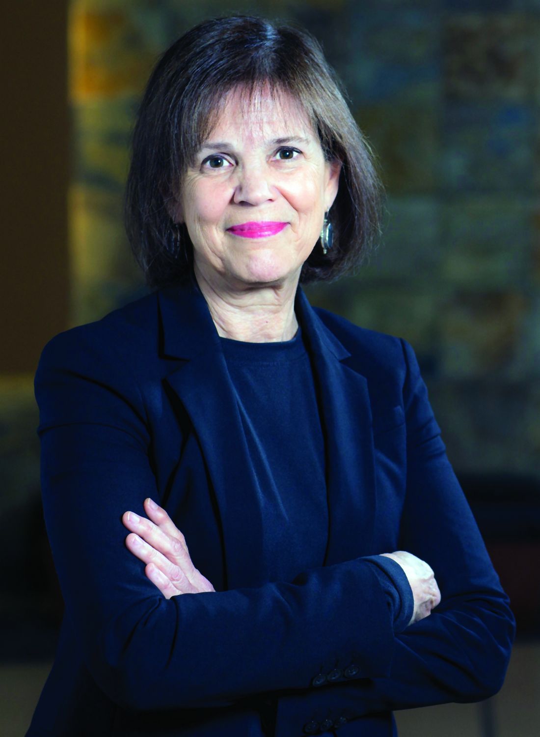
– but dermatologists, rheumatologists, and their female patients with psoriasis and psoriatic arthritis (PsA) want much more.
And women’s participation in the MotherToBaby studies conducted by the nonprofit Organization of Teratology Information Specialists (OTIS) is key, say physicians who are treating women of reproductive age. OTIS is now listed in drug labeling as the “pregnancy registry” contact for many of the medications they may be discussing with patients.
Dr. Chambers said that most women appreciate “that participating in a study may not help her with her pregnancy, but it can help her sister or her friend or someone else who has these same questions in planning a pregnancy of ‘Can I stay on my treatment?’ or, in the case of an unplanned pregnancy, ‘Should I be concerned?’ ”
OTIS has enrolled women with psoriasis and/or PsA in studies of nine medications, most of them biologics (both TNF-alpha blockers and newer anti-interleukin agents).
Four of the studies – those evaluating etanercept (Enbrel), adalimumab (Humira), abatacept (Orencia), and ustekinumab (Stelara) – are now closed to enrollment with analyses either underway or completed. The other five are currently enrolling patients and involve treatment with certolizumab pegol (Cimzia), tildrakizumab (Ilumya), apremilast (Otezla), guselkumab (Tremfya), and tofacitinib (Xeljanz).

Lisa R. Sammaritano, MD, a rheumatologist at the Hospital for Special Surgery, New York, who led the development of the American College of Rheumatology’s first guideline for the management of reproductive health in rheumatic and musculoskeletal diseases, recommends to some of her patients that they contact OTIS. “Their pregnancy registry studies have added important information to the field over the years,” she said.
Most recently, a study of the anti–TNF-alpha medication adalimumab that began in 2004 in pregnant patients with RA and Crohn’s disease culminated in a 2019 PLOS ONE paper reporting no associations between exposure to the medication and an increased risk of adverse outcomes. The outcomes studied were major structural birth defects, minor defects, spontaneous abortion, preterm delivery, prenatal and postnatal growth deficiency, serious or opportunistic infections, and malignancies.
An analysis is underway of adalimumab exposure in women with PsA – a patient subset that was added after the study started. But in the meantime, Dr. Chambers said, the 2019 research article is relevant to questions of drug safety across indications.
OTIS’s MothertoBaby studies are structured as prospective cohort studies. Dr. Chambers, a perinatal epidemiologist, is president of OTIS, which recruits women who have an exposure to the medication under study – at least one dose, for any length of time. And in most cases, it also recruits women with the underlying condition but no exposure and healthy women without the condition to represent the general population.
It’s the disease-matched comparison group that makes OTIS’s studies different from traditional pregnancy registries involving “a simple exposure series and outcomes that are described in the context of what you’d expect in the general population,” said Dr. Chambers, professor in the department of pediatrics, as well as family and preventative medicine, at UCSD and codirector of the Center for Better Beginnings at that university. “Many maternal conditions themselves [or their comorbidities] carry some risk of adverse outcomes in pregnancy.”
The OTIS studies typically involve at least 100 exposed pregnancies and a similar number of unexposed pregnancies; some have cohorts of 200-300.
The recently published study of adalimumab, for instance, included 257 women with exposure to the drug and 120 women in a disease comparison group with no exposure. In addition to finding no associations between drug exposure and adverse outcomes, the study found that women with RA or Crohn’s were at increased risk of preterm delivery, irrespective of adalimumab exposure.
“There’s insufficient [power with any of these numbers] to come to the conclusion that a drug is safe,” she said. “But what we have been able to say [through our studies] is that we’ve looked carefully at the whole array of outcomes ... and we don’t see anything unusual. That early view can be reassuring” until large population-based studies or claims analyses become possible.
Dr. Sammaritano, also with Weill Cornell Medicine, New York, said that she does not recommend registry participation for patients who stop biologics at the diagnosis of pregnancy. Since “the start of IgG antibody transfer during pregnancy is about 16 weeks,” she worries that including these patients might lead to falsely reassuring findings. “We are most interested in [knowing the outcomes of] patients who must continue the drugs through pregnancy,” she said.
Dr. Chambers, however, said that in her view, placental transfer is not a requirement for a medication to have some effect on the outcome of pregnancy. “The outcome could be influenced by an effect of the medication that doesn’t require placental transfer or require placental transfer in large amounts,” she said. “So it’s relevant to examine exposures that have occurred only in the first trimester, and this is especially true for the outcome of major birth defects, most of which are initiated in the first trimester.”
The MotherToBaby studies typically include both early, short exposures and longer exposures, she said. “And certainly, duration of use is a factor that we do consider in looking at specific outcomes such as growth, preterm delivery, and risk of serious or opportunistic infections.”
(In the published study of adalimumab, 65.3% of women in the medication-exposed cohort used the medication in all three trimesters, 10.5% in the first and second trimesters, and 22.4% in the first trimester only.)
Women participating in the MotherToBaby studies complete two to four interviews during pregnancy and may be interviewed again after delivery. They are asked for their permission to share a copy of their medical records – and their baby’s medical records – and their babies receive a follow-up pediatric exam by a pediatrician with expertise in dysmorphology/genetics (who is blinded to exposure status), most commonly in the participant’s home. Providers are not asked to enter any data.

Eliza Chakravarty, MD, a rheumatologist with the Oklahoma Medical Research Foundation in Oklahoma City who treats patients with PsA who are pregnant or considering pregnancy, said that her referrals for research participation “have been mostly to MothertoBaby.”
“Most drug companies [in the autoimmune space] are now contracting with them [for their pregnancy exposure research],” she said. “I really like that it’s become so centralized.”
She tells patients that many questions can be answered through research, that their experience matters, and that “there are benefits” to the extra pediatric examination. “I give them the information and let them decide whether or not they want to call [MotherToBaby],” she said. “I don’t want to impose. I want to make them aware.”
Dr. Chambers emphasizes to patients and physicians that the studies are strictly observational and do not require any changes in personal or medical regimens. “When people hear the word ‘research’ they think of clinical trials. We’re saying, you and your provider do everything you normally would do, just let us observe what happens during your pregnancy.”
Physicians should assure patients, moreover, that “just because the drug is being studied doesn’t mean there’s a known risk or even a suspected risk,” she said.
The MotherToBaby studies receive funding from the pharmaceutical companies, which are required by the Food and Drug Administration to conduct pregnancy exposure registries for medications used during pregnancy or in women of reproductive age. OTIS has an independent advisory board, however, and independently analyzes and publishes its findings. Progress reports are shared with the pharmaceutical companies, and in turn, the FDA, Dr. Chambers said.
To refer patients for MotherToBaby studies, physicians can use an online referral form found on the MothertoBaby web site, a service of OTIS, or call the pregnancy studies team at 877-311-8972 to provide them with the patient’s name or number. Patients may also be given the number and advised to consider calling. MotherToBaby offers medication fact sheets that answer questions about exposures during pregnancy and breastfeeding, and runs a free and confidential teratogen counseling service: 866-626-6847.
Christina Chambers, PhD, MPH, who runs the MotherToBaby Pregnancy Studies research center at the University of California, San Diego, has found most pregnant women to be “entirely altruistic” about sharing their experiences with drug treatment during pregnancy.
– but dermatologists, rheumatologists, and their female patients with psoriasis and psoriatic arthritis (PsA) want much more.
And women’s participation in the MotherToBaby studies conducted by the nonprofit Organization of Teratology Information Specialists (OTIS) is key, say physicians who are treating women of reproductive age. OTIS is now listed in drug labeling as the “pregnancy registry” contact for many of the medications they may be discussing with patients.
Dr. Chambers said that most women appreciate “that participating in a study may not help her with her pregnancy, but it can help her sister or her friend or someone else who has these same questions in planning a pregnancy of ‘Can I stay on my treatment?’ or, in the case of an unplanned pregnancy, ‘Should I be concerned?’ ”
OTIS has enrolled women with psoriasis and/or PsA in studies of nine medications, most of them biologics (both TNF-alpha blockers and newer anti-interleukin agents).
Four of the studies – those evaluating etanercept (Enbrel), adalimumab (Humira), abatacept (Orencia), and ustekinumab (Stelara) – are now closed to enrollment with analyses either underway or completed. The other five are currently enrolling patients and involve treatment with certolizumab pegol (Cimzia), tildrakizumab (Ilumya), apremilast (Otezla), guselkumab (Tremfya), and tofacitinib (Xeljanz).
Lisa R. Sammaritano, MD, a rheumatologist at the Hospital for Special Surgery, New York, who led the development of the American College of Rheumatology’s first guideline for the management of reproductive health in rheumatic and musculoskeletal diseases, recommends to some of her patients that they contact OTIS. “Their pregnancy registry studies have added important information to the field over the years,” she said.
Most recently, a study of the anti–TNF-alpha medication adalimumab that began in 2004 in pregnant patients with RA and Crohn’s disease culminated in a 2019 PLOS ONE paper reporting no associations between exposure to the medication and an increased risk of adverse outcomes. The outcomes studied were major structural birth defects, minor defects, spontaneous abortion, preterm delivery, prenatal and postnatal growth deficiency, serious or opportunistic infections, and malignancies.
An analysis is underway of adalimumab exposure in women with PsA – a patient subset that was added after the study started. But in the meantime, Dr. Chambers said, the 2019 research article is relevant to questions of drug safety across indications.
OTIS’s MothertoBaby studies are structured as prospective cohort studies. Dr. Chambers, a perinatal epidemiologist, is president of OTIS, which recruits women who have an exposure to the medication under study – at least one dose, for any length of time. And in most cases, it also recruits women with the underlying condition but no exposure and healthy women without the condition to represent the general population.
It’s the disease-matched comparison group that makes OTIS’s studies different from traditional pregnancy registries involving “a simple exposure series and outcomes that are described in the context of what you’d expect in the general population,” said Dr. Chambers, professor in the department of pediatrics, as well as family and preventative medicine, at UCSD and codirector of the Center for Better Beginnings at that university. “Many maternal conditions themselves [or their comorbidities] carry some risk of adverse outcomes in pregnancy.”
The OTIS studies typically involve at least 100 exposed pregnancies and a similar number of unexposed pregnancies; some have cohorts of 200-300.
The recently published study of adalimumab, for instance, included 257 women with exposure to the drug and 120 women in a disease comparison group with no exposure. In addition to finding no associations between drug exposure and adverse outcomes, the study found that women with RA or Crohn’s were at increased risk of preterm delivery, irrespective of adalimumab exposure.
“There’s insufficient [power with any of these numbers] to come to the conclusion that a drug is safe,” she said. “But what we have been able to say [through our studies] is that we’ve looked carefully at the whole array of outcomes ... and we don’t see anything unusual. That early view can be reassuring” until large population-based studies or claims analyses become possible.
Dr. Sammaritano, also with Weill Cornell Medicine, New York, said that she does not recommend registry participation for patients who stop biologics at the diagnosis of pregnancy. Since “the start of IgG antibody transfer during pregnancy is about 16 weeks,” she worries that including these patients might lead to falsely reassuring findings. “We are most interested in [knowing the outcomes of] patients who must continue the drugs through pregnancy,” she said.
Dr. Chambers, however, said that in her view, placental transfer is not a requirement for a medication to have some effect on the outcome of pregnancy. “The outcome could be influenced by an effect of the medication that doesn’t require placental transfer or require placental transfer in large amounts,” she said. “So it’s relevant to examine exposures that have occurred only in the first trimester, and this is especially true for the outcome of major birth defects, most of which are initiated in the first trimester.”
The MotherToBaby studies typically include both early, short exposures and longer exposures, she said. “And certainly, duration of use is a factor that we do consider in looking at specific outcomes such as growth, preterm delivery, and risk of serious or opportunistic infections.”
(In the published study of adalimumab, 65.3% of women in the medication-exposed cohort used the medication in all three trimesters, 10.5% in the first and second trimesters, and 22.4% in the first trimester only.)
Women participating in the MotherToBaby studies complete two to four interviews during pregnancy and may be interviewed again after delivery. They are asked for their permission to share a copy of their medical records – and their baby’s medical records – and their babies receive a follow-up pediatric exam by a pediatrician with expertise in dysmorphology/genetics (who is blinded to exposure status), most commonly in the participant’s home. Providers are not asked to enter any data.
Eliza Chakravarty, MD, a rheumatologist with the Oklahoma Medical Research Foundation in Oklahoma City who treats patients with PsA who are pregnant or considering pregnancy, said that her referrals for research participation “have been mostly to MothertoBaby.”
“Most drug companies [in the autoimmune space] are now contracting with them [for their pregnancy exposure research],” she said. “I really like that it’s become so centralized.”
She tells patients that many questions can be answered through research, that their experience matters, and that “there are benefits” to the extra pediatric examination. “I give them the information and let them decide whether or not they want to call [MotherToBaby],” she said. “I don’t want to impose. I want to make them aware.”
Dr. Chambers emphasizes to patients and physicians that the studies are strictly observational and do not require any changes in personal or medical regimens. “When people hear the word ‘research’ they think of clinical trials. We’re saying, you and your provider do everything you normally would do, just let us observe what happens during your pregnancy.”
Physicians should assure patients, moreover, that “just because the drug is being studied doesn’t mean there’s a known risk or even a suspected risk,” she said.
The MotherToBaby studies receive funding from the pharmaceutical companies, which are required by the Food and Drug Administration to conduct pregnancy exposure registries for medications used during pregnancy or in women of reproductive age. OTIS has an independent advisory board, however, and independently analyzes and publishes its findings. Progress reports are shared with the pharmaceutical companies, and in turn, the FDA, Dr. Chambers said.
To refer patients for MotherToBaby studies, physicians can use an online referral form found on the MothertoBaby web site, a service of OTIS, or call the pregnancy studies team at 877-311-8972 to provide them with the patient’s name or number. Patients may also be given the number and advised to consider calling. MotherToBaby offers medication fact sheets that answer questions about exposures during pregnancy and breastfeeding, and runs a free and confidential teratogen counseling service: 866-626-6847.
Study validates OSA phenotypes in Latinos
Three previously described clinical phenotypes of obstructive sleep apnea (OSA) have been validated in a large and diverse Hispanic/Latino community-based population for the first time, according to findings presented at the virtual annual meeting of the Associated Professional Sleep Societies.
The three OSA symptom profiles present in this population – labeled “minimally symptomatic,” “disturbed sleep,” and “daytime sleepiness” – are consistent with recent findings from the Sleep Apnea Global Interdisciplinary Consortium, which were published in Sleep, but there are notable differences in the prevalence of these clusters, with the minimally symptomatic cluster much more prevalent than in prior research, reported Kevin Gonzalez, of the University of California, San Diego.
“Other biopsychosocial factors may be contributing to OSA phenotypes among Hispanics and Latinos,” Mr. Gonzalez said in his presentation. Prior research to characterize the heterogeneity of sleep apnea has not included a diverse Latino population, he emphasized.
The adults studied were aged 18-74 years and participants in the multisite Hispanic Community Health Study/Study of Latinos (HCHS/SOL), a comprehensive study of Hispanic/Latino health and disease in the United States. Their respiratory events were measured overnight in HCHS/SOL sleep reading centers with an ARES Unicorder 5.2, B-Alert. Sleep patterns and risk factors were assessed using the Sleep Heart Health Study Sleep Habits Questionnaire and the Epworth Sleepiness Scale.
Participants meeting the criteria for moderate to severe OSA (with an Apnea Hypopnea Index of 15 or above) were included in the analysis (n = 1,623). Their average age was 52.4 ± 13.9 years, and 34.1% were female.
To identify phenotype clusters, investigators performed a latent class analysis using 15 common OSA symptoms and a survey weighted to adjust for selection bias. The three clusters offering the “best” fit for the data aligned with the previously reported phenotypes and identified daytime sleepiness in 15.3%, disturbed sleep (insomnia-like symptoms) in 37.7%, and minimally symptomatic (a low symptom profile) in 46.9%.
These phenotypes were reported in the European Respiratory Journal in 2014 in a cluster analysis of data from a sleep apnea cohort in Iceland and later replicated in the analysis of data from the Sleep Apnea Global Interdisciplinary Consortium published in Sleep in 2018. The consortium study also added two additional phenotypes, labeled “upper airway symptoms dominant” and “sleepiness dominant.”
The prevalence of a “minimally symptomatic group” in the new analysis of the Hispanics/Latinos in the United States is much higher than reported in these prior studies, at least partly, the investigators believed, because the “prior studies were clinical samples, and the people who were minimally symptomatic didn’t get to the sleep centers,” Mr. Gonzalez said in an interview after the meeting.
Patients with a phenotype of daytime sleepiness – the most common phenotype in prior research – constituted only a minority in the Hispanic/Latino population, he said.
Alberto Ramos, MD, of the University of Miami and the principal investigator, said in an interview that the research team is currently analyzing “if and how these different [phenotypic] clusters could affect the incidence of comorbidities” recorded in the HCHS/SOL study, such as hypertension, diabetes, cardiovascular disease, and cognitive decline.
For now, he said, the findings suggest that OSA may be especially underrecognized in Hispanics and Latinos and that there is more research to be done to better identify and stratify patients with varying symptomatology for more personalized treatment and for clinical trial selection. “Maybe we should expand our criteria ... broaden our [recognition] of the presentation of sleep apnea and the symptoms associated with it, not only in Hispanics but maybe in the general population,” Dr. Ramos said.

In commenting on the study, Krishna M. Sundar, MD, FCCP, director of the Sleep-Wake Center at the University of Utah, Salt Lake City, said that insomnia and daytime sleepiness are “key associations with obstructive sleep apnea and may predict different outcomes with untreated OSA.” Such heterogeneity is “only beginning to be appreciated,” he said. “The expression of OSA with these symptoms points to how OSA impacts quality of life” and how symptomatology in addition to Apnea Hypopnea Index “may be an important determinant of treatment benefit and compliance.”
The investigators reported no relevant disclosures. Dr. Sundar said that he is cofounder of Hypnoscure, software for population management of sleep apnea, but with no monies received.
Three previously described clinical phenotypes of obstructive sleep apnea (OSA) have been validated in a large and diverse Hispanic/Latino community-based population for the first time, according to findings presented at the virtual annual meeting of the Associated Professional Sleep Societies.
The three OSA symptom profiles present in this population – labeled “minimally symptomatic,” “disturbed sleep,” and “daytime sleepiness” – are consistent with recent findings from the Sleep Apnea Global Interdisciplinary Consortium, which were published in Sleep, but there are notable differences in the prevalence of these clusters, with the minimally symptomatic cluster much more prevalent than in prior research, reported Kevin Gonzalez, of the University of California, San Diego.
“Other biopsychosocial factors may be contributing to OSA phenotypes among Hispanics and Latinos,” Mr. Gonzalez said in his presentation. Prior research to characterize the heterogeneity of sleep apnea has not included a diverse Latino population, he emphasized.
The adults studied were aged 18-74 years and participants in the multisite Hispanic Community Health Study/Study of Latinos (HCHS/SOL), a comprehensive study of Hispanic/Latino health and disease in the United States. Their respiratory events were measured overnight in HCHS/SOL sleep reading centers with an ARES Unicorder 5.2, B-Alert. Sleep patterns and risk factors were assessed using the Sleep Heart Health Study Sleep Habits Questionnaire and the Epworth Sleepiness Scale.
Participants meeting the criteria for moderate to severe OSA (with an Apnea Hypopnea Index of 15 or above) were included in the analysis (n = 1,623). Their average age was 52.4 ± 13.9 years, and 34.1% were female.
To identify phenotype clusters, investigators performed a latent class analysis using 15 common OSA symptoms and a survey weighted to adjust for selection bias. The three clusters offering the “best” fit for the data aligned with the previously reported phenotypes and identified daytime sleepiness in 15.3%, disturbed sleep (insomnia-like symptoms) in 37.7%, and minimally symptomatic (a low symptom profile) in 46.9%.
These phenotypes were reported in the European Respiratory Journal in 2014 in a cluster analysis of data from a sleep apnea cohort in Iceland and later replicated in the analysis of data from the Sleep Apnea Global Interdisciplinary Consortium published in Sleep in 2018. The consortium study also added two additional phenotypes, labeled “upper airway symptoms dominant” and “sleepiness dominant.”
The prevalence of a “minimally symptomatic group” in the new analysis of the Hispanics/Latinos in the United States is much higher than reported in these prior studies, at least partly, the investigators believed, because the “prior studies were clinical samples, and the people who were minimally symptomatic didn’t get to the sleep centers,” Mr. Gonzalez said in an interview after the meeting.
Patients with a phenotype of daytime sleepiness – the most common phenotype in prior research – constituted only a minority in the Hispanic/Latino population, he said.
Alberto Ramos, MD, of the University of Miami and the principal investigator, said in an interview that the research team is currently analyzing “if and how these different [phenotypic] clusters could affect the incidence of comorbidities” recorded in the HCHS/SOL study, such as hypertension, diabetes, cardiovascular disease, and cognitive decline.
For now, he said, the findings suggest that OSA may be especially underrecognized in Hispanics and Latinos and that there is more research to be done to better identify and stratify patients with varying symptomatology for more personalized treatment and for clinical trial selection. “Maybe we should expand our criteria ... broaden our [recognition] of the presentation of sleep apnea and the symptoms associated with it, not only in Hispanics but maybe in the general population,” Dr. Ramos said.
In commenting on the study, Krishna M. Sundar, MD, FCCP, director of the Sleep-Wake Center at the University of Utah, Salt Lake City, said that insomnia and daytime sleepiness are “key associations with obstructive sleep apnea and may predict different outcomes with untreated OSA.” Such heterogeneity is “only beginning to be appreciated,” he said. “The expression of OSA with these symptoms points to how OSA impacts quality of life” and how symptomatology in addition to Apnea Hypopnea Index “may be an important determinant of treatment benefit and compliance.”
The investigators reported no relevant disclosures. Dr. Sundar said that he is cofounder of Hypnoscure, software for population management of sleep apnea, but with no monies received.
REPORTING FROM SLEEP 2020
Insomnia may have a role in generation of stressful life events
Insomnia disorder appears to play a causal role in the development of new stressful life events, especially “dependent” events for which individuals are at least partly responsible, said the investigators of an ongoing longitudinal study of people who have experienced involuntary job loss.
The “stress-generation hypothesis” has been applied for several decades in the context of depression. It posits that depressed individuals generate more stressful life events – events that create family conflict or disrupt careers, for instance – than individuals who are not depressed.
The new analysis of individuals with involuntary job loss suggests that the same can be said of insomnia. “Insomnia disorder is associated with fatigue, daytime sleepiness, impaired concentration, and difficulties in emotional regulation,” Iva Skobic, MSPH, MA, a PhD student at the University of Arizona, Tucson, said at the virtual annual meeting of the Associated Professional Sleep Societies.
“These may lead to impaired decision-making, interpersonal conflicts, difficulty meeting deadlines and keeping commitments, and other sources [of stressful life events],” she said. “This extension of the stress-generation hypothesis has important implications for harm reduction interventions for insomnia disorder.”
Investigators conducted a cross-lagged panel analysis using baseline and 3-month follow-up data from 137 individuals who completed a standardized, textual life event measure called the Life Events and Difficulties Schedule after having lost their jobs involuntarily. Participants were interviewed and their events were rated for severity by a consensus panel using operationalized criteria. The analysis employed linear regression controlling for covariates (age, gender, and race) and logistic regression that controlled for insomnia at baseline. Insomnia disorder was defined as meeting ICSD-2/3 criteria using the Duke Structured Interview for Sleep Disorders.
The findings: Insomnia disorder at baseline predicted the number of stressful life events (either dependent or interpersonal) generated within 3 months (beta, 0.70; standard error, 0.31; Tscore, 2.27; P = .03). Conversely, the number of stressful events at baseline did not predict insomnia (odds ratio, 0.97; 95% confidence interval, 0.73-1.29). There also was a trend toward increased generation of dependent events specifically among those with insomnia disorder.
Participants were a mean age of 42 years, and all had been in their previous place of employment for at least 6 months. Nearly 60% met the diagnostic threshold for insomnia at baseline. They were part of a larger ongoing study examining the linkages between job loss and sleep disturbances, obesity, and mental health – the Assessing Daily Activity Patterns through Occupational Transitions (ADAPT) study, supported by the National Heart, Lung, and Blood Institute.
This analysis on insomnia was completed before the COVID-19 pandemic began, but it and other analyses soon to be reported are highly relevant to the economic climate, said Patricia Haynes, PhD, principal investigator of ADAPT and a coauthor of the insomnia study, in an interview after the meeting.
Insomnia is a frequent comorbidity of depression and shares many of its symptoms, from increased fatigue to emotional dysregulation and an increased risk of maladaptive coping strategies. “Interestingly, the literature on the stress-generation hypothesis posits that these very symptoms are on the casual pathway between depression and stressful life events,” said Ms. Skobic at the meeting.
In commenting on the study, Krishna M. Sundar, MD, medical director of the Sleep-Wake Center at the University of Utah, Salt Lake City, noted that the analysis did not include any measure of the severity of insomnia. Still, he said, “finding an association [with stress generation] at [just] 3 months with the presence of insomnia disorder is quite interesting.”
There were higher rates of insomnia in the sample than depression, Dr. Haynes said, but the analysis did not control for depression or take it into account.
“We know [from prior research] that stress clearly leads to insomnia. The big [takeaway] here is that insomnia can also lead to more stress,” she said. “It’s important to think of it as a reciprocal relationship. If we can potentially treat insomnia, we may be able to stop that cycle of other stressful events that affect both [the individuals] and others as well.”
Ms. Skobic had no disclosures.
FROM SLEEP 2020