User login
M. Alexander Otto began his reporting career early in 1999 covering the pharmaceutical industry for a national pharmacists' magazine and freelancing for the Washington Post and other newspapers. He then joined BNA, now part of Bloomberg News, covering health law and the protection of people and animals in medical research. Alex next worked for the McClatchy Company. Based on his work, Alex won a year-long Knight Science Journalism Fellowship to MIT in 2008-2009. He joined the company shortly thereafter. Alex has a newspaper journalism degree from Syracuse (N.Y.) University and a master's degree in medical science -- a physician assistant degree -- from George Washington University. Alex is based in Seattle.
VIDEO: Childhood obesity predicted by infant BMI
BOSTON – Infants above the 85th percentile for body mass index at 6 months are up to nine times more likely to be severely obese by the age of 6, according to a Cincinnati Children’s Hospital investigation.
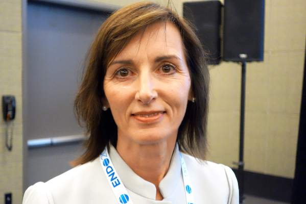
The finding means that pediatricians should routinely plot and follow body mass index (BMI) from an early age, just like height, weight, and head circumference, said investigator Dr. Allison Smego, an endocrinology fellow.
She and her colleagues reviewed the charts from birth to age 6 of 783 lean children and 480 children above the 99th BMI percentile. BMI started differentiating when children were as young as 4 months old, about a year and half before the onset of clinical obesity. The predictive value of the 85th percentile threshold held at 6, 12, and 18 months. The finding was subsequently validated in over 2,600 children.
In an interview at the annual meeting of the Endocrine Society, Dr. Smego explained how to use the findings.
The video associated with this article is no longer available on this site. Please view all of our videos on the MDedge YouTube channel
AT ENDO 2016
Proactive Endocrine Screening Urged for Pediatric Brain Tumor Survivors
BOSTON – More than a third of 419 children treated for brain tumors at Cincinnati Children’s Hospital Medical Center later developed endocrine problems, according to a review presented at the Endocrine Society annual meeting.
Over 60% of the 96 suprasellar tumor patients developed endocrine dysfunction, which isn’t surprising considering the location of the tumor, but wide-ranging endocrine problems were also common in the 145 posterior fossa, 158 supratentorial, and 20 spinal cord cases, ranging from 14% in the spinal cord group to 42% in the posterior fossa group, after some combination of radiation, chemotherapy, and surgery based on tumor location and other factors.
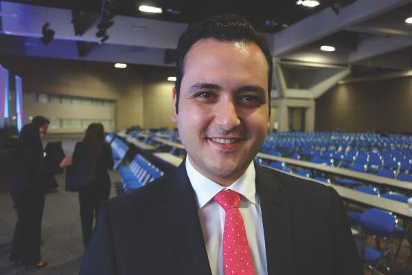
“Even with tumors that aren’t supposed to be high risk, there was a high risk of endocrinopathies. We need yearly screening of these patients” for about 6 years, after which symptom-based screening may be sufficient. The clock should be restarted if there’s a recurrence. “Not everyone does this” at Cincinnati Children’s and probably most other institutions, said investigator and endocrinology fellow Dr. Vincent Horne.
The findings are “changing how our oncology department is thinking about [screening]; there’s a concentrated effort to increase proactive screening and follow these patients long term,” he said.
“Even within our specialized, multidisciplinary center,” endocrinopathy screening referrals were low, about 61% overall and only 80% in the suprasellar group. “Patients at highest risk” – those with craniopharyngioma – “are being seen early by us,” but others aren’t being referred. It’s possible that the extent of endocrine problems after pediatric brain tumor treatment is simply unrecognized, he said.
Endocrine abnormalities were found in 114 (45%) of the 254 patients evaluated, which translated to problems in more than a third of all patients.
More than half of the children had more than one problem, and most of the issues occurred within 6 years of treatment. Central hypothyroidism was found in 53% of the children, probably because Cincinnati Children’s already has thyroid screening in place.
About 40% were growth hormone deficient, and almost a third had precocious puberty. About 30% were gonadotropin-releasing hormone deficient, over 20% had primary hypothyroidism, and about the same had diabetes insipidus. Just over 6% were hyperprolactinemic.
Of the 151 patients who completed adrenocorticotropic hormone (ACTH) testing, 14.6% were deficient. ACTH deficient children were about evenly split between the suprasellar and supratentorial groups, with the remaining in the posterior fossa cohort.
“We are probably not thinking about” the risk of radiation “to locations like the posterior fossa. That group actually had the highest risk of primary hypothyroidism [20%] because of the spinal radiation. The supratentorial group is also receiving radiation; even though we think we are missing the hypothalamus, obviously that’s not necessarily the case,” Dr. Horne said.
His team looked into endocrine screening because previous studies “were limited and done years ago.” People are living longer now after treatment, “so we need to think about how to screen for endocrine disease. This is an attempt to clarify how we should do it,” he said.
Children were a median of 8 years old at diagnosis, and the median radiation dose was 54 Gy.
There was no industry funding for the work, and the investigators had no disclosures.
AT ENDO 2016
Proactive endocrine screening urged for pediatric brain tumor survivors
BOSTON – More than a third of 419 children treated for brain tumors at Cincinnati Children’s Hospital Medical Center later developed endocrine problems, according to a review presented at the Endocrine Society annual meeting.
Over 60% of the 96 suprasellar tumor patients developed endocrine dysfunction, which isn’t surprising considering the location of the tumor, but wide-ranging endocrine problems were also common in the 145 posterior fossa, 158 supratentorial, and 20 spinal cord cases, ranging from 14% in the spinal cord group to 42% in the posterior fossa group, after some combination of radiation, chemotherapy, and surgery based on tumor location and other factors.
“Even with tumors that aren’t supposed to be high risk, there was a high risk of endocrinopathies. We need yearly screening of these patients” for about 6 years, after which symptom-based screening may be sufficient. The clock should be restarted if there’s a recurrence. “Not everyone does this” at Cincinnati Children’s and probably most other institutions, said investigator and endocrinology fellow Dr. Vincent Horne.
The findings are “changing how our oncology department is thinking about [screening]; there’s a concentrated effort to increase proactive screening and follow these patients long term,” he said.
“Even within our specialized, multidisciplinary center,” endocrinopathy screening referrals were low, about 61% overall and only 80% in the suprasellar group. “Patients at highest risk” – those with craniopharyngioma – “are being seen early by us,” but others aren’t being referred. It’s possible that the extent of endocrine problems after pediatric brain tumor treatment is simply unrecognized, he said.
Endocrine abnormalities were found in 114 (45%) of the 254 patients evaluated, which translated to problems in more than a third of all patients.
More than half of the children had more than one problem, and most of the issues occurred within 6 years of treatment. Central hypothyroidism was found in 53% of the children, probably because Cincinnati Children’s already has thyroid screening in place.
About 40% were growth hormone deficient, and almost a third had precocious puberty. About 30% were gonadotropin-releasing hormone deficient, over 20% had primary hypothyroidism, and about the same had diabetes insipidus. Just over 6% were hyperprolactinemic.
Of the 151 patients who completed adrenocorticotropic hormone (ACTH) testing, 14.6% were deficient. ACTH deficient children were about evenly split between the suprasellar and supratentorial groups, with the remaining in the posterior fossa cohort.
“We are probably not thinking about” the risk of radiation “to locations like the posterior fossa. That group actually had the highest risk of primary hypothyroidism [20%] because of the spinal radiation. The supratentorial group is also receiving radiation; even though we think we are missing the hypothalamus, obviously that’s not necessarily the case,” Dr. Horne said.
His team looked into endocrine screening because previous studies “were limited and done years ago.” People are living longer now after treatment, “so we need to think about how to screen for endocrine disease. This is an attempt to clarify how we should do it,” he said.
Children were a median of 8 years old at diagnosis, and the median radiation dose was 54 Gy.
There was no industry funding for the work, and the investigators had no disclosures.
AT ENDO 2016
Key clinical point: Screen for endocrine dysfunction for 6 years after pediatric brain tumor treatment.
Major finding: Endocrine abnormalities were found in 114 of 254 patients (45%) evaluated, which translated to problems in more than a third of all patients.
Data source: Single-center review of 419 pediatric brain tumor cases.
Disclosures: There was no industry funding for the work, and the investigators had no disclosures.
Stick with wheat flour for baked egg and milk challenges
LOS ANGELES – Children who pass oral food challenges to baked egg and milk with wheat flour are at risk for reacting to baked goods made with nonwheat flours, according to a review of more than 200 pediatric food challenges at National Jewish Health in Denver.
The children were already known to be sensitive to egg and milk, and some were being challenged to see if exposure therapy was helping. Unbeknown to the pediatric food allergy team, a kitchen worker at National Jewish had started making muffins with rice flour, thinking it would be safer.
During the month of muffins with rice flour, the failure rate for baked egg challenge muffins rose from 28% (33/120) to 58% (11/19) with rice flour. Failure to baked milk muffins rose from 14% (9/66) to 36% (5/14) (J Allergy Clin Immunol. 2016 Feb. doi: 10.1016/j.jaci.2015.12.579).
Adjusting for age, gender, and atopic dermatitis, children were more than five times more likely to fail baked eggs without wheat (odds ratio, 5.4; P = .002), and more than four times more likely to fail baked milk (OR, 4.06; P = .05).
Given that the phenomenon hasn’t been reported before, “This was very surprising to us,” said study investigator Dr. Bruce Lanser, director of the pediatric food allergy program at National Jewish. “You have to warn parents that if children pass a baked challenge with wheat, they have to continue to eat their baked milk and egg with wheat. Gluten-free products are not going to have the same effect.
“If somebody is avoiding wheat because it causes a bit of redness and itchiness, you have to clear that wheat allergy” before moving to baked egg and milk, Dr. Lanser added.
There’s also concern that “kids will go home after passing a wheat muffin challenge, eat something that’s gluten-free, and have a reaction,” he noted. Wheat-free baked goods might also not build tolerance as well, although that’s not clear from the study, Dr. Lanser said at the annual meeting of the American Academy of Allergy, Asthma, and Immunology.
Wheat seems to have something unique that alters the allergic properties of egg and milk proteins to help children outgrow their sensitivities. “Rice doesn’t have the same effect,” he observed, and it’s not known if any other grains do. Dr. Lanser said he is interested in looking into rye, barley, oats, and other alternatives.
The mean age of the children in the study was 6 years, and most children had multiple food allergies. Sensitization was confirmed by skin tests and specific IgE.
Meanwhile, there’s a new rule in the National Jewish kitchen: Unless a child has true celiac disease, “always make [challenge] muffins with wheat,” Dr. Lanser said.
There was no industry funding for the work, and the investigators had no disclosures.
AT 2016 AAAAI ANNUAL MEETING
Key clinical point: Children are more likely to react to nonwheat challenge muffins, and wheat substitutes might not work as well for oral immunotherapy.
Major finding: The failure rate for baked egg challenge muffins rose from 28% (33/120) to 58% (11/19) with rice flour. Failure to baked milk muffins rose from 14% (9/66) to 36% (5/14).
Data source: Single-center review of more than 200 pediatric food challenges.
Disclosures: There was no industry funding for the work, and the investigators had no disclosures.
Morning cortisol rules out adrenal insufficiency
BOSTON – A random morning serum cortisol above 11.1 mcg/dL safely rules out adrenal insufficiency in both inpatients and outpatients, according to a review of 3,300 adrenal insufficiency work-ups at the Edinburgh Centre for Endocrinology and Diabetes.
The finding could help eliminate the cost and hassle of unnecessary adrenocorticotropic hormone (ACTH) stimulation tests; the investigators estimated that the cut point would eliminate almost half of them without any ill effects. “You can be very confident that patients aren’t insufficient if they are above that line,” with more than 99% sensitivity. If they are below it, “they may be normal, and they may be abnormal.” Below 1.8 mcg/dL, adrenal insufficiency is almost certain, but between the cutoffs, ACTH stimulation is necessary, said lead investigator Dr. Scott Mackenzie, a trainee at the center.
In short, “basal serum cortisol as a screening test ... offers a convenient and accessible means of identifying patients who require further assessment,” he said at the annual meeting of the Endocrine Society.
Similar cut points have been suggested by previous studies, but the Scottish investigation is the first to validate its findings both inside and outside of the hospital.
The team arrived at the 11.1 mcg/dL morning cortisol cut point by comparing basal cortisol levels and synacthen results in 1,628 outpatients. They predefined a sensitivity of more than 99% for adrenal sufficiency to avoid missing anyone with true disease. The cut point’s predictive power was then validated in 875 outpatients and 797 inpatients. Morning basal cortisol levels proved superior to afternoon levels.
The investigators were thinking about cost-effectiveness, but they also wanted to increase screening. “We may be able to reduce the number of adrenal insufficiency cases we are missing because [primary care is] reluctant to send people to the clinic for synacthen tests” due to the cost and inconvenience. As with many locations in the United States, “our practice is to do [ACTH on] everyone.” If there was “a quick and easy 9 a.m. blood test” instead, it would help, Dr. Mackenzie said.
Adrenal insufficiency was on the differential for a wide variety of reasons, including hypogonadism, pituitary issues, prolactinemia, fatigue, hypoglycemia, postural hypotension, and hyponatremia. Most of the patients were middle aged, and they were about evenly split between men and women.
There was no outside funding for the work, and the investigators had no disclosures.
AT ENDO 2016
Key clinical point: Skip ACTH stimulation if morning serum cortisol is above 11.1 mcg/dL.
Major finding: A morning serum cortisol above 11.1 mcg/dL is a test of adrenal function with 99% sensitivity.
Data source: Review of 3,300 adrenal insufficiency work-ups.
Disclosures: There was no outside funding for the work, and the investigators had no disclosures.
Early estrogen likely prevents bone fractures in Turner syndrome
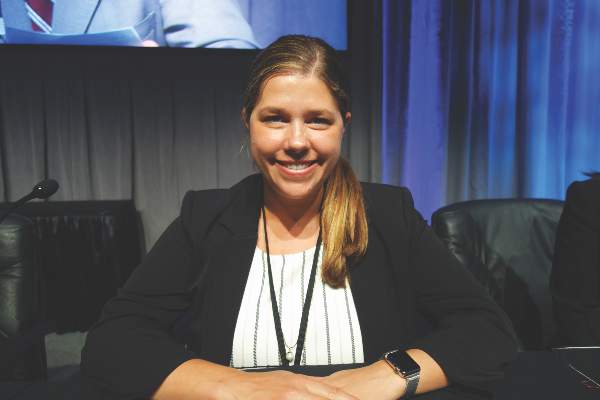
BOSTON – The longer that estrogen therapy is delayed in girls with Turner syndrome, the lower their bone density will be in subsequent years, based on results of a retrospective, cross-sectional study from Monash University, in Melbourne, Australia.
For every year after age 11 that Turner patients went without estrogen – generally due to delayed initiation, but sometimes noncompliance – there was a significant reduction in bone mineral density in both the lumbar spine (Beta -0.582, P less than 0.001) and femoral neck (Beta -0.383, P = 0.008).
Estrogen deficiency and subsequent suboptimal bone mass accrual are known to contribute to the increased risk of osteoporosis in women with Turner syndrome, and about a doubling of the risk of fragility fractures, mostly of the forearm. About a third of the 76 women in the study had at least one fracture, explained investigator Dr. Amanda Vincent, head of the Midlife Health and Menopause Program at Monash.
“Avoiding estrogen deficiency is important to optimize bone health in Turner syndrome.” It “depends on early diagnosis, age-appropriate pubertal induction, and optimization of compliance,” Dr. Vincent said at the Endocrine Society annual meeting.
The median age of Turner syndrome diagnosis was 11 years, but estrogen treatments didn’t begin until a median age of 15. The women in the study were a median of about 30 years old, which means that they were adolescents at the time when estrogen treatment was often delayed in the mistaken belief that growth hormone therapy would be more effective before puberty was induced.
It’s now known that estrogen replacement works synergistically with, and even potentiates, the effects of growth hormone. Current guidelines recommend pubertal induction by age 13 (J Clin Endocrinol Metab. 2007 Jan;92(1):10-25).
The women had at least one dual-energy x-ray absorptiometry scan at Monash since 1998. Z-scores below - 2, indicating low bone density, were found in the lumbar spines of about a quarter the subjects, and in the femoral necks of about 8%. Primary amenorrhea and premature menopause, followed by vitamin D deficiency, were the most common risk factors for low bone mass. Almost 40% of the women reported non-continuous use of estrogen. About half had undergone growth hormone therapy.
At a median height of 149 cm, the subjects were about 15 cm shorter than age-matched, healthy controls, and also had a slightly higher median body mass index of 25.6 kg/m2. Lumbar spine bone area, bone mineral content, areal bone mineral density, and bone mineral apparent density were significantly lower in Turner syndrome patients. In the femoral neck, areal bone mineral density was significantly lower.
There was no relationship between bone markers and growth hormone use or Turner syndrome karyotype; the predominant karyotype was 45XO, but the study also included mosaic karyotypes.
The investigators had no disclosures.
These are important observations. The bottom line is early recognition and early referral. It’s clear from this study and others that earlier institution of estrogen is beneficial for height, bone density, and fracture risk throughout life. It’s not just an issue of a 20 year old with low bone density; that 20 year old later becomes a 60 year old with low bone density.
|
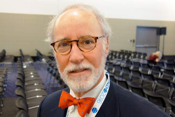
Dr. Michael Levine |
[However,] we still have a problem with delayed recognition and referral of young girls with Turner syndrome. Most girls with Turner syndrome have some typical phenotypic features, but some do not, so the diagnosis is often made too late. [To get around that problem,] we recommend that all children below the 5th percentile for height – or who flatten out too early on growth curves – be referred to rule out Turner syndrome and other problems.
Dr. Michael Levine is chief of the Division of Endocrinology at The Children’s Hospital of Philadelphia. He made his comments after the study presentation, and was not involved in the work.
These are important observations. The bottom line is early recognition and early referral. It’s clear from this study and others that earlier institution of estrogen is beneficial for height, bone density, and fracture risk throughout life. It’s not just an issue of a 20 year old with low bone density; that 20 year old later becomes a 60 year old with low bone density.

|
Dr. Michael Levine |
[However,] we still have a problem with delayed recognition and referral of young girls with Turner syndrome. Most girls with Turner syndrome have some typical phenotypic features, but some do not, so the diagnosis is often made too late. [To get around that problem,] we recommend that all children below the 5th percentile for height – or who flatten out too early on growth curves – be referred to rule out Turner syndrome and other problems.
Dr. Michael Levine is chief of the Division of Endocrinology at The Children’s Hospital of Philadelphia. He made his comments after the study presentation, and was not involved in the work.
These are important observations. The bottom line is early recognition and early referral. It’s clear from this study and others that earlier institution of estrogen is beneficial for height, bone density, and fracture risk throughout life. It’s not just an issue of a 20 year old with low bone density; that 20 year old later becomes a 60 year old with low bone density.

|
Dr. Michael Levine |
[However,] we still have a problem with delayed recognition and referral of young girls with Turner syndrome. Most girls with Turner syndrome have some typical phenotypic features, but some do not, so the diagnosis is often made too late. [To get around that problem,] we recommend that all children below the 5th percentile for height – or who flatten out too early on growth curves – be referred to rule out Turner syndrome and other problems.
Dr. Michael Levine is chief of the Division of Endocrinology at The Children’s Hospital of Philadelphia. He made his comments after the study presentation, and was not involved in the work.
BOSTON – The longer that estrogen therapy is delayed in girls with Turner syndrome, the lower their bone density will be in subsequent years, based on results of a retrospective, cross-sectional study from Monash University, in Melbourne, Australia.
For every year after age 11 that Turner patients went without estrogen – generally due to delayed initiation, but sometimes noncompliance – there was a significant reduction in bone mineral density in both the lumbar spine (Beta -0.582, P less than 0.001) and femoral neck (Beta -0.383, P = 0.008).
Estrogen deficiency and subsequent suboptimal bone mass accrual are known to contribute to the increased risk of osteoporosis in women with Turner syndrome, and about a doubling of the risk of fragility fractures, mostly of the forearm. About a third of the 76 women in the study had at least one fracture, explained investigator Dr. Amanda Vincent, head of the Midlife Health and Menopause Program at Monash.
“Avoiding estrogen deficiency is important to optimize bone health in Turner syndrome.” It “depends on early diagnosis, age-appropriate pubertal induction, and optimization of compliance,” Dr. Vincent said at the Endocrine Society annual meeting.
The median age of Turner syndrome diagnosis was 11 years, but estrogen treatments didn’t begin until a median age of 15. The women in the study were a median of about 30 years old, which means that they were adolescents at the time when estrogen treatment was often delayed in the mistaken belief that growth hormone therapy would be more effective before puberty was induced.
It’s now known that estrogen replacement works synergistically with, and even potentiates, the effects of growth hormone. Current guidelines recommend pubertal induction by age 13 (J Clin Endocrinol Metab. 2007 Jan;92(1):10-25).
The women had at least one dual-energy x-ray absorptiometry scan at Monash since 1998. Z-scores below - 2, indicating low bone density, were found in the lumbar spines of about a quarter the subjects, and in the femoral necks of about 8%. Primary amenorrhea and premature menopause, followed by vitamin D deficiency, were the most common risk factors for low bone mass. Almost 40% of the women reported non-continuous use of estrogen. About half had undergone growth hormone therapy.
At a median height of 149 cm, the subjects were about 15 cm shorter than age-matched, healthy controls, and also had a slightly higher median body mass index of 25.6 kg/m2. Lumbar spine bone area, bone mineral content, areal bone mineral density, and bone mineral apparent density were significantly lower in Turner syndrome patients. In the femoral neck, areal bone mineral density was significantly lower.
There was no relationship between bone markers and growth hormone use or Turner syndrome karyotype; the predominant karyotype was 45XO, but the study also included mosaic karyotypes.
The investigators had no disclosures.
AT ENDO 2016
Key clinical point: Induce puberty by age 13 in Turner syndrome.
Major finding: For every year after age 11 that Turner patients went without estrogen – generally due to delayed initiation, but sometimes noncompliance – there was a significant reduction in bone mineral density in both the lubar spine (Beta -0.582, P less than 0.001) and femoral neck (Beta -0.383, P = 0.008).
Data source: Retrospective, cross-section study of 76 Turner syndrome patients
Disclosures: The investigators had no disclosures.
Early biopsy predicts levonorgestrel IUD response in endometrial cancer
SAN DIEGO – Endometrial pathology findings at 3 months predicted response to levonorgestrel-releasing IUD treatment for complex atypical hyperplasia or grade 1 endometrial cancer at the MD Anderson Cancer Center in Houston.
Twenty-nine of 32 women (91%) who responded by 12 months showed stromal, glandular, or other endometrial changes indicating an effect at 3 months, vs. only 3 of 9 nonresponders (33%) (P less than .001). There were no differences in responders versus nonresponders in median age (47 vs. 56 years, P = .2) or body mass index (45 vs. 55 kg/m2, P = .16).
The finding addresses an “unmet need” for markers of response to levonorgestrel-releasing IUD therapy. “You can look at [early] pathology” and have an idea how patients will do, Dr. Shannon Westin, a study investigator who is with the department of gynecologic oncology at MD Anderson, said at the annual meeting of the Society of Gynecologic Oncology.
Twenty-seven of 29 women (93%) with complex atypical hyperplasia (CAH) responded completely to the IUD, meaning they had normal endometrium or hyperplasia without atypia at 12 months. The response rate for endometrial cancer was 67%; 7 of 12 women had a complete response, and an 8th was diagnosed at 12 months with CAH, indicating a partial response. The rest of the patients remained stable or progressed.
Endometrial biopsies were performed every 3 months; the team also did molecular testing on tumors from 20 patients. Baseline protein Ki67 – a marker of proliferation – was significantly higher in nonresponders. Expression of several estrogen-induced genes was higher in responders.
Patients opted for the IUD to retain fertility or because obesity or comorbidities precluded surgery. Exclusion criteria included prior treatment for CAH or endometrial cancer, evidence of extrauterine spread, or levonorgestrel IUD contraindications, such as uterine infection.
Adverse events – primarily irregular bleeding and cramping – were mild and tended to resolve by 12 months. Treatment had little effect on measures of social, mental, and physical function. About half of the patients were white, a third were Hispanic, and most of the remaining patients were black.
There was no external funding for the work. Dr. Westin is a consultant for AstraZeneca, Medivation, Roche, Ovation, and Vermillion, and reported receiving research funding from AstraZeneca, Critical Outcomes Technologies, and Novartis.
AT THE ANNUAL MEETING ON WOMEN’S CANCER
Key clinical point: Pathology and molecular findings identify good candidates for levonorgestrel IUD therapy.
Major finding: Twenty-nine of 32 women (91%) who responded by 12 months showed stromal, glandular, or other endometrial changes indicating an effect at 3 months, versus only 3 of 9 nonresponders (33%) (P less than .001).
Data source: A prospective investigation of 41 women.
Disclosures: There was no external funding for the work. Dr. Westin is a consultant for and receives research funding from AstraZeneca and several other companies.
Skip lymphadenectomy if SLN mapping finds low-grade endometrial cancer
SAN DIEGO – Lymphadenectomy is unnecessary if sentinel lymph node mapping successfully stages low-grade endometrial cancer, according to researchers from Johns Hopkins University in Baltimore.
Lymphadenectomy guided by frozen section remains common in the United States. But the Johns Hopkins research team found that using sentinel lymph node (SLN) mapping and biopsy instead cuts the rate of lymphadenectomy by 76%, without reducing the detection of lymphatic metastases.
It’s an important finding for cancer patients likely to survive their diagnosis. “We see low-grade patients in the clinic” who’ve had unnecessary lymphadenectomies, “and they are in terrible shape,” said investigator Dr. Abdulrahman Sinno, a gynecologic oncology fellow at Johns Hopkins. Up to half “have horrible side effects,” including crippling lymphedema and pain.
SLN mapping is “an alternative that gives us the information we need for nodal assessment without putting patients at risk. You’ll know if patients have metastases or not. If they fail to map, you do a frozen section, and if you have high-risk features, a lymphadenectomy only on [the side] that didn’t map,” Dr. Sinno said at the annual meeting of the Society of Gynecologic Oncology.
For the past several years, physicians at Johns Hopkins has been doing both SLN mapping for low-grade endometrial cancer as well as frozen sections to decide the need for lymphadenectomy. Using both approaches allowed the investigators to review how patients would have fared if they had gotten only one.
“[We could] safely study the utility of SLN mapping while maintaining the historical standard of using frozen sections to direct the need for lymphadenectomy,” Dr. Sinno said.
SLN mapping outperformed frozen section. Among 114 women, most with grade 1 disease but some with grade 2 or complex atypical hyperplasia, 8 had lymph node metastases. Mapping identified every one, five by standard hematoxylin-eosin staining, and three by ultrastaging. Frozen-section guided lymphadenectomy missed three.
Eighty four (37%) of the 224 hemi-pelvises in the study had lymphadenectomies based on worrisome frozen-section findings. If SLN mapping had been relied on to make the call, lymphadenectomies would have been performed in 20 (9%), a statistically significant difference (P = 0.004).
“Strategies that rely exclusively on uterine frozen section result in significant overtreatment. In the absence of a therapeutic benefit to lymphadenectomy, we believe” this is “unjustifiable when an alternative exists.” At Johns Hopkins these days, “if you map, you’re done,” Dr. Sinno said.
Almost two-thirds of the women had grade 1 endometrial cancer on preoperative histopathology, and about the same number on final pathology. Bilateral SLN mapping was successful in 71 cases (62%) and unilateral mapping in 27 cases (24%). At least one SLN was detected in 98 women (86%).
There were six recurrences after a median follow-up of 15 months. Four were in women who had full pelvic and periaortic lymphadenectomies that were negative. There was also a port site recurrence and a recurrence in an outlying patient with advanced disease. Overall, “recurrence was independent of whether sentinel nodes were applied,” Dr. Sinno said.
Women in the study were a median of 60 years old, with a median body mass index of 33.3 kg/m2.
Dr. Sinno reported having no relevant financial disclosures.
AT THE ANNUAL MEETING ON WOMEN’S CANCER
Key clinical point: Successful sentinel lymph node mapping gives all the information needed for nodal assessment.
Major finding: Sentinel lymph node mapping identified all eight nodal metastases; frozen-section guided lymphadenectomy missed three.
Data source: A review of 114 cases at Johns Hopkins University.
Disclosures: Dr. Sinno reported having no relevant financial disclosures.
Observation might be best in advanced-stage, low-grade ovarian CA
SAN DIEGO – Surgery, chemotherapy, and radiation do not prolong survival in advanced-stage but low-grade papillary serous ovarian cancer, according to a review of 1,159 low-grade cases in the National Cancer Data Base.
“All you are doing is exposing patients to the toxicity without any benefit. You [should] think really hard about whether or not to give [low-grade] patients radiation or chemotherapy, instead of just following them,” said investigator Dr. Allison Gockley, an ob.gyn. at Brigham and Women’s Hospital and Massachusetts General Hospital in Boston.
Seventy-four percent of the women received platinum-based chemotherapy, but it made no difference in survival (hazard ratio, 0.94; 95% CI, 0.75 -1.19); likewise, radiation made no difference in the seven women (0.6%) who received it.
Lymphadenectomy was the only thing associated with improved survival (HR, 0.5; 95% CI, 0.4-0.7). Dr. Gockley said she suspects it was a marker for careful surgery, but even surgery didn’t improve survival in low-grade cases (HR, 0.93; 95% CI, 0.51-1.70), although most patients had an operation.
The percentages of women who had surgery, chemotherapy, and radiation were almost identical among the 24,073 advanced-stage, high-grade cases also considered in the review, except that low-grade patients were more likely to get radiation. The approaches help high-grade cases, but “it feels very unsatisfying to me to treat low-grade” patients the same way when “they obviously have” different tumor biology, Dr. Gockley said at the annual meeting of the Society of Gynecologic Oncology.
She said she’s found similar issues with early-stage uterine clear cell carcinoma. The problem is that there just aren’t much data to guide treatment in less-dangerous gynecologic cancers, so patients end up being treated like they have advanced disease. “We need to step up and focus [study] on patients with these tumors so we can have specific treatments. For a lot of these rare tumors” – advanced-stage, low-grade disease represents about 5% of ovarian cancer – “these patients would probably do better if we had supertargeted chemotherapy,” she said.
In the absence of evidence, some patients opt for aggressive treatment “because it makes them feel that they can do something to control their disease.” As for doctors, “I’ve been in rooms where it’s very highly advocated, and others where physicians are more hesitant. What happens in the end is very much about how doctors [frame] the discussion,” Dr. Gockley said.
Her institutions follow national trends; very few patients get radiation, but most get chemotherapy, sometimes three cycles instead of six, or in low dose.
She and her colleagues looked into the issue because “we’ve seen a bunch of patients on our services recently who have low-grade, advanced-stage” ovarian cancer. The investigators wanted to know how other institutions handle the situation.
Most of the low-grade patients had stage III disease, and the rest had stage IV. Their mean age at diagnosis was 54 years, 9 years earlier than for high-grade diagnoses. About half of stage III patients were alive at 10 years, vs. about 20% of stage IV patients. As expected, high-grade patients died sooner.
AT THE ANNUAL MEETING ON WOMEN’S CANCER
Key clinical point: Low-grade ovarian cancer is overtreated.
Major finding: Seventy-four percent of the women received platinum-based chemotherapy, but it made no difference in survival (HR, 0.94; 95% CI, 0.75-1.19).
Data source: A review of more than 25,000 ovarian cancer cases in the National Cancer Data Base.
Disclosures: The presenter had no disclosures, and there was no outside funding for the work.
Idarucizumab may reverse dabigatran anticoagulation in intracranial hemorrhage
LOS ANGELES – It only took a few minutes for idarucizumab to normalize blood-clotting parameters in 18 patients with dabigatran-associated intracranial hemorrhages, according to interim results from an ongoing phase III trial presented at the International Stroke Conference.
“When I put patients on [dabigatran], they always ask me what happens if they bleed or need surgery. … Until now, I haven’t been able to tell them with any confidence that I have a way of reversing it. Now I think I can. ... It makes a big difference” in their comfort, said lead investigator Dr. Richard Bernstein, a Northwestern University neurology professor and director of the stroke program at Northwestern Memorial Hospital, in Chicago.
“We would love to know if hematoma expansion was limited [and outcomes improved] by giving this reversal agent,” but the study so far is too small. “We hope to have a larger cohort of brain hemorrhage patients to answer these questions,” he said.
Approved in October 2015, idarucizumab (Praxbind) was fast tracked by the Food and Drug Administration to reverse the blockbuster atrial fibrillation anticoagulant dabigatran (Pradaxa); the labeling for idarucizumab doesn’t mention intracranial hemorrhage patients specifically. Boehringer Ingelheim makes both drugs, and funded Dr. Bernstein’s work.
Eleven of the 18 patients were men, and the average age in the study was about 80 years. The patients had either subdural hematomas or bleeding into the brain itself. They were culled from the 90 subjects analyzed so far in the idarucizumab trial, dubbed RE-VERSE AD (Reversal Effects of Idarucizumab in Patients on Active Dabigatran).
The team followed label dosing: 5 g total given as two separate 2.5-g infusions. Blood samples were taken in between to check how well idarucizumab worked. The whole process took no more than 15 minutes.
The first 2.5 g completely reversed dabigatran in all 18 patients, based on their dilute thrombin or ecarin clotting times. Patients “remained reversed out to 12 hours, and all but one out to 24 hours,” Dr. Bernstein said at the conference, sponsored by the American Heart Association.
Idarucizumab is a monoclonal antibody fragment that binds dabigatran more powerfully than dabigatran binds thrombin. In vitro studies have found no prothrombotic effects. “It has no endogenous target, so the drug has no effect on any other clotting factors that we can tell. We did have, I think, five thrombotic events in our cohort, most of them many days after the dabigatran was reversed. It may have just been a reversion to [patient] clotting risks,” he said.
When – or if – to restart dabigatran “is a clinical question.” If bleeding is controlled or patients are stable after surgery, you can go back on the next day,” he said.
Idarucizumab’s labeling notes that 5% or more of patients developed hypokalemia, delirium, constipation, pyrexia, and pneumonia. It wasn’t clear these events were drug related. Patients had dabigatran reversed either for serious bleeding or emergency surgery.
Dr. Bernstein is a speaker and adviser for Boehringer Ingelheim, and reported honoraria from the company.
AT THE INTERNATIONAL STROKE CONFERENCE
Key clinical point: An ongoing investigation suggests that idarucizumab can reliably stop intracranial hemorrhage associated with dabigatran anticoagulation.
Major finding: It took only a few minutes for idarucizumab to normalize blood-clotting parameters in 18 patients with dabigatran intracranial hemorrhage.
Source: Interim results from an ongoing phase III trial.
Disclosures: The work was funded by Boehringer Ingelheim, maker of both dabigatran and idarucizumab. The lead investigator is a speaker and adviser for Boehringer, and reported honoraria from the company.


