User login
EULAR-PReS guidelines aim to aid pediatric to adult care transition
LONDON – The first European guidelines developed to help the transition of young people from pediatric to adult rheumatology care within Europe were announced at the European Congress of Rheumatology.
The key aim of the guidelines, which have been jointly written by the European League Against Rheumatism (EULAR) and the Paediatric Rheumatology European Society (PReS), is to make the transition process more consistent across rheumatology practices throughout Europe, which in turn should help to ensure both the continuity and the quality of clinical care, explained Dr. Helen E. Fosterof Newcastle (England) University.

“There is evidence that there has been a long-standing problem of young people growing up with their condition moving to adult care and either falling between the services or being lost to follow-up, or there has not been continuity of care,” she said in an interview ahead of presenting the new EULAR/PReS guidelines at the congress.
“All in all, that’s translated into poorer health outcomes for young people,” said Dr. Foster, who was one of the main convenors of the EULAR/PReS Working Party for Transitional Care Management for Adolescents and Young People.
The premise is to try to provide practical recommendations that clinicians can use to help young people in their care from the age of 11 years and older as they get ready for the transfer to adult services. The latter process can occur anywhere from 16 to 19 years of age, Dr. Foster said, but it is important to try start the transition process early and get young people more involved and responsible for their own care.
“The idea is that young people are supported to be in control of their condition, that they can cope with being seen on their own in clinic, that they are getting on with their lives, and ultimately that they have a better outcome, which includes becoming healthy, getting a job, living independently, and having a family,” she said. The age at transfer is flexible and needs to fit with the young person’s home and school life. Ideally, it occurs at a time when their disease and medication are stable, they are attending routine appointments, and generally able to be independent and cope with their condition.
EULAR/PReS transition guidelines: 12 recommendations
• Access to high-quality coordinated transition care services should be available to all young people.
• Transition should ‘start early’ (11 years of age) or directly after diagnosis.
• Direct communication is needed between young people and their families and pediatric and adult care providers.
• Each young person should have an individualized transition plan.
• There should be a written transition policy within all relevant services; this should be regularly agreed and updated.
• The multidisciplinary team involved in transitional care should be clearly defined in a written document.
• Transition services should address the complexity of adolescent and young adult development.
• There must be an agreed upon and written transfer document.
• Health care teams should be given appropriate training in adolescent and young adult rheumatic diseases.
• Secure funding is needed for uninterrupted clinical care and transition into adult services.
• An open digital platform should host the recommendations and support tools and information.
• More evidence is needed to demonstrate the outcomes of the transition to adult services.
Developing the guidelines
Together with Dr. Kirsten Minden of the German Rheumatism Research Centre Berlin (DRFZ), Dr. Foster chaired the international, multidisciplinary EULAR/PReS Working Party to review existing national and international guidelines, consensus statements, and other supporting evidence on transitional care management in childhood-onset rheumatic illness.
The remit was to develop recommendations to facilitate optimal transitional care management in rheumatology across different European countries. As such, the recommendations cover both the ideal situation as well as the bare minimum requirements to hopefully allow widespread adoption. To this end, the working party performed a systematic literature review according to EULAR standard operating procedures. They developed a set of 12 recommendations based on the evidence they reviewed.
There are 47 different health systems within Europe, all running according to different health policies set by different governments, Dr. Minden observed. In fact, only a handful of countries have specific transition care policies or pathways, so the aim was to try to develop recommendations that would work across the board while giving some ideas on how to improve existing strategies further.
She noted that some examples of existing transition programs are “Growing up and moving on” in the United Kingdom (Pediatr Transplant. 2005;9:364-72), “On your own feet ahead” in the Netherlands (BMC Health Serv Res. 2014;14:47), and “Devices for Optimization of Transfer and Transition of Adolescents with Rheumatic Disorders (DON’T RETARD)” in Belgium (Rheumatology [Oxford]. 2016;55:133-42). Of these, two are specific to the transition of young people with juvenile idiopathic arthritis (JIA) and one is for rheumatologic conditions in general.
Core elements of these programs are the need to provide written information and have a transition care plan, the allocation of a dedicated transition coordinator, and an individualized transition plan for each patient, Dr. Minden said. These elements are also part of the EULAR/PReS transition guidelines.
One of the issues to be addressed, however, is whether these transition programs actually work in the long term. “Transitional care services in rheumatology are beginning to happen and their further development can surely be facilitated by the provision of tool kits and resources for health care providers and patients,” she noted. Some of the tools already exist, so the challenge now is to get these available to all so that there can be a wider dissemination of knowledge.
The North American perspective
Both the American Academy of Pediatrics and the Canadian Pediatric Society have issued general guidance on how to transition young people from pediatric to adult services, said Dr. Lori. B. Tucker, a pediatric specialist working at BC Children’s Hospital in Vancouver, which runs the ON TRAC (Transitioning Responsibly to Adult Care) program. This is a province-wide program aimed at supporting young people between the ages of 12 and 24 years with chronic health conditions and their families to transition from pediatric to adult health care services.
The ON TRAC program includes online and mobile-enabled checklists that can be used with young people and their families, although Dr. Tucker noted that the program had perhaps not been as successful as had been hoped. Another Canadian initiative specific to rheumatology practice is the RACER (Readiness for Adult Care in Rheumatology) questionnaire. This was developed to assess how ready young people with chronic ailments were to transition to adult service.
Dr. Tucker also highlighted the YARD (Young Adult Rheumatic Diseases) clinic at her institution, set up for those aged 18 years or older with a definite diagnosis of rheumatic disease. Parents are not allowed within the clinic so as to enable young adults to take responsibility for their overall care and collaborate with their health care providers. The clinic provides education, assistance with separation independence, and other issues pertinent to this young population of patients, and it also aims to encourage adherence to appointments and treatments.
“Collaboration between pediatric rheumatologists and adult colleagues is critical to improve the outcomes of young adults with rheumatic diseases,” Dr. Tucker said. She added, “Better articulated guidelines for transition care and use of new tools have great potential to improve the care of these patients ‘lost in-between.’ ”
Why the need for the EULAR/PReS recommendations?
Dr. Foster noted that, in many countries, there is a natural break between pediatric and adult care, with young people often moving from one center to another, perhaps in another part of the country. An important part of the transition process is therefore ensuring that there are appropriately trained staff members and good communication between centers to ensure that young people don’t get lost during the move.
“This is everyone’s business,” Dr. Foster said at the congress. “It is a shared responsibility to get it right.” That means adult and pediatric health care teams work together. Care needs to be “holistic,” she added, and cover medical, psychosocial, vocational issues, and be “developmentally appropriate throughout.” Young people also need to be involved from the start of the process, beginning early and continuing into young adulthood.
The recommendations aim to be flexible so that they can be widely implemented by health care teams throughout Europe. “It is not ‘one size fits all,” Dr. Foster acknowledged in the interview, noting the importance of being realistic and recognizing the differences between health systems, resources, and access across Europe.
Dr. Foster, who trained in adult rheumatology before turning to pediatric rheumatology, noted that there are existing resources that can be used and although funding will be an issue on some levels, there are things that can be done by using existing tools and resources.
“We don’t want to reinvent the wheel. We want to share best practice and resources,” she said. Indeed, one of the recommendations is that all the guidelines and all the resources used to develop them are made publicly available via an electronic platform so that anybody involved in the care of a young person with rheumatic disease, as well as the young person and their family, can access them.
“Transitional care is key to improving long-term outcomes for young people with rheumatic disease,” Dr. Foster concluded. The EULAR/PReS transition care management guidelines have been developed with the engagement of all relevant stakeholders, she said, so they should be widely applicable and “important levers for change” throughout Europe. “Implementation will require funding, but also our will and energy to make them actually work in practice.”
The EULAR/PReS transition guidelines are being finalized and will be published soon in Annals of the Rheumatic Diseases.
Dr. Foster, Dr. Minden, and Dr. Tucker had no disclosures relevant to the development of the recommendations.
LONDON – The first European guidelines developed to help the transition of young people from pediatric to adult rheumatology care within Europe were announced at the European Congress of Rheumatology.
The key aim of the guidelines, which have been jointly written by the European League Against Rheumatism (EULAR) and the Paediatric Rheumatology European Society (PReS), is to make the transition process more consistent across rheumatology practices throughout Europe, which in turn should help to ensure both the continuity and the quality of clinical care, explained Dr. Helen E. Fosterof Newcastle (England) University.
“There is evidence that there has been a long-standing problem of young people growing up with their condition moving to adult care and either falling between the services or being lost to follow-up, or there has not been continuity of care,” she said in an interview ahead of presenting the new EULAR/PReS guidelines at the congress.
“All in all, that’s translated into poorer health outcomes for young people,” said Dr. Foster, who was one of the main convenors of the EULAR/PReS Working Party for Transitional Care Management for Adolescents and Young People.
The premise is to try to provide practical recommendations that clinicians can use to help young people in their care from the age of 11 years and older as they get ready for the transfer to adult services. The latter process can occur anywhere from 16 to 19 years of age, Dr. Foster said, but it is important to try start the transition process early and get young people more involved and responsible for their own care.
“The idea is that young people are supported to be in control of their condition, that they can cope with being seen on their own in clinic, that they are getting on with their lives, and ultimately that they have a better outcome, which includes becoming healthy, getting a job, living independently, and having a family,” she said. The age at transfer is flexible and needs to fit with the young person’s home and school life. Ideally, it occurs at a time when their disease and medication are stable, they are attending routine appointments, and generally able to be independent and cope with their condition.
EULAR/PReS transition guidelines: 12 recommendations
• Access to high-quality coordinated transition care services should be available to all young people.
• Transition should ‘start early’ (11 years of age) or directly after diagnosis.
• Direct communication is needed between young people and their families and pediatric and adult care providers.
• Each young person should have an individualized transition plan.
• There should be a written transition policy within all relevant services; this should be regularly agreed and updated.
• The multidisciplinary team involved in transitional care should be clearly defined in a written document.
• Transition services should address the complexity of adolescent and young adult development.
• There must be an agreed upon and written transfer document.
• Health care teams should be given appropriate training in adolescent and young adult rheumatic diseases.
• Secure funding is needed for uninterrupted clinical care and transition into adult services.
• An open digital platform should host the recommendations and support tools and information.
• More evidence is needed to demonstrate the outcomes of the transition to adult services.
Developing the guidelines
Together with Dr. Kirsten Minden of the German Rheumatism Research Centre Berlin (DRFZ), Dr. Foster chaired the international, multidisciplinary EULAR/PReS Working Party to review existing national and international guidelines, consensus statements, and other supporting evidence on transitional care management in childhood-onset rheumatic illness.
The remit was to develop recommendations to facilitate optimal transitional care management in rheumatology across different European countries. As such, the recommendations cover both the ideal situation as well as the bare minimum requirements to hopefully allow widespread adoption. To this end, the working party performed a systematic literature review according to EULAR standard operating procedures. They developed a set of 12 recommendations based on the evidence they reviewed.
There are 47 different health systems within Europe, all running according to different health policies set by different governments, Dr. Minden observed. In fact, only a handful of countries have specific transition care policies or pathways, so the aim was to try to develop recommendations that would work across the board while giving some ideas on how to improve existing strategies further.
She noted that some examples of existing transition programs are “Growing up and moving on” in the United Kingdom (Pediatr Transplant. 2005;9:364-72), “On your own feet ahead” in the Netherlands (BMC Health Serv Res. 2014;14:47), and “Devices for Optimization of Transfer and Transition of Adolescents with Rheumatic Disorders (DON’T RETARD)” in Belgium (Rheumatology [Oxford]. 2016;55:133-42). Of these, two are specific to the transition of young people with juvenile idiopathic arthritis (JIA) and one is for rheumatologic conditions in general.
Core elements of these programs are the need to provide written information and have a transition care plan, the allocation of a dedicated transition coordinator, and an individualized transition plan for each patient, Dr. Minden said. These elements are also part of the EULAR/PReS transition guidelines.
One of the issues to be addressed, however, is whether these transition programs actually work in the long term. “Transitional care services in rheumatology are beginning to happen and their further development can surely be facilitated by the provision of tool kits and resources for health care providers and patients,” she noted. Some of the tools already exist, so the challenge now is to get these available to all so that there can be a wider dissemination of knowledge.
The North American perspective
Both the American Academy of Pediatrics and the Canadian Pediatric Society have issued general guidance on how to transition young people from pediatric to adult services, said Dr. Lori. B. Tucker, a pediatric specialist working at BC Children’s Hospital in Vancouver, which runs the ON TRAC (Transitioning Responsibly to Adult Care) program. This is a province-wide program aimed at supporting young people between the ages of 12 and 24 years with chronic health conditions and their families to transition from pediatric to adult health care services.
The ON TRAC program includes online and mobile-enabled checklists that can be used with young people and their families, although Dr. Tucker noted that the program had perhaps not been as successful as had been hoped. Another Canadian initiative specific to rheumatology practice is the RACER (Readiness for Adult Care in Rheumatology) questionnaire. This was developed to assess how ready young people with chronic ailments were to transition to adult service.
Dr. Tucker also highlighted the YARD (Young Adult Rheumatic Diseases) clinic at her institution, set up for those aged 18 years or older with a definite diagnosis of rheumatic disease. Parents are not allowed within the clinic so as to enable young adults to take responsibility for their overall care and collaborate with their health care providers. The clinic provides education, assistance with separation independence, and other issues pertinent to this young population of patients, and it also aims to encourage adherence to appointments and treatments.
“Collaboration between pediatric rheumatologists and adult colleagues is critical to improve the outcomes of young adults with rheumatic diseases,” Dr. Tucker said. She added, “Better articulated guidelines for transition care and use of new tools have great potential to improve the care of these patients ‘lost in-between.’ ”
Why the need for the EULAR/PReS recommendations?
Dr. Foster noted that, in many countries, there is a natural break between pediatric and adult care, with young people often moving from one center to another, perhaps in another part of the country. An important part of the transition process is therefore ensuring that there are appropriately trained staff members and good communication between centers to ensure that young people don’t get lost during the move.
“This is everyone’s business,” Dr. Foster said at the congress. “It is a shared responsibility to get it right.” That means adult and pediatric health care teams work together. Care needs to be “holistic,” she added, and cover medical, psychosocial, vocational issues, and be “developmentally appropriate throughout.” Young people also need to be involved from the start of the process, beginning early and continuing into young adulthood.
The recommendations aim to be flexible so that they can be widely implemented by health care teams throughout Europe. “It is not ‘one size fits all,” Dr. Foster acknowledged in the interview, noting the importance of being realistic and recognizing the differences between health systems, resources, and access across Europe.
Dr. Foster, who trained in adult rheumatology before turning to pediatric rheumatology, noted that there are existing resources that can be used and although funding will be an issue on some levels, there are things that can be done by using existing tools and resources.
“We don’t want to reinvent the wheel. We want to share best practice and resources,” she said. Indeed, one of the recommendations is that all the guidelines and all the resources used to develop them are made publicly available via an electronic platform so that anybody involved in the care of a young person with rheumatic disease, as well as the young person and their family, can access them.
“Transitional care is key to improving long-term outcomes for young people with rheumatic disease,” Dr. Foster concluded. The EULAR/PReS transition care management guidelines have been developed with the engagement of all relevant stakeholders, she said, so they should be widely applicable and “important levers for change” throughout Europe. “Implementation will require funding, but also our will and energy to make them actually work in practice.”
The EULAR/PReS transition guidelines are being finalized and will be published soon in Annals of the Rheumatic Diseases.
Dr. Foster, Dr. Minden, and Dr. Tucker had no disclosures relevant to the development of the recommendations.
AT THE EULAR 2016 CONGRESS

VIDEO: EULAR guidance on DMARD use in RA made ‘more concise’
LONDON – The European League Against Rheumatism guidelines on the use of disease-modifying antirheumatic drugs for the treatment of rheumatoid arthritis have been updated in line with current evidence and made more concise.
Dr. Josef S. Smolen of the department of rheumatology at the Medical University of Vienna who presented the 2016 guidelines at the European Congress of Rheumatology, noted that they now consist of 12 rather than the 14 recommendations that were included in the 2013 update (Ann Rheum Dis. 2014;73:492-509) and the 15 recommendations that were in the original 2010 version.
These 12 recommendations cover treatment targets and general approaches in the management of rheumatoid arthritis that incorporate disease-modifying antirheumatic drugs (DMARDs) and the use of glucocorticoids, and present treatment options as a hierarchy to help guide clinicians through appropriate procedures when initial and subsequent treatment fails. All DMARDs are considered in the recommendations, from the long-standing conventional synthetic (cs)DMARDs, such as methotrexate, sulfasalazine, and leflunomide, and the newer biologic DMARDs, such as the anti–tumor necrosis factor (TNF)–targeting drugs, to the newer biosimilar DMARDs, and targeted synthetic (ts)DMARDS, such as the Janus kinase (JAK) inhibitors tofacitinib and baricitinib.
The recommendations have been developed in accordance with EULAR’s standard operating procedure for the development of guidelines, Dr. Smolen observed, and involved three systematic literature reviews and expert opinion garnered from a task force of 50 experts and patients.
“This was the largest task force I have ever convened,” Dr. Smolen said, noting that rheumatologists from outside Europe had been invited to contribute their expertise and knowledge for the first time. Altogether 42 rheumatologists, three clinical fellows, two health professionals, and three patients were involved in revising the recommendations.
There are now four rather than three overarching principles, two of which are shared with early inflammatory arthritis recommendations that were also presented at the congress. The first two principles state that shared decision making is key to optimizing care and that rheumatologists should be the primary specialists looking after patients. The third principle recognizes the high burden that RA can have not only on an individual level but also on health care systems and society in general, which rheumatologists should be aware of. The fourth and final principle states that treatment decisions should be based on patients’ disease activity but that other factors, such as patients’ age, risk for progression, coexisting disease, and likely tolerance of treatment should also be kept in mind.
In an interview, Dr. Smolen highlighted that the EULAR recommendations cover three main phases of DMARD treatment: First is the DMARD-naive group of patients, who may be at an early or late stage of their disease. Second is the group in whom initial treatment has failed, and third is the group for whom subsequent treatment has not worked.
“In all these phases, we have some changes,” Dr. Smolen said. As an example, he noted that in the DMARD-naive setting, the use of csDMARDs has always been recommended but that the prior advice to consider combination csDMARD treatment has been edited out.
“We now say methotrexate should be part of the first treatment strategy, and the treatment strategy encompasses the use of additional, at least conventional synthetic, DMARDs.” Glucocorticoids are also more strongly recommended as part of the initial treatment strategy in combination with methotrexate, he said, although there is the proviso to use these for as short a time as possible.
In situations where patients do not respond to methotrexate plus glucocorticoids or they cannot tolerate methotrexate, then the recommendations advise stratifying patients into two groups. Those with poor prognostic factors might be switched to a biologic therapy, such as an anti-TNF agent or a tsDMARD. In regard to the latter, there is now more evidence behind the use of JAK inhibitors, notably tofacitinib, Dr. Smolen observed. Biologic DMARDs should be combined with csDMARDs, but if the latter is not tolerated then there is the option to use an IL-6 pathway inhibitor.
“There is now compelling evidence that all biologic DMARDs, including tocilizumab, convey better clinical, functional, and structural outcomes in combination with conventional synthetic DMARDs, especially methotrexate,” Dr. Smolen observed during his presentation of the recommendations. This may not be the case for the JAK inhibitors based on the current evidence.
When asked how the EULAR recommendations match up to those issued earlier this year by the American College of Rheumatology (Arthritis Care Res. 2016;68:1-25), Dr. Smolen observed that the two had become “much closer.” There remain differences in recommendations on glucocorticoid use, which are “somewhat clearer” in the European than in the American guidelines, and EULAR proposes combining biologic DMARDs with csDMARDs rather than using them as monotherapy. The EULAR recommendations also do not distinguish patients by disease duration but by treatment phase, and use prognostic factors for stratification.
The recommendations are currently in draft format and once finalized they will be published in Annals of the Rheumatic Diseases and also made freely available via the EULAR website, joining the organization’s many other recommendations for the management of rheumatic diseases. Dr. Smolen noted that these are intended as a template to provide national societies, health systems, and regulatory bodies a guide to the best evidence-based use of DMARDS in RA throughout Europe.
Dr. Smolen has received grant support and/or honoraria for consultations and/or for presentations from: AbbVie, Amgen, AstraZeneca, Astro-Pharma, Bristol-Myers Squibb, Celgene, GlaxoSmithKline, ILTOO Pharma, Janssen, Merck Serono, Merck Sharp & Dohme, Novartis-Sandoz, Pfizer, Roche-Chugai, Samsung, and UCB.
The video associated with this article is no longer available on this site. Please view all of our videos on the MDedge YouTube channel
AT THE EULAR 2016 CONGRESS

Simplify Cardiac Risk Assessment for Rheumatologic Conditions
LONDON – Cardiovascular disease (CVD) risk assessment for patients with rheumatic diseases can be simple and integrated into general practice or rheumatology clinics, experts said during an Outcomes Science Session at the European Congress of Rheumatology
Patients with rheumatoid arthritis have a 50% higher risk of heart disease than do their counterparts without the disease, but “just having RA on its own isn’t sufficient to render that individual at high risk,” Dr. Naveed Sattar, professor of metabolic medicine at the University of Glasgow’s Institute of Cardiovascular and Medical Sciences in Scotland, said in an interview.

It’s simple enough to use traditional CVD risk factors in an RA population by including a patient’s age, gender, smoking status, and family history of heart disease, in addition to measuring blood pressure and blood lipid levels. Most risk scores will compile those features into a 10-year risk of a fatal CVD event. To account for the contribution of RA, Dr. Sattar said, simply multiply that score by 1.5.
While “there’s a fixation in some parts of Europe for [measuring] fasting lipids,” it is not necessary, Dr. Sattar said. The two lipid parameters that go in risk scores tend to be cholesterol and HDL cholesterol, he said, which change only minimally in fasting versus nonfasting states.
“The evidence overwhelmingly shows that nonfasting lipids, which can be done easily on the same sample as other clinic tests, are just as predictive of CVD risk as fasting lipids,” he said. “That really matters because many of our patients with RA or other conditions come to the hospital when they’re not fasting, and we shouldn’t be sending them away to come back fasting to do risk scores for CVD. That just doesn’t make sense.”
Updated guidelines from the European Society of Cardiology and guidelines soon to be released from the European League Against Rheumatism suggest that risk scores can be calculated every 5 years for most patients, a change from previous recommendations to calculate risk annually. Risk scoring is not perfect, however, and there is some debate about whether additional blood tests or ultrasound scanning of the carotid artery could augment the ability to predict heart disease risk. “We’re not quite there yet,” Dr. Sattar said. “I think we should do the simple things first and do them well.”
CV risk raised in all inflammatory arthritic diseases
During the same session, Dr. Paola de Pablo of the University of Birmingham, England, focused on how immune-mediated diseases predispose to premature, accelerated atherosclerosis and subsequent increased cardiovascular morbidity and mortality.
Cardiovascular risk is not only elevated in those with RA, she observed, but also in those with systemic lupus erythematosus, ankylosing spondylitis, psoriatic arthritis, vasculitides, and inflammatory myopathies. The risk varies but as a rule is more than 50% higher than the rate seen in the general population.
The underlying mechanisms are not clear, but chronic inflammation is closely linked with atherosclerosis, which in turn ups the risk for myocardial infarction and cerebrovascular accident.
Despite treatment, the risk often remains, Dr. de Pablo said. She highlighted how treatment with methotrexate and anti–tumor necrosis factor (TNF)–alpha drugs in RA had been associated with a reduction in the risk for heart attack of 20% and 40%, respectively (Ann Rheum Dis. 2014;74:480–89) so targeting inflammation with these drugs may have positive effects, at least in RA.
Managing traditional cardiovascular risk factors remains important, Dr. de Pablo said. That was a sentiment echoed by Dr. Sattar and by rheumatologist Dr. Michael Nurmohamed of the VU Medical Center in Amsterdam. This includes controlling blood pressure with antihypertensive medications and blood lipids with statins, and advocating smoking cessation and perhaps other appropriate lifestyle changes such as increasing physical exercise and controlling weight.
Dr. Nurmohamed, who was involved in the 2015 update of the EULAR recommendations on cardiovascular risk management, noted that traditional risk factor management in patients with arthritis in current clinical practice is often poor and that strategies to address this were urgently needed.
Although treating to-target and preventing disease flares in the rheumatic diseases is important, it lowers but does not normalize cardiovascular risk. “This appears to be irrespective of the drug used,” Dr. Nurmohamed said. Rheumatologists need to be careful when tapering medication, particularly the biologics, as these are perhaps helping to temper cardiovascular inflammation, which could worsen when doses are reduced. “Antirheumatic treatment only is not good enough to decrease or normalize the cardiovascular risk of our patients”, he emphasized.
Norwegian project shows how to integrate CVD assessment into routine practice
In a separate presentation, Dr. Eirik Ikdahl, a PhD student at Diakonhjemmet Hospital in Oslo, discussed how some rheumatology clinics in Norway are successfully incorporating CVD risk screening.
Through the Norwegian Collaboration on Atherosclerotic disease in patients with Rheumatic joint diseases (NOCAR), which started in April 2014, annual cardiovascular disease risk evaluations of patients with inflammatory joint diseases are being implemented into the practices of 11 rheumatology outpatient clinics. While waiting for clinic appointments, patients are given electronic devices through which they can report CVD risk factors via an electronic patient journal program called GoTreatIt Rheuma. From there, the clinic can order nonfasting lipid measurements and nurses can record patients’ blood pressure.
Then, using the ESC Systematic Coronary Risk Evaluation (SCORE) algorithm, the program automatically calculates a patient’s 10-year risk of a fatal CVD event. If the SCORE estimate is 5% or greater, the rheumatologist forwards a note to the patient’s primary care physician or cardiologist saying there is an indication for initiation of CVD-preventive measures such as medication or lifestyle changes. Rheumatologists and rheumatology nurses also deliver brief advice regarding smoking cessation and healthy diet.
“The main aim of the project is to raise awareness of the cardiovascular burden that these patients experience, and to ensure that patients with inflammatory joint diseases receive guideline-recommended cardiovascular preventive treatment,” Dr. Ikdahl said.
Of 6,150 patients defined as eligible for the NOCAR project in three of the centers, 41% (n = 2,519) received a CVD risk assessment during the first year and a half of the program, officers found in a recent review. Of those, 1,569 had RA, 418 had ankylosing spondylitis, 350 had psoriatic arthritis, and 122 had other spondyloarthritides.
Through the program, “a large number of high-risk patients have received screening that they would not otherwise have been offered,” Dr. Ikdahl said.
The major obstacles to successful implementation were time scarcity, defining a date for annual CVD risk assessment among patients who visit the clinics multiple times per year, and making sure lipids were measured before seeing the rheumatologist, he said. “We acknowledge there is room for improvement. It is challenging to implement new work tasks in an already busy rheumatology outpatient clinic, and since the project does not offer financial incentives to the participating centers, we rely on a collective effort and voluntary work based on resources already available.”
Remember CVD, but don’t forget other comorbidities
Other research presented by Dr. Laure Gossec, professor of rheumatology at Pitie-Salpétriere Hospital and Pierre & Marie Curie University in Paris highlighted the importance of identifying all comorbidities and their risk factors in patients with rheumatic diseases, and not just cardiovascular disease.
Dr. Gossec presented the results of an initiative aiming to make the collection and management of comorbidities easier in routine rheumatologic practice. The aim was to develop a simple, more pragmatic form that could be used to help rheumatologists manage selected comorbidities, and know when to refer for other specialist assessment. The focus was on ischemic cardiovascular disease, malignancies, infections such as chronic bronchitis, gastrointestinal disease such as diverticulitis, osteoporosis, and depression.
A committee of 18 experts, both physicians and nurses, was convened to examine the results of a systematic literature review of recommendations on comorbidity management and come up with concise recommendations for rheumatologists. Each of their recommendations covered whether or not the comorbidity was present (yes/no/don’t know) and if screening had been undertaken, such as measurement of blood lipids, and when this had occurred if known. There was then guidance on how to interpret these findings, calculate risk, and what to do if findings were abnormal.
The project is ongoing, and so far the expert panel has developed a pragmatic document with forms to help collect, report, and manage each specific comorbidity and its known risk factors. But it is still perhaps too long to be feasibly used in everyday practice, Dr. Gossec conceded. So the aim is to create a short, 2-page form that could summarize the recommendations briefly, and also develop a questionnaire for the patient to fill out and understand how to self-manage some comorbidities.
“We feel that this is a way to disseminate and adapt to the national context for France the EULAR comorbidities initiative,” Dr. Gossec said. “It also defines exactly what rheumatologists should be doing and when they should refer, hopefully to the benefit of our patients.”
Dr. Sattar has participated in advisory boards for Amgen, AstraZeneca, Boehringer Ingelheim, Eli Lily and UCB. He has also consulted for Merck and is a member of Roche’s speakers’ bureau. Dr. de Paolo and Dr. Nurmohamed reported no relevant financial disclosures. Dr. Ikdahl has received speaker’s honoraria from Pfizer. Dr. Gossec and coauthors have received honoraria from Abbvie France.
The video associated with this article is no longer available on this site. Please view all of our videos on the MDedge YouTube channel
The video associated with this article is no longer available on this site. Please view all of our videos on the MDedge YouTube channel
LONDON – Cardiovascular disease (CVD) risk assessment for patients with rheumatic diseases can be simple and integrated into general practice or rheumatology clinics, experts said during an Outcomes Science Session at the European Congress of Rheumatology
Patients with rheumatoid arthritis have a 50% higher risk of heart disease than do their counterparts without the disease, but “just having RA on its own isn’t sufficient to render that individual at high risk,” Dr. Naveed Sattar, professor of metabolic medicine at the University of Glasgow’s Institute of Cardiovascular and Medical Sciences in Scotland, said in an interview.
It’s simple enough to use traditional CVD risk factors in an RA population by including a patient’s age, gender, smoking status, and family history of heart disease, in addition to measuring blood pressure and blood lipid levels. Most risk scores will compile those features into a 10-year risk of a fatal CVD event. To account for the contribution of RA, Dr. Sattar said, simply multiply that score by 1.5.
While “there’s a fixation in some parts of Europe for [measuring] fasting lipids,” it is not necessary, Dr. Sattar said. The two lipid parameters that go in risk scores tend to be cholesterol and HDL cholesterol, he said, which change only minimally in fasting versus nonfasting states.
“The evidence overwhelmingly shows that nonfasting lipids, which can be done easily on the same sample as other clinic tests, are just as predictive of CVD risk as fasting lipids,” he said. “That really matters because many of our patients with RA or other conditions come to the hospital when they’re not fasting, and we shouldn’t be sending them away to come back fasting to do risk scores for CVD. That just doesn’t make sense.”
Updated guidelines from the European Society of Cardiology and guidelines soon to be released from the European League Against Rheumatism suggest that risk scores can be calculated every 5 years for most patients, a change from previous recommendations to calculate risk annually. Risk scoring is not perfect, however, and there is some debate about whether additional blood tests or ultrasound scanning of the carotid artery could augment the ability to predict heart disease risk. “We’re not quite there yet,” Dr. Sattar said. “I think we should do the simple things first and do them well.”
CV risk raised in all inflammatory arthritic diseases
During the same session, Dr. Paola de Pablo of the University of Birmingham, England, focused on how immune-mediated diseases predispose to premature, accelerated atherosclerosis and subsequent increased cardiovascular morbidity and mortality.
Cardiovascular risk is not only elevated in those with RA, she observed, but also in those with systemic lupus erythematosus, ankylosing spondylitis, psoriatic arthritis, vasculitides, and inflammatory myopathies. The risk varies but as a rule is more than 50% higher than the rate seen in the general population.
The underlying mechanisms are not clear, but chronic inflammation is closely linked with atherosclerosis, which in turn ups the risk for myocardial infarction and cerebrovascular accident.
Despite treatment, the risk often remains, Dr. de Pablo said. She highlighted how treatment with methotrexate and anti–tumor necrosis factor (TNF)–alpha drugs in RA had been associated with a reduction in the risk for heart attack of 20% and 40%, respectively (Ann Rheum Dis. 2014;74:480–89) so targeting inflammation with these drugs may have positive effects, at least in RA.
Managing traditional cardiovascular risk factors remains important, Dr. de Pablo said. That was a sentiment echoed by Dr. Sattar and by rheumatologist Dr. Michael Nurmohamed of the VU Medical Center in Amsterdam. This includes controlling blood pressure with antihypertensive medications and blood lipids with statins, and advocating smoking cessation and perhaps other appropriate lifestyle changes such as increasing physical exercise and controlling weight.
Dr. Nurmohamed, who was involved in the 2015 update of the EULAR recommendations on cardiovascular risk management, noted that traditional risk factor management in patients with arthritis in current clinical practice is often poor and that strategies to address this were urgently needed.
Although treating to-target and preventing disease flares in the rheumatic diseases is important, it lowers but does not normalize cardiovascular risk. “This appears to be irrespective of the drug used,” Dr. Nurmohamed said. Rheumatologists need to be careful when tapering medication, particularly the biologics, as these are perhaps helping to temper cardiovascular inflammation, which could worsen when doses are reduced. “Antirheumatic treatment only is not good enough to decrease or normalize the cardiovascular risk of our patients”, he emphasized.
Norwegian project shows how to integrate CVD assessment into routine practice
In a separate presentation, Dr. Eirik Ikdahl, a PhD student at Diakonhjemmet Hospital in Oslo, discussed how some rheumatology clinics in Norway are successfully incorporating CVD risk screening.
Through the Norwegian Collaboration on Atherosclerotic disease in patients with Rheumatic joint diseases (NOCAR), which started in April 2014, annual cardiovascular disease risk evaluations of patients with inflammatory joint diseases are being implemented into the practices of 11 rheumatology outpatient clinics. While waiting for clinic appointments, patients are given electronic devices through which they can report CVD risk factors via an electronic patient journal program called GoTreatIt Rheuma. From there, the clinic can order nonfasting lipid measurements and nurses can record patients’ blood pressure.
Then, using the ESC Systematic Coronary Risk Evaluation (SCORE) algorithm, the program automatically calculates a patient’s 10-year risk of a fatal CVD event. If the SCORE estimate is 5% or greater, the rheumatologist forwards a note to the patient’s primary care physician or cardiologist saying there is an indication for initiation of CVD-preventive measures such as medication or lifestyle changes. Rheumatologists and rheumatology nurses also deliver brief advice regarding smoking cessation and healthy diet.
“The main aim of the project is to raise awareness of the cardiovascular burden that these patients experience, and to ensure that patients with inflammatory joint diseases receive guideline-recommended cardiovascular preventive treatment,” Dr. Ikdahl said.
Of 6,150 patients defined as eligible for the NOCAR project in three of the centers, 41% (n = 2,519) received a CVD risk assessment during the first year and a half of the program, officers found in a recent review. Of those, 1,569 had RA, 418 had ankylosing spondylitis, 350 had psoriatic arthritis, and 122 had other spondyloarthritides.
Through the program, “a large number of high-risk patients have received screening that they would not otherwise have been offered,” Dr. Ikdahl said.
The major obstacles to successful implementation were time scarcity, defining a date for annual CVD risk assessment among patients who visit the clinics multiple times per year, and making sure lipids were measured before seeing the rheumatologist, he said. “We acknowledge there is room for improvement. It is challenging to implement new work tasks in an already busy rheumatology outpatient clinic, and since the project does not offer financial incentives to the participating centers, we rely on a collective effort and voluntary work based on resources already available.”
Remember CVD, but don’t forget other comorbidities
Other research presented by Dr. Laure Gossec, professor of rheumatology at Pitie-Salpétriere Hospital and Pierre & Marie Curie University in Paris highlighted the importance of identifying all comorbidities and their risk factors in patients with rheumatic diseases, and not just cardiovascular disease.
Dr. Gossec presented the results of an initiative aiming to make the collection and management of comorbidities easier in routine rheumatologic practice. The aim was to develop a simple, more pragmatic form that could be used to help rheumatologists manage selected comorbidities, and know when to refer for other specialist assessment. The focus was on ischemic cardiovascular disease, malignancies, infections such as chronic bronchitis, gastrointestinal disease such as diverticulitis, osteoporosis, and depression.
A committee of 18 experts, both physicians and nurses, was convened to examine the results of a systematic literature review of recommendations on comorbidity management and come up with concise recommendations for rheumatologists. Each of their recommendations covered whether or not the comorbidity was present (yes/no/don’t know) and if screening had been undertaken, such as measurement of blood lipids, and when this had occurred if known. There was then guidance on how to interpret these findings, calculate risk, and what to do if findings were abnormal.
The project is ongoing, and so far the expert panel has developed a pragmatic document with forms to help collect, report, and manage each specific comorbidity and its known risk factors. But it is still perhaps too long to be feasibly used in everyday practice, Dr. Gossec conceded. So the aim is to create a short, 2-page form that could summarize the recommendations briefly, and also develop a questionnaire for the patient to fill out and understand how to self-manage some comorbidities.
“We feel that this is a way to disseminate and adapt to the national context for France the EULAR comorbidities initiative,” Dr. Gossec said. “It also defines exactly what rheumatologists should be doing and when they should refer, hopefully to the benefit of our patients.”
Dr. Sattar has participated in advisory boards for Amgen, AstraZeneca, Boehringer Ingelheim, Eli Lily and UCB. He has also consulted for Merck and is a member of Roche’s speakers’ bureau. Dr. de Paolo and Dr. Nurmohamed reported no relevant financial disclosures. Dr. Ikdahl has received speaker’s honoraria from Pfizer. Dr. Gossec and coauthors have received honoraria from Abbvie France.
The video associated with this article is no longer available on this site. Please view all of our videos on the MDedge YouTube channel
The video associated with this article is no longer available on this site. Please view all of our videos on the MDedge YouTube channel
AT THE EULAR 2016 CONGRESS

Simplify cardiac risk assessment for rheumatologic conditions
LONDON – Cardiovascular disease (CVD) risk assessment for patients with rheumatic diseases can be simple and integrated into general practice or rheumatology clinics, experts said during an Outcomes Science Session at the European Congress of Rheumatology
Patients with rheumatoid arthritis have a 50% higher risk of heart disease than do their counterparts without the disease, but “just having RA on its own isn’t sufficient to render that individual at high risk,” Dr. Naveed Sattar, professor of metabolic medicine at the University of Glasgow’s Institute of Cardiovascular and Medical Sciences in Scotland, said in an interview.
It’s simple enough to use traditional CVD risk factors in an RA population by including a patient’s age, gender, smoking status, and family history of heart disease, in addition to measuring blood pressure and blood lipid levels. Most risk scores will compile those features into a 10-year risk of a fatal CVD event. To account for the contribution of RA, Dr. Sattar said, simply multiply that score by 1.5.
While “there’s a fixation in some parts of Europe for [measuring] fasting lipids,” it is not necessary, Dr. Sattar said. The two lipid parameters that go in risk scores tend to be cholesterol and HDL cholesterol, he said, which change only minimally in fasting versus nonfasting states.
“The evidence overwhelmingly shows that nonfasting lipids, which can be done easily on the same sample as other clinic tests, are just as predictive of CVD risk as fasting lipids,” he said. “That really matters because many of our patients with RA or other conditions come to the hospital when they’re not fasting, and we shouldn’t be sending them away to come back fasting to do risk scores for CVD. That just doesn’t make sense.”
Updated guidelines from the European Society of Cardiology and guidelines soon to be released from the European League Against Rheumatism suggest that risk scores can be calculated every 5 years for most patients, a change from previous recommendations to calculate risk annually. Risk scoring is not perfect, however, and there is some debate about whether additional blood tests or ultrasound scanning of the carotid artery could augment the ability to predict heart disease risk. “We’re not quite there yet,” Dr. Sattar said. “I think we should do the simple things first and do them well.”
CV risk raised in all inflammatory arthritic diseases
During the same session, Dr. Paola de Pablo of the University of Birmingham, England, focused on how immune-mediated diseases predispose to premature, accelerated atherosclerosis and subsequent increased cardiovascular morbidity and mortality.
Cardiovascular risk is not only elevated in those with RA, she observed, but also in those with systemic lupus erythematosus, ankylosing spondylitis, psoriatic arthritis, vasculitides, and inflammatory myopathies. The risk varies but as a rule is more than 50% higher than the rate seen in the general population.
The underlying mechanisms are not clear, but chronic inflammation is closely linked with atherosclerosis, which in turn ups the risk for myocardial infarction and cerebrovascular accident.
Despite treatment, the risk often remains, Dr. de Pablo said. She highlighted how treatment with methotrexate and anti–tumor necrosis factor (TNF)–alpha drugs in RA had been associated with a reduction in the risk for heart attack of 20% and 40%, respectively (Ann Rheum Dis. 2014;74:480–89) so targeting inflammation with these drugs may have positive effects, at least in RA.
Managing traditional cardiovascular risk factors remains important, Dr. de Pablo said. That was a sentiment echoed by Dr. Sattar and by rheumatologist Dr. Michael Nurmohamed of the VU Medical Center in Amsterdam. This includes controlling blood pressure with antihypertensive medications and blood lipids with statins, and advocating smoking cessation and perhaps other appropriate lifestyle changes such as increasing physical exercise and controlling weight.
Dr. Nurmohamed, who was involved in the 2015 update of the EULAR recommendations on cardiovascular risk management, noted that traditional risk factor management in patients with arthritis in current clinical practice is often poor and that strategies to address this were urgently needed.
Although treating to-target and preventing disease flares in the rheumatic diseases is important, it lowers but does not normalize cardiovascular risk. “This appears to be irrespective of the drug used,” Dr. Nurmohamed said. Rheumatologists need to be careful when tapering medication, particularly the biologics, as these are perhaps helping to temper cardiovascular inflammation, which could worsen when doses are reduced. “Antirheumatic treatment only is not good enough to decrease or normalize the cardiovascular risk of our patients”, he emphasized.
Norwegian project shows how to integrate CVD assessment into routine practice
In a separate presentation, Dr. Eirik Ikdahl, a PhD student at Diakonhjemmet Hospital in Oslo, discussed how some rheumatology clinics in Norway are successfully incorporating CVD risk screening.
Through the Norwegian Collaboration on Atherosclerotic disease in patients with Rheumatic joint diseases (NOCAR), which started in April 2014, annual cardiovascular disease risk evaluations of patients with inflammatory joint diseases are being implemented into the practices of 11 rheumatology outpatient clinics. While waiting for clinic appointments, patients are given electronic devices through which they can report CVD risk factors via an electronic patient journal program called GoTreatIt Rheuma. From there, the clinic can order nonfasting lipid measurements and nurses can record patients’ blood pressure.
Then, using the ESC Systematic Coronary Risk Evaluation (SCORE) algorithm, the program automatically calculates a patient’s 10-year risk of a fatal CVD event. If the SCORE estimate is 5% or greater, the rheumatologist forwards a note to the patient’s primary care physician or cardiologist saying there is an indication for initiation of CVD-preventive measures such as medication or lifestyle changes. Rheumatologists and rheumatology nurses also deliver brief advice regarding smoking cessation and healthy diet.
“The main aim of the project is to raise awareness of the cardiovascular burden that these patients experience, and to ensure that patients with inflammatory joint diseases receive guideline-recommended cardiovascular preventive treatment,” Dr. Ikdahl said.
Of 6,150 patients defined as eligible for the NOCAR project in three of the centers, 41% (n = 2,519) received a CVD risk assessment during the first year and a half of the program, officers found in a recent review. Of those, 1,569 had RA, 418 had ankylosing spondylitis, 350 had psoriatic arthritis, and 122 had other spondyloarthritides.
Through the program, “a large number of high-risk patients have received screening that they would not otherwise have been offered,” Dr. Ikdahl said.
The major obstacles to successful implementation were time scarcity, defining a date for annual CVD risk assessment among patients who visit the clinics multiple times per year, and making sure lipids were measured before seeing the rheumatologist, he said. “We acknowledge there is room for improvement. It is challenging to implement new work tasks in an already busy rheumatology outpatient clinic, and since the project does not offer financial incentives to the participating centers, we rely on a collective effort and voluntary work based on resources already available.”
Remember CVD, but don’t forget other comorbidities
Other research presented by Dr. Laure Gossec, professor of rheumatology at Pitie-Salpétriere Hospital and Pierre & Marie Curie University in Paris highlighted the importance of identifying all comorbidities and their risk factors in patients with rheumatic diseases, and not just cardiovascular disease.
Dr. Gossec presented the results of an initiative aiming to make the collection and management of comorbidities easier in routine rheumatologic practice. The aim was to develop a simple, more pragmatic form that could be used to help rheumatologists manage selected comorbidities, and know when to refer for other specialist assessment. The focus was on ischemic cardiovascular disease, malignancies, infections such as chronic bronchitis, gastrointestinal disease such as diverticulitis, osteoporosis, and depression.
A committee of 18 experts, both physicians and nurses, was convened to examine the results of a systematic literature review of recommendations on comorbidity management and come up with concise recommendations for rheumatologists. Each of their recommendations covered whether or not the comorbidity was present (yes/no/don’t know) and if screening had been undertaken, such as measurement of blood lipids, and when this had occurred if known. There was then guidance on how to interpret these findings, calculate risk, and what to do if findings were abnormal.
The project is ongoing, and so far the expert panel has developed a pragmatic document with forms to help collect, report, and manage each specific comorbidity and its known risk factors. But it is still perhaps too long to be feasibly used in everyday practice, Dr. Gossec conceded. So the aim is to create a short, 2-page form that could summarize the recommendations briefly, and also develop a questionnaire for the patient to fill out and understand how to self-manage some comorbidities.
“We feel that this is a way to disseminate and adapt to the national context for France the EULAR comorbidities initiative,” Dr. Gossec said. “It also defines exactly what rheumatologists should be doing and when they should refer, hopefully to the benefit of our patients.”
Dr. Sattar has participated in advisory boards for Amgen, AstraZeneca, Boehringer Ingelheim, Eli Lily and UCB. He has also consulted for Merck and is a member of Roche’s speakers’ bureau. Dr. de Paolo and Dr. Nurmohamed reported no relevant financial disclosures. Dr. Ikdahl has received speaker’s honoraria from Pfizer. Dr. Gossec and coauthors have received honoraria from Abbvie France.
The video associated with this article is no longer available on this site. Please view all of our videos on the MDedge YouTube channel
The video associated with this article is no longer available on this site. Please view all of our videos on the MDedge YouTube channel
AT THE EULAR 2016 CONGRESS

Early identification, treatment still key to early arthritis guidelines
LONDON, ENGLAND – Prompt referral to a rheumatologist and early initiation of treatment remain 2 of the 12 key recommendations in the updated European League Against Rheumatism guidelines for early arthritis.
There are now three specific recommendations dealing with referral and diagnosis; four that cover initial drug treatment with disease-modifying antirheumatic drugs (DMARDs), nonsteroidal anti-inflammatory drugs (NSAIDs), and glucocorticoids; two that cover management strategy and monitoring; and three that cover nonpharmacologic interventions, prevention, and patient information and education.

While many of the recommendations have not radically changed, there have been revisions to the wording. In line with other EULAR recommendations for the management of rheumatic disease, the updated early arthritis guidelines also now contain three overarching principles. The first states that the management of early arthritis should aim to achieve the best possible care and emphasizes the need for shared decision making between rheumatologist and patient. The second states that a rheumatologist should be the main specialist looking after a patient with early arthritis, and the third states that a definitive diagnosis should be made only after a careful medical history and clinical examination have been undertaken.
“[The EULAR] recommendations deal especially with early-stage inflammatory arthritis,” said Dr. Bernard Combe of Hôpital Lapeyronie in Montpellier, France, who was the convener of the task force behind the updated guidelines. As such, they are universal for all rheumatologic arthritis conditions when diagnosed early, before they differentiate into more specifics, he said in an interview. That includes progression from early inflammatory arthritis to rheumatoid arthritis, psoriatic arthritis, and spondyloarthritis.
Dr. Combe, who is also professor of rheumatology at Montpellier University and head of the bone and joint diseases department at Montpellier University Hospital, presented the updated EULAR guidance at the European Congress of Rheumatology. He noted that the guidelines were first written almost 10 years ago (Ann Rheum Dis. 2007 Jan;66(1):34-45) and so were in need of an update. A task force of 20 rheumatologists, two patients, and one health care professional representing 12 European countries were involved in the update that adhered to EULAR standard operating procedure of developing guidelines.
Although EULAR had published guidance on the management of arthritis in the intervening years, he said, this had focused more on management, and the aim of the early arthritis guidelines was to cover the “entire spectrum of the management of early inflammatory arthritis.”
The updated recommendations start and end with patient-centered statements, he observed. The first notes that patients with any joint swelling associated with pain or stiffness should be referred to and seen by a rheumatologist within 6 weeks of the onset of symptoms. The final recommendation deals with patient information and education about the disease, and programs aiming to help patients cope with pain, disability, and ensure their continued ability to work and participate in their usual social activities.
The concept of early identification and treatment is not new, Dr. Combe observed, but there is so much more evidence in support of initiating DMARD therapy within the first 3 months of referral, even if patients do not fulfill classification criteria for a specific inflammatory rheumatic disease.
In terms of diagnosis, the recommendations now hinge on performing a thorough clinical examination and using ultrasonography to confirm the presence of arthritis if needed. Magnetic resonance imaging (MRI) is no longer recommended in this initial diagnostic work up. This is due to the cost and often lack of widespread access in all European countries, Dr. Combe said. MRI might be considered later, however, if a diagnosis cannot be reached. Assessment of the number of swollen joints, acute phase reactants, and antibody tests (rheumatoid factor and anti-citrullinated protein antibody) also might be of use at this point.
Among the various DMARDs, methotrexate is recommended as the “anchor drug”; it should be used as part of the first treatment strategy in patients who are at risk of persistent disease, unless it contraindicated. The goal of treatment with DMARDs is to achieve clinical remission. Regular monitoring of disease activity, side effects, and comorbidities should be performed alongside their use. Regular monitoring of all pharmacologic therapy should include assessment of tender and swollen joint counts, global health assessments by the patient and the physician, and acute phase reactants.
As for NSAIDs, they are recommended for symptomatic relief, but “at the minimum effective dose for the shortest time possible.” The risks for gastrointestinal, renal, and cardiovascular complications should be carefully weighed against the likely benefits. Glucocorticoids also are recommended for reducing pain and swelling, and structural progression, but again these need to be used at the lowest possible dose and for no more than 6 months to avoid potential long-term side effects.
A recommendation on nonpharmacologic interventions also is included, which states that physical exercise and occupational therapy should be considered as adjunctive therapy.
In addition, the task force came up with a list of 10 research questions that need to be answered, which included items on risk prediction, optimal treatment combinations, and dosing regimens.
As with other EULAR guidelines, a flow chart is included that summarizes the recommendations to help guide physicians on when and how to treat, when to adapt dosing or change medication, and other treatments and approaches to consider.
There is a new recommendation on prevention highlighting the importance of smoking cessation, dental care, weight control, vaccination, and managing comorbidities.
Once the guidelines have been finalized, they will be published in the EULAR journal, Annals of the Rheumatic Diseases, later in the year.
Dr. Combe has received research grants and honoraria from Pfizer, Roche-Chugai, and UCB, and honoraria from Bristol-Myers Squibb, Janssen, Eli Lilly, Merck Sharpe and Dohme, and Novartis.
LONDON, ENGLAND – Prompt referral to a rheumatologist and early initiation of treatment remain 2 of the 12 key recommendations in the updated European League Against Rheumatism guidelines for early arthritis.
There are now three specific recommendations dealing with referral and diagnosis; four that cover initial drug treatment with disease-modifying antirheumatic drugs (DMARDs), nonsteroidal anti-inflammatory drugs (NSAIDs), and glucocorticoids; two that cover management strategy and monitoring; and three that cover nonpharmacologic interventions, prevention, and patient information and education.
While many of the recommendations have not radically changed, there have been revisions to the wording. In line with other EULAR recommendations for the management of rheumatic disease, the updated early arthritis guidelines also now contain three overarching principles. The first states that the management of early arthritis should aim to achieve the best possible care and emphasizes the need for shared decision making between rheumatologist and patient. The second states that a rheumatologist should be the main specialist looking after a patient with early arthritis, and the third states that a definitive diagnosis should be made only after a careful medical history and clinical examination have been undertaken.
“[The EULAR] recommendations deal especially with early-stage inflammatory arthritis,” said Dr. Bernard Combe of Hôpital Lapeyronie in Montpellier, France, who was the convener of the task force behind the updated guidelines. As such, they are universal for all rheumatologic arthritis conditions when diagnosed early, before they differentiate into more specifics, he said in an interview. That includes progression from early inflammatory arthritis to rheumatoid arthritis, psoriatic arthritis, and spondyloarthritis.
Dr. Combe, who is also professor of rheumatology at Montpellier University and head of the bone and joint diseases department at Montpellier University Hospital, presented the updated EULAR guidance at the European Congress of Rheumatology. He noted that the guidelines were first written almost 10 years ago (Ann Rheum Dis. 2007 Jan;66(1):34-45) and so were in need of an update. A task force of 20 rheumatologists, two patients, and one health care professional representing 12 European countries were involved in the update that adhered to EULAR standard operating procedure of developing guidelines.
Although EULAR had published guidance on the management of arthritis in the intervening years, he said, this had focused more on management, and the aim of the early arthritis guidelines was to cover the “entire spectrum of the management of early inflammatory arthritis.”
The updated recommendations start and end with patient-centered statements, he observed. The first notes that patients with any joint swelling associated with pain or stiffness should be referred to and seen by a rheumatologist within 6 weeks of the onset of symptoms. The final recommendation deals with patient information and education about the disease, and programs aiming to help patients cope with pain, disability, and ensure their continued ability to work and participate in their usual social activities.
The concept of early identification and treatment is not new, Dr. Combe observed, but there is so much more evidence in support of initiating DMARD therapy within the first 3 months of referral, even if patients do not fulfill classification criteria for a specific inflammatory rheumatic disease.
In terms of diagnosis, the recommendations now hinge on performing a thorough clinical examination and using ultrasonography to confirm the presence of arthritis if needed. Magnetic resonance imaging (MRI) is no longer recommended in this initial diagnostic work up. This is due to the cost and often lack of widespread access in all European countries, Dr. Combe said. MRI might be considered later, however, if a diagnosis cannot be reached. Assessment of the number of swollen joints, acute phase reactants, and antibody tests (rheumatoid factor and anti-citrullinated protein antibody) also might be of use at this point.
Among the various DMARDs, methotrexate is recommended as the “anchor drug”; it should be used as part of the first treatment strategy in patients who are at risk of persistent disease, unless it contraindicated. The goal of treatment with DMARDs is to achieve clinical remission. Regular monitoring of disease activity, side effects, and comorbidities should be performed alongside their use. Regular monitoring of all pharmacologic therapy should include assessment of tender and swollen joint counts, global health assessments by the patient and the physician, and acute phase reactants.
As for NSAIDs, they are recommended for symptomatic relief, but “at the minimum effective dose for the shortest time possible.” The risks for gastrointestinal, renal, and cardiovascular complications should be carefully weighed against the likely benefits. Glucocorticoids also are recommended for reducing pain and swelling, and structural progression, but again these need to be used at the lowest possible dose and for no more than 6 months to avoid potential long-term side effects.
A recommendation on nonpharmacologic interventions also is included, which states that physical exercise and occupational therapy should be considered as adjunctive therapy.
In addition, the task force came up with a list of 10 research questions that need to be answered, which included items on risk prediction, optimal treatment combinations, and dosing regimens.
As with other EULAR guidelines, a flow chart is included that summarizes the recommendations to help guide physicians on when and how to treat, when to adapt dosing or change medication, and other treatments and approaches to consider.
There is a new recommendation on prevention highlighting the importance of smoking cessation, dental care, weight control, vaccination, and managing comorbidities.
Once the guidelines have been finalized, they will be published in the EULAR journal, Annals of the Rheumatic Diseases, later in the year.
Dr. Combe has received research grants and honoraria from Pfizer, Roche-Chugai, and UCB, and honoraria from Bristol-Myers Squibb, Janssen, Eli Lilly, Merck Sharpe and Dohme, and Novartis.
AT THE EULAR 2016 CONGRESS

Giant cell arteritis survival doubled in the past 2 decades
LONDON – The survival of patients with giant cell arteritis has more than doubled in the past 20 years, according to the results of a population-based study presented at the European Congress of Rheumatology.
Comparing two cohorts of patients with giant cell arteritis (GCA) – one diagnosed between 1997 and 2004 and the other between 2005 and 2012 – researchers supported by Arthritis Research Canada found that the adjusted relative risks for death over the two time periods were 3.62 (95% confidence interval, 3.04-4.32) and 1.41 (95% CI, 1.15-1.74), respectively, when compared against individuals in the general population.

“The risk of death from GCA over time has decreased,” noted the principal study investigator Dr. J. Antonio Aviña-Zubieta in an interview ahead of the congress.
“We were not expecting such high mortality in the earlier GCA cohort [almost five times the general population]. GCA is a disease of older individuals, therefore the background risk for the individuals without GCA is already high, making it difficult to find statistically significant differences. In addition, we were not expecting such a dramatic improvement in the recent cohort, where the risk of death is approaching the baseline risk of the general population,” he observed.
Dr. Aviña-Zubieta, who is at the department of medicine at the University of British Columbia in Vancouver, and a scientist at Arthritis Research Canada, noted that improved mortality also was seen in individuals without GCA over the two time periods, but this was not as dramatic as in the GCA cohorts. “This suggests that at least some of the improvement in the GCA cohort is likely related to better care in general.”
Improved survival over time has been noted recently in several rheumatic diseases, such as systemic lupus erythematosus and rheumatoid arthritis, and Dr. Aviña-Zubieta’s research group wondered if the same might be true in systemic vasculitis.
“Given that GCA is the most frequent adult systemic vasculitis, we decided to test this question. Furthermore, given that our cohort is a population-based study, we thought that our result could be generalizable to the general population,” he said.
The study, which was funded by the Canadian Institutes of Health Research, involved obtaining data from an administrative health database on all newly diagnosed cases of GCA (n = 1,009) occurring between 1997 and 2012 in British Columbia, Canada. Cases were each matched by age, gender, and time of entry into the database to 10 non-GCA cases as controls (n = 10,009).
The mean age of participants in the GCA and non-GCA groups was 76 years, and 73% of participants in each group were female. As expected, individuals with GCA were more likely than those without the disease to have preexisting diseases such as hypertension (48.5% vs. 43.5%, P = .001), chronic obstructive pulmonary disease (15% vs. 11.4%, P less than .001), or angina (11.6% vs. 7%, P less than .001); to be taking medications; and to use health care resources to a greater extent.

The definition used to define a case of GCA was quite strict according to Lindsay Belvedere, a research assistant at Arthritis Research Canada and a Master in Public Health student at Brigham Young University in Provo, Utah, who presented the study’s findings. Cases were required to have one ICD-9 or ICD-10 code for GCA given to them by a rheumatologist or after hospitalization or two ICD-9 or ICD-10 codes given on two separate occasions by a nonrheumatologist physician, and also they had to have received at least one prescription for glucocorticoids.
“This definition has been used in previous studies and been found to have a positive predictive value of 91%,” she observed. “To further increase the specificity of our case definition we excluded those who, following their diagnosis for GCA, received a diagnosis for another type of inflammatory arthritis such as psoriatic arthritis.” In addition, to ensure only incident cases were included in the cohort, patients with a GCA diagnosis prior to 1996 were also excluded from the dataset.
The early (1997-2004) versus the late (2005-2012) GCA cohort was found to have “considerably higher” mortality, with 373.7 versus 87.5 cases per 1,000 person-years. By comparison, there was a much smaller improvement in mortality during the two periods in the non-GCA cohort (75.9 vs. 48.6 cases per 1,000 person-years).
“These findings suggest that health care in general has improved, but more so in individuals with a serious disease such as GCA,” Dr. Aviña-Zubieta observed. Whether this is related to patients being diagnosed earlier, different treatment approaches, better management of complications, or better strategies to prevent complications remains to be tested, he said.
What is certain is that, “in a publicly funded health care system, this is good news.” Dr. Aviña-Zubieta additionally commented: “We need to find out what were the cause-specific outcomes which have improved – for example, cardiovascular disease, infections, or perhaps cancer – and which ones did not, so we can plan how to tackle them.”
Histologic pattern on biopsy linked to survival
Also at the meeting, Dr. Pierluigi Macchioni of Arcispedale S. Maria Nuova – IRCCS in Reggio Emilia, Italy, presented separate data on the survival of patients with GCA over a 26-year period in Italy. He shared the findings of a study that indicated the histologic pattern on temporal artery biopsy (TAB) was correlated with survival.
“In a previous study, we have demonstrated that in GCA the histologic spectrum is broad and the histologic differences appear to have specific clinical correlates,” Dr. Macchioni said. So the aim of the study was to see if these differences might be linked to patient survival.
Dr. Macchioni and associates identified 280 patients with incident TAB-positive GCA diagnosed between 1986 and 2012 who were from an area of Northern Italy and had complete clinical, laboratory, and survival data available. The mean age at disease onset was 74 years.
Patients were divided into groups based on the histologic pattern seen on TAB: the majority had transmural inflammation (TMI), with 8.6% having small-vessel vasculitis, 5% inflammation limited to adventitia (ILA), and 4.6% vasa vasorum vasculitis (VVV).
Over the course of the 26-year follow-up, 159 (56.8%) died, with a median survival time from disease onset of around 8.4 years. Two histologic types were associated with a reduced overall mortality when compared with the most common TMI histologic type: The hazard ratios for death were 0.12 for the VVV and 0.38 for the ILA histologic types.
“The histological spectrum of inflammatory lesions in TAB-positive GCA has a correlation with survival,” Dr. Macchioni said. Furthermore, “the presence of arterial wall calcification at TAB has an impact on the survival of GCA patients.” Indeed, comparing those with those without arterial wall calcification the risk for death was doubled (HR, 2.05).
LONDON – The survival of patients with giant cell arteritis has more than doubled in the past 20 years, according to the results of a population-based study presented at the European Congress of Rheumatology.
Comparing two cohorts of patients with giant cell arteritis (GCA) – one diagnosed between 1997 and 2004 and the other between 2005 and 2012 – researchers supported by Arthritis Research Canada found that the adjusted relative risks for death over the two time periods were 3.62 (95% confidence interval, 3.04-4.32) and 1.41 (95% CI, 1.15-1.74), respectively, when compared against individuals in the general population.
“The risk of death from GCA over time has decreased,” noted the principal study investigator Dr. J. Antonio Aviña-Zubieta in an interview ahead of the congress.
“We were not expecting such high mortality in the earlier GCA cohort [almost five times the general population]. GCA is a disease of older individuals, therefore the background risk for the individuals without GCA is already high, making it difficult to find statistically significant differences. In addition, we were not expecting such a dramatic improvement in the recent cohort, where the risk of death is approaching the baseline risk of the general population,” he observed.
Dr. Aviña-Zubieta, who is at the department of medicine at the University of British Columbia in Vancouver, and a scientist at Arthritis Research Canada, noted that improved mortality also was seen in individuals without GCA over the two time periods, but this was not as dramatic as in the GCA cohorts. “This suggests that at least some of the improvement in the GCA cohort is likely related to better care in general.”
Improved survival over time has been noted recently in several rheumatic diseases, such as systemic lupus erythematosus and rheumatoid arthritis, and Dr. Aviña-Zubieta’s research group wondered if the same might be true in systemic vasculitis.
“Given that GCA is the most frequent adult systemic vasculitis, we decided to test this question. Furthermore, given that our cohort is a population-based study, we thought that our result could be generalizable to the general population,” he said.
The study, which was funded by the Canadian Institutes of Health Research, involved obtaining data from an administrative health database on all newly diagnosed cases of GCA (n = 1,009) occurring between 1997 and 2012 in British Columbia, Canada. Cases were each matched by age, gender, and time of entry into the database to 10 non-GCA cases as controls (n = 10,009).
The mean age of participants in the GCA and non-GCA groups was 76 years, and 73% of participants in each group were female. As expected, individuals with GCA were more likely than those without the disease to have preexisting diseases such as hypertension (48.5% vs. 43.5%, P = .001), chronic obstructive pulmonary disease (15% vs. 11.4%, P less than .001), or angina (11.6% vs. 7%, P less than .001); to be taking medications; and to use health care resources to a greater extent.
The definition used to define a case of GCA was quite strict according to Lindsay Belvedere, a research assistant at Arthritis Research Canada and a Master in Public Health student at Brigham Young University in Provo, Utah, who presented the study’s findings. Cases were required to have one ICD-9 or ICD-10 code for GCA given to them by a rheumatologist or after hospitalization or two ICD-9 or ICD-10 codes given on two separate occasions by a nonrheumatologist physician, and also they had to have received at least one prescription for glucocorticoids.
“This definition has been used in previous studies and been found to have a positive predictive value of 91%,” she observed. “To further increase the specificity of our case definition we excluded those who, following their diagnosis for GCA, received a diagnosis for another type of inflammatory arthritis such as psoriatic arthritis.” In addition, to ensure only incident cases were included in the cohort, patients with a GCA diagnosis prior to 1996 were also excluded from the dataset.
The early (1997-2004) versus the late (2005-2012) GCA cohort was found to have “considerably higher” mortality, with 373.7 versus 87.5 cases per 1,000 person-years. By comparison, there was a much smaller improvement in mortality during the two periods in the non-GCA cohort (75.9 vs. 48.6 cases per 1,000 person-years).
“These findings suggest that health care in general has improved, but more so in individuals with a serious disease such as GCA,” Dr. Aviña-Zubieta observed. Whether this is related to patients being diagnosed earlier, different treatment approaches, better management of complications, or better strategies to prevent complications remains to be tested, he said.
What is certain is that, “in a publicly funded health care system, this is good news.” Dr. Aviña-Zubieta additionally commented: “We need to find out what were the cause-specific outcomes which have improved – for example, cardiovascular disease, infections, or perhaps cancer – and which ones did not, so we can plan how to tackle them.”
Histologic pattern on biopsy linked to survival
Also at the meeting, Dr. Pierluigi Macchioni of Arcispedale S. Maria Nuova – IRCCS in Reggio Emilia, Italy, presented separate data on the survival of patients with GCA over a 26-year period in Italy. He shared the findings of a study that indicated the histologic pattern on temporal artery biopsy (TAB) was correlated with survival.
“In a previous study, we have demonstrated that in GCA the histologic spectrum is broad and the histologic differences appear to have specific clinical correlates,” Dr. Macchioni said. So the aim of the study was to see if these differences might be linked to patient survival.
Dr. Macchioni and associates identified 280 patients with incident TAB-positive GCA diagnosed between 1986 and 2012 who were from an area of Northern Italy and had complete clinical, laboratory, and survival data available. The mean age at disease onset was 74 years.
Patients were divided into groups based on the histologic pattern seen on TAB: the majority had transmural inflammation (TMI), with 8.6% having small-vessel vasculitis, 5% inflammation limited to adventitia (ILA), and 4.6% vasa vasorum vasculitis (VVV).
Over the course of the 26-year follow-up, 159 (56.8%) died, with a median survival time from disease onset of around 8.4 years. Two histologic types were associated with a reduced overall mortality when compared with the most common TMI histologic type: The hazard ratios for death were 0.12 for the VVV and 0.38 for the ILA histologic types.
“The histological spectrum of inflammatory lesions in TAB-positive GCA has a correlation with survival,” Dr. Macchioni said. Furthermore, “the presence of arterial wall calcification at TAB has an impact on the survival of GCA patients.” Indeed, comparing those with those without arterial wall calcification the risk for death was doubled (HR, 2.05).
Key clinical point: The survival of patients with giant cell arteritis has more than doubled in the past 20 years.
Major finding: Comparing two cohorts of patients with GCA – one diagnosed between 1997 and 2004 and the other between 2005 and 2012 – researchers found that the adjusted relative risks for death over the two time periods were 3.62 (95% CI, 3.04-4.32) and 1.41 (95% CI, 1.15-1.74), respectively, when compared against individuals in the general population.
Data source: A population-based study, using data from an administrative health database on all newly diagnosed cases of GCA (n = 1,009) occurring between 1997 and 2012 in British Columbia, Canada.
Disclosures: The study was funded by the Canadian Institutes of Health Research. Dr. Macchioni had no disclosures.
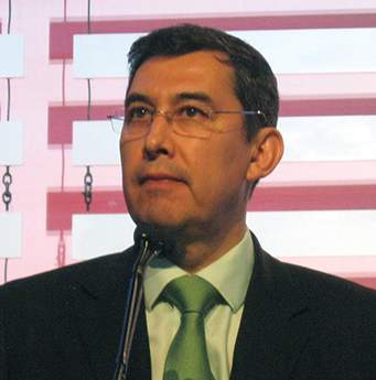
Anti-TNF agents may slow erosive hand osteoarthritis
LONDON – Tumor necrosis factor may play a role in erosive hand osteoarthritis, and treatments such as etanercept that target this cytokine may help prevent progression of the condition, according to the results of two studies presented at the European Congress of Rheumatology.
In one of the studies, immunoscintigraphic detection of radiolabeled certolizumab pegol was used to show that tumor necrosis factor (TNF) was present in swollen finger joints. In another study, researchers looked to see if treatment with etanercept would have any specific effects on the joints of patients with erosive hand osteoarthritis (OA) and performed a separate analysis of the potential effect on synovitis and the effect on bone marrow lesions.

“We previously had the idea that TNF is an important cytokine in the pathogenesis of erosive [hand] osteoarthritis; but there have been no animal studies, and it’s very difficult to take biopsies or fluid aspiration from these small finger joints,” Dr. Ruth Wittoek, a staff rheumatologist at Ghent University Hospital in Belgium and a coauthor of all three studies, explained in a precongress interview. “We needed to look for other possibilities to really identify the presence of TNF in those affected joints.”
Dr. Wittoek and her associates used immunoscintigraphy to take static images of both hands of five patients with erosive OA immediately (less than 15 minutes) after administration of radiolabeled certolizumab pegol (early phase) and 4-6 hours following the injection (late phase).
The patients studied had erosive OA for a median of 8.4 years, and their median age was 55.6 years. All patients underwent clinical examination for presence of tenderness and palpable swelling of the joints and ultrasound 1 day prior to undergoing immunoscintigraphy.
All 18 interphalangeal (IP) finger joints were scored according to the anatomical phase scoring system on x-ray, and 90 IP finger joints were studied in total. The uptake of radiolabeled certolizumab pegol was semiquantitatively described as being absent, weak, or strong.
During the early phase following administration, uptake of the radiolabeled TNF inhibitor was seen in seven (7.8%) joints, although the uptake was described as weak in all cases. The radiolabeled TNF inhibitor was seen in 24 (26.7%) joints during the late phase following administration, with five instances described as strong uptake and the remaining 19 instances being weak uptake. No uptake of the radiolabeled TNF inhibitor was seen in metacarpophalangeal joints.
Uptake of the radiolabeled TNF inhibitor was linked to signs of disease activity, including tender, swollen, and radiographically active joints.
Late uptake was present in 12 (36.4%) of 33 tender joints and in 12 (21.1%) of 57 nontender joints (odds ratio, 2.1; 95% confidence interval, 0.8-5.6; P was nonsignificant).
The relationship was most pronounced with palpable joint swelling: Late uptake was present in 14 (61%) of 23 swollen joints and 10 (14.9%) of 67 nonswollen joints (OR, 8.9; 95% CI, 3.3-26.0; P less than .001).
Late uptake was present in 18 (29%) of 62 sonographically active joints (defined as any presence of effusion or synovial proliferation) but just 6 (21.4%) of 28 noninflamed joints (OR, 1.5; 95% CI, 0.5-4.3; P was nonsignificant).
Uptake of the radiolabeled TNF inhibitor was observed in all anatomical phases of erosive hand OA, Dr. Wittoek noted, but the strongest association was found during the final remodeling (R) phase.
“Soft-tissue swelling strongly correlated with uptake of certolizumab, meaning in these joints a lot of TNF was present,” Dr. Wittoek said. “These data further solidify the rationale for cytokine-directed therapies in erosive OA.”
Although the data provide proof of concept that TNF may be involved in erosive hand OA, the lack of a control tracer was noted as a limitation of the study after the investigator’s presentation. Dr. Wittoek conceded that it would be interesting to examine that in a future study.
So, if TNF is present, what effect does anti-TNF therapy have on the joints in erosive hand OA?
That question was addressed in a multicenter, double-blind, randomized, placebo-controlled trial involving 90 patients who were randomized to receive either 50 mg of subcutaneous etanercept weekly for 24 weeks, then 25 mg weekly for the remainder of 1 year (45 patients), or placebo (45 patients). Participants were a mean age of 60 years, 81% were women, and 96% fulfilled the American College of Rheumatology hand OA criteria.
“Synovial inflammation is often present in erosive hand OA; moreover, synovitis is associated with pain and with structural damage after around 2 and a half years,” the lead study author, Dr. Margreet Kloppenburg, a professor of rheumatology at Leiden University Medical Centre in the Netherlands, said in an interview.
“Therefore, we wanted to know whether blocking of synovial inflammation by a well-known drug such as etanercept would also have a positive effect on outcomes in erosive hand OA,” Dr. Kloppenburg explained.
The primary outcome measure was the level of OA pain assessed on a visual analog scale (VAS) at 24 weeks.
Secondary endpoints included assessment of hand function, quality of life, the number of tender joints, and grip strength after 4, 8, 12, 24, and 36 weeks, and after 1 year. Radiographic progression of IP joints was scored blindly at baseline, 24 weeks, and 1 year following the quantitative Ghent University Scoring System (GUSS). VAS pain was compared between treatment groups at 24 weeks and 1 year in intention-to-treat analyses.
Although etanercept was not superior to placebo on VAS pain at 24 weeks, it was superior to placebo both on pain and structural damage assessed by GUSS in the symptomatic and inflammatory patients who completed the study. The drug was especially effective in joints with signs of inflammation.
Overall, VAS pain in all patients decreased by 24.8 mm (95% CI, –29.2 to –20.5; P less than .001) at 24 weeks. In intention-to-treat analysis, differences in pain between the groups were in favor of etanercept but did not reach statistical significance. The per-protocol analysis of GUSS showed a mean difference in favor of etanercept, indicating more remodeling in the etanercept group.
Additional analyses showed an interaction between soft swelling/erythema and etanercept treatment on GUSS, resulting in a statistically significant (P less than .05) mean difference between the two treatment groups. More patients dropped out on placebo than on etanercept (six vs. three) because of inefficacy, whereas more dropped out on etanercept than on placebo (six vs. one) because of adverse effects.
“Synovial inflammation is an interesting target for treatment in OA patients with an inflammatory hand osteoarthritis phenotype,” Dr. Kloppenburg said.
A separate analysis of the same multicenter study suggests that etanercept is effective in inhibiting bone marrow lesions (BMLs) in patients with erosive hand OA.
The researchers studied 20 participants with symptomatic erosive OA with clinical and ultrasonographic signs of inflammation in at least one IP joint. The patients underwent contrast-enhanced MRI of the eight distal and proximal IP joints of one hand at baseline and 1 year. Images were scored for synovitis and BMLs (0-3 per joint, total score 0-24), blinded for patient characteristics.
Radiographs of the same hand were scored according to the Verbruggen-Veys system. Logistic regression was used to associate the presence of an MRI feature in a joint with being in an erosive versus nonerosive anatomical phase.
“Although erosive hand OA is a condition with a high disease burden, no disease-modifying treatments are available yet,” explained Féline Kroon, a PhD student in the department of rheumatology at Leiden University Medical Center in the Netherlands, in an interview. “Research to find a new form of therapy is partly hindered by our limited knowledge on the pathophysiology of the disease.”
However, “new imaging modalities like MRI enable us to study the pathophysiology of erosive OA more closely,” she said. “This study also gave us the unique opportunity to investigate whether anti-TNF, which is known to lead to clinical improvement and improvement of inflammatory lesions on MRI in other rheumatic diseases like rheumatoid arthritis, might also be effective in erosive OA.”
The presence of BMLs, but not synovitis, was associated with the presence of an erosive anatomical phase in a joint, and treatment with etanercept appeared to be effective in inhibiting these lesions. That suggests a role for TNF in the pathophysiology of erosive OA.
The inhibitory effect of etanercept on BMLs was more pronounced in IP joints with severe synovitis at baseline, suggesting that inflamed synovial tissue could be a source of TNF production in erosive OA.
The total synovitis score was similar at baseline and 1 year in both study groups. For BMLs, the median total score at baseline was 5.4 (range 2-9) and 7.0 (0-9) at 1 year in the placebo group, versus 4.5 (3-9) and 3.7 (0-8), respectively, in the etanercept group.
The presence of BMLs was associated with being in the erosive and remodeling anatomical phases of erosive hand OA. Synovitis was not associated with those phases.
“We think that TNF-alpha plays a role in the pathophysiology of erosive OA via an effect on the subchondral bone,” Ms. Kroon said. “Because we saw that the beneficial effect of etanercept on BMLs was more pronounced in joints with synovitis at baseline, we think that, in an inflamed synovial hand joint, an interaction takes place between synovium and subchondral bone, which could be influenced by blocking TNF.”
Pfizer and UCB supported the investigator-initiated studies and provided the study drugs. Dr. Kloppenburg has received lecturing, consultancy, and investigator fees or grants from AbbVie, APPROACH, GlaxoSmithKline, Levicept, Pfizer, Servier, and UCB, all paid to her institution. All other authors declared no conflicts of interests.
LONDON – Tumor necrosis factor may play a role in erosive hand osteoarthritis, and treatments such as etanercept that target this cytokine may help prevent progression of the condition, according to the results of two studies presented at the European Congress of Rheumatology.
In one of the studies, immunoscintigraphic detection of radiolabeled certolizumab pegol was used to show that tumor necrosis factor (TNF) was present in swollen finger joints. In another study, researchers looked to see if treatment with etanercept would have any specific effects on the joints of patients with erosive hand osteoarthritis (OA) and performed a separate analysis of the potential effect on synovitis and the effect on bone marrow lesions.
“We previously had the idea that TNF is an important cytokine in the pathogenesis of erosive [hand] osteoarthritis; but there have been no animal studies, and it’s very difficult to take biopsies or fluid aspiration from these small finger joints,” Dr. Ruth Wittoek, a staff rheumatologist at Ghent University Hospital in Belgium and a coauthor of all three studies, explained in a precongress interview. “We needed to look for other possibilities to really identify the presence of TNF in those affected joints.”
Dr. Wittoek and her associates used immunoscintigraphy to take static images of both hands of five patients with erosive OA immediately (less than 15 minutes) after administration of radiolabeled certolizumab pegol (early phase) and 4-6 hours following the injection (late phase).
The patients studied had erosive OA for a median of 8.4 years, and their median age was 55.6 years. All patients underwent clinical examination for presence of tenderness and palpable swelling of the joints and ultrasound 1 day prior to undergoing immunoscintigraphy.
All 18 interphalangeal (IP) finger joints were scored according to the anatomical phase scoring system on x-ray, and 90 IP finger joints were studied in total. The uptake of radiolabeled certolizumab pegol was semiquantitatively described as being absent, weak, or strong.
During the early phase following administration, uptake of the radiolabeled TNF inhibitor was seen in seven (7.8%) joints, although the uptake was described as weak in all cases. The radiolabeled TNF inhibitor was seen in 24 (26.7%) joints during the late phase following administration, with five instances described as strong uptake and the remaining 19 instances being weak uptake. No uptake of the radiolabeled TNF inhibitor was seen in metacarpophalangeal joints.
Uptake of the radiolabeled TNF inhibitor was linked to signs of disease activity, including tender, swollen, and radiographically active joints.
Late uptake was present in 12 (36.4%) of 33 tender joints and in 12 (21.1%) of 57 nontender joints (odds ratio, 2.1; 95% confidence interval, 0.8-5.6; P was nonsignificant).
The relationship was most pronounced with palpable joint swelling: Late uptake was present in 14 (61%) of 23 swollen joints and 10 (14.9%) of 67 nonswollen joints (OR, 8.9; 95% CI, 3.3-26.0; P less than .001).
Late uptake was present in 18 (29%) of 62 sonographically active joints (defined as any presence of effusion or synovial proliferation) but just 6 (21.4%) of 28 noninflamed joints (OR, 1.5; 95% CI, 0.5-4.3; P was nonsignificant).
Uptake of the radiolabeled TNF inhibitor was observed in all anatomical phases of erosive hand OA, Dr. Wittoek noted, but the strongest association was found during the final remodeling (R) phase.
“Soft-tissue swelling strongly correlated with uptake of certolizumab, meaning in these joints a lot of TNF was present,” Dr. Wittoek said. “These data further solidify the rationale for cytokine-directed therapies in erosive OA.”
Although the data provide proof of concept that TNF may be involved in erosive hand OA, the lack of a control tracer was noted as a limitation of the study after the investigator’s presentation. Dr. Wittoek conceded that it would be interesting to examine that in a future study.
So, if TNF is present, what effect does anti-TNF therapy have on the joints in erosive hand OA?
That question was addressed in a multicenter, double-blind, randomized, placebo-controlled trial involving 90 patients who were randomized to receive either 50 mg of subcutaneous etanercept weekly for 24 weeks, then 25 mg weekly for the remainder of 1 year (45 patients), or placebo (45 patients). Participants were a mean age of 60 years, 81% were women, and 96% fulfilled the American College of Rheumatology hand OA criteria.
“Synovial inflammation is often present in erosive hand OA; moreover, synovitis is associated with pain and with structural damage after around 2 and a half years,” the lead study author, Dr. Margreet Kloppenburg, a professor of rheumatology at Leiden University Medical Centre in the Netherlands, said in an interview.
“Therefore, we wanted to know whether blocking of synovial inflammation by a well-known drug such as etanercept would also have a positive effect on outcomes in erosive hand OA,” Dr. Kloppenburg explained.
The primary outcome measure was the level of OA pain assessed on a visual analog scale (VAS) at 24 weeks.
Secondary endpoints included assessment of hand function, quality of life, the number of tender joints, and grip strength after 4, 8, 12, 24, and 36 weeks, and after 1 year. Radiographic progression of IP joints was scored blindly at baseline, 24 weeks, and 1 year following the quantitative Ghent University Scoring System (GUSS). VAS pain was compared between treatment groups at 24 weeks and 1 year in intention-to-treat analyses.
Although etanercept was not superior to placebo on VAS pain at 24 weeks, it was superior to placebo both on pain and structural damage assessed by GUSS in the symptomatic and inflammatory patients who completed the study. The drug was especially effective in joints with signs of inflammation.
Overall, VAS pain in all patients decreased by 24.8 mm (95% CI, –29.2 to –20.5; P less than .001) at 24 weeks. In intention-to-treat analysis, differences in pain between the groups were in favor of etanercept but did not reach statistical significance. The per-protocol analysis of GUSS showed a mean difference in favor of etanercept, indicating more remodeling in the etanercept group.
Additional analyses showed an interaction between soft swelling/erythema and etanercept treatment on GUSS, resulting in a statistically significant (P less than .05) mean difference between the two treatment groups. More patients dropped out on placebo than on etanercept (six vs. three) because of inefficacy, whereas more dropped out on etanercept than on placebo (six vs. one) because of adverse effects.
“Synovial inflammation is an interesting target for treatment in OA patients with an inflammatory hand osteoarthritis phenotype,” Dr. Kloppenburg said.
A separate analysis of the same multicenter study suggests that etanercept is effective in inhibiting bone marrow lesions (BMLs) in patients with erosive hand OA.
The researchers studied 20 participants with symptomatic erosive OA with clinical and ultrasonographic signs of inflammation in at least one IP joint. The patients underwent contrast-enhanced MRI of the eight distal and proximal IP joints of one hand at baseline and 1 year. Images were scored for synovitis and BMLs (0-3 per joint, total score 0-24), blinded for patient characteristics.
Radiographs of the same hand were scored according to the Verbruggen-Veys system. Logistic regression was used to associate the presence of an MRI feature in a joint with being in an erosive versus nonerosive anatomical phase.
“Although erosive hand OA is a condition with a high disease burden, no disease-modifying treatments are available yet,” explained Féline Kroon, a PhD student in the department of rheumatology at Leiden University Medical Center in the Netherlands, in an interview. “Research to find a new form of therapy is partly hindered by our limited knowledge on the pathophysiology of the disease.”
However, “new imaging modalities like MRI enable us to study the pathophysiology of erosive OA more closely,” she said. “This study also gave us the unique opportunity to investigate whether anti-TNF, which is known to lead to clinical improvement and improvement of inflammatory lesions on MRI in other rheumatic diseases like rheumatoid arthritis, might also be effective in erosive OA.”
The presence of BMLs, but not synovitis, was associated with the presence of an erosive anatomical phase in a joint, and treatment with etanercept appeared to be effective in inhibiting these lesions. That suggests a role for TNF in the pathophysiology of erosive OA.
The inhibitory effect of etanercept on BMLs was more pronounced in IP joints with severe synovitis at baseline, suggesting that inflamed synovial tissue could be a source of TNF production in erosive OA.
The total synovitis score was similar at baseline and 1 year in both study groups. For BMLs, the median total score at baseline was 5.4 (range 2-9) and 7.0 (0-9) at 1 year in the placebo group, versus 4.5 (3-9) and 3.7 (0-8), respectively, in the etanercept group.
The presence of BMLs was associated with being in the erosive and remodeling anatomical phases of erosive hand OA. Synovitis was not associated with those phases.
“We think that TNF-alpha plays a role in the pathophysiology of erosive OA via an effect on the subchondral bone,” Ms. Kroon said. “Because we saw that the beneficial effect of etanercept on BMLs was more pronounced in joints with synovitis at baseline, we think that, in an inflamed synovial hand joint, an interaction takes place between synovium and subchondral bone, which could be influenced by blocking TNF.”
Pfizer and UCB supported the investigator-initiated studies and provided the study drugs. Dr. Kloppenburg has received lecturing, consultancy, and investigator fees or grants from AbbVie, APPROACH, GlaxoSmithKline, Levicept, Pfizer, Servier, and UCB, all paid to her institution. All other authors declared no conflicts of interests.
AT THE EULAR 2016 CONGRESS
Key clinical point: Early data suggest targeting tumor necrosis factor could be of benefit in patients with erosive hand disease.
Major finding: TNF was found in the hand joints, and anti-TNF therapy reduced synovial inflammation and bone marrow lesions.
Data source: Two studies looking at the effects of anti-TNF treatments in patients with erosive hand osteoarthritis.
Disclosures: Pfizer and UCB supported the investigator-initiated studies and provided the study drugs. Dr. Kloppenburg has received lecturing, consultancy, and investigator fees or grants from AbbVie, APPROACH, GlaxoSmithKline, Levicept, Pfizer, Servier, and UCB, all paid to her institution. All other authors declared no conflicts of interests.

Lupus may confer higher risk for cervical cancer
LONDON – Women with systemic lupus erythematosus (SLE) have more than twice the risk of developing cervical neoplasia than do women in the general population, according to the results of a large Swedish registry study.
The study’s results indicate that the highest risk for cervical dysplasia or invasive cancer occurred among women with SLE who were using immunosuppressive agents, compared with those on antimalarial medication.
The results highlight the importance of women with SLE attending cervical cancer screening appointments, according to the researchers, who are from the Karolinska Institute in Stockholm, Linköping (Sweden) University, and Stanford (Calif.) University.
“At this time, we cannot comment on whether changes to screening programs are necessary, especially given there are considerable differences in cervical screening between countries,” lead study author Dr. Hjalmar Wadström said in an interview ahead of the study’s presentation at the European Congress of Rheumatology.
Dr. Wadström, who is an MD-PhD student at the Karolinska Institute working under the supervision of Dr. Johan Askling, explained that SLE is associated with various immunologic aberrations and is typically treated with immunomodulatory regimens. These regimens, however, have been linked to an increased risk of cervical neoplasia.
“Therefore, determining the risk among women with SLE is of direct clinical relevance,” he said. “Cervical cancer screening is important in the prevention of cervical cancer.”
To date, there have been few studies looking at the topic, and, as SLE is a relatively rare disease and the development of cancer is a relatively rare outcome, the study aimed to better estimate the risk.
Data from national Swedish patient and pharmacy registers were used to assemble a cohort of almost 5,000 women with SLE and a matched cohort of more than 28,000 women without SLE from the general Swedish population. The average age at the start of follow-up was 51 years, and about 40% of women were taking oral corticosteroids.
A total of 121 events occurred in the 4,550 women with SLE over the course of 23,136 total follow-up years. In comparison, there were 336 events in the 28,113 women without SLE from the general population over the course of 155,543 total years of follow-up.
This produced a hazard ratio for cervical neoplasia in women with SLE versus those without of 2.12 (95% confidence interval, 1.65-2.71). The analysis was adjusted for multiple confounding factors, including family history of cervical cancer and prior cervical screening in the 5 years before the start of follow-up.

Dr. Askling, who presented the findings during a press conference at the meeting, noted that just 5 of the 121 events seen in the overall SLE cohort were invasive cancers and the other events were premalignant changes, which included cervical intraepithelial neoplasia (CIN) stage 1, 2, and 3. “This is as it should be in the population; most events that you pick up in a screening program are premalignant,” he said.
Within the SLE cohort, two subcohorts of women also were identified: those taking hydroxychloroquine (n = 1,783) and those taking immunosuppressive drugs (n = 1,981). One of the reasons for looking at this is that treatment may serve as a proxy for the severity of disease, Dr. Wadström explained.
“SLE is a heterogeneous disease with numerous phenotypes that span from mild to life-threatening systemic disease,” he said. “Patients treated with an antimalarial, with or without oral steroids exclusively, tend to represent less severe cases, while more severe manifestations and organ involvement may necessitate potent cytotoxic immunosuppressive therapy.”
Adjusted HRs for cervical neoplasia in women with SLE were 1.52 (95% CI, 1.00-2.33) for those taking antimalarial therapy and 2.72 (95% CI, 2.01-3.67) for those taking immunosuppressive drugs, compared with women in the general population. The hazard ratio for cervical neoplasia comparing women with SLE taking immunosuppressive drugs with those taking antimalarial medication was 1.83 (95% CI, 1.15-2.91).
So what does this mean for clinical practice?
“We think it’s important that women with SLE, especially those with severe disease who are being treated with systemic immunosuppressants, attend cervical screening,” Dr. Wadström said.
Dr. Askling commented that, thankfully, women with SLE seemed to be just as adherent to attending cervical screening appointments as women in the general population. He noted it was important for clinicians to recognize that women with SLE do appear to have an increased risk for cervical changes and to ensure that they understand the need for continued adherence. “We don’t think at this stage that any additional measures should be taken,” he said.
But should all women with SLE be vaccinated against infection with the human papillomavirus, a known cause of cervical cancer? Dr. Asking said that since the women in the current study were in their 50s, it is not clear if such a move would be of benefit, certainly not when compared with vaccinating younger women. “Yes, we remember vaccination,” he said. “No, it doesn’t solve the problem for now, it solves the problem for the future.”
The researchers reported having no relevant financial disclosures.
LONDON – Women with systemic lupus erythematosus (SLE) have more than twice the risk of developing cervical neoplasia than do women in the general population, according to the results of a large Swedish registry study.
The study’s results indicate that the highest risk for cervical dysplasia or invasive cancer occurred among women with SLE who were using immunosuppressive agents, compared with those on antimalarial medication.
The results highlight the importance of women with SLE attending cervical cancer screening appointments, according to the researchers, who are from the Karolinska Institute in Stockholm, Linköping (Sweden) University, and Stanford (Calif.) University.
“At this time, we cannot comment on whether changes to screening programs are necessary, especially given there are considerable differences in cervical screening between countries,” lead study author Dr. Hjalmar Wadström said in an interview ahead of the study’s presentation at the European Congress of Rheumatology.
Dr. Wadström, who is an MD-PhD student at the Karolinska Institute working under the supervision of Dr. Johan Askling, explained that SLE is associated with various immunologic aberrations and is typically treated with immunomodulatory regimens. These regimens, however, have been linked to an increased risk of cervical neoplasia.
“Therefore, determining the risk among women with SLE is of direct clinical relevance,” he said. “Cervical cancer screening is important in the prevention of cervical cancer.”
To date, there have been few studies looking at the topic, and, as SLE is a relatively rare disease and the development of cancer is a relatively rare outcome, the study aimed to better estimate the risk.
Data from national Swedish patient and pharmacy registers were used to assemble a cohort of almost 5,000 women with SLE and a matched cohort of more than 28,000 women without SLE from the general Swedish population. The average age at the start of follow-up was 51 years, and about 40% of women were taking oral corticosteroids.
A total of 121 events occurred in the 4,550 women with SLE over the course of 23,136 total follow-up years. In comparison, there were 336 events in the 28,113 women without SLE from the general population over the course of 155,543 total years of follow-up.
This produced a hazard ratio for cervical neoplasia in women with SLE versus those without of 2.12 (95% confidence interval, 1.65-2.71). The analysis was adjusted for multiple confounding factors, including family history of cervical cancer and prior cervical screening in the 5 years before the start of follow-up.
Dr. Askling, who presented the findings during a press conference at the meeting, noted that just 5 of the 121 events seen in the overall SLE cohort were invasive cancers and the other events were premalignant changes, which included cervical intraepithelial neoplasia (CIN) stage 1, 2, and 3. “This is as it should be in the population; most events that you pick up in a screening program are premalignant,” he said.
Within the SLE cohort, two subcohorts of women also were identified: those taking hydroxychloroquine (n = 1,783) and those taking immunosuppressive drugs (n = 1,981). One of the reasons for looking at this is that treatment may serve as a proxy for the severity of disease, Dr. Wadström explained.
“SLE is a heterogeneous disease with numerous phenotypes that span from mild to life-threatening systemic disease,” he said. “Patients treated with an antimalarial, with or without oral steroids exclusively, tend to represent less severe cases, while more severe manifestations and organ involvement may necessitate potent cytotoxic immunosuppressive therapy.”
Adjusted HRs for cervical neoplasia in women with SLE were 1.52 (95% CI, 1.00-2.33) for those taking antimalarial therapy and 2.72 (95% CI, 2.01-3.67) for those taking immunosuppressive drugs, compared with women in the general population. The hazard ratio for cervical neoplasia comparing women with SLE taking immunosuppressive drugs with those taking antimalarial medication was 1.83 (95% CI, 1.15-2.91).
So what does this mean for clinical practice?
“We think it’s important that women with SLE, especially those with severe disease who are being treated with systemic immunosuppressants, attend cervical screening,” Dr. Wadström said.
Dr. Askling commented that, thankfully, women with SLE seemed to be just as adherent to attending cervical screening appointments as women in the general population. He noted it was important for clinicians to recognize that women with SLE do appear to have an increased risk for cervical changes and to ensure that they understand the need for continued adherence. “We don’t think at this stage that any additional measures should be taken,” he said.
But should all women with SLE be vaccinated against infection with the human papillomavirus, a known cause of cervical cancer? Dr. Asking said that since the women in the current study were in their 50s, it is not clear if such a move would be of benefit, certainly not when compared with vaccinating younger women. “Yes, we remember vaccination,” he said. “No, it doesn’t solve the problem for now, it solves the problem for the future.”
The researchers reported having no relevant financial disclosures.
AT THE EULAR 2016 CONGRESS
Key clinical point: Women with SLE should be encouraged to be screened for cervical cancer, particularly those treated with immunosuppressive drugs.
Major finding: The hazard ratio for cervical neoplasia in women with SLE versus those without was 2.12.
Data source: Swedish registry study of nearly 5,000 women with SLE.
Disclosures: The researchers reported having no relevant financial disclosures.

Low vasculitis risk with TNF inhibitors
GLASGOW – Treatment with tumor necrosis factor (TNF) inhibitors for rheumatoid arthritis is associated with a low risk of vasculitis-like events, according to a large analysis of data from the United Kingdom.
Investigators using data from the British Society for Rheumatology Biologics Register for Rheumatoid Arthritis (BSRBR-RA) found that the crude incidence rate was 16 cases per 10,000 person-years among TNF-inhibitor users versus seven cases per 10,000 person-years among users of nonbiologic disease-modifying antirheumatic drugs (nbDMARDs) such as methotrexate and sulfasalazine.
Although the risk was slightly higher among anti-TNF than nbDMARD users, the propensity score fully adjusted hazard ratio for a first vasculitis-like event was 1.27, comparing the anti-TNF drugs with nbDMARDs, with a 95% confidence interval of 0.40-4.04.
“This is the first prospective observational study to systematically look at the risk of vasculitis-like events” in patients with RA treated with anti-TNF agents, Dr. Meghna Jani said at the British Society for Rheumatology annual conference.
Dr. Jani of the Arthritis Research UK Centre for Epidemiology at the University of Manchester (England) explained that the reason for looking at this topic was that vasculitis-like events had been reported in case series and single-center studies, but these prior reports were too small to be able to estimate exactly how big a problem this was.
“Anti-TNF agents are associated with the development of a number of autoantibodies, including antinuclear antibodies and antidrug antibodies, and ANCA [antineutrophil cytoplasmic antibody],” she observed.
“We know that a small proportion of these patients may then go on to develop autoimmune diseases, some independent of autoantibodies,” she added. The most common of these is vasculitis, including cutaneous vasculitis.
Vasculitis is a somewhat paradoxical adverse event, she noted, in that it has been associated with anti-TNF therapy, but these drugs can also be used to treat it.
Now in its 15th year, the BSRBR-RA is the largest ongoing cohort of patients treated with biologic agents for rheumatic disease and provides one of the best sources of data to examine the risk for vasculitis-like events Dr. Jani observed. The aims were to look at the respective risks as well as to see if there were any particular predictive factors.
The current analysis included more than 16,000 patients enrolled in the BSRBR-RA between 2001 and 2015, of whom 12,745 were newly started on an anti-TNF drug and 3,640 were receiving nbDMARDs and were also biologic naive. The mean age of patients in the two groups was 56 and 60 years, 76% and 72% were female, with a mean Disease Activity Score (DAS28) of 6.5 and 5.1 and median disease duration of 11 and 6 years, respectively.
After more than 52,428 person-years of exposure and a median of 5.1 years of follow-up, 81 vasculitis-like events occurred in the anti-TNF therapy group. Vasculitis-like events were attributed to treatment only if they had occurred within 90 days of starting the drug. Follow-up stopped after a first event; if there was a switch to another biologic drug; and at death, the last clinical follow-up, or the end of the analysis period (May 31, 2015).
In comparison, there were 20,635 person-years of exposure and 6.5 years’ follow-up in the nbDMARD group, with 14 vasculitis-like events reported during this time.
A sensitivity analysis was performed excluding patients who had nail-fold vasculitis at baseline, had vasculitis due to a possible secondary cause such as infection, and were taking any other medications associated with vasculitis-like events. Results showed a similar risk for a first vasculitis event between anti-TNF and nbDMARD users (aHR = 1.05; 95% CI, 0.32-3.45).
Looking at the risk of vasculitis events for individual anti-TNF drugs, there initially appeared to be a higher risk for patients taking infliximab (n = 3,292) and etanercept (n = 4,450) but not for those taking adalimumab (n = 4,312) versus nbDMARDs, with crude incidence rates of 10, 17, and 11 per 10,000 person-years; after adjustment, these differences were not significant (aHRs of 1.55, 1.72, and 0.77, respectively, with 95% CIs crossing 1.0). A crude rate for certolizumab could not be calculated as there were no vasculitis events reported but there were only 691 patients enrolled in the BSRBR-RA at the time of the analysis who had been exposed to the drug.
“The risk of the event was highest in the first year of treatment, followed by reduction over time,” Dr. Jani reported. “Reassuringly, up to two-thirds of patients in both cohorts had manifestations that were just limited to cutaneous involvement,” she said.
The most common systemic presentation was digital ischemia, affecting 14% of patients treated with anti-TNFs and 14% of those given nbDMARDs. Neurologic involvement was also seen in both groups of patients (7% vs. 7%), but new nail-fold vasculitis (17% vs. 0%), respiratory involvement (4% vs. 0%), associated thrombotic events (5% vs. 0%), and renal involvement (2.5% vs. 0%) were seen in TNF inhibitor-treated patients only.
Ten anti-TNF–treated patients and one nbDMARD-treated patient needed treatment for the vasculitis-like event, and three patients in the anti-TNF cohort died as a result of the event, all of whom had multisystem organ involvement and one of whom had cytoplasmic ANCA-positive vasculitis.
Treatment with methotrexate or sulfasalazine at baseline was associated with a lower risk for vasculitis-like events, while seropositive status, disease duration, DAS28, and HAQ scores were associated with an increased risk for such events.
The BSRBR-RA receives restricted income financial support from Abbvie, Amgen, Swedish Orphan Biovitram (SOBI), Merck, Pfizer, Roche, and UCB Pharma. Dr. Jani disclosed she has received honoraria from Pfizer, Abbvie, and UCB Pharma.
AT RHEUMATOLOGY 2016
Key clinical point: There is a low risk of vasculitis-like events with tumor necrosis factor inhibitors.
Major finding: Crude incidence rates for vasculitis-like events were 16/10,000 person-years with TNF-inhibitor therapy and 7/10,000 person-years with nonbiologic disease-modifying antirheumatic drugs.
Data source: British Society for Rheumatology Biologics Register for Rheumatoid Arthritis of 12,745 TNF-inhibitor and 3,640 nbDMARD users.
Disclosures: The BSRBR-RA receives restricted income financial support from Abbvie, Amgen, Swedish Orphan Biovitram (SOBI), Merck, Pfizer, Roche, and UCB Pharma. Dr. Jani disclosed she has received honoraria from Pfizer, Abbvie, and UCB Pharma.
Vascular disease linked to sight loss in giant cell arteritis
GLASGOW – People with giant cell arteritis may be more likely to go blind if they have underlying vascular disease, according to an analysis of the Diagnostic and Classification Criteria in Vasculitis Study.
The results of the analysis showed that 7.9% of patients with this common type of vasculitis go blind in at least one eye within 6 months of a diagnosis, and that those with a history of peripheral vascular disease (PVD) could be up to 10 times more at risk than those without additional vascular comorbidity.
“This is the first multinational study for patients with [giant cell arteritis], and it shows that blindness is a significant problem,” said Dr. Max Yates of the University of East Anglia in Norwich, England, who presented the findings at the British Society for Rheumatology annual conference. Blindness was defined as complete visual loss rather than by a full ophthalmology assessment, so the findings probably underplay the problem in patients with some form of visual loss, he observed. Visual disturbance had been noted in 42.9% of the patients who were studied at the first clinic review.

“It is interesting that there is the association with vascular disease,” Dr. Yates said. “Perhaps we need greater vigilance in those people who already have a diagnosis of vascular disease [and] to really watch and monitor those people carefully for sight loss.”
The Diagnostic and Classification Criteria in Vasculitis Study (DCVAS) is an ongoing project designed to develop and then validate new classification and diagnostic criteria for systemic vasculitis that can be used routinely in clinical practice and in clinical trials. So far, more than 3,500 participants over age 18 have been recruited from secondary care clinics, and of those, 2,000 have a new or established diagnosis of vasculitis. The others have a similar presentation but an alternative diagnosis.
A total of 433 patients participating in the DCVAS were identified as having GCA with more than 75% diagnostic certainty, of which 93% fulfilled the 1990 American College of Rheumatology (ACR) criteria for GCA and just over half (54%) had a positive temporal artery biopsy. Visual loss was recorded by completion of the Vasculitis Damage Index (VDI) 6 months after diagnosis.
Two-thirds of patients studied were female, the median age at diagnosis was 73 years, 40% had jaw claudication, 34% had lost weight, and 16% presented with a fever. In addition, 9.2% had diabetes, 3.2% a prior stroke, and 2.5% had PVD.
Looking for predictive factors, baseline laboratory findings such as the presence of anemia, the erythrocyte sedimentation rate (ESR), C-reactive protein (CRP) levels, or platelet counts were not associated with sight loss. Dr. Yates noted in discussion that the baseline ESR range was 35-120 mm/h and the CRP ranged from 12 to over 100 mg/dL in the patients studied.
However, prior vascular disease was found to be predictive of later blindness. The odds ratio (OR) for being blind in at least one eye 6 months after a GCA diagnosis was 10.44 for PVD, with the 95% confidence interval (CI) ranging from 2.94 to 37.03. A prior diagnosis of cerebral vascular accident (OR, 4.47; 95% CI, 1.30-15.41), and diabetes (OR, 2.48; 95% CI, 0.98-6.25) also upped the risk for complete sight loss at 6 months.
Dr. Yates noted that patients were selected from multiple clinics across secondary care, so there should be better generalizability than in prior, single-center studies. However, there could be some residual referral bias.
During discussion, it was pointed out that it would be helpful to know the rate of blindness in patients taking steroids, as this was one of the major reasons for emergency rheumatology calls at one clinic, a delegate observed.
“Giant cell arteritis is really the major rheumatology emergency for practicing clinicians. We recently set up a rheumatology day service and get usually 8-10 calls about it per day,” the delegate said. “It’s often said that once a patient is on any dose of steroids, there is not risk of them going blind.” There is a lot of angst about whether it is safe to use higher doses (60 mg vs. 40 mg) and “it’s important for us as clinicians to be able to reassure people.”
Dr. Yates noted that a prospective trial would be needed to answer the question and that trials were planned. “We don’t have any data on treatment,” he said. “So we’re unable to say whether steroids were started instantly or whether there was any improvement in the visual function of these people.” Long-term complications also would be something to look at, particularly in older people who have an increased risk for eye problems such as cataracts, and could be at higher risk for visual problems if treated with steroids or other agents.
The DCVAS study is supported by the ACR and is funded by the European League Against Rheumatism and the Vasculitis Foundation. Dr. Yates reported that he had no relevant disclosures.
GLASGOW – People with giant cell arteritis may be more likely to go blind if they have underlying vascular disease, according to an analysis of the Diagnostic and Classification Criteria in Vasculitis Study.
The results of the analysis showed that 7.9% of patients with this common type of vasculitis go blind in at least one eye within 6 months of a diagnosis, and that those with a history of peripheral vascular disease (PVD) could be up to 10 times more at risk than those without additional vascular comorbidity.
“This is the first multinational study for patients with [giant cell arteritis], and it shows that blindness is a significant problem,” said Dr. Max Yates of the University of East Anglia in Norwich, England, who presented the findings at the British Society for Rheumatology annual conference. Blindness was defined as complete visual loss rather than by a full ophthalmology assessment, so the findings probably underplay the problem in patients with some form of visual loss, he observed. Visual disturbance had been noted in 42.9% of the patients who were studied at the first clinic review.
“It is interesting that there is the association with vascular disease,” Dr. Yates said. “Perhaps we need greater vigilance in those people who already have a diagnosis of vascular disease [and] to really watch and monitor those people carefully for sight loss.”
The Diagnostic and Classification Criteria in Vasculitis Study (DCVAS) is an ongoing project designed to develop and then validate new classification and diagnostic criteria for systemic vasculitis that can be used routinely in clinical practice and in clinical trials. So far, more than 3,500 participants over age 18 have been recruited from secondary care clinics, and of those, 2,000 have a new or established diagnosis of vasculitis. The others have a similar presentation but an alternative diagnosis.
A total of 433 patients participating in the DCVAS were identified as having GCA with more than 75% diagnostic certainty, of which 93% fulfilled the 1990 American College of Rheumatology (ACR) criteria for GCA and just over half (54%) had a positive temporal artery biopsy. Visual loss was recorded by completion of the Vasculitis Damage Index (VDI) 6 months after diagnosis.
Two-thirds of patients studied were female, the median age at diagnosis was 73 years, 40% had jaw claudication, 34% had lost weight, and 16% presented with a fever. In addition, 9.2% had diabetes, 3.2% a prior stroke, and 2.5% had PVD.
Looking for predictive factors, baseline laboratory findings such as the presence of anemia, the erythrocyte sedimentation rate (ESR), C-reactive protein (CRP) levels, or platelet counts were not associated with sight loss. Dr. Yates noted in discussion that the baseline ESR range was 35-120 mm/h and the CRP ranged from 12 to over 100 mg/dL in the patients studied.
However, prior vascular disease was found to be predictive of later blindness. The odds ratio (OR) for being blind in at least one eye 6 months after a GCA diagnosis was 10.44 for PVD, with the 95% confidence interval (CI) ranging from 2.94 to 37.03. A prior diagnosis of cerebral vascular accident (OR, 4.47; 95% CI, 1.30-15.41), and diabetes (OR, 2.48; 95% CI, 0.98-6.25) also upped the risk for complete sight loss at 6 months.
Dr. Yates noted that patients were selected from multiple clinics across secondary care, so there should be better generalizability than in prior, single-center studies. However, there could be some residual referral bias.
During discussion, it was pointed out that it would be helpful to know the rate of blindness in patients taking steroids, as this was one of the major reasons for emergency rheumatology calls at one clinic, a delegate observed.
“Giant cell arteritis is really the major rheumatology emergency for practicing clinicians. We recently set up a rheumatology day service and get usually 8-10 calls about it per day,” the delegate said. “It’s often said that once a patient is on any dose of steroids, there is not risk of them going blind.” There is a lot of angst about whether it is safe to use higher doses (60 mg vs. 40 mg) and “it’s important for us as clinicians to be able to reassure people.”
Dr. Yates noted that a prospective trial would be needed to answer the question and that trials were planned. “We don’t have any data on treatment,” he said. “So we’re unable to say whether steroids were started instantly or whether there was any improvement in the visual function of these people.” Long-term complications also would be something to look at, particularly in older people who have an increased risk for eye problems such as cataracts, and could be at higher risk for visual problems if treated with steroids or other agents.
The DCVAS study is supported by the ACR and is funded by the European League Against Rheumatism and the Vasculitis Foundation. Dr. Yates reported that he had no relevant disclosures.
AT RHEUMATOLOGY 2016
Key clinical point: Patients with vascular disease who develop giant cell arteritis may require careful monitoring for sight loss.
Major finding: Overall, 42.9% of patients had some visual disturbance at first clinic review; 7.9% were blind at 6 months.
Data source: Analysis of 433 patients newly diagnosed with GCA participating in the Diagnostic and Classification Criteria in Vasculitis Study (DCVAS).
Disclosures: The DCVAS study is supported by the American College of Rheumatology and is funded by the European League Against Rheumatism and the Vasculitis Foundation. Dr. Yates reported that he had no relevant disclosures.
