User login
CCJM delivers practical clinical articles relevant to internists, cardiologists, endocrinologists, and other specialists, all written by known experts.
Copyright © 2019 Cleveland Clinic. All rights reserved. The information provided is for educational purposes only. Use of this website is subject to the disclaimer and privacy policy.
gambling
compulsive behaviors
ammunition
assault rifle
black jack
Boko Haram
bondage
child abuse
cocaine
Daech
drug paraphernalia
explosion
gun
human trafficking
ISIL
ISIS
Islamic caliphate
Islamic state
mixed martial arts
MMA
molestation
national rifle association
NRA
nsfw
pedophile
pedophilia
poker
porn
pornography
psychedelic drug
recreational drug
sex slave rings
slot machine
terrorism
terrorist
Texas hold 'em
UFC
substance abuse
abuseed
abuseer
abusees
abuseing
abusely
abuses
aeolus
aeolused
aeoluser
aeoluses
aeolusing
aeolusly
aeoluss
ahole
aholeed
aholeer
aholees
aholeing
aholely
aholes
alcohol
alcoholed
alcoholer
alcoholes
alcoholing
alcoholly
alcohols
allman
allmaned
allmaner
allmanes
allmaning
allmanly
allmans
alted
altes
alting
altly
alts
analed
analer
anales
analing
anally
analprobe
analprobeed
analprobeer
analprobees
analprobeing
analprobely
analprobes
anals
anilingus
anilingused
anilinguser
anilinguses
anilingusing
anilingusly
anilinguss
anus
anused
anuser
anuses
anusing
anusly
anuss
areola
areolaed
areolaer
areolaes
areolaing
areolaly
areolas
areole
areoleed
areoleer
areolees
areoleing
areolely
areoles
arian
arianed
arianer
arianes
arianing
arianly
arians
aryan
aryaned
aryaner
aryanes
aryaning
aryanly
aryans
asiaed
asiaer
asiaes
asiaing
asialy
asias
ass
ass hole
ass lick
ass licked
ass licker
ass lickes
ass licking
ass lickly
ass licks
assbang
assbanged
assbangeded
assbangeder
assbangedes
assbangeding
assbangedly
assbangeds
assbanger
assbanges
assbanging
assbangly
assbangs
assbangsed
assbangser
assbangses
assbangsing
assbangsly
assbangss
assed
asser
asses
assesed
asseser
asseses
assesing
assesly
assess
assfuck
assfucked
assfucker
assfuckered
assfuckerer
assfuckeres
assfuckering
assfuckerly
assfuckers
assfuckes
assfucking
assfuckly
assfucks
asshat
asshated
asshater
asshates
asshating
asshatly
asshats
assholeed
assholeer
assholees
assholeing
assholely
assholes
assholesed
assholeser
assholeses
assholesing
assholesly
assholess
assing
assly
assmaster
assmastered
assmasterer
assmasteres
assmastering
assmasterly
assmasters
assmunch
assmunched
assmuncher
assmunches
assmunching
assmunchly
assmunchs
asss
asswipe
asswipeed
asswipeer
asswipees
asswipeing
asswipely
asswipes
asswipesed
asswipeser
asswipeses
asswipesing
asswipesly
asswipess
azz
azzed
azzer
azzes
azzing
azzly
azzs
babeed
babeer
babees
babeing
babely
babes
babesed
babeser
babeses
babesing
babesly
babess
ballsac
ballsaced
ballsacer
ballsaces
ballsacing
ballsack
ballsacked
ballsacker
ballsackes
ballsacking
ballsackly
ballsacks
ballsacly
ballsacs
ballsed
ballser
ballses
ballsing
ballsly
ballss
barf
barfed
barfer
barfes
barfing
barfly
barfs
bastard
bastarded
bastarder
bastardes
bastarding
bastardly
bastards
bastardsed
bastardser
bastardses
bastardsing
bastardsly
bastardss
bawdy
bawdyed
bawdyer
bawdyes
bawdying
bawdyly
bawdys
beaner
beanered
beanerer
beaneres
beanering
beanerly
beaners
beardedclam
beardedclamed
beardedclamer
beardedclames
beardedclaming
beardedclamly
beardedclams
beastiality
beastialityed
beastialityer
beastialityes
beastialitying
beastialityly
beastialitys
beatch
beatched
beatcher
beatches
beatching
beatchly
beatchs
beater
beatered
beaterer
beateres
beatering
beaterly
beaters
beered
beerer
beeres
beering
beerly
beeyotch
beeyotched
beeyotcher
beeyotches
beeyotching
beeyotchly
beeyotchs
beotch
beotched
beotcher
beotches
beotching
beotchly
beotchs
biatch
biatched
biatcher
biatches
biatching
biatchly
biatchs
big tits
big titsed
big titser
big titses
big titsing
big titsly
big titss
bigtits
bigtitsed
bigtitser
bigtitses
bigtitsing
bigtitsly
bigtitss
bimbo
bimboed
bimboer
bimboes
bimboing
bimboly
bimbos
bisexualed
bisexualer
bisexuales
bisexualing
bisexually
bisexuals
bitch
bitched
bitcheded
bitcheder
bitchedes
bitcheding
bitchedly
bitcheds
bitcher
bitches
bitchesed
bitcheser
bitcheses
bitchesing
bitchesly
bitchess
bitching
bitchly
bitchs
bitchy
bitchyed
bitchyer
bitchyes
bitchying
bitchyly
bitchys
bleached
bleacher
bleaches
bleaching
bleachly
bleachs
blow job
blow jobed
blow jober
blow jobes
blow jobing
blow jobly
blow jobs
blowed
blower
blowes
blowing
blowjob
blowjobed
blowjober
blowjobes
blowjobing
blowjobly
blowjobs
blowjobsed
blowjobser
blowjobses
blowjobsing
blowjobsly
blowjobss
blowly
blows
boink
boinked
boinker
boinkes
boinking
boinkly
boinks
bollock
bollocked
bollocker
bollockes
bollocking
bollockly
bollocks
bollocksed
bollockser
bollockses
bollocksing
bollocksly
bollockss
bollok
bolloked
bolloker
bollokes
bolloking
bollokly
bolloks
boner
bonered
bonerer
boneres
bonering
bonerly
boners
bonersed
bonerser
bonerses
bonersing
bonersly
bonerss
bong
bonged
bonger
bonges
bonging
bongly
bongs
boob
boobed
boober
boobes
boobies
boobiesed
boobieser
boobieses
boobiesing
boobiesly
boobiess
boobing
boobly
boobs
boobsed
boobser
boobses
boobsing
boobsly
boobss
booby
boobyed
boobyer
boobyes
boobying
boobyly
boobys
booger
boogered
boogerer
boogeres
boogering
boogerly
boogers
bookie
bookieed
bookieer
bookiees
bookieing
bookiely
bookies
bootee
booteeed
booteeer
booteees
booteeing
booteely
bootees
bootie
bootieed
bootieer
bootiees
bootieing
bootiely
booties
booty
bootyed
bootyer
bootyes
bootying
bootyly
bootys
boozeed
boozeer
boozees
boozeing
boozely
boozer
boozered
boozerer
boozeres
boozering
boozerly
boozers
boozes
boozy
boozyed
boozyer
boozyes
boozying
boozyly
boozys
bosomed
bosomer
bosomes
bosoming
bosomly
bosoms
bosomy
bosomyed
bosomyer
bosomyes
bosomying
bosomyly
bosomys
bugger
buggered
buggerer
buggeres
buggering
buggerly
buggers
bukkake
bukkakeed
bukkakeer
bukkakees
bukkakeing
bukkakely
bukkakes
bull shit
bull shited
bull shiter
bull shites
bull shiting
bull shitly
bull shits
bullshit
bullshited
bullshiter
bullshites
bullshiting
bullshitly
bullshits
bullshitsed
bullshitser
bullshitses
bullshitsing
bullshitsly
bullshitss
bullshitted
bullshitteded
bullshitteder
bullshittedes
bullshitteding
bullshittedly
bullshitteds
bullturds
bullturdsed
bullturdser
bullturdses
bullturdsing
bullturdsly
bullturdss
bung
bunged
bunger
bunges
bunging
bungly
bungs
busty
bustyed
bustyer
bustyes
bustying
bustyly
bustys
butt
butt fuck
butt fucked
butt fucker
butt fuckes
butt fucking
butt fuckly
butt fucks
butted
buttes
buttfuck
buttfucked
buttfucker
buttfuckered
buttfuckerer
buttfuckeres
buttfuckering
buttfuckerly
buttfuckers
buttfuckes
buttfucking
buttfuckly
buttfucks
butting
buttly
buttplug
buttpluged
buttpluger
buttpluges
buttpluging
buttplugly
buttplugs
butts
caca
cacaed
cacaer
cacaes
cacaing
cacaly
cacas
cahone
cahoneed
cahoneer
cahonees
cahoneing
cahonely
cahones
cameltoe
cameltoeed
cameltoeer
cameltoees
cameltoeing
cameltoely
cameltoes
carpetmuncher
carpetmunchered
carpetmuncherer
carpetmuncheres
carpetmunchering
carpetmuncherly
carpetmunchers
cawk
cawked
cawker
cawkes
cawking
cawkly
cawks
chinc
chinced
chincer
chinces
chincing
chincly
chincs
chincsed
chincser
chincses
chincsing
chincsly
chincss
chink
chinked
chinker
chinkes
chinking
chinkly
chinks
chode
chodeed
chodeer
chodees
chodeing
chodely
chodes
chodesed
chodeser
chodeses
chodesing
chodesly
chodess
clit
clited
cliter
clites
cliting
clitly
clitoris
clitorised
clitoriser
clitorises
clitorising
clitorisly
clitoriss
clitorus
clitorused
clitoruser
clitoruses
clitorusing
clitorusly
clitoruss
clits
clitsed
clitser
clitses
clitsing
clitsly
clitss
clitty
clittyed
clittyer
clittyes
clittying
clittyly
clittys
cocain
cocaine
cocained
cocaineed
cocaineer
cocainees
cocaineing
cocainely
cocainer
cocaines
cocaining
cocainly
cocains
cock
cock sucker
cock suckered
cock suckerer
cock suckeres
cock suckering
cock suckerly
cock suckers
cockblock
cockblocked
cockblocker
cockblockes
cockblocking
cockblockly
cockblocks
cocked
cocker
cockes
cockholster
cockholstered
cockholsterer
cockholsteres
cockholstering
cockholsterly
cockholsters
cocking
cockknocker
cockknockered
cockknockerer
cockknockeres
cockknockering
cockknockerly
cockknockers
cockly
cocks
cocksed
cockser
cockses
cocksing
cocksly
cocksmoker
cocksmokered
cocksmokerer
cocksmokeres
cocksmokering
cocksmokerly
cocksmokers
cockss
cocksucker
cocksuckered
cocksuckerer
cocksuckeres
cocksuckering
cocksuckerly
cocksuckers
coital
coitaled
coitaler
coitales
coitaling
coitally
coitals
commie
commieed
commieer
commiees
commieing
commiely
commies
condomed
condomer
condomes
condoming
condomly
condoms
coon
cooned
cooner
coones
cooning
coonly
coons
coonsed
coonser
coonses
coonsing
coonsly
coonss
corksucker
corksuckered
corksuckerer
corksuckeres
corksuckering
corksuckerly
corksuckers
cracked
crackwhore
crackwhoreed
crackwhoreer
crackwhorees
crackwhoreing
crackwhorely
crackwhores
crap
craped
craper
crapes
craping
craply
crappy
crappyed
crappyer
crappyes
crappying
crappyly
crappys
cum
cumed
cumer
cumes
cuming
cumly
cummin
cummined
cumminer
cummines
cumming
cumminged
cumminger
cumminges
cumminging
cummingly
cummings
cummining
cumminly
cummins
cums
cumshot
cumshoted
cumshoter
cumshotes
cumshoting
cumshotly
cumshots
cumshotsed
cumshotser
cumshotses
cumshotsing
cumshotsly
cumshotss
cumslut
cumsluted
cumsluter
cumslutes
cumsluting
cumslutly
cumsluts
cumstain
cumstained
cumstainer
cumstaines
cumstaining
cumstainly
cumstains
cunilingus
cunilingused
cunilinguser
cunilinguses
cunilingusing
cunilingusly
cunilinguss
cunnilingus
cunnilingused
cunnilinguser
cunnilinguses
cunnilingusing
cunnilingusly
cunnilinguss
cunny
cunnyed
cunnyer
cunnyes
cunnying
cunnyly
cunnys
cunt
cunted
cunter
cuntes
cuntface
cuntfaceed
cuntfaceer
cuntfacees
cuntfaceing
cuntfacely
cuntfaces
cunthunter
cunthuntered
cunthunterer
cunthunteres
cunthuntering
cunthunterly
cunthunters
cunting
cuntlick
cuntlicked
cuntlicker
cuntlickered
cuntlickerer
cuntlickeres
cuntlickering
cuntlickerly
cuntlickers
cuntlickes
cuntlicking
cuntlickly
cuntlicks
cuntly
cunts
cuntsed
cuntser
cuntses
cuntsing
cuntsly
cuntss
dago
dagoed
dagoer
dagoes
dagoing
dagoly
dagos
dagosed
dagoser
dagoses
dagosing
dagosly
dagoss
dammit
dammited
dammiter
dammites
dammiting
dammitly
dammits
damn
damned
damneded
damneder
damnedes
damneding
damnedly
damneds
damner
damnes
damning
damnit
damnited
damniter
damnites
damniting
damnitly
damnits
damnly
damns
dick
dickbag
dickbaged
dickbager
dickbages
dickbaging
dickbagly
dickbags
dickdipper
dickdippered
dickdipperer
dickdipperes
dickdippering
dickdipperly
dickdippers
dicked
dicker
dickes
dickface
dickfaceed
dickfaceer
dickfacees
dickfaceing
dickfacely
dickfaces
dickflipper
dickflippered
dickflipperer
dickflipperes
dickflippering
dickflipperly
dickflippers
dickhead
dickheaded
dickheader
dickheades
dickheading
dickheadly
dickheads
dickheadsed
dickheadser
dickheadses
dickheadsing
dickheadsly
dickheadss
dicking
dickish
dickished
dickisher
dickishes
dickishing
dickishly
dickishs
dickly
dickripper
dickrippered
dickripperer
dickripperes
dickrippering
dickripperly
dickrippers
dicks
dicksipper
dicksippered
dicksipperer
dicksipperes
dicksippering
dicksipperly
dicksippers
dickweed
dickweeded
dickweeder
dickweedes
dickweeding
dickweedly
dickweeds
dickwhipper
dickwhippered
dickwhipperer
dickwhipperes
dickwhippering
dickwhipperly
dickwhippers
dickzipper
dickzippered
dickzipperer
dickzipperes
dickzippering
dickzipperly
dickzippers
diddle
diddleed
diddleer
diddlees
diddleing
diddlely
diddles
dike
dikeed
dikeer
dikees
dikeing
dikely
dikes
dildo
dildoed
dildoer
dildoes
dildoing
dildoly
dildos
dildosed
dildoser
dildoses
dildosing
dildosly
dildoss
diligaf
diligafed
diligafer
diligafes
diligafing
diligafly
diligafs
dillweed
dillweeded
dillweeder
dillweedes
dillweeding
dillweedly
dillweeds
dimwit
dimwited
dimwiter
dimwites
dimwiting
dimwitly
dimwits
dingle
dingleed
dingleer
dinglees
dingleing
dinglely
dingles
dipship
dipshiped
dipshiper
dipshipes
dipshiping
dipshiply
dipships
dizzyed
dizzyer
dizzyes
dizzying
dizzyly
dizzys
doggiestyleed
doggiestyleer
doggiestylees
doggiestyleing
doggiestylely
doggiestyles
doggystyleed
doggystyleer
doggystylees
doggystyleing
doggystylely
doggystyles
dong
donged
donger
donges
donging
dongly
dongs
doofus
doofused
doofuser
doofuses
doofusing
doofusly
doofuss
doosh
dooshed
doosher
dooshes
dooshing
dooshly
dooshs
dopeyed
dopeyer
dopeyes
dopeying
dopeyly
dopeys
douchebag
douchebaged
douchebager
douchebages
douchebaging
douchebagly
douchebags
douchebagsed
douchebagser
douchebagses
douchebagsing
douchebagsly
douchebagss
doucheed
doucheer
douchees
doucheing
douchely
douches
douchey
doucheyed
doucheyer
doucheyes
doucheying
doucheyly
doucheys
drunk
drunked
drunker
drunkes
drunking
drunkly
drunks
dumass
dumassed
dumasser
dumasses
dumassing
dumassly
dumasss
dumbass
dumbassed
dumbasser
dumbasses
dumbassesed
dumbasseser
dumbasseses
dumbassesing
dumbassesly
dumbassess
dumbassing
dumbassly
dumbasss
dummy
dummyed
dummyer
dummyes
dummying
dummyly
dummys
dyke
dykeed
dykeer
dykees
dykeing
dykely
dykes
dykesed
dykeser
dykeses
dykesing
dykesly
dykess
erotic
eroticed
eroticer
erotices
eroticing
eroticly
erotics
extacy
extacyed
extacyer
extacyes
extacying
extacyly
extacys
extasy
extasyed
extasyer
extasyes
extasying
extasyly
extasys
fack
facked
facker
fackes
facking
fackly
facks
fag
faged
fager
fages
fagg
fagged
faggeded
faggeder
faggedes
faggeding
faggedly
faggeds
fagger
fagges
fagging
faggit
faggited
faggiter
faggites
faggiting
faggitly
faggits
faggly
faggot
faggoted
faggoter
faggotes
faggoting
faggotly
faggots
faggs
faging
fagly
fagot
fagoted
fagoter
fagotes
fagoting
fagotly
fagots
fags
fagsed
fagser
fagses
fagsing
fagsly
fagss
faig
faiged
faiger
faiges
faiging
faigly
faigs
faigt
faigted
faigter
faigtes
faigting
faigtly
faigts
fannybandit
fannybandited
fannybanditer
fannybandites
fannybanditing
fannybanditly
fannybandits
farted
farter
fartes
farting
fartknocker
fartknockered
fartknockerer
fartknockeres
fartknockering
fartknockerly
fartknockers
fartly
farts
felch
felched
felcher
felchered
felcherer
felcheres
felchering
felcherly
felchers
felches
felching
felchinged
felchinger
felchinges
felchinging
felchingly
felchings
felchly
felchs
fellate
fellateed
fellateer
fellatees
fellateing
fellately
fellates
fellatio
fellatioed
fellatioer
fellatioes
fellatioing
fellatioly
fellatios
feltch
feltched
feltcher
feltchered
feltcherer
feltcheres
feltchering
feltcherly
feltchers
feltches
feltching
feltchly
feltchs
feom
feomed
feomer
feomes
feoming
feomly
feoms
fisted
fisteded
fisteder
fistedes
fisteding
fistedly
fisteds
fisting
fistinged
fistinger
fistinges
fistinging
fistingly
fistings
fisty
fistyed
fistyer
fistyes
fistying
fistyly
fistys
floozy
floozyed
floozyer
floozyes
floozying
floozyly
floozys
foad
foaded
foader
foades
foading
foadly
foads
fondleed
fondleer
fondlees
fondleing
fondlely
fondles
foobar
foobared
foobarer
foobares
foobaring
foobarly
foobars
freex
freexed
freexer
freexes
freexing
freexly
freexs
frigg
frigga
friggaed
friggaer
friggaes
friggaing
friggaly
friggas
frigged
frigger
frigges
frigging
friggly
friggs
fubar
fubared
fubarer
fubares
fubaring
fubarly
fubars
fuck
fuckass
fuckassed
fuckasser
fuckasses
fuckassing
fuckassly
fuckasss
fucked
fuckeded
fuckeder
fuckedes
fuckeding
fuckedly
fuckeds
fucker
fuckered
fuckerer
fuckeres
fuckering
fuckerly
fuckers
fuckes
fuckface
fuckfaceed
fuckfaceer
fuckfacees
fuckfaceing
fuckfacely
fuckfaces
fuckin
fuckined
fuckiner
fuckines
fucking
fuckinged
fuckinger
fuckinges
fuckinging
fuckingly
fuckings
fuckining
fuckinly
fuckins
fuckly
fucknugget
fucknuggeted
fucknuggeter
fucknuggetes
fucknuggeting
fucknuggetly
fucknuggets
fucknut
fucknuted
fucknuter
fucknutes
fucknuting
fucknutly
fucknuts
fuckoff
fuckoffed
fuckoffer
fuckoffes
fuckoffing
fuckoffly
fuckoffs
fucks
fucksed
fuckser
fuckses
fucksing
fucksly
fuckss
fucktard
fucktarded
fucktarder
fucktardes
fucktarding
fucktardly
fucktards
fuckup
fuckuped
fuckuper
fuckupes
fuckuping
fuckuply
fuckups
fuckwad
fuckwaded
fuckwader
fuckwades
fuckwading
fuckwadly
fuckwads
fuckwit
fuckwited
fuckwiter
fuckwites
fuckwiting
fuckwitly
fuckwits
fudgepacker
fudgepackered
fudgepackerer
fudgepackeres
fudgepackering
fudgepackerly
fudgepackers
fuk
fuked
fuker
fukes
fuking
fukly
fuks
fvck
fvcked
fvcker
fvckes
fvcking
fvckly
fvcks
fxck
fxcked
fxcker
fxckes
fxcking
fxckly
fxcks
gae
gaeed
gaeer
gaees
gaeing
gaely
gaes
gai
gaied
gaier
gaies
gaiing
gaily
gais
ganja
ganjaed
ganjaer
ganjaes
ganjaing
ganjaly
ganjas
gayed
gayer
gayes
gaying
gayly
gays
gaysed
gayser
gayses
gaysing
gaysly
gayss
gey
geyed
geyer
geyes
geying
geyly
geys
gfc
gfced
gfcer
gfces
gfcing
gfcly
gfcs
gfy
gfyed
gfyer
gfyes
gfying
gfyly
gfys
ghay
ghayed
ghayer
ghayes
ghaying
ghayly
ghays
ghey
gheyed
gheyer
gheyes
gheying
gheyly
gheys
gigolo
gigoloed
gigoloer
gigoloes
gigoloing
gigololy
gigolos
goatse
goatseed
goatseer
goatsees
goatseing
goatsely
goatses
godamn
godamned
godamner
godamnes
godamning
godamnit
godamnited
godamniter
godamnites
godamniting
godamnitly
godamnits
godamnly
godamns
goddam
goddamed
goddamer
goddames
goddaming
goddamly
goddammit
goddammited
goddammiter
goddammites
goddammiting
goddammitly
goddammits
goddamn
goddamned
goddamner
goddamnes
goddamning
goddamnly
goddamns
goddams
goldenshower
goldenshowered
goldenshowerer
goldenshoweres
goldenshowering
goldenshowerly
goldenshowers
gonad
gonaded
gonader
gonades
gonading
gonadly
gonads
gonadsed
gonadser
gonadses
gonadsing
gonadsly
gonadss
gook
gooked
gooker
gookes
gooking
gookly
gooks
gooksed
gookser
gookses
gooksing
gooksly
gookss
gringo
gringoed
gringoer
gringoes
gringoing
gringoly
gringos
gspot
gspoted
gspoter
gspotes
gspoting
gspotly
gspots
gtfo
gtfoed
gtfoer
gtfoes
gtfoing
gtfoly
gtfos
guido
guidoed
guidoer
guidoes
guidoing
guidoly
guidos
handjob
handjobed
handjober
handjobes
handjobing
handjobly
handjobs
hard on
hard oned
hard oner
hard ones
hard oning
hard only
hard ons
hardknight
hardknighted
hardknighter
hardknightes
hardknighting
hardknightly
hardknights
hebe
hebeed
hebeer
hebees
hebeing
hebely
hebes
heeb
heebed
heeber
heebes
heebing
heebly
heebs
hell
helled
heller
helles
helling
hellly
hells
hemp
hemped
hemper
hempes
hemping
hemply
hemps
heroined
heroiner
heroines
heroining
heroinly
heroins
herp
herped
herper
herpes
herpesed
herpeser
herpeses
herpesing
herpesly
herpess
herping
herply
herps
herpy
herpyed
herpyer
herpyes
herpying
herpyly
herpys
hitler
hitlered
hitlerer
hitleres
hitlering
hitlerly
hitlers
hived
hiver
hives
hiving
hivly
hivs
hobag
hobaged
hobager
hobages
hobaging
hobagly
hobags
homey
homeyed
homeyer
homeyes
homeying
homeyly
homeys
homo
homoed
homoer
homoes
homoey
homoeyed
homoeyer
homoeyes
homoeying
homoeyly
homoeys
homoing
homoly
homos
honky
honkyed
honkyer
honkyes
honkying
honkyly
honkys
hooch
hooched
hoocher
hooches
hooching
hoochly
hoochs
hookah
hookahed
hookaher
hookahes
hookahing
hookahly
hookahs
hooker
hookered
hookerer
hookeres
hookering
hookerly
hookers
hoor
hoored
hoorer
hoores
hooring
hoorly
hoors
hootch
hootched
hootcher
hootches
hootching
hootchly
hootchs
hooter
hootered
hooterer
hooteres
hootering
hooterly
hooters
hootersed
hooterser
hooterses
hootersing
hootersly
hooterss
horny
hornyed
hornyer
hornyes
hornying
hornyly
hornys
houstoned
houstoner
houstones
houstoning
houstonly
houstons
hump
humped
humpeded
humpeder
humpedes
humpeding
humpedly
humpeds
humper
humpes
humping
humpinged
humpinger
humpinges
humpinging
humpingly
humpings
humply
humps
husbanded
husbander
husbandes
husbanding
husbandly
husbands
hussy
hussyed
hussyer
hussyes
hussying
hussyly
hussys
hymened
hymener
hymenes
hymening
hymenly
hymens
inbred
inbreded
inbreder
inbredes
inbreding
inbredly
inbreds
incest
incested
incester
incestes
incesting
incestly
incests
injun
injuned
injuner
injunes
injuning
injunly
injuns
jackass
jackassed
jackasser
jackasses
jackassing
jackassly
jackasss
jackhole
jackholeed
jackholeer
jackholees
jackholeing
jackholely
jackholes
jackoff
jackoffed
jackoffer
jackoffes
jackoffing
jackoffly
jackoffs
jap
japed
japer
japes
japing
japly
japs
japsed
japser
japses
japsing
japsly
japss
jerkoff
jerkoffed
jerkoffer
jerkoffes
jerkoffing
jerkoffly
jerkoffs
jerks
jism
jismed
jismer
jismes
jisming
jismly
jisms
jiz
jized
jizer
jizes
jizing
jizly
jizm
jizmed
jizmer
jizmes
jizming
jizmly
jizms
jizs
jizz
jizzed
jizzeded
jizzeder
jizzedes
jizzeding
jizzedly
jizzeds
jizzer
jizzes
jizzing
jizzly
jizzs
junkie
junkieed
junkieer
junkiees
junkieing
junkiely
junkies
junky
junkyed
junkyer
junkyes
junkying
junkyly
junkys
kike
kikeed
kikeer
kikees
kikeing
kikely
kikes
kikesed
kikeser
kikeses
kikesing
kikesly
kikess
killed
killer
killes
killing
killly
kills
kinky
kinkyed
kinkyer
kinkyes
kinkying
kinkyly
kinkys
kkk
kkked
kkker
kkkes
kkking
kkkly
kkks
klan
klaned
klaner
klanes
klaning
klanly
klans
knobend
knobended
knobender
knobendes
knobending
knobendly
knobends
kooch
kooched
koocher
kooches
koochesed
koocheser
koocheses
koochesing
koochesly
koochess
kooching
koochly
koochs
kootch
kootched
kootcher
kootches
kootching
kootchly
kootchs
kraut
krauted
krauter
krautes
krauting
krautly
krauts
kyke
kykeed
kykeer
kykees
kykeing
kykely
kykes
lech
leched
lecher
leches
leching
lechly
lechs
leper
lepered
leperer
leperes
lepering
leperly
lepers
lesbiansed
lesbianser
lesbianses
lesbiansing
lesbiansly
lesbianss
lesbo
lesboed
lesboer
lesboes
lesboing
lesboly
lesbos
lesbosed
lesboser
lesboses
lesbosing
lesbosly
lesboss
lez
lezbianed
lezbianer
lezbianes
lezbianing
lezbianly
lezbians
lezbiansed
lezbianser
lezbianses
lezbiansing
lezbiansly
lezbianss
lezbo
lezboed
lezboer
lezboes
lezboing
lezboly
lezbos
lezbosed
lezboser
lezboses
lezbosing
lezbosly
lezboss
lezed
lezer
lezes
lezing
lezly
lezs
lezzie
lezzieed
lezzieer
lezziees
lezzieing
lezziely
lezzies
lezziesed
lezzieser
lezzieses
lezziesing
lezziesly
lezziess
lezzy
lezzyed
lezzyer
lezzyes
lezzying
lezzyly
lezzys
lmaoed
lmaoer
lmaoes
lmaoing
lmaoly
lmaos
lmfao
lmfaoed
lmfaoer
lmfaoes
lmfaoing
lmfaoly
lmfaos
loined
loiner
loines
loining
loinly
loins
loinsed
loinser
loinses
loinsing
loinsly
loinss
lubeed
lubeer
lubees
lubeing
lubely
lubes
lusty
lustyed
lustyer
lustyes
lustying
lustyly
lustys
massa
massaed
massaer
massaes
massaing
massaly
massas
masterbate
masterbateed
masterbateer
masterbatees
masterbateing
masterbately
masterbates
masterbating
masterbatinged
masterbatinger
masterbatinges
masterbatinging
masterbatingly
masterbatings
masterbation
masterbationed
masterbationer
masterbationes
masterbationing
masterbationly
masterbations
masturbate
masturbateed
masturbateer
masturbatees
masturbateing
masturbately
masturbates
masturbating
masturbatinged
masturbatinger
masturbatinges
masturbatinging
masturbatingly
masturbatings
masturbation
masturbationed
masturbationer
masturbationes
masturbationing
masturbationly
masturbations
methed
mether
methes
mething
methly
meths
militaryed
militaryer
militaryes
militarying
militaryly
militarys
mofo
mofoed
mofoer
mofoes
mofoing
mofoly
mofos
molest
molested
molester
molestes
molesting
molestly
molests
moolie
moolieed
moolieer
mooliees
moolieing
mooliely
moolies
moron
moroned
moroner
morones
moroning
moronly
morons
motherfucka
motherfuckaed
motherfuckaer
motherfuckaes
motherfuckaing
motherfuckaly
motherfuckas
motherfucker
motherfuckered
motherfuckerer
motherfuckeres
motherfuckering
motherfuckerly
motherfuckers
motherfucking
motherfuckinged
motherfuckinger
motherfuckinges
motherfuckinging
motherfuckingly
motherfuckings
mtherfucker
mtherfuckered
mtherfuckerer
mtherfuckeres
mtherfuckering
mtherfuckerly
mtherfuckers
mthrfucker
mthrfuckered
mthrfuckerer
mthrfuckeres
mthrfuckering
mthrfuckerly
mthrfuckers
mthrfucking
mthrfuckinged
mthrfuckinger
mthrfuckinges
mthrfuckinging
mthrfuckingly
mthrfuckings
muff
muffdiver
muffdivered
muffdiverer
muffdiveres
muffdivering
muffdiverly
muffdivers
muffed
muffer
muffes
muffing
muffly
muffs
murdered
murderer
murderes
murdering
murderly
murders
muthafuckaz
muthafuckazed
muthafuckazer
muthafuckazes
muthafuckazing
muthafuckazly
muthafuckazs
muthafucker
muthafuckered
muthafuckerer
muthafuckeres
muthafuckering
muthafuckerly
muthafuckers
mutherfucker
mutherfuckered
mutherfuckerer
mutherfuckeres
mutherfuckering
mutherfuckerly
mutherfuckers
mutherfucking
mutherfuckinged
mutherfuckinger
mutherfuckinges
mutherfuckinging
mutherfuckingly
mutherfuckings
muthrfucking
muthrfuckinged
muthrfuckinger
muthrfuckinges
muthrfuckinging
muthrfuckingly
muthrfuckings
nad
naded
nader
nades
nading
nadly
nads
nadsed
nadser
nadses
nadsing
nadsly
nadss
nakeded
nakeder
nakedes
nakeding
nakedly
nakeds
napalm
napalmed
napalmer
napalmes
napalming
napalmly
napalms
nappy
nappyed
nappyer
nappyes
nappying
nappyly
nappys
nazi
nazied
nazier
nazies
naziing
nazily
nazis
nazism
nazismed
nazismer
nazismes
nazisming
nazismly
nazisms
negro
negroed
negroer
negroes
negroing
negroly
negros
nigga
niggaed
niggaer
niggaes
niggah
niggahed
niggaher
niggahes
niggahing
niggahly
niggahs
niggaing
niggaly
niggas
niggased
niggaser
niggases
niggasing
niggasly
niggass
niggaz
niggazed
niggazer
niggazes
niggazing
niggazly
niggazs
nigger
niggered
niggerer
niggeres
niggering
niggerly
niggers
niggersed
niggerser
niggerses
niggersing
niggersly
niggerss
niggle
niggleed
niggleer
nigglees
niggleing
nigglely
niggles
niglet
nigleted
nigleter
nigletes
nigleting
nigletly
niglets
nimrod
nimroded
nimroder
nimrodes
nimroding
nimrodly
nimrods
ninny
ninnyed
ninnyer
ninnyes
ninnying
ninnyly
ninnys
nooky
nookyed
nookyer
nookyes
nookying
nookyly
nookys
nuccitelli
nuccitellied
nuccitellier
nuccitellies
nuccitelliing
nuccitellily
nuccitellis
nympho
nymphoed
nymphoer
nymphoes
nymphoing
nympholy
nymphos
opium
opiumed
opiumer
opiumes
opiuming
opiumly
opiums
orgies
orgiesed
orgieser
orgieses
orgiesing
orgiesly
orgiess
orgy
orgyed
orgyer
orgyes
orgying
orgyly
orgys
paddy
paddyed
paddyer
paddyes
paddying
paddyly
paddys
paki
pakied
pakier
pakies
pakiing
pakily
pakis
pantie
pantieed
pantieer
pantiees
pantieing
pantiely
panties
pantiesed
pantieser
pantieses
pantiesing
pantiesly
pantiess
panty
pantyed
pantyer
pantyes
pantying
pantyly
pantys
pastie
pastieed
pastieer
pastiees
pastieing
pastiely
pasties
pasty
pastyed
pastyer
pastyes
pastying
pastyly
pastys
pecker
peckered
peckerer
peckeres
peckering
peckerly
peckers
pedo
pedoed
pedoer
pedoes
pedoing
pedoly
pedophile
pedophileed
pedophileer
pedophilees
pedophileing
pedophilely
pedophiles
pedophilia
pedophiliac
pedophiliaced
pedophiliacer
pedophiliaces
pedophiliacing
pedophiliacly
pedophiliacs
pedophiliaed
pedophiliaer
pedophiliaes
pedophiliaing
pedophilialy
pedophilias
pedos
penial
penialed
penialer
peniales
penialing
penially
penials
penile
penileed
penileer
penilees
penileing
penilely
peniles
penis
penised
peniser
penises
penising
penisly
peniss
perversion
perversioned
perversioner
perversiones
perversioning
perversionly
perversions
peyote
peyoteed
peyoteer
peyotees
peyoteing
peyotely
peyotes
phuck
phucked
phucker
phuckes
phucking
phuckly
phucks
pillowbiter
pillowbitered
pillowbiterer
pillowbiteres
pillowbitering
pillowbiterly
pillowbiters
pimp
pimped
pimper
pimpes
pimping
pimply
pimps
pinko
pinkoed
pinkoer
pinkoes
pinkoing
pinkoly
pinkos
pissed
pisseded
pisseder
pissedes
pisseding
pissedly
pisseds
pisser
pisses
pissing
pissly
pissoff
pissoffed
pissoffer
pissoffes
pissoffing
pissoffly
pissoffs
pisss
polack
polacked
polacker
polackes
polacking
polackly
polacks
pollock
pollocked
pollocker
pollockes
pollocking
pollockly
pollocks
poon
pooned
pooner
poones
pooning
poonly
poons
poontang
poontanged
poontanger
poontanges
poontanging
poontangly
poontangs
porn
porned
porner
pornes
porning
pornly
porno
pornoed
pornoer
pornoes
pornography
pornographyed
pornographyer
pornographyes
pornographying
pornographyly
pornographys
pornoing
pornoly
pornos
porns
prick
pricked
pricker
prickes
pricking
prickly
pricks
prig
priged
priger
priges
priging
prigly
prigs
prostitute
prostituteed
prostituteer
prostitutees
prostituteing
prostitutely
prostitutes
prude
prudeed
prudeer
prudees
prudeing
prudely
prudes
punkass
punkassed
punkasser
punkasses
punkassing
punkassly
punkasss
punky
punkyed
punkyer
punkyes
punkying
punkyly
punkys
puss
pussed
pusser
pusses
pussies
pussiesed
pussieser
pussieses
pussiesing
pussiesly
pussiess
pussing
pussly
pusss
pussy
pussyed
pussyer
pussyes
pussying
pussyly
pussypounder
pussypoundered
pussypounderer
pussypounderes
pussypoundering
pussypounderly
pussypounders
pussys
puto
putoed
putoer
putoes
putoing
putoly
putos
queaf
queafed
queafer
queafes
queafing
queafly
queafs
queef
queefed
queefer
queefes
queefing
queefly
queefs
queer
queered
queerer
queeres
queering
queerly
queero
queeroed
queeroer
queeroes
queeroing
queeroly
queeros
queers
queersed
queerser
queerses
queersing
queersly
queerss
quicky
quickyed
quickyer
quickyes
quickying
quickyly
quickys
quim
quimed
quimer
quimes
quiming
quimly
quims
racy
racyed
racyer
racyes
racying
racyly
racys
rape
raped
rapeded
rapeder
rapedes
rapeding
rapedly
rapeds
rapeed
rapeer
rapees
rapeing
rapely
raper
rapered
raperer
raperes
rapering
raperly
rapers
rapes
rapist
rapisted
rapister
rapistes
rapisting
rapistly
rapists
raunch
raunched
rauncher
raunches
raunching
raunchly
raunchs
rectus
rectused
rectuser
rectuses
rectusing
rectusly
rectuss
reefer
reefered
reeferer
reeferes
reefering
reeferly
reefers
reetard
reetarded
reetarder
reetardes
reetarding
reetardly
reetards
reich
reiched
reicher
reiches
reiching
reichly
reichs
retard
retarded
retardeded
retardeder
retardedes
retardeding
retardedly
retardeds
retarder
retardes
retarding
retardly
retards
rimjob
rimjobed
rimjober
rimjobes
rimjobing
rimjobly
rimjobs
ritard
ritarded
ritarder
ritardes
ritarding
ritardly
ritards
rtard
rtarded
rtarder
rtardes
rtarding
rtardly
rtards
rum
rumed
rumer
rumes
ruming
rumly
rump
rumped
rumper
rumpes
rumping
rumply
rumprammer
rumprammered
rumprammerer
rumprammeres
rumprammering
rumprammerly
rumprammers
rumps
rums
ruski
ruskied
ruskier
ruskies
ruskiing
ruskily
ruskis
sadism
sadismed
sadismer
sadismes
sadisming
sadismly
sadisms
sadist
sadisted
sadister
sadistes
sadisting
sadistly
sadists
scag
scaged
scager
scages
scaging
scagly
scags
scantily
scantilyed
scantilyer
scantilyes
scantilying
scantilyly
scantilys
schlong
schlonged
schlonger
schlonges
schlonging
schlongly
schlongs
scrog
scroged
scroger
scroges
scroging
scrogly
scrogs
scrot
scrote
scroted
scroteed
scroteer
scrotees
scroteing
scrotely
scroter
scrotes
scroting
scrotly
scrots
scrotum
scrotumed
scrotumer
scrotumes
scrotuming
scrotumly
scrotums
scrud
scruded
scruder
scrudes
scruding
scrudly
scruds
scum
scumed
scumer
scumes
scuming
scumly
scums
seaman
seamaned
seamaner
seamanes
seamaning
seamanly
seamans
seamen
seamened
seamener
seamenes
seamening
seamenly
seamens
seduceed
seduceer
seducees
seduceing
seducely
seduces
semen
semened
semener
semenes
semening
semenly
semens
shamedame
shamedameed
shamedameer
shamedamees
shamedameing
shamedamely
shamedames
shit
shite
shiteater
shiteatered
shiteaterer
shiteateres
shiteatering
shiteaterly
shiteaters
shited
shiteed
shiteer
shitees
shiteing
shitely
shiter
shites
shitface
shitfaceed
shitfaceer
shitfacees
shitfaceing
shitfacely
shitfaces
shithead
shitheaded
shitheader
shitheades
shitheading
shitheadly
shitheads
shithole
shitholeed
shitholeer
shitholees
shitholeing
shitholely
shitholes
shithouse
shithouseed
shithouseer
shithousees
shithouseing
shithousely
shithouses
shiting
shitly
shits
shitsed
shitser
shitses
shitsing
shitsly
shitss
shitt
shitted
shitteded
shitteder
shittedes
shitteding
shittedly
shitteds
shitter
shittered
shitterer
shitteres
shittering
shitterly
shitters
shittes
shitting
shittly
shitts
shitty
shittyed
shittyer
shittyes
shittying
shittyly
shittys
shiz
shized
shizer
shizes
shizing
shizly
shizs
shooted
shooter
shootes
shooting
shootly
shoots
sissy
sissyed
sissyer
sissyes
sissying
sissyly
sissys
skag
skaged
skager
skages
skaging
skagly
skags
skank
skanked
skanker
skankes
skanking
skankly
skanks
slave
slaveed
slaveer
slavees
slaveing
slavely
slaves
sleaze
sleazeed
sleazeer
sleazees
sleazeing
sleazely
sleazes
sleazy
sleazyed
sleazyer
sleazyes
sleazying
sleazyly
sleazys
slut
slutdumper
slutdumpered
slutdumperer
slutdumperes
slutdumpering
slutdumperly
slutdumpers
sluted
sluter
slutes
sluting
slutkiss
slutkissed
slutkisser
slutkisses
slutkissing
slutkissly
slutkisss
slutly
sluts
slutsed
slutser
slutses
slutsing
slutsly
slutss
smegma
smegmaed
smegmaer
smegmaes
smegmaing
smegmaly
smegmas
smut
smuted
smuter
smutes
smuting
smutly
smuts
smutty
smuttyed
smuttyer
smuttyes
smuttying
smuttyly
smuttys
snatch
snatched
snatcher
snatches
snatching
snatchly
snatchs
sniper
snipered
sniperer
sniperes
snipering
sniperly
snipers
snort
snorted
snorter
snortes
snorting
snortly
snorts
snuff
snuffed
snuffer
snuffes
snuffing
snuffly
snuffs
sodom
sodomed
sodomer
sodomes
sodoming
sodomly
sodoms
spic
spiced
spicer
spices
spicing
spick
spicked
spicker
spickes
spicking
spickly
spicks
spicly
spics
spik
spoof
spoofed
spoofer
spoofes
spoofing
spoofly
spoofs
spooge
spoogeed
spoogeer
spoogees
spoogeing
spoogely
spooges
spunk
spunked
spunker
spunkes
spunking
spunkly
spunks
steamyed
steamyer
steamyes
steamying
steamyly
steamys
stfu
stfued
stfuer
stfues
stfuing
stfuly
stfus
stiffy
stiffyed
stiffyer
stiffyes
stiffying
stiffyly
stiffys
stoneded
stoneder
stonedes
stoneding
stonedly
stoneds
stupided
stupider
stupides
stupiding
stupidly
stupids
suckeded
suckeder
suckedes
suckeding
suckedly
suckeds
sucker
suckes
sucking
suckinged
suckinger
suckinges
suckinging
suckingly
suckings
suckly
sucks
sumofabiatch
sumofabiatched
sumofabiatcher
sumofabiatches
sumofabiatching
sumofabiatchly
sumofabiatchs
tard
tarded
tarder
tardes
tarding
tardly
tards
tawdry
tawdryed
tawdryer
tawdryes
tawdrying
tawdryly
tawdrys
teabagging
teabagginged
teabagginger
teabagginges
teabagginging
teabaggingly
teabaggings
terd
terded
terder
terdes
terding
terdly
terds
teste
testee
testeed
testeeed
testeeer
testeees
testeeing
testeely
testeer
testees
testeing
testely
testes
testesed
testeser
testeses
testesing
testesly
testess
testicle
testicleed
testicleer
testiclees
testicleing
testiclely
testicles
testis
testised
testiser
testises
testising
testisly
testiss
thrusted
thruster
thrustes
thrusting
thrustly
thrusts
thug
thuged
thuger
thuges
thuging
thugly
thugs
tinkle
tinkleed
tinkleer
tinklees
tinkleing
tinklely
tinkles
tit
tited
titer
tites
titfuck
titfucked
titfucker
titfuckes
titfucking
titfuckly
titfucks
titi
titied
titier
tities
titiing
titily
titing
titis
titly
tits
titsed
titser
titses
titsing
titsly
titss
tittiefucker
tittiefuckered
tittiefuckerer
tittiefuckeres
tittiefuckering
tittiefuckerly
tittiefuckers
titties
tittiesed
tittieser
tittieses
tittiesing
tittiesly
tittiess
titty
tittyed
tittyer
tittyes
tittyfuck
tittyfucked
tittyfucker
tittyfuckered
tittyfuckerer
tittyfuckeres
tittyfuckering
tittyfuckerly
tittyfuckers
tittyfuckes
tittyfucking
tittyfuckly
tittyfucks
tittying
tittyly
tittys
toke
tokeed
tokeer
tokees
tokeing
tokely
tokes
toots
tootsed
tootser
tootses
tootsing
tootsly
tootss
tramp
tramped
tramper
trampes
tramping
tramply
tramps
transsexualed
transsexualer
transsexuales
transsexualing
transsexually
transsexuals
trashy
trashyed
trashyer
trashyes
trashying
trashyly
trashys
tubgirl
tubgirled
tubgirler
tubgirles
tubgirling
tubgirlly
tubgirls
turd
turded
turder
turdes
turding
turdly
turds
tush
tushed
tusher
tushes
tushing
tushly
tushs
twat
twated
twater
twates
twating
twatly
twats
twatsed
twatser
twatses
twatsing
twatsly
twatss
undies
undiesed
undieser
undieses
undiesing
undiesly
undiess
unweded
unweder
unwedes
unweding
unwedly
unweds
uzi
uzied
uzier
uzies
uziing
uzily
uzis
vag
vaged
vager
vages
vaging
vagly
vags
valium
valiumed
valiumer
valiumes
valiuming
valiumly
valiums
venous
virgined
virginer
virgines
virgining
virginly
virgins
vixen
vixened
vixener
vixenes
vixening
vixenly
vixens
vodkaed
vodkaer
vodkaes
vodkaing
vodkaly
vodkas
voyeur
voyeured
voyeurer
voyeures
voyeuring
voyeurly
voyeurs
vulgar
vulgared
vulgarer
vulgares
vulgaring
vulgarly
vulgars
wang
wanged
wanger
wanges
wanging
wangly
wangs
wank
wanked
wanker
wankered
wankerer
wankeres
wankering
wankerly
wankers
wankes
wanking
wankly
wanks
wazoo
wazooed
wazooer
wazooes
wazooing
wazooly
wazoos
wedgie
wedgieed
wedgieer
wedgiees
wedgieing
wedgiely
wedgies
weeded
weeder
weedes
weeding
weedly
weeds
weenie
weenieed
weenieer
weeniees
weenieing
weeniely
weenies
weewee
weeweeed
weeweeer
weeweees
weeweeing
weeweely
weewees
weiner
weinered
weinerer
weineres
weinering
weinerly
weiners
weirdo
weirdoed
weirdoer
weirdoes
weirdoing
weirdoly
weirdos
wench
wenched
wencher
wenches
wenching
wenchly
wenchs
wetback
wetbacked
wetbacker
wetbackes
wetbacking
wetbackly
wetbacks
whitey
whiteyed
whiteyer
whiteyes
whiteying
whiteyly
whiteys
whiz
whized
whizer
whizes
whizing
whizly
whizs
whoralicious
whoralicioused
whoraliciouser
whoraliciouses
whoraliciousing
whoraliciously
whoraliciouss
whore
whorealicious
whorealicioused
whorealiciouser
whorealiciouses
whorealiciousing
whorealiciously
whorealiciouss
whored
whoreded
whoreder
whoredes
whoreding
whoredly
whoreds
whoreed
whoreer
whorees
whoreface
whorefaceed
whorefaceer
whorefacees
whorefaceing
whorefacely
whorefaces
whorehopper
whorehoppered
whorehopperer
whorehopperes
whorehoppering
whorehopperly
whorehoppers
whorehouse
whorehouseed
whorehouseer
whorehousees
whorehouseing
whorehousely
whorehouses
whoreing
whorely
whores
whoresed
whoreser
whoreses
whoresing
whoresly
whoress
whoring
whoringed
whoringer
whoringes
whoringing
whoringly
whorings
wigger
wiggered
wiggerer
wiggeres
wiggering
wiggerly
wiggers
woody
woodyed
woodyer
woodyes
woodying
woodyly
woodys
wop
woped
woper
wopes
woping
woply
wops
wtf
wtfed
wtfer
wtfes
wtfing
wtfly
wtfs
xxx
xxxed
xxxer
xxxes
xxxing
xxxly
xxxs
yeasty
yeastyed
yeastyer
yeastyes
yeastying
yeastyly
yeastys
yobbo
yobboed
yobboer
yobboes
yobboing
yobboly
yobbos
zoophile
zoophileed
zoophileer
zoophilees
zoophileing
zoophilely
zoophiles
anal
ass
ass lick
balls
ballsac
bisexual
bleach
causas
cheap
cost of miracles
cunt
display network stats
fart
fda and death
fda AND warn
fda AND warning
fda AND warns
feom
fuck
gfc
humira AND expensive
illegal
madvocate
masturbation
nuccitelli
overdose
porn
shit
snort
texarkana
direct\-acting antivirals
assistance
ombitasvir
support path
harvoni
abbvie
direct-acting antivirals
paritaprevir
advocacy
ledipasvir
vpak
ritonavir with dasabuvir
program
gilead
greedy
financial
needy
fake-ovir
viekira pak
v pak
sofosbuvir
support
oasis
discount
dasabuvir
protest
ritonavir
section[contains(@class, 'nav-hidden')]
footer[@id='footer']
div[contains(@class, 'pane-pub-article-cleveland-clinic')]
div[contains(@class, 'pane-pub-home-cleveland-clinic')]
div[contains(@class, 'pane-pub-topic-cleveland-clinic')]
div[contains(@class, 'panel-panel-inner')]
div[contains(@class, 'pane-node-field-article-topics')]
section[contains(@class, 'footer-nav-section-wrapper')]
A randomized controlled trial of the effect of hostility reduction on cardiac autonomic regulation
Pharmacoresistant epilepsy: From pathogenesis to current and emerging therapies
Although more than 10 new antiepileptic drugs have been developed in the past decade, epilepsy remains resistant to drug therapy in about one-third of patients. Approximately 20% of patients with primary generalized epilepsy and up to 60% of patients who have focal epilepsy develop drug resistance during the course of their condition, which for many is lifelong.1
Those who get no response or only a partial response to drugs continue to have incapacitating seizures that lead to significant neuropsychiatric and social impairment, lower quality of life, greater morbidity, and a higher risk of death.
Managing these patients is a challenge and requires a structured multidisciplinary approach in specialized clinics. Newer research, particularly in pharmacogenomics, holds promise of therapy that more closely suits an individual’s profile and type of epilepsy.
This article reviews recent developments in the pathogenesis and treatment of pharmacoresistant epilepsy, placing these topics in clinical context to facilitate and enhance the physician’s ability to manage it.
THE COSTS OF RESISTANT EPILEPSY
A US study in the early 1990s estimated that the annual cost of refractory epilepsy in adults exceeds $11,745 per person2; the cost would be considerably higher today. Another study found that costs correlate with severity of illness and that patients who have intractable seizures incur a cost eight times greater than in those whose epilepsy is controlled.3
Higher risk of death
In any given interval of time, people with pharmacoresistant epilepsy are about two to 10 times more likely to die compared with the general population.4 The risk is inversely linked to seizure control.
“Sudden unexpected death in epilepsy”is the most frequent type of death in patients with pharmacoresistant epilepsy. This category excludes deaths from trauma or drowning. The death can be witnessed or unwitnessed and with or without evidence of a seizure (but not documented status epilepticus). Postmortem examination does not reveal a toxic or anatomic cause of death, and the underlying mechanisms remain unknown. However, the risk is closely associated with drug resistance (which manifests with uncontrolled convulsive seizures and need for polytherapy with antiepileptic drugs).5
Case-control studies have shown that the risk of sudden unexpected death is closely and inversely associated with seizure control; the rate is significantly higher in patients who have a higher frequency of convulsive seizures.6 In addition, freedom from seizures, achieved after successful epilepsy surgery, reduces the risk of death from all causes.7
Other causes of death in patients with epilepsy may be directly related to seizures (accidental trauma, drowning, burns) or to the underlying condition causing the seizures. Furthermore, people with epilepsy are at higher risk of suicide than the general population.
CONCEPT OF PHARMACORESISTANCE AS IT PERTAINS TO EPILEPSY
There is no uniformly accepted definition of pharmacoresistant epilepsy. Most studies defined it according to the number of antiepileptic drugs the patient had tried without success, the frequency of seizures, the duration of illness, and the period of remission. Its true definition awaits a better understanding of underlying mechanisms.
Nevertheless, a useful operational definition at present is failure to control seizures despite a trial of two or three drugs that are suitable for the type of epilepsy and have been appropriately prescribed at maximum tolerated doses. This is because the chances of controlling epilepsy decline sharply after failure of the second or third antiepileptic drug trial. In fact, some clinicians would argue against trying another antiepileptic drug in these patients, who may be candidates for surgical procedures that have high rates of success.8
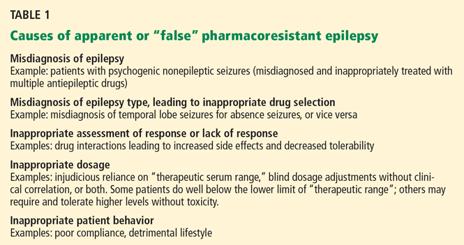
Common causes of treatment failure, such as poor compliance or inappropriate selection of first-line antiepileptic drugs, should be addressed early on by the treating physician. Nonadherence to the prescribed regimen is a very common cause of uncontrolled seizures, so it is critical to maintain a good rapport with the patient and to inquire about reasons for noncompliance.
Factors that have been associated with treatment-resistant epilepsy include:
- Early onset of seizures
- Long history of poor seizure control
- Having more than one type of seizure
- Remote symptomatic etiology (eg, patients with a history of brain infection or head trauma)
- Certain structural abnormalities (eg, cortical dysplasia)
- Certain abnormalities on electroencephalography (EEG)
- Cognitive disability
- History of status epilepticus.
When to consider referral
A topic of debate is how long a patient must have active epilepsy before he or she is considered to have pharmacoresistance and should be referred to a specialist center.
Both the rate of remission and the time needed to achieve remission depend on multiple factors such as the type and etiology of the epilepsy and the definition of sustained intractability.
Importantly, the prognosis for most patients with newly diagnosed epilepsy, whether good or bad, becomes apparent within a few years of starting treatment. Although pharmacoresistant epilepsy will become refractory within 8 years in some patients, in others a second drug may not fail for more than 1 or 2 decades after diagnosis. Nonetheless, a history of a lack of a sustained seizure-free period for 12 consecutive months, in spite of two or three suitable and tolerated antiepileptic drugs, is a definite red flag for clinicians and should prompt referral to a specialist center.9,10 The National Association of Epilepsy Centers recommends referral to a specialized epilepsy center if seizure control is not achieved by a general neurologist within a period of 9 months.11
AN APPROACH TO PHARMACORESISTANT EPILEPSY FOR THE NONSPECIALIST
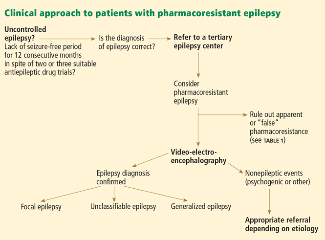
Review and confirm the diagnosis of epilepsy with the help of a careful history, video-EEG, and imaging.
When seizures cannot be controlled with drugs, it is important to verify that the events in question are indeed epileptic. Continuous video-EEG monitoring may be necessary to capture and characterize the clinical manifestations and corresponding EEG changes.14 When a typical spell is not associated with an EEG change, one can often make the diagnosis of nonepileptic events, which commonly are psychogenic nonepileptic seizures. Of note, however: scalp EEG may fail to pick up ictal EEG changes in focal seizures arising from a small or deeply situated focus: for example, as in focal sensorimotor seizures from restricted perirolandic cortex.15,16
Identify the cause, type of seizure or seizures, and syndromic classification, if any.
Review past and present medications, doses, efficacy, and side effects. Consider the possibility of drug interactions.
Different drugs may have different pharmacokinetic and dose-efficacy curves. With most of the newer-generation antiepileptic compounds such as lamotrigine (Lamictal), levetiracetam (Keppra), pregabalin (Lyrica), and topiramate (Topamax), efficacy may continue to increase in some patients as the dose is increased without reaching toxicity. An important exception is phenytoin (Dilantin): due to its nonlinear and saturable pharmacokinetics, even minor dose increases may lead to a large increase in phenytoin concentration (the drug accumulates as elimination becomes saturated). A high degree of individual variability exists, which is determined by factors that include the patient’s age, genetic and enzymatic profile, comorbidities, and concurrent medications.17 Understanding these relationships in individual patients facilitates dose selection and titration and enhances compliance. Testing serum drug levels may help when compliance is questionable.
Choose antiepileptic drugs primarily on the basis of the type of seizures and the individual clinical scenario: Which drug is likely to be most efficacious with the fewest side effects, and which one is appropriate for the patient’s comorbidities and concomitant medications?
When changing the dosage, withdrawing a drug, or adding a second antiepileptic drug, always do it in a systematic way, one step at a time. Reassess the response to the change before moving to the next step.
Discuss issues such as seizure precautions, lifestyle modifications, psychosocial dysfunction, and sudden unexpected death. Offer access to epilepsy-specialist nurses and epilepsy support groups such as those offered by the Epilepsy Foundation of America, the agency dedicated to the welfare of individuals with epilepsy in the United States (http://epilepsyfoundation.ning.com/).
PATTERNS OF DRUG RESISTANCE
Epidemiologic studies suggest three different patterns of drug resistance in epilepsy: de novo, progressive, and waxing-and-waning.
De novo drug resistance
In some patients, resistance is present from the time of onset of the very first seizure, before any antiepileptic drug is even started. One landmark study showed that patients with newly diagnosed epilepsy for whom the first drug was ineffective had only an 11% probability of future success, compared with 41% to 55% in patients who had had to stop taking the drug because of intolerable side effects or idiosyncratic reactions.10 Most patients for whom the first drug fails will be resistant to most and often all antiepileptic drugs.18 These results suggest that seizures in newly diagnosed patients are either easy to control or difficult to control right from the start.
Progressive drug resistance
In some patients, epilepsy is initially controlled but then gradually becomes refractory. This pattern may be seen, for example, in childhood epilepsies or in patients with hippocampal sclerosis.19,20
Waxing and waning resistance
In some patients, epilepsy has a waxing-and-waning pattern: ie, it alternates between a remitting (pharmacoresponsive) and relapsing (pharmacoresistant) course. Patients thought to have drug-resistant epilepsy may become seizure-free when other drugs are tried. Changes in drug bioavailability, local concentration of the drug in the brain, receptor changes, the development of tolerance, and interactions with new medications may be implicated, though the exact mechanism is not understood.21
BIOLOGIC BASIS OF PHARMACORESISTANT EPILEPSY
Pharmacoresistance is not unique to epilepsy: it is now recognized in diverse brain disorders, including depression and schizophrenia,22 and in other diseases affecting the brain, such as human immunodeficiency virus infection and many forms of cancer.23
Multiple drug resistance is characterized by insensitivity to a broad spectrum of drugs that presumably act on different receptors and by different mechanisms.24
Conceptually, the variable response to antiepileptic drugs can be attributed to factors related to the disease, the patient, and the drugs, or to other unknown factors. These factors are not mutually exclusive and may be either constitutive or acquired during the course of the disease.
Factors related to the disease (independent of the host)
These factors include etiology, epilepsy progression resulting in persistent changes of the epileptogenic network, and alterations of drug targets (ie, the “target” or pharmacodynamic hypothesis that reduced sensitivity to antiepileptic drugs is due to seizure-related alterations of specific drug targets25,26) or drug uptake into the brain27 (the “transporter” or pharmacokinetic hypothesis that the drugs are ineffective due to intrinsic or acquired overexpression of multidrug transporter proteins that hamper local drug delivery to target sites).28
Factors related to the drugs
Several drug-related factors have been implicated, such as the development of tolerance, lack of antiepileptogenic (disease-modifying) actions to interrupt the ongoing process of epileptogenesis rather than only suppressing seizures, and paucity of drugs with specific mechanisms of action tailored to difficult-tocontrol epilepsies.29
Patient characteristics
Variability in response (efficacy and adverse effects) to each antiepileptic drug can be due to interindividual differences in any of four interrelated fundamental factors: DNA, RNA, proteins, or metabolites. The field of study that aims to assess the effect of DNA variations (genotype) on a patient’s clinical response to a drug (phenotype) is known as pharmacogenetics. Age-related changes in pharmacokinetic and pharmacodynamic variables may contribute to age-dependent pharmacoresistance. Least studied are environmental factors that may play a role in the development or expression of pharmacoresistance.
NONPHARMACOLOGIC TREATMENTS
Ketogenic diet
The ketogenic diet, an important nonpharmacologic alternative, is usually reserved for young patients with difficult-to-control seizures. Originally developed almost a century ago, the diet mimics the biochemical changes associated with starvation. It is a strict regimen, high in fat and low in carbohydrate and protein (typically in a ratio of 4:1 or 3:1 in adolescents and very young children).
Such a strict regimen is difficult to implement and maintain and requires close supervision by a dietician and physician. In addition to the practical complexities, concerns also exist about the long-term effects of the diet on the child’s growth and overall health. For these reasons, the ketogenic diet is restricted to a small group of young patients with pharmacoresistant epilepsy and is not usually used long-term. There are few data indicating when it is appropriate to terminate the diet in patients who have a favorable response, but most clinicians wean the patient after 2 to 3 years.
Reports on the use of the ketogenic diet in adults are scarce, although benefit was seen in a small series.30 No long-term follow-up data exist for adults, especially regarding the risk of atherosclerosis.
Vagus nerve stimulation
Vagus nerve stimulation is a nonpharmacologic alternative for adults and for adolescents over age 12 years who have intractable focal seizures and who are not favorable surgical candidates. 31 Its effectiveness in younger patients and in those who have intractable generalized seizures is less clear, although published uncontrolled series have reported benefit (fewer seizures and better quality of life).32–34
A device consisting of a pulse generator is implanted subcutaneously in the precordium, and a lead wire is tunneled under the skin and attached to the left vagus nerve. The generator is programmed using a telemetry wand held over the device, with settings for current intensity (typically 1–2 mA), pulse width (250–300 μsec), frequency (30 Hz), and “duty cycle” (typically 30 seconds on stimulation, followed by 3 to 5 minutes off, cycling 24 hours/day). Hence, it provides “open-loop stimulation,” ie, continuous stimulation that is not modified in response to the patient’s EEG seizure activity. Patients or caregivers can also activate the device manually (“on demand”) at the first sign or warning of an impending seizure by swiping a handheld magnet.
Common side effects such as cough, voice alteration, and hoarseness are usually stimulation-dependent and tend to diminish with time. Notably, vagus nerve stimulation has none of the cognitive side effects often encountered with increasing doses of antiepileptic drugs. As with other implantable stimulators, some safety concerns exist in patients undergoing magnetic resonance imaging (MRI).
At least one-third of patients who receive this treatment show a sustained response, defined as a 50% or greater reduction in seizures. However, few achieve freedom from seizures, and therefore this therapy is considered palliative and is reserved for patients who are not candidates for surgery or for whom surgery has failed.
Unfortunately, it has not been possible to predict which patients will benefit from vagus nerve stimulation. The American Academy of Neurology recommends that this treatment be considered only after a thorough evaluation by a subspecialist to rule out nonepileptic conditions, false pharmacoresistance, and surgically treatable types of epilepsy.31
IS THE PATIENT A CANDIDATE FOR EPILEPSY SURGERY?
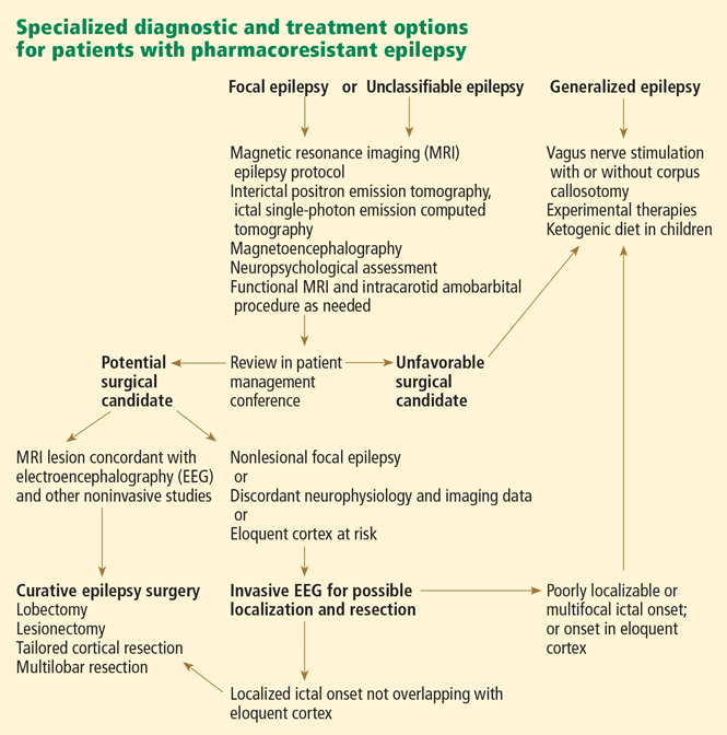
- Is the epilepsy diagnosis correct?
- Is the epilepsy focal? Have the following possibilities been excluded: generalized or multifocal epilepsy, situational or provoked seizures, or an epilepsy syndrome with spontaneous remission?
- Do seizures remain poorly controlled despite adequate pharmacologic trials?
- If so, do the seizures or medication side effects significantly affect the patient’s quality of life?
- Can an epileptogenic lesion be seen on MRI, and what is the suspected etiology?
- Is there converging evidence for a single epileptogenic focus?
- Are there abnormalities elsewhere in the brain?
- What are the chances of a good outcome in terms of seizure control and improvement in quality of life?
- What are the risks of surgery, and how do these compare with the risks of not having surgery?
- What are the patient’s perceptions and attitudes toward epilepsy surgery?
Surgical recommendations should be made after a thorough discussion of all preoperative data in a multidisciplinary patient management conference (Figure 1, Figure 2), in which epileptologists, neurosurgeons, neuroradiologists, neuropsychologists, and psychiatrists all actively participate.
Preoperative counseling is essential for the patient and his or her family, addressing the goals, risks, and benefits of the surgery. Treatment decisions should take into account the possible impact of surgery on the patient’s medical and psychosocial circumstances (risks of ongoing seizures vs surgical intervention; impact on the patient’s independence, employment status, emotional well-being, and psychiatric and other comorbidities).
EPILEPSY SURGERY: CURATIVE OR PALLIATIVE
Epilepsy surgery can be classified as curative or palliative, depending on the goal.
Curative procedures
Curative procedures include lobectomy, lesionectomy, and multilobar or hemispheric surgery (hemispherectomy).
Anterior temporal lobectomy and hippocampectomy are used for temporal lobe epilepsy and have several variations.
“Mesial temporal lobe epilepsy associated with hippocampal sclerosis,” a recognizable syndrome with complex partial seizures, stereotyped electroclinical features, and fairly typical natural history, is the most common of the focal epilepsies in adults.36 Its prognosis is poor when treated medically, but it responds well to surgery.37,38 In a landmark prospective trial, 58% of 40 patients randomized to surgical treatment were free of disabling seizures after 1 year, compared with only 8% of 40 patients treated medically.39
Lesionectomy and lobectomy are resective approaches targeting seizure foci outside the temporal lobe (most often in the frontal lobe, less commonly in the parietal or occipital lobes) or within the temporal lobe but outside the hippocampus (neocortical temporal lobe epilepsies).
The nature of the underlying substrate plays a significant role in determining the natural history,40 surgical strategy, and prognosis for freedom from seizures postoperatively. Patients with seizures due to structural lesions that are visible on MRI (“lesional epilepsies,” eg, cavernous angiomas or circumscribed lowgrade tumors) may become seizure-free after limited resections targeting the lesion itself (lesionectomy) or extending to involve part of a lobe or an entire lobe (lobectomy).
A favorable surgical outcome is much more likely if the lesion can be completely removed.41 At times, however, the lesion cannot be resected in its entirety, for example if it is located within inaccessible or essential (eloquent) cortex.
On the other hand, identifying the epileptogenic focus in patients with no visible structural abnormality on MRI (“nonlesional epilepsies”) can be challenging and usually requires intracranial investigations. In this instance, the aim of surgery is to resect regions that are electrographically abnormal. In general, the postoperative outcome is less favorable in nonlesional focal epilepsies than in lesional epilepsies.42
Multilobar resections and hemispherectomy are indicated when seizures arise from extensive, diffuse, or multiple regions of a single hemisphere.
If the neurologic function supported by the abnormal hemisphere is intact, a tailored multilobar resection aims at eliminating the epileptogenic focus without creating new deficits.
If, however, the underlying hemispheric abnormality is associated with significant contralateral hemiparesis, hemiplegia, or visual field deficits, the need to preserve function does not limit surgery, and hemispherectomy can be considered. Hemispherectomy can be the procedure of choice for selected infants and young children with catastrophic epilepsies and unilateral brain damage.43,44 The goal is to control seizures by completely disconnecting the abnormal epileptogenic hemisphere from the opposite, “good” hemisphere. A second major goal is to improve psychosocial and cognitive development by eliminating the child’s uncontrolled seizures.
Palliative procedures
Palliative procedures, in contrast to curative ones, rarely eliminate seizures entirely. It is important to determine that patients are not candidates for a more definitive, potentially curative resective procedure before considering palliative surgical options such as corpus callosotomy, multiple subpial transections, or vagus nerve stimulation.
Corpus callosotomy (transection of the corpus callosum) is performed in a small number of patients, ie, those who have disabling seizures that rapidly become generalized or injurious drop attacks and are not candidates for focal resection. By disconnecting the two hemispheres, this procedure aims to hinder the fast interhemispheric spread of seizure discharges.
Callosotomy may be complete or involve only a portion of the corpus callosum. The extent of resection has been correlated with favorable outcome.45
Some investigators report a 50% or greater reduction in seizure frequency, with drop attacks and generalized tonic-clonic seizures showing the most consistent improvement. In addition, behavior and quality of life may also improve.46
Multiple subpial transections are reserved for seizures arising from eloquent cortex (ie, from areas that cannot be removed without incurring unacceptable neurologic deficits). Therefore, the surgeon only transects the epileptogenic cortex in a vertical manner, so as to interrupt the horizontal cortical connections without resection. This approach is thought to disrupt the synchrony of seizure propagation while preserving physiologic function.
A meta-analysis of small case series suggests some decrease in seizure frequency with no or minimal neurologic compromise in up to 60% of patients.47
COMPLICATIONS OF EPILEPSY SURGERY
Resective surgery is not without risk, but often the risk is much less than that posed by uncontrolled epilepsy in the long term. Operative mortality rates vary from almost zero for temporal lobe surgery to 2.5% for hemispherectomy. The reported risk of permanent surgical morbidity varies by type of surgery from 1.1% for temporal lobe resection to about 5% for frontal lobe resection.4
NOVEL EPILEPSY THERAPIES
The failure of available antiepileptic drugs to control seizures in a substantial number of patients underscores the need to develop novel therapies such as electrical stimulation, local drug delivery, cell transplantation, and genebased therapies.48 Future targeted therapies could be coupled to seizure-forecasting systems to create “smart” implantable devices that predict, detect, and preemptively treat the seizures in a “closed-loop” fashion.
Targeted electrical stimulation
To modulate abnormal cortical hyperexcitability, electrical stimulation can be applied to the peripheral nervous system (eg, vagus nerve stimulation) or central nervous system. Central nervous system stimulation can be broadly divided into two approaches:
Direct stimulation targets presumed epileptogenic brain tissue such as the neocortex or hippocampus.
Indirect stimulation targets presumed seizure-gating networks such as in the cerebellum and various deep brain nuclei in the basal ganglia or thalamus (deep brain stimulation), which are believed to play a central role in modulating the synchronization and propagation of seizure activity.
Systematic, well-designed studies are currently under way. The budding field of electrical stimulation faces a number of challenges, which include optimizing stimulation variables and target sites, selecting favorable candidates, validating long-term safety and efficacy, evaluating long-term effects on tissue reorganization, plasticity, and epileptogenicity, and developing reliable algorithms for seizure detection and prediction.
Local drug delivery
Direct delivery of drugs into the epileptogenic brain tissue holds promise, particularly for patients whose foci cannot be surgically removed. By bypassing the systemic circulation, this approach has the potential to avoid systemic and even whole-brain side effects.
However, only a few proof-of-principle experiments have been conducted in animals, and to date no clinical study has explored the utility of intraparenchymal or intraventricular antiepileptic drug delivery in humans.
Cell and gene therapies
The emerging field of experimental cell- and gene-based neuropharmacology holds promise for location-specific therapeutic strategies. In ex vivo gene therapy, bioengineered cells capable of delivering anticonvulsant compounds might be transplanted into specific areas of the brain. On the other hand, in vivo gene therapy would involve delivering genes by viral vectors to induce the localized production of antiepileptic compounds in situ.
Endogenous anticonvulsants such as gamma-aminobutyric acid (GABA) and adenosine have been tried in various animal experiments. 49 Before they can be applied clinically, significant questions need to be addressed, including the potential for toxicity or maladaptive plasticity and long-term therapeutic safety and efficacy.
Cell transplantation is aimed at restoring the physiologic balance of neurotransmitters, and is currently being investigated for the treatment of several neurologic disorders such as Parkinson disease and Huntington disease.50 Unlike delivery of exogenous compounds, cell transplantation (heterologous fetal cell grafts or embryonic or adult stem cells) has the potential to form restorative synaptic connections and assimilate within existing cells and networks in the host tissue. An essential limitation to xenotransplantation in humans is the risk of immunologic rejection.
The future
We hope that continued progress in genomics will lead to targeted development of disease-modifying drugs that can impede or reverse the process of epileptogenesis. Advances in informatics and genetics may be harnessed to predict which patients are likely to develop pharmacoresistance, to cure certain genetic epilepsies, and to individualize antiepileptic drug selection on the basis of each person’s genetic profile.
- Siegel AM. Presurgical evaluation and surgical treatment of medically refractory epilepsy. Neurosurg Rev 2004; 27:1–18.
- Murray MI, Halpern MT, Leppik IE. Cost of refractory epilepsy in adults in the USA. Epilepsy Res 1996; 23:139–148.
- Jacoby A, Buck D, Baker G, McNamee P, Graham-Jones S, Chadwick D. Uptake and costs of care for epilepsy: findings from a U.K. regional study. Epilepsia 1998; 39:776–786.
- Chapell R, Reston J, Snyder D, Treadwell J, Treager S, Turkelson C. Management of treatment-resistant epilepsy. Evid Rep Technol Assess (Summ) 2003 Apr; 77:1–8.
- Nei M, Bagla R. Seizure-related injury and death. Curr Neurol Neurosci Rep 2007; 7:335–341.
- Langan Y, Nashef L, Sander JW. Case-control study of SUDEP. Neurology 2005; 64:1131–1133.
- Sperling MR, Feldman H, Kinman J, Liporace JD, O’Connor MJ. Seizure control and mortality in epilepsy. Ann Neurol 1999; 46:45–50.
- Berg AT. Understanding the delay before epilepsy surgery: who develops intractable focal epilepsy and when? CNS Spectr 2004; 9:136–144.
- Perucca E. Pharmacoresistance in epilepsy: how should it be defined? CNS Drugs 1998; 10:171–179.
- Kwan P, Brodie MJ. Early identification of refractory epilepsy. N Engl J Med 2000; 342:314–319.
- Gumnit RJ, Walczak TS; National Association of Epilepsy Centers. Guidelines for essential services, personnel, and facilities in specialized epilepsy centers in the United States. Epilepsia 2001; 42:804–814.
- Smith D, Defalla BA, Chadwick DW. The misdiagnosis of epilepsy and the management of refractory epilepsy in a specialist clinic. QJM 1999; 92:15–23.
- Schuele SU, Lüders HO. Intractable epilepsy: management and therapeutic alternatives. Lancet Neurol 2008; 7:514–524.
- Engel J, Burchfiel J, Ebersole J, et al. Long-term monitoring for epilepsy. Report of an IFCN committee. Electroencephalogr Clin Neurophysiol 1993; 87:437–458.
- Kanner AM, Morris HH, Lüders H, et al. Supplementary motor seizures mimicking pseudoseizures: some clinical differences. Neurology 1990; 40:1404–1407.
- Alexopoulos AV, Dinner DS. Focal motor seizures, epilepsia partialis continua, and supplementary sensorimotor seizures. In:Wyllie E, Gupta A, Lachhwani DK, editors. The Treatment of Epilepsy: Principles & Practice. Philadelphia, PA: Lippincott Williams & Wilkins, 2006:257–277.
- French JA. Refractory epilepsy: clinical overview. Epilepsia 2007; 48(suppl 1):3–7.
- Regesta G, Tanganelli P. Clinical aspects and biological bases of drug-resistant epilepsies. Epilepsy Res 1999; 34:109–122.
- Berg AT, Langfitt J, Shinnar S, et al. How long does it take for partial epilepsy to become intractable? Neurology 2003; 60:186–190.
- Berg AT, Vickrey BG, Testa FM, et al. How long does it take for epilepsy to become intractable? A prospective investigation. Ann Neurol 2006; 60:73–79.
- Löscher W, Schmidt D. Experimental and clinical evidence for loss of effect (tolerance) during prolonged treatment with antiepileptic drugs. Epilepsia 2006; 47:1253–1284.
- Löscher W, Potschka H. Drug resistance in brain diseases and the role of drug efflux transporters. Nat Rev Neurosci 2005; 6:591–602.
- Siddiqui A, Kerb R, Weale ME, et al. Association of multidrug resistance in epilepsy with a polymorphism in the drug-transporter gene ABCB1. N Engl J Med 2003; 348:1442–1448.
- Granata T, Marchi N, Carlton E, et al. Management of the patient with medically refractory epilepsy. Expert Rev Neurother 2009; 9:1791–1802.
- Marchi N, Hallene KL, Kight KM, et al. Significance of MDR1 and multiple drug resistance in refractory human epileptic brain. BMC Med 2004; 2:37.
- Oby E, Janigro D. The blood-brain barrier and epilepsy. Epilepsia 2006; 47:1761–1774.
- Schmidt D, Löscher W. New developments in antiepileptic drug resistance: an integrative view. Epilepsy Curr 2009; 9:47–52.
- Remy S, Beck H. Molecular and cellular mechanisms of pharmacoresistance in epilepsy. Brain 2006; 129:18–35.
- Löscher W, Schmidt D. New horizons in the development of antiepileptic drugs. Epilepsy Res 2002; 50:3–16.
- Sirven J, Whedon B, Caplan D, et al. The ketogenic diet for intractable epilepsy in adults: preliminary results. Epilepsia 1999; 40:1721–1726.
- Fisher RS, Handforth A. Reassessment: vagus nerve stimulation for epilepsy: a report of the Therapeutics and Technology Assessment Subcommittee of the American Academy of Neurology. Neurology 1999; 53:666–669.
- Hallböök T, Lundgren J, Stjernqvist K, Blennow G, Strömblad LG, Rosén I. Vagus nerve stimulation in 15 children with therapy resistant epilepsy; its impact on cognition, quality of life, behaviour and mood. Seizure 2005; 14:504–513.
- Holmes MD, Silbergeld DL, Drouhard D, Wilensky AJ, Ojemann LM. Effect of vagus nerve stimulation on adults with pharmacoresistant generalized epilepsy syndromes. Seizure 2004; 13:340–345.
- Murphy JV, Torkelson R, Dowler I, Simon S, Hudson S. Vagal nerve stimulation in refractory epilepsy: the first 100 patients receiving vagal nerve stimulation at a pediatric epilepsy center. Arch Pediatr Adolesc Med 2003; 157:560–564.
- Alexopoulos AV, Najm IM. Neurosurgical management of focal epilepsies in adults. In:Panayiotopoulos CP, et al, editors. Focal Epilepsies: Seizures, Syndromes and Management. Oxford, UK: Medicinae; 2009:204–220.
- Wieser HGILAE Commission on Neurosurgery of Epilepsy. ILAE Commission Report. Mesial temporal lobe epilepsy with hippocampal sclerosis. Epilepsia 2004; 45:695–714.
- Engel J. Surgery for seizures. N Engl J Med 1996; 334:647–652.
- Engel J, Wiebe S, French J, et al; Quality Standards Subcommittee of the American Academy of Neurology. Practice parameter: temporal lobe and localized neocortical resections for epilepsy: report of the Quality Standards Subcommittee of the American Academy of Neurology, in association with the American Epilepsy Society and the American Association of Neurological Surgeons. Neurology 2003; 60:538–547.
- Wiebe S, Blume WT, Girvin JP, Eliasziw M; Effectiveness and Efficiency of Surgery for Temporal Lobe Epilepsy Study Group. A randomized, controlled trial of surgery for temporal-lobe epilepsy. N Engl J Med 2001; 345:311–318.
- Semah F, Picot MC, Adam C, et al. Is the underlying cause of epilepsy a major prognostic factor for recurrence? Neurology 1998; 51:1256–1262.
- Awad IA, Rosenfeld J, Ahl J, Hahn JF, Lüders H. Intractable epilepsy and structural lesions of the brain: mapping, resection strategies, and seizure outcome. Epilepsia 1991; 32:179–186.
- Cascino GD. Surgical treatment for extratemporal epilepsy. Curr Treat Options Neurol 2004; 6:257–262.
- González-Martínez JA, Gupta A, Kotagal P, et al. Hemispherectomy for catastrophic epilepsy in infants. Epilepsia 2005; 46:1518–1525.
- Wyllie E. Surgery for catastrophic localization-related epilepsy in infants. Epilepsia 1996; 37(suppl 1):S22–S25.
- Tanriverdi T, Olivier A, Poulin N, Andermann F, Dubeau F. Long-term seizure outcome after corpus callosotomy: a retrospective analysis of 95 patients. J Neurosurg 2009; 110:332–342.
- Asadi-Pooya AA, Sharan A, Nei M, Sperling MR. Corpus callosotomy. Epilepsy Behav 2008; 13:271–278.
- Spencer SS, Schramm J, Wyler A, et al. Multiple subpial transection for intractable partial epilepsy: an international meta-analysis. Epilepsia 2002; 43:141–145.
- Alexopoulos AV, Gonugunta V, Yang J, Boulis NM. Electrical stimulation and gene-based neuromodulation for control of medically-refractory epilepsy. Acta Neurochir Suppl 2007; 97:293–309.
- Detlev B. Cell and gene therapies for refractory epilepsy. Curr Neuropharmacol 2007; 5:115–125.
- Fisher RS, Ho J. Potential new methods for antiepileptic drug delivery. CNS Drugs 2002; 16:579–593.
Although more than 10 new antiepileptic drugs have been developed in the past decade, epilepsy remains resistant to drug therapy in about one-third of patients. Approximately 20% of patients with primary generalized epilepsy and up to 60% of patients who have focal epilepsy develop drug resistance during the course of their condition, which for many is lifelong.1
Those who get no response or only a partial response to drugs continue to have incapacitating seizures that lead to significant neuropsychiatric and social impairment, lower quality of life, greater morbidity, and a higher risk of death.
Managing these patients is a challenge and requires a structured multidisciplinary approach in specialized clinics. Newer research, particularly in pharmacogenomics, holds promise of therapy that more closely suits an individual’s profile and type of epilepsy.
This article reviews recent developments in the pathogenesis and treatment of pharmacoresistant epilepsy, placing these topics in clinical context to facilitate and enhance the physician’s ability to manage it.
THE COSTS OF RESISTANT EPILEPSY
A US study in the early 1990s estimated that the annual cost of refractory epilepsy in adults exceeds $11,745 per person2; the cost would be considerably higher today. Another study found that costs correlate with severity of illness and that patients who have intractable seizures incur a cost eight times greater than in those whose epilepsy is controlled.3
Higher risk of death
In any given interval of time, people with pharmacoresistant epilepsy are about two to 10 times more likely to die compared with the general population.4 The risk is inversely linked to seizure control.
“Sudden unexpected death in epilepsy”is the most frequent type of death in patients with pharmacoresistant epilepsy. This category excludes deaths from trauma or drowning. The death can be witnessed or unwitnessed and with or without evidence of a seizure (but not documented status epilepticus). Postmortem examination does not reveal a toxic or anatomic cause of death, and the underlying mechanisms remain unknown. However, the risk is closely associated with drug resistance (which manifests with uncontrolled convulsive seizures and need for polytherapy with antiepileptic drugs).5
Case-control studies have shown that the risk of sudden unexpected death is closely and inversely associated with seizure control; the rate is significantly higher in patients who have a higher frequency of convulsive seizures.6 In addition, freedom from seizures, achieved after successful epilepsy surgery, reduces the risk of death from all causes.7
Other causes of death in patients with epilepsy may be directly related to seizures (accidental trauma, drowning, burns) or to the underlying condition causing the seizures. Furthermore, people with epilepsy are at higher risk of suicide than the general population.
CONCEPT OF PHARMACORESISTANCE AS IT PERTAINS TO EPILEPSY
There is no uniformly accepted definition of pharmacoresistant epilepsy. Most studies defined it according to the number of antiepileptic drugs the patient had tried without success, the frequency of seizures, the duration of illness, and the period of remission. Its true definition awaits a better understanding of underlying mechanisms.
Nevertheless, a useful operational definition at present is failure to control seizures despite a trial of two or three drugs that are suitable for the type of epilepsy and have been appropriately prescribed at maximum tolerated doses. This is because the chances of controlling epilepsy decline sharply after failure of the second or third antiepileptic drug trial. In fact, some clinicians would argue against trying another antiepileptic drug in these patients, who may be candidates for surgical procedures that have high rates of success.8
Common causes of treatment failure, such as poor compliance or inappropriate selection of first-line antiepileptic drugs, should be addressed early on by the treating physician. Nonadherence to the prescribed regimen is a very common cause of uncontrolled seizures, so it is critical to maintain a good rapport with the patient and to inquire about reasons for noncompliance.
Factors that have been associated with treatment-resistant epilepsy include:
- Early onset of seizures
- Long history of poor seizure control
- Having more than one type of seizure
- Remote symptomatic etiology (eg, patients with a history of brain infection or head trauma)
- Certain structural abnormalities (eg, cortical dysplasia)
- Certain abnormalities on electroencephalography (EEG)
- Cognitive disability
- History of status epilepticus.
When to consider referral
A topic of debate is how long a patient must have active epilepsy before he or she is considered to have pharmacoresistance and should be referred to a specialist center.
Both the rate of remission and the time needed to achieve remission depend on multiple factors such as the type and etiology of the epilepsy and the definition of sustained intractability.
Importantly, the prognosis for most patients with newly diagnosed epilepsy, whether good or bad, becomes apparent within a few years of starting treatment. Although pharmacoresistant epilepsy will become refractory within 8 years in some patients, in others a second drug may not fail for more than 1 or 2 decades after diagnosis. Nonetheless, a history of a lack of a sustained seizure-free period for 12 consecutive months, in spite of two or three suitable and tolerated antiepileptic drugs, is a definite red flag for clinicians and should prompt referral to a specialist center.9,10 The National Association of Epilepsy Centers recommends referral to a specialized epilepsy center if seizure control is not achieved by a general neurologist within a period of 9 months.11
AN APPROACH TO PHARMACORESISTANT EPILEPSY FOR THE NONSPECIALIST
Review and confirm the diagnosis of epilepsy with the help of a careful history, video-EEG, and imaging.
When seizures cannot be controlled with drugs, it is important to verify that the events in question are indeed epileptic. Continuous video-EEG monitoring may be necessary to capture and characterize the clinical manifestations and corresponding EEG changes.14 When a typical spell is not associated with an EEG change, one can often make the diagnosis of nonepileptic events, which commonly are psychogenic nonepileptic seizures. Of note, however: scalp EEG may fail to pick up ictal EEG changes in focal seizures arising from a small or deeply situated focus: for example, as in focal sensorimotor seizures from restricted perirolandic cortex.15,16
Identify the cause, type of seizure or seizures, and syndromic classification, if any.
Review past and present medications, doses, efficacy, and side effects. Consider the possibility of drug interactions.
Different drugs may have different pharmacokinetic and dose-efficacy curves. With most of the newer-generation antiepileptic compounds such as lamotrigine (Lamictal), levetiracetam (Keppra), pregabalin (Lyrica), and topiramate (Topamax), efficacy may continue to increase in some patients as the dose is increased without reaching toxicity. An important exception is phenytoin (Dilantin): due to its nonlinear and saturable pharmacokinetics, even minor dose increases may lead to a large increase in phenytoin concentration (the drug accumulates as elimination becomes saturated). A high degree of individual variability exists, which is determined by factors that include the patient’s age, genetic and enzymatic profile, comorbidities, and concurrent medications.17 Understanding these relationships in individual patients facilitates dose selection and titration and enhances compliance. Testing serum drug levels may help when compliance is questionable.
Choose antiepileptic drugs primarily on the basis of the type of seizures and the individual clinical scenario: Which drug is likely to be most efficacious with the fewest side effects, and which one is appropriate for the patient’s comorbidities and concomitant medications?
When changing the dosage, withdrawing a drug, or adding a second antiepileptic drug, always do it in a systematic way, one step at a time. Reassess the response to the change before moving to the next step.
Discuss issues such as seizure precautions, lifestyle modifications, psychosocial dysfunction, and sudden unexpected death. Offer access to epilepsy-specialist nurses and epilepsy support groups such as those offered by the Epilepsy Foundation of America, the agency dedicated to the welfare of individuals with epilepsy in the United States (http://epilepsyfoundation.ning.com/).
PATTERNS OF DRUG RESISTANCE
Epidemiologic studies suggest three different patterns of drug resistance in epilepsy: de novo, progressive, and waxing-and-waning.
De novo drug resistance
In some patients, resistance is present from the time of onset of the very first seizure, before any antiepileptic drug is even started. One landmark study showed that patients with newly diagnosed epilepsy for whom the first drug was ineffective had only an 11% probability of future success, compared with 41% to 55% in patients who had had to stop taking the drug because of intolerable side effects or idiosyncratic reactions.10 Most patients for whom the first drug fails will be resistant to most and often all antiepileptic drugs.18 These results suggest that seizures in newly diagnosed patients are either easy to control or difficult to control right from the start.
Progressive drug resistance
In some patients, epilepsy is initially controlled but then gradually becomes refractory. This pattern may be seen, for example, in childhood epilepsies or in patients with hippocampal sclerosis.19,20
Waxing and waning resistance
In some patients, epilepsy has a waxing-and-waning pattern: ie, it alternates between a remitting (pharmacoresponsive) and relapsing (pharmacoresistant) course. Patients thought to have drug-resistant epilepsy may become seizure-free when other drugs are tried. Changes in drug bioavailability, local concentration of the drug in the brain, receptor changes, the development of tolerance, and interactions with new medications may be implicated, though the exact mechanism is not understood.21
BIOLOGIC BASIS OF PHARMACORESISTANT EPILEPSY
Pharmacoresistance is not unique to epilepsy: it is now recognized in diverse brain disorders, including depression and schizophrenia,22 and in other diseases affecting the brain, such as human immunodeficiency virus infection and many forms of cancer.23
Multiple drug resistance is characterized by insensitivity to a broad spectrum of drugs that presumably act on different receptors and by different mechanisms.24
Conceptually, the variable response to antiepileptic drugs can be attributed to factors related to the disease, the patient, and the drugs, or to other unknown factors. These factors are not mutually exclusive and may be either constitutive or acquired during the course of the disease.
Factors related to the disease (independent of the host)
These factors include etiology, epilepsy progression resulting in persistent changes of the epileptogenic network, and alterations of drug targets (ie, the “target” or pharmacodynamic hypothesis that reduced sensitivity to antiepileptic drugs is due to seizure-related alterations of specific drug targets25,26) or drug uptake into the brain27 (the “transporter” or pharmacokinetic hypothesis that the drugs are ineffective due to intrinsic or acquired overexpression of multidrug transporter proteins that hamper local drug delivery to target sites).28
Factors related to the drugs
Several drug-related factors have been implicated, such as the development of tolerance, lack of antiepileptogenic (disease-modifying) actions to interrupt the ongoing process of epileptogenesis rather than only suppressing seizures, and paucity of drugs with specific mechanisms of action tailored to difficult-tocontrol epilepsies.29
Patient characteristics
Variability in response (efficacy and adverse effects) to each antiepileptic drug can be due to interindividual differences in any of four interrelated fundamental factors: DNA, RNA, proteins, or metabolites. The field of study that aims to assess the effect of DNA variations (genotype) on a patient’s clinical response to a drug (phenotype) is known as pharmacogenetics. Age-related changes in pharmacokinetic and pharmacodynamic variables may contribute to age-dependent pharmacoresistance. Least studied are environmental factors that may play a role in the development or expression of pharmacoresistance.
NONPHARMACOLOGIC TREATMENTS
Ketogenic diet
The ketogenic diet, an important nonpharmacologic alternative, is usually reserved for young patients with difficult-to-control seizures. Originally developed almost a century ago, the diet mimics the biochemical changes associated with starvation. It is a strict regimen, high in fat and low in carbohydrate and protein (typically in a ratio of 4:1 or 3:1 in adolescents and very young children).
Such a strict regimen is difficult to implement and maintain and requires close supervision by a dietician and physician. In addition to the practical complexities, concerns also exist about the long-term effects of the diet on the child’s growth and overall health. For these reasons, the ketogenic diet is restricted to a small group of young patients with pharmacoresistant epilepsy and is not usually used long-term. There are few data indicating when it is appropriate to terminate the diet in patients who have a favorable response, but most clinicians wean the patient after 2 to 3 years.
Reports on the use of the ketogenic diet in adults are scarce, although benefit was seen in a small series.30 No long-term follow-up data exist for adults, especially regarding the risk of atherosclerosis.
Vagus nerve stimulation
Vagus nerve stimulation is a nonpharmacologic alternative for adults and for adolescents over age 12 years who have intractable focal seizures and who are not favorable surgical candidates. 31 Its effectiveness in younger patients and in those who have intractable generalized seizures is less clear, although published uncontrolled series have reported benefit (fewer seizures and better quality of life).32–34
A device consisting of a pulse generator is implanted subcutaneously in the precordium, and a lead wire is tunneled under the skin and attached to the left vagus nerve. The generator is programmed using a telemetry wand held over the device, with settings for current intensity (typically 1–2 mA), pulse width (250–300 μsec), frequency (30 Hz), and “duty cycle” (typically 30 seconds on stimulation, followed by 3 to 5 minutes off, cycling 24 hours/day). Hence, it provides “open-loop stimulation,” ie, continuous stimulation that is not modified in response to the patient’s EEG seizure activity. Patients or caregivers can also activate the device manually (“on demand”) at the first sign or warning of an impending seizure by swiping a handheld magnet.
Common side effects such as cough, voice alteration, and hoarseness are usually stimulation-dependent and tend to diminish with time. Notably, vagus nerve stimulation has none of the cognitive side effects often encountered with increasing doses of antiepileptic drugs. As with other implantable stimulators, some safety concerns exist in patients undergoing magnetic resonance imaging (MRI).
At least one-third of patients who receive this treatment show a sustained response, defined as a 50% or greater reduction in seizures. However, few achieve freedom from seizures, and therefore this therapy is considered palliative and is reserved for patients who are not candidates for surgery or for whom surgery has failed.
Unfortunately, it has not been possible to predict which patients will benefit from vagus nerve stimulation. The American Academy of Neurology recommends that this treatment be considered only after a thorough evaluation by a subspecialist to rule out nonepileptic conditions, false pharmacoresistance, and surgically treatable types of epilepsy.31
IS THE PATIENT A CANDIDATE FOR EPILEPSY SURGERY?
- Is the epilepsy diagnosis correct?
- Is the epilepsy focal? Have the following possibilities been excluded: generalized or multifocal epilepsy, situational or provoked seizures, or an epilepsy syndrome with spontaneous remission?
- Do seizures remain poorly controlled despite adequate pharmacologic trials?
- If so, do the seizures or medication side effects significantly affect the patient’s quality of life?
- Can an epileptogenic lesion be seen on MRI, and what is the suspected etiology?
- Is there converging evidence for a single epileptogenic focus?
- Are there abnormalities elsewhere in the brain?
- What are the chances of a good outcome in terms of seizure control and improvement in quality of life?
- What are the risks of surgery, and how do these compare with the risks of not having surgery?
- What are the patient’s perceptions and attitudes toward epilepsy surgery?
Surgical recommendations should be made after a thorough discussion of all preoperative data in a multidisciplinary patient management conference (Figure 1, Figure 2), in which epileptologists, neurosurgeons, neuroradiologists, neuropsychologists, and psychiatrists all actively participate.
Preoperative counseling is essential for the patient and his or her family, addressing the goals, risks, and benefits of the surgery. Treatment decisions should take into account the possible impact of surgery on the patient’s medical and psychosocial circumstances (risks of ongoing seizures vs surgical intervention; impact on the patient’s independence, employment status, emotional well-being, and psychiatric and other comorbidities).
EPILEPSY SURGERY: CURATIVE OR PALLIATIVE
Epilepsy surgery can be classified as curative or palliative, depending on the goal.
Curative procedures
Curative procedures include lobectomy, lesionectomy, and multilobar or hemispheric surgery (hemispherectomy).
Anterior temporal lobectomy and hippocampectomy are used for temporal lobe epilepsy and have several variations.
“Mesial temporal lobe epilepsy associated with hippocampal sclerosis,” a recognizable syndrome with complex partial seizures, stereotyped electroclinical features, and fairly typical natural history, is the most common of the focal epilepsies in adults.36 Its prognosis is poor when treated medically, but it responds well to surgery.37,38 In a landmark prospective trial, 58% of 40 patients randomized to surgical treatment were free of disabling seizures after 1 year, compared with only 8% of 40 patients treated medically.39
Lesionectomy and lobectomy are resective approaches targeting seizure foci outside the temporal lobe (most often in the frontal lobe, less commonly in the parietal or occipital lobes) or within the temporal lobe but outside the hippocampus (neocortical temporal lobe epilepsies).
The nature of the underlying substrate plays a significant role in determining the natural history,40 surgical strategy, and prognosis for freedom from seizures postoperatively. Patients with seizures due to structural lesions that are visible on MRI (“lesional epilepsies,” eg, cavernous angiomas or circumscribed lowgrade tumors) may become seizure-free after limited resections targeting the lesion itself (lesionectomy) or extending to involve part of a lobe or an entire lobe (lobectomy).
A favorable surgical outcome is much more likely if the lesion can be completely removed.41 At times, however, the lesion cannot be resected in its entirety, for example if it is located within inaccessible or essential (eloquent) cortex.
On the other hand, identifying the epileptogenic focus in patients with no visible structural abnormality on MRI (“nonlesional epilepsies”) can be challenging and usually requires intracranial investigations. In this instance, the aim of surgery is to resect regions that are electrographically abnormal. In general, the postoperative outcome is less favorable in nonlesional focal epilepsies than in lesional epilepsies.42
Multilobar resections and hemispherectomy are indicated when seizures arise from extensive, diffuse, or multiple regions of a single hemisphere.
If the neurologic function supported by the abnormal hemisphere is intact, a tailored multilobar resection aims at eliminating the epileptogenic focus without creating new deficits.
If, however, the underlying hemispheric abnormality is associated with significant contralateral hemiparesis, hemiplegia, or visual field deficits, the need to preserve function does not limit surgery, and hemispherectomy can be considered. Hemispherectomy can be the procedure of choice for selected infants and young children with catastrophic epilepsies and unilateral brain damage.43,44 The goal is to control seizures by completely disconnecting the abnormal epileptogenic hemisphere from the opposite, “good” hemisphere. A second major goal is to improve psychosocial and cognitive development by eliminating the child’s uncontrolled seizures.
Palliative procedures
Palliative procedures, in contrast to curative ones, rarely eliminate seizures entirely. It is important to determine that patients are not candidates for a more definitive, potentially curative resective procedure before considering palliative surgical options such as corpus callosotomy, multiple subpial transections, or vagus nerve stimulation.
Corpus callosotomy (transection of the corpus callosum) is performed in a small number of patients, ie, those who have disabling seizures that rapidly become generalized or injurious drop attacks and are not candidates for focal resection. By disconnecting the two hemispheres, this procedure aims to hinder the fast interhemispheric spread of seizure discharges.
Callosotomy may be complete or involve only a portion of the corpus callosum. The extent of resection has been correlated with favorable outcome.45
Some investigators report a 50% or greater reduction in seizure frequency, with drop attacks and generalized tonic-clonic seizures showing the most consistent improvement. In addition, behavior and quality of life may also improve.46
Multiple subpial transections are reserved for seizures arising from eloquent cortex (ie, from areas that cannot be removed without incurring unacceptable neurologic deficits). Therefore, the surgeon only transects the epileptogenic cortex in a vertical manner, so as to interrupt the horizontal cortical connections without resection. This approach is thought to disrupt the synchrony of seizure propagation while preserving physiologic function.
A meta-analysis of small case series suggests some decrease in seizure frequency with no or minimal neurologic compromise in up to 60% of patients.47
COMPLICATIONS OF EPILEPSY SURGERY
Resective surgery is not without risk, but often the risk is much less than that posed by uncontrolled epilepsy in the long term. Operative mortality rates vary from almost zero for temporal lobe surgery to 2.5% for hemispherectomy. The reported risk of permanent surgical morbidity varies by type of surgery from 1.1% for temporal lobe resection to about 5% for frontal lobe resection.4
NOVEL EPILEPSY THERAPIES
The failure of available antiepileptic drugs to control seizures in a substantial number of patients underscores the need to develop novel therapies such as electrical stimulation, local drug delivery, cell transplantation, and genebased therapies.48 Future targeted therapies could be coupled to seizure-forecasting systems to create “smart” implantable devices that predict, detect, and preemptively treat the seizures in a “closed-loop” fashion.
Targeted electrical stimulation
To modulate abnormal cortical hyperexcitability, electrical stimulation can be applied to the peripheral nervous system (eg, vagus nerve stimulation) or central nervous system. Central nervous system stimulation can be broadly divided into two approaches:
Direct stimulation targets presumed epileptogenic brain tissue such as the neocortex or hippocampus.
Indirect stimulation targets presumed seizure-gating networks such as in the cerebellum and various deep brain nuclei in the basal ganglia or thalamus (deep brain stimulation), which are believed to play a central role in modulating the synchronization and propagation of seizure activity.
Systematic, well-designed studies are currently under way. The budding field of electrical stimulation faces a number of challenges, which include optimizing stimulation variables and target sites, selecting favorable candidates, validating long-term safety and efficacy, evaluating long-term effects on tissue reorganization, plasticity, and epileptogenicity, and developing reliable algorithms for seizure detection and prediction.
Local drug delivery
Direct delivery of drugs into the epileptogenic brain tissue holds promise, particularly for patients whose foci cannot be surgically removed. By bypassing the systemic circulation, this approach has the potential to avoid systemic and even whole-brain side effects.
However, only a few proof-of-principle experiments have been conducted in animals, and to date no clinical study has explored the utility of intraparenchymal or intraventricular antiepileptic drug delivery in humans.
Cell and gene therapies
The emerging field of experimental cell- and gene-based neuropharmacology holds promise for location-specific therapeutic strategies. In ex vivo gene therapy, bioengineered cells capable of delivering anticonvulsant compounds might be transplanted into specific areas of the brain. On the other hand, in vivo gene therapy would involve delivering genes by viral vectors to induce the localized production of antiepileptic compounds in situ.
Endogenous anticonvulsants such as gamma-aminobutyric acid (GABA) and adenosine have been tried in various animal experiments. 49 Before they can be applied clinically, significant questions need to be addressed, including the potential for toxicity or maladaptive plasticity and long-term therapeutic safety and efficacy.
Cell transplantation is aimed at restoring the physiologic balance of neurotransmitters, and is currently being investigated for the treatment of several neurologic disorders such as Parkinson disease and Huntington disease.50 Unlike delivery of exogenous compounds, cell transplantation (heterologous fetal cell grafts or embryonic or adult stem cells) has the potential to form restorative synaptic connections and assimilate within existing cells and networks in the host tissue. An essential limitation to xenotransplantation in humans is the risk of immunologic rejection.
The future
We hope that continued progress in genomics will lead to targeted development of disease-modifying drugs that can impede or reverse the process of epileptogenesis. Advances in informatics and genetics may be harnessed to predict which patients are likely to develop pharmacoresistance, to cure certain genetic epilepsies, and to individualize antiepileptic drug selection on the basis of each person’s genetic profile.
- Siegel AM. Presurgical evaluation and surgical treatment of medically refractory epilepsy. Neurosurg Rev 2004; 27:1–18.
- Murray MI, Halpern MT, Leppik IE. Cost of refractory epilepsy in adults in the USA. Epilepsy Res 1996; 23:139–148.
- Jacoby A, Buck D, Baker G, McNamee P, Graham-Jones S, Chadwick D. Uptake and costs of care for epilepsy: findings from a U.K. regional study. Epilepsia 1998; 39:776–786.
- Chapell R, Reston J, Snyder D, Treadwell J, Treager S, Turkelson C. Management of treatment-resistant epilepsy. Evid Rep Technol Assess (Summ) 2003 Apr; 77:1–8.
- Nei M, Bagla R. Seizure-related injury and death. Curr Neurol Neurosci Rep 2007; 7:335–341.
- Langan Y, Nashef L, Sander JW. Case-control study of SUDEP. Neurology 2005; 64:1131–1133.
- Sperling MR, Feldman H, Kinman J, Liporace JD, O’Connor MJ. Seizure control and mortality in epilepsy. Ann Neurol 1999; 46:45–50.
- Berg AT. Understanding the delay before epilepsy surgery: who develops intractable focal epilepsy and when? CNS Spectr 2004; 9:136–144.
- Perucca E. Pharmacoresistance in epilepsy: how should it be defined? CNS Drugs 1998; 10:171–179.
- Kwan P, Brodie MJ. Early identification of refractory epilepsy. N Engl J Med 2000; 342:314–319.
- Gumnit RJ, Walczak TS; National Association of Epilepsy Centers. Guidelines for essential services, personnel, and facilities in specialized epilepsy centers in the United States. Epilepsia 2001; 42:804–814.
- Smith D, Defalla BA, Chadwick DW. The misdiagnosis of epilepsy and the management of refractory epilepsy in a specialist clinic. QJM 1999; 92:15–23.
- Schuele SU, Lüders HO. Intractable epilepsy: management and therapeutic alternatives. Lancet Neurol 2008; 7:514–524.
- Engel J, Burchfiel J, Ebersole J, et al. Long-term monitoring for epilepsy. Report of an IFCN committee. Electroencephalogr Clin Neurophysiol 1993; 87:437–458.
- Kanner AM, Morris HH, Lüders H, et al. Supplementary motor seizures mimicking pseudoseizures: some clinical differences. Neurology 1990; 40:1404–1407.
- Alexopoulos AV, Dinner DS. Focal motor seizures, epilepsia partialis continua, and supplementary sensorimotor seizures. In:Wyllie E, Gupta A, Lachhwani DK, editors. The Treatment of Epilepsy: Principles & Practice. Philadelphia, PA: Lippincott Williams & Wilkins, 2006:257–277.
- French JA. Refractory epilepsy: clinical overview. Epilepsia 2007; 48(suppl 1):3–7.
- Regesta G, Tanganelli P. Clinical aspects and biological bases of drug-resistant epilepsies. Epilepsy Res 1999; 34:109–122.
- Berg AT, Langfitt J, Shinnar S, et al. How long does it take for partial epilepsy to become intractable? Neurology 2003; 60:186–190.
- Berg AT, Vickrey BG, Testa FM, et al. How long does it take for epilepsy to become intractable? A prospective investigation. Ann Neurol 2006; 60:73–79.
- Löscher W, Schmidt D. Experimental and clinical evidence for loss of effect (tolerance) during prolonged treatment with antiepileptic drugs. Epilepsia 2006; 47:1253–1284.
- Löscher W, Potschka H. Drug resistance in brain diseases and the role of drug efflux transporters. Nat Rev Neurosci 2005; 6:591–602.
- Siddiqui A, Kerb R, Weale ME, et al. Association of multidrug resistance in epilepsy with a polymorphism in the drug-transporter gene ABCB1. N Engl J Med 2003; 348:1442–1448.
- Granata T, Marchi N, Carlton E, et al. Management of the patient with medically refractory epilepsy. Expert Rev Neurother 2009; 9:1791–1802.
- Marchi N, Hallene KL, Kight KM, et al. Significance of MDR1 and multiple drug resistance in refractory human epileptic brain. BMC Med 2004; 2:37.
- Oby E, Janigro D. The blood-brain barrier and epilepsy. Epilepsia 2006; 47:1761–1774.
- Schmidt D, Löscher W. New developments in antiepileptic drug resistance: an integrative view. Epilepsy Curr 2009; 9:47–52.
- Remy S, Beck H. Molecular and cellular mechanisms of pharmacoresistance in epilepsy. Brain 2006; 129:18–35.
- Löscher W, Schmidt D. New horizons in the development of antiepileptic drugs. Epilepsy Res 2002; 50:3–16.
- Sirven J, Whedon B, Caplan D, et al. The ketogenic diet for intractable epilepsy in adults: preliminary results. Epilepsia 1999; 40:1721–1726.
- Fisher RS, Handforth A. Reassessment: vagus nerve stimulation for epilepsy: a report of the Therapeutics and Technology Assessment Subcommittee of the American Academy of Neurology. Neurology 1999; 53:666–669.
- Hallböök T, Lundgren J, Stjernqvist K, Blennow G, Strömblad LG, Rosén I. Vagus nerve stimulation in 15 children with therapy resistant epilepsy; its impact on cognition, quality of life, behaviour and mood. Seizure 2005; 14:504–513.
- Holmes MD, Silbergeld DL, Drouhard D, Wilensky AJ, Ojemann LM. Effect of vagus nerve stimulation on adults with pharmacoresistant generalized epilepsy syndromes. Seizure 2004; 13:340–345.
- Murphy JV, Torkelson R, Dowler I, Simon S, Hudson S. Vagal nerve stimulation in refractory epilepsy: the first 100 patients receiving vagal nerve stimulation at a pediatric epilepsy center. Arch Pediatr Adolesc Med 2003; 157:560–564.
- Alexopoulos AV, Najm IM. Neurosurgical management of focal epilepsies in adults. In:Panayiotopoulos CP, et al, editors. Focal Epilepsies: Seizures, Syndromes and Management. Oxford, UK: Medicinae; 2009:204–220.
- Wieser HGILAE Commission on Neurosurgery of Epilepsy. ILAE Commission Report. Mesial temporal lobe epilepsy with hippocampal sclerosis. Epilepsia 2004; 45:695–714.
- Engel J. Surgery for seizures. N Engl J Med 1996; 334:647–652.
- Engel J, Wiebe S, French J, et al; Quality Standards Subcommittee of the American Academy of Neurology. Practice parameter: temporal lobe and localized neocortical resections for epilepsy: report of the Quality Standards Subcommittee of the American Academy of Neurology, in association with the American Epilepsy Society and the American Association of Neurological Surgeons. Neurology 2003; 60:538–547.
- Wiebe S, Blume WT, Girvin JP, Eliasziw M; Effectiveness and Efficiency of Surgery for Temporal Lobe Epilepsy Study Group. A randomized, controlled trial of surgery for temporal-lobe epilepsy. N Engl J Med 2001; 345:311–318.
- Semah F, Picot MC, Adam C, et al. Is the underlying cause of epilepsy a major prognostic factor for recurrence? Neurology 1998; 51:1256–1262.
- Awad IA, Rosenfeld J, Ahl J, Hahn JF, Lüders H. Intractable epilepsy and structural lesions of the brain: mapping, resection strategies, and seizure outcome. Epilepsia 1991; 32:179–186.
- Cascino GD. Surgical treatment for extratemporal epilepsy. Curr Treat Options Neurol 2004; 6:257–262.
- González-Martínez JA, Gupta A, Kotagal P, et al. Hemispherectomy for catastrophic epilepsy in infants. Epilepsia 2005; 46:1518–1525.
- Wyllie E. Surgery for catastrophic localization-related epilepsy in infants. Epilepsia 1996; 37(suppl 1):S22–S25.
- Tanriverdi T, Olivier A, Poulin N, Andermann F, Dubeau F. Long-term seizure outcome after corpus callosotomy: a retrospective analysis of 95 patients. J Neurosurg 2009; 110:332–342.
- Asadi-Pooya AA, Sharan A, Nei M, Sperling MR. Corpus callosotomy. Epilepsy Behav 2008; 13:271–278.
- Spencer SS, Schramm J, Wyler A, et al. Multiple subpial transection for intractable partial epilepsy: an international meta-analysis. Epilepsia 2002; 43:141–145.
- Alexopoulos AV, Gonugunta V, Yang J, Boulis NM. Electrical stimulation and gene-based neuromodulation for control of medically-refractory epilepsy. Acta Neurochir Suppl 2007; 97:293–309.
- Detlev B. Cell and gene therapies for refractory epilepsy. Curr Neuropharmacol 2007; 5:115–125.
- Fisher RS, Ho J. Potential new methods for antiepileptic drug delivery. CNS Drugs 2002; 16:579–593.
KEY POINTS
- When seizures have failed to respond to two or three appropriate antiepileptic drugs, the chance of significant benefit from other drugs is 10% or less.
- The biologic basis of pharmacoresistance is multifactorial and varies from one patient to another.
- Social and lifestyle factors, including alcohol misuse and nonadherence to prescribed antiepileptic drugs, can contribute to or masquerade as pharmacoresistance.
- Current options for patients with pharmacoresistant epilepsy are surgery (the best option when feasible), vagus nerve stimulation, investigational drugs or devices, and aggressive combination treatment with available antiepileptic drugs.
Menstrual manipulation: Options for suppressing the cycle
If they wish, women can have more control over when and if they menstruate. By using hormonal contraceptives in extended or continuous regimens, they can have their period less often, a practice called menstrual manipulation or menstrual suppression.
Actually, with the help of their clinicians, women have been doing this for years. But now that several products have been approved by the US Food and Drug Administration (FDA) specifically for use in extended or continuous regimens, the practice has become more widely accepted.
Reasons for suppressing menstrual flow range from avoiding bleeding during a particular event (eg, a wedding, graduation, or sports competition) to finding relief from dysmenorrhea or reducing or eliminating menstruation in the treatment of endometriosis, migraine, and other medical conditions exacerbated by hormonal changes around the time of menses.1 Alternatively, some women may practice menstrual manipulation for no other reason than to simply avoid menstruation.
MENSTRUAL DISORDERS ARE TROUBLESOME, COMMON
Each year in the United States, menstrual disorders such as dysmenorrhea (painful menstruation), menorrhagia (excessive or frequent menstruation), metrorrhagia (irregular menstruation), menometrorrhagia (excessive and irregular menstruation), and premenstrual syndrome affect nearly 2.5 million women age 18 to 50 years.2 Menstrual disorders are the leading cause of gynecologic morbidity in the United States, outnumbering adnexal masses (the second most common cause) by a factor of three.2 In addition, these disorders extend into the workplace, costing US industry about 8% of its total wage bill.3
A BRIEF HISTORY OF CONTRACEPTIVE DEVELOPMENT
The idea of using progestins for birth control was first advanced in the 1950s by Dr. Gregory Pincus, who proposed a regimen of 21 days of active drug followed by 7 drug-free days to allow withdrawal bleeding, mimicking the natural cycle.4 This “21/7” regimen was designed to follow the lunar cycle in the hope it would be, in the words of Dr. John Rock, “a morally permissible variant of the rhythm method,”5 thereby making it acceptable to women, clinicians, and the Catholic Church.
In 1977, Loudon et al6 reported the results of a study in which women took active pills for 84 days instead of 21 days, which reduced the frequency of menstruation to every 3 months. Since then, extending the active pills beyond 21 days to avoid menses and other hormone-withdrawal symptoms has become popular in clinical practice, and many studies have investigated the extended or continuous use of oral and other forms of contraception to delay menses.7–18
CURRENT METHODS OF MENSTRUAL MANIPULATION
A variety of available products prevent conception by altering the menstrual cycle:
- Oral estrogen-progestin contraceptive pills
- A drug-releasing intrauterine device
- Depot medroxyprogesterone acetate injections
- A contraceptive vaginal ring
- An implantable etonogestrel contraceptive.
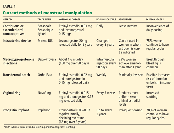
Their use in menstrual manipulation is summarized in Table 1.
Oral contraceptive pills
The most common way to manipulate the menstrual cycle is to extend the time between hormone-free weeks in an oral contraceptive regimen.
If the patient is young, you can prescribe a monophasic 21/7 oral contraceptive and tell her to take one active pill every day for 21 days and then start a new pack and keep taking active pills for up to 84 consecutive days, skipping the placebo pills until she wants to have her menstrual period. She can choose which week to have it: if the scheduled 12th week of an extended-cycle oral contraceptive regimen is inconvenient, she can plan it for week 10, or week 9, or whichever week is convenient.
The rationale for using an 84-day (12-week) cycle is that it still provides four periods per year, alleviating fears of hypertrophic endometrium.19
In this scenario, unscheduled or breakthrough bleeding can be managed by taking a “double-up pill” from a spare pack on any day breakthrough bleeding occurs and until it resolves. Menstrual periods should not be planned for intervals shorter than 21 days, owing to the risk of ovulation. Missed days of pills or use of placebo pills should also not exceed 7 days to prevent escape ovulation. 20
In some women with endometriosis and other medical reasons, continuous oral contraception with no placebo week can be prescribed.
Unfortunately, the downside to suppressing withdrawal bleeding is unscheduled or “breakthrough” bleeding. The best way to treat this unscheduled bleeding is not known. Patients who are not sexually active can be reassured that the goal of an atrophic endometrium can still be achieved, with resultant pill amenorrhea (particularly useful for those with severe dysmenorrhea or other reasons to want to avoid flow). Patients could also try to manage flow by periodically taking a 3- to 5-day break from hormone-containing pills to allow flow. They can also try switching to another oral contraceptive that has a different progestin that would spiral the arterioles of the endometrium more tightly and thus more aggressively induce atrophy.13,17,21 For instance, levonorgestrel is 10 to 20 times more potent than norethindrone. Choosing a pill with a higher monophasic dosing of levonorgestrel or a similar progestin may minimize unscheduled bleeding.
Currently, several oral contraceptives are approved for use in an extended regimen.
Seasonale was the first oral contraceptive marketed in the United States with an extended active regimen.22 It comes in a pack of 84 pills containing ethinyl estradiol 0.03 mg and levonorgestrel 0.15 mg, plus 7 placebo pills.
Seasonique is similar to Seasonale, but instead of placebo pills it has seven pills that contain ethinyl estradiol 0.010 mg.
Lybrel is a low-dose combination containing ethinyl estradiol 0.02 mg and levonorgestrel 0.09 mg. Packaged as an entire year’s worth of active pills to be taken continuously for 365 days without a placebo phase or pillfree interval,23 it is the only FDA-approved continuous oral contraceptive available in the United States.
An intrauterine device
Intrauterine devices were originally developed as contraceptives. The addition of a progestin to these devices has been shown to reduce heavy menstrual bleeding by up to 90%.24,25
Mirena IUS, a levonorgestrel-releasing device, is the only medicated intrauterine device that is currently available in the United States. (“IUS” stands for “intrauterine system.”) It was recently approved by the FDA to treat heavy menstrual bleeding in women who use intrauterine contraception as their method of pregnancy prevention.26 About 50% of women who use this device develop amenorrhea within 6 months of insertion, while 25% report oligomenorrhea.27
The Mirena device can be left in the uterus for up to 5 years. It may be a good choice for inducing amenorrhea in women with hemostatic disorders or in whom estrogen either is contraindicated or causes health concerns.18 The copper intrauterine device (Paragard; Duramed Pharmaceuticals Inc., Pomona, NY) remains a viable option for those who cannot or do not tolerate hormonal therapy. However, Mirena may provide less unscheduled bleeding than the copper intrauterine device.
Depot medroxyprogesterone acetate injections
Depo-Provera (depot medroxyprogesterone acetate) injections are given at 90-day intervals. 28 This contraceptive method inhibits ovulation and decidualizes the endometrium, thereby reducing or eliminating uterine bleeding. 29
While new users may initially experience excessive prolonged bleeding (10 or more days) while shedding their existing lining, the rate of amenorrhea has been shown to increase over time as the lining atrophies.30 Thus, prolonged use of this agent reduces the frequency of menstruation as well as menstruation-related symptoms.
Depot medroxyprogesterone acetate is ideal for patients whose menstrual periods pose a significant hygiene problem (eg, developmentally challenged girls). In our experience, the injections can be given at shorter intervals to induce atrophy of the endometrium quickly. In this scenario, the clinician might give an injection every 4 to 6 weeks for two or three doses to induce amenorrhea and then return to every-12-week dosing.
The main risk when using medroxyprogesterone injections to induce amenorrhea is the potential for bone loss. Users of this method have been shown to have lower mean bone mineral density31–33 and significantly higher levels of biomarkers of bone formation and resorption32,34 than nonusers. However, these changes are similar to those seen in breastfeeding women,35 are reversible with cessation, 36 and are not associated with increased fracture risk.37 In adolescent girls, pregnancy poses similar risks to the bones, with longerterm consequences.
Medroxyprogesterone can also stimulate appetite, causing 10 to 20 kg of weight gain in adolescents and women who are already obese and have trouble with appetite regulation.38 Slender users tend not to gain weight, however.
Given this information, depot medroxyprogesterone acetate appears to be a cost-effective contraceptive option that should be considered in the context of the clinical situation and preference of each patient.
Transdermal contraceptive patch
Ortho Evra, a transdermal patch, is designed to deliver ethinyl estradiol 0.02 mg and norelgestromin 0.150 mg daily.39 It is usually applied weekly for 3 weeks, followed by a patch-free week to induce regular monthly withdrawal bleeding.
Extended use of the patch to manipulate menstruation is an off-label use. In the only trial evaluating extended use of the patch, amenorrhea occurred in 12% of users, but unscheduled bleeding and spotting were common. 16
Although there is some evidence that the long-term use of the patch may increase the risk of venous thromboembolism,40,41 the risk in women who use the patch has been found to be similar to that in women using an oral contraceptive.42 However, serum ethinyl estradiol levels have been found to be higher with the use of the weekly patch than with oral contraceptives or the contraceptive vaginal ring39; as a result, many physicians are hesitant to recommend its continuous use.
Pending further data about the safety profile of this contraceptive, the World Health Organization Medical Eligibility Criteria for Contraceptive Use suggest that the same guidelines for the prescription of combination oral contraceptives should also apply to the patch.43
Contraceptive vaginal ring
NuvaRing, a contraceptive vaginal ring, releases a daily dose of ethinyl estradiol 0.015 mg and etonogestrel 0.12 mg.10 It is inserted, left in for 21 days, and then removed and left out for 7 days, during which withdrawal bleeding occurs.10
Vaginal administration has been shown to allow low, continuous dosing, which results in more stable serum concentrations than with the patch or oral contraceptives.39 In the only trial comparing an extended vaginal ring regimen and the traditional 28-day regimen, extended use resulted in fewer overall days of bleeding than monthly use, but with more unscheduled spotting.15
The most common side effects include headache, vaginitis, and leukorrhea,44 but there is no evidence of bacteriologic or cytologic changes in the cervicovaginal epithelium, even with extended use.45,46
Etonogestrel implantable contraceptive
Implanon, a single-rod progestin implant, is available in the United States and elsewhere. It is placed subdermally in the inner upper arm and provides contraception for as long as 3 years.
Implanon contains 68 mg of the progestin etonogestrel, which it slowly releases over time, initially at 0.06 to 0.07 mg/day, decreasing to 0.035 to 0.045 mg/day at the end of the first year, to 0.03 to 0.04 mg/day at the end of the second year, and then to 0.025 to 0.03 mg/day at the end of the third year.47
The amount of vaginal bleeding associated with the use of the implant is generally modest, but the pattern tends to be unpredictable. 48 In addition, because amenorrhea is reported as a side effect in only 22% of women during the first 2 years of its use,48 the progestin implant is a less satisfactory means of menstrual suppression than the other methods discussed above.
BENEFITS OF MENSTRUAL MANIPULATION
Menstrual manipulation has a number of benefits in terms of both overall health and lifestyle.
For most women, using a long-acting hormonal contraceptive carries low risks and substantial health benefits. Women who take oral contraceptives are less likely to develop osteoporosis, ovarian or endometrial cancer, benign breast changes, or pelvic inflammatory disease. 49 Long-term use of an oral contraceptive can also preserve fertility by reducing and delaying the incidence of endometriosis,50 and is effective at treating acne vulgaris, which tends to be common among patients with polycystic ovary syndrome.51,52 In addition, this practice can be used to reduce overall blood loss, an application that is particularly important in women with a bleeding diathesis such as von Willebrand disease, who frequently suffer from menorrhagia.53
Reduced menstruation may also prove more convenient during particular occasions, such as vacations and athletic activities. Specifically, it may be useful to women serving in the military. In a study by Schneider et al,54 a cohort of 83 female cadets reported a significant perceived impact of premenstrual and menstrual-related symptoms on academic, physical, and military activities, as well as difficulties in obtaining, changing, and disposing of menstrual materials in a military setting. Likewise, reduced menstrual frequency or amenorrhea may play an important role in female athletes, who reportedly use oral contraceptives to control premenstrual symptoms, to protect bone health, and to manipulate the menstrual cycle in order to maximize performance.55
Adolescent girls are another group who may benefit from reduced or absent menses, once they have reached near-final height. By practicing menstrual suppression, girls can avoid dysmenorrhea and the inconvenience of menstruation during the school day, when their access to painkillers, sanitary pads or tampons, and a change of clothes may be limited. 56 Clinicians who discuss with teenage patients the benefits of innovative hormonal contraceptive schedules that reduce menstrual frequency may be able to improve the quality of life for these young women.
In a very short girl just after menarche, care must be taken not to start a hormonal method too early so as not to prematurely close epiphyses and stunt final height; after menarche, most girls still have 1 to 4 inches of potential growth. For a young lady 4 feet 11 inches tall, that extra inch may be important.
Finally, menstrual manipulation may also find a niche among the developmentally challenged. Women with cognitive impairment and physical disabilities may have difficulty with hygienic practice around menses. For a number of years, contraceptives have been used to manage menstrual hygiene in patients with catamenial (ie, menstrual) epilepsy, and to address caregiver concerns in women with severe mental retardation, with improved behavior noted in some patients.57–59 In this setting, an agent that suppresses menses and also provides contraception, especially for those girls and women at risk of abuse, may offer substantial benefits.
DISADVANTAGES OF MENSTRUAL MANIPULATION
Rates of adverse events and of discontinuation of extended and continuous oral contraceptive regimens are comparable with those reported for cyclic regimens, except for higher rates of breakthrough bleeding.
In a trial of continuous oral contraceptive use in more than 2,000 patients, 396 (18.5%) withdrew from the study as a result of bothersome uterine bleeding.60 However, while breakthrough bleeding often occurs during the first few months of extended oral contraceptive use, it usually decreases with each successive cycle of therapy and is comparable to that reported by patients on the conventional oral contraceptive regimen by the fourth extended cycle.12
CONTRACEPTIVE EFFICACY
The efficacy of extended and continuous oral contraceptive regimens is comparable with that of cyclic regimens.12,60,61 One reason for this may be better adherence to continuous regimens: women using this regimen have been shown to miss fewer pills than those on a cyclic regimen, especially during the critical first week of the pill pack.21
Several studies have shown that some women ovulate during the standard 21/7 oral contraceptive regimen even if they do not miss any pills or take pills off-schedule, putting them at greater risk of pregnancy.62 Large studies evaluating the efficacy of an extendedcycle regimen have shown a pregnancy rate during the 1-year study period that was either comparable with61 or lower than12,60 rates with standard regimens.
Heterosexual couples need to be advised to use condoms to further reduce the already low failure rate and to prevent sexually transmitted diseases.
ACCEPTABILITY OF MENSTRUAL MANIPULATION
Ever since the earliest trial of an extended oral contraceptive regimen, participants have expressed a favorable response to the resulting decrease in menstrual frequency; in the 1977 study by Loudon et al,6 patients on the extended regimen cited infrequent periods (82%), fewer menstrual problems (20%), and easier pill-taking (19%) as favorable features.
In 1999, den Tonkelaar and Oddens63 surveyed 1,300 Dutch women about their preferred frequency of menstruation and found that about 70% between the ages of 15 and 49 preferred a frequency of between every 3 months and never. A similar survey in the United States indicated that 58% preferred a bleeding frequency of either every 3 months or never to more frequent periods.64
While patients find menstrual manipulation generally acceptable, clinician approval has been more varied. Loudon et al reported that “the doctors and nurses on the clinic staff were less enthusiastic about this regimen than the volunteers themselves.”6 In a survey of 222 clinicians,65 90% of responders reported ever having prescribed extended or continuous dosing regimens to adolescents, and 33% reported that extended cycles made up more than 10% of their total oral contraceptive prescriptions.
Myths and misperceptions about menstrual manipulation abound. Many clinicians believe that routine use of an extended or continuous oral contraceptive regimen is inadvisable, despite the lack of evidence to support this notion.66 Therefore, many care providers need more education about the practice and benefits of menstrual manipulation.
THE RIGHT METHOD FOR THE RIGHT PATIENT
Manipulation and suppression of menstruation through continuous or extended use of oral contraceptives or by other means may have a number of advantages to women, including fewer menstrual-related syndromes, reduced absenteeism from work or school, and greater overall satisfaction.
For women whose goal is to reduce but not necessarily to eliminate monthly bleeding, the cyclic use of estrogen-progestin contraception (rather than progestins alone or continuous use of combined hormonal preparations) is suggested.
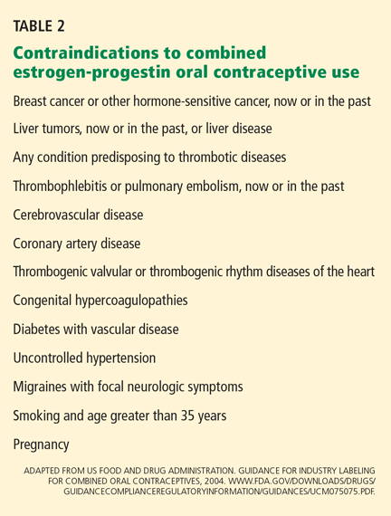
Clinicians should not overestimate the risks of oral contraceptives and other hormonal methods, but rather educate themselves so that they can utilize menstrual manipulation safely to match the individual patient’s needs.
- Association of Reproductive Health Professionals. Extended and continuous use of contraceptives to reduce menstruation. September 2004. http://www.arhp.org/publications-and-resources/clinical-proceedings/reduce-menses. Accessed May 17, 2010.
- Kjerulff KH, Erickson BA, Langenberg PW. Chronic gynecological conditions reported by US women: findings from the National Health Interview Survey, 1984 to 1992. Am J Public Health 1996; 86:195–199.
- Thomas SL, Ellertson C. Nuisance or natural and healthy: should monthly menstruation be optional for women? Lancet 2000; 355:922–924.
- Connell EB. Contraception in the prepill era. Contraception 1999; 59(suppl 1):7S–10S.
- Marks LV. Sexual chemistry: a history of the contraceptive pill. New Haven, CT: Yale University Press, 2001.
- Loudon NB, Foxwell M, Potts DM, Guild AL, Short RV. Acceptability of an oral contraceptive that reduces the frequency of menstruation: the tri-cycle pill regimen. Br Med J 1977; 2:487–490.
- Sulak PJ, Cressman BE, Waldrop E, Holleman S, Kuehl TJ. Extending the duration of active oral contraceptive pills to manage hormone withdrawal symptoms. Obstet Gynecol 1997; 89:179–183.
- Long-term reversible contraception. Twelve years of experience with the TCu380A and TCu220C. Contraception 1997; 56:341–352.
- Miller L, Notter KM. Menstrual reduction with extended use of combination oral contraceptive pills: randomized controlled trial. Obstet Gynecol 2001; 98:771–778.
- Mulders TM, Dieben TO. Use of the novel combined contraceptive vaginal ring NuvaRing for ovulation inhibition. Fertil Steril 2001; 75:865–870.
- Stanford JB, Mikolajczyk RT. Mechanisms of action of intrauterine devices: update and estimation of postfertilization effects. Am J Obstet Gynecol 2002; 187:1699–1708.
- Anderson FD, Hait H. A multicenter, randomized study of an extended cycle oral contraceptive. Contraception 2003; 68:89–96.
- Miller L, Hughes JP. Continuous combination oral contraceptive pills to eliminate withdrawal bleeding: a randomized trial. Obstet Gynecol 2003; 101:653–661.
- Sillem M, Schneidereit R, Heithecker R, Mueck AO. Use of an oral contraceptive containing drospirenone in an extended regimen. Eur J Contracept Reprod Health Care 2003; 8:162–169.
- Miller L, Verhoeven CH, Hout J. Extended regimens of the contraceptive vaginal ring: a randomized trial. Obstet Gynecol 2005; 106:473–482.
- Stewart FH, Kaunitz AM, Laguardia KD, Karvois DL, Fisher AC, Friedman AJ. Extended use of transdermal norelgestromin/ethinyl estradiol: a randomized trial. Obstet Gynecol 2005; 105:1389–1396.
- Sulak PJ, Kuehl TJ, Coffee A, Willis S. Prospective analysis of occurrence and management of breakthrough bleeding during an extended oral contraceptive regimen. Am J Obstet Gynecol 2006; 195:935–941.
- Lukes AS, Reardon B, Arepally G. Use of the levonorgestrel– releasing intrauterine system in women with hemostatic disorders. Fertil Steril 2008; 90:673–677.
- Anderson FD, Feldman R, Reape KZ. Endometrial effects of a 91-day extended-regimen oral contraceptive with low-dose estrogen in place of placebo. Contraception 2008; 77:91–96.
- Wright KP, Johnson JV. Evaluation of extended and continuous use oral contraceptives. Ther Clin Risk Manag 2008; 4:905–911.
- Edelman AB, Gallo MF, Jensen JT, Nichols MD, Schulz KF, Grimes DA. Continuous or extended cycle vs. cyclic use of combined oral contraceptives for contraception. Cochrane Database Syst Rev 2005; 3:CD004695.
- Sulak PJ, Kuehl TJ, Ortiz M, Shull BL. Acceptance of altering the standard 21-day/7-day oral contraceptive regimen to delay menses and reduce hormone withdrawal symptoms. Am J Obstet Gynecol 2002; 186:1142–1149.
- Turok D. The quest for better contraception: future methods. Obstet Gynecol Clin North Am 2007; 34:137–166.
- Bergqvist A, Rybo G. Treatment of menorrhagia with intrauterine release of progesterone. Br J Obstet Gynaecol 1983; 90:255–258.
- Andersson K, Odlind V, Rybo G. Levonorgestrel-releasing and copper-releasing (Nova T) IUDs during five years of use: a randomized comparative trial. Contraception 1994; 49:56–72.
- US Food and Drug Administration. FDA Approves Additional Use for IUD Mirena to Treat Heavy Menstrual Bleeding in IUD Users. http://www.fda.gov/NewsEvents/Newsroom/PressAnnouncements/ucm184747.htm. Accessed May 17, 2010.
- Hidalgo M, Bahamondes L, Perrotti M, Diaz J, Dantas-Monteiro C, Petta C. Bleeding patterns and clinical performance of the levonorgestrel-releasing intrauterine system (Mirena) up to two years. Contraception 2002; 65:129–132.
- Schwallie PC, Assenzo JR. Contraceptive use—efficacy study utilizing medroxyprogesterone acetate administered as an intramuscular injection once every 90 days. Fertil Steril 1973; 24:331–339.
- Kaunitz AM. Injectable contraception. New and existing options. Obstet Gynecol Clin North Am 2000; 27:741–780.
- Mainwaring R, Hales HA, Stevenson K, et al. Metabolic parameter, bleeding, and weight changes in US women using progestin only contraceptives. Contraception 1995; 51:149–153.
- Curtis KM, Martins SL. Progestogen-only contraception and bone mineral density: a systematic review. Contraception 2006; 73:470–487.
- Shaarawy M, El-Mallah SY, Seoudi S, Hassan M, Mohsen IA. Effects of the long-term use of depot medroxyprogesterone acetate as hormonal contraceptive on bone mineral density and biochemical markers of bone remodeling. Contraception 2006; 74:297–302.
- Cromer BA, Bonny AE, Stager M, et al. Bone mineral density in adolescent females using injectable or oral contraceptives: a 24-month prospective study. Fertil Steril 2008; 90:2060–2067.
- Rome E, Ziegler J, Secic M, et al. Bone biochemical markers in adolescent girls using either depot medroxyprogesterone acetate or an oral contraceptive. J Pediatr Adolesc Gynecol 2004; 17:373–377.
- More C, Bettembuk P, Bhattoa HP, Balogh A. The effects of pregnancy and lactation on bone mineral density. Osteoporos Int 2001; 12:732–737.
- Kaunitz AM, Miller PD, Rice VM, Ross D, McClung MR. Bone mineral density in women aged 25-35 years receiving depot medroxyprogesterone acetate: recovery following discontinuation. Contraception 2006; 74:90–99.
- Guilbert ER, Brown JP, Kaunitz AM, et al. The use of depot-medroxyprogesterone acetate in contraception and its potential impact on skeletal health. Contraception 2009; 79:167–177.
- Bonny AE, Ziegler J, Harvey R, Debanne SM, Secic M, Cromer BA. Weight gain in obese and nonobese adolescent girls initiating depot medroxyprogesterone, oral contraceptive pills, or no hormonal contraceptive method. Arch Pediatr Adolesc Med 2006; 160:40–45.
- van den Heuvel MW, van Bragt AJ, Alnabawy AK, Kaptein MC. Comparison of ethinylestradiol pharmacokinetics in three hormonal contraceptive formulations: the vaginal ring, the transdermal patch and an oral contraceptive. Contraception 2005; 72:168–174.
- Douketis JD, Ginsberg JS, Holbrook A, Crowther M, Duku EK, Burrows RF. A reevaluation of the risk for venous thromboembolism with the use of oral contraceptives and hormone replacement therapy. Arch Intern Med 1997; 157:1522–1530.
- Cole JA, Norman H, Doherty M, Walker AM. Venous thromboembolism, myocardial infarction, and stroke among transdermal contraceptive system users. Obstet Gynecol 2007; 109:339–346.
- Jick S, Kaye JA, Li L, Jick H. Further results on the risk of nonfatal venous thromboembolism in users of the contraceptive transdermal patch compared to users of oral contraceptives containing norgestimate and 35 microg of ethinyl estradiol. Contraception 2007; 76:4–7.
- World Health Organization. Medical Eligibility Criteria for Contraceptive Use. 3rd ed. Geneva: Reproductive Health and Research, World Health Organization; 2004.
- Dieben TO, Roumen FJ, Apter D. Efficacy, cycle control, and user acceptability of a novel combined contraceptive vaginal ring. Obstet Gynecol 2002; 100:585–593.
- Davies GC, Feng LX, Newton JR, Dieben TO, Coelingh-Bennink HJ. The effects of a combined contraceptive vaginal ring releasing ethinyloestradiol and 3-ketodesogestrel on vaginal flora. Contraception 1992; 45:511–518.
- Roumen FJ, Boon ME, van Velzen D, Dieben TO, Coelingh Bennink HJ. The cervico-vaginal epithelium during 20 cycles’ use of a combined contraceptive vaginal ring. Hum Reprod 1996; 11:2443–2448.
- Wenzl R, van Beek A, Schnabel P, Huber J. Pharmacokinetics of etonogestrel released from the contraceptive implant Implanon. Contraception 1998; 58:283–288.
- Darney P, Patel A, Rosen K, Shapiro LS, Kaunitz AM. Safety and efficacy of a single-rod etonogestrel implant (Implanon): results from 11 international clinical trials. Fertil Steril 2009; 91:1646–1653.
- Jensen JT, Speroff L. Health benefits of oral contraceptives. Obstet Gynecol Clin North Am 2000; 27:705–721.
- Seracchioli R, Mabrouk M, Frascà C, et al. Long-term cyclic and continuous oral contraceptive therapy and endometrioma recurrence: a randomized controlled trial. Fertil Steril 2010; 93:52–56.
- Falsetti L, Dordoni D, Gastaldi C, Gastaldi A. A new association of ethinylestradiol (0.035 mg) cyproterone acetate (2 mg) in the therapy of polycystic ovary syndrome. Acta Eur Fertil 1986; 17:19–25.
- Koltun W, Lucky AW, Thiboutot D, et al. Efficacy and safety of 3 mg drospirenone/20 mcg ethinylestradiol oral contraceptive administered in 24/4 regimen in the treatment of acne vulgaris: a randomized, double-blind, placebo-controlled trial. Contraception 2008; 77:249–256.
- Kadir RA, Sabin CA, Pollard D, Lee CA, Economides DL. Quality of life during menstruation in patients with inherited bleeding disorders. Haemophilia 1998; 4:836–841.
- Schneider MB, Fisher M, Friedman SB, Bijur PE, Toffler AP. Menstrual and premenstrual issues in female military cadets: a unique population with significant concerns. J Pediatr Adolesc Gynecol 1999; 12:195–201.
- Bennell K, White S, Crossley K. The oral contraceptive pill: a revolution for sportswomen? Br J Sports Med 1999; 33:231–238.
- Kaplowitz PB, Oberfield SE. Reexamination of the age limit for defining when puberty is precocious in girls in the United States: implications for evaluation and treatment. Drug and Therapeutics and Executive Committees of the Lawson Wilkins Pediatric Endocrine Society. Pediatrics 1999; 104:936–941.
- Roxburgh DR, West MJ. The use of norethisterone to suppress menstruation in the intellectually severely retarded woman. Med J Aust 1973; 2:310–313.
- Egan TM, Siegert RJ, Fairley NA. Use of hormonal contraceptives in an institutional setting: reasons for use, consent and safety in women with psychiatric and intellectual disabilities. N Z Med J 1993; 106:338–341.
- Pillai M, O’Brien K, Hill E. The levonorgestrel intrauterine system (Mirena) for the treatment of menstrual problems in adolescents with medical disorders, or physical or learning disabilities. BJOG 2010; 117:216–221.
- Archer DF, Jensen JT, Johnson JV, Borisute H, Grubb GS, Constantine GD. Evaluation of a continuous regimen of levonorgestrel/ethinyl estradiol: phase 3 study results. Contraception 2006; 74:439–445.
- Kroll R, Reape KZ, Margolis M. The efficacy and safety of a low-dose, 91-day, extended-regimen oral contraceptive with continuous ethinyl estradiol. Contraception 2010; 81:41–48.
- Archer DF. Menstrual-cycle-related symptoms: a review of the rationale for continuous use of oral contraceptives. Contraception 2006; 74:359–366.
- den Tonkelaar I, Oddens BJ. Preferred frequency and characteristics of menstrual bleeding in relation to reproductive status, oral contraceptive use, and hormone replacement therapy use. Contraception 1999; 59:357–362.
- Edelman A, Lew R, Cwiak C, Nichols M, Jensen J. Acceptability of contraceptive-induced amenorrhea in a racially diverse group of US women. Contraception 2007; 75:450–453.
- Gerschultz KL, Sucato GS, Hennon TR, Murray PJ, Gold MA. Extended cycling of combined hormonal contraceptives in adolescents: physician views and prescribing practices. J Adolesc Health 2007; 40:151–157.
- Frankovich RJ, Lebrun CM. Menstrual cycle, contraception, and performance. Clin Sports Med 2000; 19:251–271.
- Speroff L, Darney PD. A Clinical Guide for Contraception. 4th ed. Philadelphia: Lippincott Williams & Wilkins; 2005.
- Kaunitz AM. Long-acting contraceptive options. Int J Fertil Menopausal Stud 1996; 41:69–76.
- US Food and Drug Administration. Guidance for Industry Labeling for Combined Oral Contraceptives, 2004. http://www.fda.gov/downloads/Drugs/GuidanceComplianceRegulatoryInformation/Guidances/ucm075075.pdfAccessed May 17, 2010.
If they wish, women can have more control over when and if they menstruate. By using hormonal contraceptives in extended or continuous regimens, they can have their period less often, a practice called menstrual manipulation or menstrual suppression.
Actually, with the help of their clinicians, women have been doing this for years. But now that several products have been approved by the US Food and Drug Administration (FDA) specifically for use in extended or continuous regimens, the practice has become more widely accepted.
Reasons for suppressing menstrual flow range from avoiding bleeding during a particular event (eg, a wedding, graduation, or sports competition) to finding relief from dysmenorrhea or reducing or eliminating menstruation in the treatment of endometriosis, migraine, and other medical conditions exacerbated by hormonal changes around the time of menses.1 Alternatively, some women may practice menstrual manipulation for no other reason than to simply avoid menstruation.
MENSTRUAL DISORDERS ARE TROUBLESOME, COMMON
Each year in the United States, menstrual disorders such as dysmenorrhea (painful menstruation), menorrhagia (excessive or frequent menstruation), metrorrhagia (irregular menstruation), menometrorrhagia (excessive and irregular menstruation), and premenstrual syndrome affect nearly 2.5 million women age 18 to 50 years.2 Menstrual disorders are the leading cause of gynecologic morbidity in the United States, outnumbering adnexal masses (the second most common cause) by a factor of three.2 In addition, these disorders extend into the workplace, costing US industry about 8% of its total wage bill.3
A BRIEF HISTORY OF CONTRACEPTIVE DEVELOPMENT
The idea of using progestins for birth control was first advanced in the 1950s by Dr. Gregory Pincus, who proposed a regimen of 21 days of active drug followed by 7 drug-free days to allow withdrawal bleeding, mimicking the natural cycle.4 This “21/7” regimen was designed to follow the lunar cycle in the hope it would be, in the words of Dr. John Rock, “a morally permissible variant of the rhythm method,”5 thereby making it acceptable to women, clinicians, and the Catholic Church.
In 1977, Loudon et al6 reported the results of a study in which women took active pills for 84 days instead of 21 days, which reduced the frequency of menstruation to every 3 months. Since then, extending the active pills beyond 21 days to avoid menses and other hormone-withdrawal symptoms has become popular in clinical practice, and many studies have investigated the extended or continuous use of oral and other forms of contraception to delay menses.7–18
CURRENT METHODS OF MENSTRUAL MANIPULATION
A variety of available products prevent conception by altering the menstrual cycle:
- Oral estrogen-progestin contraceptive pills
- A drug-releasing intrauterine device
- Depot medroxyprogesterone acetate injections
- A contraceptive vaginal ring
- An implantable etonogestrel contraceptive.
Their use in menstrual manipulation is summarized in Table 1.
Oral contraceptive pills
The most common way to manipulate the menstrual cycle is to extend the time between hormone-free weeks in an oral contraceptive regimen.
If the patient is young, you can prescribe a monophasic 21/7 oral contraceptive and tell her to take one active pill every day for 21 days and then start a new pack and keep taking active pills for up to 84 consecutive days, skipping the placebo pills until she wants to have her menstrual period. She can choose which week to have it: if the scheduled 12th week of an extended-cycle oral contraceptive regimen is inconvenient, she can plan it for week 10, or week 9, or whichever week is convenient.
The rationale for using an 84-day (12-week) cycle is that it still provides four periods per year, alleviating fears of hypertrophic endometrium.19
In this scenario, unscheduled or breakthrough bleeding can be managed by taking a “double-up pill” from a spare pack on any day breakthrough bleeding occurs and until it resolves. Menstrual periods should not be planned for intervals shorter than 21 days, owing to the risk of ovulation. Missed days of pills or use of placebo pills should also not exceed 7 days to prevent escape ovulation. 20
In some women with endometriosis and other medical reasons, continuous oral contraception with no placebo week can be prescribed.
Unfortunately, the downside to suppressing withdrawal bleeding is unscheduled or “breakthrough” bleeding. The best way to treat this unscheduled bleeding is not known. Patients who are not sexually active can be reassured that the goal of an atrophic endometrium can still be achieved, with resultant pill amenorrhea (particularly useful for those with severe dysmenorrhea or other reasons to want to avoid flow). Patients could also try to manage flow by periodically taking a 3- to 5-day break from hormone-containing pills to allow flow. They can also try switching to another oral contraceptive that has a different progestin that would spiral the arterioles of the endometrium more tightly and thus more aggressively induce atrophy.13,17,21 For instance, levonorgestrel is 10 to 20 times more potent than norethindrone. Choosing a pill with a higher monophasic dosing of levonorgestrel or a similar progestin may minimize unscheduled bleeding.
Currently, several oral contraceptives are approved for use in an extended regimen.
Seasonale was the first oral contraceptive marketed in the United States with an extended active regimen.22 It comes in a pack of 84 pills containing ethinyl estradiol 0.03 mg and levonorgestrel 0.15 mg, plus 7 placebo pills.
Seasonique is similar to Seasonale, but instead of placebo pills it has seven pills that contain ethinyl estradiol 0.010 mg.
Lybrel is a low-dose combination containing ethinyl estradiol 0.02 mg and levonorgestrel 0.09 mg. Packaged as an entire year’s worth of active pills to be taken continuously for 365 days without a placebo phase or pillfree interval,23 it is the only FDA-approved continuous oral contraceptive available in the United States.
An intrauterine device
Intrauterine devices were originally developed as contraceptives. The addition of a progestin to these devices has been shown to reduce heavy menstrual bleeding by up to 90%.24,25
Mirena IUS, a levonorgestrel-releasing device, is the only medicated intrauterine device that is currently available in the United States. (“IUS” stands for “intrauterine system.”) It was recently approved by the FDA to treat heavy menstrual bleeding in women who use intrauterine contraception as their method of pregnancy prevention.26 About 50% of women who use this device develop amenorrhea within 6 months of insertion, while 25% report oligomenorrhea.27
The Mirena device can be left in the uterus for up to 5 years. It may be a good choice for inducing amenorrhea in women with hemostatic disorders or in whom estrogen either is contraindicated or causes health concerns.18 The copper intrauterine device (Paragard; Duramed Pharmaceuticals Inc., Pomona, NY) remains a viable option for those who cannot or do not tolerate hormonal therapy. However, Mirena may provide less unscheduled bleeding than the copper intrauterine device.
Depot medroxyprogesterone acetate injections
Depo-Provera (depot medroxyprogesterone acetate) injections are given at 90-day intervals. 28 This contraceptive method inhibits ovulation and decidualizes the endometrium, thereby reducing or eliminating uterine bleeding. 29
While new users may initially experience excessive prolonged bleeding (10 or more days) while shedding their existing lining, the rate of amenorrhea has been shown to increase over time as the lining atrophies.30 Thus, prolonged use of this agent reduces the frequency of menstruation as well as menstruation-related symptoms.
Depot medroxyprogesterone acetate is ideal for patients whose menstrual periods pose a significant hygiene problem (eg, developmentally challenged girls). In our experience, the injections can be given at shorter intervals to induce atrophy of the endometrium quickly. In this scenario, the clinician might give an injection every 4 to 6 weeks for two or three doses to induce amenorrhea and then return to every-12-week dosing.
The main risk when using medroxyprogesterone injections to induce amenorrhea is the potential for bone loss. Users of this method have been shown to have lower mean bone mineral density31–33 and significantly higher levels of biomarkers of bone formation and resorption32,34 than nonusers. However, these changes are similar to those seen in breastfeeding women,35 are reversible with cessation, 36 and are not associated with increased fracture risk.37 In adolescent girls, pregnancy poses similar risks to the bones, with longerterm consequences.
Medroxyprogesterone can also stimulate appetite, causing 10 to 20 kg of weight gain in adolescents and women who are already obese and have trouble with appetite regulation.38 Slender users tend not to gain weight, however.
Given this information, depot medroxyprogesterone acetate appears to be a cost-effective contraceptive option that should be considered in the context of the clinical situation and preference of each patient.
Transdermal contraceptive patch
Ortho Evra, a transdermal patch, is designed to deliver ethinyl estradiol 0.02 mg and norelgestromin 0.150 mg daily.39 It is usually applied weekly for 3 weeks, followed by a patch-free week to induce regular monthly withdrawal bleeding.
Extended use of the patch to manipulate menstruation is an off-label use. In the only trial evaluating extended use of the patch, amenorrhea occurred in 12% of users, but unscheduled bleeding and spotting were common. 16
Although there is some evidence that the long-term use of the patch may increase the risk of venous thromboembolism,40,41 the risk in women who use the patch has been found to be similar to that in women using an oral contraceptive.42 However, serum ethinyl estradiol levels have been found to be higher with the use of the weekly patch than with oral contraceptives or the contraceptive vaginal ring39; as a result, many physicians are hesitant to recommend its continuous use.
Pending further data about the safety profile of this contraceptive, the World Health Organization Medical Eligibility Criteria for Contraceptive Use suggest that the same guidelines for the prescription of combination oral contraceptives should also apply to the patch.43
Contraceptive vaginal ring
NuvaRing, a contraceptive vaginal ring, releases a daily dose of ethinyl estradiol 0.015 mg and etonogestrel 0.12 mg.10 It is inserted, left in for 21 days, and then removed and left out for 7 days, during which withdrawal bleeding occurs.10
Vaginal administration has been shown to allow low, continuous dosing, which results in more stable serum concentrations than with the patch or oral contraceptives.39 In the only trial comparing an extended vaginal ring regimen and the traditional 28-day regimen, extended use resulted in fewer overall days of bleeding than monthly use, but with more unscheduled spotting.15
The most common side effects include headache, vaginitis, and leukorrhea,44 but there is no evidence of bacteriologic or cytologic changes in the cervicovaginal epithelium, even with extended use.45,46
Etonogestrel implantable contraceptive
Implanon, a single-rod progestin implant, is available in the United States and elsewhere. It is placed subdermally in the inner upper arm and provides contraception for as long as 3 years.
Implanon contains 68 mg of the progestin etonogestrel, which it slowly releases over time, initially at 0.06 to 0.07 mg/day, decreasing to 0.035 to 0.045 mg/day at the end of the first year, to 0.03 to 0.04 mg/day at the end of the second year, and then to 0.025 to 0.03 mg/day at the end of the third year.47
The amount of vaginal bleeding associated with the use of the implant is generally modest, but the pattern tends to be unpredictable. 48 In addition, because amenorrhea is reported as a side effect in only 22% of women during the first 2 years of its use,48 the progestin implant is a less satisfactory means of menstrual suppression than the other methods discussed above.
BENEFITS OF MENSTRUAL MANIPULATION
Menstrual manipulation has a number of benefits in terms of both overall health and lifestyle.
For most women, using a long-acting hormonal contraceptive carries low risks and substantial health benefits. Women who take oral contraceptives are less likely to develop osteoporosis, ovarian or endometrial cancer, benign breast changes, or pelvic inflammatory disease. 49 Long-term use of an oral contraceptive can also preserve fertility by reducing and delaying the incidence of endometriosis,50 and is effective at treating acne vulgaris, which tends to be common among patients with polycystic ovary syndrome.51,52 In addition, this practice can be used to reduce overall blood loss, an application that is particularly important in women with a bleeding diathesis such as von Willebrand disease, who frequently suffer from menorrhagia.53
Reduced menstruation may also prove more convenient during particular occasions, such as vacations and athletic activities. Specifically, it may be useful to women serving in the military. In a study by Schneider et al,54 a cohort of 83 female cadets reported a significant perceived impact of premenstrual and menstrual-related symptoms on academic, physical, and military activities, as well as difficulties in obtaining, changing, and disposing of menstrual materials in a military setting. Likewise, reduced menstrual frequency or amenorrhea may play an important role in female athletes, who reportedly use oral contraceptives to control premenstrual symptoms, to protect bone health, and to manipulate the menstrual cycle in order to maximize performance.55
Adolescent girls are another group who may benefit from reduced or absent menses, once they have reached near-final height. By practicing menstrual suppression, girls can avoid dysmenorrhea and the inconvenience of menstruation during the school day, when their access to painkillers, sanitary pads or tampons, and a change of clothes may be limited. 56 Clinicians who discuss with teenage patients the benefits of innovative hormonal contraceptive schedules that reduce menstrual frequency may be able to improve the quality of life for these young women.
In a very short girl just after menarche, care must be taken not to start a hormonal method too early so as not to prematurely close epiphyses and stunt final height; after menarche, most girls still have 1 to 4 inches of potential growth. For a young lady 4 feet 11 inches tall, that extra inch may be important.
Finally, menstrual manipulation may also find a niche among the developmentally challenged. Women with cognitive impairment and physical disabilities may have difficulty with hygienic practice around menses. For a number of years, contraceptives have been used to manage menstrual hygiene in patients with catamenial (ie, menstrual) epilepsy, and to address caregiver concerns in women with severe mental retardation, with improved behavior noted in some patients.57–59 In this setting, an agent that suppresses menses and also provides contraception, especially for those girls and women at risk of abuse, may offer substantial benefits.
DISADVANTAGES OF MENSTRUAL MANIPULATION
Rates of adverse events and of discontinuation of extended and continuous oral contraceptive regimens are comparable with those reported for cyclic regimens, except for higher rates of breakthrough bleeding.
In a trial of continuous oral contraceptive use in more than 2,000 patients, 396 (18.5%) withdrew from the study as a result of bothersome uterine bleeding.60 However, while breakthrough bleeding often occurs during the first few months of extended oral contraceptive use, it usually decreases with each successive cycle of therapy and is comparable to that reported by patients on the conventional oral contraceptive regimen by the fourth extended cycle.12
CONTRACEPTIVE EFFICACY
The efficacy of extended and continuous oral contraceptive regimens is comparable with that of cyclic regimens.12,60,61 One reason for this may be better adherence to continuous regimens: women using this regimen have been shown to miss fewer pills than those on a cyclic regimen, especially during the critical first week of the pill pack.21
Several studies have shown that some women ovulate during the standard 21/7 oral contraceptive regimen even if they do not miss any pills or take pills off-schedule, putting them at greater risk of pregnancy.62 Large studies evaluating the efficacy of an extendedcycle regimen have shown a pregnancy rate during the 1-year study period that was either comparable with61 or lower than12,60 rates with standard regimens.
Heterosexual couples need to be advised to use condoms to further reduce the already low failure rate and to prevent sexually transmitted diseases.
ACCEPTABILITY OF MENSTRUAL MANIPULATION
Ever since the earliest trial of an extended oral contraceptive regimen, participants have expressed a favorable response to the resulting decrease in menstrual frequency; in the 1977 study by Loudon et al,6 patients on the extended regimen cited infrequent periods (82%), fewer menstrual problems (20%), and easier pill-taking (19%) as favorable features.
In 1999, den Tonkelaar and Oddens63 surveyed 1,300 Dutch women about their preferred frequency of menstruation and found that about 70% between the ages of 15 and 49 preferred a frequency of between every 3 months and never. A similar survey in the United States indicated that 58% preferred a bleeding frequency of either every 3 months or never to more frequent periods.64
While patients find menstrual manipulation generally acceptable, clinician approval has been more varied. Loudon et al reported that “the doctors and nurses on the clinic staff were less enthusiastic about this regimen than the volunteers themselves.”6 In a survey of 222 clinicians,65 90% of responders reported ever having prescribed extended or continuous dosing regimens to adolescents, and 33% reported that extended cycles made up more than 10% of their total oral contraceptive prescriptions.
Myths and misperceptions about menstrual manipulation abound. Many clinicians believe that routine use of an extended or continuous oral contraceptive regimen is inadvisable, despite the lack of evidence to support this notion.66 Therefore, many care providers need more education about the practice and benefits of menstrual manipulation.
THE RIGHT METHOD FOR THE RIGHT PATIENT
Manipulation and suppression of menstruation through continuous or extended use of oral contraceptives or by other means may have a number of advantages to women, including fewer menstrual-related syndromes, reduced absenteeism from work or school, and greater overall satisfaction.
For women whose goal is to reduce but not necessarily to eliminate monthly bleeding, the cyclic use of estrogen-progestin contraception (rather than progestins alone or continuous use of combined hormonal preparations) is suggested.
Clinicians should not overestimate the risks of oral contraceptives and other hormonal methods, but rather educate themselves so that they can utilize menstrual manipulation safely to match the individual patient’s needs.
- Association of Reproductive Health Professionals. Extended and continuous use of contraceptives to reduce menstruation. September 2004. http://www.arhp.org/publications-and-resources/clinical-proceedings/reduce-menses. Accessed May 17, 2010.
- Kjerulff KH, Erickson BA, Langenberg PW. Chronic gynecological conditions reported by US women: findings from the National Health Interview Survey, 1984 to 1992. Am J Public Health 1996; 86:195–199.
- Thomas SL, Ellertson C. Nuisance or natural and healthy: should monthly menstruation be optional for women? Lancet 2000; 355:922–924.
- Connell EB. Contraception in the prepill era. Contraception 1999; 59(suppl 1):7S–10S.
- Marks LV. Sexual chemistry: a history of the contraceptive pill. New Haven, CT: Yale University Press, 2001.
- Loudon NB, Foxwell M, Potts DM, Guild AL, Short RV. Acceptability of an oral contraceptive that reduces the frequency of menstruation: the tri-cycle pill regimen. Br Med J 1977; 2:487–490.
- Sulak PJ, Cressman BE, Waldrop E, Holleman S, Kuehl TJ. Extending the duration of active oral contraceptive pills to manage hormone withdrawal symptoms. Obstet Gynecol 1997; 89:179–183.
- Long-term reversible contraception. Twelve years of experience with the TCu380A and TCu220C. Contraception 1997; 56:341–352.
- Miller L, Notter KM. Menstrual reduction with extended use of combination oral contraceptive pills: randomized controlled trial. Obstet Gynecol 2001; 98:771–778.
- Mulders TM, Dieben TO. Use of the novel combined contraceptive vaginal ring NuvaRing for ovulation inhibition. Fertil Steril 2001; 75:865–870.
- Stanford JB, Mikolajczyk RT. Mechanisms of action of intrauterine devices: update and estimation of postfertilization effects. Am J Obstet Gynecol 2002; 187:1699–1708.
- Anderson FD, Hait H. A multicenter, randomized study of an extended cycle oral contraceptive. Contraception 2003; 68:89–96.
- Miller L, Hughes JP. Continuous combination oral contraceptive pills to eliminate withdrawal bleeding: a randomized trial. Obstet Gynecol 2003; 101:653–661.
- Sillem M, Schneidereit R, Heithecker R, Mueck AO. Use of an oral contraceptive containing drospirenone in an extended regimen. Eur J Contracept Reprod Health Care 2003; 8:162–169.
- Miller L, Verhoeven CH, Hout J. Extended regimens of the contraceptive vaginal ring: a randomized trial. Obstet Gynecol 2005; 106:473–482.
- Stewart FH, Kaunitz AM, Laguardia KD, Karvois DL, Fisher AC, Friedman AJ. Extended use of transdermal norelgestromin/ethinyl estradiol: a randomized trial. Obstet Gynecol 2005; 105:1389–1396.
- Sulak PJ, Kuehl TJ, Coffee A, Willis S. Prospective analysis of occurrence and management of breakthrough bleeding during an extended oral contraceptive regimen. Am J Obstet Gynecol 2006; 195:935–941.
- Lukes AS, Reardon B, Arepally G. Use of the levonorgestrel– releasing intrauterine system in women with hemostatic disorders. Fertil Steril 2008; 90:673–677.
- Anderson FD, Feldman R, Reape KZ. Endometrial effects of a 91-day extended-regimen oral contraceptive with low-dose estrogen in place of placebo. Contraception 2008; 77:91–96.
- Wright KP, Johnson JV. Evaluation of extended and continuous use oral contraceptives. Ther Clin Risk Manag 2008; 4:905–911.
- Edelman AB, Gallo MF, Jensen JT, Nichols MD, Schulz KF, Grimes DA. Continuous or extended cycle vs. cyclic use of combined oral contraceptives for contraception. Cochrane Database Syst Rev 2005; 3:CD004695.
- Sulak PJ, Kuehl TJ, Ortiz M, Shull BL. Acceptance of altering the standard 21-day/7-day oral contraceptive regimen to delay menses and reduce hormone withdrawal symptoms. Am J Obstet Gynecol 2002; 186:1142–1149.
- Turok D. The quest for better contraception: future methods. Obstet Gynecol Clin North Am 2007; 34:137–166.
- Bergqvist A, Rybo G. Treatment of menorrhagia with intrauterine release of progesterone. Br J Obstet Gynaecol 1983; 90:255–258.
- Andersson K, Odlind V, Rybo G. Levonorgestrel-releasing and copper-releasing (Nova T) IUDs during five years of use: a randomized comparative trial. Contraception 1994; 49:56–72.
- US Food and Drug Administration. FDA Approves Additional Use for IUD Mirena to Treat Heavy Menstrual Bleeding in IUD Users. http://www.fda.gov/NewsEvents/Newsroom/PressAnnouncements/ucm184747.htm. Accessed May 17, 2010.
- Hidalgo M, Bahamondes L, Perrotti M, Diaz J, Dantas-Monteiro C, Petta C. Bleeding patterns and clinical performance of the levonorgestrel-releasing intrauterine system (Mirena) up to two years. Contraception 2002; 65:129–132.
- Schwallie PC, Assenzo JR. Contraceptive use—efficacy study utilizing medroxyprogesterone acetate administered as an intramuscular injection once every 90 days. Fertil Steril 1973; 24:331–339.
- Kaunitz AM. Injectable contraception. New and existing options. Obstet Gynecol Clin North Am 2000; 27:741–780.
- Mainwaring R, Hales HA, Stevenson K, et al. Metabolic parameter, bleeding, and weight changes in US women using progestin only contraceptives. Contraception 1995; 51:149–153.
- Curtis KM, Martins SL. Progestogen-only contraception and bone mineral density: a systematic review. Contraception 2006; 73:470–487.
- Shaarawy M, El-Mallah SY, Seoudi S, Hassan M, Mohsen IA. Effects of the long-term use of depot medroxyprogesterone acetate as hormonal contraceptive on bone mineral density and biochemical markers of bone remodeling. Contraception 2006; 74:297–302.
- Cromer BA, Bonny AE, Stager M, et al. Bone mineral density in adolescent females using injectable or oral contraceptives: a 24-month prospective study. Fertil Steril 2008; 90:2060–2067.
- Rome E, Ziegler J, Secic M, et al. Bone biochemical markers in adolescent girls using either depot medroxyprogesterone acetate or an oral contraceptive. J Pediatr Adolesc Gynecol 2004; 17:373–377.
- More C, Bettembuk P, Bhattoa HP, Balogh A. The effects of pregnancy and lactation on bone mineral density. Osteoporos Int 2001; 12:732–737.
- Kaunitz AM, Miller PD, Rice VM, Ross D, McClung MR. Bone mineral density in women aged 25-35 years receiving depot medroxyprogesterone acetate: recovery following discontinuation. Contraception 2006; 74:90–99.
- Guilbert ER, Brown JP, Kaunitz AM, et al. The use of depot-medroxyprogesterone acetate in contraception and its potential impact on skeletal health. Contraception 2009; 79:167–177.
- Bonny AE, Ziegler J, Harvey R, Debanne SM, Secic M, Cromer BA. Weight gain in obese and nonobese adolescent girls initiating depot medroxyprogesterone, oral contraceptive pills, or no hormonal contraceptive method. Arch Pediatr Adolesc Med 2006; 160:40–45.
- van den Heuvel MW, van Bragt AJ, Alnabawy AK, Kaptein MC. Comparison of ethinylestradiol pharmacokinetics in three hormonal contraceptive formulations: the vaginal ring, the transdermal patch and an oral contraceptive. Contraception 2005; 72:168–174.
- Douketis JD, Ginsberg JS, Holbrook A, Crowther M, Duku EK, Burrows RF. A reevaluation of the risk for venous thromboembolism with the use of oral contraceptives and hormone replacement therapy. Arch Intern Med 1997; 157:1522–1530.
- Cole JA, Norman H, Doherty M, Walker AM. Venous thromboembolism, myocardial infarction, and stroke among transdermal contraceptive system users. Obstet Gynecol 2007; 109:339–346.
- Jick S, Kaye JA, Li L, Jick H. Further results on the risk of nonfatal venous thromboembolism in users of the contraceptive transdermal patch compared to users of oral contraceptives containing norgestimate and 35 microg of ethinyl estradiol. Contraception 2007; 76:4–7.
- World Health Organization. Medical Eligibility Criteria for Contraceptive Use. 3rd ed. Geneva: Reproductive Health and Research, World Health Organization; 2004.
- Dieben TO, Roumen FJ, Apter D. Efficacy, cycle control, and user acceptability of a novel combined contraceptive vaginal ring. Obstet Gynecol 2002; 100:585–593.
- Davies GC, Feng LX, Newton JR, Dieben TO, Coelingh-Bennink HJ. The effects of a combined contraceptive vaginal ring releasing ethinyloestradiol and 3-ketodesogestrel on vaginal flora. Contraception 1992; 45:511–518.
- Roumen FJ, Boon ME, van Velzen D, Dieben TO, Coelingh Bennink HJ. The cervico-vaginal epithelium during 20 cycles’ use of a combined contraceptive vaginal ring. Hum Reprod 1996; 11:2443–2448.
- Wenzl R, van Beek A, Schnabel P, Huber J. Pharmacokinetics of etonogestrel released from the contraceptive implant Implanon. Contraception 1998; 58:283–288.
- Darney P, Patel A, Rosen K, Shapiro LS, Kaunitz AM. Safety and efficacy of a single-rod etonogestrel implant (Implanon): results from 11 international clinical trials. Fertil Steril 2009; 91:1646–1653.
- Jensen JT, Speroff L. Health benefits of oral contraceptives. Obstet Gynecol Clin North Am 2000; 27:705–721.
- Seracchioli R, Mabrouk M, Frascà C, et al. Long-term cyclic and continuous oral contraceptive therapy and endometrioma recurrence: a randomized controlled trial. Fertil Steril 2010; 93:52–56.
- Falsetti L, Dordoni D, Gastaldi C, Gastaldi A. A new association of ethinylestradiol (0.035 mg) cyproterone acetate (2 mg) in the therapy of polycystic ovary syndrome. Acta Eur Fertil 1986; 17:19–25.
- Koltun W, Lucky AW, Thiboutot D, et al. Efficacy and safety of 3 mg drospirenone/20 mcg ethinylestradiol oral contraceptive administered in 24/4 regimen in the treatment of acne vulgaris: a randomized, double-blind, placebo-controlled trial. Contraception 2008; 77:249–256.
- Kadir RA, Sabin CA, Pollard D, Lee CA, Economides DL. Quality of life during menstruation in patients with inherited bleeding disorders. Haemophilia 1998; 4:836–841.
- Schneider MB, Fisher M, Friedman SB, Bijur PE, Toffler AP. Menstrual and premenstrual issues in female military cadets: a unique population with significant concerns. J Pediatr Adolesc Gynecol 1999; 12:195–201.
- Bennell K, White S, Crossley K. The oral contraceptive pill: a revolution for sportswomen? Br J Sports Med 1999; 33:231–238.
- Kaplowitz PB, Oberfield SE. Reexamination of the age limit for defining when puberty is precocious in girls in the United States: implications for evaluation and treatment. Drug and Therapeutics and Executive Committees of the Lawson Wilkins Pediatric Endocrine Society. Pediatrics 1999; 104:936–941.
- Roxburgh DR, West MJ. The use of norethisterone to suppress menstruation in the intellectually severely retarded woman. Med J Aust 1973; 2:310–313.
- Egan TM, Siegert RJ, Fairley NA. Use of hormonal contraceptives in an institutional setting: reasons for use, consent and safety in women with psychiatric and intellectual disabilities. N Z Med J 1993; 106:338–341.
- Pillai M, O’Brien K, Hill E. The levonorgestrel intrauterine system (Mirena) for the treatment of menstrual problems in adolescents with medical disorders, or physical or learning disabilities. BJOG 2010; 117:216–221.
- Archer DF, Jensen JT, Johnson JV, Borisute H, Grubb GS, Constantine GD. Evaluation of a continuous regimen of levonorgestrel/ethinyl estradiol: phase 3 study results. Contraception 2006; 74:439–445.
- Kroll R, Reape KZ, Margolis M. The efficacy and safety of a low-dose, 91-day, extended-regimen oral contraceptive with continuous ethinyl estradiol. Contraception 2010; 81:41–48.
- Archer DF. Menstrual-cycle-related symptoms: a review of the rationale for continuous use of oral contraceptives. Contraception 2006; 74:359–366.
- den Tonkelaar I, Oddens BJ. Preferred frequency and characteristics of menstrual bleeding in relation to reproductive status, oral contraceptive use, and hormone replacement therapy use. Contraception 1999; 59:357–362.
- Edelman A, Lew R, Cwiak C, Nichols M, Jensen J. Acceptability of contraceptive-induced amenorrhea in a racially diverse group of US women. Contraception 2007; 75:450–453.
- Gerschultz KL, Sucato GS, Hennon TR, Murray PJ, Gold MA. Extended cycling of combined hormonal contraceptives in adolescents: physician views and prescribing practices. J Adolesc Health 2007; 40:151–157.
- Frankovich RJ, Lebrun CM. Menstrual cycle, contraception, and performance. Clin Sports Med 2000; 19:251–271.
- Speroff L, Darney PD. A Clinical Guide for Contraception. 4th ed. Philadelphia: Lippincott Williams & Wilkins; 2005.
- Kaunitz AM. Long-acting contraceptive options. Int J Fertil Menopausal Stud 1996; 41:69–76.
- US Food and Drug Administration. Guidance for Industry Labeling for Combined Oral Contraceptives, 2004. http://www.fda.gov/downloads/Drugs/GuidanceComplianceRegulatoryInformation/Guidances/ucm075075.pdfAccessed May 17, 2010.
KEY POINTS
- The options for menstrual manipulation are extended or continuous regimens of oral, transdermal, or vaginal hormonal contraceptives; a levonorgestrel-releasing intrauterine device; a progestin implant; and depot medroxyprogesterone injections.
- Benefits include fewer menstrual-related syndromes, less absenteeism from work or school, and greater overall satisfaction. Medical indications for it are conditions exacerbated by hormonal changes around the time of menses.
- The main disadvantage is a higher rate of breakthrough bleeding.
- Myths and misperceptions about menstrual manipulation persist; some physicians believe it is somehow inadvisable.
Coenzyme Q10: A therapy for hypertension and statin-induced myalgia?
Coenzyme Q10 supplements have been purported to be effective for treating a variety of disorders,1,2 in particular hypertension and statin-induced myalgia.
Several studies3–7 found that coenzyme Q10 supplementation significantly lowered blood pressure in hypertensive patients. Moreover, some trials have demonstrated that statin therapy reduces serum or muscle levels of coenzyme Q10,8–14 prompting investigations to determine whether coenzyme Q10 deficiency is related to statin-induced muscle pain.15–17
In this review, we discuss the efficacy and safety of coenzyme Q10 supplementation in patients with hypertension and those taking statins, and some of the caveats about using supplements that are not approved by the US Food and Drug Administration (FDA), as well as the bioavailability and quality of available formulations.
WHAT IS COENZYME Q10?
Coenzyme Q10, also known as coenzyme Q, ubidecarenone, and ubiquinone, is found in all human cells, with the highest concentrations in the heart, liver, kidney, and pancreas.1,2 It is a potent antioxidant, a membrane stabilizer, and an integral cofactor in the mitochondrial respiratory chain, helping to generate adenosine triphosphate, the major cellular energy source.1,2,18 It may also regulate genes associated with cell metabolism.19
RATIONALE FOR SUPPLEMENTATION
Coenzyme Q10 supplementation has been used, recommended, or studied in heart failure, hypertension, parkinsonism, mitochondrial encephalomyopathies, and other ailments.
In hypertension
Depending on the class, various antihypertensive drugs can have adverse effects such as depression, cough, and cardiac and renal dysfunction. 20,21 Furthermore, many patients need to take more than one drug to control their blood pressure, increasing their risk of side effects. Some researchers believe coenzyme Q10 supplementation may reduce the need to take multiple antihypertensive drugs.5
Coenzyme Q10 appears to lower blood pressure. The exact mechanism is not known, but one theory is that it reduces peripheral resistance by preserving nitric oxide.21 Nitric oxide relaxes peripheral arteries, lowering blood pressure. In some forms of hypertension, superoxide radicals that inactivate nitric oxide are overproduced; coenzyme Q10, with its antioxidant effects, may prevent the inactivation of nitric oxide by these free radicals. Alternatively, coenzyme Q10 may boost the production of the prostaglandin prostacyclin (PGI2) a potent vasodilator and inhibitor of platelet aggregation, or it may enhance the sensitivity of arterial smooth muscle to PGI2, or both.1,22
In patients taking statins
Hydroxymethylglutaryl coenzyme A reductase inhibitors (statins), first-line agents for lowering cholesterol levels to prevent cardiovascular disease, are some of the most commonly prescribed medications.23,24 However, statin therapy carries a risk of myopathy, which can range from muscle aches to rhabdomyolysis. 23,24
In a clinical advisory,25 the American College of Cardiology, the American Heart Association, and the National Heart, Lung, and Blood Institute recommend that patients on statin therapy who experience muscle soreness, tenderness, or pain with serum creatine kinase levels 3 to 10 times the upper limit of normal should have their creatine kinase level checked weekly. If the level is 3 to 10 times the upper limit of normal, statin therapy may be continued, but if it exceeds 10 times the upper limit, then statins and other potential offending agents (eg, niacin, fibrate) need to be discontinued.
Statins inhibit the synthesis of cholesterol by reducing the production of mevalonate, a precursor of both cholesterol and coenzyme Q10. Since both cholesterol and coenzyme Q10 are produced by the same pathway, it is not surprising that statins have been reported to reduce serum and muscle coenzyme Q10 levels.9–14 However, one study did not report a significant reduction of coenzyme Q10 levels in muscle tissue in patients treated with simvastatin 20 mg for 6 months.26
Nonetheless, researchers have hypothesized that a reduction in coenzyme Q10 levels in muscle tissue causes mitochondrial dysfunction, which could increase the risk of statininduced myopathy,13–17 and some believe that treatment with coenzyme Q10 may reduce myalgic symptoms and allow patients to remain on statin therapy.13,24
Researchers have investigated the potential of coenzyme Q10 supplementation to reduce or prevent statin-induced myopathy.15–17 (More on this below.)
Interestingly, a randomized, placebo-controlled trial27 found that 6 months of daily therapy with simvastatin (Zocor) 20 mg or pravastatin (Pravachol) 40 mg lowered systolic and diastolic blood pressure significantly in patients with no documented history of cardiovascular disease or diabetes. A possible mechanism of statin-induced blood pressure reduction is the up-regulation of endothelial nitric oxide synthetase, a potent vasodilator. Coenzyme Q10 levels were not assessed during this study. Whether coenzyme Q10 supplementation used to treat statin-induced myalgia enhances or inhibits the antihypertensive effects of statins is not yet known.
EVIDENCE OF EFFECTIVENESS IN HYPERTENSION
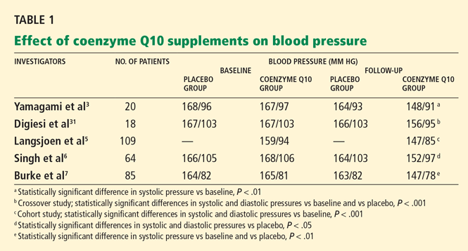
Rosenfeldt et al28 performed a meta-analysis and found that some trials documented statistically significant reductions in diastolic or systolic blood pressure or both, while others reported negligible effects.3,29 In one small trial,30 blood pressures actually went up in patients taking coenzyme Q10. Coenzyme Q10 dosages and length of therapy varied from study to study in the meta-analysis. Only minor adverse effects such as gastrointestinal upset and headache were reported.
Yamagami et al3 randomly assigned 20 patients with hypertension and a low coenzyme Q10 level to receive 100 mg of coenzyme Q10 or placebo daily for 12 weeks. Patients continued their usual antihypertensive regimen during the study period. Blood pressures, coenzyme Q10 levels, and antihypertensive drugs used were comparable between the study groups.
After 12 weeks of therapy, the mean coenzyme Q10 level in the active-treatment group had more than doubled, from 0.704 to 1.597 μg/mL. This group also experienced a statistically significant drop in systolic blood pressure, from 167 mm Hg at baseline to 148 mm Hg at 12 weeks. In the placebo group, the systolic blood pressure was 168 mm Hg at baseline and 164 mm Hg at 12 weeks; the change was not statistically significant. Diastolic pressure was not significantly lower at 12 weeks than at baseline in either group.
The authors concluded that coenzyme Q10 supplementation brought a mild reduction in high blood pressure in patients who had low coenzyme Q10 serum levels.
Digiesi et al31 randomized 18 patients with essential hypertension to receive either coenzyme Q10 100 mg or placebo daily for 10 weeks. All antihypertensive therapy was discontinued at baseline. After the first 10 weeks, patients went through a 2-week washout period and then were switched to the opposite therapy for an additional 10 weeks. Mean baseline blood pressure values were 167 mm Hg systolic and 103 mm Hg diastolic.
Those taking the supplement had a statistically significant decrease in systolic and diastolic pressures (P < .001). The antihypertensive effect was noted in the 3rd or 4th week of active treatment and persisted for the duration of therapy. The effects dissipated 7 to 10 days after coenzyme Q10 was stopped.
Langsjoen et al5 evaluated the effects of adding coenzyme Q10 to the antihypertensive drug regimen of 109 patients who had a primary diagnosis of essential hypertension in a prospective observational study. Patients with hypertension as a secondary diagnosis and other cardiovascular diseases were excluded. Variable doses of coenzyme Q10 were given, adjusted according to clinical response and to achieve serum levels greater than 2.0 μg/mL. The average dose was 225 mg/day; the mean serum level attained was 3.02 μg/mL.
Over several months, patients taking the supplement had a reduction in mean systolic pressure from 159 mm Hg at baseline to 147 mm Hg (P < .001), and a reduction in mean diastolic pressure from 94 to 85 mm Hg (P < .001). Thirty-seven percent of patients were able to discontinue one antihypertensive drug, 11% discontinued two drugs, and 4% were able to stop taking three drugs. However, 46% remained on the same antihypertensive regimen, and 3% needed an additional drug.
Singh et al6 randomized 64 patients who had coronary artery disease and who had been on antihypertensive drugs for more than 1 year to receive either B-complex vitamins or coenzyme Q10 (hydrosoluble Q-Gel) 60 mg orally once daily for 8 weeks. Five patients were not available for follow-up; therefore, only 59 patients were evaluated. Fifty-five (93%) of the 59 patients were taking only one antihypertensive drug. Initial antihypertensive drug use was similar between study groups and was continued throughout the trial.
After 8 weeks of therapy, the coenzyme Q10 group had significantly lower systolic and diastolic blood pressure than the placebo group (P < .05 for both). There was also a statistically significant decrease in the dosage of antihypertensive drugs in the coenzyme Q10 group but not in the placebo group (P < .05), reflecting coenzyme Q10’s additive antihypertensive effect.
Burke et al7 randomized 41 men and 35 women with isolated systolic hypertension (systolic pressure 150–170 mm Hg, diastolic pressure < 90 mm Hg) to receive a twice-daily dose of 60 mg of emulsified coenzyme Q10 (hydrosoluble Q-Gel) with 150 IU of vitamin E or placebo containing vitamin E alone for 12 weeks. The study also included 5 men and 4 women with normal blood pressure, all of whom received coenzyme Q10. A total of 80 patients completed treatment. The primary goal of the study was to determine the efficacy of coenzyme Q10 in the treatment of isolated systolic hypertension in patients without comorbid conditions. Blood pressures were monitored twice a week during the trial, by the same nurse.
After 12 weeks of treatment, the mean reduction in systolic pressure in hypertensive patients on coenzyme Q10 was 17.8 ± 7.3 mm Hg. There were no significant changes in diastolic pressure in any study group with treatment. Patients with isolated systolic hypertension who were taking coenzyme Q10 had a statistically significant reduction in systolic pressure compared with baseline and placebo (P < .01 for both). Approximately 55% of patients on coenzyme Q10 achieved a reduction in systolic pressure of 4 mm Hg or greater, while 45% did not respond to therapy. The mean plasma coenzyme Q10 level of the treatment group increased from 0.47 ± 0.19 μg/mL to 2.69 ± 0.54 μg/mL after 12 weeks; however, the study did not have the statistical power to demonstrate a relationship between coenzyme Q10 levels and changes in blood pressure. Twenty-seven (34%) of the 80 patients were taking a statin while on coenzyme Q10 therapy.
STUDIES IN STATIN-INDUCED MYOPATHY
Thibault et al32 and Kim et al33 reported that patients taking lovastatin (Mevacor) at dosages as high as 35 mg/kg/day to inhibit tumor growth achieved symptomatic relief of statin-induced musculoskeletal toxicity after coenzyme Q10 supplementation.
Caso et al15 performed a small pilot study in 32 patients to determine if coenzyme Q10 supplementation would improve myalgic symptoms in patients treated with statins. In this double-blind, randomized trial, patients received either coenzyme Q10 100 mg/day or vitamin E 400 IU/day for 30 days. The extent of muscle pain and its interference with daily activities were determined before and after therapy using the Brief Pain Inventory Questionnaire. The statins were atorvastatin (Lipitor) 10 mg or 20 mg, lovastatin 40 mg, pravastatin 40 mg, and simvastatin 10, 20, 40, and 80 mg. Five patients in the coenzyme Q10 group and four patients in the vitamin E group were taking nonsteroidal anti-inflammatory drugs before and during the trial. The intensity of muscle pain and its interference with daily activities were similar between study groups before the start of therapy.
After 30 days of treatment with coenzyme Q10, the pain intensity had decreased significantly from baseline (P < .001). In contrast, no change in pain intensity from baseline was noted in patients receiving vitamin E. The Pain Severity Score was significantly different between study groups, favoring the coenzyme Q10 group (P < .001). Sixteen of 18 patients on coenzyme Q10 reported a reduction in pain, while only 3 of 14 patients on vitamin E reported a similar response. Also, the interference of pain with daily activities significantly improved with coenzyme Q10 (P < .02), whereas vitamin E did not have a significant impact on this.
Young et al17 randomized 44 patients with prior statin-induced myalgia to receive increasing doses of simvastatin (10–40 mg/day) in combination with either coenzyme Q10 (Q-Gel) 200 mg/day or placebo. The primary goal was to determine if coenzyme Q10 supplementation would help improve statin tolerance in patients with a history of statininduced myalgia. Plasma coenzyme Q10 and lipid levels were measured at baseline and at the end of the study. The intensity of myalgia was assessed with a visual analogue scale.
At 12 weeks, the coenzyme Q10 plasma level was significantly higher in the treatment group than in the placebo group (P < .001). However, no differences were noted between groups in the number of patients who tolerated the 40-mg/day simvastatin dose (P = .34) or in the number of patients who remained on any simvastatin dose (P = .47). Additionally, myalgia scores did not differ between groups (P = .63). The authors acknowledged that there were only small increases in the myalgia pain scores reported in either group. Therefore, patients in the treatment group may not have experienced sufficiently severe muscle pain to have benefited from coenzyme Q10 supplementation.
IS COENZYME Q10 SAFE?
Studies have indicated that these supplements are well tolerated, with relatively few adverse effects or potential drug interactions.1,2,34
The FDA does not routinely assess the purity or quality of over-the-counter coenzyme Q10 products.35 However, the United States Pharmacopeia (USP) does test dietary supplements to make sure that they are not mislabeled and that they do not contain contaminants. 36
A USP-verified dietary supplement should:
- Contain the exact ingredients listed on the label in the listed potency and amounts
- Not include harmful levels of certain contaminants such as lead, mercury, pesticides, or bacteria
- Appropriately disintegrate and release its contents into the body within a specified period of time
- Be produced using the FDA’s current Good Manufacturing Practices.36
Side effects, contraindications, warnings
Coenzyme Q10 is a relatively safe dietary supplement. It is contraindicated in patients who are allergic to it or to any of its components.2 Most clinical trials have not reported significant adverse effects that necessitated stopping therapy.34 However, gastrointestinal effects such as abdominal discomfort, nausea, vomiting, diarrhea, and anorexia have occurred.1,2,34 Allergic rash and headache have also been reported.1,2,34 In addition, coenzyme Q10’s antiplatelet effect may increase the risk of bleeding. 37,38 It undergoes biotransformation in the liver and is eliminated primarily via the biliary tract,39 so it can accumulate in patients with hepatic impairment or biliary obstruction.
Interactions with drugs
Coenzyme Q10’s effects on platelet function may increase the risk of bleeding in patients taking antiplatelet drugs such as aspirin or clopidogrel (Plavix).37,38 On the other hand, since it acts like vitamin K, it may counteract the anticoagulant effects of warfarin (Coumadin). 1,2,40
Coenzyme Q10 may have an additive antihypertensive effect when given with antihypertensive drugs.41
Coenzyme Q10 may improve beta-cell function and enhance insulin sensitivity, which may reduce insulin requirements for diabetic patients.42,43
SLOWLY ABSORBED
Coenzyme Q10 is absorbed slowly from the gastrointestinal tract, possibly because it has a high molecular weight and is not very watersoluble. 39
One pharmacokinetic study found that after a single 100-mg oral dose of coenzyme Q10, the mean peak plasma levels of about 1 μg/mL occurred between 5 and 10 hours (mean 6.5 hours).44 Coenzyme Q10 100 mg given orally three times daily produced a mean steadystate plasma level of 5.4 μg/mL; about 90% of this steady-state concentration was achieved after 4 days.39
Some formulations have significantly better oral bioavailability and therefore produce higher plasma levels. Soft-gel capsules, especially those with vegetable oil or vitamin E, may have better absorption.43
A pharmacokinetic study showed that the area under the curve of the plasma coenzyme Q10 concentration was more than twice as high with Q-Gel soft-gel capsules, a completely solubilized formulation, than with softgel capsules with an oil suspension, powderfilled hard-shell capsules, or regular tablets.45 Another study reported that colloidal-Q10, a formulation contained in VESIsorb (a novel drug delivery system sold as CoQsource) had greater bioavailability than solubilized and oil-based preparations.46 Commercially available solubilized preparations containing ubiquinol, a metabolized form of coenzyme Q10, have been shown to produce higher serum levels than solubilized products.47
Of note: unless the manufacturer claims that its product is water-soluble, the USP does not evaluate its dissolution rate.48 Therefore, USP-verified coenzyme Q10 products that are not water-soluble may have lower bioavailability than their solubilized counterparts.
Dry dosage forms of coenzyme Q10 (eg, tablets, capsules) may be more readily absorbed if taken with a fatty meal.43
SLOWLY ELIMINATED
Taken orally, coenzyme Q10 has a low clearance rate, with an elimination half-life of about 34 hours.39
After absorption, exogenous coenzyme Q10 is taken up by chylomicrons that transport it to the liver, where it is incorporated into verylow-density lipoproteins. It is then distributed to various organs, including the adrenal glands, spleen, kidneys, lungs, and heart. Coenzyme Q10 is eliminated primarily via the biliary tract. About 60% of an oral dose is eliminated in the feces during chronic oral administration.39
TWICE-DAILY DOSING
A typical daily dose of coenzyme Q10 for treating hypertension is 120 to 200 mg, usually given orally in two divided doses.1 For statininduced myopathy, 100 to 200 mg orally daily has been used.1
Coenzyme Q10 is given in divided doses to enhance its absorption and to minimize gastrointestinal effects.1,43 Taking it with a fatty meal may also increase its absorption.43
Since solubilized forms of coenzyme Q10 and ubiquinol have significantly greater bioavailability than nonsolubilized forms, the therapeutic dose of these formulations may be lower.47
MONITORING DURING TREATMENT
Without supplementation, the mean serum level of endogenous coenzyme Q10 has been reported to be 0.99 ± 0.30 mg/L (range 0.55– 1.87).18 Serum levels above 2 μg/mL have been associated with significant reductions in blood pressure.5,7,28
The possible effects of coenzyme Q10 on blood pressure, blood glucose levels, serum creatine kinase levels, and myopathic symptoms should be kept in mind when monitoring patients who have hypertension,41 diabetes,41,42 or statin-induced myalgia.15,17 Coenzyme Q10’s possible potentiating effects on antiplatelet drugs and its inhibitory effect on warfarin should be kept in mind as well.
COST VARIES
Coenzyme Q10 is available in different dosage forms (eg, regular and rapid-release softgel capsules, regular and chewable tablets, chewable wafers, and liquid) from a variety of manufacturers. Products come in different strengths, typically ranging from 30 to 400 mg. USP-verified formulations are listed at www.usp.org/USPVerified/dietarySupplements/under “Verified Supplements.” Only USP-verified products that claim to be water-soluble meet USP dissolution requirements.
The cost varies, depending on the vendor. In general, dosage forms with greater bioavailability, such as Q-Gel and ubiquinol supplements, are more expensive. For example, a regimen of 60 mg twice daily of regular-release coenzyme Q capsules may cost approximately $20 per month, compared with $60 per month for the same supply of Q-Gel Ultra capsules. However, in some cases, supplements that produce higher serum levels may be more cost-effective.
CURRENT ROLE IN THERAPY
As an antihypertensive adjunct
Several small clinical trials have shown that coenzyme Q10 supplementation can lower blood pressure. The supplements were reported to be safe and well tolerated. Moreover, some patients with essential hypertension who were taking coenzyme Q10 were able to discontinue one or more antihypertensive drugs. A significant reduction in blood pressure with use of coenzyme Q10 would be expected to reduce the adverse consequences of hypertension in the same manner as conventional antihypertensive agents.
However, no large, double-blind, randomized study has evaluated the impact of coenzyme Q10 when taken with other antihypertensive drugs (eg, angiotensin-converting enzyme inhibitors, beta-blockers, diuretics) on specific clinical end points such as the incidence of stroke or death from a major cardiac event. Furthermore, its effects on cardiac function, exercise tolerance, and quality of life have not been determined.
The bottom line. In some cases, it seems reasonable to recommend this product as an adjunct to conventional antihypertensive therapy. Larger, well-designed clinical trials of coenzyme Q10’s antihypertensive effects on specific clinical end points such as the risk of stroke or myocardial infarction are needed to define its true therapeutic value.
As a treatment for statin-induced myalgia
Clinical evidence supporting coenzyme Q10’s use in the treatment of statin-induced myopathy is limited. Whether coenzyme Q10 is depleted from muscle tissue during statin therapy has not been confirmed. Supplementation helped reduce the severity of musculoskeletal effects of megadoses of lovastatin. However, clinical trials of coenzyme Q10 in the treatment of myalgia associated with antilipidemic statin doses did not consistently report significant improvement. Nevertheless, coenzyme Q10 has been shown to be relatively safe, with few adverse effects.
The bottom line. In some cases, coenzyme Q could be considered as a possible treatment for statin-induced myalgia, pending large-scale studies to determine if it is truly effective for this purpose.
- Jelin JM, Gregory PJ, et al. Natural medicines comprehensive database/compiled by the editors of Pharmacist’s Letter, Prescriber’s Letter. 11th ed. Stockton, CA: Therapeutic Research Faculty; 2009:452–457.
- Fetrow CW, Avila JR. Professional’s Handbook of Complementary & Alternative Medicines. 2nd ed. Springhouse, PA: Springhouse; 2001:211–215.
- Yamagami T, Takagi M, Akagami H, et al. Effect of coenzyme Q10 on essential hypertension, a double-blind controlled study. In:Folkers K, Yamamura Y, editors. Biomedical and Clinical Aspects of Coenzyme Q10: Proceedings of the Fifth International Symposium on the Biomedical and Clinical Aspects of Coenzyme Q10, vol 5. Amsterdam: Elsevier Science Publishers; 1986:337–343.
- Digiesi V, Cantini F, Oradei A, et al. Coenzyme Q10 in essential hypertension. Mol Aspects Med 1994; 15(suppl):S257–S263.
- Langsjoen P, Langsjoen P, Willis R, Folkers K. Treatment of essential hypertension with coenzyme Q10. Mol Aspects Med 1994; 15(suppl):S265–S272.
- Singh RB, Niaz MA, Rastogi SS, Shukla PK, Thakur AS. Effect of hydrosoluble coenzyme Q10 on blood pressures and insulin resistance in hypertensive patients with coronary artery disease. J Hum Hypertens 1999; 13:203–208.
- Burke BE, Neuenschwander R, Olson RD. Randomized, double-blind, placebo-controlled trial of coenzyme Q10 in isolated systolic hypertension. South Med J 2001; 94:1112–1117.
- De Pinieux G, Chariot P, Ammi-Saïd M, et al. Lipidlowering drugs and mitochondrial function: effects of HMG-CoA reductase inhibitors on serum ubiquinone and blood lactate/pyruvate ratio. Br J Clin Pharmacol 1996; 42:333–337.
- Mortensen SA, Leth A, Agner E, Rohde M. Dose-related decrease of serum coenzyme Q10 during treatment with HMG-CoA reductase inhibitors. Mol Aspects Med 1997; 18(suppl):S137–S144.
- Ghirlanda G, Oradei A, Manto A, et al. Evidence of plasma CoQ10-lowering effect by HMG-CoA reductase inhibitors: a double-blind, placebo-controlled study. J Clin Pharmacol 1993; 33:226–229.
- Folkers K, Langsjoen P, Willis R, et al. Lovastatin decreases coenzyme Q10 levels in humans. Proc Natl Acad Sci U S A 1990; 87:8931–8934.
- Watts GF, Castelluccio C, Rice-Evans C, Taub NA, Baum H, Quinn PJ. Plasma coenzyme Q10 (ubiquinone) concentrations in patients treated with simvastatin. J Clin Pathol 1993; 46:1055–1057.
- Lamperti C, Naini AB, Lucchini V, et al. Muscle coenzyme Q10 level in statin-related myopathy. Arch Neurol 2005; 62:1709–1712.
- Päivä H, Thelen KM, Van Coster R, et al. High-dose statins and skeletal muscle metabolism in humans: a randomized, controlled trial. Clin Pharmacol Ther 2005; 78:60–68.
- Caso G, Kelly P, McNurlan MA, Lawson WE. Effect of coenzyme Q10 on myopathic symptoms in patients treated with statins. Am J Cardiol 2007; 99:1409–1412.
- Marcoff L, Thompson PD. The role of coenzyme Q10 in statin-associated myopathy: a systematic review. J Am Coll Cardiol 2007; 49:2231–2237.
- Young JM, Florkowski CM, Molyneux SL, et al. Effect of coenzyme Q(10) supplementation on simvastatin-induced myalgia. Am J Cardiol 2007; 100:1400–1403.
- Berthold HK, Naini A, Di Mauro S, et al. Effect of ezetimibe and/or simvastatin on coenzyme Q10 levels in plasma: a randomised trial. Drug Saf 2006; 29:703–712.
- Groneberg DA, Kindermann B, Althammer M, et al. Coenzyme Q10 affects expression of genes involved in cell signalling, metabolism and transport in human CaCo-2 cells. Int J Biochem Cell Biol 2005; 37:1208–1218.
- Hadj A, Pepe S, Rosenfeldt F. The clinical application of metabolic therapy for cardiovascular disease. Heart Lung Circ 2007; 16(suppl 3):S56–S64.
- Pepe S, Marasco SF, Haas SJ, Sheeran FL, Krum H, Rosenfeldt FL. Coenzyme Q10 in cardiovascular disease. Mitochondrion 2007; 7(suppl 1):S154–S167.
- Lönnrot K, Pörsti I, Alho H, Wu X, Hervonen A, Tolvanen JP. Control of arterial tone after long-term coenzyme Q10 supplementation in senescent rats. Br J Pharmacol 1998; 124:1500–1506.
- Sewright KA, Clarkson PM, Thompson PD. Statin myopathy: incidence, risk factors, and pathophysiology. Curr Atheroscler Rep 2007; 9:389–396.
- Radcliffe KA, Campbell WW. Statin myopathy. Curr Neurol Neurosci Rep 2008; 8:66–72.
- Pasternak RC, Smith SC, Bairey-Merz CN, Grundy SM, Cleeman JI, Lenfant C. ACC/AHA/NHLBI clinical advisory on the use and safety of statins. Circulation 2002; 106:1024–1028.
- Laaksonen R, Jokelainen K, Laakso J, et al. The effect of simvastatin treatment on natural antioxidants in low-density lipoproteins and high-energy phosphates and ubiquinone in skeletal muscle. Am J Cardiol 1996; 77:851–854.
- Golomb BA, Dimsdale JE, White HL, Ritchie JB, Criqui MH. Reduction in blood pressure with statins: results from the UCSD Statin Study, a randomized trial. Arch Intern Med 2008; 168:721–727.
- Rosenfeldt FL, Haas SJ, Krum H, et al. Coenzyme Q10 in the treatment of hypertension: a meta-analysis of the clinical trials. J Hum Hypertens 2007; 21:297–306.
- Yamagami T, Shibata N, Folkers K. Bioenergetics in clinical medicine. Studies on coenzyme Q10 and essential hypertension. Res Commun Chem Pathol Pharmacol 1975; 11:273–288.
- Yamagami T, Shibata N, Folkers K. Study of coenzyme Q10. In:Folkers K, Yamamura Y, editors. Biomedical and clinical aspects of coenzyme Q10: proceedings of the International Symposium on Coenzyme Q10, held at Lake Yamanaka, Japan, September 16/17, 1976, a Naito Foundation symposium. Amsterdam: Elsevier Scientific Publishing Company; 1977:231–242.
- Digiesi V, Cantini F, Brodbeck B. Effect of coenzyme Q10 on essential arterial hypertension. Curr Ther Res; 1990; 47:841–845.
- Thibault A, Samid D, Tompkins AC, et al. Phase I study of lovastatin, an inhibitor of the mevalonate pathway, in patients with cancer. Clin Cancer Res 1996; 2:483–491.
- Kim WS, Kim MM, Choi HJ, et al. Phase II study of high-dose lova-statin in patients with advanced gastric adenocarcinoma. Invest New Drugs 2001; 19:81–83.
- Hidaka T, Fujii K, Funahashi I, Fukutomi N, Hosoe K. Safety assessment of coenzyme Q10 (CoQ10). Biofactors 2008; 32:199–208.
- US Food and Drug Administration. Consumer Information on Dietary Supplements. Overview of Dietary Supplements. http://www.fda.gov/Food/DietarySupplements/ConsumerInformation/. Accessed May 25, 2010.
- US Pharmacopeia. The USP Dietary Supplement Verification Program http://www.usp.org/USPVerified/dietary-Supplements/. Accessed May 25, 2010.
- Serebruany VL, Ordonez JV, Herzog WR, et al. Dietary coenzyme Q10 supplementation alters platelet size and inhibits human vitronectin (CD51/CD61) receptor expression. J Cardiovasc Pharmacol 1997; 29:16–22.
- A close look at coenzyme Q10 and policosanol. Do these supplements live up to their claims for improving heart health? Harv Heart Lett 2002; 13:6.
- Greenberg S, Frishman WH. Co-enzyme Q10: a new drug for cardiovascular disease. J Clin Pharmacol 1990; 30:596–608.
- Singh U, Devaraj S, Jialal I. Coenzyme Q10 supplementation and heart failure. Nutr Rev 2007; 65:286–293.
- Bonakdar RA, Guarneri E. Coenzyme Q10. Am Fam Physician 2005; 72:1065–1070.
- Hodgson JM, Watts GF, Playford DA, Burke V, Croft KD. Coenzyme Q10 improves blood pressure and glycaemic control: a controlled trial in subjects with type 2 diabetes. Eur J Clin Nutr 2002; 56:1137–1142.
- Pepping J. Coenzyme Q10. Am J Health Syst Pharm 1999; 56:519–521.
- Tomono Y, Hasegawa J, Seki T, Motegi K, Morishita N. Pharmacokinetic study of deuterium-labelled coenzyme Q10 in man. Int J Clin Pharmacol Ther Toxicol 1986; 24:536–541.
- Chopra RK, Goldman R, Sinatra ST, Bhagavan HN. Relative bioavailability of coenzyme Q10 formulations in human subjects. Int J Vitam Nutr Res 1998; 68:109–113.
- Liu ZX, Artmann C. Relative bioavailability comparison of different coenzyme Q10 formulations with a novel delivery system. Altern Ther Health Med 2009; 15:42–46.
- Bhagavan HN, Chopra RK. Plasma coenzyme Q10 response to oral ingestion of coenzyme Q10 formulations. Mitochondrion 2007; 7(suppl 1):S78–S88.
- The United States Pharmacopeia. Ubidecarenone Capsules Monograph. 32nd Revision. Baltimore: United Book Press, 2009:1080.
Coenzyme Q10 supplements have been purported to be effective for treating a variety of disorders,1,2 in particular hypertension and statin-induced myalgia.
Several studies3–7 found that coenzyme Q10 supplementation significantly lowered blood pressure in hypertensive patients. Moreover, some trials have demonstrated that statin therapy reduces serum or muscle levels of coenzyme Q10,8–14 prompting investigations to determine whether coenzyme Q10 deficiency is related to statin-induced muscle pain.15–17
In this review, we discuss the efficacy and safety of coenzyme Q10 supplementation in patients with hypertension and those taking statins, and some of the caveats about using supplements that are not approved by the US Food and Drug Administration (FDA), as well as the bioavailability and quality of available formulations.
WHAT IS COENZYME Q10?
Coenzyme Q10, also known as coenzyme Q, ubidecarenone, and ubiquinone, is found in all human cells, with the highest concentrations in the heart, liver, kidney, and pancreas.1,2 It is a potent antioxidant, a membrane stabilizer, and an integral cofactor in the mitochondrial respiratory chain, helping to generate adenosine triphosphate, the major cellular energy source.1,2,18 It may also regulate genes associated with cell metabolism.19
RATIONALE FOR SUPPLEMENTATION
Coenzyme Q10 supplementation has been used, recommended, or studied in heart failure, hypertension, parkinsonism, mitochondrial encephalomyopathies, and other ailments.
In hypertension
Depending on the class, various antihypertensive drugs can have adverse effects such as depression, cough, and cardiac and renal dysfunction. 20,21 Furthermore, many patients need to take more than one drug to control their blood pressure, increasing their risk of side effects. Some researchers believe coenzyme Q10 supplementation may reduce the need to take multiple antihypertensive drugs.5
Coenzyme Q10 appears to lower blood pressure. The exact mechanism is not known, but one theory is that it reduces peripheral resistance by preserving nitric oxide.21 Nitric oxide relaxes peripheral arteries, lowering blood pressure. In some forms of hypertension, superoxide radicals that inactivate nitric oxide are overproduced; coenzyme Q10, with its antioxidant effects, may prevent the inactivation of nitric oxide by these free radicals. Alternatively, coenzyme Q10 may boost the production of the prostaglandin prostacyclin (PGI2) a potent vasodilator and inhibitor of platelet aggregation, or it may enhance the sensitivity of arterial smooth muscle to PGI2, or both.1,22
In patients taking statins
Hydroxymethylglutaryl coenzyme A reductase inhibitors (statins), first-line agents for lowering cholesterol levels to prevent cardiovascular disease, are some of the most commonly prescribed medications.23,24 However, statin therapy carries a risk of myopathy, which can range from muscle aches to rhabdomyolysis. 23,24
In a clinical advisory,25 the American College of Cardiology, the American Heart Association, and the National Heart, Lung, and Blood Institute recommend that patients on statin therapy who experience muscle soreness, tenderness, or pain with serum creatine kinase levels 3 to 10 times the upper limit of normal should have their creatine kinase level checked weekly. If the level is 3 to 10 times the upper limit of normal, statin therapy may be continued, but if it exceeds 10 times the upper limit, then statins and other potential offending agents (eg, niacin, fibrate) need to be discontinued.
Statins inhibit the synthesis of cholesterol by reducing the production of mevalonate, a precursor of both cholesterol and coenzyme Q10. Since both cholesterol and coenzyme Q10 are produced by the same pathway, it is not surprising that statins have been reported to reduce serum and muscle coenzyme Q10 levels.9–14 However, one study did not report a significant reduction of coenzyme Q10 levels in muscle tissue in patients treated with simvastatin 20 mg for 6 months.26
Nonetheless, researchers have hypothesized that a reduction in coenzyme Q10 levels in muscle tissue causes mitochondrial dysfunction, which could increase the risk of statininduced myopathy,13–17 and some believe that treatment with coenzyme Q10 may reduce myalgic symptoms and allow patients to remain on statin therapy.13,24
Researchers have investigated the potential of coenzyme Q10 supplementation to reduce or prevent statin-induced myopathy.15–17 (More on this below.)
Interestingly, a randomized, placebo-controlled trial27 found that 6 months of daily therapy with simvastatin (Zocor) 20 mg or pravastatin (Pravachol) 40 mg lowered systolic and diastolic blood pressure significantly in patients with no documented history of cardiovascular disease or diabetes. A possible mechanism of statin-induced blood pressure reduction is the up-regulation of endothelial nitric oxide synthetase, a potent vasodilator. Coenzyme Q10 levels were not assessed during this study. Whether coenzyme Q10 supplementation used to treat statin-induced myalgia enhances or inhibits the antihypertensive effects of statins is not yet known.
EVIDENCE OF EFFECTIVENESS IN HYPERTENSION
Rosenfeldt et al28 performed a meta-analysis and found that some trials documented statistically significant reductions in diastolic or systolic blood pressure or both, while others reported negligible effects.3,29 In one small trial,30 blood pressures actually went up in patients taking coenzyme Q10. Coenzyme Q10 dosages and length of therapy varied from study to study in the meta-analysis. Only minor adverse effects such as gastrointestinal upset and headache were reported.
Yamagami et al3 randomly assigned 20 patients with hypertension and a low coenzyme Q10 level to receive 100 mg of coenzyme Q10 or placebo daily for 12 weeks. Patients continued their usual antihypertensive regimen during the study period. Blood pressures, coenzyme Q10 levels, and antihypertensive drugs used were comparable between the study groups.
After 12 weeks of therapy, the mean coenzyme Q10 level in the active-treatment group had more than doubled, from 0.704 to 1.597 μg/mL. This group also experienced a statistically significant drop in systolic blood pressure, from 167 mm Hg at baseline to 148 mm Hg at 12 weeks. In the placebo group, the systolic blood pressure was 168 mm Hg at baseline and 164 mm Hg at 12 weeks; the change was not statistically significant. Diastolic pressure was not significantly lower at 12 weeks than at baseline in either group.
The authors concluded that coenzyme Q10 supplementation brought a mild reduction in high blood pressure in patients who had low coenzyme Q10 serum levels.
Digiesi et al31 randomized 18 patients with essential hypertension to receive either coenzyme Q10 100 mg or placebo daily for 10 weeks. All antihypertensive therapy was discontinued at baseline. After the first 10 weeks, patients went through a 2-week washout period and then were switched to the opposite therapy for an additional 10 weeks. Mean baseline blood pressure values were 167 mm Hg systolic and 103 mm Hg diastolic.
Those taking the supplement had a statistically significant decrease in systolic and diastolic pressures (P < .001). The antihypertensive effect was noted in the 3rd or 4th week of active treatment and persisted for the duration of therapy. The effects dissipated 7 to 10 days after coenzyme Q10 was stopped.
Langsjoen et al5 evaluated the effects of adding coenzyme Q10 to the antihypertensive drug regimen of 109 patients who had a primary diagnosis of essential hypertension in a prospective observational study. Patients with hypertension as a secondary diagnosis and other cardiovascular diseases were excluded. Variable doses of coenzyme Q10 were given, adjusted according to clinical response and to achieve serum levels greater than 2.0 μg/mL. The average dose was 225 mg/day; the mean serum level attained was 3.02 μg/mL.
Over several months, patients taking the supplement had a reduction in mean systolic pressure from 159 mm Hg at baseline to 147 mm Hg (P < .001), and a reduction in mean diastolic pressure from 94 to 85 mm Hg (P < .001). Thirty-seven percent of patients were able to discontinue one antihypertensive drug, 11% discontinued two drugs, and 4% were able to stop taking three drugs. However, 46% remained on the same antihypertensive regimen, and 3% needed an additional drug.
Singh et al6 randomized 64 patients who had coronary artery disease and who had been on antihypertensive drugs for more than 1 year to receive either B-complex vitamins or coenzyme Q10 (hydrosoluble Q-Gel) 60 mg orally once daily for 8 weeks. Five patients were not available for follow-up; therefore, only 59 patients were evaluated. Fifty-five (93%) of the 59 patients were taking only one antihypertensive drug. Initial antihypertensive drug use was similar between study groups and was continued throughout the trial.
After 8 weeks of therapy, the coenzyme Q10 group had significantly lower systolic and diastolic blood pressure than the placebo group (P < .05 for both). There was also a statistically significant decrease in the dosage of antihypertensive drugs in the coenzyme Q10 group but not in the placebo group (P < .05), reflecting coenzyme Q10’s additive antihypertensive effect.
Burke et al7 randomized 41 men and 35 women with isolated systolic hypertension (systolic pressure 150–170 mm Hg, diastolic pressure < 90 mm Hg) to receive a twice-daily dose of 60 mg of emulsified coenzyme Q10 (hydrosoluble Q-Gel) with 150 IU of vitamin E or placebo containing vitamin E alone for 12 weeks. The study also included 5 men and 4 women with normal blood pressure, all of whom received coenzyme Q10. A total of 80 patients completed treatment. The primary goal of the study was to determine the efficacy of coenzyme Q10 in the treatment of isolated systolic hypertension in patients without comorbid conditions. Blood pressures were monitored twice a week during the trial, by the same nurse.
After 12 weeks of treatment, the mean reduction in systolic pressure in hypertensive patients on coenzyme Q10 was 17.8 ± 7.3 mm Hg. There were no significant changes in diastolic pressure in any study group with treatment. Patients with isolated systolic hypertension who were taking coenzyme Q10 had a statistically significant reduction in systolic pressure compared with baseline and placebo (P < .01 for both). Approximately 55% of patients on coenzyme Q10 achieved a reduction in systolic pressure of 4 mm Hg or greater, while 45% did not respond to therapy. The mean plasma coenzyme Q10 level of the treatment group increased from 0.47 ± 0.19 μg/mL to 2.69 ± 0.54 μg/mL after 12 weeks; however, the study did not have the statistical power to demonstrate a relationship between coenzyme Q10 levels and changes in blood pressure. Twenty-seven (34%) of the 80 patients were taking a statin while on coenzyme Q10 therapy.
STUDIES IN STATIN-INDUCED MYOPATHY
Thibault et al32 and Kim et al33 reported that patients taking lovastatin (Mevacor) at dosages as high as 35 mg/kg/day to inhibit tumor growth achieved symptomatic relief of statin-induced musculoskeletal toxicity after coenzyme Q10 supplementation.
Caso et al15 performed a small pilot study in 32 patients to determine if coenzyme Q10 supplementation would improve myalgic symptoms in patients treated with statins. In this double-blind, randomized trial, patients received either coenzyme Q10 100 mg/day or vitamin E 400 IU/day for 30 days. The extent of muscle pain and its interference with daily activities were determined before and after therapy using the Brief Pain Inventory Questionnaire. The statins were atorvastatin (Lipitor) 10 mg or 20 mg, lovastatin 40 mg, pravastatin 40 mg, and simvastatin 10, 20, 40, and 80 mg. Five patients in the coenzyme Q10 group and four patients in the vitamin E group were taking nonsteroidal anti-inflammatory drugs before and during the trial. The intensity of muscle pain and its interference with daily activities were similar between study groups before the start of therapy.
After 30 days of treatment with coenzyme Q10, the pain intensity had decreased significantly from baseline (P < .001). In contrast, no change in pain intensity from baseline was noted in patients receiving vitamin E. The Pain Severity Score was significantly different between study groups, favoring the coenzyme Q10 group (P < .001). Sixteen of 18 patients on coenzyme Q10 reported a reduction in pain, while only 3 of 14 patients on vitamin E reported a similar response. Also, the interference of pain with daily activities significantly improved with coenzyme Q10 (P < .02), whereas vitamin E did not have a significant impact on this.
Young et al17 randomized 44 patients with prior statin-induced myalgia to receive increasing doses of simvastatin (10–40 mg/day) in combination with either coenzyme Q10 (Q-Gel) 200 mg/day or placebo. The primary goal was to determine if coenzyme Q10 supplementation would help improve statin tolerance in patients with a history of statininduced myalgia. Plasma coenzyme Q10 and lipid levels were measured at baseline and at the end of the study. The intensity of myalgia was assessed with a visual analogue scale.
At 12 weeks, the coenzyme Q10 plasma level was significantly higher in the treatment group than in the placebo group (P < .001). However, no differences were noted between groups in the number of patients who tolerated the 40-mg/day simvastatin dose (P = .34) or in the number of patients who remained on any simvastatin dose (P = .47). Additionally, myalgia scores did not differ between groups (P = .63). The authors acknowledged that there were only small increases in the myalgia pain scores reported in either group. Therefore, patients in the treatment group may not have experienced sufficiently severe muscle pain to have benefited from coenzyme Q10 supplementation.
IS COENZYME Q10 SAFE?
Studies have indicated that these supplements are well tolerated, with relatively few adverse effects or potential drug interactions.1,2,34
The FDA does not routinely assess the purity or quality of over-the-counter coenzyme Q10 products.35 However, the United States Pharmacopeia (USP) does test dietary supplements to make sure that they are not mislabeled and that they do not contain contaminants. 36
A USP-verified dietary supplement should:
- Contain the exact ingredients listed on the label in the listed potency and amounts
- Not include harmful levels of certain contaminants such as lead, mercury, pesticides, or bacteria
- Appropriately disintegrate and release its contents into the body within a specified period of time
- Be produced using the FDA’s current Good Manufacturing Practices.36
Side effects, contraindications, warnings
Coenzyme Q10 is a relatively safe dietary supplement. It is contraindicated in patients who are allergic to it or to any of its components.2 Most clinical trials have not reported significant adverse effects that necessitated stopping therapy.34 However, gastrointestinal effects such as abdominal discomfort, nausea, vomiting, diarrhea, and anorexia have occurred.1,2,34 Allergic rash and headache have also been reported.1,2,34 In addition, coenzyme Q10’s antiplatelet effect may increase the risk of bleeding. 37,38 It undergoes biotransformation in the liver and is eliminated primarily via the biliary tract,39 so it can accumulate in patients with hepatic impairment or biliary obstruction.
Interactions with drugs
Coenzyme Q10’s effects on platelet function may increase the risk of bleeding in patients taking antiplatelet drugs such as aspirin or clopidogrel (Plavix).37,38 On the other hand, since it acts like vitamin K, it may counteract the anticoagulant effects of warfarin (Coumadin). 1,2,40
Coenzyme Q10 may have an additive antihypertensive effect when given with antihypertensive drugs.41
Coenzyme Q10 may improve beta-cell function and enhance insulin sensitivity, which may reduce insulin requirements for diabetic patients.42,43
SLOWLY ABSORBED
Coenzyme Q10 is absorbed slowly from the gastrointestinal tract, possibly because it has a high molecular weight and is not very watersoluble. 39
One pharmacokinetic study found that after a single 100-mg oral dose of coenzyme Q10, the mean peak plasma levels of about 1 μg/mL occurred between 5 and 10 hours (mean 6.5 hours).44 Coenzyme Q10 100 mg given orally three times daily produced a mean steadystate plasma level of 5.4 μg/mL; about 90% of this steady-state concentration was achieved after 4 days.39
Some formulations have significantly better oral bioavailability and therefore produce higher plasma levels. Soft-gel capsules, especially those with vegetable oil or vitamin E, may have better absorption.43
A pharmacokinetic study showed that the area under the curve of the plasma coenzyme Q10 concentration was more than twice as high with Q-Gel soft-gel capsules, a completely solubilized formulation, than with softgel capsules with an oil suspension, powderfilled hard-shell capsules, or regular tablets.45 Another study reported that colloidal-Q10, a formulation contained in VESIsorb (a novel drug delivery system sold as CoQsource) had greater bioavailability than solubilized and oil-based preparations.46 Commercially available solubilized preparations containing ubiquinol, a metabolized form of coenzyme Q10, have been shown to produce higher serum levels than solubilized products.47
Of note: unless the manufacturer claims that its product is water-soluble, the USP does not evaluate its dissolution rate.48 Therefore, USP-verified coenzyme Q10 products that are not water-soluble may have lower bioavailability than their solubilized counterparts.
Dry dosage forms of coenzyme Q10 (eg, tablets, capsules) may be more readily absorbed if taken with a fatty meal.43
SLOWLY ELIMINATED
Taken orally, coenzyme Q10 has a low clearance rate, with an elimination half-life of about 34 hours.39
After absorption, exogenous coenzyme Q10 is taken up by chylomicrons that transport it to the liver, where it is incorporated into verylow-density lipoproteins. It is then distributed to various organs, including the adrenal glands, spleen, kidneys, lungs, and heart. Coenzyme Q10 is eliminated primarily via the biliary tract. About 60% of an oral dose is eliminated in the feces during chronic oral administration.39
TWICE-DAILY DOSING
A typical daily dose of coenzyme Q10 for treating hypertension is 120 to 200 mg, usually given orally in two divided doses.1 For statininduced myopathy, 100 to 200 mg orally daily has been used.1
Coenzyme Q10 is given in divided doses to enhance its absorption and to minimize gastrointestinal effects.1,43 Taking it with a fatty meal may also increase its absorption.43
Since solubilized forms of coenzyme Q10 and ubiquinol have significantly greater bioavailability than nonsolubilized forms, the therapeutic dose of these formulations may be lower.47
MONITORING DURING TREATMENT
Without supplementation, the mean serum level of endogenous coenzyme Q10 has been reported to be 0.99 ± 0.30 mg/L (range 0.55– 1.87).18 Serum levels above 2 μg/mL have been associated with significant reductions in blood pressure.5,7,28
The possible effects of coenzyme Q10 on blood pressure, blood glucose levels, serum creatine kinase levels, and myopathic symptoms should be kept in mind when monitoring patients who have hypertension,41 diabetes,41,42 or statin-induced myalgia.15,17 Coenzyme Q10’s possible potentiating effects on antiplatelet drugs and its inhibitory effect on warfarin should be kept in mind as well.
COST VARIES
Coenzyme Q10 is available in different dosage forms (eg, regular and rapid-release softgel capsules, regular and chewable tablets, chewable wafers, and liquid) from a variety of manufacturers. Products come in different strengths, typically ranging from 30 to 400 mg. USP-verified formulations are listed at www.usp.org/USPVerified/dietarySupplements/under “Verified Supplements.” Only USP-verified products that claim to be water-soluble meet USP dissolution requirements.
The cost varies, depending on the vendor. In general, dosage forms with greater bioavailability, such as Q-Gel and ubiquinol supplements, are more expensive. For example, a regimen of 60 mg twice daily of regular-release coenzyme Q capsules may cost approximately $20 per month, compared with $60 per month for the same supply of Q-Gel Ultra capsules. However, in some cases, supplements that produce higher serum levels may be more cost-effective.
CURRENT ROLE IN THERAPY
As an antihypertensive adjunct
Several small clinical trials have shown that coenzyme Q10 supplementation can lower blood pressure. The supplements were reported to be safe and well tolerated. Moreover, some patients with essential hypertension who were taking coenzyme Q10 were able to discontinue one or more antihypertensive drugs. A significant reduction in blood pressure with use of coenzyme Q10 would be expected to reduce the adverse consequences of hypertension in the same manner as conventional antihypertensive agents.
However, no large, double-blind, randomized study has evaluated the impact of coenzyme Q10 when taken with other antihypertensive drugs (eg, angiotensin-converting enzyme inhibitors, beta-blockers, diuretics) on specific clinical end points such as the incidence of stroke or death from a major cardiac event. Furthermore, its effects on cardiac function, exercise tolerance, and quality of life have not been determined.
The bottom line. In some cases, it seems reasonable to recommend this product as an adjunct to conventional antihypertensive therapy. Larger, well-designed clinical trials of coenzyme Q10’s antihypertensive effects on specific clinical end points such as the risk of stroke or myocardial infarction are needed to define its true therapeutic value.
As a treatment for statin-induced myalgia
Clinical evidence supporting coenzyme Q10’s use in the treatment of statin-induced myopathy is limited. Whether coenzyme Q10 is depleted from muscle tissue during statin therapy has not been confirmed. Supplementation helped reduce the severity of musculoskeletal effects of megadoses of lovastatin. However, clinical trials of coenzyme Q10 in the treatment of myalgia associated with antilipidemic statin doses did not consistently report significant improvement. Nevertheless, coenzyme Q10 has been shown to be relatively safe, with few adverse effects.
The bottom line. In some cases, coenzyme Q could be considered as a possible treatment for statin-induced myalgia, pending large-scale studies to determine if it is truly effective for this purpose.
- Jelin JM, Gregory PJ, et al. Natural medicines comprehensive database/compiled by the editors of Pharmacist’s Letter, Prescriber’s Letter. 11th ed. Stockton, CA: Therapeutic Research Faculty; 2009:452–457.
- Fetrow CW, Avila JR. Professional’s Handbook of Complementary & Alternative Medicines. 2nd ed. Springhouse, PA: Springhouse; 2001:211–215.
- Yamagami T, Takagi M, Akagami H, et al. Effect of coenzyme Q10 on essential hypertension, a double-blind controlled study. In:Folkers K, Yamamura Y, editors. Biomedical and Clinical Aspects of Coenzyme Q10: Proceedings of the Fifth International Symposium on the Biomedical and Clinical Aspects of Coenzyme Q10, vol 5. Amsterdam: Elsevier Science Publishers; 1986:337–343.
- Digiesi V, Cantini F, Oradei A, et al. Coenzyme Q10 in essential hypertension. Mol Aspects Med 1994; 15(suppl):S257–S263.
- Langsjoen P, Langsjoen P, Willis R, Folkers K. Treatment of essential hypertension with coenzyme Q10. Mol Aspects Med 1994; 15(suppl):S265–S272.
- Singh RB, Niaz MA, Rastogi SS, Shukla PK, Thakur AS. Effect of hydrosoluble coenzyme Q10 on blood pressures and insulin resistance in hypertensive patients with coronary artery disease. J Hum Hypertens 1999; 13:203–208.
- Burke BE, Neuenschwander R, Olson RD. Randomized, double-blind, placebo-controlled trial of coenzyme Q10 in isolated systolic hypertension. South Med J 2001; 94:1112–1117.
- De Pinieux G, Chariot P, Ammi-Saïd M, et al. Lipidlowering drugs and mitochondrial function: effects of HMG-CoA reductase inhibitors on serum ubiquinone and blood lactate/pyruvate ratio. Br J Clin Pharmacol 1996; 42:333–337.
- Mortensen SA, Leth A, Agner E, Rohde M. Dose-related decrease of serum coenzyme Q10 during treatment with HMG-CoA reductase inhibitors. Mol Aspects Med 1997; 18(suppl):S137–S144.
- Ghirlanda G, Oradei A, Manto A, et al. Evidence of plasma CoQ10-lowering effect by HMG-CoA reductase inhibitors: a double-blind, placebo-controlled study. J Clin Pharmacol 1993; 33:226–229.
- Folkers K, Langsjoen P, Willis R, et al. Lovastatin decreases coenzyme Q10 levels in humans. Proc Natl Acad Sci U S A 1990; 87:8931–8934.
- Watts GF, Castelluccio C, Rice-Evans C, Taub NA, Baum H, Quinn PJ. Plasma coenzyme Q10 (ubiquinone) concentrations in patients treated with simvastatin. J Clin Pathol 1993; 46:1055–1057.
- Lamperti C, Naini AB, Lucchini V, et al. Muscle coenzyme Q10 level in statin-related myopathy. Arch Neurol 2005; 62:1709–1712.
- Päivä H, Thelen KM, Van Coster R, et al. High-dose statins and skeletal muscle metabolism in humans: a randomized, controlled trial. Clin Pharmacol Ther 2005; 78:60–68.
- Caso G, Kelly P, McNurlan MA, Lawson WE. Effect of coenzyme Q10 on myopathic symptoms in patients treated with statins. Am J Cardiol 2007; 99:1409–1412.
- Marcoff L, Thompson PD. The role of coenzyme Q10 in statin-associated myopathy: a systematic review. J Am Coll Cardiol 2007; 49:2231–2237.
- Young JM, Florkowski CM, Molyneux SL, et al. Effect of coenzyme Q(10) supplementation on simvastatin-induced myalgia. Am J Cardiol 2007; 100:1400–1403.
- Berthold HK, Naini A, Di Mauro S, et al. Effect of ezetimibe and/or simvastatin on coenzyme Q10 levels in plasma: a randomised trial. Drug Saf 2006; 29:703–712.
- Groneberg DA, Kindermann B, Althammer M, et al. Coenzyme Q10 affects expression of genes involved in cell signalling, metabolism and transport in human CaCo-2 cells. Int J Biochem Cell Biol 2005; 37:1208–1218.
- Hadj A, Pepe S, Rosenfeldt F. The clinical application of metabolic therapy for cardiovascular disease. Heart Lung Circ 2007; 16(suppl 3):S56–S64.
- Pepe S, Marasco SF, Haas SJ, Sheeran FL, Krum H, Rosenfeldt FL. Coenzyme Q10 in cardiovascular disease. Mitochondrion 2007; 7(suppl 1):S154–S167.
- Lönnrot K, Pörsti I, Alho H, Wu X, Hervonen A, Tolvanen JP. Control of arterial tone after long-term coenzyme Q10 supplementation in senescent rats. Br J Pharmacol 1998; 124:1500–1506.
- Sewright KA, Clarkson PM, Thompson PD. Statin myopathy: incidence, risk factors, and pathophysiology. Curr Atheroscler Rep 2007; 9:389–396.
- Radcliffe KA, Campbell WW. Statin myopathy. Curr Neurol Neurosci Rep 2008; 8:66–72.
- Pasternak RC, Smith SC, Bairey-Merz CN, Grundy SM, Cleeman JI, Lenfant C. ACC/AHA/NHLBI clinical advisory on the use and safety of statins. Circulation 2002; 106:1024–1028.
- Laaksonen R, Jokelainen K, Laakso J, et al. The effect of simvastatin treatment on natural antioxidants in low-density lipoproteins and high-energy phosphates and ubiquinone in skeletal muscle. Am J Cardiol 1996; 77:851–854.
- Golomb BA, Dimsdale JE, White HL, Ritchie JB, Criqui MH. Reduction in blood pressure with statins: results from the UCSD Statin Study, a randomized trial. Arch Intern Med 2008; 168:721–727.
- Rosenfeldt FL, Haas SJ, Krum H, et al. Coenzyme Q10 in the treatment of hypertension: a meta-analysis of the clinical trials. J Hum Hypertens 2007; 21:297–306.
- Yamagami T, Shibata N, Folkers K. Bioenergetics in clinical medicine. Studies on coenzyme Q10 and essential hypertension. Res Commun Chem Pathol Pharmacol 1975; 11:273–288.
- Yamagami T, Shibata N, Folkers K. Study of coenzyme Q10. In:Folkers K, Yamamura Y, editors. Biomedical and clinical aspects of coenzyme Q10: proceedings of the International Symposium on Coenzyme Q10, held at Lake Yamanaka, Japan, September 16/17, 1976, a Naito Foundation symposium. Amsterdam: Elsevier Scientific Publishing Company; 1977:231–242.
- Digiesi V, Cantini F, Brodbeck B. Effect of coenzyme Q10 on essential arterial hypertension. Curr Ther Res; 1990; 47:841–845.
- Thibault A, Samid D, Tompkins AC, et al. Phase I study of lovastatin, an inhibitor of the mevalonate pathway, in patients with cancer. Clin Cancer Res 1996; 2:483–491.
- Kim WS, Kim MM, Choi HJ, et al. Phase II study of high-dose lova-statin in patients with advanced gastric adenocarcinoma. Invest New Drugs 2001; 19:81–83.
- Hidaka T, Fujii K, Funahashi I, Fukutomi N, Hosoe K. Safety assessment of coenzyme Q10 (CoQ10). Biofactors 2008; 32:199–208.
- US Food and Drug Administration. Consumer Information on Dietary Supplements. Overview of Dietary Supplements. http://www.fda.gov/Food/DietarySupplements/ConsumerInformation/. Accessed May 25, 2010.
- US Pharmacopeia. The USP Dietary Supplement Verification Program http://www.usp.org/USPVerified/dietary-Supplements/. Accessed May 25, 2010.
- Serebruany VL, Ordonez JV, Herzog WR, et al. Dietary coenzyme Q10 supplementation alters platelet size and inhibits human vitronectin (CD51/CD61) receptor expression. J Cardiovasc Pharmacol 1997; 29:16–22.
- A close look at coenzyme Q10 and policosanol. Do these supplements live up to their claims for improving heart health? Harv Heart Lett 2002; 13:6.
- Greenberg S, Frishman WH. Co-enzyme Q10: a new drug for cardiovascular disease. J Clin Pharmacol 1990; 30:596–608.
- Singh U, Devaraj S, Jialal I. Coenzyme Q10 supplementation and heart failure. Nutr Rev 2007; 65:286–293.
- Bonakdar RA, Guarneri E. Coenzyme Q10. Am Fam Physician 2005; 72:1065–1070.
- Hodgson JM, Watts GF, Playford DA, Burke V, Croft KD. Coenzyme Q10 improves blood pressure and glycaemic control: a controlled trial in subjects with type 2 diabetes. Eur J Clin Nutr 2002; 56:1137–1142.
- Pepping J. Coenzyme Q10. Am J Health Syst Pharm 1999; 56:519–521.
- Tomono Y, Hasegawa J, Seki T, Motegi K, Morishita N. Pharmacokinetic study of deuterium-labelled coenzyme Q10 in man. Int J Clin Pharmacol Ther Toxicol 1986; 24:536–541.
- Chopra RK, Goldman R, Sinatra ST, Bhagavan HN. Relative bioavailability of coenzyme Q10 formulations in human subjects. Int J Vitam Nutr Res 1998; 68:109–113.
- Liu ZX, Artmann C. Relative bioavailability comparison of different coenzyme Q10 formulations with a novel delivery system. Altern Ther Health Med 2009; 15:42–46.
- Bhagavan HN, Chopra RK. Plasma coenzyme Q10 response to oral ingestion of coenzyme Q10 formulations. Mitochondrion 2007; 7(suppl 1):S78–S88.
- The United States Pharmacopeia. Ubidecarenone Capsules Monograph. 32nd Revision. Baltimore: United Book Press, 2009:1080.
KEY POINTS
- In some clinical trials, coenzyme Q10 supplements significantly lowered diastolic and systolic blood pressure.
- Statins may lower coenzyme Q10 serum levels, and some investigators have evaluated the relationship between coenzyme Q10 deficiency and statin-related myalgia, but more evidence is needed to support the use of coenzyme Q10 supplements to prevent or treat myalgia.
- Coenzyme Q10 supplementation appears to be relatively safe. Most clinical trials have not reported significant side effects that necessitated stopping therapy. Gastrointestinal effects include abdominal discomfort, nausea, vomiting, diarrhea, and anorexia. Allergic rash and headache have also been reported.
Pyogenic liver abscess
A 58-year-old woman presents with fever, chills, vomiting, and right-upperquadrant abdominal pain. She has had diarrhea for several days and has lost 15 lb over the last 6 weeks. Six months ago, she took a cruise through the Panama Canal to Nicaragua, Costa Rica, Mexico, and El Salvador.
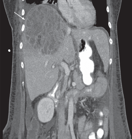
The patient undergoes CT-guided liver biopsy with drain placement. Cultures grow Klebsiella pneumoniae, and she is started on intravenous piperacillin-tazobactam for 4 weeks, followed by 4 weeks of oral ciprofloxacin.
Follow-up evaluation at 4 weeks and at 8 weeks shows improvement in the patient’s condition, with CT of the abdomen and pelvis showing a gradual decrease in the size of the abscess.
INCREASING PREVALENCE
K pneumoniae has emerged as the most common organism seen in pyogenic liver abscess.1 Initially seen in Taiwan in the 1980s, K pneumoniae liver abscess is becoming more common in the United States.2 Diabetes and impaired fasting glucose have both been implicated as potential risk factors, but the condition is also seen in nondiabetic patients.3 Although pyogenic liver abscess is commonly a sequela of biliary disease, K pneumoniae liver abscess is more often cryptogenic. 1 Clinical manifestations usually include fever, abdominal pain in the right upper quadrant, nausea, and vomiting.
In this patient, no clear relationship was established between her travel and her illness.
GREATER RISK OF SPREAD
K pneumoniae liver abscess is more likely to spread than polymicrobial liver abscess.3 It is associated with endophthalmitis, meningitis, brain abscess, and septic pulmonary embolism.3 Diabetic patients are particularly susceptible to metastatic foci.3 The reason is not well understood, but poor glycemic control leading to impaired neutrophil phagocytosis is thought to play a role.4
DIAGNOSIS AND TREATMENT
Liver abscesses are associated with elevated alkaline phosphatase levels, hyperbilirubinemia, leukocytosis, hypoalbuminemia, and anemia. As bacteremia is often seen with K pneumoniae liver abscess, blood cultures should be obtained.1 Imaging studies should include right-upper-quadrant ultrasonography if suspicion is high for concomitant biliary disease, and also CT with intravenous contrast to better quantify the dimensions of the abscess.5
Treatment includes empiric parenteral antibiotics and percutaneous drainage. In addition, culture of purulent material for aerobic and anaerobic organisms helps guide antibiotic treatment.
The antibiotic regimen should consist of a first- or third-generation beta-lactamase inhibitor, with or without an aminoglycoside.3 Patients unable to tolerate beta-lactam antibiotics can be given a fluoroquinolone.
The results of cultures and determination of antibiotic sensitivities help to further modifiy antibiotic therapy. Antibiotic therapy may be needed for 4 to 6 weeks. Parenteral antibiotics are recommended initially, and if a patient responds to therapy, treatment can be switched to oral antibiotics to complete the course of treatment.
Although antibiotics with percutaneous drainage are the recommended course of therapy, surgical drainage is sometimes necessary and is best done with the input of a hepatobiliary surgeon. Patients with abscesses larger than 5 cm who had surgical drainage had better clinical outcomes than those who had percutaneous drainage,6 but monitoring the response to antibiotics and the patient’s clinical course is very important when determining the need for emergency surgical intervention vs percutaneous drainage.6
Follow-up imaging is necessary to evaluate the response to therapy, to determine the continued need for antibiotics, and to assess for any further need for drainage.
- Pope JV, Teich DL, Clardy P, McGillicuddy DC. Klebsiella pneumoniae liver abscess: an emerging problem in North America. J Emerg Med 2008; Epub ahead of print.
- Frazee BW, Hansen S, Lambert L. Invasive infection with hypermucoviscous Klebsiella pneumoniae: multiple cases presenting to a single emergency department in the United States. Ann Emerg Med 2009; 53:639–642.
- Lee SS, Chen YS, Tsai HC, et al. Predictors of septic metastatic infection and mortality among patients with Klebsiella pneumoniae liver abscess. Clin Infect Dis 2008; 47:642–650.
- Lin JC, Siu LK, Fung CP, et al. Impaired phagocytosis of capsular serotypes K1 or K2 Klebsiella pneumoniae in type 2 diabetes mellitus patients with poor glycemic control. J Clin Endocrinol Metab 2006; 91:3084–3087.
- Golia P, Sadler M. Pyogenic liver abscess: Klebsiella as an emerging pathogen. Emerg Radiol 2006; 13:87–88.
- Tan YM, Chung AY, Chow PK, et al. An appraisal of surgical and percutaneous drainage for pyogenic liver abscesses larger than 5 cm. Ann Surg 2005; 241:485–490.
A 58-year-old woman presents with fever, chills, vomiting, and right-upperquadrant abdominal pain. She has had diarrhea for several days and has lost 15 lb over the last 6 weeks. Six months ago, she took a cruise through the Panama Canal to Nicaragua, Costa Rica, Mexico, and El Salvador.
The patient undergoes CT-guided liver biopsy with drain placement. Cultures grow Klebsiella pneumoniae, and she is started on intravenous piperacillin-tazobactam for 4 weeks, followed by 4 weeks of oral ciprofloxacin.
Follow-up evaluation at 4 weeks and at 8 weeks shows improvement in the patient’s condition, with CT of the abdomen and pelvis showing a gradual decrease in the size of the abscess.
INCREASING PREVALENCE
K pneumoniae has emerged as the most common organism seen in pyogenic liver abscess.1 Initially seen in Taiwan in the 1980s, K pneumoniae liver abscess is becoming more common in the United States.2 Diabetes and impaired fasting glucose have both been implicated as potential risk factors, but the condition is also seen in nondiabetic patients.3 Although pyogenic liver abscess is commonly a sequela of biliary disease, K pneumoniae liver abscess is more often cryptogenic. 1 Clinical manifestations usually include fever, abdominal pain in the right upper quadrant, nausea, and vomiting.
In this patient, no clear relationship was established between her travel and her illness.
GREATER RISK OF SPREAD
K pneumoniae liver abscess is more likely to spread than polymicrobial liver abscess.3 It is associated with endophthalmitis, meningitis, brain abscess, and septic pulmonary embolism.3 Diabetic patients are particularly susceptible to metastatic foci.3 The reason is not well understood, but poor glycemic control leading to impaired neutrophil phagocytosis is thought to play a role.4
DIAGNOSIS AND TREATMENT
Liver abscesses are associated with elevated alkaline phosphatase levels, hyperbilirubinemia, leukocytosis, hypoalbuminemia, and anemia. As bacteremia is often seen with K pneumoniae liver abscess, blood cultures should be obtained.1 Imaging studies should include right-upper-quadrant ultrasonography if suspicion is high for concomitant biliary disease, and also CT with intravenous contrast to better quantify the dimensions of the abscess.5
Treatment includes empiric parenteral antibiotics and percutaneous drainage. In addition, culture of purulent material for aerobic and anaerobic organisms helps guide antibiotic treatment.
The antibiotic regimen should consist of a first- or third-generation beta-lactamase inhibitor, with or without an aminoglycoside.3 Patients unable to tolerate beta-lactam antibiotics can be given a fluoroquinolone.
The results of cultures and determination of antibiotic sensitivities help to further modifiy antibiotic therapy. Antibiotic therapy may be needed for 4 to 6 weeks. Parenteral antibiotics are recommended initially, and if a patient responds to therapy, treatment can be switched to oral antibiotics to complete the course of treatment.
Although antibiotics with percutaneous drainage are the recommended course of therapy, surgical drainage is sometimes necessary and is best done with the input of a hepatobiliary surgeon. Patients with abscesses larger than 5 cm who had surgical drainage had better clinical outcomes than those who had percutaneous drainage,6 but monitoring the response to antibiotics and the patient’s clinical course is very important when determining the need for emergency surgical intervention vs percutaneous drainage.6
Follow-up imaging is necessary to evaluate the response to therapy, to determine the continued need for antibiotics, and to assess for any further need for drainage.
- Pope JV, Teich DL, Clardy P, McGillicuddy DC. Klebsiella pneumoniae liver abscess: an emerging problem in North America. J Emerg Med 2008; Epub ahead of print.
- Frazee BW, Hansen S, Lambert L. Invasive infection with hypermucoviscous Klebsiella pneumoniae: multiple cases presenting to a single emergency department in the United States. Ann Emerg Med 2009; 53:639–642.
- Lee SS, Chen YS, Tsai HC, et al. Predictors of septic metastatic infection and mortality among patients with Klebsiella pneumoniae liver abscess. Clin Infect Dis 2008; 47:642–650.
- Lin JC, Siu LK, Fung CP, et al. Impaired phagocytosis of capsular serotypes K1 or K2 Klebsiella pneumoniae in type 2 diabetes mellitus patients with poor glycemic control. J Clin Endocrinol Metab 2006; 91:3084–3087.
- Golia P, Sadler M. Pyogenic liver abscess: Klebsiella as an emerging pathogen. Emerg Radiol 2006; 13:87–88.
- Tan YM, Chung AY, Chow PK, et al. An appraisal of surgical and percutaneous drainage for pyogenic liver abscesses larger than 5 cm. Ann Surg 2005; 241:485–490.
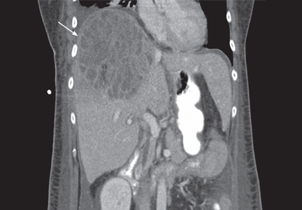
The eyes: A window into the past
A 34-year-old woman living in southern California presents for a routine physical examination. During the eye examination, the physician notices “spots” on the retina and refers the patient to a retinal specialist.
The patient has no complaints about her vision. She has myopia (−4.5 diopters), corrected with glasses. She has no family history of ocular disease. Her medical history is unremarkable, and she is taking no medications.
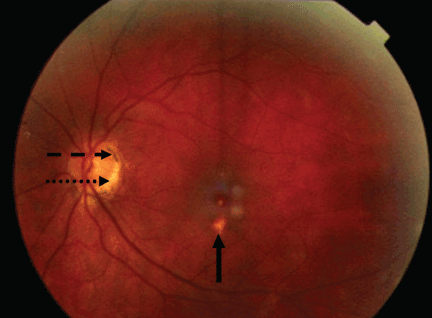
THE MOST LIKELY DIAGNOSIS
The lesions raise the suspicion of histoplasmosis, but since the patient has no other evidence of histoplasmosis, the likely diagnosis is presumed ocular histoplasmosis syndrome (POHS).
Further questioning reveals that the woman grew up on a farm in the Ohio River valley, one of two areas in the United States where Histoplasma capsulatum is highly endemic.1 (The other area is the Mississippi River valley.)
As this case shows, POHS is important to consider, especially in areas where H capsulatum is not endemic, to avert a lengthy workup for other causes of retinal lesions. It also shows the importance of a thorough history, including previous residences and travel.2
Pathogenesis is uncertain
In histoplasmosis, the infection is acquired by inhalation of microconidia of H capsulatum, usually via disruption of the soil (as in farming) and especially in areas where there are bird roosts. Infection is often asymptomatic, and fewer than 1% of people exposed develop a clinical illness 7 to 21 days after exposure.3
In disseminated histoplasmosis, eye involvement manifests as panophthalmitis or uveitis, caused by yeast implantation. The finding of eye lesions typical of histoplasmosis but in the absence of signs of disseminated histoplasmosis— as in POHS—is much more common, seen in perhaps 1% to 5% of residents in highly endemic areas.4
The pathogenesis of POHS and its association with histoplasmosis are still unclear.4–7 Some have proposed a cellular immune response to deposited fungal antigens. Others contend that patients with specific human leukocyte antigen types (eg, B7, DR2) may be susceptible.4H capsulatum DNA has been isolated in one case of POHS,6 and the classic ocular lesions are prevalent in people who live in the Ohio River valley.7 Therefore, even though a definitive causative relationship between H capsulatum exposure and POHS has not been proven, the ocular lesions are presumed to be the result of previous exposure to H capsulatum, as in this case.
Establishing the diagnosis of POHS
Most cases of POHS are detected on routine eye examination. The diagnosis is confirmed by a dilated eye examination showing peripheral, punched-out, atrophic scars (“histospots”), which represent focal defects in the Bruch membrane, along with a history of living in an area endemic for H capsulatum. Histo-spots range from 0.2 to 0.7 disk diameters and can occur as single or multiple lesions.8 Most often, both eyes are involved, albeit asymmetrically.9 Some areas may contain pigment deposits, as in this case.8 Peripapillary atrophy, a thinning of the retina immediately surrounding the head of the optic nerve, is also characteristic of POHS. Active inflammation in the anterior chamber and vitreous are absent.
Although most patients with POHS do not have a documented diagnosis of previous clinical Histoplasma infection, they may have a positive histoplasmin skin test, as well as lung, liver, or spleen calcifications. However, skin testing is not recommended as it may exacerbate POHS,9 and serologic testing is usually negative.10
POHS has most commonly been diagnosed in whites ages 20 to 50 (mean age 35). Men and women appear to be equally affected.
The potential for vision loss
Very few patients with ophthalmoscopic evidence of POHS develop visual symptoms.7 Still, there is a risk of choroidal neovascularization at the site of the choroidal scars. These new vessels can hemorrhage, causing impaired central vision (distorted vision, blind spots).
The trigger for choroidal neovascularization is unknown; exposure to fungal antigens and eye surgery such as LASIK have been proposed. 11 Choroidal neovascularization usually occurs 10 to 20 years after scar formation and occurs in fewer than 5% of POHS patients.10,12
HOW THE PATIENT WAS MANAGED
Given that the patient had POHS with no evidence of neovascularization, she was followed with serial visual assessments using an Amsler grid.11 For POHS with choroidal neovascularization, treatment focuses on reducing the risk of vascular complications and includes oral corticosteroids, intravitreal corticosteroid injections, laser photocoagulation, and photodynamic therapy with verteporfin (Visudyne). 4,10,13–15 Antifungal treatment is not useful, as the lesions are not proven to be caused by active infection.10
Future treatments may include antiangiogenic drugs and gene therapy.9
Since her diagnosis, the patient’s visual tests have been stable, with no neovascularization.
- Edwards LB, Acquaviva FA, Livesay VT, Cross FW, Palmer CE. An atlas of sensitivity to tuberculin, PPD-B, and histoplasmin in the United States. Am Rev Respir Dis 1969; 99: suppl:1–132.
- Wheat LJ. Histoplasmosis: a review for clinicians from non-endemic areas. Mycoses 2006; 49:274–282.
- Kauffman CA. Histoplasmosis: a clinical and laboratory update. Clin Microbiol Rev 2007; 20:115–132.
- Prasad AG, Van Gelder RN. Presumed ocular histoplasmosis syndrome. Curr Opin Ophthalmol 2005; 16:364–368.
- Ongkosuwito JV, Kortbeek LM, Van der Lelij A, et al. Aetiological study of the presumed ocular histoplasmosis syndrome in the Netherlands. Br J Ophthalmol 1999; 83:535–539.
- Spencer WH, Chan CC, Shen DF, Rao NA. Detection of Histoplasma capsulatum DNA in lesions of chronic ocular histoplasmosis syndrome. Arch Ophthalmol 2003; 121:1551–1555.
- Find SL. Ocular histoplasmosis syndrome. Int Ophthalmol Clin 1977; 17:75–87.
- McMillan TA, Lashkari K. Ocular histoplasmosis. Int Ophthalmol Clin 1996; 36:179–186.
- Ciulla TA, Piper HC, Xiao M, Wheat LJ. Presumed ocular histoplasmosis syndrome: update on epidemiology, pathogenesis, and photodynamic, antiangiogenic, and surgical therapies. Curr Opin Ophthalmol 2001; 12:442–449.
- Oliver A, Ciulla TA, Comer GM. New and classic insights into presumed ocular histoplasmosis syndrome and its treatment. Curr Opin Ophthalmol 2005; 16:160–165.
- Trevino R, Salvat R. Preventing reactivation of ocular histoplasmosis: guidance for patients at risk. Optometry 2006; 77:10–16.
- Macular Photocoagulation Study Group. Five-year followup of fellow eyes of individuals with ocular histoplasmosis and unilateral extrafoveal or juxtafoveal choroidal neovascularization. Arch Ophthalmol 1996; 114:677–688.
- Rosenfeld PJ, Saperstein DA, Bressler NM, et al; Verteporfin in Ocular Histoplasmosis Study Group. Photodynamic therapy with verteporfin in ocular histoplasmosis: uncontrolled, open-label 2-year study. Ophthalmology 2004; 111:1725–1733.
- Shah GK, Blinder KJ, Hariprasad SM, et al. Photodynamic therapy for juxtafoveal choroidal neovascularization due to ocular histoplasmosis syndrome. Retina 2005; 25:26–32.
- Rechtman E, Allen VD, Danis RP, Pratt LM, Harris A, Speicher MA. Intravitreal triamcinolone for choroidal neovascularization in ocular histoplasmosis syndrome. Am J Ophthalmol 2003; 136:739–741.
A 34-year-old woman living in southern California presents for a routine physical examination. During the eye examination, the physician notices “spots” on the retina and refers the patient to a retinal specialist.
The patient has no complaints about her vision. She has myopia (−4.5 diopters), corrected with glasses. She has no family history of ocular disease. Her medical history is unremarkable, and she is taking no medications.
THE MOST LIKELY DIAGNOSIS
The lesions raise the suspicion of histoplasmosis, but since the patient has no other evidence of histoplasmosis, the likely diagnosis is presumed ocular histoplasmosis syndrome (POHS).
Further questioning reveals that the woman grew up on a farm in the Ohio River valley, one of two areas in the United States where Histoplasma capsulatum is highly endemic.1 (The other area is the Mississippi River valley.)
As this case shows, POHS is important to consider, especially in areas where H capsulatum is not endemic, to avert a lengthy workup for other causes of retinal lesions. It also shows the importance of a thorough history, including previous residences and travel.2
Pathogenesis is uncertain
In histoplasmosis, the infection is acquired by inhalation of microconidia of H capsulatum, usually via disruption of the soil (as in farming) and especially in areas where there are bird roosts. Infection is often asymptomatic, and fewer than 1% of people exposed develop a clinical illness 7 to 21 days after exposure.3
In disseminated histoplasmosis, eye involvement manifests as panophthalmitis or uveitis, caused by yeast implantation. The finding of eye lesions typical of histoplasmosis but in the absence of signs of disseminated histoplasmosis— as in POHS—is much more common, seen in perhaps 1% to 5% of residents in highly endemic areas.4
The pathogenesis of POHS and its association with histoplasmosis are still unclear.4–7 Some have proposed a cellular immune response to deposited fungal antigens. Others contend that patients with specific human leukocyte antigen types (eg, B7, DR2) may be susceptible.4H capsulatum DNA has been isolated in one case of POHS,6 and the classic ocular lesions are prevalent in people who live in the Ohio River valley.7 Therefore, even though a definitive causative relationship between H capsulatum exposure and POHS has not been proven, the ocular lesions are presumed to be the result of previous exposure to H capsulatum, as in this case.
Establishing the diagnosis of POHS
Most cases of POHS are detected on routine eye examination. The diagnosis is confirmed by a dilated eye examination showing peripheral, punched-out, atrophic scars (“histospots”), which represent focal defects in the Bruch membrane, along with a history of living in an area endemic for H capsulatum. Histo-spots range from 0.2 to 0.7 disk diameters and can occur as single or multiple lesions.8 Most often, both eyes are involved, albeit asymmetrically.9 Some areas may contain pigment deposits, as in this case.8 Peripapillary atrophy, a thinning of the retina immediately surrounding the head of the optic nerve, is also characteristic of POHS. Active inflammation in the anterior chamber and vitreous are absent.
Although most patients with POHS do not have a documented diagnosis of previous clinical Histoplasma infection, they may have a positive histoplasmin skin test, as well as lung, liver, or spleen calcifications. However, skin testing is not recommended as it may exacerbate POHS,9 and serologic testing is usually negative.10
POHS has most commonly been diagnosed in whites ages 20 to 50 (mean age 35). Men and women appear to be equally affected.
The potential for vision loss
Very few patients with ophthalmoscopic evidence of POHS develop visual symptoms.7 Still, there is a risk of choroidal neovascularization at the site of the choroidal scars. These new vessels can hemorrhage, causing impaired central vision (distorted vision, blind spots).
The trigger for choroidal neovascularization is unknown; exposure to fungal antigens and eye surgery such as LASIK have been proposed. 11 Choroidal neovascularization usually occurs 10 to 20 years after scar formation and occurs in fewer than 5% of POHS patients.10,12
HOW THE PATIENT WAS MANAGED
Given that the patient had POHS with no evidence of neovascularization, she was followed with serial visual assessments using an Amsler grid.11 For POHS with choroidal neovascularization, treatment focuses on reducing the risk of vascular complications and includes oral corticosteroids, intravitreal corticosteroid injections, laser photocoagulation, and photodynamic therapy with verteporfin (Visudyne). 4,10,13–15 Antifungal treatment is not useful, as the lesions are not proven to be caused by active infection.10
Future treatments may include antiangiogenic drugs and gene therapy.9
Since her diagnosis, the patient’s visual tests have been stable, with no neovascularization.
- Edwards LB, Acquaviva FA, Livesay VT, Cross FW, Palmer CE. An atlas of sensitivity to tuberculin, PPD-B, and histoplasmin in the United States. Am Rev Respir Dis 1969; 99: suppl:1–132.
- Wheat LJ. Histoplasmosis: a review for clinicians from non-endemic areas. Mycoses 2006; 49:274–282.
- Kauffman CA. Histoplasmosis: a clinical and laboratory update. Clin Microbiol Rev 2007; 20:115–132.
- Prasad AG, Van Gelder RN. Presumed ocular histoplasmosis syndrome. Curr Opin Ophthalmol 2005; 16:364–368.
- Ongkosuwito JV, Kortbeek LM, Van der Lelij A, et al. Aetiological study of the presumed ocular histoplasmosis syndrome in the Netherlands. Br J Ophthalmol 1999; 83:535–539.
- Spencer WH, Chan CC, Shen DF, Rao NA. Detection of Histoplasma capsulatum DNA in lesions of chronic ocular histoplasmosis syndrome. Arch Ophthalmol 2003; 121:1551–1555.
- Find SL. Ocular histoplasmosis syndrome. Int Ophthalmol Clin 1977; 17:75–87.
- McMillan TA, Lashkari K. Ocular histoplasmosis. Int Ophthalmol Clin 1996; 36:179–186.
- Ciulla TA, Piper HC, Xiao M, Wheat LJ. Presumed ocular histoplasmosis syndrome: update on epidemiology, pathogenesis, and photodynamic, antiangiogenic, and surgical therapies. Curr Opin Ophthalmol 2001; 12:442–449.
- Oliver A, Ciulla TA, Comer GM. New and classic insights into presumed ocular histoplasmosis syndrome and its treatment. Curr Opin Ophthalmol 2005; 16:160–165.
- Trevino R, Salvat R. Preventing reactivation of ocular histoplasmosis: guidance for patients at risk. Optometry 2006; 77:10–16.
- Macular Photocoagulation Study Group. Five-year followup of fellow eyes of individuals with ocular histoplasmosis and unilateral extrafoveal or juxtafoveal choroidal neovascularization. Arch Ophthalmol 1996; 114:677–688.
- Rosenfeld PJ, Saperstein DA, Bressler NM, et al; Verteporfin in Ocular Histoplasmosis Study Group. Photodynamic therapy with verteporfin in ocular histoplasmosis: uncontrolled, open-label 2-year study. Ophthalmology 2004; 111:1725–1733.
- Shah GK, Blinder KJ, Hariprasad SM, et al. Photodynamic therapy for juxtafoveal choroidal neovascularization due to ocular histoplasmosis syndrome. Retina 2005; 25:26–32.
- Rechtman E, Allen VD, Danis RP, Pratt LM, Harris A, Speicher MA. Intravitreal triamcinolone for choroidal neovascularization in ocular histoplasmosis syndrome. Am J Ophthalmol 2003; 136:739–741.
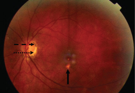
Escaping the heat in the EMR pool

In reply, based on his experience, Dr. Tom Abelson defends EMRs, predicting that their current weaknesses will be overcome as technology improves, and the faster we embrace this technologic advance, the faster fixes for initial limitations will be developed. We are, he says, just learning to swim.
The EMR is a tool, but a tool performs at the skill level of the user. The EMR is more powerful than the paper record it replaces, offering the promise of being searchable, interactive, and able to prompt us to perform in predefined ways, and also linking us at the point of care with reference materials. But the information contained in the EMR can be no better than what we enter. An EMR cannot supplant our need to think and act as physicians defending our patients’ best interests.
Our skill in using the EMR is evolving. Thus far, cut-and-paste and other electronic shortcuts are rampant and are a detriment to quality care, but these are examples of misuse and are not an intrinsic fault of the tool. These physician behaviors can be curtailed.
The EMR cannot be read like a book. Events, consultations, and nursing interventions do not readily unfold in chronologic order. Suggestions of consultants can be missed, and perhaps due to limited typing skills, clinical reasoning is not fully explained (was it always clearly explained on paper?). The notes, however, are legible.
We must not be enticed to let EMRs overly influence our billing practices. Rather, the EMR should be a tool to improve the accuracy of the record of the patient encounter.
The EMR can come between the doctor and patient, but it need not. We need to be a bit more attentive to the patient, push back intermittently from the keyboard, and make eye contact. We need to engage the patient with his or her EMR on screen—show some trended lab results or radiographic images and, when they leave, present them with a typed set of legible instructions and their drug list (noting that all this information, and more, can be available to the next physician that they see, by cyberlink or via fax).
For those of you adopting an EMR system, I suggest you make sure you get adequate, continued access to on-site support from your vendor. The tool must be trained to swim in your pool.
The electronic medical record: Learning to swim
While Dr. J. Timothy hanlon raises compelling issues about electronic medical records (EMRs),1 I think that his goslow approach can lead to lost opportunities. The push toward implementing EMR systems does not amount to a dangerous dive into a shallow pool. Rather, we should be optimistic that we are just learning to swim.
Addressing these concerns during these early years of the EMR is constructive and necessary. But his commentary may leave physicians wondering what to do. Should the EMR be scrapped until it is fully developed outside the clinical realm? Is that goal even attainable? Are physicians who use EMR systems putting patient care and information security at risk? I believe there is a more positive way to look at each of the issues.
No new technology comes into the world 100% formed and vetted. The nature of progress is evolution based on experience, as unforeseen problems are corrected. Skepticism and vigilance are warranted, but so is optimism.
CONNECTIVITY WILL IMPROVE
Dr. Hanlon notes that many EMR systems are available and that, at this point, they do not communicate with one another. That is often true. But Google, Microsoft, and others are working on the issues of connectivity and “portability” of the EMR. It is reasonable to expect that eventually there will be winners and losers, just as when VHS won out over Beta in the early days of video recording. More efficient sharing of information will eventually be possible. It would be counterproductive to legislate a single EMR system nationwide before a number of EMRs can be fully tried and tested in the trenches of patient care.
In the meantime, it is no harder—in fact, it is easier than ever—for one physician to send information to another, either by printing it out and mailing or faxing it or by e-mailing it. Furthermore, that information is much more legible than the handwritten records we continue to receive from physicians who do not use EMRs.
Therefore, while one can criticize the current lack of complete intersystem communication, this is only a temporary limitation, and developing complete interconnectivity of EMR systems is a key goal.
STAYING VIGILANT ABOUT SECURITY
The security of EMRs has always been a concern. However, improvements in security have prevented a large-scale privacy breach since an incident in 2006 in which a laptop computer containing information on 26.5 million people was stolen from the home of a Veterans Administration employee.2 For instance, Cleveland Clinic recently encrypted all of its laptop computers containing patient data, to protect patient information should a laptop be lost or stolen. This is only one of many security innovations that are being implemented. While we must remain highly vigilant and continue to improve security, we must remember that the paper chart is not immune to privacy breaches either, and when paper charts were stolen, the medical record was irretrievably lost and was not reproducible. This is not the case with the EMR.
QUALITY OF CARE IS PARAMOUNT
As Dr. Hanlon accurately notes, evidence that the EMR improves the quality of care is mixed so far. He is concerned that most of the studies showing improved outcomes came from “benchmark” institutions, and that the results may not be broadly applicable. Such pessimism is unwarranted, given that the EMR is in its relative infancy and the motivation to improve quality of care is paramount, especially in this era of health care reform. While benchmark institutions are in an ideal position to do the studies on quality, there is no reason to assume that the results will not be applicable to other institutions as well.
EDUCATION: AN AREA FOR INNOVATION
Dr. Hanlon notes that research on EMRs for medical education is in its infancy. But infants grow rapidly. While it may be true that students might have to learn to use different EMR systems at different institutions, these students have grown up with rapidly changing computer systems and can learn and adapt at a remarkable rate. Therefore, education is a wonderful area for innovation and research on the EMR. It is not a reason to fear the EMR or the present diversity of EMR systems.
ACCURACY CAN BE IMPROVED
Dr. Hanlon is correct that the problem of cutting and pasting of previous notes, potentially propagating an initial error (so-called high-risk copying3) is profound within the EMR. But I prefer to look at this as an area for innovation— such as nonerasable tags to identify copied material.
While errors in medication lists are possible, especially if practitioners use cut-and-paste methods and thus perpetuate a previous error, systems and workflows are being developed to overcome such problems. Some of these include special alerts when certain high-risk drugs are ordered, drop-down menus with drug dosing included, and links to databases that allow quick access to information on drug interactions.
And again, medication errors are not unique to the EMR. They also occur in paper charts as a result of photocopying, illegible handwriting, and transcription errors.
Compared with the paper chart, the EMR is more legible, and the ability to instantaneously transfer unchanged important and valid information potentially enhances the completeness and logic of a given note and provides the physician more time to spend evaluating (and looking at) the patient. So, rather than focusing on the negatives of the current problem of cutting and pasting, I prefer to focus on how to improve it. That is, how can we make the information in the EMR more accurate, catch errors, and then make the latest information easily accessible to users?
STAYING FOCUSED ON THE PATIENT, EVEN WITH A COMPUTER IN THE ROOM
A major complaint by patients and caregivers is that using an EMR makes the physician focus on a computer screen rather than looking at the patient. This concern is valid, but I think we can learn to stay focused on the patient, even with a computer in the examination room, and still take advantage of everything technology has to offer.
This issue will disappear in less than one generation. Young people are remarkably able to multitask while typing. They are able to talk with their patients while typing and to look them in the eyes. And typing letter by letter will become obsolete as soon as voice-recognition software and ways to edit its output accurately are perfected. Many of us at Cleveland Clinic use a combination of templates, typing, and voice-recognition dictation, and find this to be effective.
When we tell our grandchildren that we used to type each individual letter on a page, they will be as amazed as we are to hear that cars used to be started with an external crank.
DOCTOR-DOCTOR COMMUNICATION IS ENHANCED
I agree that template notes written by physicians who cannot type very well can lack the substance and color found in a well-reported medical history and examination. But voice-recognition transcription can help flesh out key parts of the history, differential diagnosis, and management plan. Further, the note can be produced on the spot, the patient can check the note for accuracy, and the conclusions can be shared instantaneously with all involved caregivers. Doctor-doctor communication is thus enhanced.
EVERY REASON TO MOVE FORWARD
Dr. Hanlon is also concerned about EMRs and the potential for “billing creep” and outright fraud. But fraud is as old as billing. What is required is continued vigilance and system controls, which actually might be more effective in an EMR system than in a paper billing system. Integrity will be neither enhanced nor diminished by digitization, unfortunately.
In summary, while Dr. Hanlon sees reason to slow down the move to EMRs, I see every reason to move forward. The problems he describes are part of the growing pains of any new technology. He is right that we cannot move blindly, ignoring the challenges of this technology. But slowing down will only delay its benefits.
- Hanlon JT. The electronic medical record: diving into a shallow pool? Cleve Clin J Med 2010; 77:408–411.
- Lemos R. Veterans Affairs warns of massive privacy breach. SecurityFocus 2006 (May 22). http://www.securityfocus.com/news/11393. Accessed May 19, 2010.
- Hammond KW, Helbig ST, Benson CC, Brathwaite-Sketoe BM. Are Electronic Medical Records Trustworthy? Observations on Copying, Pasting and Duplication. AMIA Annu Symp Proc 2003 http://www.ncbi.nlm.nih.gov/pmc/articles/PMC1480345/. Accessed May 19, 2010.
While Dr. J. Timothy hanlon raises compelling issues about electronic medical records (EMRs),1 I think that his goslow approach can lead to lost opportunities. The push toward implementing EMR systems does not amount to a dangerous dive into a shallow pool. Rather, we should be optimistic that we are just learning to swim.
Addressing these concerns during these early years of the EMR is constructive and necessary. But his commentary may leave physicians wondering what to do. Should the EMR be scrapped until it is fully developed outside the clinical realm? Is that goal even attainable? Are physicians who use EMR systems putting patient care and information security at risk? I believe there is a more positive way to look at each of the issues.
No new technology comes into the world 100% formed and vetted. The nature of progress is evolution based on experience, as unforeseen problems are corrected. Skepticism and vigilance are warranted, but so is optimism.
CONNECTIVITY WILL IMPROVE
Dr. Hanlon notes that many EMR systems are available and that, at this point, they do not communicate with one another. That is often true. But Google, Microsoft, and others are working on the issues of connectivity and “portability” of the EMR. It is reasonable to expect that eventually there will be winners and losers, just as when VHS won out over Beta in the early days of video recording. More efficient sharing of information will eventually be possible. It would be counterproductive to legislate a single EMR system nationwide before a number of EMRs can be fully tried and tested in the trenches of patient care.
In the meantime, it is no harder—in fact, it is easier than ever—for one physician to send information to another, either by printing it out and mailing or faxing it or by e-mailing it. Furthermore, that information is much more legible than the handwritten records we continue to receive from physicians who do not use EMRs.
Therefore, while one can criticize the current lack of complete intersystem communication, this is only a temporary limitation, and developing complete interconnectivity of EMR systems is a key goal.
STAYING VIGILANT ABOUT SECURITY
The security of EMRs has always been a concern. However, improvements in security have prevented a large-scale privacy breach since an incident in 2006 in which a laptop computer containing information on 26.5 million people was stolen from the home of a Veterans Administration employee.2 For instance, Cleveland Clinic recently encrypted all of its laptop computers containing patient data, to protect patient information should a laptop be lost or stolen. This is only one of many security innovations that are being implemented. While we must remain highly vigilant and continue to improve security, we must remember that the paper chart is not immune to privacy breaches either, and when paper charts were stolen, the medical record was irretrievably lost and was not reproducible. This is not the case with the EMR.
QUALITY OF CARE IS PARAMOUNT
As Dr. Hanlon accurately notes, evidence that the EMR improves the quality of care is mixed so far. He is concerned that most of the studies showing improved outcomes came from “benchmark” institutions, and that the results may not be broadly applicable. Such pessimism is unwarranted, given that the EMR is in its relative infancy and the motivation to improve quality of care is paramount, especially in this era of health care reform. While benchmark institutions are in an ideal position to do the studies on quality, there is no reason to assume that the results will not be applicable to other institutions as well.
EDUCATION: AN AREA FOR INNOVATION
Dr. Hanlon notes that research on EMRs for medical education is in its infancy. But infants grow rapidly. While it may be true that students might have to learn to use different EMR systems at different institutions, these students have grown up with rapidly changing computer systems and can learn and adapt at a remarkable rate. Therefore, education is a wonderful area for innovation and research on the EMR. It is not a reason to fear the EMR or the present diversity of EMR systems.
ACCURACY CAN BE IMPROVED
Dr. Hanlon is correct that the problem of cutting and pasting of previous notes, potentially propagating an initial error (so-called high-risk copying3) is profound within the EMR. But I prefer to look at this as an area for innovation— such as nonerasable tags to identify copied material.
While errors in medication lists are possible, especially if practitioners use cut-and-paste methods and thus perpetuate a previous error, systems and workflows are being developed to overcome such problems. Some of these include special alerts when certain high-risk drugs are ordered, drop-down menus with drug dosing included, and links to databases that allow quick access to information on drug interactions.
And again, medication errors are not unique to the EMR. They also occur in paper charts as a result of photocopying, illegible handwriting, and transcription errors.
Compared with the paper chart, the EMR is more legible, and the ability to instantaneously transfer unchanged important and valid information potentially enhances the completeness and logic of a given note and provides the physician more time to spend evaluating (and looking at) the patient. So, rather than focusing on the negatives of the current problem of cutting and pasting, I prefer to focus on how to improve it. That is, how can we make the information in the EMR more accurate, catch errors, and then make the latest information easily accessible to users?
STAYING FOCUSED ON THE PATIENT, EVEN WITH A COMPUTER IN THE ROOM
A major complaint by patients and caregivers is that using an EMR makes the physician focus on a computer screen rather than looking at the patient. This concern is valid, but I think we can learn to stay focused on the patient, even with a computer in the examination room, and still take advantage of everything technology has to offer.
This issue will disappear in less than one generation. Young people are remarkably able to multitask while typing. They are able to talk with their patients while typing and to look them in the eyes. And typing letter by letter will become obsolete as soon as voice-recognition software and ways to edit its output accurately are perfected. Many of us at Cleveland Clinic use a combination of templates, typing, and voice-recognition dictation, and find this to be effective.
When we tell our grandchildren that we used to type each individual letter on a page, they will be as amazed as we are to hear that cars used to be started with an external crank.
DOCTOR-DOCTOR COMMUNICATION IS ENHANCED
I agree that template notes written by physicians who cannot type very well can lack the substance and color found in a well-reported medical history and examination. But voice-recognition transcription can help flesh out key parts of the history, differential diagnosis, and management plan. Further, the note can be produced on the spot, the patient can check the note for accuracy, and the conclusions can be shared instantaneously with all involved caregivers. Doctor-doctor communication is thus enhanced.
EVERY REASON TO MOVE FORWARD
Dr. Hanlon is also concerned about EMRs and the potential for “billing creep” and outright fraud. But fraud is as old as billing. What is required is continued vigilance and system controls, which actually might be more effective in an EMR system than in a paper billing system. Integrity will be neither enhanced nor diminished by digitization, unfortunately.
In summary, while Dr. Hanlon sees reason to slow down the move to EMRs, I see every reason to move forward. The problems he describes are part of the growing pains of any new technology. He is right that we cannot move blindly, ignoring the challenges of this technology. But slowing down will only delay its benefits.
- Hanlon JT. The electronic medical record: diving into a shallow pool? Cleve Clin J Med 2010; 77:408–411.
- Lemos R. Veterans Affairs warns of massive privacy breach. SecurityFocus 2006 (May 22). http://www.securityfocus.com/news/11393. Accessed May 19, 2010.
- Hammond KW, Helbig ST, Benson CC, Brathwaite-Sketoe BM. Are Electronic Medical Records Trustworthy? Observations on Copying, Pasting and Duplication. AMIA Annu Symp Proc 2003 http://www.ncbi.nlm.nih.gov/pmc/articles/PMC1480345/. Accessed May 19, 2010.
Bariatric surgery for type 2 diabetes: Weighing the impact for obese patients
Evidence is mounting for the use of bariatric surgery to treat type 2 diabetes mellitus in patients whose body mass index (BMI) is 35 kg/m2 or higher. In obese patients who also have type 2 diabetes, bariatric surgery sends it into remission (defined as normoglycemic control without the need for diabetic medications) in more than three-fourths of cases, with higher rates with the Roux-en-Y gastric bypass procedure than with the laparoscopic adjustable gastric banding procedure.
However, data on the effects of this surgery on type 2 diabetes come primarily from observational studies that lacked appropriate control groups, and the relative benefit of bariatric surgery vs aggressive medical antidiabetic therapy is not yet known. Needed are randomized trials comparing the two types of therapy (and the various types of bariatric surgery) in diabetic patients with less-severe obesity.
Further, why would bariatric surgery help with diabetes, and why would one procedure do it better than another? To be honest, we are not sure, but evidence points not only to weight loss but also to better insulin sensitivity and to alterations in levels of hormones secreted by the gut that increase insulin secretion.
OBESITY PROMOTES DIABETES; WEIGHT LOSS COUNTERACTS IT
Type 2 diabetes mellitus is a complex metabolic disease characterized by insulin resistance and progressive failure of pancreatic beta cells, resulting in hyperglycemia.1,2
Obesity, a potent risk factor for type 2 diabetes, contributes to its development by inducing insulin resistance and inflammation, which in turn impair glucose regulation.3,4 Fat deposits in the abdomen, muscles, and liver contribute to elevations of circulating free fatty acids and adipocyte-derived cytokines that mediate insulin resistance and inflammatory pathways.5
In the Diabetes Prevention Program,6 modest weight loss (5% to 10% of body weight) through diet and exercise reduced the incidence of type 2 diabetes, and in the ongoing Action for Health in Diabetes (Look AHEAD) study of the National Institutes of Health, it improved glucose homeostasis.7,8
The current medical approach to type 2 diabetes includes advising the patient to lose weight through lifestyle modification, and prescribing drugs that restore glycemic control by reducing insulin resistance (biguanides, glitazones) and improving insulin secretion (incretin mimetics and analogues and sulfonylureas). 9,10
However, several factors make type 2 diabetes challenging to treat in obese people. Patients who lose weight via behavioral changes and weight-loss drugs tend to gain the weight back. Antidiabetic drugs pose the risk of hypoglycemia. Moreover, although many new classes of drugs have been developed to treat type 2 diabetes, most patients fail to achieve the American Diabetes Association goal for glycemic control, ie, a hemoglobin A1c level lower than 7%.11
BARIATRIC PROCEDURES AND THEIR EFFECT ON DIABETES CONTROL
After bariatric surgery, patients lose more weight than with traditional weight-loss methods—up to 25% of their total body weight. Furthermore, of those with type 2 diabetes, 87% achieve at least better glucose control and need fewer antidiabetic medications,12 and an average of 78% achieve normal glycemic control without taking any antidiabetic medications at all.12,13
But not all bariatric procedures have the same effect on weight and diabetes: certain procedures have a greater effect.
The two major types are classified as gastric restrictive procedures and intestinal bypass procedures. The classification was initially based on the presumed mechanism of weight loss.
Gastric restrictive procedures (laparoscopic adjustable gastric banding, sleeve gastrectomy, vertical gastroplasty) limit gastric volume and, hence, restrict the intake of calories by inducing satiety. Afterward, patients lose approximately 10% to 20% of their total body weight.
Furthermore, multiple studies, including a randomized controlled trial14 (more about this below), have shown remission of type 2 diabetes with laparoscopic adjustable gastric banding but not with conventional medical therapy. The effect is primarily mediated by weight loss and improved insulin sensitivity, both of which occur several months following surgery. Of note, however: in this trial,14 all the patients had diabetes of short duration, less than 2 years.
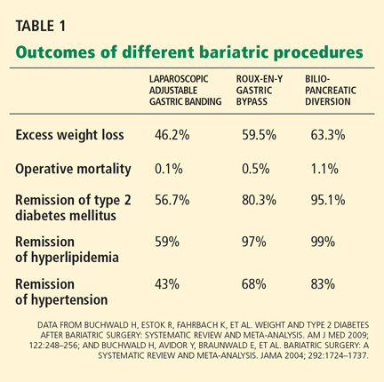
Hence, different procedures have different effects on diabetes.12 The speed at which type 2 diabetes goes into remission differs with restrictive vs malabsorptive procedures. After Roux-en-Y gastric bypass and biliopancreatic diversion, diabetes remits within days, even before the patient has lost much weight.15 This does not happen after gastric restrictive procedures.12,16
Observational studies of the effect of Roux-en-Y surgery on diabetes
Several observational studies have evaluated the benefit of Roux-en-Y surgery for patients with type 2 diabetes mellitus.
Pories et al15 followed 608 severely obese patients, of whom 165 (27%) had type 2 diabetes or impaired glucose tolerance.
At a mean follow-up of 7.6 years after surgery, 83% of the diabetic patients were off their antidiabetic drugs, and 99% of those with impaired glucose tolerance were normoglycemic, with normal fasting glucose and hemoglobin A1c levels. Marked improvements in hyperlipidemia, hypertension, fertility, osteoarthritis, and obstructive sleep apnea were also noted.
Schauer et al17 observed similar results in 1,160 morbidly obese patients, of whom 240 (21%) had type 2 diabetes or impaired fasting glucose.
After laparoscopic Roux-en-Y gastric bypass surgery, fasting glucose and hemoglobin A1c levels returned to normal levels in 83% of cases and were markedly improved in the remaining 17%. Significantly (80%) fewer patients needed oral antidiabetic agents or insulin (79% fewer). Patients most likely to achieve complete remission of diabetes were those with the shortest duration of diabetes (< 5 years), the mildest severity of diabetes (diet-controlled), and the greatest weight loss after surgery. The rate of diabetes remission in patients who had been diabetic for 5 years or less was 95%, compared with 75% in those who had been diabetic for 6 to 10 years and 54% in those who had been diabetic for more than 10 years (P < .001).
The Swedish Obese Subjects (SOS) study18 prospectively followed 1,703 patients, of whom 118 had type 2 diabetes, for 10 years after various bariatric surgery procedures (primarily vertical gastroplasty). In a control group that received medical therapy, 77 patients had type 2 diabetes. Medical therapy was ill-defined with respect to aggressiveness and adherence to intervention with lifestyle and pharmacotherapy.
At 2 years, the surgical group had lost a mean of 28 kg, glycemic control had improved in the diabetic patients, and many of them had been able to stop taking oral hypoglycemic drugs or insulin. In contrast, the need for these agents increased in the medically treated patients. The proportion treated by diet alone rose from 59% to 73% in the surgical group, but declined from 55% to 34% in the nonsurgical group.13
In these studies, surgery also reduced the risk of progressing from impaired glucose tolerance to type 2 diabetes; the risk was 30 times lower in the study by Pories et al.15 In the SOS study,18 the frequency of diabetes was 30 times lower at 2 years and five times lower at 8 years after surgery.
Studies of biliopancreatic diversion
Data on the effects of biliopancreatic diversion, a primarily malabsorptive procedure, are limited to European studies.
Scopinaro et al19,20 reported long-term follow-up data on 312 patients with type 2 diabetes who underwent biliopancreatic diversion; 310 patients (99%) achieved normal fasting glucose values by 1 year after surgery. At 10 years after surgery, 98% of the patients were still in complete remission of diabetes, defined as normal glucose values without the use of antidiabetic medications.
Others have noted similar findings.21,22
Limitations of the studies
Although these data seem encouraging, these studies had major limitations.
The patients were mostly white women with severe obesity, ie, a BMI greater than 40 kg/m2, which is not representative of patients with type 2 diabetes in the community. Only about 20% had glucose intolerance or overt type 2 diabetes mellitus. Would other groups benefit, particularly men and those with lesssevere obesity?
Moreover, these studies were observational, with no randomized control groups. Many reports consisted of large case series. It is not clear how specific bariatric procedures were chosen or what criteria were used for performing bariatric surgery. A lack of complete follow-up data is also a concern.
Needed are large randomized trials evaluating the effects of various bariatric procedures in a less obese cohort with type 2 diabetes, ie, typical patients seen in the community. Moreover, surgery has not been compared directly with more vigorous medical weight-loss strategies, such as those used in the Diabetes Prevention Project6 and the Look AHEAD trial.7,8
A randomized controlled trial of gastric banding
The only randomized controlled trial to date that compared standard medical diabetes therapy with bariatric surgery was conducted by Dixon et al.14
Sixty patients with type 2 diabetes (duration < 2 years and mean hemoglobin A1c 7.7%) were randomized either to receive medical management as defined by the American Diabetes Association guidelines or to undergo laparoscopic adjustable gastric banding.
At 2 years, the rate of remission (defined as hemoglobin A1c < 6.2% and a normal fasting glucose level) was 13% in the medical treatment group vs 73% in the surgery group (P < .001). Patients receiving medical treatment had lost a mean of 1.7% of their body weight, vs 20.7% in the surgical patients (P < .001). Weight loss was strongly associated with remission of type 2 diabetes after surgery.
This study was controversial in that the medical intervention in this trial was not as aggressive as in the Diabetes Prevention Project and Look AHEAD trials.
INDICATIONS FOR BARIATRIC SURGERY IN PATIENTS WITH DIABETES
According to guidelines from the National Institutes of Health,23 the current indications for bariatric surgery include a BMI of 40 kg/m2 or higher, or a BMI between 35 and 40 kg/m2 with at least two obesity-related comorbidities. Diabetes is considered a key comorbidity that justifies the risk of surgery. The guidelines suggest that bariatric surgery be discussed with all severely obese patients (BMI > 35 kg/m2) with type 2 diabetes who have not been able to lose weight with other weight-control approaches.
Since type 2 diabetes mellitus is a progressive disease characterized by relentless deterioration of beta-cell function, many endocrinologists favor aggressive weight-loss approaches early in the course of the disease. We believe that bariatric surgery should be considered early, as it may help preserve pancreatic betacell function and slow the progression of microvascular and macrovascular complications.
HOW DOES BARIATRIC SURGERY IMPROVE TYPE 2 DIABETES?
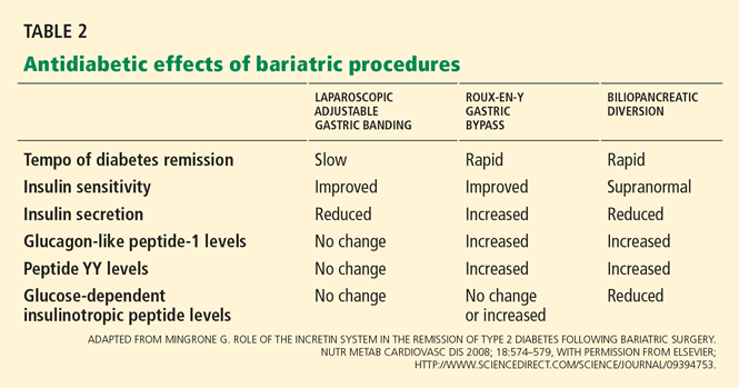
Hypothesis 1: Weight loss increases insulin sensitivity
The enforced caloric restriction, negative energy balance, and weight loss after bariatric surgery reduce insulin resistance. Consequently, the beta cells can rest because they don’t need to produce as much insulin. These effects have been observed after both gastric restrictive procedures and gastric bypass procedures.
Hypothesis 2: Less lipotoxicity, inflammation
Another theory is that bariatric surgery lessens insulin resistance by reducing “lipotoxicity,” a condition related to dysregulated fatty acid flux, lipid metabolites in tissues, and direct and indirect effects of hormones secreted by adipocytes.
The strongest evidence for this theory comes from Bikman et al,26 who found that insulin sensitivity increased after Roux-en-Y surgery more than expected from weight loss alone. One year after surgery, even though they remained anthropometrically obese (BMI > 30 kg/m2), the patients had insulin sensitivity levels similar to those in a control group of lean people (BMI < 25 kg/m2).
Insulin sensitivity begins to improve within 1 week of intestinal bypass procedures,15,27 suggesting that these procedures are doing something more than simply forcing weight loss via caloric restriction, as gastric restrictive procedures do.
Hypothesis 3: An effect on gut hormones

The “hindgut hypothesis” raised by Cummings et al24 suggests that accelerated transit of concentrated nutrients (particularly glucose) to the distal intestine results in increased production of insulinotropic and appetite-controlling substances, which account for the reversal of hyperglycemia and obesity.
In contrast, the “foregut hypothesis” raised by Rubino et al28 suggests that nutrient interactions in the duodenum are diabetogenic and, hence, bypassing the duodenum would reverse this defect. Their conclusions come from experiments in rodents that underwent jejunoileal bypass and subsequent refeeding through the bypassed intestine.
GUT HORMONES AND OTHER PEPTIDES ALTERED BY BARIATRIC SURGERY
Incretin hormones: GLP-1, GIP
Gastrointestinal hormones that increase insulin release after a meal are known as incretins. Of interest, they have this effect only when glucose is ingested orally—not when it is infused intravenously.29,30
Glucagon-like peptide 1 (GLP-1) and glucose-dependent insulinotropic peptide (GIP) account for 50% to 60% of nutrient-related insulin secretion. In addition to stimulating insulin, GLP-1 suppresses glucagon and slows gastric emptying, which delays digestion and reduces postprandial glycemia. GLP-1 also acts on the hypothalamus to induce satiety.
Laferrère et al31 and others32,33 documented robust increases in postprandial levels of GLP-1 within 4 weeks after Roux-en-Y surgery. GLP-1 levels did not increase with comparable weight loss induced by diet.
Rubino et al28,34 documented similar findings that occurred prior to marked weight loss, suggesting that the benefit of Roux-en-Y surgery on remission of diabetes may not be completely attributable to reduced caloric intake and weight loss. Insulin secretion is generally reduced after gastric restrictive procedures (eg, laparoscopic adjustable gastric banding) and biliopancreatic diversion,35 and is increased after Roux-en-Y gastric bypass.32,33,36
Noninsulinotropic peptides: Ghrelin, peptide YY
Noninsulinotropic gut peptides that are altered after Roux-en-Y surgery include ghrelin and peptide YY.
Ghrelin, a hormone derived from the gastric fundus, stimulates appetite. Ghrelin concentrations are lower after Roux-en-Y surgery, indicating that suppression of hunger signals helps sustain weight loss. In contrast, ghrelin levels increase with diet-induced weight loss.37 However, the data on ghrelin levels at various times after bariatric surgical procedures are not consistent.33,38
Peptide YY, like GLP-1, is secreted by L cells of the distal small intestine and is responsible for increasing satiety and delaying gastric emptying after meals. Numerous studies have consistently documented increases in postprandial peptide YY and GLP-1 levels after gastric bypass.32,33,39–41
ACUTE EFFECTS OF BARIATRIC SURGERY ON INSULIN SECRETION, SENSITIVITY
Bariatric surgery alters both insulin secretion and insulin sensitivity, thus improving glucose regulation.
The relationship between insulin secretion and sensitivity is a hyperbolic curve, so that any change in insulin sensitivity is balanced by a reciprocal and proportionate change in insulin secretion. The development of type 2 diabetes is characterized by a reduction in insulin secretion (decompensation) relative to the severity of insulin resistance.
In the first 6 weeks after Roux-en-Y gastric bypass or biliopancreatic diversion, insulin sensitivity improves while insulin secretion increases disproportionately, associated with a robust increase in GLP-1, and resulting in normal glucose homeostasis.16,31,42
In contrast, patients who lose weight by dieting or undergoing gastric restrictive procedures show a modest increase in insulin sensitivity and a compensatory reduction in insulin secretion, termed “beta-cell rest.”16,31,42
RISKS OF BARIATRIC SURGERY
Short-term risks
An important concern about using bariatric surgery to treat type 2 diabetes is the risk of morbidity and death associated with these procedures.
Buchwald et al13 performed a meta-analysis of 136 bariatric studies that included 22,094 patients. The 30-day operative death rates were 1.1% with biliopancreatic diversion, 0.5% with Roux-en-Y surgery, and 0.1% with restrictive procedures.
Laparoscopic adjustable gastric banding is considered the safest of the current bariatric procedures. It does not involve bowel anastomosis, and the risks of major hemorrhage, gastric perforation, and pulmonary embolism are less than 1%. Late complications requiring reoperation include band slippage or prolapse (5%–10%) and band erosion (1%–3%). The entire intestinal tract is left intact, so subsequent nutritional deficiencies are rare.43
Roux-en-Y gastric bypass carries an overall risk of major complications of 10% to 15%. Anastomotic leak (1%–5%), pulmonary embolism (< 1%), and hemorrhage (1%–4%) can be life-threatening but are rare if the staff are experienced. Late complications such as ulcer or stricture formation at the gastrojejunostomy site occur in 5% to 10% of cases and are managed nonoperatively.
Nutritional deficiencies
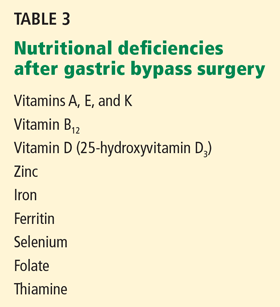
Protein-calorie malnutrition is recognized by signs such as edema, hypoalbuminemia, anemia, and hair loss. To minimize this problem after Roux-en-Y surgery, we suggest that patients take in 60 to 80 g of protein and 700 to 800 kcal a day.
Vitamin deficiencies can lead to Wernicke encephalopathy (due to thiamine deficiency), peripheral neuropathy (due to vitamin B12 deficiency),45,46 and metabolic bone disease (due to long-term deficiencies of vitamin D and calcium). Often, vitamin deficiencies are present before surgery and require prompt supplementation to avoid exacerbation of these deficiencies afterward.
Biliopancreatic diversion procedures are performed at relatively few centers worldwide, largely because of the massive amounts of protein, fat, and carbohydrate malabsorption they cause. Long-term deficiencies of fat-soluble vitamins, iron, calcium, and vitamins B12 and D have been reported in one-third to one-half of patients undergoing these procedures, and nutritional supplementation is mandatory.43 Protein-calorie malnutrition occurs in 7% of cases, and 2% of patients require operative revision to lengthen the common channel.

In rare cases, severe hypoglycemia has been noted after Roux-en-Y surgery and is associated with prandial hyperinsulinemia related to elevated GLP-1 levels.36,47 Neuroglycopenia and seizures have been reported in severe cases. Initial treatment of hypoglycemia involves dietary modification targeting carbohydrate restriction, the use of alpha glucosidase inhibitors such as acarbose (Precose), and referral to an endocrinologist for further management.
Long-term death rates
Death rates after bariatric surgery must be weighed against the long-term cardiovascular risks of continued obesity and type 2 diabetes.
Strong evidence now exists that bariatric surgery increases life expectancy48 and that this is largely attributable to reduction in cardiovascular risk factors such as diabetes and cancer. Recent studies have found that the long-term death rate is 32% to 73% lower for patients undergoing bariatric surgery than in matched controls who do not undergo surgery.49 A decrease in the death rate related to diabetes has played an important role in these results.
Acknowledgments: We acknowledge support from the National Institutes of Health, Multidisciplinary Clinical Research Career Development Programs Grant 5K12RR023264 (SRK), National Center for Research Resources, CTSA 1UL1RR024989, and research grants from Ethicon Endo-Surgery (PS,SRK).
- DeFronzo RA. Pathogenesis of type 2 diabetes mellitus. Med Clin North Am 2004; 88:787–835.
- Kashyap SR, Defronzo RA. The insulin resistance syndrome: physiological considerations. Diab Vasc Dis Res 2007; 4:13–19.
- Mokdad AH, Ford ES, Bowman BA, et al. Prevalence of obesity, diabetes, and obesity-related health risk factors, 2001. JAMA 2003; 289:76–79.
- Unger RH. Minireview: weapons of lean body mass destruction: the role of ectopic lipids in the metabolic syndrome. Endocrinology 2003; 144:5159–5165.
- Itani SI, Ruderman NB, Schmieder F, Boden G. Lipid-induced insulin resistance in human muscle is associated with changes in diacylglycerol, protein kinase C, and IkappaB-alpha. Diabetes 2002; 51:2005–2011.
- Diabetes Prevention Program Research Group. Reduction in the incidence of type 2 diabetes with lifestyle intervention or metformin. N Engl J Med 2002; 346:393–403.
- Look AHEAD Research Group; Pi-Sunyer X, Blackburn G, Brancati FL, et al. Reduction in weight and cardiovascular disease risk factors in individuals with type 2 diabetes: one-year results of the look AHEAD trial. Diabetes Care 2007; 30:1374–1383.
- Look AHEAD Research Group; Wadden TA, West DS, Delahanty L, et al. The Look AHEAD study: a description of the lifestyle intervention and the evidence supporting it. Obesity (Silver Spring) 2006; 14:737–752.
- Nathan DM. Clinical practice. Initial management of glycemia in type 2 diabetes mellitus. N Engl J Med 2002; 347:1342–1349.
- Nathan DM, Buse JB, Davidson MB, et al. Management of hyperglycemia in type 2 diabetes: a consensus algorithm for the initiation and adjustment of therapy: update regarding thiazolidinediones: a consensus statement from the American Diabetes Association and the European Association for the Study of Diabetes. Diabetes Care 2008; 31:173–175.
- Spann SJ, Nutting PA, Galliher JM, et al. Management of type 2 diabetes in the primary care setting: a practice-based research network study. Ann Fam Med 2006; 4:23–31.
- Buchwald H, Estok R, Fahrbach K, et al. Weight and type 2 diabetes after bariatric surgery: systematic review and meta-analysis. Am J Med 2009; 122:248–256.
- Buchwald H, Avidor Y, Braunwald E, et al. Bariatric surgery: a systematic review and meta-analysis. JAMA 2004; 292:1724–1737.
- Dixon JB, O’Brien PE, Playfair J, et al. Adjustable gastric banding and conventional therapy for type 2 diabetes. JAMA 2008; 299:316–323.
- Pories WJ, Swanson MS, MacDonald KG, et al. Who would have thought it? An operation proves to be the most effective therapy for adult-onset diabetes mellitus. Ann Surg 1995; 222:339–350.
- Kashyap SR, Daud S, Kelly KR, et al. Acute effects of gastric bypass versus gastric restrictive surgery on beta-cell function and insulinotropic hormones in severely obese patients with type 2 diabetes. Int J Obes (Lond) 2009; epub ahead of print
- Schauer PR, Burguera B, Ikramuddin S, et al. Effect of laparoscopic Roux-en Y gastric bypass on type 2 diabetes mellitus. Ann Surg 2003; 238:467–484.
- Sjöström L, Lindroos AK, Peltonen M, et al; Swedish Obese Subjects Study Scientific Group. Lifestyle, diabetes, and cardiovascular risk factors 10 years after bariatric surgery. N Engl J Med 2004; 351:2683–2693.
- Scopinaro N, Marinari GM, Camerini GB, Papadia FS, Adami GF. Specific effects of biliopancreatic diversion on the major components of metabolic syndrome: a long-term follow-up study. Diabetes Care 2005; 28:2406–2411.
- Scopinaro N, Papadia F, Marinari G, Camerini G, Adami G. Long-term control of type 2 diabetes mellitus and the other major components of the metabolic syndrome after biliopancreatic diversion in patients with BMI < 35 kg/m2. Obes Surg 2007; 17:185–192.
- Alexandrides TK, Skroubis G, Kalfarentzos F. Resolution of diabetes mellitus and metabolic syndrome following Roux-en-Y gastric bypass and a variant of biliopancreatic diversion in patients with morbid obesity. Obes Surg 2007; 17:176–184.
- Chiellini C, Rubino F, Castagneto M, Nanni G, Mingrone G. The effect of bilio-pancreatic diversion on type 2 diabetes in patients with BMI < 35 kg/m2. Diabetologia 2009; 52:1027–1030.
- Consensus Development Conference Panel. NIH conference. Gastrointestinal surgery for severe obesity. Ann Intern Med 1991; 115:956–961.
- Cummings DE, Overduin J, Foster-Schubert KE. Gastric bypass for obesity: mechanisms of weight loss and diabetes resolution. J Clin Endocrinol Metab 2004; 89:2608–2615.
- Cummings DE, Flum DR. Gastrointestinal surgery as a treatment for diabetes. JAMA 2008; 299:341–343.
- Bikman BT, Zheng D, Pories WJ, et al. Mechanism for improved insulin sensitivity after gastric bypass surgery. J Clin Endocrinol Metab 2008; 93:4656–4663.
- Guidone C, Manco M, Valera-Mora E, et al. Mechanisms of recovery from type 2 diabetes after malabsorptive bariatric surgery. Diabetes 2006; 55:2025–2031.
- Rubino F, Forgione A, Cummings DE, et al. The mechanism of diabetes control after gastrointestinal bypass surgery reveals a role of the proximal small intestine in the pathophysiology of type 2 diabetes. Ann Surg 2006; 244:741–749.
- Vilsbøll T, Krarup T, Madsbad S, Holst JJ. Both GLP-1 and GIP are insulinotropic at basal and postprandial glucose levels and contribute nearly equally to the incretin effect of a meal in healthy subjects. Regul Pept 2003; 114:115–121.
- Vollmer K, Holst JJ, Baller B, et al. Predictors of incretin concentrations in subjects with normal, impaired, and diabetic glucose tolerance. Diabetes 2008; 57:678–687.
- Laferrère B, Teixeira J, McGinty J, et al. Effect of weight loss by gastric bypass surgery versus hypocaloric diet on glucose and incretin levels in patients with type 2 diabetes. J Clin Endocrinol Metab 2008; 93:2479–2485.
- Korner J, Bessler M, Inabnet W, Taveras C, Holst JJ. Exaggerated glucagon-like peptide-1 and blunted glucose-dependent insulinotropic peptide secretion are associated with Roux-en-Y gastric bypass but not adjustable gastric banding. Surg Obes Relat Dis 2007; 3:597–601.
- le Roux CW, Aylwin SJ, Batterham RL, et al. Gut hormone profiles following bariatric surgery favor an anorectic state, facilitate weight loss, and improve metabolic parameters. Ann Surg 2006; 243:108–114.
- Rubino F, Gagner M, Gentileschi P, et al. The early effect of the Roux-en-Y gastric bypass on hormones involved in body weight regulation and glucose metabolism. Ann Surg 2004; 240:236–242.
- Salinari S, Bertuzzi A, Asnaghi S, Guidone C, Manco M, Mingrone G. First-phase insulin secretion restoration and differential response to glucose load depending on the route of administration in type 2 diabetic subjects after bariatric surgery. Diabetes Care 2009; 32:375–380.
- Goldfine AB, Mun EC, Devine E, et al. Patients with neuroglycopenia after gastric bypass surgery have exaggerated incretin and insulin secretory responses to a mixed meal. J Clin Endocrinol Metab 2007; 92:4678–4685.
- Cummings DE, Weigle DS, Frayo RS, et al. Plasma ghrelin levels after diet-induced weight loss or gastric bypass surgery. N Engl J Med 2002; 346:1623–1630.
- Chandarana K, Drew ME, Emmanuel J, et al. Subject standardization, acclimatization, and sample processing affect gut hormone levels and appetite in humans. Gastroenterology 2009; 136:2115–2126.
- Korner J, Inabnet W, Febres G, et al. Prospective study of gut hormone and metabolic changes after adjustable gastric banding and Roux-en-Y gastric bypass. Int J Obes (Lond) 2009; 33:786–795.
- Boey D, Sainsbury A, Herzog H. The role of peptide YY in regulating glucose homeostasis. Peptides 2007; 28:390–395.
- Hanusch-Enserer U, Ghatei MA, Cauza E, Bloom SR, Prager R, Roden M. Relation of fasting plasma peptide YY to glucose metabolism and cardiovascular risk factors after restrictive bariatric surgery. Wien Klin Wochenschr 2007; 119:291–296.
- Laferrère B, Heshka S, Wang K, et al. Incretin levels and effect are markedly enhanced 1 month after Roux-en-Y gastric bypass surgery in obese patients with type 2 diabetes. Diabetes Care 2007; 30:1709–1716.
- Tucker ON, Szomstein S, Rosenthal RJ. Nutritional consequences of weight-loss surgery. Med Clin North Am 2007; 91:499–514.
- Davies DJ, Baxter JM, Baxter JN. Nutritional deficiencies after bariatric surgery. Obes Surg 2007; 17:1150–1158.
- Angstadt JD, Bodziner RA. Peripheral polyneuropathy from thiamine deficiency following laparoscopic Roux-en-Y gastric bypass. Obes Surg 2005; 15:890–892.
- Ritz P, Becouarn G, Douay O, Sallé A, Topart P, Rohmer V. Gastric bypass is not associated with protein malnutrition in morbidly obese patients. Obes Surg 2009; 19:840–844.
- Service GJ, Thompson GB, Service FJ, Andrews JC, Collazo-Clavell ML, Lloyd RV. Hyperinsulinemic hypoglycemia with nesidioblastosis after gastric-bypass surgery. N Engl J Med 2005; 353:249–254.
- Sjöström L, Narbro K, Sjöström CD, et al;Swedish Obese Subjects Study. Effects of bariatric surgery on mortality in Swedish obese subjects. N Engl J Med 2007; 357:741–752.
- Adams TD, Gress RE, Smith SC, et al. Long-term mortality after gastric bypass surgery. N Engl J Med 2007; 357:753–761.
Evidence is mounting for the use of bariatric surgery to treat type 2 diabetes mellitus in patients whose body mass index (BMI) is 35 kg/m2 or higher. In obese patients who also have type 2 diabetes, bariatric surgery sends it into remission (defined as normoglycemic control without the need for diabetic medications) in more than three-fourths of cases, with higher rates with the Roux-en-Y gastric bypass procedure than with the laparoscopic adjustable gastric banding procedure.
However, data on the effects of this surgery on type 2 diabetes come primarily from observational studies that lacked appropriate control groups, and the relative benefit of bariatric surgery vs aggressive medical antidiabetic therapy is not yet known. Needed are randomized trials comparing the two types of therapy (and the various types of bariatric surgery) in diabetic patients with less-severe obesity.
Further, why would bariatric surgery help with diabetes, and why would one procedure do it better than another? To be honest, we are not sure, but evidence points not only to weight loss but also to better insulin sensitivity and to alterations in levels of hormones secreted by the gut that increase insulin secretion.
OBESITY PROMOTES DIABETES; WEIGHT LOSS COUNTERACTS IT
Type 2 diabetes mellitus is a complex metabolic disease characterized by insulin resistance and progressive failure of pancreatic beta cells, resulting in hyperglycemia.1,2
Obesity, a potent risk factor for type 2 diabetes, contributes to its development by inducing insulin resistance and inflammation, which in turn impair glucose regulation.3,4 Fat deposits in the abdomen, muscles, and liver contribute to elevations of circulating free fatty acids and adipocyte-derived cytokines that mediate insulin resistance and inflammatory pathways.5
In the Diabetes Prevention Program,6 modest weight loss (5% to 10% of body weight) through diet and exercise reduced the incidence of type 2 diabetes, and in the ongoing Action for Health in Diabetes (Look AHEAD) study of the National Institutes of Health, it improved glucose homeostasis.7,8
The current medical approach to type 2 diabetes includes advising the patient to lose weight through lifestyle modification, and prescribing drugs that restore glycemic control by reducing insulin resistance (biguanides, glitazones) and improving insulin secretion (incretin mimetics and analogues and sulfonylureas). 9,10
However, several factors make type 2 diabetes challenging to treat in obese people. Patients who lose weight via behavioral changes and weight-loss drugs tend to gain the weight back. Antidiabetic drugs pose the risk of hypoglycemia. Moreover, although many new classes of drugs have been developed to treat type 2 diabetes, most patients fail to achieve the American Diabetes Association goal for glycemic control, ie, a hemoglobin A1c level lower than 7%.11
BARIATRIC PROCEDURES AND THEIR EFFECT ON DIABETES CONTROL
After bariatric surgery, patients lose more weight than with traditional weight-loss methods—up to 25% of their total body weight. Furthermore, of those with type 2 diabetes, 87% achieve at least better glucose control and need fewer antidiabetic medications,12 and an average of 78% achieve normal glycemic control without taking any antidiabetic medications at all.12,13
But not all bariatric procedures have the same effect on weight and diabetes: certain procedures have a greater effect.
The two major types are classified as gastric restrictive procedures and intestinal bypass procedures. The classification was initially based on the presumed mechanism of weight loss.
Gastric restrictive procedures (laparoscopic adjustable gastric banding, sleeve gastrectomy, vertical gastroplasty) limit gastric volume and, hence, restrict the intake of calories by inducing satiety. Afterward, patients lose approximately 10% to 20% of their total body weight.
Furthermore, multiple studies, including a randomized controlled trial14 (more about this below), have shown remission of type 2 diabetes with laparoscopic adjustable gastric banding but not with conventional medical therapy. The effect is primarily mediated by weight loss and improved insulin sensitivity, both of which occur several months following surgery. Of note, however: in this trial,14 all the patients had diabetes of short duration, less than 2 years.
Hence, different procedures have different effects on diabetes.12 The speed at which type 2 diabetes goes into remission differs with restrictive vs malabsorptive procedures. After Roux-en-Y gastric bypass and biliopancreatic diversion, diabetes remits within days, even before the patient has lost much weight.15 This does not happen after gastric restrictive procedures.12,16
Observational studies of the effect of Roux-en-Y surgery on diabetes
Several observational studies have evaluated the benefit of Roux-en-Y surgery for patients with type 2 diabetes mellitus.
Pories et al15 followed 608 severely obese patients, of whom 165 (27%) had type 2 diabetes or impaired glucose tolerance.
At a mean follow-up of 7.6 years after surgery, 83% of the diabetic patients were off their antidiabetic drugs, and 99% of those with impaired glucose tolerance were normoglycemic, with normal fasting glucose and hemoglobin A1c levels. Marked improvements in hyperlipidemia, hypertension, fertility, osteoarthritis, and obstructive sleep apnea were also noted.
Schauer et al17 observed similar results in 1,160 morbidly obese patients, of whom 240 (21%) had type 2 diabetes or impaired fasting glucose.
After laparoscopic Roux-en-Y gastric bypass surgery, fasting glucose and hemoglobin A1c levels returned to normal levels in 83% of cases and were markedly improved in the remaining 17%. Significantly (80%) fewer patients needed oral antidiabetic agents or insulin (79% fewer). Patients most likely to achieve complete remission of diabetes were those with the shortest duration of diabetes (< 5 years), the mildest severity of diabetes (diet-controlled), and the greatest weight loss after surgery. The rate of diabetes remission in patients who had been diabetic for 5 years or less was 95%, compared with 75% in those who had been diabetic for 6 to 10 years and 54% in those who had been diabetic for more than 10 years (P < .001).
The Swedish Obese Subjects (SOS) study18 prospectively followed 1,703 patients, of whom 118 had type 2 diabetes, for 10 years after various bariatric surgery procedures (primarily vertical gastroplasty). In a control group that received medical therapy, 77 patients had type 2 diabetes. Medical therapy was ill-defined with respect to aggressiveness and adherence to intervention with lifestyle and pharmacotherapy.
At 2 years, the surgical group had lost a mean of 28 kg, glycemic control had improved in the diabetic patients, and many of them had been able to stop taking oral hypoglycemic drugs or insulin. In contrast, the need for these agents increased in the medically treated patients. The proportion treated by diet alone rose from 59% to 73% in the surgical group, but declined from 55% to 34% in the nonsurgical group.13
In these studies, surgery also reduced the risk of progressing from impaired glucose tolerance to type 2 diabetes; the risk was 30 times lower in the study by Pories et al.15 In the SOS study,18 the frequency of diabetes was 30 times lower at 2 years and five times lower at 8 years after surgery.
Studies of biliopancreatic diversion
Data on the effects of biliopancreatic diversion, a primarily malabsorptive procedure, are limited to European studies.
Scopinaro et al19,20 reported long-term follow-up data on 312 patients with type 2 diabetes who underwent biliopancreatic diversion; 310 patients (99%) achieved normal fasting glucose values by 1 year after surgery. At 10 years after surgery, 98% of the patients were still in complete remission of diabetes, defined as normal glucose values without the use of antidiabetic medications.
Others have noted similar findings.21,22
Limitations of the studies
Although these data seem encouraging, these studies had major limitations.
The patients were mostly white women with severe obesity, ie, a BMI greater than 40 kg/m2, which is not representative of patients with type 2 diabetes in the community. Only about 20% had glucose intolerance or overt type 2 diabetes mellitus. Would other groups benefit, particularly men and those with lesssevere obesity?
Moreover, these studies were observational, with no randomized control groups. Many reports consisted of large case series. It is not clear how specific bariatric procedures were chosen or what criteria were used for performing bariatric surgery. A lack of complete follow-up data is also a concern.
Needed are large randomized trials evaluating the effects of various bariatric procedures in a less obese cohort with type 2 diabetes, ie, typical patients seen in the community. Moreover, surgery has not been compared directly with more vigorous medical weight-loss strategies, such as those used in the Diabetes Prevention Project6 and the Look AHEAD trial.7,8
A randomized controlled trial of gastric banding
The only randomized controlled trial to date that compared standard medical diabetes therapy with bariatric surgery was conducted by Dixon et al.14
Sixty patients with type 2 diabetes (duration < 2 years and mean hemoglobin A1c 7.7%) were randomized either to receive medical management as defined by the American Diabetes Association guidelines or to undergo laparoscopic adjustable gastric banding.
At 2 years, the rate of remission (defined as hemoglobin A1c < 6.2% and a normal fasting glucose level) was 13% in the medical treatment group vs 73% in the surgery group (P < .001). Patients receiving medical treatment had lost a mean of 1.7% of their body weight, vs 20.7% in the surgical patients (P < .001). Weight loss was strongly associated with remission of type 2 diabetes after surgery.
This study was controversial in that the medical intervention in this trial was not as aggressive as in the Diabetes Prevention Project and Look AHEAD trials.
INDICATIONS FOR BARIATRIC SURGERY IN PATIENTS WITH DIABETES
According to guidelines from the National Institutes of Health,23 the current indications for bariatric surgery include a BMI of 40 kg/m2 or higher, or a BMI between 35 and 40 kg/m2 with at least two obesity-related comorbidities. Diabetes is considered a key comorbidity that justifies the risk of surgery. The guidelines suggest that bariatric surgery be discussed with all severely obese patients (BMI > 35 kg/m2) with type 2 diabetes who have not been able to lose weight with other weight-control approaches.
Since type 2 diabetes mellitus is a progressive disease characterized by relentless deterioration of beta-cell function, many endocrinologists favor aggressive weight-loss approaches early in the course of the disease. We believe that bariatric surgery should be considered early, as it may help preserve pancreatic betacell function and slow the progression of microvascular and macrovascular complications.
HOW DOES BARIATRIC SURGERY IMPROVE TYPE 2 DIABETES?
Hypothesis 1: Weight loss increases insulin sensitivity
The enforced caloric restriction, negative energy balance, and weight loss after bariatric surgery reduce insulin resistance. Consequently, the beta cells can rest because they don’t need to produce as much insulin. These effects have been observed after both gastric restrictive procedures and gastric bypass procedures.
Hypothesis 2: Less lipotoxicity, inflammation
Another theory is that bariatric surgery lessens insulin resistance by reducing “lipotoxicity,” a condition related to dysregulated fatty acid flux, lipid metabolites in tissues, and direct and indirect effects of hormones secreted by adipocytes.
The strongest evidence for this theory comes from Bikman et al,26 who found that insulin sensitivity increased after Roux-en-Y surgery more than expected from weight loss alone. One year after surgery, even though they remained anthropometrically obese (BMI > 30 kg/m2), the patients had insulin sensitivity levels similar to those in a control group of lean people (BMI < 25 kg/m2).
Insulin sensitivity begins to improve within 1 week of intestinal bypass procedures,15,27 suggesting that these procedures are doing something more than simply forcing weight loss via caloric restriction, as gastric restrictive procedures do.
Hypothesis 3: An effect on gut hormones
The “hindgut hypothesis” raised by Cummings et al24 suggests that accelerated transit of concentrated nutrients (particularly glucose) to the distal intestine results in increased production of insulinotropic and appetite-controlling substances, which account for the reversal of hyperglycemia and obesity.
In contrast, the “foregut hypothesis” raised by Rubino et al28 suggests that nutrient interactions in the duodenum are diabetogenic and, hence, bypassing the duodenum would reverse this defect. Their conclusions come from experiments in rodents that underwent jejunoileal bypass and subsequent refeeding through the bypassed intestine.
GUT HORMONES AND OTHER PEPTIDES ALTERED BY BARIATRIC SURGERY
Incretin hormones: GLP-1, GIP
Gastrointestinal hormones that increase insulin release after a meal are known as incretins. Of interest, they have this effect only when glucose is ingested orally—not when it is infused intravenously.29,30
Glucagon-like peptide 1 (GLP-1) and glucose-dependent insulinotropic peptide (GIP) account for 50% to 60% of nutrient-related insulin secretion. In addition to stimulating insulin, GLP-1 suppresses glucagon and slows gastric emptying, which delays digestion and reduces postprandial glycemia. GLP-1 also acts on the hypothalamus to induce satiety.
Laferrère et al31 and others32,33 documented robust increases in postprandial levels of GLP-1 within 4 weeks after Roux-en-Y surgery. GLP-1 levels did not increase with comparable weight loss induced by diet.
Rubino et al28,34 documented similar findings that occurred prior to marked weight loss, suggesting that the benefit of Roux-en-Y surgery on remission of diabetes may not be completely attributable to reduced caloric intake and weight loss. Insulin secretion is generally reduced after gastric restrictive procedures (eg, laparoscopic adjustable gastric banding) and biliopancreatic diversion,35 and is increased after Roux-en-Y gastric bypass.32,33,36
Noninsulinotropic peptides: Ghrelin, peptide YY
Noninsulinotropic gut peptides that are altered after Roux-en-Y surgery include ghrelin and peptide YY.
Ghrelin, a hormone derived from the gastric fundus, stimulates appetite. Ghrelin concentrations are lower after Roux-en-Y surgery, indicating that suppression of hunger signals helps sustain weight loss. In contrast, ghrelin levels increase with diet-induced weight loss.37 However, the data on ghrelin levels at various times after bariatric surgical procedures are not consistent.33,38
Peptide YY, like GLP-1, is secreted by L cells of the distal small intestine and is responsible for increasing satiety and delaying gastric emptying after meals. Numerous studies have consistently documented increases in postprandial peptide YY and GLP-1 levels after gastric bypass.32,33,39–41
ACUTE EFFECTS OF BARIATRIC SURGERY ON INSULIN SECRETION, SENSITIVITY
Bariatric surgery alters both insulin secretion and insulin sensitivity, thus improving glucose regulation.
The relationship between insulin secretion and sensitivity is a hyperbolic curve, so that any change in insulin sensitivity is balanced by a reciprocal and proportionate change in insulin secretion. The development of type 2 diabetes is characterized by a reduction in insulin secretion (decompensation) relative to the severity of insulin resistance.
In the first 6 weeks after Roux-en-Y gastric bypass or biliopancreatic diversion, insulin sensitivity improves while insulin secretion increases disproportionately, associated with a robust increase in GLP-1, and resulting in normal glucose homeostasis.16,31,42
In contrast, patients who lose weight by dieting or undergoing gastric restrictive procedures show a modest increase in insulin sensitivity and a compensatory reduction in insulin secretion, termed “beta-cell rest.”16,31,42
RISKS OF BARIATRIC SURGERY
Short-term risks
An important concern about using bariatric surgery to treat type 2 diabetes is the risk of morbidity and death associated with these procedures.
Buchwald et al13 performed a meta-analysis of 136 bariatric studies that included 22,094 patients. The 30-day operative death rates were 1.1% with biliopancreatic diversion, 0.5% with Roux-en-Y surgery, and 0.1% with restrictive procedures.
Laparoscopic adjustable gastric banding is considered the safest of the current bariatric procedures. It does not involve bowel anastomosis, and the risks of major hemorrhage, gastric perforation, and pulmonary embolism are less than 1%. Late complications requiring reoperation include band slippage or prolapse (5%–10%) and band erosion (1%–3%). The entire intestinal tract is left intact, so subsequent nutritional deficiencies are rare.43
Roux-en-Y gastric bypass carries an overall risk of major complications of 10% to 15%. Anastomotic leak (1%–5%), pulmonary embolism (< 1%), and hemorrhage (1%–4%) can be life-threatening but are rare if the staff are experienced. Late complications such as ulcer or stricture formation at the gastrojejunostomy site occur in 5% to 10% of cases and are managed nonoperatively.
Nutritional deficiencies
Protein-calorie malnutrition is recognized by signs such as edema, hypoalbuminemia, anemia, and hair loss. To minimize this problem after Roux-en-Y surgery, we suggest that patients take in 60 to 80 g of protein and 700 to 800 kcal a day.
Vitamin deficiencies can lead to Wernicke encephalopathy (due to thiamine deficiency), peripheral neuropathy (due to vitamin B12 deficiency),45,46 and metabolic bone disease (due to long-term deficiencies of vitamin D and calcium). Often, vitamin deficiencies are present before surgery and require prompt supplementation to avoid exacerbation of these deficiencies afterward.
Biliopancreatic diversion procedures are performed at relatively few centers worldwide, largely because of the massive amounts of protein, fat, and carbohydrate malabsorption they cause. Long-term deficiencies of fat-soluble vitamins, iron, calcium, and vitamins B12 and D have been reported in one-third to one-half of patients undergoing these procedures, and nutritional supplementation is mandatory.43 Protein-calorie malnutrition occurs in 7% of cases, and 2% of patients require operative revision to lengthen the common channel.
In rare cases, severe hypoglycemia has been noted after Roux-en-Y surgery and is associated with prandial hyperinsulinemia related to elevated GLP-1 levels.36,47 Neuroglycopenia and seizures have been reported in severe cases. Initial treatment of hypoglycemia involves dietary modification targeting carbohydrate restriction, the use of alpha glucosidase inhibitors such as acarbose (Precose), and referral to an endocrinologist for further management.
Long-term death rates
Death rates after bariatric surgery must be weighed against the long-term cardiovascular risks of continued obesity and type 2 diabetes.
Strong evidence now exists that bariatric surgery increases life expectancy48 and that this is largely attributable to reduction in cardiovascular risk factors such as diabetes and cancer. Recent studies have found that the long-term death rate is 32% to 73% lower for patients undergoing bariatric surgery than in matched controls who do not undergo surgery.49 A decrease in the death rate related to diabetes has played an important role in these results.
Acknowledgments: We acknowledge support from the National Institutes of Health, Multidisciplinary Clinical Research Career Development Programs Grant 5K12RR023264 (SRK), National Center for Research Resources, CTSA 1UL1RR024989, and research grants from Ethicon Endo-Surgery (PS,SRK).
- DeFronzo RA. Pathogenesis of type 2 diabetes mellitus. Med Clin North Am 2004; 88:787–835.
- Kashyap SR, Defronzo RA. The insulin resistance syndrome: physiological considerations. Diab Vasc Dis Res 2007; 4:13–19.
- Mokdad AH, Ford ES, Bowman BA, et al. Prevalence of obesity, diabetes, and obesity-related health risk factors, 2001. JAMA 2003; 289:76–79.
- Unger RH. Minireview: weapons of lean body mass destruction: the role of ectopic lipids in the metabolic syndrome. Endocrinology 2003; 144:5159–5165.
- Itani SI, Ruderman NB, Schmieder F, Boden G. Lipid-induced insulin resistance in human muscle is associated with changes in diacylglycerol, protein kinase C, and IkappaB-alpha. Diabetes 2002; 51:2005–2011.
- Diabetes Prevention Program Research Group. Reduction in the incidence of type 2 diabetes with lifestyle intervention or metformin. N Engl J Med 2002; 346:393–403.
- Look AHEAD Research Group; Pi-Sunyer X, Blackburn G, Brancati FL, et al. Reduction in weight and cardiovascular disease risk factors in individuals with type 2 diabetes: one-year results of the look AHEAD trial. Diabetes Care 2007; 30:1374–1383.
- Look AHEAD Research Group; Wadden TA, West DS, Delahanty L, et al. The Look AHEAD study: a description of the lifestyle intervention and the evidence supporting it. Obesity (Silver Spring) 2006; 14:737–752.
- Nathan DM. Clinical practice. Initial management of glycemia in type 2 diabetes mellitus. N Engl J Med 2002; 347:1342–1349.
- Nathan DM, Buse JB, Davidson MB, et al. Management of hyperglycemia in type 2 diabetes: a consensus algorithm for the initiation and adjustment of therapy: update regarding thiazolidinediones: a consensus statement from the American Diabetes Association and the European Association for the Study of Diabetes. Diabetes Care 2008; 31:173–175.
- Spann SJ, Nutting PA, Galliher JM, et al. Management of type 2 diabetes in the primary care setting: a practice-based research network study. Ann Fam Med 2006; 4:23–31.
- Buchwald H, Estok R, Fahrbach K, et al. Weight and type 2 diabetes after bariatric surgery: systematic review and meta-analysis. Am J Med 2009; 122:248–256.
- Buchwald H, Avidor Y, Braunwald E, et al. Bariatric surgery: a systematic review and meta-analysis. JAMA 2004; 292:1724–1737.
- Dixon JB, O’Brien PE, Playfair J, et al. Adjustable gastric banding and conventional therapy for type 2 diabetes. JAMA 2008; 299:316–323.
- Pories WJ, Swanson MS, MacDonald KG, et al. Who would have thought it? An operation proves to be the most effective therapy for adult-onset diabetes mellitus. Ann Surg 1995; 222:339–350.
- Kashyap SR, Daud S, Kelly KR, et al. Acute effects of gastric bypass versus gastric restrictive surgery on beta-cell function and insulinotropic hormones in severely obese patients with type 2 diabetes. Int J Obes (Lond) 2009; epub ahead of print
- Schauer PR, Burguera B, Ikramuddin S, et al. Effect of laparoscopic Roux-en Y gastric bypass on type 2 diabetes mellitus. Ann Surg 2003; 238:467–484.
- Sjöström L, Lindroos AK, Peltonen M, et al; Swedish Obese Subjects Study Scientific Group. Lifestyle, diabetes, and cardiovascular risk factors 10 years after bariatric surgery. N Engl J Med 2004; 351:2683–2693.
- Scopinaro N, Marinari GM, Camerini GB, Papadia FS, Adami GF. Specific effects of biliopancreatic diversion on the major components of metabolic syndrome: a long-term follow-up study. Diabetes Care 2005; 28:2406–2411.
- Scopinaro N, Papadia F, Marinari G, Camerini G, Adami G. Long-term control of type 2 diabetes mellitus and the other major components of the metabolic syndrome after biliopancreatic diversion in patients with BMI < 35 kg/m2. Obes Surg 2007; 17:185–192.
- Alexandrides TK, Skroubis G, Kalfarentzos F. Resolution of diabetes mellitus and metabolic syndrome following Roux-en-Y gastric bypass and a variant of biliopancreatic diversion in patients with morbid obesity. Obes Surg 2007; 17:176–184.
- Chiellini C, Rubino F, Castagneto M, Nanni G, Mingrone G. The effect of bilio-pancreatic diversion on type 2 diabetes in patients with BMI < 35 kg/m2. Diabetologia 2009; 52:1027–1030.
- Consensus Development Conference Panel. NIH conference. Gastrointestinal surgery for severe obesity. Ann Intern Med 1991; 115:956–961.
- Cummings DE, Overduin J, Foster-Schubert KE. Gastric bypass for obesity: mechanisms of weight loss and diabetes resolution. J Clin Endocrinol Metab 2004; 89:2608–2615.
- Cummings DE, Flum DR. Gastrointestinal surgery as a treatment for diabetes. JAMA 2008; 299:341–343.
- Bikman BT, Zheng D, Pories WJ, et al. Mechanism for improved insulin sensitivity after gastric bypass surgery. J Clin Endocrinol Metab 2008; 93:4656–4663.
- Guidone C, Manco M, Valera-Mora E, et al. Mechanisms of recovery from type 2 diabetes after malabsorptive bariatric surgery. Diabetes 2006; 55:2025–2031.
- Rubino F, Forgione A, Cummings DE, et al. The mechanism of diabetes control after gastrointestinal bypass surgery reveals a role of the proximal small intestine in the pathophysiology of type 2 diabetes. Ann Surg 2006; 244:741–749.
- Vilsbøll T, Krarup T, Madsbad S, Holst JJ. Both GLP-1 and GIP are insulinotropic at basal and postprandial glucose levels and contribute nearly equally to the incretin effect of a meal in healthy subjects. Regul Pept 2003; 114:115–121.
- Vollmer K, Holst JJ, Baller B, et al. Predictors of incretin concentrations in subjects with normal, impaired, and diabetic glucose tolerance. Diabetes 2008; 57:678–687.
- Laferrère B, Teixeira J, McGinty J, et al. Effect of weight loss by gastric bypass surgery versus hypocaloric diet on glucose and incretin levels in patients with type 2 diabetes. J Clin Endocrinol Metab 2008; 93:2479–2485.
- Korner J, Bessler M, Inabnet W, Taveras C, Holst JJ. Exaggerated glucagon-like peptide-1 and blunted glucose-dependent insulinotropic peptide secretion are associated with Roux-en-Y gastric bypass but not adjustable gastric banding. Surg Obes Relat Dis 2007; 3:597–601.
- le Roux CW, Aylwin SJ, Batterham RL, et al. Gut hormone profiles following bariatric surgery favor an anorectic state, facilitate weight loss, and improve metabolic parameters. Ann Surg 2006; 243:108–114.
- Rubino F, Gagner M, Gentileschi P, et al. The early effect of the Roux-en-Y gastric bypass on hormones involved in body weight regulation and glucose metabolism. Ann Surg 2004; 240:236–242.
- Salinari S, Bertuzzi A, Asnaghi S, Guidone C, Manco M, Mingrone G. First-phase insulin secretion restoration and differential response to glucose load depending on the route of administration in type 2 diabetic subjects after bariatric surgery. Diabetes Care 2009; 32:375–380.
- Goldfine AB, Mun EC, Devine E, et al. Patients with neuroglycopenia after gastric bypass surgery have exaggerated incretin and insulin secretory responses to a mixed meal. J Clin Endocrinol Metab 2007; 92:4678–4685.
- Cummings DE, Weigle DS, Frayo RS, et al. Plasma ghrelin levels after diet-induced weight loss or gastric bypass surgery. N Engl J Med 2002; 346:1623–1630.
- Chandarana K, Drew ME, Emmanuel J, et al. Subject standardization, acclimatization, and sample processing affect gut hormone levels and appetite in humans. Gastroenterology 2009; 136:2115–2126.
- Korner J, Inabnet W, Febres G, et al. Prospective study of gut hormone and metabolic changes after adjustable gastric banding and Roux-en-Y gastric bypass. Int J Obes (Lond) 2009; 33:786–795.
- Boey D, Sainsbury A, Herzog H. The role of peptide YY in regulating glucose homeostasis. Peptides 2007; 28:390–395.
- Hanusch-Enserer U, Ghatei MA, Cauza E, Bloom SR, Prager R, Roden M. Relation of fasting plasma peptide YY to glucose metabolism and cardiovascular risk factors after restrictive bariatric surgery. Wien Klin Wochenschr 2007; 119:291–296.
- Laferrère B, Heshka S, Wang K, et al. Incretin levels and effect are markedly enhanced 1 month after Roux-en-Y gastric bypass surgery in obese patients with type 2 diabetes. Diabetes Care 2007; 30:1709–1716.
- Tucker ON, Szomstein S, Rosenthal RJ. Nutritional consequences of weight-loss surgery. Med Clin North Am 2007; 91:499–514.
- Davies DJ, Baxter JM, Baxter JN. Nutritional deficiencies after bariatric surgery. Obes Surg 2007; 17:1150–1158.
- Angstadt JD, Bodziner RA. Peripheral polyneuropathy from thiamine deficiency following laparoscopic Roux-en-Y gastric bypass. Obes Surg 2005; 15:890–892.
- Ritz P, Becouarn G, Douay O, Sallé A, Topart P, Rohmer V. Gastric bypass is not associated with protein malnutrition in morbidly obese patients. Obes Surg 2009; 19:840–844.
- Service GJ, Thompson GB, Service FJ, Andrews JC, Collazo-Clavell ML, Lloyd RV. Hyperinsulinemic hypoglycemia with nesidioblastosis after gastric-bypass surgery. N Engl J Med 2005; 353:249–254.
- Sjöström L, Narbro K, Sjöström CD, et al;Swedish Obese Subjects Study. Effects of bariatric surgery on mortality in Swedish obese subjects. N Engl J Med 2007; 357:741–752.
- Adams TD, Gress RE, Smith SC, et al. Long-term mortality after gastric bypass surgery. N Engl J Med 2007; 357:753–761.
KEY POINTS
- After Roux-en-Y gastric bypass and biliopancreatic diversion, normoglycemia is restored within days, even before the patient has lost much weight.
- Alterations in postprandial levels of intestine-derived hormones (glucagon-like peptide 1, peptide YY, and ghrelin) contribute to the robust metabolic benefits of intestinal bypass procedures.
- Nutritional deficiencies are common after bariatric surgery, and long-term follow-up is mandatory for surveillance of metabolic status.
- Although curing diabetes cannot yet be considered a goal of bariatric surgery, it may be a serendipitous benefit.

The electronic medical record: Diving into a shallow pool?
The rush to adopt the electronic medical record (EMR) has accelerated since the signing of the Health Information Technology for Economic and Clinical Health (HITECH) Act, part of the American Recovery and Reinvestment (ie, the Stimulus) Act of 2009. The HITECH Act provides incentives for physicians to adopt EMRs. However, I fear that our mad rush to complete adoption of the hodgepodge of currently available EMR systems may have unforeseen and unintended consequences. A skeptical look at several unresolved issues is warranted.
SO FAR, ELECTRONIC SYSTEMS ARE NOT INTERCONNECTABLE
More than 300 EMR systems are available, but only about two dozen account for most systems in use.1 So far, these systems are not interconnectable, ie, they are unable to share information, so patients seen by different physicians may still have a fragmented electronic record.
EMRs can also be inefficient to use. Many systems require logging on to a separate, password-protected system to view images. These problems are likely to go away over time with Internet-based solutions under development by Google and others, but the current lack of interconnectivity leaves much to be desired.
ELECTRONIC RECORDS ARE AT RISK
EMRs are at considerable security risk. About 13% of medical offices in the United States are using some form of EMR.2 A 1995 Harris poll revealed that 70% of Americans were concerned about the security of EMR systems.3 In 2007, the New York Times reported that more than 250,000 patients each year are victims of medical identity theft.4 A New Zealand survey revealed that 73.3% of patients were “highly concerned” about security and privacy.5 Even more troubling to physicians is the reported 13% incidence of patients withholding medical information because of security concerns. Furthermore, multiple breaches of electronic records have already been reported, including an extensive breach of the Veterans Administration system.6
DO ELECTRONIC RECORDS IMPROVE OR WORSEN THE QUALITY OF CAR E?
Proponents have repeatedly touted that EMRs improve the quality of medical care, and these claims have been used to accelerate the adoption of the EMR. The contention that EMRs improve the accuracy of billing, coding, and administrative functions is supported by considerable data; however, the evidence of the effect of EMRs on quality of care is mixed, with some information suggesting quality may not improve.
In an analysis of 750,000 patient records for a 2-year period as part of the National Ambulatory Care Survey, Linder et al7 found that the EMR was superior in one quality area, worse in another area, and the same as paper-based records in 14 other areas. They pointed out that previous studies showing improved outcomes were mainly from large institutions with internally developed EMR systems, and that outcomes reported from these “benchmark” institutions may not be broadly applicable.7 Linder et al concluded that use of electronic records “was generally not associated with improved quality of ambulatory care,”7 and that increased use of EMRs does not imply an automatic improvement in quality of care.7
Crosson et al8 evaluated diabetes care in a cross-sectional analysis of 50 ambulatory care practices from 2003 and 2004 and reported that “after controlling for potential practice- and patient-level confounders and for the clustering of patients within practices, patients with diabetes in practices that did not have an EMR were significantly more likely to have received care that met the guidelines for processes of care, treatment, and intermediate outcomes.”8
The Palo Alto Medical Foundation reported on the sources and types of discrepancies between EMR-listed medications and actual patient medications and found that 79.8% of the time the errors were generated by the EMR system.9 And an outpatient study that videotaped medical encounters to evaluate the accuracy of EMR in an area in which accuracy would be expected (medication lists) found that fewer than one-fifth of exchanges “ended with clear conclusions by both parties regarding prescribed medication regimens.”10 Never mind the lingering questions regarding our ability to define quality: these data provide at least some cause for concern and caution in our rush to adopt innovation in health care without proper consideration of the possible unintended consequences.
WHAT EFFECT ON MEDICAL EDUCATION?
Almost no information is available on the effects of the EMR on the process of medical student education. One could postulate and hope that embedded diagnostic algorithms and drug interaction software would facilitate the education process.
In a paper in Academic Psychiatry, Keenan et al noted that research on EMRs for education is in its infancy.11 A 2008 study of the effects of EMR on third-year medical students’ clinical experience found that students reported significant concerns about the potential impact of EMRs on their ability to conduct the doctor-patient encounter.12 Furthermore, 48% reported spending less time with patients face to face because of the EMR, and 34% reported less time talking to patients.12 In today’s world of off-site rotations and with nearly two dozen EMR systems in outpatient use alone, it is likely that a considerable amount of medical students’ time and effort is expended learning how to use different systems, which may detract from their actual medical experience.
Lastly, a survey of Canadian and US medical schools13 found that only 44% of schools had a policy regarding medical students’ documentation of progress notes in the EMR during ambulatory internal medicine clerkships. In an era when the medical student has been relegated to an observer in the education process, 14 the EMR has introduced yet another poorly understood variable in student education, which clearly begs for a thorough evaluation as the use of EMRs becomes more widespread. How can we maximize rather than dilute student education through the vehicle of electronic records?
ACCURACY VS COPYING AND PASTING
A recent Veterans Adminstration study found that 99% of progress notes in EMRs that were examined contained copied or duplicated text.15 Ten percent of 98,753 examined records contained an instance of what was considered “high-risk copying.” Weir et al16 manually reviewed a set of 60 inpatient charts at the Salt Lake City VA Health Care System and found an average of one factual error introduced into the electronic record per episode of copying.16 The clinical accuracy of the EMR is therefore questionable. Physicians pressed for time are more likely to introduce errors in the EMR, and the information put into the EMR is unlikely to be questioned—and may well be perpetuated by copy-and-paste methodology.
A THIRD PARTY IN THE EXAMINATION ROOM
Considerable information is available about the effect of the EMR on doctor-patient interaction. Margalit et al17 studied videotapes of physician encounters and noted that physicians spent an average of 25% (in some cases as much as 42%) of each visit gazing at the computer screen. They also noted that screengazing seemed to be particularly disruptive to psychological and emotional exchange.
Ventres et al18 reported that in the examination room the EMR is “much like a third party to a conversation”18 and contended that the widespread use of EMRs would have intended and unintended consquences on the cognitive and social dimensions of the physician-patient encounter. They concluded that these issues demand thoughtful consideration as the use of the EMR proliferates, “not only to forestall problems but to maximize the effectiveness of this burgeoning medical technology.”18
DEVOID OF REAL MEDICAL THOUGHT
Notwithstanding data errors and the cutting and pasting of prior notes in the EMR, we still know very little about how the EMR affects how doctors express their thoughts and communicate with one another. My particular concern is with menu-driven or templatedriven notes: they produce reams of important data, and they help ensure that coding requirements are met. But this way of writing notes about a patient is devoid of real medical thought. To describe a patient in templatedriven fashion as “an 88-year-old white male” pales next to a personalized description such as “an 88-year-old World War II B-17 bomber pilot shot down three times over Europe.”
A colleague of mine recently lamented, “I can no longer make use of my partners’ templated notes, as they convey no real information.” I do believe we should be concerned about the undesirable effects that such changes in record-keeping may produce.
LET’S CHECK THE WATER BEFORE DIVING IN
What should we do as we face these issues?
First, we should be aware that governmental and financial pressures and the availability of new technology are pushing us rapidly into new, poorly understood territory. This awareness is critical, as it at least permits a more open mind and allows the potential for honest dialogue, rather than just following directives from above.
Second, we should recognize the gaps in our understanding of the overall effects of the EMR on medicine as a profession and begin to more critically study these effects: ie, we need to be proactive rather than reactive. Denying that we lack answers to key questions about EMRs is clearly counterproductive.
We live in the electronic age. EMRs will continue to proliferate, and they have the potential to be cost-effective, care-enhancing, and time-saving. Obviously, there is no turning back the clock. However, the issues I have raised here—and other issues such as additional physician time,1 potential “billing creep,” and the opportunity for outright fraud (rarely discussed in physician circles), not to mention cost—are deeply concerning and worthy of notice and careful consideration.
My thoughts here are meant to serve as a call to reassess the possible unintended consequences of the federally mandated rush toward an as-yet poorly integrated system of EMRs. Perhaps we should check the water first, lest we find we are diving into a shallow pool.
- Prosser K. Sonoma County Medical Association. The true costs of EMRs. Sonoma Medicine Spring 2009. http://www.scma.org/magazine/articles/?articleid=398. Accessed April 13, 2010.
- DesRoches CM, Campbell EG, Rao SR, et al. Electronic health records in ambulatory care—a national survey of physicians. N Engl J Med 2008; 359:50–60.
- Cummings J. The benefits of electronic medical records sound good, but privacy could become a difficult issue. Harris Interactive Feb 8, 2007. http://news.harrisinteractive.com/profiles/investor/fullpage.asp?f=1&BzID=1963&to=cp&Nav=0&LangID=1&s=0&ID=11259. Accessed May 31, 2010.
- Konrad W. Medical problems could include identity theft. The New York Times. June 12, 2009.
- Chhanabhi P, Holt A. Consumers are ready to accept the transition to online and electronic records if they can be assured of the security measures. MedGenMed 2007; 9:8.
- Lemos R. Veterans Affairs warns of massive privacy breach. SecurityFocus 2006 (May 22). http://www.securityfocus.com/news/11393. Accessed May 31, 2010.
- Linder JA, Ma J, Bates DW, Middleton B, Stafford RS. Electronic health record use and the quality of ambulatory care in the United States. Arch Intern Med 2007; 167:1400–1405.
- Crosson JC, Ohman-Strickland PA, Hahn KA, et al. Electronic medical records and diabetes quality of care: results from a sample of family medicine practices. Ann Fam Med 2007; 5:209–215.
- Orrico KB. Sources and types of discrepancies between electronic medical records and actual outpatient medication use. J Manag Care Pharm 2008; 14:626–631.
- Arar NH, Wen L, McGrath J, Steinbach R, Pugh JA. Communicating about medications during primary care outpatient visits: the role of electronic medical records. Inform Prim Care 2005; 13:13–22.
- Keenan CR, Nguyen HH, Srinivasan M. Electronic medical records and their impact on resident and medical student education. Acad Psychiatry 2006; 30:522–527.
- Rouf E, Chumley HS, Dobbie AE. Electronic health records in outpatient clinics: perspectives of third year medical students. BMC Med Educ 2008; 8:13.
- Mintz M, Narvarte HJ, O’Brien KE, Papp KK, Thomas M, Durning SJ. Use of electronic medical records by physicians and students in academic internal medicine settings. Acad Med 2009; 84:1698–1704.
- Alpert JS, Mandell BF. Back to the future: medical students can matter again. Am J Med 2009; 122:971–972.
- Hammond KW, Helbig ST, Benson CC, Brathwaite-Sketoe BM. Are electronic medical records trustworthy? Observations on copying, pasting and duplication. AMIA Annu Symp Proc 2003:269–273.
- Weir CR, Hurdle JF, Felgar MA, Hoffman JM, Roth B, Nebeker JR. Direct text entry in electronic progress notes. An evaluation of input errors. Methods Inf Med 2003; 42:61–67.
- Margalit RS, Roter D, Dunevant MA, Larson S, Reis S. Electronic medical record use and physician-patient communication: an observational study of Israeli primary care encounters. Patient Educ Couns 2006; 61:134–141.
- Ventres W, Kooienga S, Vuckovic N, Marlin R, Nygren P, Stewart V. Physicians, patients, and the electronic health record: an ethnographic analysis. Ann Fam Med 2006; 4:124–131.
The rush to adopt the electronic medical record (EMR) has accelerated since the signing of the Health Information Technology for Economic and Clinical Health (HITECH) Act, part of the American Recovery and Reinvestment (ie, the Stimulus) Act of 2009. The HITECH Act provides incentives for physicians to adopt EMRs. However, I fear that our mad rush to complete adoption of the hodgepodge of currently available EMR systems may have unforeseen and unintended consequences. A skeptical look at several unresolved issues is warranted.
SO FAR, ELECTRONIC SYSTEMS ARE NOT INTERCONNECTABLE
More than 300 EMR systems are available, but only about two dozen account for most systems in use.1 So far, these systems are not interconnectable, ie, they are unable to share information, so patients seen by different physicians may still have a fragmented electronic record.
EMRs can also be inefficient to use. Many systems require logging on to a separate, password-protected system to view images. These problems are likely to go away over time with Internet-based solutions under development by Google and others, but the current lack of interconnectivity leaves much to be desired.
ELECTRONIC RECORDS ARE AT RISK
EMRs are at considerable security risk. About 13% of medical offices in the United States are using some form of EMR.2 A 1995 Harris poll revealed that 70% of Americans were concerned about the security of EMR systems.3 In 2007, the New York Times reported that more than 250,000 patients each year are victims of medical identity theft.4 A New Zealand survey revealed that 73.3% of patients were “highly concerned” about security and privacy.5 Even more troubling to physicians is the reported 13% incidence of patients withholding medical information because of security concerns. Furthermore, multiple breaches of electronic records have already been reported, including an extensive breach of the Veterans Administration system.6
DO ELECTRONIC RECORDS IMPROVE OR WORSEN THE QUALITY OF CAR E?
Proponents have repeatedly touted that EMRs improve the quality of medical care, and these claims have been used to accelerate the adoption of the EMR. The contention that EMRs improve the accuracy of billing, coding, and administrative functions is supported by considerable data; however, the evidence of the effect of EMRs on quality of care is mixed, with some information suggesting quality may not improve.
In an analysis of 750,000 patient records for a 2-year period as part of the National Ambulatory Care Survey, Linder et al7 found that the EMR was superior in one quality area, worse in another area, and the same as paper-based records in 14 other areas. They pointed out that previous studies showing improved outcomes were mainly from large institutions with internally developed EMR systems, and that outcomes reported from these “benchmark” institutions may not be broadly applicable.7 Linder et al concluded that use of electronic records “was generally not associated with improved quality of ambulatory care,”7 and that increased use of EMRs does not imply an automatic improvement in quality of care.7
Crosson et al8 evaluated diabetes care in a cross-sectional analysis of 50 ambulatory care practices from 2003 and 2004 and reported that “after controlling for potential practice- and patient-level confounders and for the clustering of patients within practices, patients with diabetes in practices that did not have an EMR were significantly more likely to have received care that met the guidelines for processes of care, treatment, and intermediate outcomes.”8
The Palo Alto Medical Foundation reported on the sources and types of discrepancies between EMR-listed medications and actual patient medications and found that 79.8% of the time the errors were generated by the EMR system.9 And an outpatient study that videotaped medical encounters to evaluate the accuracy of EMR in an area in which accuracy would be expected (medication lists) found that fewer than one-fifth of exchanges “ended with clear conclusions by both parties regarding prescribed medication regimens.”10 Never mind the lingering questions regarding our ability to define quality: these data provide at least some cause for concern and caution in our rush to adopt innovation in health care without proper consideration of the possible unintended consequences.
WHAT EFFECT ON MEDICAL EDUCATION?
Almost no information is available on the effects of the EMR on the process of medical student education. One could postulate and hope that embedded diagnostic algorithms and drug interaction software would facilitate the education process.
In a paper in Academic Psychiatry, Keenan et al noted that research on EMRs for education is in its infancy.11 A 2008 study of the effects of EMR on third-year medical students’ clinical experience found that students reported significant concerns about the potential impact of EMRs on their ability to conduct the doctor-patient encounter.12 Furthermore, 48% reported spending less time with patients face to face because of the EMR, and 34% reported less time talking to patients.12 In today’s world of off-site rotations and with nearly two dozen EMR systems in outpatient use alone, it is likely that a considerable amount of medical students’ time and effort is expended learning how to use different systems, which may detract from their actual medical experience.
Lastly, a survey of Canadian and US medical schools13 found that only 44% of schools had a policy regarding medical students’ documentation of progress notes in the EMR during ambulatory internal medicine clerkships. In an era when the medical student has been relegated to an observer in the education process, 14 the EMR has introduced yet another poorly understood variable in student education, which clearly begs for a thorough evaluation as the use of EMRs becomes more widespread. How can we maximize rather than dilute student education through the vehicle of electronic records?
ACCURACY VS COPYING AND PASTING
A recent Veterans Adminstration study found that 99% of progress notes in EMRs that were examined contained copied or duplicated text.15 Ten percent of 98,753 examined records contained an instance of what was considered “high-risk copying.” Weir et al16 manually reviewed a set of 60 inpatient charts at the Salt Lake City VA Health Care System and found an average of one factual error introduced into the electronic record per episode of copying.16 The clinical accuracy of the EMR is therefore questionable. Physicians pressed for time are more likely to introduce errors in the EMR, and the information put into the EMR is unlikely to be questioned—and may well be perpetuated by copy-and-paste methodology.
A THIRD PARTY IN THE EXAMINATION ROOM
Considerable information is available about the effect of the EMR on doctor-patient interaction. Margalit et al17 studied videotapes of physician encounters and noted that physicians spent an average of 25% (in some cases as much as 42%) of each visit gazing at the computer screen. They also noted that screengazing seemed to be particularly disruptive to psychological and emotional exchange.
Ventres et al18 reported that in the examination room the EMR is “much like a third party to a conversation”18 and contended that the widespread use of EMRs would have intended and unintended consquences on the cognitive and social dimensions of the physician-patient encounter. They concluded that these issues demand thoughtful consideration as the use of the EMR proliferates, “not only to forestall problems but to maximize the effectiveness of this burgeoning medical technology.”18
DEVOID OF REAL MEDICAL THOUGHT
Notwithstanding data errors and the cutting and pasting of prior notes in the EMR, we still know very little about how the EMR affects how doctors express their thoughts and communicate with one another. My particular concern is with menu-driven or templatedriven notes: they produce reams of important data, and they help ensure that coding requirements are met. But this way of writing notes about a patient is devoid of real medical thought. To describe a patient in templatedriven fashion as “an 88-year-old white male” pales next to a personalized description such as “an 88-year-old World War II B-17 bomber pilot shot down three times over Europe.”
A colleague of mine recently lamented, “I can no longer make use of my partners’ templated notes, as they convey no real information.” I do believe we should be concerned about the undesirable effects that such changes in record-keeping may produce.
LET’S CHECK THE WATER BEFORE DIVING IN
What should we do as we face these issues?
First, we should be aware that governmental and financial pressures and the availability of new technology are pushing us rapidly into new, poorly understood territory. This awareness is critical, as it at least permits a more open mind and allows the potential for honest dialogue, rather than just following directives from above.
Second, we should recognize the gaps in our understanding of the overall effects of the EMR on medicine as a profession and begin to more critically study these effects: ie, we need to be proactive rather than reactive. Denying that we lack answers to key questions about EMRs is clearly counterproductive.
We live in the electronic age. EMRs will continue to proliferate, and they have the potential to be cost-effective, care-enhancing, and time-saving. Obviously, there is no turning back the clock. However, the issues I have raised here—and other issues such as additional physician time,1 potential “billing creep,” and the opportunity for outright fraud (rarely discussed in physician circles), not to mention cost—are deeply concerning and worthy of notice and careful consideration.
My thoughts here are meant to serve as a call to reassess the possible unintended consequences of the federally mandated rush toward an as-yet poorly integrated system of EMRs. Perhaps we should check the water first, lest we find we are diving into a shallow pool.
- Prosser K. Sonoma County Medical Association. The true costs of EMRs. Sonoma Medicine Spring 2009. http://www.scma.org/magazine/articles/?articleid=398. Accessed April 13, 2010.
- DesRoches CM, Campbell EG, Rao SR, et al. Electronic health records in ambulatory care—a national survey of physicians. N Engl J Med 2008; 359:50–60.
- Cummings J. The benefits of electronic medical records sound good, but privacy could become a difficult issue. Harris Interactive Feb 8, 2007. http://news.harrisinteractive.com/profiles/investor/fullpage.asp?f=1&BzID=1963&to=cp&Nav=0&LangID=1&s=0&ID=11259. Accessed May 31, 2010.
- Konrad W. Medical problems could include identity theft. The New York Times. June 12, 2009.
- Chhanabhi P, Holt A. Consumers are ready to accept the transition to online and electronic records if they can be assured of the security measures. MedGenMed 2007; 9:8.
- Lemos R. Veterans Affairs warns of massive privacy breach. SecurityFocus 2006 (May 22). http://www.securityfocus.com/news/11393. Accessed May 31, 2010.
- Linder JA, Ma J, Bates DW, Middleton B, Stafford RS. Electronic health record use and the quality of ambulatory care in the United States. Arch Intern Med 2007; 167:1400–1405.
- Crosson JC, Ohman-Strickland PA, Hahn KA, et al. Electronic medical records and diabetes quality of care: results from a sample of family medicine practices. Ann Fam Med 2007; 5:209–215.
- Orrico KB. Sources and types of discrepancies between electronic medical records and actual outpatient medication use. J Manag Care Pharm 2008; 14:626–631.
- Arar NH, Wen L, McGrath J, Steinbach R, Pugh JA. Communicating about medications during primary care outpatient visits: the role of electronic medical records. Inform Prim Care 2005; 13:13–22.
- Keenan CR, Nguyen HH, Srinivasan M. Electronic medical records and their impact on resident and medical student education. Acad Psychiatry 2006; 30:522–527.
- Rouf E, Chumley HS, Dobbie AE. Electronic health records in outpatient clinics: perspectives of third year medical students. BMC Med Educ 2008; 8:13.
- Mintz M, Narvarte HJ, O’Brien KE, Papp KK, Thomas M, Durning SJ. Use of electronic medical records by physicians and students in academic internal medicine settings. Acad Med 2009; 84:1698–1704.
- Alpert JS, Mandell BF. Back to the future: medical students can matter again. Am J Med 2009; 122:971–972.
- Hammond KW, Helbig ST, Benson CC, Brathwaite-Sketoe BM. Are electronic medical records trustworthy? Observations on copying, pasting and duplication. AMIA Annu Symp Proc 2003:269–273.
- Weir CR, Hurdle JF, Felgar MA, Hoffman JM, Roth B, Nebeker JR. Direct text entry in electronic progress notes. An evaluation of input errors. Methods Inf Med 2003; 42:61–67.
- Margalit RS, Roter D, Dunevant MA, Larson S, Reis S. Electronic medical record use and physician-patient communication: an observational study of Israeli primary care encounters. Patient Educ Couns 2006; 61:134–141.
- Ventres W, Kooienga S, Vuckovic N, Marlin R, Nygren P, Stewart V. Physicians, patients, and the electronic health record: an ethnographic analysis. Ann Fam Med 2006; 4:124–131.