User login
CCJM delivers practical clinical articles relevant to internists, cardiologists, endocrinologists, and other specialists, all written by known experts.
Copyright © 2019 Cleveland Clinic. All rights reserved. The information provided is for educational purposes only. Use of this website is subject to the disclaimer and privacy policy.
gambling
compulsive behaviors
ammunition
assault rifle
black jack
Boko Haram
bondage
child abuse
cocaine
Daech
drug paraphernalia
explosion
gun
human trafficking
ISIL
ISIS
Islamic caliphate
Islamic state
mixed martial arts
MMA
molestation
national rifle association
NRA
nsfw
pedophile
pedophilia
poker
porn
pornography
psychedelic drug
recreational drug
sex slave rings
slot machine
terrorism
terrorist
Texas hold 'em
UFC
substance abuse
abuseed
abuseer
abusees
abuseing
abusely
abuses
aeolus
aeolused
aeoluser
aeoluses
aeolusing
aeolusly
aeoluss
ahole
aholeed
aholeer
aholees
aholeing
aholely
aholes
alcohol
alcoholed
alcoholer
alcoholes
alcoholing
alcoholly
alcohols
allman
allmaned
allmaner
allmanes
allmaning
allmanly
allmans
alted
altes
alting
altly
alts
analed
analer
anales
analing
anally
analprobe
analprobeed
analprobeer
analprobees
analprobeing
analprobely
analprobes
anals
anilingus
anilingused
anilinguser
anilinguses
anilingusing
anilingusly
anilinguss
anus
anused
anuser
anuses
anusing
anusly
anuss
areola
areolaed
areolaer
areolaes
areolaing
areolaly
areolas
areole
areoleed
areoleer
areolees
areoleing
areolely
areoles
arian
arianed
arianer
arianes
arianing
arianly
arians
aryan
aryaned
aryaner
aryanes
aryaning
aryanly
aryans
asiaed
asiaer
asiaes
asiaing
asialy
asias
ass
ass hole
ass lick
ass licked
ass licker
ass lickes
ass licking
ass lickly
ass licks
assbang
assbanged
assbangeded
assbangeder
assbangedes
assbangeding
assbangedly
assbangeds
assbanger
assbanges
assbanging
assbangly
assbangs
assbangsed
assbangser
assbangses
assbangsing
assbangsly
assbangss
assed
asser
asses
assesed
asseser
asseses
assesing
assesly
assess
assfuck
assfucked
assfucker
assfuckered
assfuckerer
assfuckeres
assfuckering
assfuckerly
assfuckers
assfuckes
assfucking
assfuckly
assfucks
asshat
asshated
asshater
asshates
asshating
asshatly
asshats
assholeed
assholeer
assholees
assholeing
assholely
assholes
assholesed
assholeser
assholeses
assholesing
assholesly
assholess
assing
assly
assmaster
assmastered
assmasterer
assmasteres
assmastering
assmasterly
assmasters
assmunch
assmunched
assmuncher
assmunches
assmunching
assmunchly
assmunchs
asss
asswipe
asswipeed
asswipeer
asswipees
asswipeing
asswipely
asswipes
asswipesed
asswipeser
asswipeses
asswipesing
asswipesly
asswipess
azz
azzed
azzer
azzes
azzing
azzly
azzs
babeed
babeer
babees
babeing
babely
babes
babesed
babeser
babeses
babesing
babesly
babess
ballsac
ballsaced
ballsacer
ballsaces
ballsacing
ballsack
ballsacked
ballsacker
ballsackes
ballsacking
ballsackly
ballsacks
ballsacly
ballsacs
ballsed
ballser
ballses
ballsing
ballsly
ballss
barf
barfed
barfer
barfes
barfing
barfly
barfs
bastard
bastarded
bastarder
bastardes
bastarding
bastardly
bastards
bastardsed
bastardser
bastardses
bastardsing
bastardsly
bastardss
bawdy
bawdyed
bawdyer
bawdyes
bawdying
bawdyly
bawdys
beaner
beanered
beanerer
beaneres
beanering
beanerly
beaners
beardedclam
beardedclamed
beardedclamer
beardedclames
beardedclaming
beardedclamly
beardedclams
beastiality
beastialityed
beastialityer
beastialityes
beastialitying
beastialityly
beastialitys
beatch
beatched
beatcher
beatches
beatching
beatchly
beatchs
beater
beatered
beaterer
beateres
beatering
beaterly
beaters
beered
beerer
beeres
beering
beerly
beeyotch
beeyotched
beeyotcher
beeyotches
beeyotching
beeyotchly
beeyotchs
beotch
beotched
beotcher
beotches
beotching
beotchly
beotchs
biatch
biatched
biatcher
biatches
biatching
biatchly
biatchs
big tits
big titsed
big titser
big titses
big titsing
big titsly
big titss
bigtits
bigtitsed
bigtitser
bigtitses
bigtitsing
bigtitsly
bigtitss
bimbo
bimboed
bimboer
bimboes
bimboing
bimboly
bimbos
bisexualed
bisexualer
bisexuales
bisexualing
bisexually
bisexuals
bitch
bitched
bitcheded
bitcheder
bitchedes
bitcheding
bitchedly
bitcheds
bitcher
bitches
bitchesed
bitcheser
bitcheses
bitchesing
bitchesly
bitchess
bitching
bitchly
bitchs
bitchy
bitchyed
bitchyer
bitchyes
bitchying
bitchyly
bitchys
bleached
bleacher
bleaches
bleaching
bleachly
bleachs
blow job
blow jobed
blow jober
blow jobes
blow jobing
blow jobly
blow jobs
blowed
blower
blowes
blowing
blowjob
blowjobed
blowjober
blowjobes
blowjobing
blowjobly
blowjobs
blowjobsed
blowjobser
blowjobses
blowjobsing
blowjobsly
blowjobss
blowly
blows
boink
boinked
boinker
boinkes
boinking
boinkly
boinks
bollock
bollocked
bollocker
bollockes
bollocking
bollockly
bollocks
bollocksed
bollockser
bollockses
bollocksing
bollocksly
bollockss
bollok
bolloked
bolloker
bollokes
bolloking
bollokly
bolloks
boner
bonered
bonerer
boneres
bonering
bonerly
boners
bonersed
bonerser
bonerses
bonersing
bonersly
bonerss
bong
bonged
bonger
bonges
bonging
bongly
bongs
boob
boobed
boober
boobes
boobies
boobiesed
boobieser
boobieses
boobiesing
boobiesly
boobiess
boobing
boobly
boobs
boobsed
boobser
boobses
boobsing
boobsly
boobss
booby
boobyed
boobyer
boobyes
boobying
boobyly
boobys
booger
boogered
boogerer
boogeres
boogering
boogerly
boogers
bookie
bookieed
bookieer
bookiees
bookieing
bookiely
bookies
bootee
booteeed
booteeer
booteees
booteeing
booteely
bootees
bootie
bootieed
bootieer
bootiees
bootieing
bootiely
booties
booty
bootyed
bootyer
bootyes
bootying
bootyly
bootys
boozeed
boozeer
boozees
boozeing
boozely
boozer
boozered
boozerer
boozeres
boozering
boozerly
boozers
boozes
boozy
boozyed
boozyer
boozyes
boozying
boozyly
boozys
bosomed
bosomer
bosomes
bosoming
bosomly
bosoms
bosomy
bosomyed
bosomyer
bosomyes
bosomying
bosomyly
bosomys
bugger
buggered
buggerer
buggeres
buggering
buggerly
buggers
bukkake
bukkakeed
bukkakeer
bukkakees
bukkakeing
bukkakely
bukkakes
bull shit
bull shited
bull shiter
bull shites
bull shiting
bull shitly
bull shits
bullshit
bullshited
bullshiter
bullshites
bullshiting
bullshitly
bullshits
bullshitsed
bullshitser
bullshitses
bullshitsing
bullshitsly
bullshitss
bullshitted
bullshitteded
bullshitteder
bullshittedes
bullshitteding
bullshittedly
bullshitteds
bullturds
bullturdsed
bullturdser
bullturdses
bullturdsing
bullturdsly
bullturdss
bung
bunged
bunger
bunges
bunging
bungly
bungs
busty
bustyed
bustyer
bustyes
bustying
bustyly
bustys
butt
butt fuck
butt fucked
butt fucker
butt fuckes
butt fucking
butt fuckly
butt fucks
butted
buttes
buttfuck
buttfucked
buttfucker
buttfuckered
buttfuckerer
buttfuckeres
buttfuckering
buttfuckerly
buttfuckers
buttfuckes
buttfucking
buttfuckly
buttfucks
butting
buttly
buttplug
buttpluged
buttpluger
buttpluges
buttpluging
buttplugly
buttplugs
butts
caca
cacaed
cacaer
cacaes
cacaing
cacaly
cacas
cahone
cahoneed
cahoneer
cahonees
cahoneing
cahonely
cahones
cameltoe
cameltoeed
cameltoeer
cameltoees
cameltoeing
cameltoely
cameltoes
carpetmuncher
carpetmunchered
carpetmuncherer
carpetmuncheres
carpetmunchering
carpetmuncherly
carpetmunchers
cawk
cawked
cawker
cawkes
cawking
cawkly
cawks
chinc
chinced
chincer
chinces
chincing
chincly
chincs
chincsed
chincser
chincses
chincsing
chincsly
chincss
chink
chinked
chinker
chinkes
chinking
chinkly
chinks
chode
chodeed
chodeer
chodees
chodeing
chodely
chodes
chodesed
chodeser
chodeses
chodesing
chodesly
chodess
clit
clited
cliter
clites
cliting
clitly
clitoris
clitorised
clitoriser
clitorises
clitorising
clitorisly
clitoriss
clitorus
clitorused
clitoruser
clitoruses
clitorusing
clitorusly
clitoruss
clits
clitsed
clitser
clitses
clitsing
clitsly
clitss
clitty
clittyed
clittyer
clittyes
clittying
clittyly
clittys
cocain
cocaine
cocained
cocaineed
cocaineer
cocainees
cocaineing
cocainely
cocainer
cocaines
cocaining
cocainly
cocains
cock
cock sucker
cock suckered
cock suckerer
cock suckeres
cock suckering
cock suckerly
cock suckers
cockblock
cockblocked
cockblocker
cockblockes
cockblocking
cockblockly
cockblocks
cocked
cocker
cockes
cockholster
cockholstered
cockholsterer
cockholsteres
cockholstering
cockholsterly
cockholsters
cocking
cockknocker
cockknockered
cockknockerer
cockknockeres
cockknockering
cockknockerly
cockknockers
cockly
cocks
cocksed
cockser
cockses
cocksing
cocksly
cocksmoker
cocksmokered
cocksmokerer
cocksmokeres
cocksmokering
cocksmokerly
cocksmokers
cockss
cocksucker
cocksuckered
cocksuckerer
cocksuckeres
cocksuckering
cocksuckerly
cocksuckers
coital
coitaled
coitaler
coitales
coitaling
coitally
coitals
commie
commieed
commieer
commiees
commieing
commiely
commies
condomed
condomer
condomes
condoming
condomly
condoms
coon
cooned
cooner
coones
cooning
coonly
coons
coonsed
coonser
coonses
coonsing
coonsly
coonss
corksucker
corksuckered
corksuckerer
corksuckeres
corksuckering
corksuckerly
corksuckers
cracked
crackwhore
crackwhoreed
crackwhoreer
crackwhorees
crackwhoreing
crackwhorely
crackwhores
crap
craped
craper
crapes
craping
craply
crappy
crappyed
crappyer
crappyes
crappying
crappyly
crappys
cum
cumed
cumer
cumes
cuming
cumly
cummin
cummined
cumminer
cummines
cumming
cumminged
cumminger
cumminges
cumminging
cummingly
cummings
cummining
cumminly
cummins
cums
cumshot
cumshoted
cumshoter
cumshotes
cumshoting
cumshotly
cumshots
cumshotsed
cumshotser
cumshotses
cumshotsing
cumshotsly
cumshotss
cumslut
cumsluted
cumsluter
cumslutes
cumsluting
cumslutly
cumsluts
cumstain
cumstained
cumstainer
cumstaines
cumstaining
cumstainly
cumstains
cunilingus
cunilingused
cunilinguser
cunilinguses
cunilingusing
cunilingusly
cunilinguss
cunnilingus
cunnilingused
cunnilinguser
cunnilinguses
cunnilingusing
cunnilingusly
cunnilinguss
cunny
cunnyed
cunnyer
cunnyes
cunnying
cunnyly
cunnys
cunt
cunted
cunter
cuntes
cuntface
cuntfaceed
cuntfaceer
cuntfacees
cuntfaceing
cuntfacely
cuntfaces
cunthunter
cunthuntered
cunthunterer
cunthunteres
cunthuntering
cunthunterly
cunthunters
cunting
cuntlick
cuntlicked
cuntlicker
cuntlickered
cuntlickerer
cuntlickeres
cuntlickering
cuntlickerly
cuntlickers
cuntlickes
cuntlicking
cuntlickly
cuntlicks
cuntly
cunts
cuntsed
cuntser
cuntses
cuntsing
cuntsly
cuntss
dago
dagoed
dagoer
dagoes
dagoing
dagoly
dagos
dagosed
dagoser
dagoses
dagosing
dagosly
dagoss
dammit
dammited
dammiter
dammites
dammiting
dammitly
dammits
damn
damned
damneded
damneder
damnedes
damneding
damnedly
damneds
damner
damnes
damning
damnit
damnited
damniter
damnites
damniting
damnitly
damnits
damnly
damns
dick
dickbag
dickbaged
dickbager
dickbages
dickbaging
dickbagly
dickbags
dickdipper
dickdippered
dickdipperer
dickdipperes
dickdippering
dickdipperly
dickdippers
dicked
dicker
dickes
dickface
dickfaceed
dickfaceer
dickfacees
dickfaceing
dickfacely
dickfaces
dickflipper
dickflippered
dickflipperer
dickflipperes
dickflippering
dickflipperly
dickflippers
dickhead
dickheaded
dickheader
dickheades
dickheading
dickheadly
dickheads
dickheadsed
dickheadser
dickheadses
dickheadsing
dickheadsly
dickheadss
dicking
dickish
dickished
dickisher
dickishes
dickishing
dickishly
dickishs
dickly
dickripper
dickrippered
dickripperer
dickripperes
dickrippering
dickripperly
dickrippers
dicks
dicksipper
dicksippered
dicksipperer
dicksipperes
dicksippering
dicksipperly
dicksippers
dickweed
dickweeded
dickweeder
dickweedes
dickweeding
dickweedly
dickweeds
dickwhipper
dickwhippered
dickwhipperer
dickwhipperes
dickwhippering
dickwhipperly
dickwhippers
dickzipper
dickzippered
dickzipperer
dickzipperes
dickzippering
dickzipperly
dickzippers
diddle
diddleed
diddleer
diddlees
diddleing
diddlely
diddles
dike
dikeed
dikeer
dikees
dikeing
dikely
dikes
dildo
dildoed
dildoer
dildoes
dildoing
dildoly
dildos
dildosed
dildoser
dildoses
dildosing
dildosly
dildoss
diligaf
diligafed
diligafer
diligafes
diligafing
diligafly
diligafs
dillweed
dillweeded
dillweeder
dillweedes
dillweeding
dillweedly
dillweeds
dimwit
dimwited
dimwiter
dimwites
dimwiting
dimwitly
dimwits
dingle
dingleed
dingleer
dinglees
dingleing
dinglely
dingles
dipship
dipshiped
dipshiper
dipshipes
dipshiping
dipshiply
dipships
dizzyed
dizzyer
dizzyes
dizzying
dizzyly
dizzys
doggiestyleed
doggiestyleer
doggiestylees
doggiestyleing
doggiestylely
doggiestyles
doggystyleed
doggystyleer
doggystylees
doggystyleing
doggystylely
doggystyles
dong
donged
donger
donges
donging
dongly
dongs
doofus
doofused
doofuser
doofuses
doofusing
doofusly
doofuss
doosh
dooshed
doosher
dooshes
dooshing
dooshly
dooshs
dopeyed
dopeyer
dopeyes
dopeying
dopeyly
dopeys
douchebag
douchebaged
douchebager
douchebages
douchebaging
douchebagly
douchebags
douchebagsed
douchebagser
douchebagses
douchebagsing
douchebagsly
douchebagss
doucheed
doucheer
douchees
doucheing
douchely
douches
douchey
doucheyed
doucheyer
doucheyes
doucheying
doucheyly
doucheys
drunk
drunked
drunker
drunkes
drunking
drunkly
drunks
dumass
dumassed
dumasser
dumasses
dumassing
dumassly
dumasss
dumbass
dumbassed
dumbasser
dumbasses
dumbassesed
dumbasseser
dumbasseses
dumbassesing
dumbassesly
dumbassess
dumbassing
dumbassly
dumbasss
dummy
dummyed
dummyer
dummyes
dummying
dummyly
dummys
dyke
dykeed
dykeer
dykees
dykeing
dykely
dykes
dykesed
dykeser
dykeses
dykesing
dykesly
dykess
erotic
eroticed
eroticer
erotices
eroticing
eroticly
erotics
extacy
extacyed
extacyer
extacyes
extacying
extacyly
extacys
extasy
extasyed
extasyer
extasyes
extasying
extasyly
extasys
fack
facked
facker
fackes
facking
fackly
facks
fag
faged
fager
fages
fagg
fagged
faggeded
faggeder
faggedes
faggeding
faggedly
faggeds
fagger
fagges
fagging
faggit
faggited
faggiter
faggites
faggiting
faggitly
faggits
faggly
faggot
faggoted
faggoter
faggotes
faggoting
faggotly
faggots
faggs
faging
fagly
fagot
fagoted
fagoter
fagotes
fagoting
fagotly
fagots
fags
fagsed
fagser
fagses
fagsing
fagsly
fagss
faig
faiged
faiger
faiges
faiging
faigly
faigs
faigt
faigted
faigter
faigtes
faigting
faigtly
faigts
fannybandit
fannybandited
fannybanditer
fannybandites
fannybanditing
fannybanditly
fannybandits
farted
farter
fartes
farting
fartknocker
fartknockered
fartknockerer
fartknockeres
fartknockering
fartknockerly
fartknockers
fartly
farts
felch
felched
felcher
felchered
felcherer
felcheres
felchering
felcherly
felchers
felches
felching
felchinged
felchinger
felchinges
felchinging
felchingly
felchings
felchly
felchs
fellate
fellateed
fellateer
fellatees
fellateing
fellately
fellates
fellatio
fellatioed
fellatioer
fellatioes
fellatioing
fellatioly
fellatios
feltch
feltched
feltcher
feltchered
feltcherer
feltcheres
feltchering
feltcherly
feltchers
feltches
feltching
feltchly
feltchs
feom
feomed
feomer
feomes
feoming
feomly
feoms
fisted
fisteded
fisteder
fistedes
fisteding
fistedly
fisteds
fisting
fistinged
fistinger
fistinges
fistinging
fistingly
fistings
fisty
fistyed
fistyer
fistyes
fistying
fistyly
fistys
floozy
floozyed
floozyer
floozyes
floozying
floozyly
floozys
foad
foaded
foader
foades
foading
foadly
foads
fondleed
fondleer
fondlees
fondleing
fondlely
fondles
foobar
foobared
foobarer
foobares
foobaring
foobarly
foobars
freex
freexed
freexer
freexes
freexing
freexly
freexs
frigg
frigga
friggaed
friggaer
friggaes
friggaing
friggaly
friggas
frigged
frigger
frigges
frigging
friggly
friggs
fubar
fubared
fubarer
fubares
fubaring
fubarly
fubars
fuck
fuckass
fuckassed
fuckasser
fuckasses
fuckassing
fuckassly
fuckasss
fucked
fuckeded
fuckeder
fuckedes
fuckeding
fuckedly
fuckeds
fucker
fuckered
fuckerer
fuckeres
fuckering
fuckerly
fuckers
fuckes
fuckface
fuckfaceed
fuckfaceer
fuckfacees
fuckfaceing
fuckfacely
fuckfaces
fuckin
fuckined
fuckiner
fuckines
fucking
fuckinged
fuckinger
fuckinges
fuckinging
fuckingly
fuckings
fuckining
fuckinly
fuckins
fuckly
fucknugget
fucknuggeted
fucknuggeter
fucknuggetes
fucknuggeting
fucknuggetly
fucknuggets
fucknut
fucknuted
fucknuter
fucknutes
fucknuting
fucknutly
fucknuts
fuckoff
fuckoffed
fuckoffer
fuckoffes
fuckoffing
fuckoffly
fuckoffs
fucks
fucksed
fuckser
fuckses
fucksing
fucksly
fuckss
fucktard
fucktarded
fucktarder
fucktardes
fucktarding
fucktardly
fucktards
fuckup
fuckuped
fuckuper
fuckupes
fuckuping
fuckuply
fuckups
fuckwad
fuckwaded
fuckwader
fuckwades
fuckwading
fuckwadly
fuckwads
fuckwit
fuckwited
fuckwiter
fuckwites
fuckwiting
fuckwitly
fuckwits
fudgepacker
fudgepackered
fudgepackerer
fudgepackeres
fudgepackering
fudgepackerly
fudgepackers
fuk
fuked
fuker
fukes
fuking
fukly
fuks
fvck
fvcked
fvcker
fvckes
fvcking
fvckly
fvcks
fxck
fxcked
fxcker
fxckes
fxcking
fxckly
fxcks
gae
gaeed
gaeer
gaees
gaeing
gaely
gaes
gai
gaied
gaier
gaies
gaiing
gaily
gais
ganja
ganjaed
ganjaer
ganjaes
ganjaing
ganjaly
ganjas
gayed
gayer
gayes
gaying
gayly
gays
gaysed
gayser
gayses
gaysing
gaysly
gayss
gey
geyed
geyer
geyes
geying
geyly
geys
gfc
gfced
gfcer
gfces
gfcing
gfcly
gfcs
gfy
gfyed
gfyer
gfyes
gfying
gfyly
gfys
ghay
ghayed
ghayer
ghayes
ghaying
ghayly
ghays
ghey
gheyed
gheyer
gheyes
gheying
gheyly
gheys
gigolo
gigoloed
gigoloer
gigoloes
gigoloing
gigololy
gigolos
goatse
goatseed
goatseer
goatsees
goatseing
goatsely
goatses
godamn
godamned
godamner
godamnes
godamning
godamnit
godamnited
godamniter
godamnites
godamniting
godamnitly
godamnits
godamnly
godamns
goddam
goddamed
goddamer
goddames
goddaming
goddamly
goddammit
goddammited
goddammiter
goddammites
goddammiting
goddammitly
goddammits
goddamn
goddamned
goddamner
goddamnes
goddamning
goddamnly
goddamns
goddams
goldenshower
goldenshowered
goldenshowerer
goldenshoweres
goldenshowering
goldenshowerly
goldenshowers
gonad
gonaded
gonader
gonades
gonading
gonadly
gonads
gonadsed
gonadser
gonadses
gonadsing
gonadsly
gonadss
gook
gooked
gooker
gookes
gooking
gookly
gooks
gooksed
gookser
gookses
gooksing
gooksly
gookss
gringo
gringoed
gringoer
gringoes
gringoing
gringoly
gringos
gspot
gspoted
gspoter
gspotes
gspoting
gspotly
gspots
gtfo
gtfoed
gtfoer
gtfoes
gtfoing
gtfoly
gtfos
guido
guidoed
guidoer
guidoes
guidoing
guidoly
guidos
handjob
handjobed
handjober
handjobes
handjobing
handjobly
handjobs
hard on
hard oned
hard oner
hard ones
hard oning
hard only
hard ons
hardknight
hardknighted
hardknighter
hardknightes
hardknighting
hardknightly
hardknights
hebe
hebeed
hebeer
hebees
hebeing
hebely
hebes
heeb
heebed
heeber
heebes
heebing
heebly
heebs
hell
helled
heller
helles
helling
hellly
hells
hemp
hemped
hemper
hempes
hemping
hemply
hemps
heroined
heroiner
heroines
heroining
heroinly
heroins
herp
herped
herper
herpes
herpesed
herpeser
herpeses
herpesing
herpesly
herpess
herping
herply
herps
herpy
herpyed
herpyer
herpyes
herpying
herpyly
herpys
hitler
hitlered
hitlerer
hitleres
hitlering
hitlerly
hitlers
hived
hiver
hives
hiving
hivly
hivs
hobag
hobaged
hobager
hobages
hobaging
hobagly
hobags
homey
homeyed
homeyer
homeyes
homeying
homeyly
homeys
homo
homoed
homoer
homoes
homoey
homoeyed
homoeyer
homoeyes
homoeying
homoeyly
homoeys
homoing
homoly
homos
honky
honkyed
honkyer
honkyes
honkying
honkyly
honkys
hooch
hooched
hoocher
hooches
hooching
hoochly
hoochs
hookah
hookahed
hookaher
hookahes
hookahing
hookahly
hookahs
hooker
hookered
hookerer
hookeres
hookering
hookerly
hookers
hoor
hoored
hoorer
hoores
hooring
hoorly
hoors
hootch
hootched
hootcher
hootches
hootching
hootchly
hootchs
hooter
hootered
hooterer
hooteres
hootering
hooterly
hooters
hootersed
hooterser
hooterses
hootersing
hootersly
hooterss
horny
hornyed
hornyer
hornyes
hornying
hornyly
hornys
houstoned
houstoner
houstones
houstoning
houstonly
houstons
hump
humped
humpeded
humpeder
humpedes
humpeding
humpedly
humpeds
humper
humpes
humping
humpinged
humpinger
humpinges
humpinging
humpingly
humpings
humply
humps
husbanded
husbander
husbandes
husbanding
husbandly
husbands
hussy
hussyed
hussyer
hussyes
hussying
hussyly
hussys
hymened
hymener
hymenes
hymening
hymenly
hymens
inbred
inbreded
inbreder
inbredes
inbreding
inbredly
inbreds
incest
incested
incester
incestes
incesting
incestly
incests
injun
injuned
injuner
injunes
injuning
injunly
injuns
jackass
jackassed
jackasser
jackasses
jackassing
jackassly
jackasss
jackhole
jackholeed
jackholeer
jackholees
jackholeing
jackholely
jackholes
jackoff
jackoffed
jackoffer
jackoffes
jackoffing
jackoffly
jackoffs
jap
japed
japer
japes
japing
japly
japs
japsed
japser
japses
japsing
japsly
japss
jerkoff
jerkoffed
jerkoffer
jerkoffes
jerkoffing
jerkoffly
jerkoffs
jerks
jism
jismed
jismer
jismes
jisming
jismly
jisms
jiz
jized
jizer
jizes
jizing
jizly
jizm
jizmed
jizmer
jizmes
jizming
jizmly
jizms
jizs
jizz
jizzed
jizzeded
jizzeder
jizzedes
jizzeding
jizzedly
jizzeds
jizzer
jizzes
jizzing
jizzly
jizzs
junkie
junkieed
junkieer
junkiees
junkieing
junkiely
junkies
junky
junkyed
junkyer
junkyes
junkying
junkyly
junkys
kike
kikeed
kikeer
kikees
kikeing
kikely
kikes
kikesed
kikeser
kikeses
kikesing
kikesly
kikess
killed
killer
killes
killing
killly
kills
kinky
kinkyed
kinkyer
kinkyes
kinkying
kinkyly
kinkys
kkk
kkked
kkker
kkkes
kkking
kkkly
kkks
klan
klaned
klaner
klanes
klaning
klanly
klans
knobend
knobended
knobender
knobendes
knobending
knobendly
knobends
kooch
kooched
koocher
kooches
koochesed
koocheser
koocheses
koochesing
koochesly
koochess
kooching
koochly
koochs
kootch
kootched
kootcher
kootches
kootching
kootchly
kootchs
kraut
krauted
krauter
krautes
krauting
krautly
krauts
kyke
kykeed
kykeer
kykees
kykeing
kykely
kykes
lech
leched
lecher
leches
leching
lechly
lechs
leper
lepered
leperer
leperes
lepering
leperly
lepers
lesbiansed
lesbianser
lesbianses
lesbiansing
lesbiansly
lesbianss
lesbo
lesboed
lesboer
lesboes
lesboing
lesboly
lesbos
lesbosed
lesboser
lesboses
lesbosing
lesbosly
lesboss
lez
lezbianed
lezbianer
lezbianes
lezbianing
lezbianly
lezbians
lezbiansed
lezbianser
lezbianses
lezbiansing
lezbiansly
lezbianss
lezbo
lezboed
lezboer
lezboes
lezboing
lezboly
lezbos
lezbosed
lezboser
lezboses
lezbosing
lezbosly
lezboss
lezed
lezer
lezes
lezing
lezly
lezs
lezzie
lezzieed
lezzieer
lezziees
lezzieing
lezziely
lezzies
lezziesed
lezzieser
lezzieses
lezziesing
lezziesly
lezziess
lezzy
lezzyed
lezzyer
lezzyes
lezzying
lezzyly
lezzys
lmaoed
lmaoer
lmaoes
lmaoing
lmaoly
lmaos
lmfao
lmfaoed
lmfaoer
lmfaoes
lmfaoing
lmfaoly
lmfaos
loined
loiner
loines
loining
loinly
loins
loinsed
loinser
loinses
loinsing
loinsly
loinss
lubeed
lubeer
lubees
lubeing
lubely
lubes
lusty
lustyed
lustyer
lustyes
lustying
lustyly
lustys
massa
massaed
massaer
massaes
massaing
massaly
massas
masterbate
masterbateed
masterbateer
masterbatees
masterbateing
masterbately
masterbates
masterbating
masterbatinged
masterbatinger
masterbatinges
masterbatinging
masterbatingly
masterbatings
masterbation
masterbationed
masterbationer
masterbationes
masterbationing
masterbationly
masterbations
masturbate
masturbateed
masturbateer
masturbatees
masturbateing
masturbately
masturbates
masturbating
masturbatinged
masturbatinger
masturbatinges
masturbatinging
masturbatingly
masturbatings
masturbation
masturbationed
masturbationer
masturbationes
masturbationing
masturbationly
masturbations
methed
mether
methes
mething
methly
meths
militaryed
militaryer
militaryes
militarying
militaryly
militarys
mofo
mofoed
mofoer
mofoes
mofoing
mofoly
mofos
molest
molested
molester
molestes
molesting
molestly
molests
moolie
moolieed
moolieer
mooliees
moolieing
mooliely
moolies
moron
moroned
moroner
morones
moroning
moronly
morons
motherfucka
motherfuckaed
motherfuckaer
motherfuckaes
motherfuckaing
motherfuckaly
motherfuckas
motherfucker
motherfuckered
motherfuckerer
motherfuckeres
motherfuckering
motherfuckerly
motherfuckers
motherfucking
motherfuckinged
motherfuckinger
motherfuckinges
motherfuckinging
motherfuckingly
motherfuckings
mtherfucker
mtherfuckered
mtherfuckerer
mtherfuckeres
mtherfuckering
mtherfuckerly
mtherfuckers
mthrfucker
mthrfuckered
mthrfuckerer
mthrfuckeres
mthrfuckering
mthrfuckerly
mthrfuckers
mthrfucking
mthrfuckinged
mthrfuckinger
mthrfuckinges
mthrfuckinging
mthrfuckingly
mthrfuckings
muff
muffdiver
muffdivered
muffdiverer
muffdiveres
muffdivering
muffdiverly
muffdivers
muffed
muffer
muffes
muffing
muffly
muffs
murdered
murderer
murderes
murdering
murderly
murders
muthafuckaz
muthafuckazed
muthafuckazer
muthafuckazes
muthafuckazing
muthafuckazly
muthafuckazs
muthafucker
muthafuckered
muthafuckerer
muthafuckeres
muthafuckering
muthafuckerly
muthafuckers
mutherfucker
mutherfuckered
mutherfuckerer
mutherfuckeres
mutherfuckering
mutherfuckerly
mutherfuckers
mutherfucking
mutherfuckinged
mutherfuckinger
mutherfuckinges
mutherfuckinging
mutherfuckingly
mutherfuckings
muthrfucking
muthrfuckinged
muthrfuckinger
muthrfuckinges
muthrfuckinging
muthrfuckingly
muthrfuckings
nad
naded
nader
nades
nading
nadly
nads
nadsed
nadser
nadses
nadsing
nadsly
nadss
nakeded
nakeder
nakedes
nakeding
nakedly
nakeds
napalm
napalmed
napalmer
napalmes
napalming
napalmly
napalms
nappy
nappyed
nappyer
nappyes
nappying
nappyly
nappys
nazi
nazied
nazier
nazies
naziing
nazily
nazis
nazism
nazismed
nazismer
nazismes
nazisming
nazismly
nazisms
negro
negroed
negroer
negroes
negroing
negroly
negros
nigga
niggaed
niggaer
niggaes
niggah
niggahed
niggaher
niggahes
niggahing
niggahly
niggahs
niggaing
niggaly
niggas
niggased
niggaser
niggases
niggasing
niggasly
niggass
niggaz
niggazed
niggazer
niggazes
niggazing
niggazly
niggazs
nigger
niggered
niggerer
niggeres
niggering
niggerly
niggers
niggersed
niggerser
niggerses
niggersing
niggersly
niggerss
niggle
niggleed
niggleer
nigglees
niggleing
nigglely
niggles
niglet
nigleted
nigleter
nigletes
nigleting
nigletly
niglets
nimrod
nimroded
nimroder
nimrodes
nimroding
nimrodly
nimrods
ninny
ninnyed
ninnyer
ninnyes
ninnying
ninnyly
ninnys
nooky
nookyed
nookyer
nookyes
nookying
nookyly
nookys
nuccitelli
nuccitellied
nuccitellier
nuccitellies
nuccitelliing
nuccitellily
nuccitellis
nympho
nymphoed
nymphoer
nymphoes
nymphoing
nympholy
nymphos
opium
opiumed
opiumer
opiumes
opiuming
opiumly
opiums
orgies
orgiesed
orgieser
orgieses
orgiesing
orgiesly
orgiess
orgy
orgyed
orgyer
orgyes
orgying
orgyly
orgys
paddy
paddyed
paddyer
paddyes
paddying
paddyly
paddys
paki
pakied
pakier
pakies
pakiing
pakily
pakis
pantie
pantieed
pantieer
pantiees
pantieing
pantiely
panties
pantiesed
pantieser
pantieses
pantiesing
pantiesly
pantiess
panty
pantyed
pantyer
pantyes
pantying
pantyly
pantys
pastie
pastieed
pastieer
pastiees
pastieing
pastiely
pasties
pasty
pastyed
pastyer
pastyes
pastying
pastyly
pastys
pecker
peckered
peckerer
peckeres
peckering
peckerly
peckers
pedo
pedoed
pedoer
pedoes
pedoing
pedoly
pedophile
pedophileed
pedophileer
pedophilees
pedophileing
pedophilely
pedophiles
pedophilia
pedophiliac
pedophiliaced
pedophiliacer
pedophiliaces
pedophiliacing
pedophiliacly
pedophiliacs
pedophiliaed
pedophiliaer
pedophiliaes
pedophiliaing
pedophilialy
pedophilias
pedos
penial
penialed
penialer
peniales
penialing
penially
penials
penile
penileed
penileer
penilees
penileing
penilely
peniles
penis
penised
peniser
penises
penising
penisly
peniss
perversion
perversioned
perversioner
perversiones
perversioning
perversionly
perversions
peyote
peyoteed
peyoteer
peyotees
peyoteing
peyotely
peyotes
phuck
phucked
phucker
phuckes
phucking
phuckly
phucks
pillowbiter
pillowbitered
pillowbiterer
pillowbiteres
pillowbitering
pillowbiterly
pillowbiters
pimp
pimped
pimper
pimpes
pimping
pimply
pimps
pinko
pinkoed
pinkoer
pinkoes
pinkoing
pinkoly
pinkos
pissed
pisseded
pisseder
pissedes
pisseding
pissedly
pisseds
pisser
pisses
pissing
pissly
pissoff
pissoffed
pissoffer
pissoffes
pissoffing
pissoffly
pissoffs
pisss
polack
polacked
polacker
polackes
polacking
polackly
polacks
pollock
pollocked
pollocker
pollockes
pollocking
pollockly
pollocks
poon
pooned
pooner
poones
pooning
poonly
poons
poontang
poontanged
poontanger
poontanges
poontanging
poontangly
poontangs
porn
porned
porner
pornes
porning
pornly
porno
pornoed
pornoer
pornoes
pornography
pornographyed
pornographyer
pornographyes
pornographying
pornographyly
pornographys
pornoing
pornoly
pornos
porns
prick
pricked
pricker
prickes
pricking
prickly
pricks
prig
priged
priger
priges
priging
prigly
prigs
prostitute
prostituteed
prostituteer
prostitutees
prostituteing
prostitutely
prostitutes
prude
prudeed
prudeer
prudees
prudeing
prudely
prudes
punkass
punkassed
punkasser
punkasses
punkassing
punkassly
punkasss
punky
punkyed
punkyer
punkyes
punkying
punkyly
punkys
puss
pussed
pusser
pusses
pussies
pussiesed
pussieser
pussieses
pussiesing
pussiesly
pussiess
pussing
pussly
pusss
pussy
pussyed
pussyer
pussyes
pussying
pussyly
pussypounder
pussypoundered
pussypounderer
pussypounderes
pussypoundering
pussypounderly
pussypounders
pussys
puto
putoed
putoer
putoes
putoing
putoly
putos
queaf
queafed
queafer
queafes
queafing
queafly
queafs
queef
queefed
queefer
queefes
queefing
queefly
queefs
queer
queered
queerer
queeres
queering
queerly
queero
queeroed
queeroer
queeroes
queeroing
queeroly
queeros
queers
queersed
queerser
queerses
queersing
queersly
queerss
quicky
quickyed
quickyer
quickyes
quickying
quickyly
quickys
quim
quimed
quimer
quimes
quiming
quimly
quims
racy
racyed
racyer
racyes
racying
racyly
racys
rape
raped
rapeded
rapeder
rapedes
rapeding
rapedly
rapeds
rapeed
rapeer
rapees
rapeing
rapely
raper
rapered
raperer
raperes
rapering
raperly
rapers
rapes
rapist
rapisted
rapister
rapistes
rapisting
rapistly
rapists
raunch
raunched
rauncher
raunches
raunching
raunchly
raunchs
rectus
rectused
rectuser
rectuses
rectusing
rectusly
rectuss
reefer
reefered
reeferer
reeferes
reefering
reeferly
reefers
reetard
reetarded
reetarder
reetardes
reetarding
reetardly
reetards
reich
reiched
reicher
reiches
reiching
reichly
reichs
retard
retarded
retardeded
retardeder
retardedes
retardeding
retardedly
retardeds
retarder
retardes
retarding
retardly
retards
rimjob
rimjobed
rimjober
rimjobes
rimjobing
rimjobly
rimjobs
ritard
ritarded
ritarder
ritardes
ritarding
ritardly
ritards
rtard
rtarded
rtarder
rtardes
rtarding
rtardly
rtards
rum
rumed
rumer
rumes
ruming
rumly
rump
rumped
rumper
rumpes
rumping
rumply
rumprammer
rumprammered
rumprammerer
rumprammeres
rumprammering
rumprammerly
rumprammers
rumps
rums
ruski
ruskied
ruskier
ruskies
ruskiing
ruskily
ruskis
sadism
sadismed
sadismer
sadismes
sadisming
sadismly
sadisms
sadist
sadisted
sadister
sadistes
sadisting
sadistly
sadists
scag
scaged
scager
scages
scaging
scagly
scags
scantily
scantilyed
scantilyer
scantilyes
scantilying
scantilyly
scantilys
schlong
schlonged
schlonger
schlonges
schlonging
schlongly
schlongs
scrog
scroged
scroger
scroges
scroging
scrogly
scrogs
scrot
scrote
scroted
scroteed
scroteer
scrotees
scroteing
scrotely
scroter
scrotes
scroting
scrotly
scrots
scrotum
scrotumed
scrotumer
scrotumes
scrotuming
scrotumly
scrotums
scrud
scruded
scruder
scrudes
scruding
scrudly
scruds
scum
scumed
scumer
scumes
scuming
scumly
scums
seaman
seamaned
seamaner
seamanes
seamaning
seamanly
seamans
seamen
seamened
seamener
seamenes
seamening
seamenly
seamens
seduceed
seduceer
seducees
seduceing
seducely
seduces
semen
semened
semener
semenes
semening
semenly
semens
shamedame
shamedameed
shamedameer
shamedamees
shamedameing
shamedamely
shamedames
shit
shite
shiteater
shiteatered
shiteaterer
shiteateres
shiteatering
shiteaterly
shiteaters
shited
shiteed
shiteer
shitees
shiteing
shitely
shiter
shites
shitface
shitfaceed
shitfaceer
shitfacees
shitfaceing
shitfacely
shitfaces
shithead
shitheaded
shitheader
shitheades
shitheading
shitheadly
shitheads
shithole
shitholeed
shitholeer
shitholees
shitholeing
shitholely
shitholes
shithouse
shithouseed
shithouseer
shithousees
shithouseing
shithousely
shithouses
shiting
shitly
shits
shitsed
shitser
shitses
shitsing
shitsly
shitss
shitt
shitted
shitteded
shitteder
shittedes
shitteding
shittedly
shitteds
shitter
shittered
shitterer
shitteres
shittering
shitterly
shitters
shittes
shitting
shittly
shitts
shitty
shittyed
shittyer
shittyes
shittying
shittyly
shittys
shiz
shized
shizer
shizes
shizing
shizly
shizs
shooted
shooter
shootes
shooting
shootly
shoots
sissy
sissyed
sissyer
sissyes
sissying
sissyly
sissys
skag
skaged
skager
skages
skaging
skagly
skags
skank
skanked
skanker
skankes
skanking
skankly
skanks
slave
slaveed
slaveer
slavees
slaveing
slavely
slaves
sleaze
sleazeed
sleazeer
sleazees
sleazeing
sleazely
sleazes
sleazy
sleazyed
sleazyer
sleazyes
sleazying
sleazyly
sleazys
slut
slutdumper
slutdumpered
slutdumperer
slutdumperes
slutdumpering
slutdumperly
slutdumpers
sluted
sluter
slutes
sluting
slutkiss
slutkissed
slutkisser
slutkisses
slutkissing
slutkissly
slutkisss
slutly
sluts
slutsed
slutser
slutses
slutsing
slutsly
slutss
smegma
smegmaed
smegmaer
smegmaes
smegmaing
smegmaly
smegmas
smut
smuted
smuter
smutes
smuting
smutly
smuts
smutty
smuttyed
smuttyer
smuttyes
smuttying
smuttyly
smuttys
snatch
snatched
snatcher
snatches
snatching
snatchly
snatchs
sniper
snipered
sniperer
sniperes
snipering
sniperly
snipers
snort
snorted
snorter
snortes
snorting
snortly
snorts
snuff
snuffed
snuffer
snuffes
snuffing
snuffly
snuffs
sodom
sodomed
sodomer
sodomes
sodoming
sodomly
sodoms
spic
spiced
spicer
spices
spicing
spick
spicked
spicker
spickes
spicking
spickly
spicks
spicly
spics
spik
spoof
spoofed
spoofer
spoofes
spoofing
spoofly
spoofs
spooge
spoogeed
spoogeer
spoogees
spoogeing
spoogely
spooges
spunk
spunked
spunker
spunkes
spunking
spunkly
spunks
steamyed
steamyer
steamyes
steamying
steamyly
steamys
stfu
stfued
stfuer
stfues
stfuing
stfuly
stfus
stiffy
stiffyed
stiffyer
stiffyes
stiffying
stiffyly
stiffys
stoneded
stoneder
stonedes
stoneding
stonedly
stoneds
stupided
stupider
stupides
stupiding
stupidly
stupids
suckeded
suckeder
suckedes
suckeding
suckedly
suckeds
sucker
suckes
sucking
suckinged
suckinger
suckinges
suckinging
suckingly
suckings
suckly
sucks
sumofabiatch
sumofabiatched
sumofabiatcher
sumofabiatches
sumofabiatching
sumofabiatchly
sumofabiatchs
tard
tarded
tarder
tardes
tarding
tardly
tards
tawdry
tawdryed
tawdryer
tawdryes
tawdrying
tawdryly
tawdrys
teabagging
teabagginged
teabagginger
teabagginges
teabagginging
teabaggingly
teabaggings
terd
terded
terder
terdes
terding
terdly
terds
teste
testee
testeed
testeeed
testeeer
testeees
testeeing
testeely
testeer
testees
testeing
testely
testes
testesed
testeser
testeses
testesing
testesly
testess
testicle
testicleed
testicleer
testiclees
testicleing
testiclely
testicles
testis
testised
testiser
testises
testising
testisly
testiss
thrusted
thruster
thrustes
thrusting
thrustly
thrusts
thug
thuged
thuger
thuges
thuging
thugly
thugs
tinkle
tinkleed
tinkleer
tinklees
tinkleing
tinklely
tinkles
tit
tited
titer
tites
titfuck
titfucked
titfucker
titfuckes
titfucking
titfuckly
titfucks
titi
titied
titier
tities
titiing
titily
titing
titis
titly
tits
titsed
titser
titses
titsing
titsly
titss
tittiefucker
tittiefuckered
tittiefuckerer
tittiefuckeres
tittiefuckering
tittiefuckerly
tittiefuckers
titties
tittiesed
tittieser
tittieses
tittiesing
tittiesly
tittiess
titty
tittyed
tittyer
tittyes
tittyfuck
tittyfucked
tittyfucker
tittyfuckered
tittyfuckerer
tittyfuckeres
tittyfuckering
tittyfuckerly
tittyfuckers
tittyfuckes
tittyfucking
tittyfuckly
tittyfucks
tittying
tittyly
tittys
toke
tokeed
tokeer
tokees
tokeing
tokely
tokes
toots
tootsed
tootser
tootses
tootsing
tootsly
tootss
tramp
tramped
tramper
trampes
tramping
tramply
tramps
transsexualed
transsexualer
transsexuales
transsexualing
transsexually
transsexuals
trashy
trashyed
trashyer
trashyes
trashying
trashyly
trashys
tubgirl
tubgirled
tubgirler
tubgirles
tubgirling
tubgirlly
tubgirls
turd
turded
turder
turdes
turding
turdly
turds
tush
tushed
tusher
tushes
tushing
tushly
tushs
twat
twated
twater
twates
twating
twatly
twats
twatsed
twatser
twatses
twatsing
twatsly
twatss
undies
undiesed
undieser
undieses
undiesing
undiesly
undiess
unweded
unweder
unwedes
unweding
unwedly
unweds
uzi
uzied
uzier
uzies
uziing
uzily
uzis
vag
vaged
vager
vages
vaging
vagly
vags
valium
valiumed
valiumer
valiumes
valiuming
valiumly
valiums
venous
virgined
virginer
virgines
virgining
virginly
virgins
vixen
vixened
vixener
vixenes
vixening
vixenly
vixens
vodkaed
vodkaer
vodkaes
vodkaing
vodkaly
vodkas
voyeur
voyeured
voyeurer
voyeures
voyeuring
voyeurly
voyeurs
vulgar
vulgared
vulgarer
vulgares
vulgaring
vulgarly
vulgars
wang
wanged
wanger
wanges
wanging
wangly
wangs
wank
wanked
wanker
wankered
wankerer
wankeres
wankering
wankerly
wankers
wankes
wanking
wankly
wanks
wazoo
wazooed
wazooer
wazooes
wazooing
wazooly
wazoos
wedgie
wedgieed
wedgieer
wedgiees
wedgieing
wedgiely
wedgies
weeded
weeder
weedes
weeding
weedly
weeds
weenie
weenieed
weenieer
weeniees
weenieing
weeniely
weenies
weewee
weeweeed
weeweeer
weeweees
weeweeing
weeweely
weewees
weiner
weinered
weinerer
weineres
weinering
weinerly
weiners
weirdo
weirdoed
weirdoer
weirdoes
weirdoing
weirdoly
weirdos
wench
wenched
wencher
wenches
wenching
wenchly
wenchs
wetback
wetbacked
wetbacker
wetbackes
wetbacking
wetbackly
wetbacks
whitey
whiteyed
whiteyer
whiteyes
whiteying
whiteyly
whiteys
whiz
whized
whizer
whizes
whizing
whizly
whizs
whoralicious
whoralicioused
whoraliciouser
whoraliciouses
whoraliciousing
whoraliciously
whoraliciouss
whore
whorealicious
whorealicioused
whorealiciouser
whorealiciouses
whorealiciousing
whorealiciously
whorealiciouss
whored
whoreded
whoreder
whoredes
whoreding
whoredly
whoreds
whoreed
whoreer
whorees
whoreface
whorefaceed
whorefaceer
whorefacees
whorefaceing
whorefacely
whorefaces
whorehopper
whorehoppered
whorehopperer
whorehopperes
whorehoppering
whorehopperly
whorehoppers
whorehouse
whorehouseed
whorehouseer
whorehousees
whorehouseing
whorehousely
whorehouses
whoreing
whorely
whores
whoresed
whoreser
whoreses
whoresing
whoresly
whoress
whoring
whoringed
whoringer
whoringes
whoringing
whoringly
whorings
wigger
wiggered
wiggerer
wiggeres
wiggering
wiggerly
wiggers
woody
woodyed
woodyer
woodyes
woodying
woodyly
woodys
wop
woped
woper
wopes
woping
woply
wops
wtf
wtfed
wtfer
wtfes
wtfing
wtfly
wtfs
xxx
xxxed
xxxer
xxxes
xxxing
xxxly
xxxs
yeasty
yeastyed
yeastyer
yeastyes
yeastying
yeastyly
yeastys
yobbo
yobboed
yobboer
yobboes
yobboing
yobboly
yobbos
zoophile
zoophileed
zoophileer
zoophilees
zoophileing
zoophilely
zoophiles
anal
ass
ass lick
balls
ballsac
bisexual
bleach
causas
cheap
cost of miracles
cunt
display network stats
fart
fda and death
fda AND warn
fda AND warning
fda AND warns
feom
fuck
gfc
humira AND expensive
illegal
madvocate
masturbation
nuccitelli
overdose
porn
shit
snort
texarkana
direct\-acting antivirals
assistance
ombitasvir
support path
harvoni
abbvie
direct-acting antivirals
paritaprevir
advocacy
ledipasvir
vpak
ritonavir with dasabuvir
program
gilead
greedy
financial
needy
fake-ovir
viekira pak
v pak
sofosbuvir
support
oasis
discount
dasabuvir
protest
ritonavir
section[contains(@class, 'nav-hidden')]
footer[@id='footer']
div[contains(@class, 'pane-pub-article-cleveland-clinic')]
div[contains(@class, 'pane-pub-home-cleveland-clinic')]
div[contains(@class, 'pane-pub-topic-cleveland-clinic')]
div[contains(@class, 'panel-panel-inner')]
div[contains(@class, 'pane-node-field-article-topics')]
section[contains(@class, 'footer-nav-section-wrapper')]
Preventing and treating acute gout attacks across the clinical spectrum
Supplement Editor:
Brian F. Mandell, MD, PhD
Contents
Preventing and treating acute gout attacks across the clinical spectrum: A roundtable discussion
Brian F. Mandell, MD, PhD; N. Lawrence Edwards, MD; John S. Sundy, MD, PhD; Peter A. Simkin, MD; and James C. Pile, MD
Supplement Editor:
Brian F. Mandell, MD, PhD
Contents
Preventing and treating acute gout attacks across the clinical spectrum: A roundtable discussion
Brian F. Mandell, MD, PhD; N. Lawrence Edwards, MD; John S. Sundy, MD, PhD; Peter A. Simkin, MD; and James C. Pile, MD
Supplement Editor:
Brian F. Mandell, MD, PhD
Contents
Preventing and treating acute gout attacks across the clinical spectrum: A roundtable discussion
Brian F. Mandell, MD, PhD; N. Lawrence Edwards, MD; John S. Sundy, MD, PhD; Peter A. Simkin, MD; and James C. Pile, MD

Preventing and treating acute gout attacks across the clinical spectrum: A roundtable discussion
Gout is the most common form of inflammatory arthritis in men, and both gout and its root cause, hyperuricemia, are increasing in prevalence in both men and women. The result is that all medical generalists and specialists undoubtedly encounter patients with acute gouty flares in the outpatient or inpatient setting.
Gout increasingly presents in patients with a number of comorbidities, including chronic kidney disease, coronary artery disease, congestive heart failure, diabetes, and hypertension. As a result, medical management of the acute attack of gout, a notably painful and debilitating condition, is growing more complicated.
I asked three of my “goutophile” rheumatology colleagues from around the country and an infectious disease specialist/hospitalist to join me earlier this year in an unscripted roundtable discussion of the management of the acute gout attack. All have extensive real-world clinical experience, and the spirit of our conversation was clinically pragmatic. We aimed to put the relevant, though limited in number, clinical trials into a very practical perspective. We focused our conversation on the diagnosis and management of the acute attack in the outpatient and inpatient settings, essentially leaving undiscussed the long-term management of hyperuricemia—the underlying cause of acute gout attacks. The roundtable and this resulting journal supplement were supported by a CME grant from URL Pharma, Inc., the manufacturer of the branded formulation of colchicine.
I believe our discussion, transcribed and published here without external editing or input, will be of value to all practitioners faced with patients enduring an acute gout attack. We’ve added references as appropriate, and I hope the result is a balanced mix of experience-based medicine from thoughtful, seasoned clinicians with quantitative supporting data provided as available from the published clinical studies. This supplement has been independently peer reviewed by a rheumatologist content expert to exclude any content that could be perceived as unjustified bias for or against any specific therapy.
Gout is a disease that has fascinated me for 30 years, and my colleagues share my sense of wonder with the dramatic nature of the acute gouty flare (and its auto-resolution). I hope our shared interest shines through here and will assist all of us in managing patients with this chronic disorder punctuated by dramatically painful attacks.
As with all issues and supplements of Cleveland Clinic Journal of Medicine, I, as the journal’s editor-in-chief, welcome your feedback and comments.
—Brian F. Mandell, MD, PhD, FACR
HOW DO WE KNOW IT’S GOUT? WHEN PRESUMPTIVE DIAGNOSIS IS (AND ISN’T) ENOUGH
Brian F. Mandell, MD, PhD: In addressing acute gouty arthritis, let’s start with diagnosis. The gold standard for diagnosis of gout is synovial fluid aspiration and analysis for the presence of monosodium urate crystals. But what’s the practicality of aspiration and the need for synovial fluid analysis in a given patient at a given time? When is fluid analysis useful, and when is it absolutely necessary?
N. Lawrence Edwards, MD: It’s important to emphasize that synovial fluid analysis remains the gold standard of diagnosis. Ultrasonography of the peripheral joints is probably specific enough that it may soon be considered on par with fluid analysis for crystals, but we’re not there yet and it is not uniformly available. Less than 10% of patients who are diagnosed with gout have ever undergone synovial fluid aspiration, however, so presumptive, or “clinical,” diagnosis is very common.
A reliable presumptive diagnosis should be based on the image of a classic gout attack. The further an episode strays from a classic attack, the more presumptive the diagnosis is. So if a patient experiences the typical rapid explosion of symptoms from no pain to maximum pain over less than half a day, and if the symptoms are in the typical joints—the midfoot, the ankle, the first metatarsophalangeal joint (great toe)—that’s very consistent with a classic gout attack. Symptoms in the knee are more problematic because the two main conditions in the differential diagnosis of gout—septic arthritis and calcium pyrophosphate dihydrate (CPPD) crystal deposition disease (pseudogout)—also commonly affect the knee.
If the patient has had acute attacks that have self-resolved or gone away with therapy over a 5- to 8-day period, that too is a profile that’s typical enough for a presumptive diagnosis of gout, especially since septic arthritis doesn’t tend to do that. Pseudogout usually is a bit slower in onset. It can look virtually identical to gout, but the affected joints often aren’t the same and the attacks tend to last for weeks rather than days, which ultimately simplifies the differential diagnosis.
The setting where synovial fluid analysis is absolutely essential is when a septic process is suspected. If there’s any nagging concern that an infection might be present, the clinician should aspirate the joint or refer the patient to someone who can. The features of a septic process may include joint swelling, rigors, and fever, but all of those can be seen in gout, so they are not distinguishing.
Dr. Mandell: John, how comfortable can you be in making the distinction between crystal-induced arthritis versus infection at the start of a given attack?
John S. Sundy, MD, PhD: Larry’s point about the features typical for gout is key, but I also consider the patient’s comorbidities and general situation. For instance, if the patient is a healthy middle-aged man with no relevant past medical history, it’s easier to feel comfortable with a presumptive diagnosis of gout. But if I’m dealing with a hospitalized patient who’s had invasive procedures and has risk factors for bacteremia, it’s imperative to get synovial fluid to rule out a septic joint. There is, of course, a continuum between these two examples. It’s important to have a construct of what typical gout is. When a case is close to that construct, it’s reasonable to work with a presumptive diagnosis; the further a case is from the construct, the more important it is to get fluid. From there it’s just a matter of overcoming the practical issues that a lot of clinicians contend with in obtaining the aspiration and fluid analysis.
Although it’s fine to think about a presumptive diagnosis and initiate treatment accordingly, it’s still incumbent upon the clinician to strive to get a firm diagnosis at some point so that the patient and physician can be 100% confident in the diagnosis.
Peter A. Simkin, MD: When we focus on the initial diagnosis we may overlook the fact that a chronically gouty joint is more susceptible to infection, so that patients who have well-established gout may still require joint aspiration to exclude acquired infection. I agree that the knee is a major site to focus on, and the olecranon bursa is another common site of infection in gouty patients.
Dr. Mandell: That’s convenient, since both of those areas are easily aspirated by rheumatologists and nonrheumatologists alike. So I’m hearing a consensus that we look at the entire clinical picture, including the historical features and the location of symptoms, to assess whether an acute attack is more likely to be gout or infection. I would add that crystal disease, especially gout, is far more common than infection, and infection is particularly unusual in the midfoot.
No objective analysis has shown that any single feature, such as leukocytosis or fever, is reliably indicative of infection versus crystal disease. Yet when we look at the entire pattern, if there isn’t significant white blood cell elevation, if there aren’t rigors, if there isn’t significant fever, and if the patient has a history of self-limited attacks, that points to a clinical diagnosis of gout that we probably can be comfortable with. That said, joint aspiration is still required if there is not a rapid response to treatment.
There are settings, as John noted, where vigilance for infection is particularly indicated, notably in hospitalized patients. Jim, as a hospitalist, you may be less apt than us rheumatologists to stick a needle into a joint. What’s your comfort level in assessing the hospitalized patient with sudden eruption of a red, swollen, painful joint?
James C. Pile, MD: I agree that the setting is very important. Let’s say I have a patient with a well-established history of gout to whom I’ve been giving aggressive diuresis for heart failure exacerbation for 2 or 3 days. If he develops a hot, swollen ankle that’s consistent with his prior episodes, I won’t typically feel obligated to stick a needle in that joint, particularly not in an ankle, for which I’d want to consult a rheumatologist in any event. If it’s a patient who underwent cardiac catheterization or other instrumentation a couple of days ago, and if this patient is febrile and has sudden eruption of a hot, swollen knee, that’s a different story. Aspiration is absolutely required in that patient, who needs to be treated as having septic arthritis until proven otherwise. But there is a lot of gray area between these two scenarios.
Dr. Mandell: Speaking of willingness to aspirate joints, there’s a concern among nonrheumatologists about putting a needle into a joint that appears to have cellulitis over it. This can stand in the way of appropriate diagnosis since the response to crystals in a joint can produce a response that looks like cellulitis. If my examination suggests joint involvement, a cellulitislike appearance does not stand in the way of aspirating the joint or bursa, which might reveal a closed-space infection.
Dr. Edwards: One population that should make us nervous any time they get an acutely swollen joint are organ transplant recipients. When these patients get gout, it’s often atypical. Not only are they usually on cyclosporine, which can cause hyperuricemia and accelerated gout, but they’re also often taking corticosteroids, which dampen the acute symptoms of gout. So whether the acute swelling is caused by gout or infection, it may not appear to be quite as profound as it actually is. Additionally, these patients may not get fever or rigors. Despite all this, they are at high risk for infection, in light of their immunosuppression. When any transplant recipient presents with acute monoarthritis, the burden of proof is on us to show that it’s not an infected joint.
Dr. Mandell: I’d add that Larry’s caveat extends to patients on any immunosuppression, including the anti–tumor necrosis factor agents, and we have seen both infection and gout in these patients.
Dr. Simkin: It’s also worth noting that joint aspiration can be therapeutic. The exquisitely painful joint often is so painful because of distention of the joint capsule. If the joint is tapped, the capsule becomes decompressed and the needle track may leave a vent through which it can remain decompressed.
Dr. Edwards: Pointing out the therapeutic benefit of joint aspiration may even be one way to get patients to allow us to put a needle into an excruciatingly painful joint. So while we advocate joint aspiration as a diagnostic procedure, often an effective approach is to explain to patients that if you tap the joint, you can treat their pain much more rapidly. Instead of having another 18 to 36 hours of pretty significant pain even on the best oral treatments, with aspiration patients might be relatively free of pain within 18 to 24 hours.
Dr. Mandell: So I’m hearing that a history of prior events consistent with attacks of crystal-induced arthritis suggests that an acute flare is likely to be another attack of crystal disease. Almost all patients with acute postoperative flares have a history of gout, though that history rarely is noted in the chart and is usually obtained later. The problem with this reliance on history is that if it’s a previously damaged joint, it may have altered microvasculature and may be more prone to infection, as Peter noted. The literature on coexistent infection in crystal disease is small, which attests to its relative infrequency, but we need to be wary of this potential for coexistence, particularly in patients at risk of infection, such as transplant recipients and those with recent infection elsewhere and bacteremia. Acute joint swelling in such patients calls for arthrocentesis.
Dr. Edwards: One last criterion for when to do a joint aspiration is when a patient with well-established gout tells you an attack is different from previous attacks. If a patient says, “This isn’t like my previous gout attacks,” that’s a red flag that usually merits joint aspiration.
Dr. Mandell: We’d be remiss if we didn’t mention the longstanding “preliminary” criteria for the diagnosis of acute gout published by the American Rheumatism Association (now the American College of Rheumatology) in 1977.1 They’ve been used for clinical research studies and also applied in principle to clinical practice. There have been a couple of recent papers examining the limitations of these criteria. How do you use these “criteria”?
Dr. Edwards: Their biggest drawback is that they’re cumbersome. They also have recently been shown to lack specificity and, to some degree, sensitivity.2
Dr. Mandell: Testing of the ACR criteria has shown that we’ve had unfounded confidence in using them in practice. As you said, their sensitivity has been reported to be 80% and their specificity even lower, at 64%.3 So even a panel of clinical indicators of gout cannot be relied on in a setting where we have concern about the possible alternative diagnosis of infection.
CAN TRIALS OF SPECIFIC THERAPIES OFFER DIAGNOSTIC UTILITY?
Dr. Mandell: What about the response to anti-inflammatory therapy? Can we use the specificity of response to a drug like colchicine to determine whether an attack is more or less likely to be attributable to crystal disease?
Dr. Simkin: It’s certainly helpful, but entities other than crystal disease can respond to anti-inflammatory drugs, of course. For instance, some people with paraneoplastic problems will have acute arthritis that goes away with anti-inflammatory therapy. So I think it’s more a matter of how fast and complete the response is rather than whether there is a response.
Dr. Mandell: Consider the cases you’ve seen that ultimately turned out to be septic arthritis. What was the initial response in those cases to either nonsteroidal antiinflammatory drugs (NSAIDs), colchicine, or corticosteroids, and what conclusions can you draw from those cases?
Dr. Sundy: In my experience, signs and symptoms are often more lingering in these infectious cases. I think it’s fair to generalize that the response is not as rapid as with a typical gout flare. This underscores the importance of follow-up. If a patient presents to the emergency room and is treated and released, the critical question is how careful the follow-up is going to be at 24, 48, and 72 hours to see if improvement and resolution are occurring as expected. If you’re going to proceed on the basis of a presumptive diagnosis, there must be planned and frequent follow-up.
Dr. Edwards: I don’t think anyone thinks that a patient with an acute septic joint is going to get much improvement at all from oral colchicine or much more than a little fever reduction from NSAIDs without parenteral antibiotics. That joint is going to progress toward destruction within days. So John’s point is crucial that any time you make a presumptive diagnosis of gout, it’s incumbent on you to make sure that the pattern thereafter is one of resolution over time—not worsening—on the medications you use.
Dr. Pile: Even with corticosteroids, which are probably most likely to mask the clinical picture, in most cases I think it’s still fairly easy to sort things out based on response in the first 24 to 48 hours after initiating therapy. I don’t think patients with bacterial septic arthritis are going to remain better on corticosteroids.
Dr. Mandell: The time frame is crucial. Therapy for acute arthritis is ideally started fast, and patients frequently will be treated initially with an anti-inflammatory—in the community, probably more often with an NSAID than with colchicine or a steroid. Even if they actually have a septic joint, they may initially get a bit better on the anti-inflammatory, perhaps from the analgesic or antipyretic effects, but the benefit will clearly plateau and the attack will worsen again. So the period for peak vigilance is probably 1 to 2 days after therapy is started; if improvement is not maintained in this period, you should be circumspect regarding diagnosis of crystal disease and you must aspirate, or reaspirate, the joint.
Let’s explore specificity a bit further. Diagnostic specificity has historically been attributed to colchicine. People have even suggested that a diagnostic colchicine trial can be useful. How does that jibe with your experience with response to colchicine therapy in patients with entities other than gout?
Dr. Edwards: The main disease in the differential diagnosis of gout other than a septic process is pseudogout, or CPPD arthropathy, and patients with pseudogout may also respond to colchicine.
Dr. Simkin: Another entity that reportedly responds to colchicine is sarcoid, particularly sarcoid of the ankle.
Dr. Edwards: In fact, colchicine has been tried for treating the acute inflammatory symptoms of many of the diseases we see as rheumatologists. Some people swear by it, while others believe it doesn’t do much for these conditions. In any case, I don’t think its specificity for gout is very strong, and we have no basis to say that response to colchicine should serve as a diagnostic test.
Dr. Mandell: I agree, and the literature over the past few years on colchicine for treatment of acute and chronic pericarditis further argues against specificity of this drug for crystal-induced inflammation.
SERUM URATE LEVEL IS UNRELIABLE FOR DIAGNOSIS
Dr. Edwards: While we’re discussing diagnosis, nothing’s been said about serum urate levels, which I think many primary care physicians rely on heavily in the diagnosis of gout. We need to underscore that a lot of people who are hyperuricemic will never develop gout. At the same time, there’s also the phenomenon during an acute attack in which an acute uricosuria accompanies the initial inflammation, causing serum urate values to fall from what would otherwise be a high baseline to a level that looks normal. These declines may be between about 1.5 to 2.5 mg/dL, so that a patient presenting to the emergency room with a serum urate of, say, 7 mg/dL might actually have a baseline chronic level of 9 mg/dL.
Relying too heavily on serum urate levels can be misleading in either direction: someone with joint pain with serum urate elevation may be diagnosed with gout inappropriately, whereas someone who comes in with a gouty attack who has a serum urate level in the normal range may be thought not to have the disease.
Dr. Mandell: The fact that urate level didn’t come up in a conversation among rheumatologists with an interest in gout testifies that none of us uses serum urate in the diagnosis of acute arthritis. Your point is well taken, though, that serum urate is used for this purpose in the community but shouldn’t be, especially not in an acute setting.
Dr. Simkin: Indeed, I’ve seen a serum urate of 3.7 mg/dL during an acute, crystal-confirmed gout attack in a patient who was not on urate-lowering therapy.
Dr. Mandell: There’s also the issue that laboratory-defined normal levels of serum urate are not the same as biologically “normal” levels in the context of urate deposition. Levels that are in the normal range in the laboratory clearly can be above the saturation point of urate in physiologic tissues, which is about 6.7 mg/dL.
Dr. Simkin: Plus, many labs report their normal range as being up to 8 mg/dL, yet most gouty arthritis in the community probably occurs in patients with urate levels below 8 mg/dL, and this misstatement of normality certainly deserves attention.
Dr. Sundy: The issue of serum urate further underscores the importance of looking at gout as a longitudinal condition and not just as an acute episodic one. We would never treat and release a patient who came in with a severe hypertensive episode without insisting that further follow-up was indicated for the hypertension. Similarly, for a person who presents in the emergency room with gout there should be a longer-term strategy to make sure the patient understands the importance of followup to address the underlying hyperuricemia.
Dr. Mandell: Yes, there is a challenge with the system of care; the providers faced with the acute attack are not the ones who will ultimately be treating the disease and its associated comorbidities.
GENERAL APPROACH TO THE ACUTE GOUT ATTACK
Dr. Mandell: Let’s return to the patient who presents with an acutely swollen, painful joint. Let’s say gout is diagnosed with confidence, supported either by synovial fluid analysis or by the overall clinical details. What are your general considerations, Jim, for initial treatment in the hospital?
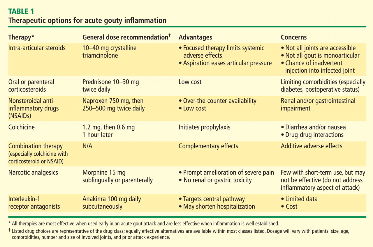
Dr. Mandell: We’ll come back and talk about each of these drug classes specifically, but what do my fellow rheumatologists tend to reach for as first-choice therapy?
Dr. Simkin: My thinking is very similar to Jim’s. As far as NSAIDs are concerned, so many of our patients have significant renal compromise that it is absolutely mandatory that we know what a patient’s renal function is before we treat acute gout with NSAIDs. I’ve seen more catastrophes from the use of NSAIDs in patients with renal compromise than from any other gout treatment scenario. So I probably wind up using steroids more often, but in the hospital setting you often have to deal with a surgeon who doesn’t want to use steroids. In such a case, if the patient also has renal problems, an agent that has entered the picture in our hospital is the interleukin-1 (IL-1) receptor antagonist anakinra, given in daily subcutaneous injections of 100 mg. When a patient is hospitalized for gout, it’s his severely painful joint that’s keeping him from going home, and in this situation anakinra in fact becomes a relatively inexpensive option, despite its absolute cost, when compared with the cost of the hospital bed.
Dr. Mandell: I think that’s the first time I’ve heard “anakinra” and “inexpensive” used in the same sentence.
Dr. Simkin: Of course biologics are terribly expensive, but so is hospitalization, and hospitals are bad places. We want to get our patients home, and in our experience this has been a very useful way to make that happen sooner.
Dr. Pile: I agree that an emphasis on hospital throughput is incredibly important. For the hospitalized patient with gout that’s preventing ambulation, that’s the issue that must be addressed before discharge is possible. Certainly the agent with the fastest onset of action is going to be very attractive.
Dr. Edwards: All of these agents for acute gout have a relatively fast onset of action if they’re given early in the disease process. Once you get out to a day and a half from the onset of symptoms or beyond, you’re fighting an uphill battle in terms of making a difference in the natural course of the attack. I believe that’s true of all three of the medication classes that are typically used.
My approach to the initial therapy choice is highly individualized, depending on what the patient’s been on before. If they’ve been on maintenance colchicine to prevent flares and then they flare, I usually won’t use colchicine for the acute attack; I will go with a steroid or an NSAID. If they’ve been on NSAIDs as preventive therapy and they flare, I might try low-dose colchicine for the flare or use steroids. In cases of a prolonged course, where the patient is in the hospital and it’s 3 or 4 days since symptom onset and a steroid taper or a trial of colchicine has failed, I’ve been very impressed with the ability of anakinra to suddenly bring the attack to a halt. Like Peter, I am on the cusp of looking at acute treatments a little differently, although there still aren’t a lot of data on IL-1 inhibition in this setting.
Dr. Sundy: There are data showing that a gout flare adds about 3 days to the hospital stay,4 so that’s a huge burden that intervention with IL-1 inhibition can really help to address.
Dr. Mandell: I think we’ve seen a trend over time toward corticosteroid therapy becoming more common, particularly in hospitalized patients. I think that’s partially related to more widespread use of appropriate deep vein thrombosis (DVT) prophylaxis, which means that more patients are on anticoagulant therapy, which is one more reason to shy away from NSAIDs, particularly the nonselective ones, in the hospital setting. Do you sense that trends in the outpatient setting have shifted, or do most clinicians still reach for NSAIDs?
Dr. Sundy: I think most people still reach for the NSAID, but it really depends on the comorbidities. I sense we’re seeing chronic kidney disease in a greater proportion of patients, and that’s probably creating a shift toward a bit more corticosteroid use. I like to reach for an NSAID as first-line therapy, but we really have to understand our patient’s overall comorbidity profile, including renal function, before doing so, as Peter said.
Dr. Edwards: I think NSAIDs are still the most commonly used acute treatment for gout. I used to calm myself when I used them by saying, “It’s only a 7- to 10-day course; how much trouble can you get the patient into?” Well, in that short a time I’ve had patients tip over into congestive heart failure because they had some renal decompensation beforehand and then had another 20% or 25% of their renal function knocked out with the NSAID, and they’d have extra sodium retention. I’ve seen GI bleeds develop in patients over that short a time. I don’t prescribe nonselective cyclooxygenase (COX) inhibitors anymore without also giving a proton pump inhibitor for stomach protection. I think that’s becoming a standard of how to use NSAIDs among rheumatologists, and it’s hard to get that stomach protection up and running in the very short time frame of an acute gout attack. So I’m personally tending away from NSAIDs; I use steroids more often and then the IL-1 inhibitor for special cases.
Dr. Mandell: Studies assessing the gastric risk of NSAIDs have shown that a GI bleed can be induced in as little as 2 or 3 days. I agree that there is a trend, at least among rheumatologists, to use a proton pump inhibitor when initiating outpatient NSAID therapy. This combination may still be cost-effective for many patients, as it can speed the return to their usual activities. But even in the outpatient setting steroids are becoming more common than they used to be.
DOSING FOR THE ACUTE ATTACK: BE AGGRESSIVE FROM THE START
Dr. Simkin: Whatever dose of steroids we use in the outpatient setting, I think it’s highly desirable to divide it. Rheumatologists are taught to use a single daily steroid dose in the morning, primarily to spare the adrenal glands. While that makes sense in patients on long-term steroid therapy, such as for lupus, it doesn’t necessarily make sense in gouty arthritis. I’ve seen patients whose gout flared up every night despite taking sufficient prednisone; when they divided the dose, their gout was controlled.
Dr. Mandell: I would generalize that further to stress the importance of using an adequate and aggressive dose of either steroids or NSAIDs for treating acute gout. A common mistake I notice in the community is the use of too low a dose of either steroids or NSAIDs. We should treat aggressively from the start with a full dose of these agents.
Dr. Edwards: Even more than the usual full dose, I would say. Many generalists have a concept of what an analgesic dose of an NSAID is, which is pretty low—in the case of ibuprofen, perhaps 400 mg three times daily. An anti-inflammatory dose is higher—perhaps 800 mg three times daily for ibuprofen. Most of us who’ve used NSAIDs for gout realize that we have to get even a little bolder than that from the start. It can be hard to convince some generalists to exceed what has been their ceiling of comfort for an NSAID, but doing so for the first 24 or 36 hours is important to getting the gout attack under control. Of course, the need for such aggressive dosing is all the more reason to make sure not to use an NSAID in a patient with significant renal disease or other comorbidities for which NSAIDs are problematic.
Dr. Mandell: And all the more reason to provide gastric protection.
Dr. Sundy: I would add that if a clinician is not comfortable using doses that high, that may be a good reason to think about choosing a different initial therapy, be it colchicine or steroids (if the reluctance is with NSAIDs) or NSAIDs (if the reluctance is with steroids).
ACUTE ANALGESIA: ANYTHING MORE THAN AN ADJUNCT?
Dr. Mandell: What about treating gout with acute analgesia alone? Consider a setting where you may want to avoid a drug with anti-inflammatory or antipyretic effects, perhaps in the postoperative period or when coexistent infection is a concern and you want to monitor the fever. Can you get by with using narcotic analgesia alone?
Dr. Sundy: I’m not impressed with that approach. Narcotics can play a role in managing a patient’s pain, but a narcotics-only approach will allow the flare to linger and not address complications of the flare or back-to-work issues. I view analgesics as cotherapy as opposed to single-agent therapy.
Dr. Mandell: Is narcotic analgesia effective even at treating the pain acutely?
Dr. Sundy: No. Gout pain is an exquisitely inflammatory pain, and the key is to tackle that inflammatory response. Analgesics are really just an adjunct.
Dr. Simkin: I totally agree, but they’re an important adjunct that we often overlook.
Dr. Edwards: I’ve been singularly unimpressed with narcotics. Gout is a cytokine-driven process that is intensely inflammatory. It’s like a lot of other types of pain that don’t respond fully to narcotics, such as herpetic neuralgia or uterine pain. Perhaps the benefit of narcotics in gout is their soporific effect—patients sleep through their attack. Yet if you touch the gouty joint or the patient moves it, the pain is just as intense as if the patient weren’t taking anything. So I don’t know that narcotics add anything unless the patient has other causes of pain. I don’t use them at all.
Dr. Sundy: I tend to make the option available to the patient. I say, “Here’s what we need to do to knock down this flare. And here’s something additional you can try for pain; we can see if it helps.” But I’ll emphasize the importance of improving the inflammatory piece.
Getting back to the postoperative setting, we should recognize that a strategy to just ride out a perioperative gout flare with an analgesic medication, perhaps because of concern about the effect of corticosteroids on wound healing or infection, can carry important risks. A patient treated that way will end up bedbound at precisely the time you want him or her to be moving around, so there’s now the risk of postoperative pneumonia and other complications that won’t get documented as complications of gout even though they actually are.
Dr. Mandell: Not to mention the untreated postoperative fever from the crystal-induced attack, which then leads to a work-up looking for DVTs, more blood cultures, and more radiographs, all the while extending the hospital stay and wasting resources. Jim, as a hospitalist, do you still see narcotics used initially as the primary treatment for gout flares in the hospital?
Dr. Pile: It depends on which hat I’m wearing. If I’m the hospitalist and the patient’s on my service, usually my antennae are up for the appearance of gout in the hospital, and the house staff may or may not recognize it if the process starts overnight. When I’m serving as the medical consultant, I sometimes encounter cases where the surgeons don’t recognize a gouty flare when it first appears, and I’ve had multiple patients in whom gout-induced postoperative fever triggers a consultation. In the latter case, it’s not uncommon to see narcotics being used in an attempt to treat gouty pain.
RELATIVE DRUG EFFICACY FOR ACUTE ATTACKS: HOW MUCH DO WE KNOW?
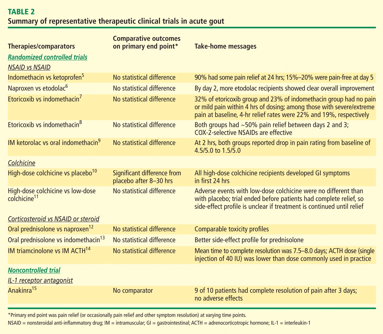
Dr. Edwards: If they’re given early, all of them have good—if not necessarily fast and ideal—effectiveness. The recent AGREE trial of colchicine (Acute Gout Flare Receiving Colchicine Evaluation) assessed pain reduction at 24 hours in patients randomized to three treatment groups: a low-dose colchicine regimen consisting of three 0.6-mg tablets (1.2 mg initially, followed by 0.6 mg 1 hour later); a more traditional high-dose colchicine regimen consisting of eight 0.6-mg tablets (1.2 mg initially, followed by 0.6 mg every hour for 6 hours), and placebo.11 Response, defined as a 50% or greater reduction in pain at 24 hours without rescue medication, was reported in 37.8% of patients in the low-dose colchicine group, 32.7% of patients in the high-dose colchicine group, and 15.5% of placebo recipients.
This is an excellent study that should finally take highdose colchicine for the treatment of acute gout off the table since it offered no improvement in efficacy but was significantly more toxic. However, while 50% improvement in 24 hours is a hard target to achieve, the fact that barely more than a third of subjects hit the target means we should still be looking for more effective approaches.
Dr. Simkin: Similarly, in the first controlled study of colchicine in acute gout, published by Ahern et al in 1987,10 23% of colchicine recipients noted a 50% reduction in their pain at 12 hours after starting treatment. That leaves more than three-quarters with little or no response within 12 hours, and gout is one of the most painful conditions we treat. We’d very much like to help our patients sooner than that. So while there are no head-to-head data comparing colchicine versus NSAIDs versus steroids, my impression too is that oral colchicine is a second-line choice for the acute gout attack.
Dr. Sundy: When we try to understand this literature, it’s important to look closely at how patients were ascertained. The AGREE trial evaluated patients who were enrolled when they were asymptomatic; they were given study medication and instructed on how to start it once a flare began. In contrast, most of the well-controlled NSAID trials captured patients as they came in with a flare, and they had to have had the flare no longer than 48 hours. I believe there’s a big difference between having had a flare for 12 hours versus 48 hours—and even as long as 72 hours in some corticosteroid trials. The longer the clock has been allowed to tick before treatment is started, the harder it is to achieve rapid symptom reduction.
Dr. Mandell: Obviously it’s difficult to compare between studies, but it’s interesting that in a study of the COX-2 inhibitor etoricoxib versus indomethacin,7 one of the largest studies of NSAID therapy for acute gout, approximately one-third of patients taking etoricoxib reported no pain or mild pain within 4 hours. This very rapid pain control is what we really want, since pain is the concern, along with eventual complete resolution, of course. So there really may be value in having something that has analgesic as well as anti-inflammatory effect, to Peter’s earlier point. Tackling both the pain and the inflammation that’s causing the pain certainly makes sense.
Dr. Edwards: Yes, and that etoricoxib study was designed the way people often are treated, with the unfortunate delay before the doctor is seen and the medication is started. The AGREE trial is how I hope everybody would be treated regardless of what they’re treated with. We’re probably never going to see a good head-to-head trial among the three main types of drugs we use for acute gout—the unpredictability of flares makes it extremely difficult.
Dr. Pile: I’m curious whether or not you, as rheumatologists who specialize in gout, are surprised by the results of AGREE. My experience with colchicine over time has been more favorable than what’s suggested by AGREE.
Dr. Edwards: How do you use the colchicine?
Dr. Pile: For more than a decade I’ve been using 0.6 mg three times daily. I thought that practice was unusual until I recently read the AGREE study and realized that a lot of you have been using that regimen for a while.
Dr. Mandell: Some gout patients will take a colchicine tablet when they feel the wisp of an attack coming on and it immediately will stop the attack. That gets to the lore that if you treat a flare very early on, you may be able to abort and treat an attack very quickly. Years ago, however, I was involved in an analysis of 100 patients treated with intravenous (IV) colchicine, and we found that treatment response was unaffected by whether patients were treated early or after a delay of 48 hours. I believe colchicine behaves like a different drug when given IV—and the IV form has since been withdrawn from the market for safety concerns—but this underscores that individual responses to various agents are quite unique. I think there are some patients who are exquisitely sensitive to colchicine, while others are exquisitely sensitive to an NSAID, and so on. This means we’d need a very large sample size to tease out that variation in a trial, and that’s not likely to happen.
Dr. Edwards: I too have had gout patients who tell me they get this premonition of an attack—a feeling that “something just isn’t right”—hours before they actually feel any pain from the attack. A lot of them tell me they’ve learned over time that if they take a colchicine tablet when they get this premonition—or an extra colchicine tablet, as some are on colchicine maintenance therapy (typically one or two tablets a day) plus whatever other background therapy they’re on—they don’t get the flare or the flare is diminished.
Dr. Mandell: I’d like to wrap up this portion by getting your sense of whether there’s a difference in efficacy for treating acute attacks among the drug classes we’ve discussed.
Dr. Edwards: I believe the IL-1 inhibitors are probably the most potent agents for aborting a gout attack, followed by steroids, which I think have a leg up on both colchicine and NSAIDs. The latter two options probably are equally effective in aborting an acute attack.
Dr. Sundy: Yes, I would rank them the same way.
THE ROLE OF COMORBIDITIES IN THERAPY CHOICE
Chronic kidney disease
Dr. Mandell: Let’s get more specific about comorbidities and therapy choice, starting with chronic kidney disease. What’s the threshold at which renal compromise starts to influence your choice of therapy for an acute attack of gout?
Dr. Sundy: I have a very low threshold for avoiding NSAIDs in that setting, mainly because when I want to use NSAIDs, I want to use them at a very high dose, and even short-term use of high-dose NSAIDs can reduce creatinine clearance. So I would certainly avoid NSAIDs when the patient’s creatinine clearance is less than 60 mL/min, and I’d be even more cautious if the patient had underlying hypertension or congestive heart failure.
Dr. Mandell: So even with the reversibility of almost all of the NSAIDs’ renal effects, you tend to stay away from them in that setting?
Dr. Sundy: Yes.
Dr. Edwards: I’m less guided by a single creatinine clearance level. I tend to look at patients in light of the 20% to 25% reduction in the glomerular filtration rate (GFR) that most NSAIDs will cause, even when used acutely. Though the NSAID effect is reversible, if that GFR reduction is going to make a difference to other compensated systems, primarily the heart, then the NSAID should be avoided.
Dr. Simkin: It’s my understanding that plasma flow to the kidney becomes prostaglandin dependent with renal insufficiency, and when we use antiprostaglandin agents we get into trouble on a mechanistic level. I’m not sure of the exact point at which that occurs, but I think we all recognize that serum creatinine is a fairly weak indicator of which patients have some limitation.
Dr. Mandell: So we agree on the need to be very cautious with the use of NSAIDs, recognizing that in the acute setting there is reversibly depressed renal blood flow from NSAIDs. The decreased blood flow will get better, but there’s the issue of what happens during fluid retention when there is coexistent congestive heart failure, diabetes, and chronic kidney disease—you may also occasionally bump up the potassium level. In general, most rheumatologists shy away from selective or nonselective NSAIDs in the setting of chronic kidney disease. Is that consistent with the use you see in the hospital, Jim?
Dr. Pile: It is. I would add that if patients are on certain other medications—especially angiotensin-converting enzyme inhibitors, angiotensin receptor blockers, or beta-blockers—there may be additive negative effects because of the potential for hyperkalemia if an NSAID were added to the mix.
Dr. Mandell: I would add to that list of concerning medications aminoglycosides, cyclosporine, and other nephrotoxic agents.
Diabetes
Dr. Mandell: What about diabetes? How does a patient’s diabetes enter into your choice of acute gout therapy in the outpatient and inpatient settings?
Dr. Edwards: Diabetes is especially problematic because it significantly affects two of our treatment choices—steroids and NSAIDs. The steroid doses needed to effectively treat an acute gout attack have a pretty profound effect on glucose levels in diabetic patients. That’s always of concern to me, and it’s certainly of concern to my patients with diabetes who are vigilant about monitoring their glucose levels, which may spike from 130 or 140 mg/dL to values in the high 200s. These elevations will last for the 7 to 10 days of treatment, and the longterm adverse effects of that period of hyperglycemia are not clear. If we put these patients on short courses of corticosteroids, perhaps we should be treating their diabetes more aggressively during that period, but I don’t think we have a history of doing that.
Dr. Pile: It’s very difficult to do in the short term. That’s especially the case with the diabetic patients I see as a hospitalist at a safety-net hospital. Many of my patients don’t have good glucose control at baseline: their levels of 250 mg/dL may go to 400 mg/dL or higher with acute corticosteroid therapy, so that’s clearly a problem.
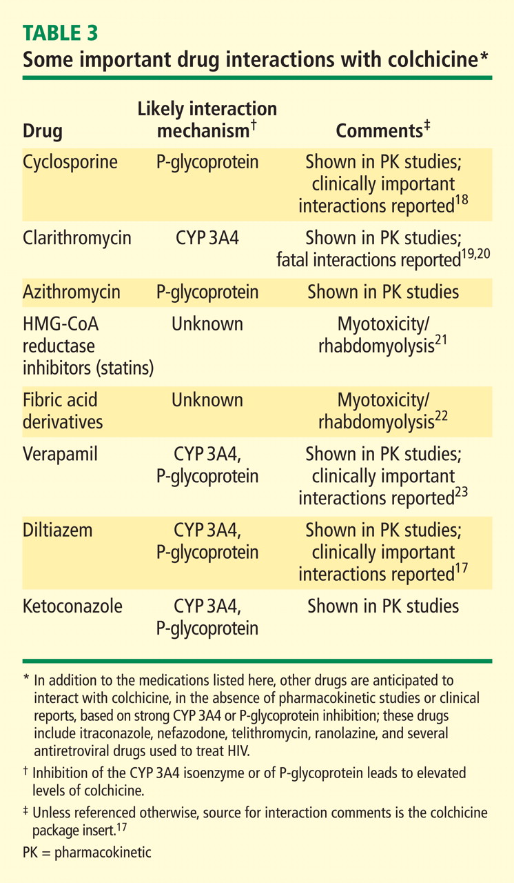
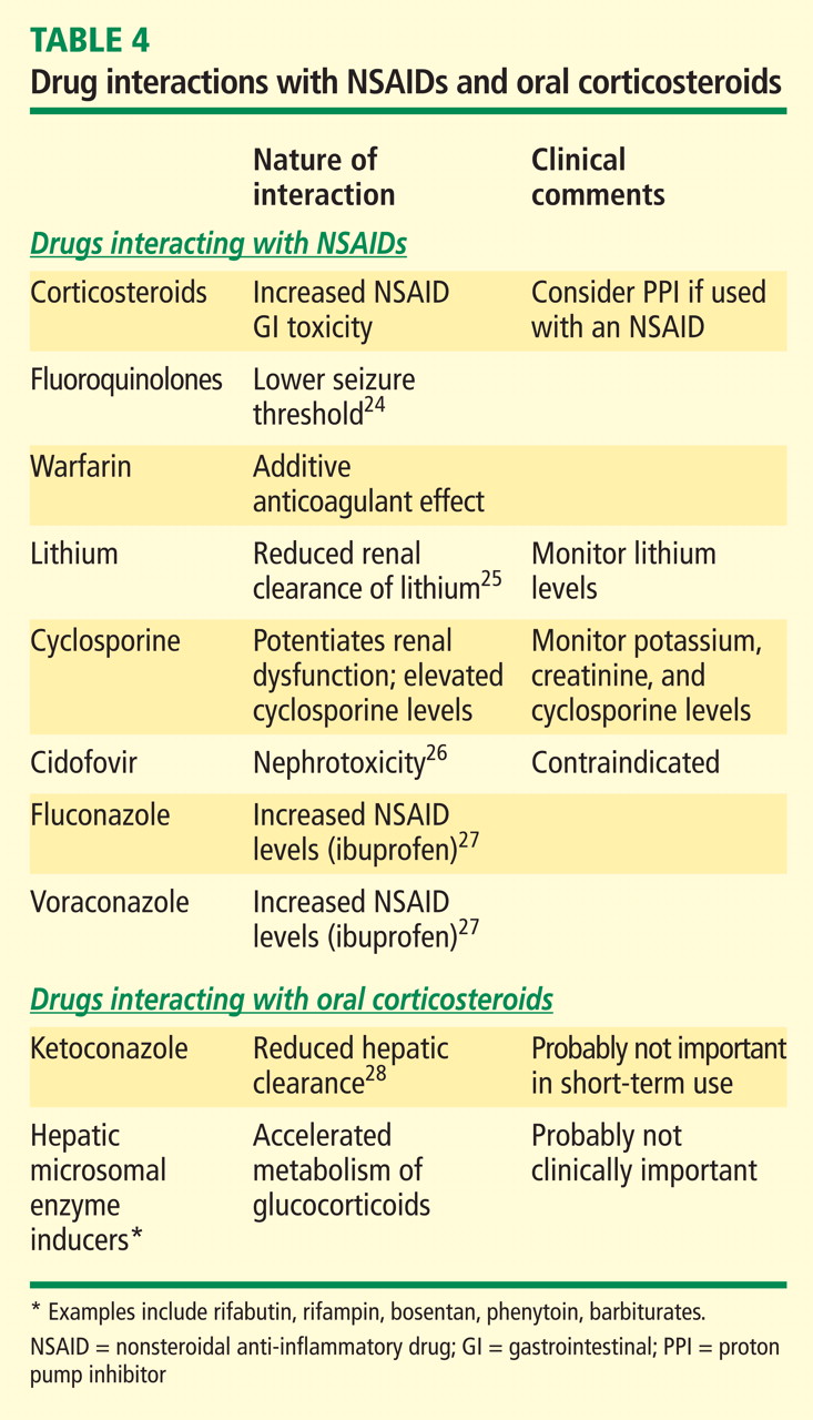
Dr. Mandell: I try to avoid giving corticosteroids in the outpatient setting to a diabetic patient who is being maintained on oral hypoglycemic medications, particularly if efforts are being made to avoid more aggressive diabetes therapy. Steroids may be an option, though, for a patient who is on insulin, uses a pump, and is very comfortable measuring and managing his or her glucose at home. In the hospital setting, I’m more comfortable using corticosteroids in diabetics because I have much better control over their glucose; I can just increase the basal and premeal coverage.
Dr. Pile: I agree. With vigilance I can control almost anyone’s blood sugar in the hospital, but when patients leave the hospital on steroids, it becomes much more problematic.
Dr. Mandell: Right. You don’t know what’s going to happen when they go out and liberalize their diet on top of the steroids, because often it’s the postprandial surge that’s exacerbated by steroids.
Dr. Sundy: It can be very helpful to know at baseline what a patient’s spot blood sugar is before using a steroid, just as it’s important to know what the creatinine is before considering an NSAID. In the setting of an acute flare, the patient might already be quite hyperglycemic, and many of these patients probably don’t have the tools to manage this at home, so careful follow-up—coming back the next day for at least a blood sugar check—is critical to the proper care of these patients.
Dr. Mandell: When we talk about steroids we tend to think mostly about prednisone, but Peter mentioned injectable steroid. What about injectable adrenocorticotropic hormone (ACTH), which goes in and out of vogue as an agent to treat acute gout? What is your experience with using ACTH injections, specifically related to the issues of diabetes and heart failure?
Dr. Edwards: Before it got reformulated about 3 or 4 years ago and its price went way up, injectable ACTH was my drug of choice for treating acute gout. I had hardly any failures on it over the 10 to 15 years when it was my main means of treating acute gout. I would typically use 80 IU of ACTH gel, given subcutaneously, and the response within 12 hours was quite dramatic. About one in four patients would require a second injection at that 12-hour point, but almost everyone was symptomfree at 24 hours. This response was seen even in patients who were on fairly high-dose chronic steroids already, such as transplant patients.
This suggested that a mechanism other than just adrenocortical stimulation explained the benefit from ACTH. In fact, a lot of data have emerged suggesting that ACTH has a specific effect that is not related to adrenocortical stimulation.
Ten years ago an injection of ACTH gel would cost about $3 or $4, whereas today it costs $2,000 or more.29 I don’t think it’s justifiable at that cost.
Dr. Mandell: There’s also the issue of fluid retention with ACTH, and the higher doses, which seem to be more effective, are more likely to exacerbate congestive heart failure, which has to be a concern if you go this route.
Coronary artery disease
Dr. Mandell: Atherosclerosis, diabetes, and chronic kidney disease are increasingly recognized in the gout population, and coronary artery disease links with all of those. Does the presence of coronary artery disease enter into your choice of agents for a patient with an acute attack of gout?
Dr. Edwards: It doesn’t have as big an impact as diabetes does. To me, the priority in this setting is resolving the acute attack as quickly as possible, as I’ve seen my share of patients for whom the acute gout attack has been so painful and stressful that they’ve developed angina at the same time. So I might use steroids in patients with coronary disease because I’ll want to go with what’s likely to be most efficacious for quick resolution of the attack even though steroids may increase glucose and blood pressure and thus raise fluid retention as an issue.
Dr. Mandell: The presence of coronary disease implies the use of low-dose aspirin therapy. Despite the slight elevation in serum urate from low-dose aspirin, I recommend that my gout patients continue this for coronary protection. But even low-dose aspirin raises concerns of GI safety and even possible drug interactions with ibuprofen that could reduce the aspirin’s efficacy. Does this lessen your tendency to reach for an NSAID in these patients?
Dr. Edwards: I think the issue of cardiovascular safety and NSAIDs is still up in the air. The data that came out around the time the selective COX-2 inhibitors were released made us all think again about the role of prostaglandins and coronary blood flow and how blocking prostaglandins might have some bad effects. I don’t often use NSAIDs in patients with significant heart disease, simply because they don’t act fast enough to get patients through the attack quickly and I worry about the sodium retention and other problems putting stress on the heart.
Dr. Sundy: While coronary disease doesn’t create the acute dilemmas that diabetes does, it is a consideration in my therapy choice, and I generally avoid using NSAIDs.
Dr. Mandell: It is important to remember the additive gastric toxicity effect from low-dose aspirin and NSAIDs.
Infection
Dr. Mandell: Let’s turn to the hospitalized patient—a patient admitted to the medical service with infection. This patient is going to get fluids and drugs, and these are likely to raise or drop the serum urate level, which may precipitate a gout attack. From your perspective as an infectious disease consultant, Jim, how does the coexistence of a documented infection in the hospital—which you’re treating—factor into which agents to use or avoid when treating acute gout?
Dr. Pile: In practice, I try to avoid steroids in that situation. Theoretically, however, I’m not sure this scenario is really a contraindication to steroid use. I’m not aware of much solid data showing, for example, that patients with community-acquired pneumonia on appropriate antibiotic therapy have worse outcomes if they receive steroids than if they do not. Certainly there are a variety of serious infections for which we use steroids as a matter of course, including pneumococcal or tuberculous meningitis, tuberculous pericarditis, severe typhoid, and severe septic shock (the data for septic shock are murky, but there’s a role for at least steroid supplementation). As long as the antimicrobial therapy is appropriate, patients with serious infections are not necessarily going to do any worse with steroids.
Dr. Edwards: The biggest risk from steroids would be masking the symptoms of an unrecognized infection. If you know the infection is there and you’re treating it appropriately, I don’t think steroids are contraindicated.
Dr. Pile: A related risk is that steroids can make it more difficult to monitor the course of a known infection by blunting the overall response and bumping up the white blood cell count.
Dr. Mandell: My practice would fall in the realm of what Jim described as the theoretical course of action. If I knew what the infection was or felt comfortable in treating it, I would not shy away from systemic corticosteroids in the hospital setting, where I could monitor the patient. I think a corticosteroid may often be the safest drug to use in that setting, given the likelihood of comorbidities, at least compared with NSAIDs.
Dr. Simkin: Let’s say a patient comes in with acute knee pain and you aspirate the knee. The question arises whether you should wait for the culture results or go ahead and inject steroid while your needle is in the knee. I certainly have a much lower threshold to go ahead and inject while I’m in there, although I’d always be sure to get the culture results and revisit as needed. But if I pull out extremely inflammatory fluid during the aspiration, I’m probably not going to inject the steroid.
Dr. Edwards: And what if you actually do inject an infected joint with a corticosteroid—are you doing harm?
Dr. Simkin: I would like to think I’m not, and I may be helping.
Dr. Edwards: Yes, there are a lot of theoretical reasons why you might be helping, and there’s a small literature on steroids and infected joints that suggests you might actually slow down some bone resorptive steps and be doing various similar things.
Dr. Mandell: But in humans (a series of pediatric patients with septic arthritis30), those were systemic steroids, not intra-articular.
Dr. Edwards: In animal studies there is some amelioration of the destructive component with intra-articular steroids. The main point is that we must send off the fluid for culture when we inject steroid into a joint, and we must follow up on the cultures to make sure nothing is growing in a joint because we’re going to be calming it down. Whatever the inflammatory process is—whether infection or crystalline or rheumatoid—the patient is initially going to get better with a steroid injection. So we can’t use initial clinical improvement as the marker that we’ve done the right thing. We need to make sure the aspirate culture is negative.
Dr. Mandell: What about the use of colchicine or an IL-1 antagonist in the setting of infection?
Dr. Pile: There may be some theoretical concern, but I think low-dose colchicine would be a reasonable agent to use in the setting of infection.
Dr. Edwards: I don’t think there’s much concern since IV colchicine has been taken off the market. IV colchicine had a much more profound effect on bone marrow, and this resulted in a number of deaths from infection in immunosuppressed patients such as chemotherapy recipients. But oral colchicine in the doses we use for acute attacks and for maintenance probably doesn’t have a very profound effect on response to infectious processes or on white blood cell production (in the absence of chronic kidney disease).
Dr. Pile: Which raises the point that if the patient were on a potent inhibitor of the cytochrome P450 3A4 system, you might conceivably get into the same situation. For example, serious toxicity and even fatalities have been reported in patients taking colchicine and clarithromycin concurrently.31
Dr. Mandell: Peter, would you be comfortable giving an IL-1 antagonist to a hospitalized patient with pneumonia that’s being treated with antibiotics? Let’s say the patient has chronic kidney disease and diabetes and you’re looking for an agent to use.
Dr. Simkin: Well, I wouldn’t be comfortable, but I might feel I had to do it. If the organism were known, as you said, and the antibiotic therapy were appropriate, I might depend on that antibiotic coverage.
Dr. Sundy: Although I can envision some scenarios where I’d use the IL-1 antagonist in this situation, I’d be looking for an alternative. My concern is that people with any source of infection were excluded from clinical trials of anakinra, so we don’t have a good understanding of how it behaves in the setting of infection.
Dr. Edwards: And the fact that anakinra is not approved by the Food and Drug Administration (FDA) for treatment of acute gout should make us a bit uncomfortable.
Dr. Mandell: It’s interesting that the limited data do not show a lot of infections with IL-1 antagonist therapy other than when combined with anti–tumor necrosis factor therapy. The experience in periodic fever patients, who are on IL-1 antagonist therapy for many years, has not really shown this to be an agent predisposing to infection. I think this goes back to the point that if you document an infection and you’re treating it appropriately, some immunosuppressive or anti-inflammatory therapies may not be bad if you need to treat the attack of gout, particularly if it’s in a setting where you can monitor the patient well.
NSAIDs: SPECIAL CONSIDERATIONS FOR USE
Dr. Mandell: Let’s drill down on the use of the specific classes of drugs, starting with NSAIDs. Larry made the point that NSAIDs should be used at high doses for acute gout attacks. What else is worth noting about the dosing and use of NSAIDs in this setting?
Dr. Sundy: In addition to that distinction between analgesic and anti-inflammatory dosing, it’s important that patients be told to treat at the appropriate dose intervals for whichever NSAID is chosen so that the anti-inflammatory benefit is maintained. I generally recommend that patients continue on that dosing regimen until they have substantial symptom improvement, at which point they can begin to back off, but that’s generally a 7- to 10-day treatment course, and typically closer to 10 days. Finally, I’m quick to use proton pump inhibitors or some sort of gastroprotective regimen with most patients that I start on NSAIDs in this setting.
Dr. Mandell: Your point about duration of therapy bears emphasizing. A common mistake with the use of NSAIDs is that they’re stopped too soon out of an understandable concern about their toxicity. Often they’re stopped when symptoms are still present, which means the attack hasn’t really resolved, and so it comes back. I tell patients to wait until their attack is completely gone, continue for a few days longer, and then stop the NSAID at that point.
Dr. Edwards: If we look at the natural course of disease, a patient’s first four to six gout attacks tend to be a bit shorter in duration, in the range of 5 to 7 days. As recurrent attacks occur over the decades, they tend to get more drawn out, lasting perhaps a couple or few weeks. We want treatment to at least cover the anticipated period of the natural attack since we’re not doing anything profound to the disease process itself, except perhaps with an IL-1 inhibitor. For instance, a typical treatment in emergency departments is an intramuscular injection of ketorolac. That may make patients feel good for the next 24 or 36 hours, but they’ll have a resumption of their acute attack at that point. I generally stick to 2 weeks as a reasonable treatment period. I might shorten that for a patient with diabetes in whom I have to use steroids or for a patient with congestive heart failure that I needed to put on an NSAID, but I generally try to cover patients with anti-inflammatory therapy for a couple of weeks.
Dr. Mandell: Your point about intramuscular ketorolac is important. A single shot is not likely to resolve the attack, and the intramuscular route will not ameliorate the gastric toxicity of this drug. Those are two common misperceptions.
What about the COX-2-selective NSAIDs? There’s only one that’s still marketed in the United States, celecoxib, but what’s your sense of these drugs as a class in terms of efficacy for treating gout?
Dr. Sundy: I think they’re effective. Only one COX-2 inhibitor, etoricoxib, has been studied in clinical trials for acute gout, and it’s not available in the United States, but I think the class as a whole is a reasonable choice. That’s especially true since the course of treatment is only 7 to 14 days. Even so, it’s still reasonable to use some gastric protection when giving celecoxib, as it does no harm and can add some reassurance. As with other NSAIDs, dosing of celecoxib for acute gout is higher than for other typical uses in the drug’s labeling.
Dr. Edwards: Yes, when I use celecoxib for acute gout I use 400 mg every 12 hours for the first 2 days and then take it down to 200 mg every 12 hours for the remainder of the treatment period.
Dr. Mandell: And the COX-2-selective NSAIDs do adversely affect renal function.
Dr. Simkin: One thing that troubles me when we talk about dosage for any of our agents is the large individual variation we encounter. I don’t think there’s a patient population with a wider variation of body sizes than the gout population. We see enormous patients with metabolic syndrome and several large joints, and we see frail elderly patients who have a single small joint affected. In fact, in addition to body size, the size of the joints involved can tell us a lot about the amount of inflammation we’re dealing with. Someone with two knees and an ankle affected is probably different from someone who has just a great toe affected. In light of these variables and so many others that come into play, such as age and comorbidities, I’m hesitant to recommend any particular dose because I try to make adjustments. Of course, there are no data that cover all these variables.
Dr. Mandell: But would you agree with the generality that high doses—higher than those we typically use for other musculoskeletal pains—are warranted in treating acute gout?
Dr. Simkin: Yes, but a high dose for a small elderly woman is different from a high dose for a young man who’s quite large.
Dr. Sundy: This sensitivity to individual variation can also apply to duration of therapy. As we noted, it’s not uncommon for veteran gout patients to find that starting treatment early—within 24 or 48 hours—may be sufficient to tamp down their symptoms, yet that’s an observation that is developed only over time in an individual patient. So it’s not a recommendation, but many patients will do fine with that. In those situations, I may not be so adamant about the need to continue treatment for a full 10 or 14 days.
STEROIDS: SPECIAL CONSIDERATIONS FOR USE
Dr. Mandell: Let’s move to corticosteroids. A very common treatment used for acute gout is a tapered-dose regimen like the Medrol Dosepak (blister-pack) formulation of methylprednisolone. What strikes me is that this is a one-size-fits-all formulation, yet you’ve all just argued that there’s no one-size-fits-all approach. What are your thoughts?
Dr. Edwards: The Medrol Dosepak contains 21 4-mg tablets of methylprednisolone to be taken over the course of 6 days. Six tablets are taken the first day, and then one fewer tablet is taken each successive day through day 6. An obvious potential drawback is that the patient is off of medicine after 6 days, though that will be enough for some patients if they start treatment promptly.
I do use the Medrol Dosepak, and I always have patients fill a prescription ahead of time so that it’s available for quick initiation when they start to have their next attack. I instruct them to avoid storing it in a hot place, such as a car, but that it can be stored for up to 5 years at room temperature. Most of my patients like this approach, and if they do well with it they’re good about getting a new refill in advance of their next attack. This is a pretty handy way of letting patients be in charge of their attacks. If it doesn’t work and their symptoms continue, they call me and I’ll see them promptly.
A different approach is more appropriate for patients whose attacks tend to be more recalcitrant, perhaps because they’ve had gout for a long time or have bulky disease with lots of tophi. In those cases I’ll go with a steroid regimen that’s essentially double what a Medrol Dosepak would be—namely, prednisone 30 mg for the first 2 days then tapered down by 5 mg every other day. As Peter said, this approach would be overwhelming to a frail 82-year-old woman with a single tophus in her distal interphalangeal joint, but it tends to work well for a more typical 50-year-old obese male gout patient.
Dr. Sundy: I do some of the same things you describe, though my background in allergy has accustomed me to the steroid burst, so I’ll tend to use a dose of about 0.5 to 1.0 mg/kg/day for 4 or 5 days and then try to taper rapidly over another 2 to 4 days. I’m curious whether you think methylprednisolone offers advantages over prednisone.
Dr. Edwards: There are a few patients—roughly 5% to 15% of the population, according to various studies—who have trouble converting prednisone to its active form in their liver because they’re missing an enzyme. So I tend to use methylprednisolone since it has a benefit in that small segment of the population, but it otherwise doesn’t offer anything special beyond the convenient packaging that we discussed.
Dr. Mandell: I think convenience is the major factor. The blister-pack preparations are useful for patients who get confused with tapering. For patients who can make adjustments based on their symptoms, I find those personalized adjustments to be better than just giving them an absolute number of pills.
Dr. Pile: This issue of dosing duration is very important for generalists; just listening to your schedules is very instructive for me. I think a lot of nonrheumatologists, myself included, are inclined to use a one-size-fits-all approach. The mistake that I’ve been most apt to make, apparently, is using an insufficient duration of therapy. I’ve tended to use 40 mg prednisone for 5 or 6 days and then to stop rather than tapering the dose.
Dr. Mandell: From the cost perspective, steroids seem to be a very cost-effective option. The Medrol Dosepak, as a branded product, costs a bit more. Similarly, generic NSAIDs are inexpensive. There has been some feeling that nongeneric NSAIDs may offer advantages over generic NSAIDs, but I don’t know of any published evidence to support that. What are your thoughts?
Dr. Edwards: I agree with you. There has been this folklore about indomethacin and, before that, phenylbutazone being rather remarkable and specific drugs for gout. That hasn’t been borne out by the data, and virtually all of the NSAIDs work fairly comparably.
COLCHICINE: SPECIAL CONSIDERATIONS FOR USE
Dr. Mandell: Let’s move to colchicine, which represents a unique class of anti-inflammatory agent. It has been available and widely used for many years, but it had not been formally licensed by the FDA until last year. Without corporate imperative and funding, data on its clinical use have been limited. As we mentioned, an IV form of colchicine was withdrawn from the market a few years ago after being associated with multisystem organ failure and death. Low-dose short-term colchicine doesn’t have those risks, and its efficacy in treating gout has been accepted for many years despite the dearth of clinical trial data until recently. How has colchicine been used in the past, and do the new data guide us on how it should be used differently, specifically for treating acute gout attacks?
Dr. Simkin: The so-called traditional regimen of starting with a pair of colchicine tablets and then giving another tablet every 1 or 2 hours until relief (or until GI toxicity developed) is one of the great embarrassments in the history of our field. The more recent trial evidence clearly indicates in the short term that there is no additional benefit beyond a very low dose. As we noted, this has been in the wind anecdotally for quite a few years, and it was confirmed by the recent AGREE trial.11 I find it interesting that the dosage used in AGREE—three 0.6-mg tablets—is very close to what we use for prophylaxis. We may well wind up concluding that any patient we’re seeing for the first time with an acute attack should be treated with that dose of colchicine, as an adjunct to another anti-inflammatory drug.
The use of colchicine as an adjunct for acute gout attacks was advanced in a review by Cronstein and Terkeltaub several years ago.32 Since we know that the mechanisms of action of the various drugs for acute gout are sufficiently different from each other, it makes sense to consider hitting the process at more than one spot. Having said that, one of the most crucial concerns in using colchicine is what other medications the patient is taking, given colchicine’s multiple drug interactions (Table 3). These include interactions with most statins, some antibiotics, and some calcium channel blockers, which share drug transporters with colchicine. So we have to be cautious about using colchicine with those other therapies.
Dr. Mandell: That’s also important in patients on chronic prophylactic colchicine therapy, as we know from recent pharmacokinetic studies and anecdotal case reports of colchicine and some of these other drugs.16–23 Do you think it’s as important in a patient on, say, chronic statin therapy who’s not on baseline colchicine, in whom you use a colchicine regimen of just two tablets followed by one tablet and then stop the colchicine right there? Is the potential for meaningful drug interactions as important in that setting?
Dr. Simkin: Probably not. One of the key issues is to educate patients about what to look for. Tell patients that if they notice numbness, weakness, or myalgia, you want to know about it. The evidence I’ve seen is that people who go into colchicine-induced rhabdomyolysis, for instance, develop it over a period of days, not hours, and if they stop the drug early, they probably will be okay. That’s the biggest concern. Neutropenia is the second concern.
Dr. Mandell: But fortunately, assuming no chronic kidney disease, those are generally patients taking colchicine chronically, not just acutely. We talked a bit before about our panel’s concerns about the efficacy of colchicine. Say you have a patient who has no history of prior colchicine use. How comfortable are you that treating such a patient with just the three-tablet colchicine regimen is likely to give the desired effect of rapid pain control and resolution of the attack?
Dr. Edwards: I’d expect that there wouldn’t be a profound or rapid improvement on that regimen, although the patient would certainly do better than if he or she were on nothing. I think that’s what the AGREE trial showed—that patients improve but still have significant morbidity days after initiating therapy.
Dr. Mandell: I think the AGREE trial did what it was intended to do: it showed that a low-dose colchicine regimen was as effective as and better tolerated than a high-dose colchicine regimen. This was something most of us believed, but it had never been demonstrated in a trial, and now there are good data to show it. And AGREE was consistent with the 1987 Ahern study10 in showing that the high-dose regimen is better than placebo. However, it in no way convinced me that colchicine in this dosing regimen is a monotherapy I’d be comfortable using to treat a severe acute attack. AGREE also didn’t give me any information as to how likely this approach would be to resolve an attack. For all the reasons we discussed, continued anti-inflammatory therapy is likely necessary beyond three pills, and I can’t be confident that a course of three colchicine pills is sufficient to maintain a drug level inside white cells to prevent other attacks from happening or a rebound attack once the regimen is over. I’m sure this approach will work for some patients; I just don’t know at the outset which ones or what percentage of patients will respond completely.
Dr. Edwards: I like the idea of combination therapy, of using colchicine as an adjunct. We’ve been talking up to now about monotherapies for acute gout because monotherapy is simple and patients and physicians alike prefer things that are fairly simple to use. But there is good theoretical reason to support using colchicine in combination with either NSAIDs or steroids, though there are probably no experimental data. I can’t imagine a reason for using corticosteroids and NSAIDs together, but pairing up colchicine with a steroid or an NSAID might allow you to keep the dose of the other agent lower and perhaps reduce the overall toxicity of your therapy. I’ve occasionally put patients on that type of combination therapy, and it’s probably a helpful way to go.
Dr. Mandell: For years my approach has been one of cotherapy, counting on either NSAIDs or corticosteroids to treat the acute attack by rapidly relieving the pain and inflammation and using colchicine in low doses—at a prophylactic dose, as Peter said—and extending it over a much longer period. I’m counting on the colchicine not to treat the acute attack but to prevent the next one and hopefully, for all the reasons we talked about, shorten the duration of therapy with the steroid or NSAID.
Dr. Edwards: Sure. The way I use colchicine is mostly for maintenance and for prophylaxis while I’m initiating urate-lowering therapy. I use it for a bit longer than most others might—I draw it out to at least 6 months after a complete resolution of symptoms, sometimes to 9 or 12 months. When a patient has a flare, I certainly have them maintain the colchicine if I’m going to treat them with something else, as I almost always do, and I believe that lets me get by with lower doses of the other agent I’m using for the acute attack.
Dr. Sundy: When I use colchicine as monotherapy for an acute flare, it’s because the patient has concluded from experience that it has worked effectively for him or her. But that’s not very common.
Dr. Mandell: There are times when the patient’s comorbidities lead me to conclude that colchicine may be the best drug to try for acute treatment. But I’m not comfortable assuming that three pills will do it all.
Dr. Pile: From the perspective of the generalist, who typically treats a simpler gout population than you do, what is the recommended treatment duration if colchicine is used as monotherapy for an acute attack? We now have this recommendation to use a single day’s worth of therapy, but you’re all expressing reservations with that.
Dr. Edwards: The recommendation in the drug’s current labeling is to take two 0.6-mg tablets at the first sign of a flare and a third 0.6-mg tablet 1 hour later. That was the low-dose regimen in AGREE, and I think it’s a good one, as only a very small percentage of patients will get the GI side effects with this regimen that occur with more prolonged therapy. The real question is what you do on day 2 and day 3 on out to day 7 or 10. If I were to use colchicine monotherapy, after that first day I’d prescribe one tablet three times daily for another 3 or 4 days and then drop down to one tablet twice daily for the rest of whatever duration of therapy I felt was justified. Of course, this is highly individualized and depends on how the drug is tolerated; probably more than 25% or 30% of patients can’t tolerate a three-times-daily regimen even for 3 or 4 days. But the total duration would be at least 7 days and up to 10 or even 14 days.
Dr. Simkin: Or months, because you’ve established a diagnosis.
Dr. Edwards: Yes—as prophylaxis, for which the dosage is one tablet once or twice daily. And then you’d leave the patient on prophylaxis and start urate-lowering therapy, if it hadn’t been started already.
Dr. Mandell: We’re now way past having trial data to support this, and this approach may negate the purported safety advantages of the low-dose regimen supported by AGREE.
Dr. Edwards: Yes, this is rank speculation.
Dr. Simkin: But there is evidence of the value of prophylaxis.
Dr. Mandell: There is certainly evidence for the value of prophylaxis, from a number of studies. We’ll get to that shortly. However, if we think just about treating the acute attack and the AGREE findings, I don’t think we can extend those results with confidence to a setting where we switch immediately from acute therapy into a prophylactic mode. Not that we shouldn’t consider doing that—it’s similar to my practice as well, though I tend to use lower doses as I extend out—but the existing trial data are limited to that very short window of acute treatment. Anything we do beyond that is an extension from our belief that the inflammatory response to crystals in a joint lasts longer. I believe it generally requires longer anti-inflammatory therapy, but we need to expect some frequency of side effects and drug interactions as we put patients on chronic colchicine therapy.
The other factor that used to be an advantage of this type of regimen was its low cost. For a long time colchicine was available from multiple manufacturers and was exceedingly inexpensive, although with less FDA guidance on its use and less oversight of its manufacturing. This enabled even patients with no insurance to go on a prophylactic therapy and remain on it. The environment is now quite different with the introduction of a single FDA-approved and -regulated branded product. So how do we deal with cost in the current environment?
Dr. Edwards: In terms of specific numbers, once-daily and twice-daily doses of unbranded colchicine used to cost about $6 and $12 a month, respectively, based on the average wholesale price. Now the cost of branded colchicine (Colcrys) is about $175 a month for the once-daily dose and $350 a month for the twice-daily dose, again based on the average wholesale price.
Dr. Simkin: I think all of us are deeply troubled by the high cost of this preparation and hope that the alternatives will still remain available to us.
Dr. Edwards: Yes, especially since it sounds like most of us consider colchicine our go-to drug for maintenance. It used to be that patients might be on maintenance colchicine therapy for 5 or 10 years without any addition of urate-lowering therapy. Today, however, when most of us put a patient on prophylactic therapy with colchicine, it’s with the intent of also addressing the hyperuricemia that’s at the heart of the disease process. In that case we still cover patients with colchicine prophylaxis during the initial months of urate-lowering therapy because of the elevated rate of gout flares—so-called mobilization flares—that occur during the process of uric acid reduction.
Dr. Mandell: The manufacturer of branded colchicine has introduced a patient assistance program to ease the drug’s cost for the financially needy. We will have to see how practical it is and how it affects our patients’ use of this medication.
ACUTE FLARES IN THE SETTING OF PROPHYLAXIS AND URATE LOWERING: WHAT TO DO?
Dr. Mandell: It sounds like most of us would treat patients prophylactically with colchicine for a while after an attack, certainly after several attacks, and then continue on chronic low-dose colchicine during the introduction and adjustment of the urate-lowering therapy that constitutes comprehensive gout treatment. Yet some patients will still have flares. So how does this baseline low-dose colchicine prophylaxis—a 0.6-mg tablet once or twice daily—influence your choice of therapy for those attacks? Do you bump up the colchicine dose, or do you absolutely avoid increasing the colchicine?
Dr. Sundy: I wouldn’t absolutely avoid an increase, but I would tend not to adjust it. I would maintain the dose and add a different agent on top of it—an NSAID or corticosteroid—to manage the acute attack. In general, if a patient is on regular colchicine prophylaxis, the assumption is that they have sufficient renal function to support that use, so such a patient may do fine with an NSAID.
Dr. Simkin: I trust we all agree that it’s critically important to continue the urate-lowering therapy the patient is taking if he suffers an attack. The same thing pertains to the prophylactic regimen. Both of these components should continue through the flare, but I agree that we usually want to add something to treat a flare, and it should be something the patient has on hand and can take without needing to talk to his physician if it’s the middle of the night.
Dr. Mandell: So I’m hearing that most of us would add something else for the short term on top of the prophylactic colchicine dose when a flare developed rather than increasing the colchicine dose.
Dr. Simkin: Yes, although one exception I’d be comfortable with is the example we discussed earlier of the patient who has found that taking an extra colchicine pill or two can help abort or diminish a flare without causing diarrhea.
Dr. Mandell: We cannot extrapolate the AGREE data to support a short-course regimen for an acute attack in patients who are already on chronic colchicine for prophylaxis.
PROPHYLAXIS IN THE PERIOPERATIVE SETTING
Dr. Mandell: Admission to the hospital for acute medical or surgical reasons is not infrequently associated with gout flares. Jim, have we in the rheumatologic community made it clear that chronic colchicine prophylaxis and urate-lowering therapy should ideally be continued when patients with gout are hospitalized for any reason, or is it a reflex for these drugs be held?
Dr. Pile: I think there’s a general appreciation, at least in the hospital medicine community, that these therapies should be continued in that situation. My sense is that these prophylactic gout therapies are viewed by most hospitalists as somewhat analogous to beta-blockers for chronic heart failure, which are understood to be necessary to continue when a patient is admitted with an acute exacerbation of heart failure. I don’t have data to support this contention, however.
Dr. Edwards: My experience suggests that discontinuing urate-lowering therapy during an acute gout attack is a common mistake in general practice. I think that’s been demonstrated in surveys of primary care physicians. And when the uratelowering therapy is stopped, the attack is often prolonged and made worse. Widespread misperception about this remains—it’s a big problem.
Dr. Mandell: Based on educational lectures I’ve given to primary care audiences, I agree with Larry. If I present a case question on a scenario like this, many physicians in the audience will anonymously indicate via the audience response system that they would stop a patient’s allopurinol if an acute attack occurred.
Let’s turn to the scenario of a patient who’s admitted for surgery—elective or emergent—who has a history of gout and is on prophylaxis. What’s the general routine in managing these medications in patients who are going to surgery?
Dr. Pile: In my preoperative consultations I’ve always stressed to gout patients and their surgeons that the postoperative period is a high-risk setting for acute exacerbation of gout. My routine recommendation in this setting is that patients continue any of their goutrelated medications through the postoperative period, but that often doesn’t happen. All sorts of medications are inappropriately stopped perioperatively, including statins and beta-blockers in addition to colchicine or allopurinol. Something I haven’t done routinely is to recommend introducing prophylaxis in the postoperative period for patients who were not already receiving it. Is that something you consider doing, given the likelihood of postoperative flare?
Dr. Edwards: It’s important to appreciate the reasons why gout flares postoperatively. It has a lot to do with why uric acid levels go up postoperatively: fluid changes, starvation, ketosis for any reason, lactic acidosis—these all cause pretty remarkable changes in systemic pH, which certainly is a trigger for that. The perioperative state also changes the exchange of uric acid in the kidney so that there is greater accumulation, and anything that rapidly bumps up serum urate levels while also creating a change in the pH is a setup for flares. So any recommendations about whether or not to give prophylaxis have a lot to do with the anticipated impact of the surgery on all of those physiologic parameters.
Dr. Sundy: As rheumatologists we’re usually called after the fact, so I can’t say I’ve given the issue of prophylaxis in the perioperative setting a lot of thought. I would add that patients who are undergoing cardiac catheterization, especially with stenting procedures that may require a large dye load, are another group that tends to be at increased risk of flare, especially in the setting of acute myocardial infarction.
Dr. Edwards: They could get a subtle change in renal function related to the dye load that isn’t throwing them into acute tubular necrosis or acute renal failure but is enough to reduce elimination of urate for long enough that they get this bump of uric acid that triggers the flare.
Dr. Mandell: The retrospective analyses of perioperative gout show that patients who come in with lower urate to begin with are less likely to have flares,33 which suggests that perioperative flares are less likely in patients who have been better controlled and managed. The question of whether to introduce prophylaxis at admission, given the likelihood of postoperative flare, hasn’t been studied formally. Despite this absence of data, I would be comfortable introducing colchicine at a low dose for prophylaxis in this setting, especially in a patient who has had frequent or recent attacks. The major exception would be for patients undergoing bowel surgery or similar procedures, since colchicine’s potential to cause diarrhea and other GI side effects is a main concern with its perioperative use. But a study of prophylactic colchicine before surgery in a “high-risk” population is a trial begging to be done. The challenge is that because the event rate is low, the sample size needed would be so large as to potentially make the study impossible.
Dr. Pile: One issue I encounter from time to time as a preoperative consultant involves patients who should be on chronic urate-lowering therapy but are not. Obviously, when I’m seeing them 7 days before surgery it’s not the time to contemplate doing anything besides perhaps starting colchicine.
Dr. Mandell: Yes, that would be a time to avoid starting urate-lowering therapy since a drop in serum urate is likely to precipitate gout flares, and that’s something you don’t want to happen in a postoperative setting. So just as we shouldn’t stop urate-lowering therapy preoperatively, we shouldn’t initiate it either. When the patient is already on urate-lowering therapy, it should be continued as close as possible to the time of surgery and restarted immediately thereafter. The fluids that are given perioperatively tend to drop the patient’s urate levels, so we often can get away with the brief window of discontinuation around the time of surgery so long as we restart the allopurinol postoperatively.
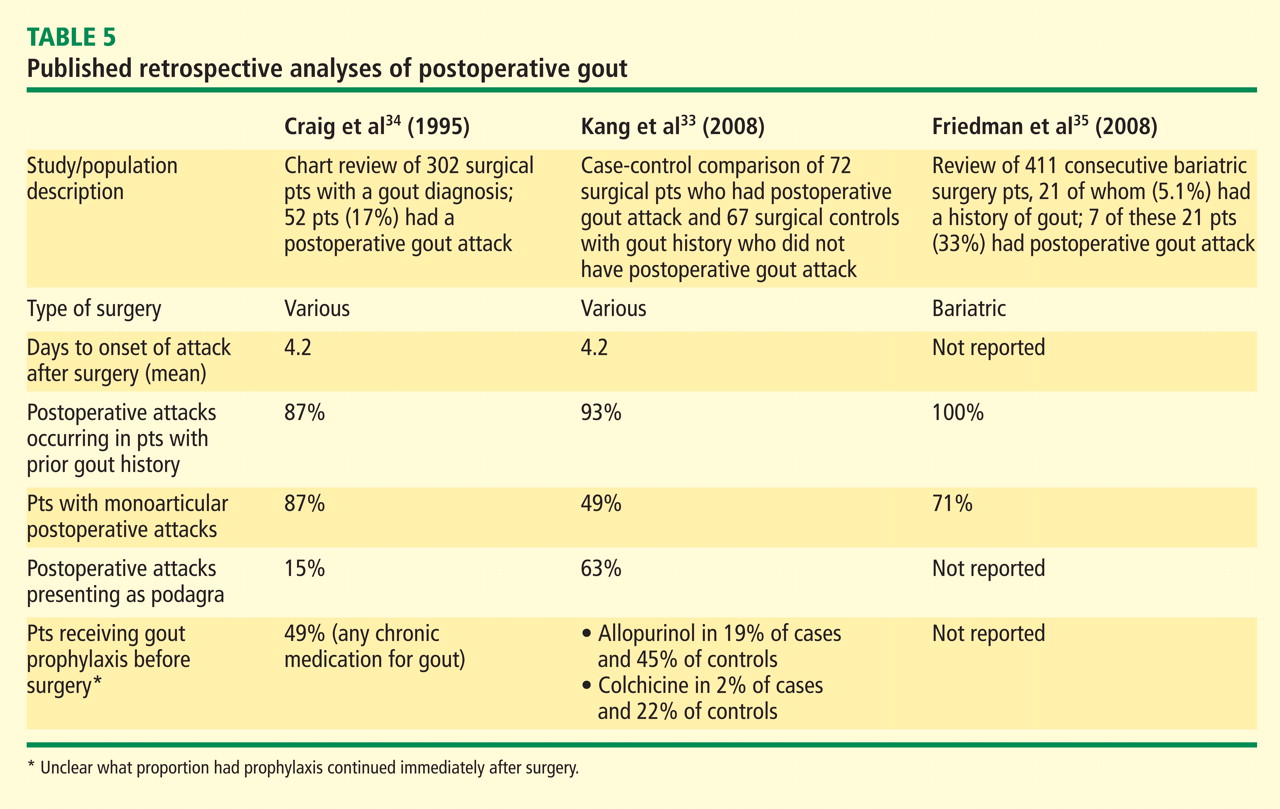
Dr. Edwards: There are certain surgeries, such as many cardiac procedures, for which the rooms are kept hypothermic, and that can be one more physiologic stimulus for an attack of gout, particularly in the body’s cooler joints. That’s a setting where podagra might be more common, whereas major abdominal surgery with a lot of bowel ischemia and lactic acidosis may render a wide range of joints more equally susceptible, although there are no data on this question.
Dr. Simkin: I’m not certain that postoperative gout is significantly different from hospitalization gout. If patients who are admitted for medical reasons—for instance, cardiac disease or pneumonia or ketoacidosis—also have an established diagnosis of gouty arthritis, they’re at elevated risk too.
Dr. Mandell: Yes, and that point—that preexisting gout is a major risk factor, whether or not it was known at the time of admission—came through from the studies Jim noted above and our experience. That piece of the history is often not obtained until an attack occurs, although more widespread use of electronic medical records may lessen this problem. It’s frustrating that attacks tend to occur about 4 days after surgery, which is typically around the time of planned discharge.
AVOIDING MOBILIZATION FLARES WHEN LOWERING URATE: START LOW, GO SLOW
Dr. Mandell: Let’s shift from this focus on the hospital setting as a precipitant for acute gout attacks and recall that the most predictable precipitant is when we pharmacologically reduce the serum urate level in an effort to decrease the total body load. As Larry noted, the down side of this very necessary intervention is that “mobilization flares” of gout are fairly predictable in this setting. While many of us have used prophylaxis to reduce the likelihood of these flares, we hadn’t had much data on duration of prophylaxis prior to some recent trials of urate-lowering therapy.36 So what are your practices now for trying to reduce the chance of acute attacks as you introduce urate-lowering therapy?
Dr. Edwards: A trigger for gouty attacks is the fluctuation of serum urate levels, be it from initiating or discontinuing urate-lowering therapy. So the approach when initiating urate-lowering therapy is to start low and go slow with your dose escalation. Most of us now buy into the notion of trying to lower the serum urate below 6.0 mg/dL for most patients, and perhaps by another 1.5 mg/dL or so for patients with bulky tophaceous disease. To get to that target, however, you should start with a fairly modest dose of whichever urate-lowering therapy you’re using—allopurinol, febuxostat, or (in rare cases) probenecid—and increase it over time. I tend to let several weeks pass between each dose escalation to ensure that the prior escalation has had time to drop the urate to its new nadir, and then I see what that level is. If it’s not at target, I will modestly escalate further.
Dr. Mandell: And we should emphasize that frequent initial monitoring of urate is appropriate to determine whether you’re reaching your target.
Dr. Edwards: That’s a critical element that isn’t done well in this country. Many studies show that regular monitoring of serum urate after initiation of urate-lowering therapy is actually a rarity. The approach to urate monitoring should be the same as for glucose monitoring with oral diabetes medications or blood pressure monitoring with antihypertensives. We should know what impact we’re having on the patient and continue to adjust the dose to find the appropriate level for the individual patient.
As with the therapies for acute gout, there is no onesize-fits-all dose regimen for urate-lowering therapy. A minority of patients will do fine on 100 mg/day of allopurinol. Most patients will still not reach their serum urate target at 300 mg/day of allopurinol, and we should consider gradual escalation to more generous doses as necessary, beyond 400 mg/day and up to the “maximum” recommended dose of 800 mg/day. Monitoring of serum urate should then continue even after we’ve found an appropriate dose to reach the target level since certain factors may change, altering concentrations of any of the urate-lowering therapies. For instance, a new drug may be added, or the patient may develop new comorbidities or a change in renal function. At this point the monitoring need not be as tight as during the initial period, but it should continue on a semiannual or yearly basis.
Dr. Simkin: I completely agree that going slow is desirable, and it’s very important for our patients to understand what we’re doing and what we’re aiming for. Every gout patient should know what his or her last serum urate level was, yet very rarely is that the case.
Dr. Edwards: Not only that, but they should know what the target level is so that they become an equal partner in their treatment plan, just as diabetics might be unsatisfied until their hemoglobin A1c is down to near 6.0%.
WHAT ROLE FOR PROPHYLAXIS DURING URATE LOWERING?
Dr. Mandell: Larry’s point that dropping the serum urate level slowly over time is less likely to induce an attack of gout is something we had suspected for a while and now have some direct trial evidence for. But no matter what we do, there’s always some likelihood of inducing an attack as we lower the urate level. So what do you do, Peter, to prevent those attacks?
Dr. Simkin: I try to get the patient on a regular dosage of colchicine prophylaxis. There’s good evidence that it’s effective. The dose is usually one or two tablets a day, largely depending on GI tolerance. It’s worth noting that more than a few of my elderly patients appreciate colchicine’s GI effects, as they provide some welcome relief from their chronic constipation. While that’s certainly not welcome by everyone, for some patients it makes colchicine prophylaxis an easier sell.
Dr. Sundy: Colchicine is my standard for prophylaxis as well. I think it’s highly effective.
Dr. Edwards: For how and when to start prophylaxis, my general recommendation is that it should precede the start of urate-lowering therapy by about 1 or 2 weeks. I think starting colchicine 2 weeks in advance is probably a good buffer for ensuring that it’s in the system. The urate-lowering therapies have very rapid onset—allopurinol will cause fluctuations in urate levels within a day or two—so the prophylaxis has to be fully on board.
Dr. Pile: Do you have a sense of how much more effective twice-daily dosing of colchicine is compared with once-daily dosing?
Dr. Edwards: I don’t have a good sense of it, but I tend to use twice-daily dosing unless renal function is a concern—specifically, if the patient’s GFR is between 50 and 60 mL/min/1.73 m2. And in such cases, you’re not really diminishing the dose with a once-daily regimen because the patient is keeping more of the drug around, in light of the renal insufficiency. That hearkens back to Peter’s earlier comments about baseline therapy with statins, some calcium channel blockers, and other drugs that will also require dose modification.
Dr. Mandell: The point is that we want to maintain a certain level of colchicine in all our patients for prophylaxis. It’s not clear that the blood level matters as much as the intracellular level, but we can’t routinely measure either. I think the answer to Jim’s question is that we don’t have data on whether once daily, twice daily, or even three times daily is the right regimen for colchicine prophylaxis; patients differ. I believe many of us start with twice-daily dosing on an empirical basis. If it’s not tolerated, typically for GI reasons, we’ll go down to once a day. If it’s tolerated but ineffective from the point of view of having flares, we may try to go to three times a day, assuming the patient’s not on other medications that would preclude that and doesn’t have chronic kidney disease or hepatobiliary disease.
WHAT ABOUT ALTERNATIVE PROPHYLAXIS OPTIONS?
Dr. Mandell: If a patient is truly intolerant of colchicine but has a reasonable GFR and no comorbidities, what about alternative prophylactic agents? The class to think about would seem to be NSAIDs, but we have no trial data to guide us with their use for prophylaxis. What’s been your experience with NSAIDs for prophylaxis of gout flares during the urate-lowering process?
Dr. Sundy: My experience is that NSAIDs are effective for this. I don’t have a notion of comparative effectiveness relative to colchicine, but I will use an NSAID in this setting when there has been GI intolerance to colchicine. The trick is the added responsibility of monitoring toxicity in terms of blood pressure, renal function, and hyperkalemia, as well as offering gastric protection, but I certainly have patients on NSAID prophylaxis.
Dr. Mandell: Do you feel compelled to use the same very high doses that we talked about for treating acute attacks of gout?
Dr. Sundy: No. I tend to use doses in more of a midrange area and make adjustments based on how the patient is doing. For example, I might increase the dose if a patient has a breakthrough flare.
Dr. Simkin: A situation where I’ve more often seen NSAIDs used for prophylaxis is in patients who have gout but are also taking NSAIDs on a regular basis for osteoarthritis. In that setting I’d be less inclined to add colchicine.
Dr. Pile: And I guess cost becomes an issue now, with the advent of branded colchicine, in a way that it hadn’t been before. At my local retail pharmacy generic naproxen costs 6 cents per 200-mg tablet, so some NSAIDs are very inexpensive.
Dr. Mandell: Colchicine used to have a similar cost invisibility as well. As Peter said before, our ability to provide cost-efficient prophylaxis is disappearing, and this may affect our ability to provide prophylaxis to some patients.
Dr. Sundy: I agree. Many times patients will stop taking drugs that are too expensive for them without telling their doctor. I sense that not using prophylaxis is a major impediment to adherence with urate-lowering drugs as well, as patients will stop those drugs because they sense appropriately that their gout has gotten worse without having been adequately educated about this risk. At the end of the day, we’re trying to eliminate gout as a medical issue for our patients, and anything that makes nonadherence more likely can be a big impediment to this goal.
Dr. Edwards: Patient education is absolutely crucial in this regard. We should step back and appreciate that these regimens can be very complicated and not terribly obvious from the patient’s perspective. First you have the disease gout, for which the treatment is allopurinol. You take that treatment and then your gout gets worse. Without good education to prepare patients for that, how can we expect them to respond, other than wanting to stop the allopurinol and probably thinking poorly of their doctor? Physicians have to educate patients on what to expect and how to stay the course. We must make clear from the start that the treatment of gout is a long-term process. We must explain that when acute flares occur the emphasis is on getting rid of the pain quickly but that the larger job of getting their disease under control by lowering their uric acid levels over time is a long haul. Both patient and physician have to be patient, go slowly, and make sure they don’t disrupt therapy just because symptoms are occurring.
Dr. Sundy: It sounds funny, but sometimes I’ve taken to congratulating patients whose gout flares after they start urate-lowering therapy. I say, “This is great—it means the drug is working, even if it doesn’t seem like it.”
SHOULD WE INTRODUCE URATE-LOWERING THERAPY AT THE TIME OF AN ACUTE ATTACK?
Dr. Mandell: Let’s consider the patient who presents with a first or second or third attack of gout but is not yet being treated for the underlying hyperuricemia. Should we introduce urate-lowering therapy at the time of the attack? After all, it’s a moment of opportunity, as the patient is “captive” and needs to be treated for his or her pain, so should we use that as a chance to also start the urate-lowering therapy that will be needed? There seem to be two schools of thought on this question. One is that if we start urate-lowering therapy immediately and drop the urate, the attack is likely to last longer. The other is that as we drop the urate by starting uratelowering therapy, we’re already treating them very aggressively for inflammation, so it may be an acceptable time to introduce urate-lowering therapy. What are your individual approaches?
Dr. Edwards: I’ve heard that argument of “we have them here now, let’s get it started.” I’d counter that when you start on this course of urate lowering, you need a very close connection to the patient during the long period of dose adjustment. Both the patient and physician need to understand that clearly. I’ve seen too many cases of a bad flare turning into a terrible flare when urate-lowering therapy is started during an acute attack. Patients are unhappy and get turned off of the idea of reintroducing allopurinol or any other urate-lowering therapy. So I don’t use that approach. Instead, I try to optimize the conditions for initiating urate-lowering therapy to minimize the chance that patients will have a bad experience. There’s no reason to rush, since most patients presenting with their first few flares have been hyperuricemic for 30 years, with urate slowly accumulating and depositing around their bodies. I’d promote an approach of letting the acute attack resolve and then getting the uratelowering therapy started about a month later.
Dr. Simkin: My approach is along the same lines as Larry’s. We all encounter gouty patients who say, “I can’t take allopurinol.” When you ask why, the most common response is, “It makes my arthritis worse.” Patients must understand that this is a hump they have to get over; we need to do everything we can to lessen that hump, and that involves good prophylaxis. But it’s also essential to educate them to expect the hump and to forewarn and forearm them in terms of how they’ll cope with it.
Dr. Sundy: A common rationale for starting urate-lowering therapy during the acute attack is that the patient may not follow up and you’ve got them on hand for intervention right now. But if follow-up is that unlikely, urate-lowering therapy is not going to be successful in any case, since frequent monitoring and dose adjustments are needed over many months. The notion that a patient can just be put on 300 mg of allopurinol and check back in 6 months is unrealistic.
Dr. Mandell: It sounds like we’re unanimous on this. One other consideration I would add is that I will avoid adding more than one drug in a patient at the same time whenever I can, to avoid confusion about which drug is causing any side effects that may develop.
REFINING THE PARADIGM: ACUTE ATTACKS AS THE ENTRY POINT TO A PROGRESSIVE DISEASE
Dr. Mandell: So our recommendation to the hospitalist who’s confronting acute gout is to treat the acute attack, start the education process, and make sure the patient understands this is a chronic disease that you’re going to make better in the hospital but which requires lifelong attention to prevent flaring up again. At that point the recommendation is to refer the patient back to the primary care physician or a rheumatologist with the understanding that the underlying hyperuricemia needs to be addressed and that the patient should make sure that happens.
Dr. Sundy: This raises some interesting handoff issues. How do we best ensure that the physician who’s going to pick up responsibility for chronic management is aware of these issues and on board with the necessary patient education efforts? There’s clearly an opportunity for doctor-to-doctor education here as well.
Dr. Pile: Yes, this is a microcosm of what we deal with all the time in the hospitalist world—safe transitions of care and trying to avoid information “voltage drop” as patients return to the care of their usual physicians.
Dr. Simkin: I think our education of both our colleagues and our patients needs to emphasize that this is not only a chronic disease but a progressive disease.
Dr. Edwards: That’s a great point, and it raises the question of the stage in the disease process where we should start urate-lowering therapy—a question without a clear-cut answer. Three decades ago, the thinking was that you should do it before the patient transitioned from acute intermittent gout to a more chronic and advanced tophaceous gout because of how terrible those joints looked and how destructive advanced gout is. Yet the destruction begins much earlier than that, and changes probably occur within the joint even before a patient has his or her first gout attack. There are soft-tissue changes around the joint as monosodium urate crystals accumulate, and the effects on bone and cartilage are occurring throughout the period of acute intermittent gout if the serum urate level hasn’t been lowered. As we’ve come to recognize this, we’ve pushed the initiation of uratelowering therapy earlier and earlier. The standard recommendation now is to start it as soon as a patient has two gout attacks within a year. Since 60% of patients have their second attack within a year of their first, that means initiating urate-lowering therapy after the second attack for about 60% of patients. An even larger majority of patients will be at that stage to qualify for urate-lowering therapy within the first few years after their initial attack.
Dr. Mandell: This is a good place to conclude, with a reminder that the acute gout attack is just one part of a chronic and potentially progressive disease. Although the acute attack is what gets the patient’s attention—and often the physician’s attention as well—it is really just the entrance into therapy for gout, a chronic disease.
- Wallace SL, Robinson H, Masi AT, et al Preliminary criteria for the classification of the acute arthritis of primary gout. Arthritis Rheum 1977; 20:895–900.
- Malik A, Schumacher HR, Dinnella JE, Clayburne GM. Clinical diagnostic criteria for gout: comparison with the gold standard of synovial fluid crystal analysis. J Clin Rheumatol 2009; 15:22–24.
- Janssens HJEM, Janssen M, van de Lisdonk EH, et al Limited validity of the American College of Rheumatology criteria for classifying patients with gout in primary care [letter]. Ann Rheum Dis 2010; Mar 16[Epub ahead of print].
- Lin Y-H, Hsu H-L, Huang Y-C, et al Gouty arthritis in acute cerebrovascular disease. Cerebrovasc Dis 2009; 28:391–396.
- Altman RD, Honig S, Levin JM, Lightfoot RW. Ketoprofen versus indomethacin in patients with acute gouty arthritis: a multicenter, double blind comparative study. J Rheumatol 1988; 15:1422–1426.
- Maccagno A, Di Giorgio E, Romanowicz A. Effectiveness of etodolac (‘Lodine’) compared with naproxen in patients with acute gout. Curr Med Res Opin 1991; 12:423–429.
- Schumacher HR, Boice JA, Daikh DI, et al Randomised double blind trial of etoricoxib and indometacin in treatment of acute gouty arthritis. BMJ 2002; 324:1488–1492.
- Rubin BR, Burton R, Navarra S, et al Efficacy and safety profile of treatment with etoricoxib 120 mg once daily compared with indomethacin 50 mg three times daily in acute gout: a randomized controlled trial. Arthritis Rheum 2004; 50:598–606.
- Shrestha M, Morgan DL, Moreden JM, et al Randomized doubleblind comparison of the analgesic efficacy of intramuscular ketorolac and oral indomethacin in the treatment of acute gouty arthritis. Ann Emerg Med 1995; 26:682–686.
- Ahern MJ, Reid C, Gordon TP, et al Does colchicine work? The results of the first controlled study in acute gout. Aust N Z J Med 1987; 17:301–304.
- Terkeltaub RA, Furst DE, Bennett K, et al High vs low dosing of oral colchicine for early acute gout flare: twenty-four-hour outcome of the first multicenter, randomized, double-blind, placebo-controlled, parallel-group, dose-comparison colchicine study. Arthritis Rheum 2010; 62:1060–1068.
- Janssens HJ, Janssen M, van de Lisdonk EH, et al Use of oral prednisolone or naproxen for the treatment of gout arthritis: a doubleblind, randomised equivalence trial. Lancet 2008; 371:1854–1860.
- Man CY, Cheung IT, Cameron PA, et al Comparison of oral prednisolone/paracetamol and oral indomethacin/paracetamol combination therapy in the treatment of acute goutlike arthritis: a double-blind, randomized, controlled trial. Ann Emerg Med 2007; 49:670–677.
- Siegel LB, Alloway JA, Nashel DJ. Comparison of adrenocorticotropic hormone and triamcinolone acetonide in the treatment of acute gouty arthritis. J Rheumatol 1994; 21:1325–1327.
- So A, De Smedt T, Revaz S, Tschopp J. A pilot study of IL-1 inhibition by anakinra in acute gout. Arthritis Res Ther 2007; 9:R28.
- Terkeltaub RA. Colchicine update: 2008. Semin Arthritis Rheum 2009; 38:411–419.
- Colcrys [package insert]. Philadelphia, PA: Mutual Pharmaceutical Company; 2009.
- Simkin PA, Gardner GC. Colchicine use in cyclosporine treated transplant recipients: how little is too much? J Rheumatol 2000; 27:1334–1337.
- Dogukan A, Oymak FS, Taskapan H, et al Acute fatal colchicine intoxication in a patient on continuous ambulatory peritoneal dialysis (CAPD): possible role of clarithromycin administration. Clin Nephrol 2001; 55:181–182.
- Rollot F, Pajot O, Chauvelot-Moachon L, et al Acute colchicine intoxication during clarithromycin administration. Ann Pharmacother 2004; 38:2074–2077.
- Alayli G, Cengiz K, Canturk F, Durmus D, Akyol Y, Menekse EB. Acute myopathy in a patient with concomitant use of pravastatin and colchicine. Ann Pharmacother 2005; 39:1358–1361.
- Atmaca H, Sayarlioglu H, Kulah E, Demircan N, Akpolat T. Rhabdomyolysis associated with gemfibrozil-colchicine therapy. Ann Pharmacother 2002; 36:1719–1721.
- Roger U, Lins H, Scherrmann JM, et al Tetraparesis associated with colchicine is probably due to inhibition by verapamil of the P-glycoprotein efflux pump in the blood-brain barrier. BMJ 2005; 331:613.
- Levaquin [package insert]. Raritan, NJ: Ortho-McNeil; 2009.
- Phelan KM, Mosholder AD, Lu S. Lithium interaction with the cyclooxygenase 2 inhibitors rofecoxib and celecoxib and other nonsteroidal anti-inflammatory drugs. J Clin Psychiatry 2003; 64:1328–1334.
- Vistide [package insert]. Foster City, CA: Gilead Sciences; 2001.
- Hynninen V, Olkkola KT, Leino K, et al Effects of the antifungals voriconazole and fluconazole on the pharmacokinetics of S+ and R−-ibuprofen. Antimicrob Agents Chemother 2006; 50:1967–1972.
- Albengres E, Le Louet H, Tillement JP. Systemic antifungal agents. Drug interactions of clinical significance. Drug Saf 1998; 18:83–97.
- 2009 Red Book: Pharmacy’s Fundamental Reference. 113th ed. Los Angeles, CA: RAmEx Ars Medica; 2009.
- Odio CM, Ramirez T, Arias G, et al Double blind, randomized, placebo-controlled study of dexamethasone therapy for hematogenous septic arthritis in children. Pediatr Infect Dis J 2003; 22:883–888.
- Hung IF, Wu AK, Cheng VC, et al Fatal interaction between clarithromycin and colchicine in patients with renal insufficiency: a retrospective study. Clin Infect Dis 2005; 41:291–300.
- Cronstein BN, Terkeltaub R. The inflammatory process of gout and its treatment. Arthritis Res Ther 2006; 8( suppl 1):S3–S9.
- Kang EH, Lee EY, Lee YJ, et al Clinical features and risk factors of postsurgical gout. Ann Rheum Dis 2008; 67:1271–1275.
- Craig MH, Poole GV, Hauser CJ. Postsurgical gout. Am Surg 1995; 61:56–59.
- Friedman JE, Dallal RM, Lord JL. Gouty attacks occur frequently in postoperative gastric bypass patients. Surg Obes Relat Dis 2008; 4:11–15.
- Becker MA, Schumacher HR, Wortmann RL, et al Febuxostat compared with allopurinol in patients with hyperuricemia and gout. N Engl J Med 2005; 353:2450–2461.
Gout is the most common form of inflammatory arthritis in men, and both gout and its root cause, hyperuricemia, are increasing in prevalence in both men and women. The result is that all medical generalists and specialists undoubtedly encounter patients with acute gouty flares in the outpatient or inpatient setting.
Gout increasingly presents in patients with a number of comorbidities, including chronic kidney disease, coronary artery disease, congestive heart failure, diabetes, and hypertension. As a result, medical management of the acute attack of gout, a notably painful and debilitating condition, is growing more complicated.
I asked three of my “goutophile” rheumatology colleagues from around the country and an infectious disease specialist/hospitalist to join me earlier this year in an unscripted roundtable discussion of the management of the acute gout attack. All have extensive real-world clinical experience, and the spirit of our conversation was clinically pragmatic. We aimed to put the relevant, though limited in number, clinical trials into a very practical perspective. We focused our conversation on the diagnosis and management of the acute attack in the outpatient and inpatient settings, essentially leaving undiscussed the long-term management of hyperuricemia—the underlying cause of acute gout attacks. The roundtable and this resulting journal supplement were supported by a CME grant from URL Pharma, Inc., the manufacturer of the branded formulation of colchicine.
I believe our discussion, transcribed and published here without external editing or input, will be of value to all practitioners faced with patients enduring an acute gout attack. We’ve added references as appropriate, and I hope the result is a balanced mix of experience-based medicine from thoughtful, seasoned clinicians with quantitative supporting data provided as available from the published clinical studies. This supplement has been independently peer reviewed by a rheumatologist content expert to exclude any content that could be perceived as unjustified bias for or against any specific therapy.
Gout is a disease that has fascinated me for 30 years, and my colleagues share my sense of wonder with the dramatic nature of the acute gouty flare (and its auto-resolution). I hope our shared interest shines through here and will assist all of us in managing patients with this chronic disorder punctuated by dramatically painful attacks.
As with all issues and supplements of Cleveland Clinic Journal of Medicine, I, as the journal’s editor-in-chief, welcome your feedback and comments.
—Brian F. Mandell, MD, PhD, FACR
HOW DO WE KNOW IT’S GOUT? WHEN PRESUMPTIVE DIAGNOSIS IS (AND ISN’T) ENOUGH
Brian F. Mandell, MD, PhD: In addressing acute gouty arthritis, let’s start with diagnosis. The gold standard for diagnosis of gout is synovial fluid aspiration and analysis for the presence of monosodium urate crystals. But what’s the practicality of aspiration and the need for synovial fluid analysis in a given patient at a given time? When is fluid analysis useful, and when is it absolutely necessary?
N. Lawrence Edwards, MD: It’s important to emphasize that synovial fluid analysis remains the gold standard of diagnosis. Ultrasonography of the peripheral joints is probably specific enough that it may soon be considered on par with fluid analysis for crystals, but we’re not there yet and it is not uniformly available. Less than 10% of patients who are diagnosed with gout have ever undergone synovial fluid aspiration, however, so presumptive, or “clinical,” diagnosis is very common.
A reliable presumptive diagnosis should be based on the image of a classic gout attack. The further an episode strays from a classic attack, the more presumptive the diagnosis is. So if a patient experiences the typical rapid explosion of symptoms from no pain to maximum pain over less than half a day, and if the symptoms are in the typical joints—the midfoot, the ankle, the first metatarsophalangeal joint (great toe)—that’s very consistent with a classic gout attack. Symptoms in the knee are more problematic because the two main conditions in the differential diagnosis of gout—septic arthritis and calcium pyrophosphate dihydrate (CPPD) crystal deposition disease (pseudogout)—also commonly affect the knee.
If the patient has had acute attacks that have self-resolved or gone away with therapy over a 5- to 8-day period, that too is a profile that’s typical enough for a presumptive diagnosis of gout, especially since septic arthritis doesn’t tend to do that. Pseudogout usually is a bit slower in onset. It can look virtually identical to gout, but the affected joints often aren’t the same and the attacks tend to last for weeks rather than days, which ultimately simplifies the differential diagnosis.
The setting where synovial fluid analysis is absolutely essential is when a septic process is suspected. If there’s any nagging concern that an infection might be present, the clinician should aspirate the joint or refer the patient to someone who can. The features of a septic process may include joint swelling, rigors, and fever, but all of those can be seen in gout, so they are not distinguishing.
Dr. Mandell: John, how comfortable can you be in making the distinction between crystal-induced arthritis versus infection at the start of a given attack?
John S. Sundy, MD, PhD: Larry’s point about the features typical for gout is key, but I also consider the patient’s comorbidities and general situation. For instance, if the patient is a healthy middle-aged man with no relevant past medical history, it’s easier to feel comfortable with a presumptive diagnosis of gout. But if I’m dealing with a hospitalized patient who’s had invasive procedures and has risk factors for bacteremia, it’s imperative to get synovial fluid to rule out a septic joint. There is, of course, a continuum between these two examples. It’s important to have a construct of what typical gout is. When a case is close to that construct, it’s reasonable to work with a presumptive diagnosis; the further a case is from the construct, the more important it is to get fluid. From there it’s just a matter of overcoming the practical issues that a lot of clinicians contend with in obtaining the aspiration and fluid analysis.
Although it’s fine to think about a presumptive diagnosis and initiate treatment accordingly, it’s still incumbent upon the clinician to strive to get a firm diagnosis at some point so that the patient and physician can be 100% confident in the diagnosis.
Peter A. Simkin, MD: When we focus on the initial diagnosis we may overlook the fact that a chronically gouty joint is more susceptible to infection, so that patients who have well-established gout may still require joint aspiration to exclude acquired infection. I agree that the knee is a major site to focus on, and the olecranon bursa is another common site of infection in gouty patients.
Dr. Mandell: That’s convenient, since both of those areas are easily aspirated by rheumatologists and nonrheumatologists alike. So I’m hearing a consensus that we look at the entire clinical picture, including the historical features and the location of symptoms, to assess whether an acute attack is more likely to be gout or infection. I would add that crystal disease, especially gout, is far more common than infection, and infection is particularly unusual in the midfoot.
No objective analysis has shown that any single feature, such as leukocytosis or fever, is reliably indicative of infection versus crystal disease. Yet when we look at the entire pattern, if there isn’t significant white blood cell elevation, if there aren’t rigors, if there isn’t significant fever, and if the patient has a history of self-limited attacks, that points to a clinical diagnosis of gout that we probably can be comfortable with. That said, joint aspiration is still required if there is not a rapid response to treatment.
There are settings, as John noted, where vigilance for infection is particularly indicated, notably in hospitalized patients. Jim, as a hospitalist, you may be less apt than us rheumatologists to stick a needle into a joint. What’s your comfort level in assessing the hospitalized patient with sudden eruption of a red, swollen, painful joint?
James C. Pile, MD: I agree that the setting is very important. Let’s say I have a patient with a well-established history of gout to whom I’ve been giving aggressive diuresis for heart failure exacerbation for 2 or 3 days. If he develops a hot, swollen ankle that’s consistent with his prior episodes, I won’t typically feel obligated to stick a needle in that joint, particularly not in an ankle, for which I’d want to consult a rheumatologist in any event. If it’s a patient who underwent cardiac catheterization or other instrumentation a couple of days ago, and if this patient is febrile and has sudden eruption of a hot, swollen knee, that’s a different story. Aspiration is absolutely required in that patient, who needs to be treated as having septic arthritis until proven otherwise. But there is a lot of gray area between these two scenarios.
Dr. Mandell: Speaking of willingness to aspirate joints, there’s a concern among nonrheumatologists about putting a needle into a joint that appears to have cellulitis over it. This can stand in the way of appropriate diagnosis since the response to crystals in a joint can produce a response that looks like cellulitis. If my examination suggests joint involvement, a cellulitislike appearance does not stand in the way of aspirating the joint or bursa, which might reveal a closed-space infection.
Dr. Edwards: One population that should make us nervous any time they get an acutely swollen joint are organ transplant recipients. When these patients get gout, it’s often atypical. Not only are they usually on cyclosporine, which can cause hyperuricemia and accelerated gout, but they’re also often taking corticosteroids, which dampen the acute symptoms of gout. So whether the acute swelling is caused by gout or infection, it may not appear to be quite as profound as it actually is. Additionally, these patients may not get fever or rigors. Despite all this, they are at high risk for infection, in light of their immunosuppression. When any transplant recipient presents with acute monoarthritis, the burden of proof is on us to show that it’s not an infected joint.
Dr. Mandell: I’d add that Larry’s caveat extends to patients on any immunosuppression, including the anti–tumor necrosis factor agents, and we have seen both infection and gout in these patients.
Dr. Simkin: It’s also worth noting that joint aspiration can be therapeutic. The exquisitely painful joint often is so painful because of distention of the joint capsule. If the joint is tapped, the capsule becomes decompressed and the needle track may leave a vent through which it can remain decompressed.
Dr. Edwards: Pointing out the therapeutic benefit of joint aspiration may even be one way to get patients to allow us to put a needle into an excruciatingly painful joint. So while we advocate joint aspiration as a diagnostic procedure, often an effective approach is to explain to patients that if you tap the joint, you can treat their pain much more rapidly. Instead of having another 18 to 36 hours of pretty significant pain even on the best oral treatments, with aspiration patients might be relatively free of pain within 18 to 24 hours.
Dr. Mandell: So I’m hearing that a history of prior events consistent with attacks of crystal-induced arthritis suggests that an acute flare is likely to be another attack of crystal disease. Almost all patients with acute postoperative flares have a history of gout, though that history rarely is noted in the chart and is usually obtained later. The problem with this reliance on history is that if it’s a previously damaged joint, it may have altered microvasculature and may be more prone to infection, as Peter noted. The literature on coexistent infection in crystal disease is small, which attests to its relative infrequency, but we need to be wary of this potential for coexistence, particularly in patients at risk of infection, such as transplant recipients and those with recent infection elsewhere and bacteremia. Acute joint swelling in such patients calls for arthrocentesis.
Dr. Edwards: One last criterion for when to do a joint aspiration is when a patient with well-established gout tells you an attack is different from previous attacks. If a patient says, “This isn’t like my previous gout attacks,” that’s a red flag that usually merits joint aspiration.
Dr. Mandell: We’d be remiss if we didn’t mention the longstanding “preliminary” criteria for the diagnosis of acute gout published by the American Rheumatism Association (now the American College of Rheumatology) in 1977.1 They’ve been used for clinical research studies and also applied in principle to clinical practice. There have been a couple of recent papers examining the limitations of these criteria. How do you use these “criteria”?
Dr. Edwards: Their biggest drawback is that they’re cumbersome. They also have recently been shown to lack specificity and, to some degree, sensitivity.2
Dr. Mandell: Testing of the ACR criteria has shown that we’ve had unfounded confidence in using them in practice. As you said, their sensitivity has been reported to be 80% and their specificity even lower, at 64%.3 So even a panel of clinical indicators of gout cannot be relied on in a setting where we have concern about the possible alternative diagnosis of infection.
CAN TRIALS OF SPECIFIC THERAPIES OFFER DIAGNOSTIC UTILITY?
Dr. Mandell: What about the response to anti-inflammatory therapy? Can we use the specificity of response to a drug like colchicine to determine whether an attack is more or less likely to be attributable to crystal disease?
Dr. Simkin: It’s certainly helpful, but entities other than crystal disease can respond to anti-inflammatory drugs, of course. For instance, some people with paraneoplastic problems will have acute arthritis that goes away with anti-inflammatory therapy. So I think it’s more a matter of how fast and complete the response is rather than whether there is a response.
Dr. Mandell: Consider the cases you’ve seen that ultimately turned out to be septic arthritis. What was the initial response in those cases to either nonsteroidal antiinflammatory drugs (NSAIDs), colchicine, or corticosteroids, and what conclusions can you draw from those cases?
Dr. Sundy: In my experience, signs and symptoms are often more lingering in these infectious cases. I think it’s fair to generalize that the response is not as rapid as with a typical gout flare. This underscores the importance of follow-up. If a patient presents to the emergency room and is treated and released, the critical question is how careful the follow-up is going to be at 24, 48, and 72 hours to see if improvement and resolution are occurring as expected. If you’re going to proceed on the basis of a presumptive diagnosis, there must be planned and frequent follow-up.
Dr. Edwards: I don’t think anyone thinks that a patient with an acute septic joint is going to get much improvement at all from oral colchicine or much more than a little fever reduction from NSAIDs without parenteral antibiotics. That joint is going to progress toward destruction within days. So John’s point is crucial that any time you make a presumptive diagnosis of gout, it’s incumbent on you to make sure that the pattern thereafter is one of resolution over time—not worsening—on the medications you use.
Dr. Pile: Even with corticosteroids, which are probably most likely to mask the clinical picture, in most cases I think it’s still fairly easy to sort things out based on response in the first 24 to 48 hours after initiating therapy. I don’t think patients with bacterial septic arthritis are going to remain better on corticosteroids.
Dr. Mandell: The time frame is crucial. Therapy for acute arthritis is ideally started fast, and patients frequently will be treated initially with an anti-inflammatory—in the community, probably more often with an NSAID than with colchicine or a steroid. Even if they actually have a septic joint, they may initially get a bit better on the anti-inflammatory, perhaps from the analgesic or antipyretic effects, but the benefit will clearly plateau and the attack will worsen again. So the period for peak vigilance is probably 1 to 2 days after therapy is started; if improvement is not maintained in this period, you should be circumspect regarding diagnosis of crystal disease and you must aspirate, or reaspirate, the joint.
Let’s explore specificity a bit further. Diagnostic specificity has historically been attributed to colchicine. People have even suggested that a diagnostic colchicine trial can be useful. How does that jibe with your experience with response to colchicine therapy in patients with entities other than gout?
Dr. Edwards: The main disease in the differential diagnosis of gout other than a septic process is pseudogout, or CPPD arthropathy, and patients with pseudogout may also respond to colchicine.
Dr. Simkin: Another entity that reportedly responds to colchicine is sarcoid, particularly sarcoid of the ankle.
Dr. Edwards: In fact, colchicine has been tried for treating the acute inflammatory symptoms of many of the diseases we see as rheumatologists. Some people swear by it, while others believe it doesn’t do much for these conditions. In any case, I don’t think its specificity for gout is very strong, and we have no basis to say that response to colchicine should serve as a diagnostic test.
Dr. Mandell: I agree, and the literature over the past few years on colchicine for treatment of acute and chronic pericarditis further argues against specificity of this drug for crystal-induced inflammation.
SERUM URATE LEVEL IS UNRELIABLE FOR DIAGNOSIS
Dr. Edwards: While we’re discussing diagnosis, nothing’s been said about serum urate levels, which I think many primary care physicians rely on heavily in the diagnosis of gout. We need to underscore that a lot of people who are hyperuricemic will never develop gout. At the same time, there’s also the phenomenon during an acute attack in which an acute uricosuria accompanies the initial inflammation, causing serum urate values to fall from what would otherwise be a high baseline to a level that looks normal. These declines may be between about 1.5 to 2.5 mg/dL, so that a patient presenting to the emergency room with a serum urate of, say, 7 mg/dL might actually have a baseline chronic level of 9 mg/dL.
Relying too heavily on serum urate levels can be misleading in either direction: someone with joint pain with serum urate elevation may be diagnosed with gout inappropriately, whereas someone who comes in with a gouty attack who has a serum urate level in the normal range may be thought not to have the disease.
Dr. Mandell: The fact that urate level didn’t come up in a conversation among rheumatologists with an interest in gout testifies that none of us uses serum urate in the diagnosis of acute arthritis. Your point is well taken, though, that serum urate is used for this purpose in the community but shouldn’t be, especially not in an acute setting.
Dr. Simkin: Indeed, I’ve seen a serum urate of 3.7 mg/dL during an acute, crystal-confirmed gout attack in a patient who was not on urate-lowering therapy.
Dr. Mandell: There’s also the issue that laboratory-defined normal levels of serum urate are not the same as biologically “normal” levels in the context of urate deposition. Levels that are in the normal range in the laboratory clearly can be above the saturation point of urate in physiologic tissues, which is about 6.7 mg/dL.
Dr. Simkin: Plus, many labs report their normal range as being up to 8 mg/dL, yet most gouty arthritis in the community probably occurs in patients with urate levels below 8 mg/dL, and this misstatement of normality certainly deserves attention.
Dr. Sundy: The issue of serum urate further underscores the importance of looking at gout as a longitudinal condition and not just as an acute episodic one. We would never treat and release a patient who came in with a severe hypertensive episode without insisting that further follow-up was indicated for the hypertension. Similarly, for a person who presents in the emergency room with gout there should be a longer-term strategy to make sure the patient understands the importance of followup to address the underlying hyperuricemia.
Dr. Mandell: Yes, there is a challenge with the system of care; the providers faced with the acute attack are not the ones who will ultimately be treating the disease and its associated comorbidities.
GENERAL APPROACH TO THE ACUTE GOUT ATTACK
Dr. Mandell: Let’s return to the patient who presents with an acutely swollen, painful joint. Let’s say gout is diagnosed with confidence, supported either by synovial fluid analysis or by the overall clinical details. What are your general considerations, Jim, for initial treatment in the hospital?
Dr. Mandell: We’ll come back and talk about each of these drug classes specifically, but what do my fellow rheumatologists tend to reach for as first-choice therapy?
Dr. Simkin: My thinking is very similar to Jim’s. As far as NSAIDs are concerned, so many of our patients have significant renal compromise that it is absolutely mandatory that we know what a patient’s renal function is before we treat acute gout with NSAIDs. I’ve seen more catastrophes from the use of NSAIDs in patients with renal compromise than from any other gout treatment scenario. So I probably wind up using steroids more often, but in the hospital setting you often have to deal with a surgeon who doesn’t want to use steroids. In such a case, if the patient also has renal problems, an agent that has entered the picture in our hospital is the interleukin-1 (IL-1) receptor antagonist anakinra, given in daily subcutaneous injections of 100 mg. When a patient is hospitalized for gout, it’s his severely painful joint that’s keeping him from going home, and in this situation anakinra in fact becomes a relatively inexpensive option, despite its absolute cost, when compared with the cost of the hospital bed.
Dr. Mandell: I think that’s the first time I’ve heard “anakinra” and “inexpensive” used in the same sentence.
Dr. Simkin: Of course biologics are terribly expensive, but so is hospitalization, and hospitals are bad places. We want to get our patients home, and in our experience this has been a very useful way to make that happen sooner.
Dr. Pile: I agree that an emphasis on hospital throughput is incredibly important. For the hospitalized patient with gout that’s preventing ambulation, that’s the issue that must be addressed before discharge is possible. Certainly the agent with the fastest onset of action is going to be very attractive.
Dr. Edwards: All of these agents for acute gout have a relatively fast onset of action if they’re given early in the disease process. Once you get out to a day and a half from the onset of symptoms or beyond, you’re fighting an uphill battle in terms of making a difference in the natural course of the attack. I believe that’s true of all three of the medication classes that are typically used.
My approach to the initial therapy choice is highly individualized, depending on what the patient’s been on before. If they’ve been on maintenance colchicine to prevent flares and then they flare, I usually won’t use colchicine for the acute attack; I will go with a steroid or an NSAID. If they’ve been on NSAIDs as preventive therapy and they flare, I might try low-dose colchicine for the flare or use steroids. In cases of a prolonged course, where the patient is in the hospital and it’s 3 or 4 days since symptom onset and a steroid taper or a trial of colchicine has failed, I’ve been very impressed with the ability of anakinra to suddenly bring the attack to a halt. Like Peter, I am on the cusp of looking at acute treatments a little differently, although there still aren’t a lot of data on IL-1 inhibition in this setting.
Dr. Sundy: There are data showing that a gout flare adds about 3 days to the hospital stay,4 so that’s a huge burden that intervention with IL-1 inhibition can really help to address.
Dr. Mandell: I think we’ve seen a trend over time toward corticosteroid therapy becoming more common, particularly in hospitalized patients. I think that’s partially related to more widespread use of appropriate deep vein thrombosis (DVT) prophylaxis, which means that more patients are on anticoagulant therapy, which is one more reason to shy away from NSAIDs, particularly the nonselective ones, in the hospital setting. Do you sense that trends in the outpatient setting have shifted, or do most clinicians still reach for NSAIDs?
Dr. Sundy: I think most people still reach for the NSAID, but it really depends on the comorbidities. I sense we’re seeing chronic kidney disease in a greater proportion of patients, and that’s probably creating a shift toward a bit more corticosteroid use. I like to reach for an NSAID as first-line therapy, but we really have to understand our patient’s overall comorbidity profile, including renal function, before doing so, as Peter said.
Dr. Edwards: I think NSAIDs are still the most commonly used acute treatment for gout. I used to calm myself when I used them by saying, “It’s only a 7- to 10-day course; how much trouble can you get the patient into?” Well, in that short a time I’ve had patients tip over into congestive heart failure because they had some renal decompensation beforehand and then had another 20% or 25% of their renal function knocked out with the NSAID, and they’d have extra sodium retention. I’ve seen GI bleeds develop in patients over that short a time. I don’t prescribe nonselective cyclooxygenase (COX) inhibitors anymore without also giving a proton pump inhibitor for stomach protection. I think that’s becoming a standard of how to use NSAIDs among rheumatologists, and it’s hard to get that stomach protection up and running in the very short time frame of an acute gout attack. So I’m personally tending away from NSAIDs; I use steroids more often and then the IL-1 inhibitor for special cases.
Dr. Mandell: Studies assessing the gastric risk of NSAIDs have shown that a GI bleed can be induced in as little as 2 or 3 days. I agree that there is a trend, at least among rheumatologists, to use a proton pump inhibitor when initiating outpatient NSAID therapy. This combination may still be cost-effective for many patients, as it can speed the return to their usual activities. But even in the outpatient setting steroids are becoming more common than they used to be.
DOSING FOR THE ACUTE ATTACK: BE AGGRESSIVE FROM THE START
Dr. Simkin: Whatever dose of steroids we use in the outpatient setting, I think it’s highly desirable to divide it. Rheumatologists are taught to use a single daily steroid dose in the morning, primarily to spare the adrenal glands. While that makes sense in patients on long-term steroid therapy, such as for lupus, it doesn’t necessarily make sense in gouty arthritis. I’ve seen patients whose gout flared up every night despite taking sufficient prednisone; when they divided the dose, their gout was controlled.
Dr. Mandell: I would generalize that further to stress the importance of using an adequate and aggressive dose of either steroids or NSAIDs for treating acute gout. A common mistake I notice in the community is the use of too low a dose of either steroids or NSAIDs. We should treat aggressively from the start with a full dose of these agents.
Dr. Edwards: Even more than the usual full dose, I would say. Many generalists have a concept of what an analgesic dose of an NSAID is, which is pretty low—in the case of ibuprofen, perhaps 400 mg three times daily. An anti-inflammatory dose is higher—perhaps 800 mg three times daily for ibuprofen. Most of us who’ve used NSAIDs for gout realize that we have to get even a little bolder than that from the start. It can be hard to convince some generalists to exceed what has been their ceiling of comfort for an NSAID, but doing so for the first 24 or 36 hours is important to getting the gout attack under control. Of course, the need for such aggressive dosing is all the more reason to make sure not to use an NSAID in a patient with significant renal disease or other comorbidities for which NSAIDs are problematic.
Dr. Mandell: And all the more reason to provide gastric protection.
Dr. Sundy: I would add that if a clinician is not comfortable using doses that high, that may be a good reason to think about choosing a different initial therapy, be it colchicine or steroids (if the reluctance is with NSAIDs) or NSAIDs (if the reluctance is with steroids).
ACUTE ANALGESIA: ANYTHING MORE THAN AN ADJUNCT?
Dr. Mandell: What about treating gout with acute analgesia alone? Consider a setting where you may want to avoid a drug with anti-inflammatory or antipyretic effects, perhaps in the postoperative period or when coexistent infection is a concern and you want to monitor the fever. Can you get by with using narcotic analgesia alone?
Dr. Sundy: I’m not impressed with that approach. Narcotics can play a role in managing a patient’s pain, but a narcotics-only approach will allow the flare to linger and not address complications of the flare or back-to-work issues. I view analgesics as cotherapy as opposed to single-agent therapy.
Dr. Mandell: Is narcotic analgesia effective even at treating the pain acutely?
Dr. Sundy: No. Gout pain is an exquisitely inflammatory pain, and the key is to tackle that inflammatory response. Analgesics are really just an adjunct.
Dr. Simkin: I totally agree, but they’re an important adjunct that we often overlook.
Dr. Edwards: I’ve been singularly unimpressed with narcotics. Gout is a cytokine-driven process that is intensely inflammatory. It’s like a lot of other types of pain that don’t respond fully to narcotics, such as herpetic neuralgia or uterine pain. Perhaps the benefit of narcotics in gout is their soporific effect—patients sleep through their attack. Yet if you touch the gouty joint or the patient moves it, the pain is just as intense as if the patient weren’t taking anything. So I don’t know that narcotics add anything unless the patient has other causes of pain. I don’t use them at all.
Dr. Sundy: I tend to make the option available to the patient. I say, “Here’s what we need to do to knock down this flare. And here’s something additional you can try for pain; we can see if it helps.” But I’ll emphasize the importance of improving the inflammatory piece.
Getting back to the postoperative setting, we should recognize that a strategy to just ride out a perioperative gout flare with an analgesic medication, perhaps because of concern about the effect of corticosteroids on wound healing or infection, can carry important risks. A patient treated that way will end up bedbound at precisely the time you want him or her to be moving around, so there’s now the risk of postoperative pneumonia and other complications that won’t get documented as complications of gout even though they actually are.
Dr. Mandell: Not to mention the untreated postoperative fever from the crystal-induced attack, which then leads to a work-up looking for DVTs, more blood cultures, and more radiographs, all the while extending the hospital stay and wasting resources. Jim, as a hospitalist, do you still see narcotics used initially as the primary treatment for gout flares in the hospital?
Dr. Pile: It depends on which hat I’m wearing. If I’m the hospitalist and the patient’s on my service, usually my antennae are up for the appearance of gout in the hospital, and the house staff may or may not recognize it if the process starts overnight. When I’m serving as the medical consultant, I sometimes encounter cases where the surgeons don’t recognize a gouty flare when it first appears, and I’ve had multiple patients in whom gout-induced postoperative fever triggers a consultation. In the latter case, it’s not uncommon to see narcotics being used in an attempt to treat gouty pain.
RELATIVE DRUG EFFICACY FOR ACUTE ATTACKS: HOW MUCH DO WE KNOW?
Dr. Edwards: If they’re given early, all of them have good—if not necessarily fast and ideal—effectiveness. The recent AGREE trial of colchicine (Acute Gout Flare Receiving Colchicine Evaluation) assessed pain reduction at 24 hours in patients randomized to three treatment groups: a low-dose colchicine regimen consisting of three 0.6-mg tablets (1.2 mg initially, followed by 0.6 mg 1 hour later); a more traditional high-dose colchicine regimen consisting of eight 0.6-mg tablets (1.2 mg initially, followed by 0.6 mg every hour for 6 hours), and placebo.11 Response, defined as a 50% or greater reduction in pain at 24 hours without rescue medication, was reported in 37.8% of patients in the low-dose colchicine group, 32.7% of patients in the high-dose colchicine group, and 15.5% of placebo recipients.
This is an excellent study that should finally take highdose colchicine for the treatment of acute gout off the table since it offered no improvement in efficacy but was significantly more toxic. However, while 50% improvement in 24 hours is a hard target to achieve, the fact that barely more than a third of subjects hit the target means we should still be looking for more effective approaches.
Dr. Simkin: Similarly, in the first controlled study of colchicine in acute gout, published by Ahern et al in 1987,10 23% of colchicine recipients noted a 50% reduction in their pain at 12 hours after starting treatment. That leaves more than three-quarters with little or no response within 12 hours, and gout is one of the most painful conditions we treat. We’d very much like to help our patients sooner than that. So while there are no head-to-head data comparing colchicine versus NSAIDs versus steroids, my impression too is that oral colchicine is a second-line choice for the acute gout attack.
Dr. Sundy: When we try to understand this literature, it’s important to look closely at how patients were ascertained. The AGREE trial evaluated patients who were enrolled when they were asymptomatic; they were given study medication and instructed on how to start it once a flare began. In contrast, most of the well-controlled NSAID trials captured patients as they came in with a flare, and they had to have had the flare no longer than 48 hours. I believe there’s a big difference between having had a flare for 12 hours versus 48 hours—and even as long as 72 hours in some corticosteroid trials. The longer the clock has been allowed to tick before treatment is started, the harder it is to achieve rapid symptom reduction.
Dr. Mandell: Obviously it’s difficult to compare between studies, but it’s interesting that in a study of the COX-2 inhibitor etoricoxib versus indomethacin,7 one of the largest studies of NSAID therapy for acute gout, approximately one-third of patients taking etoricoxib reported no pain or mild pain within 4 hours. This very rapid pain control is what we really want, since pain is the concern, along with eventual complete resolution, of course. So there really may be value in having something that has analgesic as well as anti-inflammatory effect, to Peter’s earlier point. Tackling both the pain and the inflammation that’s causing the pain certainly makes sense.
Dr. Edwards: Yes, and that etoricoxib study was designed the way people often are treated, with the unfortunate delay before the doctor is seen and the medication is started. The AGREE trial is how I hope everybody would be treated regardless of what they’re treated with. We’re probably never going to see a good head-to-head trial among the three main types of drugs we use for acute gout—the unpredictability of flares makes it extremely difficult.
Dr. Pile: I’m curious whether or not you, as rheumatologists who specialize in gout, are surprised by the results of AGREE. My experience with colchicine over time has been more favorable than what’s suggested by AGREE.
Dr. Edwards: How do you use the colchicine?
Dr. Pile: For more than a decade I’ve been using 0.6 mg three times daily. I thought that practice was unusual until I recently read the AGREE study and realized that a lot of you have been using that regimen for a while.
Dr. Mandell: Some gout patients will take a colchicine tablet when they feel the wisp of an attack coming on and it immediately will stop the attack. That gets to the lore that if you treat a flare very early on, you may be able to abort and treat an attack very quickly. Years ago, however, I was involved in an analysis of 100 patients treated with intravenous (IV) colchicine, and we found that treatment response was unaffected by whether patients were treated early or after a delay of 48 hours. I believe colchicine behaves like a different drug when given IV—and the IV form has since been withdrawn from the market for safety concerns—but this underscores that individual responses to various agents are quite unique. I think there are some patients who are exquisitely sensitive to colchicine, while others are exquisitely sensitive to an NSAID, and so on. This means we’d need a very large sample size to tease out that variation in a trial, and that’s not likely to happen.
Dr. Edwards: I too have had gout patients who tell me they get this premonition of an attack—a feeling that “something just isn’t right”—hours before they actually feel any pain from the attack. A lot of them tell me they’ve learned over time that if they take a colchicine tablet when they get this premonition—or an extra colchicine tablet, as some are on colchicine maintenance therapy (typically one or two tablets a day) plus whatever other background therapy they’re on—they don’t get the flare or the flare is diminished.
Dr. Mandell: I’d like to wrap up this portion by getting your sense of whether there’s a difference in efficacy for treating acute attacks among the drug classes we’ve discussed.
Dr. Edwards: I believe the IL-1 inhibitors are probably the most potent agents for aborting a gout attack, followed by steroids, which I think have a leg up on both colchicine and NSAIDs. The latter two options probably are equally effective in aborting an acute attack.
Dr. Sundy: Yes, I would rank them the same way.
THE ROLE OF COMORBIDITIES IN THERAPY CHOICE
Chronic kidney disease
Dr. Mandell: Let’s get more specific about comorbidities and therapy choice, starting with chronic kidney disease. What’s the threshold at which renal compromise starts to influence your choice of therapy for an acute attack of gout?
Dr. Sundy: I have a very low threshold for avoiding NSAIDs in that setting, mainly because when I want to use NSAIDs, I want to use them at a very high dose, and even short-term use of high-dose NSAIDs can reduce creatinine clearance. So I would certainly avoid NSAIDs when the patient’s creatinine clearance is less than 60 mL/min, and I’d be even more cautious if the patient had underlying hypertension or congestive heart failure.
Dr. Mandell: So even with the reversibility of almost all of the NSAIDs’ renal effects, you tend to stay away from them in that setting?
Dr. Sundy: Yes.
Dr. Edwards: I’m less guided by a single creatinine clearance level. I tend to look at patients in light of the 20% to 25% reduction in the glomerular filtration rate (GFR) that most NSAIDs will cause, even when used acutely. Though the NSAID effect is reversible, if that GFR reduction is going to make a difference to other compensated systems, primarily the heart, then the NSAID should be avoided.
Dr. Simkin: It’s my understanding that plasma flow to the kidney becomes prostaglandin dependent with renal insufficiency, and when we use antiprostaglandin agents we get into trouble on a mechanistic level. I’m not sure of the exact point at which that occurs, but I think we all recognize that serum creatinine is a fairly weak indicator of which patients have some limitation.
Dr. Mandell: So we agree on the need to be very cautious with the use of NSAIDs, recognizing that in the acute setting there is reversibly depressed renal blood flow from NSAIDs. The decreased blood flow will get better, but there’s the issue of what happens during fluid retention when there is coexistent congestive heart failure, diabetes, and chronic kidney disease—you may also occasionally bump up the potassium level. In general, most rheumatologists shy away from selective or nonselective NSAIDs in the setting of chronic kidney disease. Is that consistent with the use you see in the hospital, Jim?
Dr. Pile: It is. I would add that if patients are on certain other medications—especially angiotensin-converting enzyme inhibitors, angiotensin receptor blockers, or beta-blockers—there may be additive negative effects because of the potential for hyperkalemia if an NSAID were added to the mix.
Dr. Mandell: I would add to that list of concerning medications aminoglycosides, cyclosporine, and other nephrotoxic agents.
Diabetes
Dr. Mandell: What about diabetes? How does a patient’s diabetes enter into your choice of acute gout therapy in the outpatient and inpatient settings?
Dr. Edwards: Diabetes is especially problematic because it significantly affects two of our treatment choices—steroids and NSAIDs. The steroid doses needed to effectively treat an acute gout attack have a pretty profound effect on glucose levels in diabetic patients. That’s always of concern to me, and it’s certainly of concern to my patients with diabetes who are vigilant about monitoring their glucose levels, which may spike from 130 or 140 mg/dL to values in the high 200s. These elevations will last for the 7 to 10 days of treatment, and the longterm adverse effects of that period of hyperglycemia are not clear. If we put these patients on short courses of corticosteroids, perhaps we should be treating their diabetes more aggressively during that period, but I don’t think we have a history of doing that.
Dr. Pile: It’s very difficult to do in the short term. That’s especially the case with the diabetic patients I see as a hospitalist at a safety-net hospital. Many of my patients don’t have good glucose control at baseline: their levels of 250 mg/dL may go to 400 mg/dL or higher with acute corticosteroid therapy, so that’s clearly a problem.
Dr. Mandell: I try to avoid giving corticosteroids in the outpatient setting to a diabetic patient who is being maintained on oral hypoglycemic medications, particularly if efforts are being made to avoid more aggressive diabetes therapy. Steroids may be an option, though, for a patient who is on insulin, uses a pump, and is very comfortable measuring and managing his or her glucose at home. In the hospital setting, I’m more comfortable using corticosteroids in diabetics because I have much better control over their glucose; I can just increase the basal and premeal coverage.
Dr. Pile: I agree. With vigilance I can control almost anyone’s blood sugar in the hospital, but when patients leave the hospital on steroids, it becomes much more problematic.
Dr. Mandell: Right. You don’t know what’s going to happen when they go out and liberalize their diet on top of the steroids, because often it’s the postprandial surge that’s exacerbated by steroids.
Dr. Sundy: It can be very helpful to know at baseline what a patient’s spot blood sugar is before using a steroid, just as it’s important to know what the creatinine is before considering an NSAID. In the setting of an acute flare, the patient might already be quite hyperglycemic, and many of these patients probably don’t have the tools to manage this at home, so careful follow-up—coming back the next day for at least a blood sugar check—is critical to the proper care of these patients.
Dr. Mandell: When we talk about steroids we tend to think mostly about prednisone, but Peter mentioned injectable steroid. What about injectable adrenocorticotropic hormone (ACTH), which goes in and out of vogue as an agent to treat acute gout? What is your experience with using ACTH injections, specifically related to the issues of diabetes and heart failure?
Dr. Edwards: Before it got reformulated about 3 or 4 years ago and its price went way up, injectable ACTH was my drug of choice for treating acute gout. I had hardly any failures on it over the 10 to 15 years when it was my main means of treating acute gout. I would typically use 80 IU of ACTH gel, given subcutaneously, and the response within 12 hours was quite dramatic. About one in four patients would require a second injection at that 12-hour point, but almost everyone was symptomfree at 24 hours. This response was seen even in patients who were on fairly high-dose chronic steroids already, such as transplant patients.
This suggested that a mechanism other than just adrenocortical stimulation explained the benefit from ACTH. In fact, a lot of data have emerged suggesting that ACTH has a specific effect that is not related to adrenocortical stimulation.
Ten years ago an injection of ACTH gel would cost about $3 or $4, whereas today it costs $2,000 or more.29 I don’t think it’s justifiable at that cost.
Dr. Mandell: There’s also the issue of fluid retention with ACTH, and the higher doses, which seem to be more effective, are more likely to exacerbate congestive heart failure, which has to be a concern if you go this route.
Coronary artery disease
Dr. Mandell: Atherosclerosis, diabetes, and chronic kidney disease are increasingly recognized in the gout population, and coronary artery disease links with all of those. Does the presence of coronary artery disease enter into your choice of agents for a patient with an acute attack of gout?
Dr. Edwards: It doesn’t have as big an impact as diabetes does. To me, the priority in this setting is resolving the acute attack as quickly as possible, as I’ve seen my share of patients for whom the acute gout attack has been so painful and stressful that they’ve developed angina at the same time. So I might use steroids in patients with coronary disease because I’ll want to go with what’s likely to be most efficacious for quick resolution of the attack even though steroids may increase glucose and blood pressure and thus raise fluid retention as an issue.
Dr. Mandell: The presence of coronary disease implies the use of low-dose aspirin therapy. Despite the slight elevation in serum urate from low-dose aspirin, I recommend that my gout patients continue this for coronary protection. But even low-dose aspirin raises concerns of GI safety and even possible drug interactions with ibuprofen that could reduce the aspirin’s efficacy. Does this lessen your tendency to reach for an NSAID in these patients?
Dr. Edwards: I think the issue of cardiovascular safety and NSAIDs is still up in the air. The data that came out around the time the selective COX-2 inhibitors were released made us all think again about the role of prostaglandins and coronary blood flow and how blocking prostaglandins might have some bad effects. I don’t often use NSAIDs in patients with significant heart disease, simply because they don’t act fast enough to get patients through the attack quickly and I worry about the sodium retention and other problems putting stress on the heart.
Dr. Sundy: While coronary disease doesn’t create the acute dilemmas that diabetes does, it is a consideration in my therapy choice, and I generally avoid using NSAIDs.
Dr. Mandell: It is important to remember the additive gastric toxicity effect from low-dose aspirin and NSAIDs.
Infection
Dr. Mandell: Let’s turn to the hospitalized patient—a patient admitted to the medical service with infection. This patient is going to get fluids and drugs, and these are likely to raise or drop the serum urate level, which may precipitate a gout attack. From your perspective as an infectious disease consultant, Jim, how does the coexistence of a documented infection in the hospital—which you’re treating—factor into which agents to use or avoid when treating acute gout?
Dr. Pile: In practice, I try to avoid steroids in that situation. Theoretically, however, I’m not sure this scenario is really a contraindication to steroid use. I’m not aware of much solid data showing, for example, that patients with community-acquired pneumonia on appropriate antibiotic therapy have worse outcomes if they receive steroids than if they do not. Certainly there are a variety of serious infections for which we use steroids as a matter of course, including pneumococcal or tuberculous meningitis, tuberculous pericarditis, severe typhoid, and severe septic shock (the data for septic shock are murky, but there’s a role for at least steroid supplementation). As long as the antimicrobial therapy is appropriate, patients with serious infections are not necessarily going to do any worse with steroids.
Dr. Edwards: The biggest risk from steroids would be masking the symptoms of an unrecognized infection. If you know the infection is there and you’re treating it appropriately, I don’t think steroids are contraindicated.
Dr. Pile: A related risk is that steroids can make it more difficult to monitor the course of a known infection by blunting the overall response and bumping up the white blood cell count.
Dr. Mandell: My practice would fall in the realm of what Jim described as the theoretical course of action. If I knew what the infection was or felt comfortable in treating it, I would not shy away from systemic corticosteroids in the hospital setting, where I could monitor the patient. I think a corticosteroid may often be the safest drug to use in that setting, given the likelihood of comorbidities, at least compared with NSAIDs.
Dr. Simkin: Let’s say a patient comes in with acute knee pain and you aspirate the knee. The question arises whether you should wait for the culture results or go ahead and inject steroid while your needle is in the knee. I certainly have a much lower threshold to go ahead and inject while I’m in there, although I’d always be sure to get the culture results and revisit as needed. But if I pull out extremely inflammatory fluid during the aspiration, I’m probably not going to inject the steroid.
Dr. Edwards: And what if you actually do inject an infected joint with a corticosteroid—are you doing harm?
Dr. Simkin: I would like to think I’m not, and I may be helping.
Dr. Edwards: Yes, there are a lot of theoretical reasons why you might be helping, and there’s a small literature on steroids and infected joints that suggests you might actually slow down some bone resorptive steps and be doing various similar things.
Dr. Mandell: But in humans (a series of pediatric patients with septic arthritis30), those were systemic steroids, not intra-articular.
Dr. Edwards: In animal studies there is some amelioration of the destructive component with intra-articular steroids. The main point is that we must send off the fluid for culture when we inject steroid into a joint, and we must follow up on the cultures to make sure nothing is growing in a joint because we’re going to be calming it down. Whatever the inflammatory process is—whether infection or crystalline or rheumatoid—the patient is initially going to get better with a steroid injection. So we can’t use initial clinical improvement as the marker that we’ve done the right thing. We need to make sure the aspirate culture is negative.
Dr. Mandell: What about the use of colchicine or an IL-1 antagonist in the setting of infection?
Dr. Pile: There may be some theoretical concern, but I think low-dose colchicine would be a reasonable agent to use in the setting of infection.
Dr. Edwards: I don’t think there’s much concern since IV colchicine has been taken off the market. IV colchicine had a much more profound effect on bone marrow, and this resulted in a number of deaths from infection in immunosuppressed patients such as chemotherapy recipients. But oral colchicine in the doses we use for acute attacks and for maintenance probably doesn’t have a very profound effect on response to infectious processes or on white blood cell production (in the absence of chronic kidney disease).
Dr. Pile: Which raises the point that if the patient were on a potent inhibitor of the cytochrome P450 3A4 system, you might conceivably get into the same situation. For example, serious toxicity and even fatalities have been reported in patients taking colchicine and clarithromycin concurrently.31
Dr. Mandell: Peter, would you be comfortable giving an IL-1 antagonist to a hospitalized patient with pneumonia that’s being treated with antibiotics? Let’s say the patient has chronic kidney disease and diabetes and you’re looking for an agent to use.
Dr. Simkin: Well, I wouldn’t be comfortable, but I might feel I had to do it. If the organism were known, as you said, and the antibiotic therapy were appropriate, I might depend on that antibiotic coverage.
Dr. Sundy: Although I can envision some scenarios where I’d use the IL-1 antagonist in this situation, I’d be looking for an alternative. My concern is that people with any source of infection were excluded from clinical trials of anakinra, so we don’t have a good understanding of how it behaves in the setting of infection.
Dr. Edwards: And the fact that anakinra is not approved by the Food and Drug Administration (FDA) for treatment of acute gout should make us a bit uncomfortable.
Dr. Mandell: It’s interesting that the limited data do not show a lot of infections with IL-1 antagonist therapy other than when combined with anti–tumor necrosis factor therapy. The experience in periodic fever patients, who are on IL-1 antagonist therapy for many years, has not really shown this to be an agent predisposing to infection. I think this goes back to the point that if you document an infection and you’re treating it appropriately, some immunosuppressive or anti-inflammatory therapies may not be bad if you need to treat the attack of gout, particularly if it’s in a setting where you can monitor the patient well.
NSAIDs: SPECIAL CONSIDERATIONS FOR USE
Dr. Mandell: Let’s drill down on the use of the specific classes of drugs, starting with NSAIDs. Larry made the point that NSAIDs should be used at high doses for acute gout attacks. What else is worth noting about the dosing and use of NSAIDs in this setting?
Dr. Sundy: In addition to that distinction between analgesic and anti-inflammatory dosing, it’s important that patients be told to treat at the appropriate dose intervals for whichever NSAID is chosen so that the anti-inflammatory benefit is maintained. I generally recommend that patients continue on that dosing regimen until they have substantial symptom improvement, at which point they can begin to back off, but that’s generally a 7- to 10-day treatment course, and typically closer to 10 days. Finally, I’m quick to use proton pump inhibitors or some sort of gastroprotective regimen with most patients that I start on NSAIDs in this setting.
Dr. Mandell: Your point about duration of therapy bears emphasizing. A common mistake with the use of NSAIDs is that they’re stopped too soon out of an understandable concern about their toxicity. Often they’re stopped when symptoms are still present, which means the attack hasn’t really resolved, and so it comes back. I tell patients to wait until their attack is completely gone, continue for a few days longer, and then stop the NSAID at that point.
Dr. Edwards: If we look at the natural course of disease, a patient’s first four to six gout attacks tend to be a bit shorter in duration, in the range of 5 to 7 days. As recurrent attacks occur over the decades, they tend to get more drawn out, lasting perhaps a couple or few weeks. We want treatment to at least cover the anticipated period of the natural attack since we’re not doing anything profound to the disease process itself, except perhaps with an IL-1 inhibitor. For instance, a typical treatment in emergency departments is an intramuscular injection of ketorolac. That may make patients feel good for the next 24 or 36 hours, but they’ll have a resumption of their acute attack at that point. I generally stick to 2 weeks as a reasonable treatment period. I might shorten that for a patient with diabetes in whom I have to use steroids or for a patient with congestive heart failure that I needed to put on an NSAID, but I generally try to cover patients with anti-inflammatory therapy for a couple of weeks.
Dr. Mandell: Your point about intramuscular ketorolac is important. A single shot is not likely to resolve the attack, and the intramuscular route will not ameliorate the gastric toxicity of this drug. Those are two common misperceptions.
What about the COX-2-selective NSAIDs? There’s only one that’s still marketed in the United States, celecoxib, but what’s your sense of these drugs as a class in terms of efficacy for treating gout?
Dr. Sundy: I think they’re effective. Only one COX-2 inhibitor, etoricoxib, has been studied in clinical trials for acute gout, and it’s not available in the United States, but I think the class as a whole is a reasonable choice. That’s especially true since the course of treatment is only 7 to 14 days. Even so, it’s still reasonable to use some gastric protection when giving celecoxib, as it does no harm and can add some reassurance. As with other NSAIDs, dosing of celecoxib for acute gout is higher than for other typical uses in the drug’s labeling.
Dr. Edwards: Yes, when I use celecoxib for acute gout I use 400 mg every 12 hours for the first 2 days and then take it down to 200 mg every 12 hours for the remainder of the treatment period.
Dr. Mandell: And the COX-2-selective NSAIDs do adversely affect renal function.
Dr. Simkin: One thing that troubles me when we talk about dosage for any of our agents is the large individual variation we encounter. I don’t think there’s a patient population with a wider variation of body sizes than the gout population. We see enormous patients with metabolic syndrome and several large joints, and we see frail elderly patients who have a single small joint affected. In fact, in addition to body size, the size of the joints involved can tell us a lot about the amount of inflammation we’re dealing with. Someone with two knees and an ankle affected is probably different from someone who has just a great toe affected. In light of these variables and so many others that come into play, such as age and comorbidities, I’m hesitant to recommend any particular dose because I try to make adjustments. Of course, there are no data that cover all these variables.
Dr. Mandell: But would you agree with the generality that high doses—higher than those we typically use for other musculoskeletal pains—are warranted in treating acute gout?
Dr. Simkin: Yes, but a high dose for a small elderly woman is different from a high dose for a young man who’s quite large.
Dr. Sundy: This sensitivity to individual variation can also apply to duration of therapy. As we noted, it’s not uncommon for veteran gout patients to find that starting treatment early—within 24 or 48 hours—may be sufficient to tamp down their symptoms, yet that’s an observation that is developed only over time in an individual patient. So it’s not a recommendation, but many patients will do fine with that. In those situations, I may not be so adamant about the need to continue treatment for a full 10 or 14 days.
STEROIDS: SPECIAL CONSIDERATIONS FOR USE
Dr. Mandell: Let’s move to corticosteroids. A very common treatment used for acute gout is a tapered-dose regimen like the Medrol Dosepak (blister-pack) formulation of methylprednisolone. What strikes me is that this is a one-size-fits-all formulation, yet you’ve all just argued that there’s no one-size-fits-all approach. What are your thoughts?
Dr. Edwards: The Medrol Dosepak contains 21 4-mg tablets of methylprednisolone to be taken over the course of 6 days. Six tablets are taken the first day, and then one fewer tablet is taken each successive day through day 6. An obvious potential drawback is that the patient is off of medicine after 6 days, though that will be enough for some patients if they start treatment promptly.
I do use the Medrol Dosepak, and I always have patients fill a prescription ahead of time so that it’s available for quick initiation when they start to have their next attack. I instruct them to avoid storing it in a hot place, such as a car, but that it can be stored for up to 5 years at room temperature. Most of my patients like this approach, and if they do well with it they’re good about getting a new refill in advance of their next attack. This is a pretty handy way of letting patients be in charge of their attacks. If it doesn’t work and their symptoms continue, they call me and I’ll see them promptly.
A different approach is more appropriate for patients whose attacks tend to be more recalcitrant, perhaps because they’ve had gout for a long time or have bulky disease with lots of tophi. In those cases I’ll go with a steroid regimen that’s essentially double what a Medrol Dosepak would be—namely, prednisone 30 mg for the first 2 days then tapered down by 5 mg every other day. As Peter said, this approach would be overwhelming to a frail 82-year-old woman with a single tophus in her distal interphalangeal joint, but it tends to work well for a more typical 50-year-old obese male gout patient.
Dr. Sundy: I do some of the same things you describe, though my background in allergy has accustomed me to the steroid burst, so I’ll tend to use a dose of about 0.5 to 1.0 mg/kg/day for 4 or 5 days and then try to taper rapidly over another 2 to 4 days. I’m curious whether you think methylprednisolone offers advantages over prednisone.
Dr. Edwards: There are a few patients—roughly 5% to 15% of the population, according to various studies—who have trouble converting prednisone to its active form in their liver because they’re missing an enzyme. So I tend to use methylprednisolone since it has a benefit in that small segment of the population, but it otherwise doesn’t offer anything special beyond the convenient packaging that we discussed.
Dr. Mandell: I think convenience is the major factor. The blister-pack preparations are useful for patients who get confused with tapering. For patients who can make adjustments based on their symptoms, I find those personalized adjustments to be better than just giving them an absolute number of pills.
Dr. Pile: This issue of dosing duration is very important for generalists; just listening to your schedules is very instructive for me. I think a lot of nonrheumatologists, myself included, are inclined to use a one-size-fits-all approach. The mistake that I’ve been most apt to make, apparently, is using an insufficient duration of therapy. I’ve tended to use 40 mg prednisone for 5 or 6 days and then to stop rather than tapering the dose.
Dr. Mandell: From the cost perspective, steroids seem to be a very cost-effective option. The Medrol Dosepak, as a branded product, costs a bit more. Similarly, generic NSAIDs are inexpensive. There has been some feeling that nongeneric NSAIDs may offer advantages over generic NSAIDs, but I don’t know of any published evidence to support that. What are your thoughts?
Dr. Edwards: I agree with you. There has been this folklore about indomethacin and, before that, phenylbutazone being rather remarkable and specific drugs for gout. That hasn’t been borne out by the data, and virtually all of the NSAIDs work fairly comparably.
COLCHICINE: SPECIAL CONSIDERATIONS FOR USE
Dr. Mandell: Let’s move to colchicine, which represents a unique class of anti-inflammatory agent. It has been available and widely used for many years, but it had not been formally licensed by the FDA until last year. Without corporate imperative and funding, data on its clinical use have been limited. As we mentioned, an IV form of colchicine was withdrawn from the market a few years ago after being associated with multisystem organ failure and death. Low-dose short-term colchicine doesn’t have those risks, and its efficacy in treating gout has been accepted for many years despite the dearth of clinical trial data until recently. How has colchicine been used in the past, and do the new data guide us on how it should be used differently, specifically for treating acute gout attacks?
Dr. Simkin: The so-called traditional regimen of starting with a pair of colchicine tablets and then giving another tablet every 1 or 2 hours until relief (or until GI toxicity developed) is one of the great embarrassments in the history of our field. The more recent trial evidence clearly indicates in the short term that there is no additional benefit beyond a very low dose. As we noted, this has been in the wind anecdotally for quite a few years, and it was confirmed by the recent AGREE trial.11 I find it interesting that the dosage used in AGREE—three 0.6-mg tablets—is very close to what we use for prophylaxis. We may well wind up concluding that any patient we’re seeing for the first time with an acute attack should be treated with that dose of colchicine, as an adjunct to another anti-inflammatory drug.
The use of colchicine as an adjunct for acute gout attacks was advanced in a review by Cronstein and Terkeltaub several years ago.32 Since we know that the mechanisms of action of the various drugs for acute gout are sufficiently different from each other, it makes sense to consider hitting the process at more than one spot. Having said that, one of the most crucial concerns in using colchicine is what other medications the patient is taking, given colchicine’s multiple drug interactions (Table 3). These include interactions with most statins, some antibiotics, and some calcium channel blockers, which share drug transporters with colchicine. So we have to be cautious about using colchicine with those other therapies.
Dr. Mandell: That’s also important in patients on chronic prophylactic colchicine therapy, as we know from recent pharmacokinetic studies and anecdotal case reports of colchicine and some of these other drugs.16–23 Do you think it’s as important in a patient on, say, chronic statin therapy who’s not on baseline colchicine, in whom you use a colchicine regimen of just two tablets followed by one tablet and then stop the colchicine right there? Is the potential for meaningful drug interactions as important in that setting?
Dr. Simkin: Probably not. One of the key issues is to educate patients about what to look for. Tell patients that if they notice numbness, weakness, or myalgia, you want to know about it. The evidence I’ve seen is that people who go into colchicine-induced rhabdomyolysis, for instance, develop it over a period of days, not hours, and if they stop the drug early, they probably will be okay. That’s the biggest concern. Neutropenia is the second concern.
Dr. Mandell: But fortunately, assuming no chronic kidney disease, those are generally patients taking colchicine chronically, not just acutely. We talked a bit before about our panel’s concerns about the efficacy of colchicine. Say you have a patient who has no history of prior colchicine use. How comfortable are you that treating such a patient with just the three-tablet colchicine regimen is likely to give the desired effect of rapid pain control and resolution of the attack?
Dr. Edwards: I’d expect that there wouldn’t be a profound or rapid improvement on that regimen, although the patient would certainly do better than if he or she were on nothing. I think that’s what the AGREE trial showed—that patients improve but still have significant morbidity days after initiating therapy.
Dr. Mandell: I think the AGREE trial did what it was intended to do: it showed that a low-dose colchicine regimen was as effective as and better tolerated than a high-dose colchicine regimen. This was something most of us believed, but it had never been demonstrated in a trial, and now there are good data to show it. And AGREE was consistent with the 1987 Ahern study10 in showing that the high-dose regimen is better than placebo. However, it in no way convinced me that colchicine in this dosing regimen is a monotherapy I’d be comfortable using to treat a severe acute attack. AGREE also didn’t give me any information as to how likely this approach would be to resolve an attack. For all the reasons we discussed, continued anti-inflammatory therapy is likely necessary beyond three pills, and I can’t be confident that a course of three colchicine pills is sufficient to maintain a drug level inside white cells to prevent other attacks from happening or a rebound attack once the regimen is over. I’m sure this approach will work for some patients; I just don’t know at the outset which ones or what percentage of patients will respond completely.
Dr. Edwards: I like the idea of combination therapy, of using colchicine as an adjunct. We’ve been talking up to now about monotherapies for acute gout because monotherapy is simple and patients and physicians alike prefer things that are fairly simple to use. But there is good theoretical reason to support using colchicine in combination with either NSAIDs or steroids, though there are probably no experimental data. I can’t imagine a reason for using corticosteroids and NSAIDs together, but pairing up colchicine with a steroid or an NSAID might allow you to keep the dose of the other agent lower and perhaps reduce the overall toxicity of your therapy. I’ve occasionally put patients on that type of combination therapy, and it’s probably a helpful way to go.
Dr. Mandell: For years my approach has been one of cotherapy, counting on either NSAIDs or corticosteroids to treat the acute attack by rapidly relieving the pain and inflammation and using colchicine in low doses—at a prophylactic dose, as Peter said—and extending it over a much longer period. I’m counting on the colchicine not to treat the acute attack but to prevent the next one and hopefully, for all the reasons we talked about, shorten the duration of therapy with the steroid or NSAID.
Dr. Edwards: Sure. The way I use colchicine is mostly for maintenance and for prophylaxis while I’m initiating urate-lowering therapy. I use it for a bit longer than most others might—I draw it out to at least 6 months after a complete resolution of symptoms, sometimes to 9 or 12 months. When a patient has a flare, I certainly have them maintain the colchicine if I’m going to treat them with something else, as I almost always do, and I believe that lets me get by with lower doses of the other agent I’m using for the acute attack.
Dr. Sundy: When I use colchicine as monotherapy for an acute flare, it’s because the patient has concluded from experience that it has worked effectively for him or her. But that’s not very common.
Dr. Mandell: There are times when the patient’s comorbidities lead me to conclude that colchicine may be the best drug to try for acute treatment. But I’m not comfortable assuming that three pills will do it all.
Dr. Pile: From the perspective of the generalist, who typically treats a simpler gout population than you do, what is the recommended treatment duration if colchicine is used as monotherapy for an acute attack? We now have this recommendation to use a single day’s worth of therapy, but you’re all expressing reservations with that.
Dr. Edwards: The recommendation in the drug’s current labeling is to take two 0.6-mg tablets at the first sign of a flare and a third 0.6-mg tablet 1 hour later. That was the low-dose regimen in AGREE, and I think it’s a good one, as only a very small percentage of patients will get the GI side effects with this regimen that occur with more prolonged therapy. The real question is what you do on day 2 and day 3 on out to day 7 or 10. If I were to use colchicine monotherapy, after that first day I’d prescribe one tablet three times daily for another 3 or 4 days and then drop down to one tablet twice daily for the rest of whatever duration of therapy I felt was justified. Of course, this is highly individualized and depends on how the drug is tolerated; probably more than 25% or 30% of patients can’t tolerate a three-times-daily regimen even for 3 or 4 days. But the total duration would be at least 7 days and up to 10 or even 14 days.
Dr. Simkin: Or months, because you’ve established a diagnosis.
Dr. Edwards: Yes—as prophylaxis, for which the dosage is one tablet once or twice daily. And then you’d leave the patient on prophylaxis and start urate-lowering therapy, if it hadn’t been started already.
Dr. Mandell: We’re now way past having trial data to support this, and this approach may negate the purported safety advantages of the low-dose regimen supported by AGREE.
Dr. Edwards: Yes, this is rank speculation.
Dr. Simkin: But there is evidence of the value of prophylaxis.
Dr. Mandell: There is certainly evidence for the value of prophylaxis, from a number of studies. We’ll get to that shortly. However, if we think just about treating the acute attack and the AGREE findings, I don’t think we can extend those results with confidence to a setting where we switch immediately from acute therapy into a prophylactic mode. Not that we shouldn’t consider doing that—it’s similar to my practice as well, though I tend to use lower doses as I extend out—but the existing trial data are limited to that very short window of acute treatment. Anything we do beyond that is an extension from our belief that the inflammatory response to crystals in a joint lasts longer. I believe it generally requires longer anti-inflammatory therapy, but we need to expect some frequency of side effects and drug interactions as we put patients on chronic colchicine therapy.
The other factor that used to be an advantage of this type of regimen was its low cost. For a long time colchicine was available from multiple manufacturers and was exceedingly inexpensive, although with less FDA guidance on its use and less oversight of its manufacturing. This enabled even patients with no insurance to go on a prophylactic therapy and remain on it. The environment is now quite different with the introduction of a single FDA-approved and -regulated branded product. So how do we deal with cost in the current environment?
Dr. Edwards: In terms of specific numbers, once-daily and twice-daily doses of unbranded colchicine used to cost about $6 and $12 a month, respectively, based on the average wholesale price. Now the cost of branded colchicine (Colcrys) is about $175 a month for the once-daily dose and $350 a month for the twice-daily dose, again based on the average wholesale price.
Dr. Simkin: I think all of us are deeply troubled by the high cost of this preparation and hope that the alternatives will still remain available to us.
Dr. Edwards: Yes, especially since it sounds like most of us consider colchicine our go-to drug for maintenance. It used to be that patients might be on maintenance colchicine therapy for 5 or 10 years without any addition of urate-lowering therapy. Today, however, when most of us put a patient on prophylactic therapy with colchicine, it’s with the intent of also addressing the hyperuricemia that’s at the heart of the disease process. In that case we still cover patients with colchicine prophylaxis during the initial months of urate-lowering therapy because of the elevated rate of gout flares—so-called mobilization flares—that occur during the process of uric acid reduction.
Dr. Mandell: The manufacturer of branded colchicine has introduced a patient assistance program to ease the drug’s cost for the financially needy. We will have to see how practical it is and how it affects our patients’ use of this medication.
ACUTE FLARES IN THE SETTING OF PROPHYLAXIS AND URATE LOWERING: WHAT TO DO?
Dr. Mandell: It sounds like most of us would treat patients prophylactically with colchicine for a while after an attack, certainly after several attacks, and then continue on chronic low-dose colchicine during the introduction and adjustment of the urate-lowering therapy that constitutes comprehensive gout treatment. Yet some patients will still have flares. So how does this baseline low-dose colchicine prophylaxis—a 0.6-mg tablet once or twice daily—influence your choice of therapy for those attacks? Do you bump up the colchicine dose, or do you absolutely avoid increasing the colchicine?
Dr. Sundy: I wouldn’t absolutely avoid an increase, but I would tend not to adjust it. I would maintain the dose and add a different agent on top of it—an NSAID or corticosteroid—to manage the acute attack. In general, if a patient is on regular colchicine prophylaxis, the assumption is that they have sufficient renal function to support that use, so such a patient may do fine with an NSAID.
Dr. Simkin: I trust we all agree that it’s critically important to continue the urate-lowering therapy the patient is taking if he suffers an attack. The same thing pertains to the prophylactic regimen. Both of these components should continue through the flare, but I agree that we usually want to add something to treat a flare, and it should be something the patient has on hand and can take without needing to talk to his physician if it’s the middle of the night.
Dr. Mandell: So I’m hearing that most of us would add something else for the short term on top of the prophylactic colchicine dose when a flare developed rather than increasing the colchicine dose.
Dr. Simkin: Yes, although one exception I’d be comfortable with is the example we discussed earlier of the patient who has found that taking an extra colchicine pill or two can help abort or diminish a flare without causing diarrhea.
Dr. Mandell: We cannot extrapolate the AGREE data to support a short-course regimen for an acute attack in patients who are already on chronic colchicine for prophylaxis.
PROPHYLAXIS IN THE PERIOPERATIVE SETTING
Dr. Mandell: Admission to the hospital for acute medical or surgical reasons is not infrequently associated with gout flares. Jim, have we in the rheumatologic community made it clear that chronic colchicine prophylaxis and urate-lowering therapy should ideally be continued when patients with gout are hospitalized for any reason, or is it a reflex for these drugs be held?
Dr. Pile: I think there’s a general appreciation, at least in the hospital medicine community, that these therapies should be continued in that situation. My sense is that these prophylactic gout therapies are viewed by most hospitalists as somewhat analogous to beta-blockers for chronic heart failure, which are understood to be necessary to continue when a patient is admitted with an acute exacerbation of heart failure. I don’t have data to support this contention, however.
Dr. Edwards: My experience suggests that discontinuing urate-lowering therapy during an acute gout attack is a common mistake in general practice. I think that’s been demonstrated in surveys of primary care physicians. And when the uratelowering therapy is stopped, the attack is often prolonged and made worse. Widespread misperception about this remains—it’s a big problem.
Dr. Mandell: Based on educational lectures I’ve given to primary care audiences, I agree with Larry. If I present a case question on a scenario like this, many physicians in the audience will anonymously indicate via the audience response system that they would stop a patient’s allopurinol if an acute attack occurred.
Let’s turn to the scenario of a patient who’s admitted for surgery—elective or emergent—who has a history of gout and is on prophylaxis. What’s the general routine in managing these medications in patients who are going to surgery?
Dr. Pile: In my preoperative consultations I’ve always stressed to gout patients and their surgeons that the postoperative period is a high-risk setting for acute exacerbation of gout. My routine recommendation in this setting is that patients continue any of their goutrelated medications through the postoperative period, but that often doesn’t happen. All sorts of medications are inappropriately stopped perioperatively, including statins and beta-blockers in addition to colchicine or allopurinol. Something I haven’t done routinely is to recommend introducing prophylaxis in the postoperative period for patients who were not already receiving it. Is that something you consider doing, given the likelihood of postoperative flare?
Dr. Edwards: It’s important to appreciate the reasons why gout flares postoperatively. It has a lot to do with why uric acid levels go up postoperatively: fluid changes, starvation, ketosis for any reason, lactic acidosis—these all cause pretty remarkable changes in systemic pH, which certainly is a trigger for that. The perioperative state also changes the exchange of uric acid in the kidney so that there is greater accumulation, and anything that rapidly bumps up serum urate levels while also creating a change in the pH is a setup for flares. So any recommendations about whether or not to give prophylaxis have a lot to do with the anticipated impact of the surgery on all of those physiologic parameters.
Dr. Sundy: As rheumatologists we’re usually called after the fact, so I can’t say I’ve given the issue of prophylaxis in the perioperative setting a lot of thought. I would add that patients who are undergoing cardiac catheterization, especially with stenting procedures that may require a large dye load, are another group that tends to be at increased risk of flare, especially in the setting of acute myocardial infarction.
Dr. Edwards: They could get a subtle change in renal function related to the dye load that isn’t throwing them into acute tubular necrosis or acute renal failure but is enough to reduce elimination of urate for long enough that they get this bump of uric acid that triggers the flare.
Dr. Mandell: The retrospective analyses of perioperative gout show that patients who come in with lower urate to begin with are less likely to have flares,33 which suggests that perioperative flares are less likely in patients who have been better controlled and managed. The question of whether to introduce prophylaxis at admission, given the likelihood of postoperative flare, hasn’t been studied formally. Despite this absence of data, I would be comfortable introducing colchicine at a low dose for prophylaxis in this setting, especially in a patient who has had frequent or recent attacks. The major exception would be for patients undergoing bowel surgery or similar procedures, since colchicine’s potential to cause diarrhea and other GI side effects is a main concern with its perioperative use. But a study of prophylactic colchicine before surgery in a “high-risk” population is a trial begging to be done. The challenge is that because the event rate is low, the sample size needed would be so large as to potentially make the study impossible.
Dr. Pile: One issue I encounter from time to time as a preoperative consultant involves patients who should be on chronic urate-lowering therapy but are not. Obviously, when I’m seeing them 7 days before surgery it’s not the time to contemplate doing anything besides perhaps starting colchicine.
Dr. Mandell: Yes, that would be a time to avoid starting urate-lowering therapy since a drop in serum urate is likely to precipitate gout flares, and that’s something you don’t want to happen in a postoperative setting. So just as we shouldn’t stop urate-lowering therapy preoperatively, we shouldn’t initiate it either. When the patient is already on urate-lowering therapy, it should be continued as close as possible to the time of surgery and restarted immediately thereafter. The fluids that are given perioperatively tend to drop the patient’s urate levels, so we often can get away with the brief window of discontinuation around the time of surgery so long as we restart the allopurinol postoperatively.
Dr. Edwards: There are certain surgeries, such as many cardiac procedures, for which the rooms are kept hypothermic, and that can be one more physiologic stimulus for an attack of gout, particularly in the body’s cooler joints. That’s a setting where podagra might be more common, whereas major abdominal surgery with a lot of bowel ischemia and lactic acidosis may render a wide range of joints more equally susceptible, although there are no data on this question.
Dr. Simkin: I’m not certain that postoperative gout is significantly different from hospitalization gout. If patients who are admitted for medical reasons—for instance, cardiac disease or pneumonia or ketoacidosis—also have an established diagnosis of gouty arthritis, they’re at elevated risk too.
Dr. Mandell: Yes, and that point—that preexisting gout is a major risk factor, whether or not it was known at the time of admission—came through from the studies Jim noted above and our experience. That piece of the history is often not obtained until an attack occurs, although more widespread use of electronic medical records may lessen this problem. It’s frustrating that attacks tend to occur about 4 days after surgery, which is typically around the time of planned discharge.
AVOIDING MOBILIZATION FLARES WHEN LOWERING URATE: START LOW, GO SLOW
Dr. Mandell: Let’s shift from this focus on the hospital setting as a precipitant for acute gout attacks and recall that the most predictable precipitant is when we pharmacologically reduce the serum urate level in an effort to decrease the total body load. As Larry noted, the down side of this very necessary intervention is that “mobilization flares” of gout are fairly predictable in this setting. While many of us have used prophylaxis to reduce the likelihood of these flares, we hadn’t had much data on duration of prophylaxis prior to some recent trials of urate-lowering therapy.36 So what are your practices now for trying to reduce the chance of acute attacks as you introduce urate-lowering therapy?
Dr. Edwards: A trigger for gouty attacks is the fluctuation of serum urate levels, be it from initiating or discontinuing urate-lowering therapy. So the approach when initiating urate-lowering therapy is to start low and go slow with your dose escalation. Most of us now buy into the notion of trying to lower the serum urate below 6.0 mg/dL for most patients, and perhaps by another 1.5 mg/dL or so for patients with bulky tophaceous disease. To get to that target, however, you should start with a fairly modest dose of whichever urate-lowering therapy you’re using—allopurinol, febuxostat, or (in rare cases) probenecid—and increase it over time. I tend to let several weeks pass between each dose escalation to ensure that the prior escalation has had time to drop the urate to its new nadir, and then I see what that level is. If it’s not at target, I will modestly escalate further.
Dr. Mandell: And we should emphasize that frequent initial monitoring of urate is appropriate to determine whether you’re reaching your target.
Dr. Edwards: That’s a critical element that isn’t done well in this country. Many studies show that regular monitoring of serum urate after initiation of urate-lowering therapy is actually a rarity. The approach to urate monitoring should be the same as for glucose monitoring with oral diabetes medications or blood pressure monitoring with antihypertensives. We should know what impact we’re having on the patient and continue to adjust the dose to find the appropriate level for the individual patient.
As with the therapies for acute gout, there is no onesize-fits-all dose regimen for urate-lowering therapy. A minority of patients will do fine on 100 mg/day of allopurinol. Most patients will still not reach their serum urate target at 300 mg/day of allopurinol, and we should consider gradual escalation to more generous doses as necessary, beyond 400 mg/day and up to the “maximum” recommended dose of 800 mg/day. Monitoring of serum urate should then continue even after we’ve found an appropriate dose to reach the target level since certain factors may change, altering concentrations of any of the urate-lowering therapies. For instance, a new drug may be added, or the patient may develop new comorbidities or a change in renal function. At this point the monitoring need not be as tight as during the initial period, but it should continue on a semiannual or yearly basis.
Dr. Simkin: I completely agree that going slow is desirable, and it’s very important for our patients to understand what we’re doing and what we’re aiming for. Every gout patient should know what his or her last serum urate level was, yet very rarely is that the case.
Dr. Edwards: Not only that, but they should know what the target level is so that they become an equal partner in their treatment plan, just as diabetics might be unsatisfied until their hemoglobin A1c is down to near 6.0%.
WHAT ROLE FOR PROPHYLAXIS DURING URATE LOWERING?
Dr. Mandell: Larry’s point that dropping the serum urate level slowly over time is less likely to induce an attack of gout is something we had suspected for a while and now have some direct trial evidence for. But no matter what we do, there’s always some likelihood of inducing an attack as we lower the urate level. So what do you do, Peter, to prevent those attacks?
Dr. Simkin: I try to get the patient on a regular dosage of colchicine prophylaxis. There’s good evidence that it’s effective. The dose is usually one or two tablets a day, largely depending on GI tolerance. It’s worth noting that more than a few of my elderly patients appreciate colchicine’s GI effects, as they provide some welcome relief from their chronic constipation. While that’s certainly not welcome by everyone, for some patients it makes colchicine prophylaxis an easier sell.
Dr. Sundy: Colchicine is my standard for prophylaxis as well. I think it’s highly effective.
Dr. Edwards: For how and when to start prophylaxis, my general recommendation is that it should precede the start of urate-lowering therapy by about 1 or 2 weeks. I think starting colchicine 2 weeks in advance is probably a good buffer for ensuring that it’s in the system. The urate-lowering therapies have very rapid onset—allopurinol will cause fluctuations in urate levels within a day or two—so the prophylaxis has to be fully on board.
Dr. Pile: Do you have a sense of how much more effective twice-daily dosing of colchicine is compared with once-daily dosing?
Dr. Edwards: I don’t have a good sense of it, but I tend to use twice-daily dosing unless renal function is a concern—specifically, if the patient’s GFR is between 50 and 60 mL/min/1.73 m2. And in such cases, you’re not really diminishing the dose with a once-daily regimen because the patient is keeping more of the drug around, in light of the renal insufficiency. That hearkens back to Peter’s earlier comments about baseline therapy with statins, some calcium channel blockers, and other drugs that will also require dose modification.
Dr. Mandell: The point is that we want to maintain a certain level of colchicine in all our patients for prophylaxis. It’s not clear that the blood level matters as much as the intracellular level, but we can’t routinely measure either. I think the answer to Jim’s question is that we don’t have data on whether once daily, twice daily, or even three times daily is the right regimen for colchicine prophylaxis; patients differ. I believe many of us start with twice-daily dosing on an empirical basis. If it’s not tolerated, typically for GI reasons, we’ll go down to once a day. If it’s tolerated but ineffective from the point of view of having flares, we may try to go to three times a day, assuming the patient’s not on other medications that would preclude that and doesn’t have chronic kidney disease or hepatobiliary disease.
WHAT ABOUT ALTERNATIVE PROPHYLAXIS OPTIONS?
Dr. Mandell: If a patient is truly intolerant of colchicine but has a reasonable GFR and no comorbidities, what about alternative prophylactic agents? The class to think about would seem to be NSAIDs, but we have no trial data to guide us with their use for prophylaxis. What’s been your experience with NSAIDs for prophylaxis of gout flares during the urate-lowering process?
Dr. Sundy: My experience is that NSAIDs are effective for this. I don’t have a notion of comparative effectiveness relative to colchicine, but I will use an NSAID in this setting when there has been GI intolerance to colchicine. The trick is the added responsibility of monitoring toxicity in terms of blood pressure, renal function, and hyperkalemia, as well as offering gastric protection, but I certainly have patients on NSAID prophylaxis.
Dr. Mandell: Do you feel compelled to use the same very high doses that we talked about for treating acute attacks of gout?
Dr. Sundy: No. I tend to use doses in more of a midrange area and make adjustments based on how the patient is doing. For example, I might increase the dose if a patient has a breakthrough flare.
Dr. Simkin: A situation where I’ve more often seen NSAIDs used for prophylaxis is in patients who have gout but are also taking NSAIDs on a regular basis for osteoarthritis. In that setting I’d be less inclined to add colchicine.
Dr. Pile: And I guess cost becomes an issue now, with the advent of branded colchicine, in a way that it hadn’t been before. At my local retail pharmacy generic naproxen costs 6 cents per 200-mg tablet, so some NSAIDs are very inexpensive.
Dr. Mandell: Colchicine used to have a similar cost invisibility as well. As Peter said before, our ability to provide cost-efficient prophylaxis is disappearing, and this may affect our ability to provide prophylaxis to some patients.
Dr. Sundy: I agree. Many times patients will stop taking drugs that are too expensive for them without telling their doctor. I sense that not using prophylaxis is a major impediment to adherence with urate-lowering drugs as well, as patients will stop those drugs because they sense appropriately that their gout has gotten worse without having been adequately educated about this risk. At the end of the day, we’re trying to eliminate gout as a medical issue for our patients, and anything that makes nonadherence more likely can be a big impediment to this goal.
Dr. Edwards: Patient education is absolutely crucial in this regard. We should step back and appreciate that these regimens can be very complicated and not terribly obvious from the patient’s perspective. First you have the disease gout, for which the treatment is allopurinol. You take that treatment and then your gout gets worse. Without good education to prepare patients for that, how can we expect them to respond, other than wanting to stop the allopurinol and probably thinking poorly of their doctor? Physicians have to educate patients on what to expect and how to stay the course. We must make clear from the start that the treatment of gout is a long-term process. We must explain that when acute flares occur the emphasis is on getting rid of the pain quickly but that the larger job of getting their disease under control by lowering their uric acid levels over time is a long haul. Both patient and physician have to be patient, go slowly, and make sure they don’t disrupt therapy just because symptoms are occurring.
Dr. Sundy: It sounds funny, but sometimes I’ve taken to congratulating patients whose gout flares after they start urate-lowering therapy. I say, “This is great—it means the drug is working, even if it doesn’t seem like it.”
SHOULD WE INTRODUCE URATE-LOWERING THERAPY AT THE TIME OF AN ACUTE ATTACK?
Dr. Mandell: Let’s consider the patient who presents with a first or second or third attack of gout but is not yet being treated for the underlying hyperuricemia. Should we introduce urate-lowering therapy at the time of the attack? After all, it’s a moment of opportunity, as the patient is “captive” and needs to be treated for his or her pain, so should we use that as a chance to also start the urate-lowering therapy that will be needed? There seem to be two schools of thought on this question. One is that if we start urate-lowering therapy immediately and drop the urate, the attack is likely to last longer. The other is that as we drop the urate by starting uratelowering therapy, we’re already treating them very aggressively for inflammation, so it may be an acceptable time to introduce urate-lowering therapy. What are your individual approaches?
Dr. Edwards: I’ve heard that argument of “we have them here now, let’s get it started.” I’d counter that when you start on this course of urate lowering, you need a very close connection to the patient during the long period of dose adjustment. Both the patient and physician need to understand that clearly. I’ve seen too many cases of a bad flare turning into a terrible flare when urate-lowering therapy is started during an acute attack. Patients are unhappy and get turned off of the idea of reintroducing allopurinol or any other urate-lowering therapy. So I don’t use that approach. Instead, I try to optimize the conditions for initiating urate-lowering therapy to minimize the chance that patients will have a bad experience. There’s no reason to rush, since most patients presenting with their first few flares have been hyperuricemic for 30 years, with urate slowly accumulating and depositing around their bodies. I’d promote an approach of letting the acute attack resolve and then getting the uratelowering therapy started about a month later.
Dr. Simkin: My approach is along the same lines as Larry’s. We all encounter gouty patients who say, “I can’t take allopurinol.” When you ask why, the most common response is, “It makes my arthritis worse.” Patients must understand that this is a hump they have to get over; we need to do everything we can to lessen that hump, and that involves good prophylaxis. But it’s also essential to educate them to expect the hump and to forewarn and forearm them in terms of how they’ll cope with it.
Dr. Sundy: A common rationale for starting urate-lowering therapy during the acute attack is that the patient may not follow up and you’ve got them on hand for intervention right now. But if follow-up is that unlikely, urate-lowering therapy is not going to be successful in any case, since frequent monitoring and dose adjustments are needed over many months. The notion that a patient can just be put on 300 mg of allopurinol and check back in 6 months is unrealistic.
Dr. Mandell: It sounds like we’re unanimous on this. One other consideration I would add is that I will avoid adding more than one drug in a patient at the same time whenever I can, to avoid confusion about which drug is causing any side effects that may develop.
REFINING THE PARADIGM: ACUTE ATTACKS AS THE ENTRY POINT TO A PROGRESSIVE DISEASE
Dr. Mandell: So our recommendation to the hospitalist who’s confronting acute gout is to treat the acute attack, start the education process, and make sure the patient understands this is a chronic disease that you’re going to make better in the hospital but which requires lifelong attention to prevent flaring up again. At that point the recommendation is to refer the patient back to the primary care physician or a rheumatologist with the understanding that the underlying hyperuricemia needs to be addressed and that the patient should make sure that happens.
Dr. Sundy: This raises some interesting handoff issues. How do we best ensure that the physician who’s going to pick up responsibility for chronic management is aware of these issues and on board with the necessary patient education efforts? There’s clearly an opportunity for doctor-to-doctor education here as well.
Dr. Pile: Yes, this is a microcosm of what we deal with all the time in the hospitalist world—safe transitions of care and trying to avoid information “voltage drop” as patients return to the care of their usual physicians.
Dr. Simkin: I think our education of both our colleagues and our patients needs to emphasize that this is not only a chronic disease but a progressive disease.
Dr. Edwards: That’s a great point, and it raises the question of the stage in the disease process where we should start urate-lowering therapy—a question without a clear-cut answer. Three decades ago, the thinking was that you should do it before the patient transitioned from acute intermittent gout to a more chronic and advanced tophaceous gout because of how terrible those joints looked and how destructive advanced gout is. Yet the destruction begins much earlier than that, and changes probably occur within the joint even before a patient has his or her first gout attack. There are soft-tissue changes around the joint as monosodium urate crystals accumulate, and the effects on bone and cartilage are occurring throughout the period of acute intermittent gout if the serum urate level hasn’t been lowered. As we’ve come to recognize this, we’ve pushed the initiation of uratelowering therapy earlier and earlier. The standard recommendation now is to start it as soon as a patient has two gout attacks within a year. Since 60% of patients have their second attack within a year of their first, that means initiating urate-lowering therapy after the second attack for about 60% of patients. An even larger majority of patients will be at that stage to qualify for urate-lowering therapy within the first few years after their initial attack.
Dr. Mandell: This is a good place to conclude, with a reminder that the acute gout attack is just one part of a chronic and potentially progressive disease. Although the acute attack is what gets the patient’s attention—and often the physician’s attention as well—it is really just the entrance into therapy for gout, a chronic disease.
- Wallace SL, Robinson H, Masi AT, et al Preliminary criteria for the classification of the acute arthritis of primary gout. Arthritis Rheum 1977; 20:895–900.
- Malik A, Schumacher HR, Dinnella JE, Clayburne GM. Clinical diagnostic criteria for gout: comparison with the gold standard of synovial fluid crystal analysis. J Clin Rheumatol 2009; 15:22–24.
- Janssens HJEM, Janssen M, van de Lisdonk EH, et al Limited validity of the American College of Rheumatology criteria for classifying patients with gout in primary care [letter]. Ann Rheum Dis 2010; Mar 16[Epub ahead of print].
- Lin Y-H, Hsu H-L, Huang Y-C, et al Gouty arthritis in acute cerebrovascular disease. Cerebrovasc Dis 2009; 28:391–396.
- Altman RD, Honig S, Levin JM, Lightfoot RW. Ketoprofen versus indomethacin in patients with acute gouty arthritis: a multicenter, double blind comparative study. J Rheumatol 1988; 15:1422–1426.
- Maccagno A, Di Giorgio E, Romanowicz A. Effectiveness of etodolac (‘Lodine’) compared with naproxen in patients with acute gout. Curr Med Res Opin 1991; 12:423–429.
- Schumacher HR, Boice JA, Daikh DI, et al Randomised double blind trial of etoricoxib and indometacin in treatment of acute gouty arthritis. BMJ 2002; 324:1488–1492.
- Rubin BR, Burton R, Navarra S, et al Efficacy and safety profile of treatment with etoricoxib 120 mg once daily compared with indomethacin 50 mg three times daily in acute gout: a randomized controlled trial. Arthritis Rheum 2004; 50:598–606.
- Shrestha M, Morgan DL, Moreden JM, et al Randomized doubleblind comparison of the analgesic efficacy of intramuscular ketorolac and oral indomethacin in the treatment of acute gouty arthritis. Ann Emerg Med 1995; 26:682–686.
- Ahern MJ, Reid C, Gordon TP, et al Does colchicine work? The results of the first controlled study in acute gout. Aust N Z J Med 1987; 17:301–304.
- Terkeltaub RA, Furst DE, Bennett K, et al High vs low dosing of oral colchicine for early acute gout flare: twenty-four-hour outcome of the first multicenter, randomized, double-blind, placebo-controlled, parallel-group, dose-comparison colchicine study. Arthritis Rheum 2010; 62:1060–1068.
- Janssens HJ, Janssen M, van de Lisdonk EH, et al Use of oral prednisolone or naproxen for the treatment of gout arthritis: a doubleblind, randomised equivalence trial. Lancet 2008; 371:1854–1860.
- Man CY, Cheung IT, Cameron PA, et al Comparison of oral prednisolone/paracetamol and oral indomethacin/paracetamol combination therapy in the treatment of acute goutlike arthritis: a double-blind, randomized, controlled trial. Ann Emerg Med 2007; 49:670–677.
- Siegel LB, Alloway JA, Nashel DJ. Comparison of adrenocorticotropic hormone and triamcinolone acetonide in the treatment of acute gouty arthritis. J Rheumatol 1994; 21:1325–1327.
- So A, De Smedt T, Revaz S, Tschopp J. A pilot study of IL-1 inhibition by anakinra in acute gout. Arthritis Res Ther 2007; 9:R28.
- Terkeltaub RA. Colchicine update: 2008. Semin Arthritis Rheum 2009; 38:411–419.
- Colcrys [package insert]. Philadelphia, PA: Mutual Pharmaceutical Company; 2009.
- Simkin PA, Gardner GC. Colchicine use in cyclosporine treated transplant recipients: how little is too much? J Rheumatol 2000; 27:1334–1337.
- Dogukan A, Oymak FS, Taskapan H, et al Acute fatal colchicine intoxication in a patient on continuous ambulatory peritoneal dialysis (CAPD): possible role of clarithromycin administration. Clin Nephrol 2001; 55:181–182.
- Rollot F, Pajot O, Chauvelot-Moachon L, et al Acute colchicine intoxication during clarithromycin administration. Ann Pharmacother 2004; 38:2074–2077.
- Alayli G, Cengiz K, Canturk F, Durmus D, Akyol Y, Menekse EB. Acute myopathy in a patient with concomitant use of pravastatin and colchicine. Ann Pharmacother 2005; 39:1358–1361.
- Atmaca H, Sayarlioglu H, Kulah E, Demircan N, Akpolat T. Rhabdomyolysis associated with gemfibrozil-colchicine therapy. Ann Pharmacother 2002; 36:1719–1721.
- Roger U, Lins H, Scherrmann JM, et al Tetraparesis associated with colchicine is probably due to inhibition by verapamil of the P-glycoprotein efflux pump in the blood-brain barrier. BMJ 2005; 331:613.
- Levaquin [package insert]. Raritan, NJ: Ortho-McNeil; 2009.
- Phelan KM, Mosholder AD, Lu S. Lithium interaction with the cyclooxygenase 2 inhibitors rofecoxib and celecoxib and other nonsteroidal anti-inflammatory drugs. J Clin Psychiatry 2003; 64:1328–1334.
- Vistide [package insert]. Foster City, CA: Gilead Sciences; 2001.
- Hynninen V, Olkkola KT, Leino K, et al Effects of the antifungals voriconazole and fluconazole on the pharmacokinetics of S+ and R−-ibuprofen. Antimicrob Agents Chemother 2006; 50:1967–1972.
- Albengres E, Le Louet H, Tillement JP. Systemic antifungal agents. Drug interactions of clinical significance. Drug Saf 1998; 18:83–97.
- 2009 Red Book: Pharmacy’s Fundamental Reference. 113th ed. Los Angeles, CA: RAmEx Ars Medica; 2009.
- Odio CM, Ramirez T, Arias G, et al Double blind, randomized, placebo-controlled study of dexamethasone therapy for hematogenous septic arthritis in children. Pediatr Infect Dis J 2003; 22:883–888.
- Hung IF, Wu AK, Cheng VC, et al Fatal interaction between clarithromycin and colchicine in patients with renal insufficiency: a retrospective study. Clin Infect Dis 2005; 41:291–300.
- Cronstein BN, Terkeltaub R. The inflammatory process of gout and its treatment. Arthritis Res Ther 2006; 8( suppl 1):S3–S9.
- Kang EH, Lee EY, Lee YJ, et al Clinical features and risk factors of postsurgical gout. Ann Rheum Dis 2008; 67:1271–1275.
- Craig MH, Poole GV, Hauser CJ. Postsurgical gout. Am Surg 1995; 61:56–59.
- Friedman JE, Dallal RM, Lord JL. Gouty attacks occur frequently in postoperative gastric bypass patients. Surg Obes Relat Dis 2008; 4:11–15.
- Becker MA, Schumacher HR, Wortmann RL, et al Febuxostat compared with allopurinol in patients with hyperuricemia and gout. N Engl J Med 2005; 353:2450–2461.
Update on the management of hirsutism
Hirsutism causes significant anxiety and lack of self-esteem in women. Although it is itself a benign condition, it is often the sign of an underlying and possibly serious endocrine condition.
As we will discuss, the diagnosis begins with a detailed history and physical examination, with laboratory testing and imaging as needed to confirm or rule out underlying causes. Management begins with patient education and support and includes hair removal and drug treatment of any underlying metabolic derangement.
PREVALENCE AND IMPACT
Hirsutism is a common disorder of excess growth of terminal hair in an androgen-dependent male distribution in women, including the chin, upper lip, breasts, upper back, and abdomen.1 It affects 5% to 10% of women of reproductive age.1,2
Hirsutism should be differentiated from hypertrichosis, which can be hereditary or acquired, and which is defined as increased general hair growth in androgen-independent areas.1
Excess hair is cosmetically concerning for women and can significantly affect self-esteem. 3 Normal or acceptable hair growth depends on a woman’s ethnicity and her perception of familial, cultural, and societal norms for the quantity and distribution of hair. Mediterranean women generally have a medium amount of body and facial hair, whereas Asian women have a minimal amount.1,4,5
Hirsutism can be clinically graded according to the Ferriman-Gallwey scale2,6 and is defined as a Ferriman-Gallwey score of 8 or higher.1
HOW CIRCULATING ANDROGENS AFFECT HAIR FOLLICLES
In androgen-dependent areas, circulating androgens influence hair follicle characteristics. Androgens increase the size and diameter of the hair fibers in certain androgen-dependent sites, as seen in puberty with the transformation of vellus hairs (small, nonpigmented hairs) into terminal hairs (large, pigmented hairs) in the pubic and axillary regions in women, as well as the beard area in men.2,7 Interestingly, the same circulating androgens cause miniaturization of the susceptible hair follicles of the central scalp.7 The susceptibility of the hair follicle to the effects of the androgens may be genetically determined.7,8
Hirsutism is a sign of hyperandrogenism and increased action of androgens on hair follicles. In women, about half of circulating testosterone arises from the ovaries and adrenal glands; the rest originates from peripheral conversion of weaker androgens (such as androstenedione produced by the adrenals and ovaries) into testosterone.9 Dehydroepiandrosterone sulfate (DHEAS) originates mainly in the adrenal glands.9,10 Testosterone is converted to the more potent dihydrotestosterone (DHT) by type II 5-alpha reductase in the skin, which can then act on susceptible hair follicles.7,11 Therefore, hirsutism can be a consequence of endogenous androgen over-production from the ovaries or the adrenal glands (or both), of exposure to an exogenous source of androgen such as a drug, or of heightened hair follicle sensitivity and metabolism of normal circulating androgen levels (target end-organ dysfunction).1
‘IDIOPATHIC’ HIRSUTISM: A MISLEADING DIAGNOSIS
Many women with hirsutism are found to have polycystic ovary syndrome as the underlying cause, but hirsutism is also commonly labeled as idiopathic when it occurs without an obvious cause, eg, in women with regular menses and normal androgen levels and without features suspicious for other causes of hirsutism. 1,2,12,13 But while this term is commonly used,1,12 it may be misleading, especially if the diagnosis of idiopathic hirsutism is based on standard laboratory tests, which do not always detect androgen excess.2,13 Minor ovarian or adrenal functional hyperandrogenism,14 increased peripheral activity of 5-alpha reductase in the hair follicle, or abnormalities in the androgen receptor have been implicated in the pathogenesis of so-called idiopathic hirsutism. 2,15
HIRSUTISM AND POLYCYSTIC OVARY SYNDROME
Polycystic ovary syndrome, a metabolic syndrome, presents clinically with menstrual irregularities such as oligomenorrhea or amenorrhea, infertility, and signs of hyperandrogenism such as hirsutism, acne, or androgenetic alopecia.16,17 Metabolic disturbances including insulin resistance, impaired glucose tolerance, hyperlipidemia, and obesity (body mass index > 30 kg/m2) also can occur, thus increasing cardiovascular risk.16–18
The finding of polycystic ovaries is not required to make the diagnosis of polycystic ovary syndrome, and their presence does not prove the diagnosis.16,19 Gonadotropin-dependent functional ovarian hyperandrogenism is believed to cause this syndrome; however, mild adrenocorticotropic-dependent functional adrenal hyperandrogenism also is a feature in many cases. In rare cases, polycystic ovary syndrome presents with an isolated elevation of DHEAS.16,20
OTHER CONDITIONS OF EXCESS ANDROGEN
The syndrome of hyperandrogenism, insulin resistance, and acanthosis nigricans, abbreviated as HAIR-AN, is separate from polycystic ovary syndrome; it characterizes a group of inherited syndromes associated with severe metabolic abnormalities of insulin and glucose metabolism and with marked clinical signs of hyperandrogenism.12
The syndrome of seborrhea, acne, hirsutism, and acanthosis nigricans, abbreviated as SAHA, while not itself a diagnosis, is a clinical spectrum of dermatologic signs and symptoms also associated with hyperandrogenism. These are signs that may present with the HAIR-AN syndrome or with another cause of excess androgens, such as idiopathic, ovarian, adrenal, or hyperprolactinemic hyperandrogenism.21
Thyroid disease, hyperprolactinemia, acromegaly, Cushing syndrome, exogenous factors such as androgenic drugs, and nonclassical congenital adrenal hyperplasia can also produce hirsutism.12 In nonclassical congenital adrenal hyperplasia, which is typically caused by a deficiency of 21-hydroxylase, patients present with premature pubarche, hirsutism in the prepubertal years, and menstrual irregularities including primary amenorrhea.22,23
Important rare causes of hirsutism include benign and malignant androgen-secreting tumors of adrenal or ovarian origin. In such cases, hirsutism can have an acute onset or rapid progression and may be associated with features of virilization, such as deepened voice, increased muscle mass, androgenetic alopecia, clitoromegaly, and increased libido.12
A THOROUGH HISTORY IS CRITICAL TO DIAGNOSIS
A thorough medical history can provide important diagnostic clues in women with hirsutism. The clinician should elicit details about the onset and progression of the hair growth,12,15 previous treatments, and any cutaneous signs of hyperandrogenism, such as acne, seborrhea, acanthosis nigricans, or patterned hair loss.
Also important are the menstrual history and a history of infertility. Primary amenorrhea is defined as failure to menstruate by 16 years of age if secondary sexual characteristics have developed, or by 14 years of age if no secondary sexual characteristics have developed, and it can indicate nonclassical congenital adrenal hyperplasia.
The clinician should also try to determine if the patient has a history of galactorrhea or symptoms of virilization (eg, deepened voice, clitoromegaly, increased muscle mass); a family history of hirsutism, polycystic ovary syndrome, HAIR-AN syndrome, metabolic conditions such as type 2 diabetes mellitus, or cardiovascular disease12,15; or a history of symptoms of any condition known to produce hirsutism, such as Cushing disease, acromegaly, or a thyroid disorder. Also important is a drug history to determine if the patient has taken drugs such as androgens, anabolic steroids, or valproic acid (Depakote).20
THE PHYSICAL EXAMINATION
Another proposed predictor of hirsutism is that terminal hair on the chin or the lower abdomen (Ferriman-Gallwey score ≥ 2) is nearly 100% sensitive and 27% specific at predicting total-body hirsutism.24
As part of the physical examination, the clinician should also look for other cutaneous signs of hyperandrogenism, such as acne, androgenetic alopecia, and seborrhea. Acanthosis nigricans is a sign of insulin resistance. Height and weight should be measured and the body mass index calculated. Blood pressure should be recorded, as high blood pressure may be seen in Cushing syndrome and is an important cardiovascular risk factor. Signs of virilization should be identified. Indicators of Cushing disease such as striae, moon facies, fat redistribution, fragile skin, and proximal myopathy should be noted as well as signs of thyroid disease, such as textural skin changes, goiter, and hair loss. Expressible or spontaneous galactorrhea suggests hyperprolactinemia. Acromegaly is associated with coarse facies and enlarged hands and feet. Many of the endocrinopathies can be caused by a pituitary adenoma, which can manifest as a visual field defect, so visual fields should be examined.25 The examination should also exclude any palpable ovarian or adrenal mass.12
WHEN IS ADDITIONAL TESTING NEEDED?
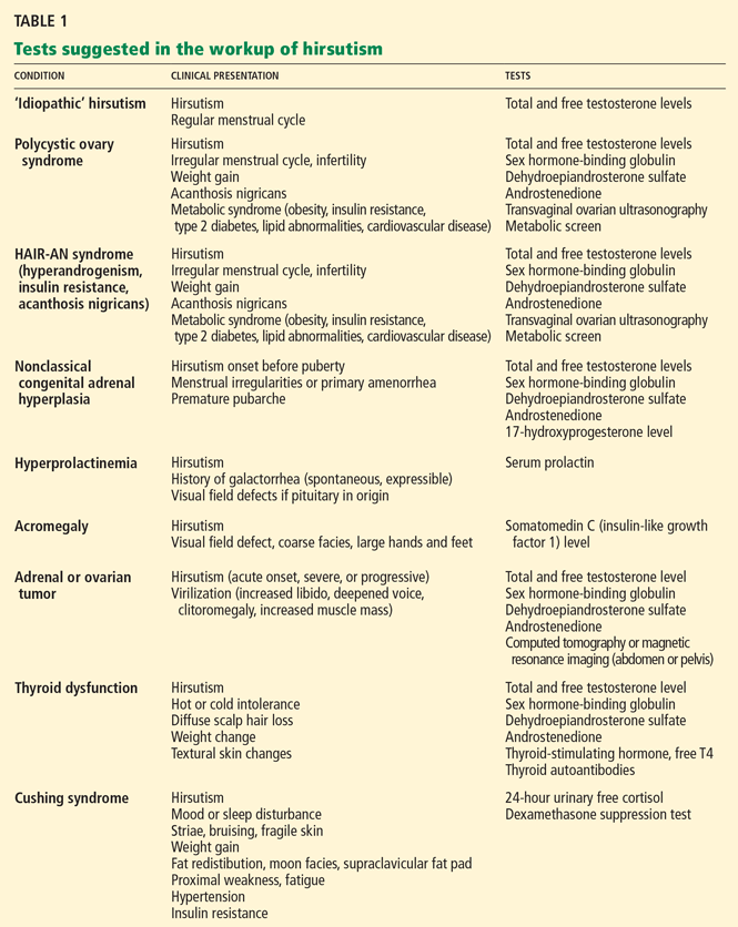
The current Endocrine Society guidelines20 recommend obtaining an early-morning testosterone blood level in the following patients:
- Women with moderate or severe hirsutism
- Women with hirsutism of any degree with sudden onset or rapid progression, or accompanied by signs or symptoms suggesting malignancy or polycystic ovary syndrome: eg, menstrual irregularity, infertility, central obesity, clitoromegaly, or acanthosis nigricans.15,20
Testing androgen levels in mild, isolated hirsutism has not been proven to be useful or to alter management.20
Free testosterone level
An early-morning total or free testosterone level is the initial test in the laboratory evaluation of hirsutism.12,15 Additional specialized laboratory testing may be needed to determine the free testosterone level,15 as the free testosterone test is not available at all laboratories. A normal total testosterone level does not exclude hyperandrogenism but can suggest the diagnosis of idiopathic hirsutism.15
Further testing is needed if the total testosterone level is normal or only slightly elevated, or if there is a strong clinical suspicion of an underlying condition such as endocrinopathy or tumor. It is also useful in patients whose hirsutism responds poorly to medical treatments15 (see discussion below).
If the total testosterone level is elevated, if the hirsutism is moderate to severe, if there are associated symptoms, or if hirsutism is acute or progressive, a further endocrinologic workup is needed,15 possibly including measurement of free testosterone, sex hormone-binding globulin, DHEAS, and androstenedione.15 Free testosterone, unbound to sex hormone-binding globulin, is the biologically active fraction, with the levels of binding globulin increased by drugs such as oral contraceptives15 and decreased by high insulin levels in insulin resistance.25
Test in patients with mild hirsutism?
Although the guidelines suggest that no additional workup is necessary for women with mild hirsutism, we evaluate all patients with hirsutism and those with the SAHA clinical spectrum by measuring free and total testosterone and DHEAS. In our experience, even women with mild hirsutism with subtle symptoms and signs of hyperandrogenism and mild hirsutism often have elevated androgen levels.
Test in women with idiopathic hirsutism?
In women with idiopathic hirsutism, minor forms of functional ovarian and adrenal hyperandrogenism are believed to play a role and are thought to be undetectable with conventional testing.25 The gonadotropin-releasing hormone (GnRH) analogue stimulation test may uncover occult hyperandrogenism in this setting, but it is used as a research tool and does not currently have application in routine clinical practice.14
It is important to remember that some women with apparent idiopathic hirsutism and a history of regular menstrual cycles are actually oligo-ovulatory or anovulatory. In these instances, another diagnosis should be considered,13 and referral to an endocrinologist for further evaluation of ovulatory function is recommended.13
CURRENT USE OF DIAGNOSTIC IMAGING
When malignancy is suspected
A testosterone level above 200 ng/dL suggests an ovarian tumor, and a DHEAS level above 700 μg/dL suggests an adrenal tumor.26 However, not all tumors present with such high androgen levels, and sudden onset of hirsutism, rapid progression of hirsutism, or signs of virilization suggest a tumor.15 In such cases, transvaginal ultrasonography, computed tomography, or magnetic resonance imaging (MRI) of the abdomen can exclude an ovarian or adrenal tumor.
When polycystic ovary syndrome is suspected
The diagnosis of polycystic ovary syndrome is confirmed by two out of three criteria:
- Oligo-ovulation or anovulation
- Clinical or laboratory signs of hyperandrogenism
- Ultrasonographic evidence of polycystic ovaries, with exclusion of other causes of hyperandrogenism.
ADDITIONAL LABORATORY TESTING
Tests for polycystic ovary syndrome
Assessment of polycystic ovary syndrome involves transvaginal ultrasonography, but ultrasonographic evidence of a polycystic ovary is not necessary for the diagnosis.16 A fasting lipid profile and fasting serum glucose are recommended, and if the fasting serum glucose is normal, an oral glucose tolerance test is recommended. 17
Some have reported measuring the ratio of luteinizing hormone to follicle-stimulating hormone in the workup of polycystic ovary syndrome, and a ratio greater than 2 has been considered indicative but not diagnostic.16,25 The individual levels of luteinizing hormone, follicle-stimulating hormone, and estradiol are more important in the evaluation of infertility and ovulatory dysfunction. In patients with elevations of these hormones or with these symptoms, referral for infertility screening with an endocrinologist or gynecologist is recommended. 25
Additional testing and referral for Cushing syndrome, other conditions
Cushing syndrome can be tested for with a 24-hour urine cortisol, overnight low-dose dexamethasone suppression test, and late-night salivary cortisol.27,28 Referral to an endocrinologist for further testing can differentiate between corticotropin-dependent or corticotropin-independent Cushing syndrome.25 Cushing syndrome is often associated with hyperandrogenism, particularly in those cases caused by adrenal tumors.29
The prolactin level and the level of somatomedin C (insulin-like growth factor 1) can be used to rule out hyperprolactinemia and acromegaly, respectively.12 If Cushing syndrome, hyperprolactinemia, or acromegaly is diagnosed by endocrinologic testing, pituitary MRI should be performed.12,25
Referral to specialist centers with experience with these conditions is essential. Nonclassical congenital adrenal hyperplasia can be screened for by a serum 17-hydroxyprogesterone level measured in the follicular phase.12 Measurement of thyroid-stimulating hormone, free thyroxine, and thyroid peroxidase antibodies screens for thyroid disease.12 Hirsutism has been reported with the commencement of L-thyroxine therapy.30
THE PRINCIPLES OF TREATMENT
Patient education regarding the cause of hirsutism and reasonable treatment expectations and emotional support are important in the management of hirsutism. Also important is regular follow-up to measure and document the response to treatment; this can include repeating Ferriman-Gallwey scoring, taking photographs of affected areas, and retesting androgen levels after 3 to 6 months.12
Treatment must be continued for an ongoing effect, and most pharmacologic treatments can take up to 3 to 6 months to produce significant improvement.1
When an underlying condition is diagnosed, treatment of the condition is essential. Androgen-secreting tumors require surgical management.12 Cushing disease, hyperprolactinemia, and acromegaly should be clinically apparent from examination and testing, and appropriate referral and standard management should be instigated. Exogenous sources of androgen such as androgenic progestins or anabolic steroids should be discontinued. Lifestyle management is important, and weight loss in obese patients with polycystic ovary syndrome can improve hirsutism as well as mitigate cardiovascular risk factors.31
In classic congenital adrenal hyperplasia, glucocorticoid therapy manages both ovulation induction and hirsutism.20 However, in nonclassical congenital adrenal hyperplasia, glucocorticoid therapy supports ovulation induction, but hirsutism usually requires both systemic antiandrogen and hair removal.20
CURRENT OPTIONS FOR HAIR REMOVAL
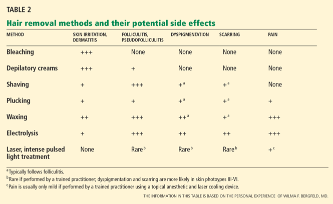
The choice of method depends on patient preference, adverse effects, the degree of hirsutism, the level of distress, previous treatments, and cost.1,15,32
Self-care methods
Self-care methods offer only temporary reduction of excess hairs.
Plucking removes the entire hair, including the root, but it is painful and time-consuming, and it is only practical for areas where few hairs exist, such as on the face.1
Shaving is an easy, inexpensive, and painless choice for hair removal. Although a common belief is that shaving causes faster or thicker hair regrowth, shaving affects neither the diameter nor the rate of growth of the hair.32 Given its masculine association, shaving is not acceptable to most women except perhaps for use on the legs and axillae.1,32 Shaving can cause irritation, folliculitis, pseudofolliculitis, and infection.1
Waxing removes the entire hair. While it is more expensive than plucking, regrowth is slower, occurring over weeks. It is painful and can cause thermal burns, irritation, folliculitis, scarring, and postinflammatory dyspigmentation.1
Chemical depilatories, usually thioglycollic acid preparations, are inexpensive, painless, and easy to use. However, the resulting hair reduction is of short duration because the hair shafts are only removed at the level of the skin surface.1 They can also cause irritant dermatitis. 1
Bleaching with hydrogen peroxide is inexpensive and can camouflage dark facial hair, but it can also cause skin discoloration and irritation. 1
Clinic-based methods
Electrolysis often results in a permanent reduction in hair growth.1,32 A fine needle is placed into the hair follicle and an electrical current is applied. Each follicle is treated individually. 1,32 Best results are seen on darker hairs in patients with lighter skin, but it can be used on all skin types and hair colors.1,32
Electrolysis is operator-dependent, and there are US Food and Drug Administration (FDA) regulations regarding electrolysis techniques. It requires multiple treatments, and it is painful and can cause erythema, folliculitis, pseudofolliculitis, infection, scarring, and postinflammatory dyspigmentation.1,32 Some reports suggest that prior waxing and plucking of hairs damages the hair by twisting the hair shaft, making electrolysis more difficult.32
Laser treatment uses light of certain wavelengths to damage the hair follicles. While laser hair removal does not result in complete or persistent hair removal, it is more effective than shaving, waxing, and electrolysis, producing partial hair reduction for up to 6 months; the effect is enhanced with multiple treatments.33,34 The number of treatments required depends on the laser type and on the nature of the patient’s hair follicles.35
Laser systems for hair removal are of various wavelengths and also include intense pulsed light systems. The choice of system depends on the patient’s skin type and hair color. Women with fair skin and dark hair are ideal candidates; longer-wavelength lasers are preferred for darker or tanned skin types.
Adverse effects of laser hair removal include pain, erythema, burns, dyspigmentation, and scarring. Laser cooling devices can prevent or minimize some of these effects. Laser treatment has also been known to cause a paradoxical increase in hair growth.1,33,34
DRUG THERAPIES FOR HIRSUTISM
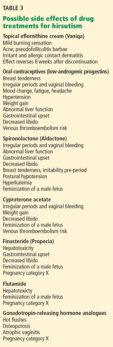
The drugs most commonly used for hirsutism are oral contraceptives (off-label use) and antiandrogenic drugs (off-label use). Topical eflornithine cream (Vaniqa) is FDA-approved for hirsutism but is less commonly used. Insulin sensitizers, GnRH analogues, and other drugs are occasionally used (off-label) to treat hirsutism.
Topical eflornithine cream
Topical eflornithine cream treats facial hirsutism by slowing the rate of hair growth; it does this by irreversibly inhibiting ornithine decarboxylase, an enzyme essential for hair growth.39,40 Studies showed that twice-daily application reduced unwanted facial hair in women after 24 weeks of treatment.39,40 Treatment must be continuous, since hair growth rapidly returns to the pretreatment rate by 8 weeks after discontinuing eflornithine.39,40 White women have been shown to respond better than black women.39 Adverse effects include a mild burning sensation, acne, pseudofolliculitis barbae, irritation, and allergic contact dermatitis.39,40 Improved outcomes have been suggested when eflornithine cream is combined with laser hair removal.41
Oral contraceptives
Oral contraceptives are commonly used off-label for the management of hirsutism.20 Oral contraceptives suppress the secretion of luteinizing hormone and, hence, the synthesis of ovarian androgen, thereby increasing levels of sex hormone-binding globulin and decreasing free plasma testosterone.1,20 Adrenal androgen production is also slightly reduced.20
Oral contraceptives usually combine a synthetic estrogen and a progestin. Certain progestins are more androgenic and should be avoided.1
For treating hirsutism, oral contraceptives should be used that contain low-androgenic progestins such as cyproterone acetate (not available in the United States), drosperinone (eg, in Yasmin), norgestimate (eg, in Ortho Tri-Cyclen), or desogestrel (eg, in Mircette).1,20
Side effects of oral contraceptives include breast tenderness, gastrointestinal upset, headache, loss of libido, hypertension, and the potential risk of venous thromboembolism.1,15,32,36
Antiandrogenic drugs
Several antiandrogenic drugs are used off-label to treat hirsutism.
Spironolactone (Aldactone), a competitive inhibitor of the androgen receptor and 5-alpha reductase activity,20 can be effective in the treatment of hirsutism. Monotherapy with spironolactone, without an oral contraceptive or other reliable form of contraception, is not recommended because of the teratogenic potential of all antiandrogens to feminize a developing male fetus.20 Thus, reliable contraception should be used in females of childbearing age when starting antiandrogen therapy.
The dosage of spironolactone for hirsutism is usually 100 mg to 200 mg daily.1,20 Hyperkalemia, polyuria, postural hypotension, irregular menses, and liver abnormalities are among the possible adverse effects (Table 3). Spironolactone was found to be tumorigenic in animal studies, although this has unknown relevance in humans.36
Cyproterone, an antiandrogen not available in the United States,42 competitively inhibits the androgen receptor and 5-alpha-reductase activity.1,20,36 It can be used for only the first 10 days of the menstrual cycle (50-mg or 100-mg dose) with an oral contraceptive pill, or in a low dose in a combined oral contraceptive pill (Diane-35 in Canada and the United Kingdom).1
Side effects are similar to those of oral contraceptives and include fatigue, mood change, risk of venous thromboembolism, and decreased libido.1,15,36 Importantly, in woman of childbearing age, there is the potential risk of feminization of a male fetus, so reliable contraception must be used.15,36
Flutamide, an investigational antiandrogen, has shown promise in the treatment of hirsutism.20 Flutamide is a nonsteroidal competitive inhibitor of androgen receptor binding. It carries a significant risk of hepatotoxicity. 1,15
Finasteride (Propecia) 1 mg is only occasionally used in the treatment of hirsutism (off-label usage). It inhibits type II 5-alphareductase to suppress dihydrotestosterone levels. 32 It carries a risk of gastrointestinal disturbance, decreased libido, hepatotoxicity, and feminization of a male fetus (pregnancy category X), so reliable contraception is required in all females of childbearing age, as with all antiandrogens1 (Table 3).
Dutasteride (Avodart), a type I and II 5-alpha-reductase inhibitor, has not been studied for the treatment of hirsutism (pregnancy category X).
Insulin sensitizers
Metformin (Glucophage) and other insulin sensitizers are less effective than antiandrogens at reducing hirsutism.20,38 However, metformin is effective at inducing ovulation in patients with polycystic ovary syndrome.38 Gastrointestinal upset is a common side effect; lactic acidosis is a serious but rare adverse effect.1
Gonadotropin-releasing hormone analogues
GnRH analogues are an option only if oral contraceptives and antiandrogen drugs are unsuccessful in patients with severe hyperandrogenism. 20 They suppress secretion of luteinizing hormone and the synthesis of ovarian androgen.1,20 These drugs are given as monthly intramuscular injections, usually with some form of estrogen-progestin replacement, since GnRH analogues cause estrogen levels to fall to menopausal levels.1
Side effects include signs and symptoms of menopause including hot flushes, atrophic vaginitis, and osteoporosis.1,15 These drugs completely inhibit ovulation, and some endocrinologists and gynecologists do not suggest further contraception in women of childbearing years for this reason. However, GnRH analogues are not approved as a contraceptive and are pregnancy category X.
Other drugs
Other drugs with antiandrogen activity include cimetidine and ketoconazole.12 Cimetidine (Tagamet) is not effective for the treatment of hirsutism, and ketoconazole (Nizoral) is associated with significant risk for adrenocortical suppression12 and hepatotoxicity in addition to multiple drug interactions, given its effect on the hepatic P450 enzyme system.
Acknowledgment: Many thanks to Rebecca Tung, MD, dermatologic surgeon, Cleveland Clinic, for her advice on lasers.
- Mofid A, Seyyed Alinaghi SA, Zandieh S, Yazdani T. Hirsutism. Int J Clin Pract 2008; 62:433–443.
- Azziz R, Carmina E, Sawaya ME. Idiopathic hirsutism. Endocr Rev 2000; 21:347–362.
- Himelein MJ, Thatcher SS. Polycystic ovary syndrome and mental health: a review. Obstet Gynecol Surv 2006; 61:723–732.
- Williamson K, Gunn AJ, Johnson N, Milsom SR. The impact of ethnicity on the presentation of polycystic ovarian syndrome. Aust N Z J Obstet Gynaecol 2001; 41:202–206.
- Diamanti-Kandarakis E, Kouli CR, Bergiele AT, et al. A survey of the polycystic ovary syndrome in the Greek island of Lesbos: hormonal and metabolic profile. J Clin Endocrinol Metab 1999; 84:4006–4011.
- Ferriman D, Gallwey JD. Clinical assessment of body hair growth in women. J Clin Endocrinol Metab 1961; 21:1440–1447.
- Messenger AG. The control of hair growth: an overview. J Invest Dermatol 1993; 101(suppl 1):4S–9S.
- Rosenfield RL. Hirsutism and the variable response of the pilosebaceous unit to androgen. J Investig Dermatol Symp Proc 2005; 10:205–208.
- Longcope C. Adrenal and gonadal androgen secretion in normal females. Clin Endocrinol Metab 1986; 15:213–228.
- Braunstein GD. Testis. In:Gardner DG, Shoback D, editors. Greenspan’s Basic & Clinical Endocrinology. 8th ed. New York: McGraw-Hill, 2007.
- Deplewski D, Rosenfield RL. Role of hormones in pilosebaceous unit development. Endocr Rev 2000; 21:363–392.
- Practice Committee of the American Society for Reproductive Medicine. The evaluation and treatment of androgen excess. Fertil Steril 2006; 86(suppl 5):S241–S247.
- Azziz R, Waggoner WT, Ochoa T, Knochenhauer ES, Boots LR. Idiopathic hirsutism: an uncommon cause of hirsutism in Alabama. Fertil Steril 1998; 70:274–278.
- Rossi R, Tauchmanovà L, Luciano A, et al. Functional hyperandrogenism detected by corticotropin and GnRH-analogue stimulation tests in women affected by apparently idiopathic hirsutism. J Endocrinol Invest 2001; 24:491–498.
- Rosenfield RL. Clinical practice. Hirsutism. N Engl J Med 2005; 353:2578–2588.
- Rotterdam ESHRE/ASRM-Sponsored PCOS consensus workshop group. Revised 2003 consensus on diagnostic criteria and long-term health risks related to polycystic ovary syndrome (PCOS). Hum Reprod 2004; 19:41–47.
- Salley KE, Wickham EP, Cheang KI, Essah PA, Karjane NW, Nestler JE. Glucose intolerance in polycystic ovary syndrome—a position statement of the Androgen Excess Society. J Clin Endocrinol Metab 2007; 92:4546–4556.
- Eckel RH, Grundy SM, Zimmet PZ. The metabolic syndrome. Lancet 2005; 365:1415–1428.
- Azziz R. Diagnostic criteria for polycystic ovary syndrome: a reappraisal. Fertil Steril 2005; 83:1343–1346.
- Martin KA, Chang RJ, Ehrmann DA, et al. Evaluation and treatment of hirsutism in premenopausal women: an endocrine society clinical practice guideline. http://www.endo-society.org/guidelines/final/upload/Hirsutism_Guideline.pdf. Accessed March 30, 2010.
- Orfanos CE, Adler YD, Zouboulis CC. The SAHA syndrome. Horm Res 2000; 54:251–258.
- New MI. Extensive clinical experience: nonclassical 21-hydroxylase deficiency. J Clin Endocrinol Metab 2006; 91:4205–4214.
- Kohn B, Levine LS, Pollack MS, et al. Late-onset steroid 21-hydroxylase deficiency: a variant of classical congenital adrenal hyperplasia. J Clin Endocrinol Metab 1982; 55:817–827.
- Knochenhauer ES, Hines G, Conway-Myers BA, Azziz R. Examination of the chin or lower abdomen only for the prediction of hirsutism. Fertil Steril 2000; 74:980–983.
- Somani N, Harrison S, Bergfeld WF. The clinical evaluation of hirsutism. Dermatol Ther 2008; 21:376–391.
- Waggoner W, Boots LR, Azziz R. Total testosterone and DHEAS levels as predictors of androgen-secreting neoplasms: a populational study. Gynecol Endocrinol 1999; 13:394–400.
- Crapo L. Cushing’s syndrome: a review of diagnostic tests. Metabolism 1979; 28:955–977.
- Blethen SL, Chasalow FI. Overnight dexamethasone suppression test: normal responses and the diagnosis of Cushing’s syndrome. Steroids 1989; 54:185–193.
- Bertagna C, Orth DN. Clinical and laboratory findings and results of therapy in 58 patients with adrenocortical tumors admitted to a single medical center (1951 to 1978). Am J Med 1981; 71:855–875.
- Kologlu S, Baskal N, Kologlu LB, Laleli Y, Tuccar E. Hirsutism due to the treatment with L-thyroxine in patients with thyroid pathology. Endocrinologie 1988; 26:179–185.
- Gambineri A, Patton L, Vaccina A, et al. Treatment with flutamide, metformin, and their combination added to a hypocaloric diet in overweight-obese women with polycystic ovary syndrome: a randomized, 12-month, placebo-controlled study. J Clin Endocrinol Metab 2006; 91:3970–3980.
- Dawber RP. Guidance for the management of hirsutism. Curr Med Res Opin 2005; 21:1227–1234.
- Haedersdal M, Wulf HC. Evidence based review of hair removal using lasers and light sources. J Eur Acad Dermatol Venereol 2006; 20:9–20.
- Sadighha A, Mohaghegh Zahed G. Meta-analysis of hair removal laser trials. Lasers Med Sci 2009; 24:21–25.
- Casey AS, Goldberg D. Guidelines for laser hair removal. J Cosmet Laser Ther 2008; 10:24–33.
- Wakelin SH, Maibach HI, editors. Handbook of Systemic Drug Treatment in Dermatology. London: Manson Publishing Ltd, 2004.
- Swiglo BA, Cosma M, Flynn DN, et al. Clinical review: antiandrogens for the treatment of hirsutism: a systematic review and metaanalyses of randomized controlled trials. J Clin Endocrinol Metab 2008; 93:1153–1160.
- Cosma M, Swiglo BA, Flynn DN, et al. Clinical review: insulin sensitizers for the treatment of hirsutism: a systematic review and metaanalyses of randomized controlled trials. J Clin Endocrinol Metab 2008; 93:1135–1142.
- Balfour JA, McClellan K. Topical eflornithine. Am J Clin Dermatol 2001; 2:197–201.
- Wolf JE, Shander D, Huber F, et al; Eflornithine HCl Study Group. Randomized, double-blind clinical evaluation of the efficacy and safety of topical eflornithine HCl 13.9% cream in the treatment of women with facial hair. Int J Dematol 2007; 46:94–98.
- Hamzavi I, Tan E, Shapiro J, Lui H. A randomized bilateral vehicle-controlled study of eflornithine cream combined with laser treatment versus laser treatment alone for facial hirsutism in women. J Am Acad Dermatol 2007; 57:54–59.
- Van der Spuy ZM, le Roux PA. Cyproterone acetate for hirsutism. Cochrane Database Syst Rev 2003; 4:CD001125.
Hirsutism causes significant anxiety and lack of self-esteem in women. Although it is itself a benign condition, it is often the sign of an underlying and possibly serious endocrine condition.
As we will discuss, the diagnosis begins with a detailed history and physical examination, with laboratory testing and imaging as needed to confirm or rule out underlying causes. Management begins with patient education and support and includes hair removal and drug treatment of any underlying metabolic derangement.
PREVALENCE AND IMPACT
Hirsutism is a common disorder of excess growth of terminal hair in an androgen-dependent male distribution in women, including the chin, upper lip, breasts, upper back, and abdomen.1 It affects 5% to 10% of women of reproductive age.1,2
Hirsutism should be differentiated from hypertrichosis, which can be hereditary or acquired, and which is defined as increased general hair growth in androgen-independent areas.1
Excess hair is cosmetically concerning for women and can significantly affect self-esteem. 3 Normal or acceptable hair growth depends on a woman’s ethnicity and her perception of familial, cultural, and societal norms for the quantity and distribution of hair. Mediterranean women generally have a medium amount of body and facial hair, whereas Asian women have a minimal amount.1,4,5
Hirsutism can be clinically graded according to the Ferriman-Gallwey scale2,6 and is defined as a Ferriman-Gallwey score of 8 or higher.1
HOW CIRCULATING ANDROGENS AFFECT HAIR FOLLICLES
In androgen-dependent areas, circulating androgens influence hair follicle characteristics. Androgens increase the size and diameter of the hair fibers in certain androgen-dependent sites, as seen in puberty with the transformation of vellus hairs (small, nonpigmented hairs) into terminal hairs (large, pigmented hairs) in the pubic and axillary regions in women, as well as the beard area in men.2,7 Interestingly, the same circulating androgens cause miniaturization of the susceptible hair follicles of the central scalp.7 The susceptibility of the hair follicle to the effects of the androgens may be genetically determined.7,8
Hirsutism is a sign of hyperandrogenism and increased action of androgens on hair follicles. In women, about half of circulating testosterone arises from the ovaries and adrenal glands; the rest originates from peripheral conversion of weaker androgens (such as androstenedione produced by the adrenals and ovaries) into testosterone.9 Dehydroepiandrosterone sulfate (DHEAS) originates mainly in the adrenal glands.9,10 Testosterone is converted to the more potent dihydrotestosterone (DHT) by type II 5-alpha reductase in the skin, which can then act on susceptible hair follicles.7,11 Therefore, hirsutism can be a consequence of endogenous androgen over-production from the ovaries or the adrenal glands (or both), of exposure to an exogenous source of androgen such as a drug, or of heightened hair follicle sensitivity and metabolism of normal circulating androgen levels (target end-organ dysfunction).1
‘IDIOPATHIC’ HIRSUTISM: A MISLEADING DIAGNOSIS
Many women with hirsutism are found to have polycystic ovary syndrome as the underlying cause, but hirsutism is also commonly labeled as idiopathic when it occurs without an obvious cause, eg, in women with regular menses and normal androgen levels and without features suspicious for other causes of hirsutism. 1,2,12,13 But while this term is commonly used,1,12 it may be misleading, especially if the diagnosis of idiopathic hirsutism is based on standard laboratory tests, which do not always detect androgen excess.2,13 Minor ovarian or adrenal functional hyperandrogenism,14 increased peripheral activity of 5-alpha reductase in the hair follicle, or abnormalities in the androgen receptor have been implicated in the pathogenesis of so-called idiopathic hirsutism. 2,15
HIRSUTISM AND POLYCYSTIC OVARY SYNDROME
Polycystic ovary syndrome, a metabolic syndrome, presents clinically with menstrual irregularities such as oligomenorrhea or amenorrhea, infertility, and signs of hyperandrogenism such as hirsutism, acne, or androgenetic alopecia.16,17 Metabolic disturbances including insulin resistance, impaired glucose tolerance, hyperlipidemia, and obesity (body mass index > 30 kg/m2) also can occur, thus increasing cardiovascular risk.16–18
The finding of polycystic ovaries is not required to make the diagnosis of polycystic ovary syndrome, and their presence does not prove the diagnosis.16,19 Gonadotropin-dependent functional ovarian hyperandrogenism is believed to cause this syndrome; however, mild adrenocorticotropic-dependent functional adrenal hyperandrogenism also is a feature in many cases. In rare cases, polycystic ovary syndrome presents with an isolated elevation of DHEAS.16,20
OTHER CONDITIONS OF EXCESS ANDROGEN
The syndrome of hyperandrogenism, insulin resistance, and acanthosis nigricans, abbreviated as HAIR-AN, is separate from polycystic ovary syndrome; it characterizes a group of inherited syndromes associated with severe metabolic abnormalities of insulin and glucose metabolism and with marked clinical signs of hyperandrogenism.12
The syndrome of seborrhea, acne, hirsutism, and acanthosis nigricans, abbreviated as SAHA, while not itself a diagnosis, is a clinical spectrum of dermatologic signs and symptoms also associated with hyperandrogenism. These are signs that may present with the HAIR-AN syndrome or with another cause of excess androgens, such as idiopathic, ovarian, adrenal, or hyperprolactinemic hyperandrogenism.21
Thyroid disease, hyperprolactinemia, acromegaly, Cushing syndrome, exogenous factors such as androgenic drugs, and nonclassical congenital adrenal hyperplasia can also produce hirsutism.12 In nonclassical congenital adrenal hyperplasia, which is typically caused by a deficiency of 21-hydroxylase, patients present with premature pubarche, hirsutism in the prepubertal years, and menstrual irregularities including primary amenorrhea.22,23
Important rare causes of hirsutism include benign and malignant androgen-secreting tumors of adrenal or ovarian origin. In such cases, hirsutism can have an acute onset or rapid progression and may be associated with features of virilization, such as deepened voice, increased muscle mass, androgenetic alopecia, clitoromegaly, and increased libido.12
A THOROUGH HISTORY IS CRITICAL TO DIAGNOSIS
A thorough medical history can provide important diagnostic clues in women with hirsutism. The clinician should elicit details about the onset and progression of the hair growth,12,15 previous treatments, and any cutaneous signs of hyperandrogenism, such as acne, seborrhea, acanthosis nigricans, or patterned hair loss.
Also important are the menstrual history and a history of infertility. Primary amenorrhea is defined as failure to menstruate by 16 years of age if secondary sexual characteristics have developed, or by 14 years of age if no secondary sexual characteristics have developed, and it can indicate nonclassical congenital adrenal hyperplasia.
The clinician should also try to determine if the patient has a history of galactorrhea or symptoms of virilization (eg, deepened voice, clitoromegaly, increased muscle mass); a family history of hirsutism, polycystic ovary syndrome, HAIR-AN syndrome, metabolic conditions such as type 2 diabetes mellitus, or cardiovascular disease12,15; or a history of symptoms of any condition known to produce hirsutism, such as Cushing disease, acromegaly, or a thyroid disorder. Also important is a drug history to determine if the patient has taken drugs such as androgens, anabolic steroids, or valproic acid (Depakote).20
THE PHYSICAL EXAMINATION
Another proposed predictor of hirsutism is that terminal hair on the chin or the lower abdomen (Ferriman-Gallwey score ≥ 2) is nearly 100% sensitive and 27% specific at predicting total-body hirsutism.24
As part of the physical examination, the clinician should also look for other cutaneous signs of hyperandrogenism, such as acne, androgenetic alopecia, and seborrhea. Acanthosis nigricans is a sign of insulin resistance. Height and weight should be measured and the body mass index calculated. Blood pressure should be recorded, as high blood pressure may be seen in Cushing syndrome and is an important cardiovascular risk factor. Signs of virilization should be identified. Indicators of Cushing disease such as striae, moon facies, fat redistribution, fragile skin, and proximal myopathy should be noted as well as signs of thyroid disease, such as textural skin changes, goiter, and hair loss. Expressible or spontaneous galactorrhea suggests hyperprolactinemia. Acromegaly is associated with coarse facies and enlarged hands and feet. Many of the endocrinopathies can be caused by a pituitary adenoma, which can manifest as a visual field defect, so visual fields should be examined.25 The examination should also exclude any palpable ovarian or adrenal mass.12
WHEN IS ADDITIONAL TESTING NEEDED?
The current Endocrine Society guidelines20 recommend obtaining an early-morning testosterone blood level in the following patients:
- Women with moderate or severe hirsutism
- Women with hirsutism of any degree with sudden onset or rapid progression, or accompanied by signs or symptoms suggesting malignancy or polycystic ovary syndrome: eg, menstrual irregularity, infertility, central obesity, clitoromegaly, or acanthosis nigricans.15,20
Testing androgen levels in mild, isolated hirsutism has not been proven to be useful or to alter management.20
Free testosterone level
An early-morning total or free testosterone level is the initial test in the laboratory evaluation of hirsutism.12,15 Additional specialized laboratory testing may be needed to determine the free testosterone level,15 as the free testosterone test is not available at all laboratories. A normal total testosterone level does not exclude hyperandrogenism but can suggest the diagnosis of idiopathic hirsutism.15
Further testing is needed if the total testosterone level is normal or only slightly elevated, or if there is a strong clinical suspicion of an underlying condition such as endocrinopathy or tumor. It is also useful in patients whose hirsutism responds poorly to medical treatments15 (see discussion below).
If the total testosterone level is elevated, if the hirsutism is moderate to severe, if there are associated symptoms, or if hirsutism is acute or progressive, a further endocrinologic workup is needed,15 possibly including measurement of free testosterone, sex hormone-binding globulin, DHEAS, and androstenedione.15 Free testosterone, unbound to sex hormone-binding globulin, is the biologically active fraction, with the levels of binding globulin increased by drugs such as oral contraceptives15 and decreased by high insulin levels in insulin resistance.25
Test in patients with mild hirsutism?
Although the guidelines suggest that no additional workup is necessary for women with mild hirsutism, we evaluate all patients with hirsutism and those with the SAHA clinical spectrum by measuring free and total testosterone and DHEAS. In our experience, even women with mild hirsutism with subtle symptoms and signs of hyperandrogenism and mild hirsutism often have elevated androgen levels.
Test in women with idiopathic hirsutism?
In women with idiopathic hirsutism, minor forms of functional ovarian and adrenal hyperandrogenism are believed to play a role and are thought to be undetectable with conventional testing.25 The gonadotropin-releasing hormone (GnRH) analogue stimulation test may uncover occult hyperandrogenism in this setting, but it is used as a research tool and does not currently have application in routine clinical practice.14
It is important to remember that some women with apparent idiopathic hirsutism and a history of regular menstrual cycles are actually oligo-ovulatory or anovulatory. In these instances, another diagnosis should be considered,13 and referral to an endocrinologist for further evaluation of ovulatory function is recommended.13
CURRENT USE OF DIAGNOSTIC IMAGING
When malignancy is suspected
A testosterone level above 200 ng/dL suggests an ovarian tumor, and a DHEAS level above 700 μg/dL suggests an adrenal tumor.26 However, not all tumors present with such high androgen levels, and sudden onset of hirsutism, rapid progression of hirsutism, or signs of virilization suggest a tumor.15 In such cases, transvaginal ultrasonography, computed tomography, or magnetic resonance imaging (MRI) of the abdomen can exclude an ovarian or adrenal tumor.
When polycystic ovary syndrome is suspected
The diagnosis of polycystic ovary syndrome is confirmed by two out of three criteria:
- Oligo-ovulation or anovulation
- Clinical or laboratory signs of hyperandrogenism
- Ultrasonographic evidence of polycystic ovaries, with exclusion of other causes of hyperandrogenism.
ADDITIONAL LABORATORY TESTING
Tests for polycystic ovary syndrome
Assessment of polycystic ovary syndrome involves transvaginal ultrasonography, but ultrasonographic evidence of a polycystic ovary is not necessary for the diagnosis.16 A fasting lipid profile and fasting serum glucose are recommended, and if the fasting serum glucose is normal, an oral glucose tolerance test is recommended. 17
Some have reported measuring the ratio of luteinizing hormone to follicle-stimulating hormone in the workup of polycystic ovary syndrome, and a ratio greater than 2 has been considered indicative but not diagnostic.16,25 The individual levels of luteinizing hormone, follicle-stimulating hormone, and estradiol are more important in the evaluation of infertility and ovulatory dysfunction. In patients with elevations of these hormones or with these symptoms, referral for infertility screening with an endocrinologist or gynecologist is recommended. 25
Additional testing and referral for Cushing syndrome, other conditions
Cushing syndrome can be tested for with a 24-hour urine cortisol, overnight low-dose dexamethasone suppression test, and late-night salivary cortisol.27,28 Referral to an endocrinologist for further testing can differentiate between corticotropin-dependent or corticotropin-independent Cushing syndrome.25 Cushing syndrome is often associated with hyperandrogenism, particularly in those cases caused by adrenal tumors.29
The prolactin level and the level of somatomedin C (insulin-like growth factor 1) can be used to rule out hyperprolactinemia and acromegaly, respectively.12 If Cushing syndrome, hyperprolactinemia, or acromegaly is diagnosed by endocrinologic testing, pituitary MRI should be performed.12,25
Referral to specialist centers with experience with these conditions is essential. Nonclassical congenital adrenal hyperplasia can be screened for by a serum 17-hydroxyprogesterone level measured in the follicular phase.12 Measurement of thyroid-stimulating hormone, free thyroxine, and thyroid peroxidase antibodies screens for thyroid disease.12 Hirsutism has been reported with the commencement of L-thyroxine therapy.30
THE PRINCIPLES OF TREATMENT
Patient education regarding the cause of hirsutism and reasonable treatment expectations and emotional support are important in the management of hirsutism. Also important is regular follow-up to measure and document the response to treatment; this can include repeating Ferriman-Gallwey scoring, taking photographs of affected areas, and retesting androgen levels after 3 to 6 months.12
Treatment must be continued for an ongoing effect, and most pharmacologic treatments can take up to 3 to 6 months to produce significant improvement.1
When an underlying condition is diagnosed, treatment of the condition is essential. Androgen-secreting tumors require surgical management.12 Cushing disease, hyperprolactinemia, and acromegaly should be clinically apparent from examination and testing, and appropriate referral and standard management should be instigated. Exogenous sources of androgen such as androgenic progestins or anabolic steroids should be discontinued. Lifestyle management is important, and weight loss in obese patients with polycystic ovary syndrome can improve hirsutism as well as mitigate cardiovascular risk factors.31
In classic congenital adrenal hyperplasia, glucocorticoid therapy manages both ovulation induction and hirsutism.20 However, in nonclassical congenital adrenal hyperplasia, glucocorticoid therapy supports ovulation induction, but hirsutism usually requires both systemic antiandrogen and hair removal.20
CURRENT OPTIONS FOR HAIR REMOVAL
The choice of method depends on patient preference, adverse effects, the degree of hirsutism, the level of distress, previous treatments, and cost.1,15,32
Self-care methods
Self-care methods offer only temporary reduction of excess hairs.
Plucking removes the entire hair, including the root, but it is painful and time-consuming, and it is only practical for areas where few hairs exist, such as on the face.1
Shaving is an easy, inexpensive, and painless choice for hair removal. Although a common belief is that shaving causes faster or thicker hair regrowth, shaving affects neither the diameter nor the rate of growth of the hair.32 Given its masculine association, shaving is not acceptable to most women except perhaps for use on the legs and axillae.1,32 Shaving can cause irritation, folliculitis, pseudofolliculitis, and infection.1
Waxing removes the entire hair. While it is more expensive than plucking, regrowth is slower, occurring over weeks. It is painful and can cause thermal burns, irritation, folliculitis, scarring, and postinflammatory dyspigmentation.1
Chemical depilatories, usually thioglycollic acid preparations, are inexpensive, painless, and easy to use. However, the resulting hair reduction is of short duration because the hair shafts are only removed at the level of the skin surface.1 They can also cause irritant dermatitis. 1
Bleaching with hydrogen peroxide is inexpensive and can camouflage dark facial hair, but it can also cause skin discoloration and irritation. 1
Clinic-based methods
Electrolysis often results in a permanent reduction in hair growth.1,32 A fine needle is placed into the hair follicle and an electrical current is applied. Each follicle is treated individually. 1,32 Best results are seen on darker hairs in patients with lighter skin, but it can be used on all skin types and hair colors.1,32
Electrolysis is operator-dependent, and there are US Food and Drug Administration (FDA) regulations regarding electrolysis techniques. It requires multiple treatments, and it is painful and can cause erythema, folliculitis, pseudofolliculitis, infection, scarring, and postinflammatory dyspigmentation.1,32 Some reports suggest that prior waxing and plucking of hairs damages the hair by twisting the hair shaft, making electrolysis more difficult.32
Laser treatment uses light of certain wavelengths to damage the hair follicles. While laser hair removal does not result in complete or persistent hair removal, it is more effective than shaving, waxing, and electrolysis, producing partial hair reduction for up to 6 months; the effect is enhanced with multiple treatments.33,34 The number of treatments required depends on the laser type and on the nature of the patient’s hair follicles.35
Laser systems for hair removal are of various wavelengths and also include intense pulsed light systems. The choice of system depends on the patient’s skin type and hair color. Women with fair skin and dark hair are ideal candidates; longer-wavelength lasers are preferred for darker or tanned skin types.
Adverse effects of laser hair removal include pain, erythema, burns, dyspigmentation, and scarring. Laser cooling devices can prevent or minimize some of these effects. Laser treatment has also been known to cause a paradoxical increase in hair growth.1,33,34
DRUG THERAPIES FOR HIRSUTISM
The drugs most commonly used for hirsutism are oral contraceptives (off-label use) and antiandrogenic drugs (off-label use). Topical eflornithine cream (Vaniqa) is FDA-approved for hirsutism but is less commonly used. Insulin sensitizers, GnRH analogues, and other drugs are occasionally used (off-label) to treat hirsutism.
Topical eflornithine cream
Topical eflornithine cream treats facial hirsutism by slowing the rate of hair growth; it does this by irreversibly inhibiting ornithine decarboxylase, an enzyme essential for hair growth.39,40 Studies showed that twice-daily application reduced unwanted facial hair in women after 24 weeks of treatment.39,40 Treatment must be continuous, since hair growth rapidly returns to the pretreatment rate by 8 weeks after discontinuing eflornithine.39,40 White women have been shown to respond better than black women.39 Adverse effects include a mild burning sensation, acne, pseudofolliculitis barbae, irritation, and allergic contact dermatitis.39,40 Improved outcomes have been suggested when eflornithine cream is combined with laser hair removal.41
Oral contraceptives
Oral contraceptives are commonly used off-label for the management of hirsutism.20 Oral contraceptives suppress the secretion of luteinizing hormone and, hence, the synthesis of ovarian androgen, thereby increasing levels of sex hormone-binding globulin and decreasing free plasma testosterone.1,20 Adrenal androgen production is also slightly reduced.20
Oral contraceptives usually combine a synthetic estrogen and a progestin. Certain progestins are more androgenic and should be avoided.1
For treating hirsutism, oral contraceptives should be used that contain low-androgenic progestins such as cyproterone acetate (not available in the United States), drosperinone (eg, in Yasmin), norgestimate (eg, in Ortho Tri-Cyclen), or desogestrel (eg, in Mircette).1,20
Side effects of oral contraceptives include breast tenderness, gastrointestinal upset, headache, loss of libido, hypertension, and the potential risk of venous thromboembolism.1,15,32,36
Antiandrogenic drugs
Several antiandrogenic drugs are used off-label to treat hirsutism.
Spironolactone (Aldactone), a competitive inhibitor of the androgen receptor and 5-alpha reductase activity,20 can be effective in the treatment of hirsutism. Monotherapy with spironolactone, without an oral contraceptive or other reliable form of contraception, is not recommended because of the teratogenic potential of all antiandrogens to feminize a developing male fetus.20 Thus, reliable contraception should be used in females of childbearing age when starting antiandrogen therapy.
The dosage of spironolactone for hirsutism is usually 100 mg to 200 mg daily.1,20 Hyperkalemia, polyuria, postural hypotension, irregular menses, and liver abnormalities are among the possible adverse effects (Table 3). Spironolactone was found to be tumorigenic in animal studies, although this has unknown relevance in humans.36
Cyproterone, an antiandrogen not available in the United States,42 competitively inhibits the androgen receptor and 5-alpha-reductase activity.1,20,36 It can be used for only the first 10 days of the menstrual cycle (50-mg or 100-mg dose) with an oral contraceptive pill, or in a low dose in a combined oral contraceptive pill (Diane-35 in Canada and the United Kingdom).1
Side effects are similar to those of oral contraceptives and include fatigue, mood change, risk of venous thromboembolism, and decreased libido.1,15,36 Importantly, in woman of childbearing age, there is the potential risk of feminization of a male fetus, so reliable contraception must be used.15,36
Flutamide, an investigational antiandrogen, has shown promise in the treatment of hirsutism.20 Flutamide is a nonsteroidal competitive inhibitor of androgen receptor binding. It carries a significant risk of hepatotoxicity. 1,15
Finasteride (Propecia) 1 mg is only occasionally used in the treatment of hirsutism (off-label usage). It inhibits type II 5-alphareductase to suppress dihydrotestosterone levels. 32 It carries a risk of gastrointestinal disturbance, decreased libido, hepatotoxicity, and feminization of a male fetus (pregnancy category X), so reliable contraception is required in all females of childbearing age, as with all antiandrogens1 (Table 3).
Dutasteride (Avodart), a type I and II 5-alpha-reductase inhibitor, has not been studied for the treatment of hirsutism (pregnancy category X).
Insulin sensitizers
Metformin (Glucophage) and other insulin sensitizers are less effective than antiandrogens at reducing hirsutism.20,38 However, metformin is effective at inducing ovulation in patients with polycystic ovary syndrome.38 Gastrointestinal upset is a common side effect; lactic acidosis is a serious but rare adverse effect.1
Gonadotropin-releasing hormone analogues
GnRH analogues are an option only if oral contraceptives and antiandrogen drugs are unsuccessful in patients with severe hyperandrogenism. 20 They suppress secretion of luteinizing hormone and the synthesis of ovarian androgen.1,20 These drugs are given as monthly intramuscular injections, usually with some form of estrogen-progestin replacement, since GnRH analogues cause estrogen levels to fall to menopausal levels.1
Side effects include signs and symptoms of menopause including hot flushes, atrophic vaginitis, and osteoporosis.1,15 These drugs completely inhibit ovulation, and some endocrinologists and gynecologists do not suggest further contraception in women of childbearing years for this reason. However, GnRH analogues are not approved as a contraceptive and are pregnancy category X.
Other drugs
Other drugs with antiandrogen activity include cimetidine and ketoconazole.12 Cimetidine (Tagamet) is not effective for the treatment of hirsutism, and ketoconazole (Nizoral) is associated with significant risk for adrenocortical suppression12 and hepatotoxicity in addition to multiple drug interactions, given its effect on the hepatic P450 enzyme system.
Acknowledgment: Many thanks to Rebecca Tung, MD, dermatologic surgeon, Cleveland Clinic, for her advice on lasers.
- Mofid A, Seyyed Alinaghi SA, Zandieh S, Yazdani T. Hirsutism. Int J Clin Pract 2008; 62:433–443.
- Azziz R, Carmina E, Sawaya ME. Idiopathic hirsutism. Endocr Rev 2000; 21:347–362.
- Himelein MJ, Thatcher SS. Polycystic ovary syndrome and mental health: a review. Obstet Gynecol Surv 2006; 61:723–732.
- Williamson K, Gunn AJ, Johnson N, Milsom SR. The impact of ethnicity on the presentation of polycystic ovarian syndrome. Aust N Z J Obstet Gynaecol 2001; 41:202–206.
- Diamanti-Kandarakis E, Kouli CR, Bergiele AT, et al. A survey of the polycystic ovary syndrome in the Greek island of Lesbos: hormonal and metabolic profile. J Clin Endocrinol Metab 1999; 84:4006–4011.
- Ferriman D, Gallwey JD. Clinical assessment of body hair growth in women. J Clin Endocrinol Metab 1961; 21:1440–1447.
- Messenger AG. The control of hair growth: an overview. J Invest Dermatol 1993; 101(suppl 1):4S–9S.
- Rosenfield RL. Hirsutism and the variable response of the pilosebaceous unit to androgen. J Investig Dermatol Symp Proc 2005; 10:205–208.
- Longcope C. Adrenal and gonadal androgen secretion in normal females. Clin Endocrinol Metab 1986; 15:213–228.
- Braunstein GD. Testis. In:Gardner DG, Shoback D, editors. Greenspan’s Basic & Clinical Endocrinology. 8th ed. New York: McGraw-Hill, 2007.
- Deplewski D, Rosenfield RL. Role of hormones in pilosebaceous unit development. Endocr Rev 2000; 21:363–392.
- Practice Committee of the American Society for Reproductive Medicine. The evaluation and treatment of androgen excess. Fertil Steril 2006; 86(suppl 5):S241–S247.
- Azziz R, Waggoner WT, Ochoa T, Knochenhauer ES, Boots LR. Idiopathic hirsutism: an uncommon cause of hirsutism in Alabama. Fertil Steril 1998; 70:274–278.
- Rossi R, Tauchmanovà L, Luciano A, et al. Functional hyperandrogenism detected by corticotropin and GnRH-analogue stimulation tests in women affected by apparently idiopathic hirsutism. J Endocrinol Invest 2001; 24:491–498.
- Rosenfield RL. Clinical practice. Hirsutism. N Engl J Med 2005; 353:2578–2588.
- Rotterdam ESHRE/ASRM-Sponsored PCOS consensus workshop group. Revised 2003 consensus on diagnostic criteria and long-term health risks related to polycystic ovary syndrome (PCOS). Hum Reprod 2004; 19:41–47.
- Salley KE, Wickham EP, Cheang KI, Essah PA, Karjane NW, Nestler JE. Glucose intolerance in polycystic ovary syndrome—a position statement of the Androgen Excess Society. J Clin Endocrinol Metab 2007; 92:4546–4556.
- Eckel RH, Grundy SM, Zimmet PZ. The metabolic syndrome. Lancet 2005; 365:1415–1428.
- Azziz R. Diagnostic criteria for polycystic ovary syndrome: a reappraisal. Fertil Steril 2005; 83:1343–1346.
- Martin KA, Chang RJ, Ehrmann DA, et al. Evaluation and treatment of hirsutism in premenopausal women: an endocrine society clinical practice guideline. http://www.endo-society.org/guidelines/final/upload/Hirsutism_Guideline.pdf. Accessed March 30, 2010.
- Orfanos CE, Adler YD, Zouboulis CC. The SAHA syndrome. Horm Res 2000; 54:251–258.
- New MI. Extensive clinical experience: nonclassical 21-hydroxylase deficiency. J Clin Endocrinol Metab 2006; 91:4205–4214.
- Kohn B, Levine LS, Pollack MS, et al. Late-onset steroid 21-hydroxylase deficiency: a variant of classical congenital adrenal hyperplasia. J Clin Endocrinol Metab 1982; 55:817–827.
- Knochenhauer ES, Hines G, Conway-Myers BA, Azziz R. Examination of the chin or lower abdomen only for the prediction of hirsutism. Fertil Steril 2000; 74:980–983.
- Somani N, Harrison S, Bergfeld WF. The clinical evaluation of hirsutism. Dermatol Ther 2008; 21:376–391.
- Waggoner W, Boots LR, Azziz R. Total testosterone and DHEAS levels as predictors of androgen-secreting neoplasms: a populational study. Gynecol Endocrinol 1999; 13:394–400.
- Crapo L. Cushing’s syndrome: a review of diagnostic tests. Metabolism 1979; 28:955–977.
- Blethen SL, Chasalow FI. Overnight dexamethasone suppression test: normal responses and the diagnosis of Cushing’s syndrome. Steroids 1989; 54:185–193.
- Bertagna C, Orth DN. Clinical and laboratory findings and results of therapy in 58 patients with adrenocortical tumors admitted to a single medical center (1951 to 1978). Am J Med 1981; 71:855–875.
- Kologlu S, Baskal N, Kologlu LB, Laleli Y, Tuccar E. Hirsutism due to the treatment with L-thyroxine in patients with thyroid pathology. Endocrinologie 1988; 26:179–185.
- Gambineri A, Patton L, Vaccina A, et al. Treatment with flutamide, metformin, and their combination added to a hypocaloric diet in overweight-obese women with polycystic ovary syndrome: a randomized, 12-month, placebo-controlled study. J Clin Endocrinol Metab 2006; 91:3970–3980.
- Dawber RP. Guidance for the management of hirsutism. Curr Med Res Opin 2005; 21:1227–1234.
- Haedersdal M, Wulf HC. Evidence based review of hair removal using lasers and light sources. J Eur Acad Dermatol Venereol 2006; 20:9–20.
- Sadighha A, Mohaghegh Zahed G. Meta-analysis of hair removal laser trials. Lasers Med Sci 2009; 24:21–25.
- Casey AS, Goldberg D. Guidelines for laser hair removal. J Cosmet Laser Ther 2008; 10:24–33.
- Wakelin SH, Maibach HI, editors. Handbook of Systemic Drug Treatment in Dermatology. London: Manson Publishing Ltd, 2004.
- Swiglo BA, Cosma M, Flynn DN, et al. Clinical review: antiandrogens for the treatment of hirsutism: a systematic review and metaanalyses of randomized controlled trials. J Clin Endocrinol Metab 2008; 93:1153–1160.
- Cosma M, Swiglo BA, Flynn DN, et al. Clinical review: insulin sensitizers for the treatment of hirsutism: a systematic review and metaanalyses of randomized controlled trials. J Clin Endocrinol Metab 2008; 93:1135–1142.
- Balfour JA, McClellan K. Topical eflornithine. Am J Clin Dermatol 2001; 2:197–201.
- Wolf JE, Shander D, Huber F, et al; Eflornithine HCl Study Group. Randomized, double-blind clinical evaluation of the efficacy and safety of topical eflornithine HCl 13.9% cream in the treatment of women with facial hair. Int J Dematol 2007; 46:94–98.
- Hamzavi I, Tan E, Shapiro J, Lui H. A randomized bilateral vehicle-controlled study of eflornithine cream combined with laser treatment versus laser treatment alone for facial hirsutism in women. J Am Acad Dermatol 2007; 57:54–59.
- Van der Spuy ZM, le Roux PA. Cyproterone acetate for hirsutism. Cochrane Database Syst Rev 2003; 4:CD001125.
KEY POINTS
- The finding of polycystic ovaries is not required for the diagnosis of polycystic ovary syndrome, nor does their presence prove the diagnosis. Gonadotropin-dependent functional ovarian hyperandrogenism is believed to cause this syndrome; however, mild adrenocorticotropic-dependent functional adrenal hyperandrogenism also is a feature in many cases.
- Even women with mild hirsutism with subtle symptoms and signs of hyperandrogenism can have elevated androgen levels, and thus, they deserve a laboratory evaluation.
- Laser treatment does not result in complete, permanent hair reduction, but it is more effective than shaving, waxing, and electrolysis, producing partial hair reduction for up to 6 months.
Incidence, outcomes, and management of bleeding in non-ST-elevation acute coronary syndromes
The medical management of non-ST-elevation acute coronary syndromes focuses on blocking the coagulation cascade and inhibiting platelets. This—plus diagnostic angiography followed, if needed, by revascularization—has reduced the rates of death and recurrent ischemic events.1 However, the combination of potent antithrombotic drugs and invasive procedures also increases the risk of bleeding.
This review discusses the incidence and complications associated with bleeding during the treatment of acute coronary syndromes and summarizes recommendations for preventing and managing bleeding in this setting.
THE TRUE INCIDENCE OF BLEEDING IS HARD TO DETERMINE
The optimal way to detect and analyze bleeding events in clinical trials and registries is highly debated. The reported incidences of bleeding during antithrombotic and antiplatelet therapy for non-ST-elevation acute coronary syndromes depend on how bleeding was defined, how the acute coronary syndromes were treated, and on other factors such as how the study was designed.
How was bleeding defined?
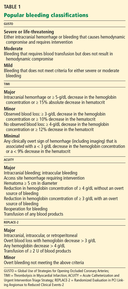
Since these classification schemes are based on different types of data, they yield different numbers when applied to the same study population. For instance, Rao et al4 pooled the data from the PURSUIT and PARAGON B trials (15,454 patients in all) and found that the incidence of severe bleeding (by the GUSTO criteria) was 1.2%, while the rate of major bleeding (by the TIMI criteria) was 8.2%.
What was the treatment strategy?
Another reason that the true incidence of bleeding is hard to determine is that different studies used treatment strategies that differed in the type, timing, and dose of antithrombotic agents and whether invasive procedures were used early. For example, if unfractionated heparin is used aggressively in regimens that are not adjusted for weight and with a higher target for the activated clotting time, the risk of bleeding is higher than with conservative dosing.5–7
Subherwal et al8 evaluated the effect of treatment strategy on the incidence of bleeding in patients with non-ST-elevation acute coronary syndromes who received two or more antithrombotic drugs in the CRUSADE Quality Improvement Initiative. The risk of bleeding was higher with an invasive approach (catheterization) than with a conservative approach (no catheterization), regardless of baseline bleeding risk.
What type of study was it?
Another source of variation is the design of the study. Registries differ from clinical trials in patient characteristics and in the way data are gathered (prospectively vs retrospectively).
In registries, data are often collected retrospectively, whereas in clinical trials the data are prospectively collected. For this reason, the definition of bleeding in registries is often based on events that are easily identified through chart review, such as transfusion. This may lead to a lower reported rate of bleeding, since other, less serious bleeding events such as access-site hematomas and epistaxis may not be documented in the medical record.
On the other hand, registries often include older and sicker patients, who may be more prone to bleeding and who are often excluded from clinical trials. This may lead to a higher rate of reported bleeding.9
Where the study was conducted makes a difference as well, owing to regional practice differences. For example, Moscucci et al10 reported that the incidence of major bleeding in 24,045 patients with non-ST-elevation acute coronary syndromes in the GRACE registry (in 14 countries worldwide) was 3.9%. In contrast, Yang et al11 reported that the rate of bleeding in the CRUSADE registry (in the United States) was 10.3%.
This difference was partly influenced by different definitions of bleeding. The GRACE registry defined major bleeding as life-threatening events requiring transfusion of two or more units of packed red blood cells, or resulting in an absolute decrease in the hematocrit of 10% or more or death, or hemorrhagic subdural hematoma. In contrast, the CRUSADE data reflect bleeding requiring transfusion. However, practice patterns such as greater use of invasive procedures in the United States may also be responsible.
Rao and colleagues12 examined international variation in blood transfusion rates among patients with acute coronary syndromes. Patients outside the United States were significantly less likely to receive transfusions, even after adjusting for patient and practice differences.
Taking these confounders into account, it is reasonable to estimate that the frequency of bleeding in patients with non-ST-elevation acute coronary syndromes ranges from less than 1% to 10%.13
BLEEDING IS ASSOCIATED WITH POOR OUTCOMES
Regardless of the definition or the data source, hemorrhagic complications are associated with a higher risk of death and nonfatal adverse events, both in the short term and in the long term.
Short-term outcomes
A higher risk of death. In the GRACE registry study by Moscucci et al10 discussed above, patients who had major bleeding were significantly more likely to die during their hospitalization than those who did not (odds ratio [OR] 1.64, 95% confidence interval [CI] 1.18–2.28).
Rao et al14 evaluated pooled data from the multicenter international GUSTO IIb, PURSUIT, and PARAGON A and B trials and found that the effects of bleeding in non-ST-elevation acute coronary syndromes extended beyond the hospital stay. The more severe the bleeding (by the GUSTO criteria), the greater the adjusted hazard ratio (HR) for death within 30 days:
- With mild bleeding—HR 1.6, 95% CI 1.3–1.9
- With moderate bleeding—HR 2.7, 95% CI 2.3–3.4
- With severe bleeding—HR 10.6, 95% CI 8.3–13.6.
The pattern was the same for death within 6 months:
- With mild bleeding—HR 1.4, 95% CI 1.2–1.6
- With moderate bleeding—HR 2.1, 95% CI 1.8–2.4
- With severe bleeding, HR 7.5, 95% CI 6.1–9.3.
These findings were confirmed by Eikelboom et al15 in 34,146 patients with acute coronary syndromes in the OASIS registry, the OASIS-2 trial, and the CURE randomized trial. In the first 30 days, five times as many patients died (12.8% vs 2.5%; P < .0009) among those who developed major bleeding compared with those who did not. These investigators defined major bleeding as bleeding that was life-threatening or significantly disabling or that required transfusion of two or more units of packed red blood cells.
A higher risk of nonfatal adverse events. Bleeding after antithrombotic therapy for non-ST-elevation acute coronary syndromes has also been associated with nonfatal adverse events such as stroke and stent thrombosis.
For example, in the study by Eikelboom et al,15 major bleeding was associated with a higher risk of recurrent ischemic events. Approximately 1 in 5 patients in the OASIS trials who developed major bleeding during the first 30 days died or had a myocardial infarction or stroke by 30 days, compared with 1 in 20 of those who did not develop major bleeding during the first 30 days. However, after events that occurred during the first 30 days were excluded, the association between major bleeding and both myocardial infarction and stroke was no longer evident between 30 days and 6 months.
Manoukian et al16 evaluated the impact of major bleeding in 13,819 patients with highrisk acute coronary syndromes undergoing treatment with an early invasive strategy in the ACUITY trial. At 30 days, patients with major bleeding had higher rates of the composite end point of death, myocardial infarction, or unplanned revascularization for ischemia (23.1% vs 6.8%, P < .0001) and of stent thrombosis (3.4% vs 0.6%, P < .0001).
Long-term outcomes
The association between bleeding and adverse outcomes persists in the long term as well, although the mechanisms underlying this association are not well studied.
Kinnaird et al17 examined the data from 10,974 unselected patients who underwent percutaneous coronary intervention. At 1 year, the following percentages of patients had died:
- After TIMI major bleeding—17.2%
- After TIMI minor bleeding—9.1%
- After no bleeding—5.5%.
However, after adjustment for potential confounders, only transfusion remained a significant predictor of 1-year mortality.
Mehran et al18 evaluated 1-year mortality data from the ACUITY trial. Compared with the rate in patients who had no major bleeding and no myocardial infarction, the hazard ratios for death were:
- After major bleeding—HR 3.5, 95% CI 2.7–4.4
- After myocardial infarction—HR 3.1, 95% CI 2.4–3.9.
Interestingly, the risk of death associated with myocardial infarction abated after 7 days, while the risk associated with bleeding persisted beyond 30 days and remained constant throughout the first year following the bleeding event.
Similarly, Ndrepepa and colleagues19 examined pooled data from four ISAR trials using the TIMI bleeding scale and found that myocardial infarction, target vessel revascularization, and major bleeding all had similar discriminatory ability at predicting 1-year mortality.
In patients undergoing elective or urgent percutaneous coronary intervention in the REPLACE-2 trial,20 independent predictors of death by 1 year were21:
- Major hemorrhage (OR 2.66, 95% CI 1.44–4.92)
- Periprocedural myocardial infarction (OR 2.46, 95% CI 1.44–4.20).
THEORIES OF HOW BLEEDING MAY CAUSE ADVERSE OUTCOMES
Several mechanisms have been proposed to explain the association between bleeding during treatment for acute coronary syndromes and adverse clinical outcomes.13,22
The immediate effects of bleeding are thought to be hypotension and a reflex hyperadrenergic state to compensate for the loss of intravascular volume.23 This physiologic response is believed to contribute to myocardial ischemia by further decreasing myocardial oxygen supply in obstructive coronary disease.
Trying to minimize blood loss, physicians may withhold anticoagulation and antiplatelet therapy, which in turn may lead to further ischemia.24 To compensate for blood loss, physicians may also resort to blood transfusion. However, depletion of 2,3-diphosphoglycerate and nitric oxide in stored donor red blood cells is postulated to reduce oxygen delivery by increasing hemoglobin’s affinity for oxygen, leading to induced microvascular obstruction and adverse inflammatory reactions.15,25
Recent data have also begun to elucidate the long-term effects of bleeding during acute coronary syndrome management. Patients with anemia during the acute phase of infarction have greater neurohormonal activation.26 These adaptive responses to anemia may lead to eccentric left ventricular remodeling that may lead to higher oxygen consumption, increased diastolic wall stress, interstitial fibrosis, and accelerated myocyte loss.27–30
Nevertheless, we must point out that although strong associations between bleeding and adverse outcomes have been established, direct causality has not.
TO PREVENT BLEEDING, START BY ASSESSING RISK
The CRUSADE bleeding risk score
The CRUSADE bleeding score (calculator available at http://www.crusadebleedingscore.org/) was developed and validated in more than 89,000 community-treated patients with non-ST-elevation acute coronary syndromes.8 It is based on eight variables:
- Sex (higher risk in women)
- History of diabetes (higher risk)
- Prior vascular disease (higher risk)
- Heart rate (the higher the rate, the higher the risk)
- Systolic blood pressure (higher risk with pressures above or below the 121–180 mm Hg range)
- Signs of congestive heart failure (higher risk)
- Baseline hematocrit (the lower the hematocrit, the higher the risk)
- Creatinine clearance (by the Cockcroft-Gault formula; the lower the creatinine clearance, the higher the risk).
Patients who are found to have bleeding scores suggesting a moderate or higher risk of bleeding should be considered for medications associated with a favorable bleeding profile, and for radial access at the time of coronary angiography. Scores are graded as follows8:
- < 21: Very low risk
- 21–30: Low risk
- 31–40: Moderate risk
- 41–50: High risk
- > 50: Very high risk.
The CRUSADE bleeding score is unique in that, unlike earlier risk stratification tools, it was developed in a “real world” population, not in subgroups or in a clinical trial. It can be calculated at baseline to help guide the selection of treatment.8
Adjusting the heparin regimen in patients at risk of bleeding
Both the joint American College of Cardiology/American Heart Association1 and the European Society of Cardiology guidelines31 for the treatment of non-ST-elevation acute coronary syndromes recommend taking steps to prevent bleeding, such as adjusting the dosage of unfractionated heparin, using safer drugs, reducing the duration of antithrombotic treatment, and using combinations of antithrombotic and antiplatelet agents according to proven indications.31
In the CRUSADE registry, 42% of patients with non-ST-elevation acute coronary syndromes received at least one initial dose of antithrombotic drug outside the recommended range, resulting in an estimated 15% excess of bleeding events.32 Thus, proper dosing is a target for prevention.
Appropriate antithrombotic dosing takes into account the patient’s age, weight, and renal function. However, heparin dosage in the current guidelines1 is based on weight only: a loading dose of 60 U/kg (maximum 4,000 U) by intravenous bolus, then 12 U/kg/hour (maximum 1,000 U/hour) to maintain an activated partial thromboplastin time of 50 to 70 seconds.1
Renal dysfunction is particularly worrisome in patients with non-ST-elevation acute coronary syndromes because it is associated with higher rates of major bleeding and death. In the OASIS-5 trial,33 the overall risk of death was approximately five times higher in patients in the lowest quartile of renal function (glomerular filtration rate < 58 mL/min/1.73 m2) than in the highest quartile (glomerular filtration rate ≥ 86 mL/min/1.73 m2).
Renal function must be evaluated not only on admission but also throughout the hospital stay. Patients presenting with acute coronary syndromes often experience fluctuations in renal function that would call for adjustment of heparin dosing, either increasing the dose to maximize the drug’s efficacy if renal function is recovering or decreasing the dose to prevent bleeding if renal function is deteriorating.
Clopidogrel vs prasugrel
Certain medications should be avoided when the risk of bleeding outweighs any potential benefit in terms of ischemia.
For example, in a randomized trial,34 prasugrel (Effient), a potent thienopyridine, was associated with a significantly lower rate of the composite end point of stroke, myocardial infarction, or death than clopidogrel (Plavix) in patients with acute coronary syndromes undergoing percutaneous coronary interventions. However, it did not seem to offer any advantage in patients 75 years old and older, those with prior stroke or transient ischemic attack, or those weighing less than 60 kg, and it posed a substantially higher risk of bleeding.
With clopidogrel, the risk of acute bleeding is primarily in patients who undergo coronary artery bypass grafting within 5 days of receiving a dose.35,36 Therefore, clopidogrel should be stopped 5 to 7 days before bypass surgery.1 Importantly, there is no increased risk of recurrent ischemic events during this 5-day waiting period in patients who receive clopidogrel early. Therefore, the recommendation to stop clopidogrel before surgery does not negate the benefits of early treatment.36
Lower-risk drugs: Fondaparinux and bivalirudin
At this time, only two agents have been studied in clinical trials that have specifically focused on reducing bleeding risk: fondaparinux (Arixtra) and bivalirudin (Angiomax).20,37–39
Fondaparinux
OASIS-5 was a randomized, double-blind trial that compared fondaparinux and enoxaparin (Lovenox) in patients with acute coronary syndromes.38 Fondaparinux was similar to enoxaparin in terms of the combined end point of death, myocardial infarction, or refractory ischemia at 9 days, and fewer patients on fondaparinux developed bleeding (2.2% vs 4.1%, HR 0.52; 95% CI 0.44–0.61). This difference persisted during long-term follow-up.
Importantly, fewer patients died in the fondaparinux group. At 180 days, 638 (6.5%) of the patients in the enoxaparin group had died, compared with 574 (5.8%) in the fondaparinux group, a difference of 64 deaths (P = .05). The authors found that 41 fewer patients in the fondaparinux group than in the enoxaparin group died after major bleeding, and 20 fewer patients in the fondaparinux group died after minor bleeding.38 Thus, most of the difference in mortality rates between the two groups was attributed to a lower incidence of bleeding with fondaparinux.
Unfortunately, despite its safe bleeding profile, fondaparinux has fallen out of favor for use in acute coronary syndromes, owing to a higher risk of catheter thrombosis in the fondaparinux group (0.9%) than in those undergoing percutaneous coronary interventions with enoxaparin alone (0.4%) in the OASIS-5 trial.40
Bivalirudin
The direct thrombin inhibitor bivalirudin has been studied in three large randomized trials in patients undergoing percutaneous coronary interventions.20,37,41
The ACUITY trial37 was a prospective, open-label, randomized, multicenter trial that compared three regimens in patients with moderate or high-risk non-ST-elevation acute coronary syndromes:
- Heparin plus a glycoprotein IIb/IIIa inhibitor
- Bivalirudin plus a glycoprotein IIb/IIIa inhibitor
- Bivalirudin alone.
Bivalirudin alone was as effective as heparin plus a glycoprotein IIb/IIIa inhibitor with respect to the composite ischemia end point, which at 30 days had occurred in 7.8% vs 7.3% of the patients in these treatment groups (P = .32, RR 1.08; 95% CI 0.93–1.24), and it was superior with respect to major bleeding (3.0% vs 5.7%, P < .001, RR 0.53; 95% CI 0.43–0.65).
The HORIZONS-AMI study41 was a prospective, open-label, randomized, multicenter trial that compared bivalirudin alone vs heparin plus a glycoprotein IIb/IIIa inhibitor in patients with ST-elevation acute coronary syndromes who were undergoing primary percutaneous coronary interventions. The two primary end points were major bleeding and net adverse events.
At 1 year, patients assigned to bivalirudin had a lower rate of major bleeding than did controls (5.8% vs 9.2%, HR 0.61, 95% CI 0.48–0.78, P < .0001), with similar rates of major adverse cardiac events in both groups (11.9% vs 11.9%, HR 1.00, 95% CI 0.82– 1.21, P = .98).41
Both OASIS 5 and HORIZONS-AMI are examples of clinical trials in which strategies that reduced bleeding risk were also associated with improved survival.
For cardiac catheterization, inserting the catheter in the wrist poses less risk
Bleeding is currently the most common noncardiac complication in patients undergoing percutaneous coronary interventions, and it most often occurs at the vascular access site.17
Rao et al12 evaluated data from 593,094 procedures in the National Cardiovascular Data Registry and found that, compared with the femoral approach, the use of transradial percutaneous coronary intervention was associated with a similar rate of procedural success (OR 1.02, 95% CI 0.93–1.12) but a significantly lower risk of bleeding complications (OR 0.42, 95% CI 0.31–0.56) after multivariable adjustment.
The use of smaller sheath sizes (4F–6F) and preferential use of bivalirudin over unfractionated heparin and glycoprotein IIb/IIIa inhibitor therapy are other methods described to decrease the risk of bleeding after percutaneous coronary interventions.20,41–49
IF BLEEDING OCCURS
Once a bleeding complication occurs, cessation of therapy is a potential option. Stopping or reversing antithrombotic and antiplatelet therapy is warranted in the event of major bleeding (eg, gastrointestinal, retroperitoneal, intracranial).31
Stopping antithrombotic and antiplatelet therapy
Whether bleeding is minor or major, the risk of a recurrent thrombotic event must be considered, especially in patients who have undergone revascularization, stent implantation, or both. The risk of acute thrombotic events after interrupting antithrombotic or antiplatelet agents is considered greatest 4 to 5 days following revascularization or percutaneous coronary intervention.15 If bleeding can be controlled with local treatment such as pressure, packing, or dressing, antithrombotic and antiplatelet therapy need not be interrupted.50
Current guidelines recommend strict control of hemorrhage for at least 24 hours before reintroducing antiplatelet or antithrombotic agents.
It is also important to remember that in the setting of gastrointestinal bleeding due to peptic ulcer disease, adjunctive proton pump inhibitors are recommended after restarting antiplatelet or antithrombotic therapy or both.
Importantly, evidence-based antithrombotic medications (especially dual antiplatelet therapy) should be restarted once the acute bleeding event has resolved.31
Reversal of anticoagulant and antiplatelet therapies
Unfractionated heparin is reversed with infusion of protamine sulfate at a dose of 1 mg per 100 U of unfractionated heparin given over the previous 4 hours.51,52 The rate of protamine sulfate infusion should be less than 100 mg over 2 hours, with 50% of the dose given initially and subsequent doses titrated according to bleeding response.52,53 Protamine sulfate is associated with a risk of hypotension and bradycardia, and for this reason it should be given no faster than 5 mg/min.
Low-molecular-weight heparin (LMWH) can be inhibited by 1 mg of protamine sulfate for each 1 mg of LMWH given over the previous 4 hours.51,52
However, protamine sulfate only partially neutralizes the anticoagulant effect of LMWH. In cases in which protamine sulfate is unsuccessful in abating bleeding associated with LMWH use, guidelines allow for the use of recombinant factor VIIa (NovoSeven).31 In healthy volunteers given fondaparinux, recombinant factor VIIa normalized coagulation times and thrombin generation within 1.5 hours, with a sustained effect for 6 hours.52
It is important to note that the use of this agent has not been fully studied, it is very costly (a single dose of 40 μg/kg costs from $3,000 to $4,000), and it is linked to reports of increased risk of thrombotic complications.54,55
Antiplatelet agents are more complex to reverse. The antiplatelet actions of aspirin and clopidogrel wear off as new platelets are produced. Approximately 10% of a patient’s platelet count is produced daily; thus, the antiplatelet effects of aspirin and clopidogrel can persist for 5 to 10 days.31,56
If these agents need to be reversed quickly to stop bleeding, according to expert consensus the aspirin effect can be reversed by transfusion of one unit of platelets. The antiplatelet effect of clopidogrel is more significant than that of aspirin; thus, two units of platelets are recommended.56
Glycoprotein IIb/IIIa inhibitors. If a major bleeding event requires the reversal of glycoprotein IIb/IIIa inhibitor therapy, the treatment must take into consideration the pharmacodynamics of the target drug. Both eptifibatide (Integrilin) and tirofiban (Aggrastat) competitively inhibit glycoprotein IIb/IIIa receptors; thus, their effects depend on dosing, elimination, and time. Due to the stoichiometry of both drugs, transfusion of platelets is ineffective. Both eptifibatide and tirofiban are eliminated by the kidney; thus, normal renal function is key to the amount of time it takes for platelet function to return to baseline.57 Evidence suggests that fibrinogen-rich plasma can be administered to restore platelet function.31,58,59
Abciximab (ReoPro). Whereas reversal of eptifibatide and tirofiban focuses on overcoming competitive inhibition, neutralization of abciximab involves overcoming its high receptor affinity. At 24 hours after abciximab infusion is stopped, platelet aggregation may still be inhibited by up to 50%. Fortunately, owing to abciximab’s short plasma half-life and its dilution in serum, platelet transfusion is effective in reversing its antiplatelet effects.31,57
Blood transfusion
Long considered beneficial to critically ill patients, blood transfusion to maintain hematocrit levels during acute coronary syndromes has come under intense scrutiny. Randomized trials have shown that transfusion should not be given aggressively to critically ill patients.60 In acute coronary syndromes, there are only observational data.
Rao et al61 used detailed clinical data from 24,112 patients with acute coronary syndromes in the GUSTO IIb, PURSUIT, and PARAGON B trials to determine the association between blood transfusion and outcomes in patients who developed moderate to severe bleeding, anemia, or both during their hospitalization. The rates of death in the hospital and at 30 days were significantly higher in patients who received a transfusion (30-day mortality HR 3.94; 95% CI 3.36–4.75). However, there was no significant association between transfusion and the 30-day mortality rate if the nadir hematocrit was 25% or less.
Of note: no randomized clinical trial has evaluated transfusion strategies in acute coronary syndromes at this time. Until such data are available, it is reasonable to follow published guidelines and to avoid transfusion in stable patients with ischemic heart disease unless the hematocrit is 25% or less.31 The risks and benefits of blood transfusion should be carefully weighed. Routine use of transfusion to maintain predefined hemoglobin levels is not recommended in stable patients.
- Anderson JL, Adams CD, Antman EM, et al. ACC/AHA 2007 guidelines for the management of patients with unstable angina/non-ST-elevation myocardial infarction. J Am Coll Cardiol 2007; 50:e1–e157.
- The GUSTO Investigators. An international randomized trial comparing four thrombolytic strategies for acute myocardial infarction. N Engl J Med 1993; 329:673–682.
- Chesebro JH, Knatterud G, Roberts R, et al. Thrombolysis in Myocardial Infarction (TIMI) Trial, Phase I: a comparison between intravenous tissue plasminogen activator and intravenous streptokinase. Clinical findings through hospital discharge. Circulation 1987; 76:142–154.
- Rao SV, O’Grady K, Pieper KS, et al. A comparison of the clinical impact of bleeding measured by two different classifications among patients with acute coronary syndromes. J Am Coll Cardiol 2006; 47:809–816.
- Granger CB, Hirsch J, Califf RM, et al. Activated partial thromboplastin time and outcome after thrombolytic therapy for acute myocardial infarction: results from the GUSTO-I trial. Circulation 1996; 93:870–878.
- Gilchrist IC, Berkowitz SD, Thompson TD, Califf RM, Granger CB. Heparin dosing and outcome in acute coronary syndromes: the GUSTO-IIb experience. Global Use of Strategies to Open Occluded Coronary Arteries. Am Heart J 2002; 144:73–80.
- Tolleson TR, O’Shea JC, Bittl JA, et al. Relationship between heparin anticoagulation and clinical outcomes in coronary stent intervention: observations from the ESPRIT trial. J Am Coll Cardiol 2003; 41:386–393.
- Subherwal S, Bach RG, Chen AY, et al. Baseline risk of major bleeding in non-ST-segment-elevation myocardial infarction: the CRUSADE (Can Rapid risk stratification of Unstable angina patients Suppress ADverse outcomes with Early implementation of the ACC/AHA Guidelines) Bleeding Score. Circulation 2009; 119:1873–1882.
- Bassand JP. Bleeding and transfusion in acute coronary syndromes: a shift in the paradigm. Heart 2008; 94:661–666.
- Moscucci M, Fox KA, Cannon CP, et al. Predictors of major bleeding in acute coronary syndromes: the Global Registry of Acute Coronary Events (GRACE). Eur Heart J 2003; 24:1815–1823.
- Yang X, Alexander KP, Chen AY, et al; CRUSADE Investigators. The implications of blood transfusions for patients with non-ST-segment elevation acute coronary syndromes: results from the CRUSADE National Quality Improvement Initiative. J Am Coll Cardiol 2005; 46:1490–1495.
- Rao SV, Ou FS, Wang TY, et al. Trends in the prevalence and outcomes of radial and femoral approaches to percutaneous coronary intervention: a report from the National Cardiovascular Data Registry. JACC Cardiovasc Interv 2008; 1:379–386.
- Rao SV, Eikelboom JA, Granger CB, Harrington RA, Califf RM, Bassand JP. Bleeding and blood transfusion issues in patients with non-ST-segment elevation acute coronary syndromes. Eur Heart J 2007; 28:1193–1204.
- Rao SV, O’Grady K, Pieper KS, et al. Impact of bleeding severity on clinical outcomes among patients with acute coronary syndromes. Am J Cardiol 2005; 96:1200–1206.
- Eikelboom JW, Mehta SR, Anand SS, Xie C, Fox KA, Yusuf S. Adverse impact of bleeding on prognosis in patients with acute coronary syndromes. Circulation 2006; 114:774–782.
- Manoukian SV, Feit F, Mehran R, et al. Impact of major bleeding on 30-day mortality and clinical outcomes in patients with acute coronary syndromes: an analysis from the ACUITY Trial. J Am Coll Cardiol 2007; 49:1362–1368.
- Kinnaird TD, Stabile E, Mintz GS, et al. Incidence, predictors, and prognostic implications of bleeding and blood transfusion following percutaneous coronary interventions. Am J Cardiol 2003; 92:930–935.
- Mehran R, Pocock SJ, Stone GW, et al. Associations of major bleeding and myocardial infarction with the incidence and timing of mortality in patients presenting with non-ST-elevation acute coronary syndromes: a risk model from the ACUITY trial. Eur Heart J 2009; 30:1457–1466.
- Ndrepepa G, Berger PB, Mehilli J, et al. Periprocedural bleeding and 1-year outcome after percutaneous coronary interventions: appropriateness of including bleeding as a component of a quadruple end point. J Am Coll Cardiol 2008; 51:690–697.
- Lincoff AM, Bittl JA, Harrington RA, et al; REPLACE-2 Investigators. Bivalirudin and provisional glycoprotein IIb/IIIa blockade compared with heparin and planned glycoprotein IIb/IIIa blockade during percutaneous coronary intervention: REPLACE-2 randomized trial. JAMA 2003; 289:853–863.
- Feit F, Voeltz MD, Attubato MJ, et al. Predictors and impact of major hemorrhage on mortality following percutaneous coronary intervention from the REPLACE-2 Trial. Am J Cardiol 2007; 100:1364–1369.
- Fitchett D. The impact of bleeding in patients with acute coronary syndromes: how to optimize the benefits of treatment and minimize the risk. Can J Cardiol 2007; 23:663–671.
- Bassand JP. Impact of anaemia, bleeding, and transfusions in acute coronary syndromes: a shift in the paradigm. Eur Heart J 2007; 28:1273–1274.
- Yan AT, Yan RT, Huynh T, et al; INTERACT Investigators. Bleeding and outcome in acute coronary syndrome: insights from continuous electrocardiogram monitoring in the Integrilin and Enoxaparin Randomized Assessment of Acute Coronary Syndrome Treatment (INTERACT) Trial. Am Heart J 2008; 156:769–775.
- Jolicoeur EM, O’Neill WW, Hellkamp A, et al; APEX-AMI Investigators. Transfusion and mortality in patients with ST-segment elevation myocardial infarction treated with primary percutaneous coronary intervention. Eur Heart J 2009; 30:2575–2583.
- Gehi A, Ix J, Shlipak M, Pipkin SS, Whooley MA. Relation of anemia to low heart rate variability in patients with coronary heart disease (from the Heart and Soul study). Am J Cardiol 2005; 95:1474–1477.
- Anand I, McMurray JJ, Whitmore J, et al. Anemia and its relationship to clinical outcome in heart failure. Circulation 2004; 110:149–154.
- O’Riordan E, Foley RN. Effects of anaemia on cardiovascular status. Nephrol Dial Transplant 2000; 15(suppl 3):19–22.
- Olivetti G, Quaini F, Lagrasta C, et al. Myocyte cellular hypertrophy and hyperplasia contribute to ventricular wall remodeling in anemia-induced cardiac hypertrophy in rats. Am J Pathol 1992; 141:227–239.
- Aronson D, Suleiman M, Agmon Y, et al. Changes in haemoglobin levels during hospital course and long-term outcome after acute myocardial infarction. Eur Heart J 2007; 28:1289–1296.
- Task Force for Diagnosis and Treatment of Non-ST-Segment Elevation Acute Coronary Syndromes of European Society of Cardiology; Bassand JP, Hamm CW, Ardissino D, et al. Guidelines for the diagnosis and treatment of non-ST-segment elevation acute coronary syndromes. Eur Heart J 2007; 28:1598–1660.
- Alexander KP, Chen AY, Roe MT, et al; CRUSADE Investigators. Excess dosing of antiplatelet and antithrombin agents in the treatment of non-ST-segment elevation acute coronary syndromes. JAMA 2005; 294:3108–3116.
- Fox KA, Bassand JP, Mehta SR, et al; OASIS 5 Investigators. Influence of renal function on the efficacy and safety of fondaparinux relative to enoxaparin in non ST-segment elevation acute coronary syndromes. Ann Intern Med 2007; 147:304–310.
- Wiviott SD, Braunwald E, McCabe CH, et al; TRITON-TIMI 38 Investigators. Prasugrel versus clopidogrel in patients with acute coronary syndromes. N Engl J Med 2007; 357:2001–2015.
- Berger JS, Frye CB, Harshaw Q, Edwards FH, Steinhubl SR, Becker RC. Impact of clopidogrel in patients with acute coronary syndromes requiring coronary artery bypass surgery: a multicenter analysis. J Am Coll Cardiol 2008; 52:1693–1701.
- Fox KA, Mehta SR, Peters R, et al; Clopidogrel in Unstable angina to prevent Recurrent ischemic Events Trial. Benefits and risks of the combination of clopidogrel and aspirin in patients undergoing surgical revascularization for non-ST-elevation acute coronary syndrome: the Clopidogrel in Unstable angina to prevent Recurrent ischemic Events (CURE) Trial. Circulation 2004; 110:1202–1208.
- Stone GW, McLaurin BT, Cox DA, et al; ACUITY Investigators. Bivalirudin for patients with acute coronary syndromes. N Engl J Med 2006; 355:2203–2216.
- Fifth Organization to Assess Strategies in Acute Ischemic Syndromes Investigators; Yusuf S, Mehta SR, Chrolavicius S, et al. Comparison of fondaparinux and enoxaparin in acute coronary syndromes. N Engl J Med 2006; 354:1464–1476.
- Potsis TZ, Katsouras C, Goudevenos JA. Avoiding and managing bleeding complications in patients with non-ST-segment elevation acute coronary syndromes. Angiology 2009; 60:148–158.
- Mehta SR, Granger CB, Eikelboom JW, et al. Efficacy and safety of fondaparinux versus enoxaparin in patients with acute coronary syndromes undergoing percutaneous coronary intervention: results from the OASIS-5 trial. J Am Coll Cardiol 2007; 50:1742–1751.
- Mehran R, Lansky AJ, Witzenbichler B, et al; HORIZONS-AMI Trial Investigators. Bivalirudin in patients undergoing primary angioplasty for acute myocardial infarction (HORIZONS-AMI): 1-year results of a randomised controlled trial. Lancet 2009; 374:1149–1159.
- Stone GW, Ware JH, Bertrand ME, et al; ACUITY Investigators. Antithrombotic strategies in patients with acute coronary syndromes undergoing early invasive management: one-year results from the ACUITY trial. JAMA 2007; 298:2497–2506.
- Cantor WJ, Mahaffey KW, Huang Z, et al. Bleeding complications in patients with acute coronary syndrome undergoing early invasive management can be reduced with radial access, smaller sheath sizes, and timely sheath removal. Catheter Cardiovasc Interv 2007; 69:73–83.
- Büchler JR, Ribeiro EE, Falcão JL, et al. A randomized trial of 5 versus 7 French guiding catheters for transfemoral percutaneous coronary stent implantation. J Interv Cardiol 2008; 21:50–55.
- Shammas NW, Allie D, Hall P, et al; APPROVE Investigators. Predictors of in-hospital and 30-day complications of peripheral vascular interventions using bivalirudin as the primary anticoagulant: results from the APPROVE Registry. J Invasive Cardiol 2005; 17:356–359.
- Doyle BJ, Ting HH, Bell MR, et al. Major femoral bleeding complications after percutaneous coronary intervention: incidence, predictors, and impact on long-term survival among 17,901 patients treated at the Mayo Clinic from 1994 to 2005. JACC Cardiovasc Interv 2008; 1:202–209.
- Stone GW, White HD, Ohman EM, et al; Acute Catheterization and Urgent Intervention Triage strategy (ACUITY) trial investigators. Bivalirudin in patients with acute coronary syndromes undergoing percutaneous coronary intervention: a subgroup analysis from the Acute Catheterization and Urgent Intervention Triage strategy (ACUITY) trial. Lancet 2007; 369:907–919.
- Stone GW, Bertrand ME, Moses JW, et al; ACUITY Investigators. Routine upstream initiation vs deferred selective use of glycoprotein IIb/IIIa inhibitors in acute coronary syndromes: the ACUITY Timing trial. JAMA 2007; 297:591–602.
- Lincoff AM, Bittl JA, Kleiman NS, et al; REPLACE-1 Investigators. Comparison of bivalirudin versus heparin during percutaneous coronary intervention (the Randomized Evaluation of PCI Linking Angiomax to Reduced Clinical Events [REPLACE]-1 trial). Am J Cardiol 2004; 93:1092–1096.
- Barkun A, Bardou M, Marshall JK; Nonvariceal Upper GI Bleeding Consensus Conference Group. Consensus recommendations for managing patients with nonvariceal upper gastrointestinal bleeding. Ann Intern Med 2003; 139:843–857.
- Warkentin TE, Crowther MA. Reversing anticoagulants both old and new. Can J Anaesth 2002; 49:S11–S25.
- Crowther MA, Warkentin TE. Bleeding risk and the management of bleeding complications in patients undergoing anticoagulant therapy: focus on new anticoagulant agents. Blood 2008; 111:4871–4879.
- Kessler CM. Current and future challenges of antithrombotic agents and anticoagulants: strategies for reversal of hemorrhagic complications. Semin Hematol 2004; 41(suppl 1):44–50.
- Ganguly S, Spengel K, Tilzer LL, O’Neal B, Simpson SQ. Recombinant factor VIIa: unregulated continuous use in patients with bleeding and coagulopathy does not alter mortality and outcome. Clin Lab Haematol 2006; 28:309–312.
- O’Connell KA, Wood JJ, Wise RP, Lozier JN, Braun MM. Thromboembolic adverse events after use of recombinant human coagulation factor VIIa. JAMA 2006; 295:293–298.
- Beshay JE, Morgan H, Madden C, Yu W, Sarode R. Emergency reversal of anticoagulation and antiplatelet therapies in neurosurgical patients. J Neurosurg 2010; 112:307–318.
- Tcheng JE. Clinical challenges of platelet glycoprotein IIb/IIIa receptor inhibitor therapy: bleeding, reversal, thrombocytopenia, and retreatment. Am Heart J 2000; 139:S38–S45.
- Li YF, Spencer FA, Becker RC. Comparative efficacy of fibrinogen and platelet supplementation on the in vitro reversibility of competitive glycoprotein IIb/IIIa receptor-directed platelet inhibition. Am Heart J 2002; 143:725–732.
- Schroeder WS, Gandhi PJ. Emergency management of hemorrhagic complications in the era of glycoprotein IIb/IIIa receptor antagonists, clopidogrel, low molecular weight heparin, and third-generation fibrinolytic agents. Curr Cardiol Rep 2003; 5:310–317.
- Hébert PC, Wells G, Blajchman MA, et al. A multicenter, randomized, controlled clinical trial of transfusion requirements in critical care. Transfusion Requirements in Critical Care Investigators, Canadian Critical Care Trials Group. N Engl J Med 1999; 340:409–417.
- Rao SV, Jollis JG, Harrington RA, et al. Relationship of blood transfusion and clinical outcomes in patients with acute coronary syndromes. JAMA 2004; 292:1555–1562.
The medical management of non-ST-elevation acute coronary syndromes focuses on blocking the coagulation cascade and inhibiting platelets. This—plus diagnostic angiography followed, if needed, by revascularization—has reduced the rates of death and recurrent ischemic events.1 However, the combination of potent antithrombotic drugs and invasive procedures also increases the risk of bleeding.
This review discusses the incidence and complications associated with bleeding during the treatment of acute coronary syndromes and summarizes recommendations for preventing and managing bleeding in this setting.
THE TRUE INCIDENCE OF BLEEDING IS HARD TO DETERMINE
The optimal way to detect and analyze bleeding events in clinical trials and registries is highly debated. The reported incidences of bleeding during antithrombotic and antiplatelet therapy for non-ST-elevation acute coronary syndromes depend on how bleeding was defined, how the acute coronary syndromes were treated, and on other factors such as how the study was designed.
How was bleeding defined?
Since these classification schemes are based on different types of data, they yield different numbers when applied to the same study population. For instance, Rao et al4 pooled the data from the PURSUIT and PARAGON B trials (15,454 patients in all) and found that the incidence of severe bleeding (by the GUSTO criteria) was 1.2%, while the rate of major bleeding (by the TIMI criteria) was 8.2%.
What was the treatment strategy?
Another reason that the true incidence of bleeding is hard to determine is that different studies used treatment strategies that differed in the type, timing, and dose of antithrombotic agents and whether invasive procedures were used early. For example, if unfractionated heparin is used aggressively in regimens that are not adjusted for weight and with a higher target for the activated clotting time, the risk of bleeding is higher than with conservative dosing.5–7
Subherwal et al8 evaluated the effect of treatment strategy on the incidence of bleeding in patients with non-ST-elevation acute coronary syndromes who received two or more antithrombotic drugs in the CRUSADE Quality Improvement Initiative. The risk of bleeding was higher with an invasive approach (catheterization) than with a conservative approach (no catheterization), regardless of baseline bleeding risk.
What type of study was it?
Another source of variation is the design of the study. Registries differ from clinical trials in patient characteristics and in the way data are gathered (prospectively vs retrospectively).
In registries, data are often collected retrospectively, whereas in clinical trials the data are prospectively collected. For this reason, the definition of bleeding in registries is often based on events that are easily identified through chart review, such as transfusion. This may lead to a lower reported rate of bleeding, since other, less serious bleeding events such as access-site hematomas and epistaxis may not be documented in the medical record.
On the other hand, registries often include older and sicker patients, who may be more prone to bleeding and who are often excluded from clinical trials. This may lead to a higher rate of reported bleeding.9
Where the study was conducted makes a difference as well, owing to regional practice differences. For example, Moscucci et al10 reported that the incidence of major bleeding in 24,045 patients with non-ST-elevation acute coronary syndromes in the GRACE registry (in 14 countries worldwide) was 3.9%. In contrast, Yang et al11 reported that the rate of bleeding in the CRUSADE registry (in the United States) was 10.3%.
This difference was partly influenced by different definitions of bleeding. The GRACE registry defined major bleeding as life-threatening events requiring transfusion of two or more units of packed red blood cells, or resulting in an absolute decrease in the hematocrit of 10% or more or death, or hemorrhagic subdural hematoma. In contrast, the CRUSADE data reflect bleeding requiring transfusion. However, practice patterns such as greater use of invasive procedures in the United States may also be responsible.
Rao and colleagues12 examined international variation in blood transfusion rates among patients with acute coronary syndromes. Patients outside the United States were significantly less likely to receive transfusions, even after adjusting for patient and practice differences.
Taking these confounders into account, it is reasonable to estimate that the frequency of bleeding in patients with non-ST-elevation acute coronary syndromes ranges from less than 1% to 10%.13
BLEEDING IS ASSOCIATED WITH POOR OUTCOMES
Regardless of the definition or the data source, hemorrhagic complications are associated with a higher risk of death and nonfatal adverse events, both in the short term and in the long term.
Short-term outcomes
A higher risk of death. In the GRACE registry study by Moscucci et al10 discussed above, patients who had major bleeding were significantly more likely to die during their hospitalization than those who did not (odds ratio [OR] 1.64, 95% confidence interval [CI] 1.18–2.28).
Rao et al14 evaluated pooled data from the multicenter international GUSTO IIb, PURSUIT, and PARAGON A and B trials and found that the effects of bleeding in non-ST-elevation acute coronary syndromes extended beyond the hospital stay. The more severe the bleeding (by the GUSTO criteria), the greater the adjusted hazard ratio (HR) for death within 30 days:
- With mild bleeding—HR 1.6, 95% CI 1.3–1.9
- With moderate bleeding—HR 2.7, 95% CI 2.3–3.4
- With severe bleeding—HR 10.6, 95% CI 8.3–13.6.
The pattern was the same for death within 6 months:
- With mild bleeding—HR 1.4, 95% CI 1.2–1.6
- With moderate bleeding—HR 2.1, 95% CI 1.8–2.4
- With severe bleeding, HR 7.5, 95% CI 6.1–9.3.
These findings were confirmed by Eikelboom et al15 in 34,146 patients with acute coronary syndromes in the OASIS registry, the OASIS-2 trial, and the CURE randomized trial. In the first 30 days, five times as many patients died (12.8% vs 2.5%; P < .0009) among those who developed major bleeding compared with those who did not. These investigators defined major bleeding as bleeding that was life-threatening or significantly disabling or that required transfusion of two or more units of packed red blood cells.
A higher risk of nonfatal adverse events. Bleeding after antithrombotic therapy for non-ST-elevation acute coronary syndromes has also been associated with nonfatal adverse events such as stroke and stent thrombosis.
For example, in the study by Eikelboom et al,15 major bleeding was associated with a higher risk of recurrent ischemic events. Approximately 1 in 5 patients in the OASIS trials who developed major bleeding during the first 30 days died or had a myocardial infarction or stroke by 30 days, compared with 1 in 20 of those who did not develop major bleeding during the first 30 days. However, after events that occurred during the first 30 days were excluded, the association between major bleeding and both myocardial infarction and stroke was no longer evident between 30 days and 6 months.
Manoukian et al16 evaluated the impact of major bleeding in 13,819 patients with highrisk acute coronary syndromes undergoing treatment with an early invasive strategy in the ACUITY trial. At 30 days, patients with major bleeding had higher rates of the composite end point of death, myocardial infarction, or unplanned revascularization for ischemia (23.1% vs 6.8%, P < .0001) and of stent thrombosis (3.4% vs 0.6%, P < .0001).
Long-term outcomes
The association between bleeding and adverse outcomes persists in the long term as well, although the mechanisms underlying this association are not well studied.
Kinnaird et al17 examined the data from 10,974 unselected patients who underwent percutaneous coronary intervention. At 1 year, the following percentages of patients had died:
- After TIMI major bleeding—17.2%
- After TIMI minor bleeding—9.1%
- After no bleeding—5.5%.
However, after adjustment for potential confounders, only transfusion remained a significant predictor of 1-year mortality.
Mehran et al18 evaluated 1-year mortality data from the ACUITY trial. Compared with the rate in patients who had no major bleeding and no myocardial infarction, the hazard ratios for death were:
- After major bleeding—HR 3.5, 95% CI 2.7–4.4
- After myocardial infarction—HR 3.1, 95% CI 2.4–3.9.
Interestingly, the risk of death associated with myocardial infarction abated after 7 days, while the risk associated with bleeding persisted beyond 30 days and remained constant throughout the first year following the bleeding event.
Similarly, Ndrepepa and colleagues19 examined pooled data from four ISAR trials using the TIMI bleeding scale and found that myocardial infarction, target vessel revascularization, and major bleeding all had similar discriminatory ability at predicting 1-year mortality.
In patients undergoing elective or urgent percutaneous coronary intervention in the REPLACE-2 trial,20 independent predictors of death by 1 year were21:
- Major hemorrhage (OR 2.66, 95% CI 1.44–4.92)
- Periprocedural myocardial infarction (OR 2.46, 95% CI 1.44–4.20).
THEORIES OF HOW BLEEDING MAY CAUSE ADVERSE OUTCOMES
Several mechanisms have been proposed to explain the association between bleeding during treatment for acute coronary syndromes and adverse clinical outcomes.13,22
The immediate effects of bleeding are thought to be hypotension and a reflex hyperadrenergic state to compensate for the loss of intravascular volume.23 This physiologic response is believed to contribute to myocardial ischemia by further decreasing myocardial oxygen supply in obstructive coronary disease.
Trying to minimize blood loss, physicians may withhold anticoagulation and antiplatelet therapy, which in turn may lead to further ischemia.24 To compensate for blood loss, physicians may also resort to blood transfusion. However, depletion of 2,3-diphosphoglycerate and nitric oxide in stored donor red blood cells is postulated to reduce oxygen delivery by increasing hemoglobin’s affinity for oxygen, leading to induced microvascular obstruction and adverse inflammatory reactions.15,25
Recent data have also begun to elucidate the long-term effects of bleeding during acute coronary syndrome management. Patients with anemia during the acute phase of infarction have greater neurohormonal activation.26 These adaptive responses to anemia may lead to eccentric left ventricular remodeling that may lead to higher oxygen consumption, increased diastolic wall stress, interstitial fibrosis, and accelerated myocyte loss.27–30
Nevertheless, we must point out that although strong associations between bleeding and adverse outcomes have been established, direct causality has not.
TO PREVENT BLEEDING, START BY ASSESSING RISK
The CRUSADE bleeding risk score
The CRUSADE bleeding score (calculator available at http://www.crusadebleedingscore.org/) was developed and validated in more than 89,000 community-treated patients with non-ST-elevation acute coronary syndromes.8 It is based on eight variables:
- Sex (higher risk in women)
- History of diabetes (higher risk)
- Prior vascular disease (higher risk)
- Heart rate (the higher the rate, the higher the risk)
- Systolic blood pressure (higher risk with pressures above or below the 121–180 mm Hg range)
- Signs of congestive heart failure (higher risk)
- Baseline hematocrit (the lower the hematocrit, the higher the risk)
- Creatinine clearance (by the Cockcroft-Gault formula; the lower the creatinine clearance, the higher the risk).
Patients who are found to have bleeding scores suggesting a moderate or higher risk of bleeding should be considered for medications associated with a favorable bleeding profile, and for radial access at the time of coronary angiography. Scores are graded as follows8:
- < 21: Very low risk
- 21–30: Low risk
- 31–40: Moderate risk
- 41–50: High risk
- > 50: Very high risk.
The CRUSADE bleeding score is unique in that, unlike earlier risk stratification tools, it was developed in a “real world” population, not in subgroups or in a clinical trial. It can be calculated at baseline to help guide the selection of treatment.8
Adjusting the heparin regimen in patients at risk of bleeding
Both the joint American College of Cardiology/American Heart Association1 and the European Society of Cardiology guidelines31 for the treatment of non-ST-elevation acute coronary syndromes recommend taking steps to prevent bleeding, such as adjusting the dosage of unfractionated heparin, using safer drugs, reducing the duration of antithrombotic treatment, and using combinations of antithrombotic and antiplatelet agents according to proven indications.31
In the CRUSADE registry, 42% of patients with non-ST-elevation acute coronary syndromes received at least one initial dose of antithrombotic drug outside the recommended range, resulting in an estimated 15% excess of bleeding events.32 Thus, proper dosing is a target for prevention.
Appropriate antithrombotic dosing takes into account the patient’s age, weight, and renal function. However, heparin dosage in the current guidelines1 is based on weight only: a loading dose of 60 U/kg (maximum 4,000 U) by intravenous bolus, then 12 U/kg/hour (maximum 1,000 U/hour) to maintain an activated partial thromboplastin time of 50 to 70 seconds.1
Renal dysfunction is particularly worrisome in patients with non-ST-elevation acute coronary syndromes because it is associated with higher rates of major bleeding and death. In the OASIS-5 trial,33 the overall risk of death was approximately five times higher in patients in the lowest quartile of renal function (glomerular filtration rate < 58 mL/min/1.73 m2) than in the highest quartile (glomerular filtration rate ≥ 86 mL/min/1.73 m2).
Renal function must be evaluated not only on admission but also throughout the hospital stay. Patients presenting with acute coronary syndromes often experience fluctuations in renal function that would call for adjustment of heparin dosing, either increasing the dose to maximize the drug’s efficacy if renal function is recovering or decreasing the dose to prevent bleeding if renal function is deteriorating.
Clopidogrel vs prasugrel
Certain medications should be avoided when the risk of bleeding outweighs any potential benefit in terms of ischemia.
For example, in a randomized trial,34 prasugrel (Effient), a potent thienopyridine, was associated with a significantly lower rate of the composite end point of stroke, myocardial infarction, or death than clopidogrel (Plavix) in patients with acute coronary syndromes undergoing percutaneous coronary interventions. However, it did not seem to offer any advantage in patients 75 years old and older, those with prior stroke or transient ischemic attack, or those weighing less than 60 kg, and it posed a substantially higher risk of bleeding.
With clopidogrel, the risk of acute bleeding is primarily in patients who undergo coronary artery bypass grafting within 5 days of receiving a dose.35,36 Therefore, clopidogrel should be stopped 5 to 7 days before bypass surgery.1 Importantly, there is no increased risk of recurrent ischemic events during this 5-day waiting period in patients who receive clopidogrel early. Therefore, the recommendation to stop clopidogrel before surgery does not negate the benefits of early treatment.36
Lower-risk drugs: Fondaparinux and bivalirudin
At this time, only two agents have been studied in clinical trials that have specifically focused on reducing bleeding risk: fondaparinux (Arixtra) and bivalirudin (Angiomax).20,37–39
Fondaparinux
OASIS-5 was a randomized, double-blind trial that compared fondaparinux and enoxaparin (Lovenox) in patients with acute coronary syndromes.38 Fondaparinux was similar to enoxaparin in terms of the combined end point of death, myocardial infarction, or refractory ischemia at 9 days, and fewer patients on fondaparinux developed bleeding (2.2% vs 4.1%, HR 0.52; 95% CI 0.44–0.61). This difference persisted during long-term follow-up.
Importantly, fewer patients died in the fondaparinux group. At 180 days, 638 (6.5%) of the patients in the enoxaparin group had died, compared with 574 (5.8%) in the fondaparinux group, a difference of 64 deaths (P = .05). The authors found that 41 fewer patients in the fondaparinux group than in the enoxaparin group died after major bleeding, and 20 fewer patients in the fondaparinux group died after minor bleeding.38 Thus, most of the difference in mortality rates between the two groups was attributed to a lower incidence of bleeding with fondaparinux.
Unfortunately, despite its safe bleeding profile, fondaparinux has fallen out of favor for use in acute coronary syndromes, owing to a higher risk of catheter thrombosis in the fondaparinux group (0.9%) than in those undergoing percutaneous coronary interventions with enoxaparin alone (0.4%) in the OASIS-5 trial.40
Bivalirudin
The direct thrombin inhibitor bivalirudin has been studied in three large randomized trials in patients undergoing percutaneous coronary interventions.20,37,41
The ACUITY trial37 was a prospective, open-label, randomized, multicenter trial that compared three regimens in patients with moderate or high-risk non-ST-elevation acute coronary syndromes:
- Heparin plus a glycoprotein IIb/IIIa inhibitor
- Bivalirudin plus a glycoprotein IIb/IIIa inhibitor
- Bivalirudin alone.
Bivalirudin alone was as effective as heparin plus a glycoprotein IIb/IIIa inhibitor with respect to the composite ischemia end point, which at 30 days had occurred in 7.8% vs 7.3% of the patients in these treatment groups (P = .32, RR 1.08; 95% CI 0.93–1.24), and it was superior with respect to major bleeding (3.0% vs 5.7%, P < .001, RR 0.53; 95% CI 0.43–0.65).
The HORIZONS-AMI study41 was a prospective, open-label, randomized, multicenter trial that compared bivalirudin alone vs heparin plus a glycoprotein IIb/IIIa inhibitor in patients with ST-elevation acute coronary syndromes who were undergoing primary percutaneous coronary interventions. The two primary end points were major bleeding and net adverse events.
At 1 year, patients assigned to bivalirudin had a lower rate of major bleeding than did controls (5.8% vs 9.2%, HR 0.61, 95% CI 0.48–0.78, P < .0001), with similar rates of major adverse cardiac events in both groups (11.9% vs 11.9%, HR 1.00, 95% CI 0.82– 1.21, P = .98).41
Both OASIS 5 and HORIZONS-AMI are examples of clinical trials in which strategies that reduced bleeding risk were also associated with improved survival.
For cardiac catheterization, inserting the catheter in the wrist poses less risk
Bleeding is currently the most common noncardiac complication in patients undergoing percutaneous coronary interventions, and it most often occurs at the vascular access site.17
Rao et al12 evaluated data from 593,094 procedures in the National Cardiovascular Data Registry and found that, compared with the femoral approach, the use of transradial percutaneous coronary intervention was associated with a similar rate of procedural success (OR 1.02, 95% CI 0.93–1.12) but a significantly lower risk of bleeding complications (OR 0.42, 95% CI 0.31–0.56) after multivariable adjustment.
The use of smaller sheath sizes (4F–6F) and preferential use of bivalirudin over unfractionated heparin and glycoprotein IIb/IIIa inhibitor therapy are other methods described to decrease the risk of bleeding after percutaneous coronary interventions.20,41–49
IF BLEEDING OCCURS
Once a bleeding complication occurs, cessation of therapy is a potential option. Stopping or reversing antithrombotic and antiplatelet therapy is warranted in the event of major bleeding (eg, gastrointestinal, retroperitoneal, intracranial).31
Stopping antithrombotic and antiplatelet therapy
Whether bleeding is minor or major, the risk of a recurrent thrombotic event must be considered, especially in patients who have undergone revascularization, stent implantation, or both. The risk of acute thrombotic events after interrupting antithrombotic or antiplatelet agents is considered greatest 4 to 5 days following revascularization or percutaneous coronary intervention.15 If bleeding can be controlled with local treatment such as pressure, packing, or dressing, antithrombotic and antiplatelet therapy need not be interrupted.50
Current guidelines recommend strict control of hemorrhage for at least 24 hours before reintroducing antiplatelet or antithrombotic agents.
It is also important to remember that in the setting of gastrointestinal bleeding due to peptic ulcer disease, adjunctive proton pump inhibitors are recommended after restarting antiplatelet or antithrombotic therapy or both.
Importantly, evidence-based antithrombotic medications (especially dual antiplatelet therapy) should be restarted once the acute bleeding event has resolved.31
Reversal of anticoagulant and antiplatelet therapies
Unfractionated heparin is reversed with infusion of protamine sulfate at a dose of 1 mg per 100 U of unfractionated heparin given over the previous 4 hours.51,52 The rate of protamine sulfate infusion should be less than 100 mg over 2 hours, with 50% of the dose given initially and subsequent doses titrated according to bleeding response.52,53 Protamine sulfate is associated with a risk of hypotension and bradycardia, and for this reason it should be given no faster than 5 mg/min.
Low-molecular-weight heparin (LMWH) can be inhibited by 1 mg of protamine sulfate for each 1 mg of LMWH given over the previous 4 hours.51,52
However, protamine sulfate only partially neutralizes the anticoagulant effect of LMWH. In cases in which protamine sulfate is unsuccessful in abating bleeding associated with LMWH use, guidelines allow for the use of recombinant factor VIIa (NovoSeven).31 In healthy volunteers given fondaparinux, recombinant factor VIIa normalized coagulation times and thrombin generation within 1.5 hours, with a sustained effect for 6 hours.52
It is important to note that the use of this agent has not been fully studied, it is very costly (a single dose of 40 μg/kg costs from $3,000 to $4,000), and it is linked to reports of increased risk of thrombotic complications.54,55
Antiplatelet agents are more complex to reverse. The antiplatelet actions of aspirin and clopidogrel wear off as new platelets are produced. Approximately 10% of a patient’s platelet count is produced daily; thus, the antiplatelet effects of aspirin and clopidogrel can persist for 5 to 10 days.31,56
If these agents need to be reversed quickly to stop bleeding, according to expert consensus the aspirin effect can be reversed by transfusion of one unit of platelets. The antiplatelet effect of clopidogrel is more significant than that of aspirin; thus, two units of platelets are recommended.56
Glycoprotein IIb/IIIa inhibitors. If a major bleeding event requires the reversal of glycoprotein IIb/IIIa inhibitor therapy, the treatment must take into consideration the pharmacodynamics of the target drug. Both eptifibatide (Integrilin) and tirofiban (Aggrastat) competitively inhibit glycoprotein IIb/IIIa receptors; thus, their effects depend on dosing, elimination, and time. Due to the stoichiometry of both drugs, transfusion of platelets is ineffective. Both eptifibatide and tirofiban are eliminated by the kidney; thus, normal renal function is key to the amount of time it takes for platelet function to return to baseline.57 Evidence suggests that fibrinogen-rich plasma can be administered to restore platelet function.31,58,59
Abciximab (ReoPro). Whereas reversal of eptifibatide and tirofiban focuses on overcoming competitive inhibition, neutralization of abciximab involves overcoming its high receptor affinity. At 24 hours after abciximab infusion is stopped, platelet aggregation may still be inhibited by up to 50%. Fortunately, owing to abciximab’s short plasma half-life and its dilution in serum, platelet transfusion is effective in reversing its antiplatelet effects.31,57
Blood transfusion
Long considered beneficial to critically ill patients, blood transfusion to maintain hematocrit levels during acute coronary syndromes has come under intense scrutiny. Randomized trials have shown that transfusion should not be given aggressively to critically ill patients.60 In acute coronary syndromes, there are only observational data.
Rao et al61 used detailed clinical data from 24,112 patients with acute coronary syndromes in the GUSTO IIb, PURSUIT, and PARAGON B trials to determine the association between blood transfusion and outcomes in patients who developed moderate to severe bleeding, anemia, or both during their hospitalization. The rates of death in the hospital and at 30 days were significantly higher in patients who received a transfusion (30-day mortality HR 3.94; 95% CI 3.36–4.75). However, there was no significant association between transfusion and the 30-day mortality rate if the nadir hematocrit was 25% or less.
Of note: no randomized clinical trial has evaluated transfusion strategies in acute coronary syndromes at this time. Until such data are available, it is reasonable to follow published guidelines and to avoid transfusion in stable patients with ischemic heart disease unless the hematocrit is 25% or less.31 The risks and benefits of blood transfusion should be carefully weighed. Routine use of transfusion to maintain predefined hemoglobin levels is not recommended in stable patients.
- Anderson JL, Adams CD, Antman EM, et al. ACC/AHA 2007 guidelines for the management of patients with unstable angina/non-ST-elevation myocardial infarction. J Am Coll Cardiol 2007; 50:e1–e157.
- The GUSTO Investigators. An international randomized trial comparing four thrombolytic strategies for acute myocardial infarction. N Engl J Med 1993; 329:673–682.
- Chesebro JH, Knatterud G, Roberts R, et al. Thrombolysis in Myocardial Infarction (TIMI) Trial, Phase I: a comparison between intravenous tissue plasminogen activator and intravenous streptokinase. Clinical findings through hospital discharge. Circulation 1987; 76:142–154.
- Rao SV, O’Grady K, Pieper KS, et al. A comparison of the clinical impact of bleeding measured by two different classifications among patients with acute coronary syndromes. J Am Coll Cardiol 2006; 47:809–816.
- Granger CB, Hirsch J, Califf RM, et al. Activated partial thromboplastin time and outcome after thrombolytic therapy for acute myocardial infarction: results from the GUSTO-I trial. Circulation 1996; 93:870–878.
- Gilchrist IC, Berkowitz SD, Thompson TD, Califf RM, Granger CB. Heparin dosing and outcome in acute coronary syndromes: the GUSTO-IIb experience. Global Use of Strategies to Open Occluded Coronary Arteries. Am Heart J 2002; 144:73–80.
- Tolleson TR, O’Shea JC, Bittl JA, et al. Relationship between heparin anticoagulation and clinical outcomes in coronary stent intervention: observations from the ESPRIT trial. J Am Coll Cardiol 2003; 41:386–393.
- Subherwal S, Bach RG, Chen AY, et al. Baseline risk of major bleeding in non-ST-segment-elevation myocardial infarction: the CRUSADE (Can Rapid risk stratification of Unstable angina patients Suppress ADverse outcomes with Early implementation of the ACC/AHA Guidelines) Bleeding Score. Circulation 2009; 119:1873–1882.
- Bassand JP. Bleeding and transfusion in acute coronary syndromes: a shift in the paradigm. Heart 2008; 94:661–666.
- Moscucci M, Fox KA, Cannon CP, et al. Predictors of major bleeding in acute coronary syndromes: the Global Registry of Acute Coronary Events (GRACE). Eur Heart J 2003; 24:1815–1823.
- Yang X, Alexander KP, Chen AY, et al; CRUSADE Investigators. The implications of blood transfusions for patients with non-ST-segment elevation acute coronary syndromes: results from the CRUSADE National Quality Improvement Initiative. J Am Coll Cardiol 2005; 46:1490–1495.
- Rao SV, Ou FS, Wang TY, et al. Trends in the prevalence and outcomes of radial and femoral approaches to percutaneous coronary intervention: a report from the National Cardiovascular Data Registry. JACC Cardiovasc Interv 2008; 1:379–386.
- Rao SV, Eikelboom JA, Granger CB, Harrington RA, Califf RM, Bassand JP. Bleeding and blood transfusion issues in patients with non-ST-segment elevation acute coronary syndromes. Eur Heart J 2007; 28:1193–1204.
- Rao SV, O’Grady K, Pieper KS, et al. Impact of bleeding severity on clinical outcomes among patients with acute coronary syndromes. Am J Cardiol 2005; 96:1200–1206.
- Eikelboom JW, Mehta SR, Anand SS, Xie C, Fox KA, Yusuf S. Adverse impact of bleeding on prognosis in patients with acute coronary syndromes. Circulation 2006; 114:774–782.
- Manoukian SV, Feit F, Mehran R, et al. Impact of major bleeding on 30-day mortality and clinical outcomes in patients with acute coronary syndromes: an analysis from the ACUITY Trial. J Am Coll Cardiol 2007; 49:1362–1368.
- Kinnaird TD, Stabile E, Mintz GS, et al. Incidence, predictors, and prognostic implications of bleeding and blood transfusion following percutaneous coronary interventions. Am J Cardiol 2003; 92:930–935.
- Mehran R, Pocock SJ, Stone GW, et al. Associations of major bleeding and myocardial infarction with the incidence and timing of mortality in patients presenting with non-ST-elevation acute coronary syndromes: a risk model from the ACUITY trial. Eur Heart J 2009; 30:1457–1466.
- Ndrepepa G, Berger PB, Mehilli J, et al. Periprocedural bleeding and 1-year outcome after percutaneous coronary interventions: appropriateness of including bleeding as a component of a quadruple end point. J Am Coll Cardiol 2008; 51:690–697.
- Lincoff AM, Bittl JA, Harrington RA, et al; REPLACE-2 Investigators. Bivalirudin and provisional glycoprotein IIb/IIIa blockade compared with heparin and planned glycoprotein IIb/IIIa blockade during percutaneous coronary intervention: REPLACE-2 randomized trial. JAMA 2003; 289:853–863.
- Feit F, Voeltz MD, Attubato MJ, et al. Predictors and impact of major hemorrhage on mortality following percutaneous coronary intervention from the REPLACE-2 Trial. Am J Cardiol 2007; 100:1364–1369.
- Fitchett D. The impact of bleeding in patients with acute coronary syndromes: how to optimize the benefits of treatment and minimize the risk. Can J Cardiol 2007; 23:663–671.
- Bassand JP. Impact of anaemia, bleeding, and transfusions in acute coronary syndromes: a shift in the paradigm. Eur Heart J 2007; 28:1273–1274.
- Yan AT, Yan RT, Huynh T, et al; INTERACT Investigators. Bleeding and outcome in acute coronary syndrome: insights from continuous electrocardiogram monitoring in the Integrilin and Enoxaparin Randomized Assessment of Acute Coronary Syndrome Treatment (INTERACT) Trial. Am Heart J 2008; 156:769–775.
- Jolicoeur EM, O’Neill WW, Hellkamp A, et al; APEX-AMI Investigators. Transfusion and mortality in patients with ST-segment elevation myocardial infarction treated with primary percutaneous coronary intervention. Eur Heart J 2009; 30:2575–2583.
- Gehi A, Ix J, Shlipak M, Pipkin SS, Whooley MA. Relation of anemia to low heart rate variability in patients with coronary heart disease (from the Heart and Soul study). Am J Cardiol 2005; 95:1474–1477.
- Anand I, McMurray JJ, Whitmore J, et al. Anemia and its relationship to clinical outcome in heart failure. Circulation 2004; 110:149–154.
- O’Riordan E, Foley RN. Effects of anaemia on cardiovascular status. Nephrol Dial Transplant 2000; 15(suppl 3):19–22.
- Olivetti G, Quaini F, Lagrasta C, et al. Myocyte cellular hypertrophy and hyperplasia contribute to ventricular wall remodeling in anemia-induced cardiac hypertrophy in rats. Am J Pathol 1992; 141:227–239.
- Aronson D, Suleiman M, Agmon Y, et al. Changes in haemoglobin levels during hospital course and long-term outcome after acute myocardial infarction. Eur Heart J 2007; 28:1289–1296.
- Task Force for Diagnosis and Treatment of Non-ST-Segment Elevation Acute Coronary Syndromes of European Society of Cardiology; Bassand JP, Hamm CW, Ardissino D, et al. Guidelines for the diagnosis and treatment of non-ST-segment elevation acute coronary syndromes. Eur Heart J 2007; 28:1598–1660.
- Alexander KP, Chen AY, Roe MT, et al; CRUSADE Investigators. Excess dosing of antiplatelet and antithrombin agents in the treatment of non-ST-segment elevation acute coronary syndromes. JAMA 2005; 294:3108–3116.
- Fox KA, Bassand JP, Mehta SR, et al; OASIS 5 Investigators. Influence of renal function on the efficacy and safety of fondaparinux relative to enoxaparin in non ST-segment elevation acute coronary syndromes. Ann Intern Med 2007; 147:304–310.
- Wiviott SD, Braunwald E, McCabe CH, et al; TRITON-TIMI 38 Investigators. Prasugrel versus clopidogrel in patients with acute coronary syndromes. N Engl J Med 2007; 357:2001–2015.
- Berger JS, Frye CB, Harshaw Q, Edwards FH, Steinhubl SR, Becker RC. Impact of clopidogrel in patients with acute coronary syndromes requiring coronary artery bypass surgery: a multicenter analysis. J Am Coll Cardiol 2008; 52:1693–1701.
- Fox KA, Mehta SR, Peters R, et al; Clopidogrel in Unstable angina to prevent Recurrent ischemic Events Trial. Benefits and risks of the combination of clopidogrel and aspirin in patients undergoing surgical revascularization for non-ST-elevation acute coronary syndrome: the Clopidogrel in Unstable angina to prevent Recurrent ischemic Events (CURE) Trial. Circulation 2004; 110:1202–1208.
- Stone GW, McLaurin BT, Cox DA, et al; ACUITY Investigators. Bivalirudin for patients with acute coronary syndromes. N Engl J Med 2006; 355:2203–2216.
- Fifth Organization to Assess Strategies in Acute Ischemic Syndromes Investigators; Yusuf S, Mehta SR, Chrolavicius S, et al. Comparison of fondaparinux and enoxaparin in acute coronary syndromes. N Engl J Med 2006; 354:1464–1476.
- Potsis TZ, Katsouras C, Goudevenos JA. Avoiding and managing bleeding complications in patients with non-ST-segment elevation acute coronary syndromes. Angiology 2009; 60:148–158.
- Mehta SR, Granger CB, Eikelboom JW, et al. Efficacy and safety of fondaparinux versus enoxaparin in patients with acute coronary syndromes undergoing percutaneous coronary intervention: results from the OASIS-5 trial. J Am Coll Cardiol 2007; 50:1742–1751.
- Mehran R, Lansky AJ, Witzenbichler B, et al; HORIZONS-AMI Trial Investigators. Bivalirudin in patients undergoing primary angioplasty for acute myocardial infarction (HORIZONS-AMI): 1-year results of a randomised controlled trial. Lancet 2009; 374:1149–1159.
- Stone GW, Ware JH, Bertrand ME, et al; ACUITY Investigators. Antithrombotic strategies in patients with acute coronary syndromes undergoing early invasive management: one-year results from the ACUITY trial. JAMA 2007; 298:2497–2506.
- Cantor WJ, Mahaffey KW, Huang Z, et al. Bleeding complications in patients with acute coronary syndrome undergoing early invasive management can be reduced with radial access, smaller sheath sizes, and timely sheath removal. Catheter Cardiovasc Interv 2007; 69:73–83.
- Büchler JR, Ribeiro EE, Falcão JL, et al. A randomized trial of 5 versus 7 French guiding catheters for transfemoral percutaneous coronary stent implantation. J Interv Cardiol 2008; 21:50–55.
- Shammas NW, Allie D, Hall P, et al; APPROVE Investigators. Predictors of in-hospital and 30-day complications of peripheral vascular interventions using bivalirudin as the primary anticoagulant: results from the APPROVE Registry. J Invasive Cardiol 2005; 17:356–359.
- Doyle BJ, Ting HH, Bell MR, et al. Major femoral bleeding complications after percutaneous coronary intervention: incidence, predictors, and impact on long-term survival among 17,901 patients treated at the Mayo Clinic from 1994 to 2005. JACC Cardiovasc Interv 2008; 1:202–209.
- Stone GW, White HD, Ohman EM, et al; Acute Catheterization and Urgent Intervention Triage strategy (ACUITY) trial investigators. Bivalirudin in patients with acute coronary syndromes undergoing percutaneous coronary intervention: a subgroup analysis from the Acute Catheterization and Urgent Intervention Triage strategy (ACUITY) trial. Lancet 2007; 369:907–919.
- Stone GW, Bertrand ME, Moses JW, et al; ACUITY Investigators. Routine upstream initiation vs deferred selective use of glycoprotein IIb/IIIa inhibitors in acute coronary syndromes: the ACUITY Timing trial. JAMA 2007; 297:591–602.
- Lincoff AM, Bittl JA, Kleiman NS, et al; REPLACE-1 Investigators. Comparison of bivalirudin versus heparin during percutaneous coronary intervention (the Randomized Evaluation of PCI Linking Angiomax to Reduced Clinical Events [REPLACE]-1 trial). Am J Cardiol 2004; 93:1092–1096.
- Barkun A, Bardou M, Marshall JK; Nonvariceal Upper GI Bleeding Consensus Conference Group. Consensus recommendations for managing patients with nonvariceal upper gastrointestinal bleeding. Ann Intern Med 2003; 139:843–857.
- Warkentin TE, Crowther MA. Reversing anticoagulants both old and new. Can J Anaesth 2002; 49:S11–S25.
- Crowther MA, Warkentin TE. Bleeding risk and the management of bleeding complications in patients undergoing anticoagulant therapy: focus on new anticoagulant agents. Blood 2008; 111:4871–4879.
- Kessler CM. Current and future challenges of antithrombotic agents and anticoagulants: strategies for reversal of hemorrhagic complications. Semin Hematol 2004; 41(suppl 1):44–50.
- Ganguly S, Spengel K, Tilzer LL, O’Neal B, Simpson SQ. Recombinant factor VIIa: unregulated continuous use in patients with bleeding and coagulopathy does not alter mortality and outcome. Clin Lab Haematol 2006; 28:309–312.
- O’Connell KA, Wood JJ, Wise RP, Lozier JN, Braun MM. Thromboembolic adverse events after use of recombinant human coagulation factor VIIa. JAMA 2006; 295:293–298.
- Beshay JE, Morgan H, Madden C, Yu W, Sarode R. Emergency reversal of anticoagulation and antiplatelet therapies in neurosurgical patients. J Neurosurg 2010; 112:307–318.
- Tcheng JE. Clinical challenges of platelet glycoprotein IIb/IIIa receptor inhibitor therapy: bleeding, reversal, thrombocytopenia, and retreatment. Am Heart J 2000; 139:S38–S45.
- Li YF, Spencer FA, Becker RC. Comparative efficacy of fibrinogen and platelet supplementation on the in vitro reversibility of competitive glycoprotein IIb/IIIa receptor-directed platelet inhibition. Am Heart J 2002; 143:725–732.
- Schroeder WS, Gandhi PJ. Emergency management of hemorrhagic complications in the era of glycoprotein IIb/IIIa receptor antagonists, clopidogrel, low molecular weight heparin, and third-generation fibrinolytic agents. Curr Cardiol Rep 2003; 5:310–317.
- Hébert PC, Wells G, Blajchman MA, et al. A multicenter, randomized, controlled clinical trial of transfusion requirements in critical care. Transfusion Requirements in Critical Care Investigators, Canadian Critical Care Trials Group. N Engl J Med 1999; 340:409–417.
- Rao SV, Jollis JG, Harrington RA, et al. Relationship of blood transfusion and clinical outcomes in patients with acute coronary syndromes. JAMA 2004; 292:1555–1562.
KEY POINTS
- The reported incidence of bleeding after treatment for non-ST-elevation acute coronary syndromes ranges from less than 1% to 10%, depending on a number of factors.
- Bleeding is strongly associated with adverse outcomes, although a causal relationship has not been established.
- Patients should be assessed for risk of bleeding so that the antithrombotic and antiplatelet regimen can be adjusted, safer alternatives can be considered, and percutaneous interventions can be used less aggressively for those at high risk.
- If bleeding develops and the risk of continued bleeding outweighs the risk of recurrent ischemia, antithrombotic and antiplatelet drug therapy can be interrupted and other agents given to reverse the effects of these drugs.
Should patients with mild asthma use inhaled steroids?
Yes. A number of large randomized controlled trials have shown inhaled corticosteroids to be beneficial in low doses for patients who have mild persistent asthma, and therefore these drugs are strongly recommended in this situation.1
Asthma care providers should, however, consider this “yes” in the context of asthma severity, the goals of therapy, and the benefits and risks associated with inhaled corticosteroids.
CLASSIFICATION OF ASTHMA SEVERITY
Although the studies of asthma prevalence had methodologic limitations and therefore the true prevalence of mild persistent asthma cannot be determined, it is common. Fuhlbrigge et al2 reported that most asthma patients have some form of persistent asthma. In contrast, Dusser et al3 reviewed available studies and concluded that most patients with asthma have either intermittent or mild persistent asthma.
GOALS: REDUCE IMPAIRMENT AND RISK
The goals of asthma management are to:
Reduce impairment by controlling symptoms so that normal activity levels can be maintained, by minimizing the need for short-acting bronchodilator use, and by maintaining normal pulmonary function; and to
Reduce risk by preventing progressive loss of lung function and recurrent exacerbations, and by optimizing pharmacotherapy while minimizing potential adverse effects.1
EVIDENCE OF BENEFIT
The OPTIMA trial4 (Low Dose Inhaled Budesonide and Formoterol in Mild Persistent Asthma) was a double-blind, randomized trial carried out in 198 centers in 17 countries. Compared with those randomized to receive placebo, patients who were randomized to receive an inhaled corticosteroid, ie, budesonide (Pulmicort) 100 μg twice daily, had 60% fewer severe exacerbations (relative risk [RR] 0.4, 95% confidence interval [CI] 0.27–0.59) and 48% fewer days when their asthma was poorly controlled (RR 0.52, 95% CI 0.4–0.67). Adding a long-acting beta-agonist did not change this outcome.
The START study5 (Inhaled Steroid Treatment as Regular Therapy in Early Asthma) showed that, compared with placebo, starting inhaled budesonide within the first 2 years of asthma symptoms in patients with mild persistent asthma was associated with better asthma control and less need for additional asthma medication.
The IMPACT study6 (Improving Asthma Control Trial) showed that inhaled steroids need to be taken daily, on a regular schedule, rather than intermittently as needed. Patients received either inhaled budesonide as needed, budesonide 200 μg twice daily every day, or zafirlukast (Accolate) 20 mg twice daily. Daily budesonide therapy resulted in better asthma control, less bronchial hyperresponsiveness, and less airway inflammation compared with intermittent use, zafirlukast therapy, or placebo. Daily zafirlukast and intermittent steroid treatment produced similar results for all outcomes measured.
Despite this strong evidence supporting regular use of inhaled corticosteroids in patients with mild persistent asthma, many patients choose to take them intermittently.
Suissa et al7 found, in a large observational cohort study, that fewer patients died of asthma if they were receiving low-dose inhaled corticosteroids than if they were not. The rate of death due to asthma was lower in patients who had used more inhaled corticosteroids over the previous year, and the death rate was higher in those who had discontinued inhaled corticosteroids in the previous 3 months than in those who continued using them.
STEROIDS DO NOT SLOW THE LOSS OF LUNG FUNCTION
Compared with people without asthma, asthma patients have substantially lower values of forced expiratory volume in the first second of expiration (FEV1). They also have a faster rate of functional decline: the average decrease in FEV1 in asthma patients is 38 mL per year, compared with 22 mL per year in nonasthmatic people.9
Although inhaled corticosteroids have been shown to increase lung function in asthma patients in the short term, there is little convincing evidence to suggest that they affect the rate of decline in the long term.10 In fact, airway inflammation and bronchial hyperresponsiveness return to baseline within 2 weeks after inhaled corticosteroids are discontinued.10
DO INHALED CORTICOSTEROIDS STUNT CHILDREN’S GROWTH?
The safety of long-term low-dose inhaled corticosteroids is well established in adults. However, two large randomized controlled trials found that children treated with low-dose inhaled steroids (budesonide 200–400 μg per day) grew 1 to 1.5 cm less over 3 to 5 years of treatment than children receiving placebo.11 However, this effect was primarily evident within the first year of therapy, and growth velocity was similar to that with placebo at the end of the treatment period (4 to 6 years).12
Agertoft and Pedersen13 found that taking inhaled corticosteroids long-term is unlikely to have an effect on final height. Children who took inhaled budesonide (up to an average daily dose of 500 μg) into adulthood ended up no shorter than those who did not.
Based on these and other data, inhaled corticosteroids are generally considered safe at recommended doses. However, the decision to prescribe them for long-term therapy should be based on the risks and benefits to the individual patient.1
ALTERNATIVE DRUGS FOR MILD PERSISTENT ASTHMA
Leukotriene-modifying drugs include the leukotriene receptor antagonists montelukast (Singulair) and zafirlukast and the 5-lipoxygenase inhibitor zileuton (Zyflo CR). These drugs have been associated with statistically significant improvement in FEV1 compared with placebo in patients with mild to moderate asthma, reductions in both blood and sputum eosinophils,14 and attenuation of bronchoconstriction with exercise.11
Large randomized trials comparing leukotriene modifier therapy with low-dose inhaled steroids in adults and children with mild persistent asthma have found that although outcomes improve with either therapy, the improvement is statistically superior with inhaled steroids for most asthma-control measures. 6,8 Low-dose inhaled steroid therapy in patients with mild persistent and moderate persistent asthma has been associated with superior clinical outcomes as well as greater improvement in pulmonary function than treatment with antileukotriene drugs (Table 2).8
Asthma is heterogeneous, and properly selected patients with mild persistent asthma may achieve good control with leukotrienemodifier monotherapy.15 Alternatives for patients with mild persistent asthma include the methylxanthine theophylline, but this drug is less desirable due to its narrow therapeutic index. 1 The inhaled cromones nedocromil (Tilade) and cromolyn (Intal) were other options in this patient population, but their short half-lives made them less practical, and US production has been discontinued.
THE BOTTOM LINE
Though leukotriene receptor antagonists can be effective, the daily use of inhaled corticosteroids results in higher asthma control test scores, more symptom-free days, greater pre-bronchodilator FEV1, and decreased percentage of sputum eosinophils6 in patients with mild persistent asthma, and the addition of a long-acting beta agonist does not provide additional benefit.4 Furthermore, daily use of inhaled corticosteroids in these patients has also been associated with a lower rate of asthma-related deaths and with less need for systemic corticosteroid therapy,7,8 even though inhaled corticosteroids have not yet been shown to alter the progressive loss of lung function.10
- National Heart, Lung, and Blood Institute. Guidelines for the Diagnosis and Management of Asthma (EPR-3). www.nhlbi.nih.gov/guidelines/asthma/. Accessed March 26, 2010.
- Fuhlbrigge AL, Adams RJ, Guilbert TW, et al. The burden of asthma in the United States: level and distribution are dependent on interpretation of the National Asthma Education and Prevention Program. Am J Respir Crit Care Med 2002; 166:1044–1049.
- Dusser D, Montani D, Chanez P, et al. Mild asthma: an expert review on epidemiology, clinical characteristics and treatment recommendations. Allergy 2007; 62:591–604.
- O’Byrne PM, Barnes PJ, Rodriguez-Roisin R, et al. Low dose inhaled budesonide and formoterol in mild persistent asthma: the OPTIMA randomized trial. Am J Respir Crit Care Med 2001; 164:1392–1397.
- Busse WW, Pedersen S, Pauwels RA, et al; START Investigators Group. The Inhaled Steroid Treatment As Regular Therapy in Early Asthma (START) study 5-year follow-up: effectiveness of early intervention with budesonide in mild persistent asthma. J Allergy Clin Immunol 2008; 121:1167–1174.
- Boushey HA, Sorkness CA, King TS, et al; National Heart, Lung, and Blood Institute’s Asthma Clinical Research Network. Daily versus as-needed corticosteroids for mild persistent asthma. N Engl J Med 2005; 352:1519–1528.
- Suissa S, Ernst P, Benayoun S, Baltzan M, Cai B. Low-dose inhaled corticosteroids and the prevention of death from asthma. N Engl J Med 2000; 343:332–356.
- Busse W, Wolfe J, Storms W, et al. Fluticasone propionate compared with zafirlukast in controlling persistent asthma: a randomized double-blind, placebo-controlled trial. J Fam Pract 2001; 50:595–602.
- Lange P, Parner J, Vestbo J, Schnohr P, Jensen G. A 15-year follow-up study of ventilatory function in adults with asthma. N Engl J Med 1998; 339:1194–1200.
- Fanta CH. Asthma. N Engl J Med 2009; 360:1002–1014.
- O’Byrne PM, Parameswaran K. Pharmacological management of mild or moderate persistent asthma. Lancet 2006; 368:794–803.
- The Childhood Asthma Management Program Research Group. Long-term effects of budesonide or nedocromil in children with asthma. N Engl J Med 2000; 343:1054–1063.
- Agertoft L, Pedersen S. Effect of long-term treatment with inhaled budesonide on adult height in children with asthma. N Engl J Med 2000; 343:1064–1069.
- Pizzichini E, Leff JA, Reiss TF, et al. Montelukast reduces airway eosinophilic inflammation in asthma: a randomized, controlled trial. Eur Respir J 1999; 14:12–18.
- Kraft M, Israel E, O’Connor GT. Clinical decisions. Treatment of mild persistent asthma. N Engl J Med 2007; 356:2096–2100.
Yes. A number of large randomized controlled trials have shown inhaled corticosteroids to be beneficial in low doses for patients who have mild persistent asthma, and therefore these drugs are strongly recommended in this situation.1
Asthma care providers should, however, consider this “yes” in the context of asthma severity, the goals of therapy, and the benefits and risks associated with inhaled corticosteroids.
CLASSIFICATION OF ASTHMA SEVERITY
Although the studies of asthma prevalence had methodologic limitations and therefore the true prevalence of mild persistent asthma cannot be determined, it is common. Fuhlbrigge et al2 reported that most asthma patients have some form of persistent asthma. In contrast, Dusser et al3 reviewed available studies and concluded that most patients with asthma have either intermittent or mild persistent asthma.
GOALS: REDUCE IMPAIRMENT AND RISK
The goals of asthma management are to:
Reduce impairment by controlling symptoms so that normal activity levels can be maintained, by minimizing the need for short-acting bronchodilator use, and by maintaining normal pulmonary function; and to
Reduce risk by preventing progressive loss of lung function and recurrent exacerbations, and by optimizing pharmacotherapy while minimizing potential adverse effects.1
EVIDENCE OF BENEFIT
The OPTIMA trial4 (Low Dose Inhaled Budesonide and Formoterol in Mild Persistent Asthma) was a double-blind, randomized trial carried out in 198 centers in 17 countries. Compared with those randomized to receive placebo, patients who were randomized to receive an inhaled corticosteroid, ie, budesonide (Pulmicort) 100 μg twice daily, had 60% fewer severe exacerbations (relative risk [RR] 0.4, 95% confidence interval [CI] 0.27–0.59) and 48% fewer days when their asthma was poorly controlled (RR 0.52, 95% CI 0.4–0.67). Adding a long-acting beta-agonist did not change this outcome.
The START study5 (Inhaled Steroid Treatment as Regular Therapy in Early Asthma) showed that, compared with placebo, starting inhaled budesonide within the first 2 years of asthma symptoms in patients with mild persistent asthma was associated with better asthma control and less need for additional asthma medication.
The IMPACT study6 (Improving Asthma Control Trial) showed that inhaled steroids need to be taken daily, on a regular schedule, rather than intermittently as needed. Patients received either inhaled budesonide as needed, budesonide 200 μg twice daily every day, or zafirlukast (Accolate) 20 mg twice daily. Daily budesonide therapy resulted in better asthma control, less bronchial hyperresponsiveness, and less airway inflammation compared with intermittent use, zafirlukast therapy, or placebo. Daily zafirlukast and intermittent steroid treatment produced similar results for all outcomes measured.
Despite this strong evidence supporting regular use of inhaled corticosteroids in patients with mild persistent asthma, many patients choose to take them intermittently.
Suissa et al7 found, in a large observational cohort study, that fewer patients died of asthma if they were receiving low-dose inhaled corticosteroids than if they were not. The rate of death due to asthma was lower in patients who had used more inhaled corticosteroids over the previous year, and the death rate was higher in those who had discontinued inhaled corticosteroids in the previous 3 months than in those who continued using them.
STEROIDS DO NOT SLOW THE LOSS OF LUNG FUNCTION
Compared with people without asthma, asthma patients have substantially lower values of forced expiratory volume in the first second of expiration (FEV1). They also have a faster rate of functional decline: the average decrease in FEV1 in asthma patients is 38 mL per year, compared with 22 mL per year in nonasthmatic people.9
Although inhaled corticosteroids have been shown to increase lung function in asthma patients in the short term, there is little convincing evidence to suggest that they affect the rate of decline in the long term.10 In fact, airway inflammation and bronchial hyperresponsiveness return to baseline within 2 weeks after inhaled corticosteroids are discontinued.10
DO INHALED CORTICOSTEROIDS STUNT CHILDREN’S GROWTH?
The safety of long-term low-dose inhaled corticosteroids is well established in adults. However, two large randomized controlled trials found that children treated with low-dose inhaled steroids (budesonide 200–400 μg per day) grew 1 to 1.5 cm less over 3 to 5 years of treatment than children receiving placebo.11 However, this effect was primarily evident within the first year of therapy, and growth velocity was similar to that with placebo at the end of the treatment period (4 to 6 years).12
Agertoft and Pedersen13 found that taking inhaled corticosteroids long-term is unlikely to have an effect on final height. Children who took inhaled budesonide (up to an average daily dose of 500 μg) into adulthood ended up no shorter than those who did not.
Based on these and other data, inhaled corticosteroids are generally considered safe at recommended doses. However, the decision to prescribe them for long-term therapy should be based on the risks and benefits to the individual patient.1
ALTERNATIVE DRUGS FOR MILD PERSISTENT ASTHMA
Leukotriene-modifying drugs include the leukotriene receptor antagonists montelukast (Singulair) and zafirlukast and the 5-lipoxygenase inhibitor zileuton (Zyflo CR). These drugs have been associated with statistically significant improvement in FEV1 compared with placebo in patients with mild to moderate asthma, reductions in both blood and sputum eosinophils,14 and attenuation of bronchoconstriction with exercise.11
Large randomized trials comparing leukotriene modifier therapy with low-dose inhaled steroids in adults and children with mild persistent asthma have found that although outcomes improve with either therapy, the improvement is statistically superior with inhaled steroids for most asthma-control measures. 6,8 Low-dose inhaled steroid therapy in patients with mild persistent and moderate persistent asthma has been associated with superior clinical outcomes as well as greater improvement in pulmonary function than treatment with antileukotriene drugs (Table 2).8
Asthma is heterogeneous, and properly selected patients with mild persistent asthma may achieve good control with leukotrienemodifier monotherapy.15 Alternatives for patients with mild persistent asthma include the methylxanthine theophylline, but this drug is less desirable due to its narrow therapeutic index. 1 The inhaled cromones nedocromil (Tilade) and cromolyn (Intal) were other options in this patient population, but their short half-lives made them less practical, and US production has been discontinued.
THE BOTTOM LINE
Though leukotriene receptor antagonists can be effective, the daily use of inhaled corticosteroids results in higher asthma control test scores, more symptom-free days, greater pre-bronchodilator FEV1, and decreased percentage of sputum eosinophils6 in patients with mild persistent asthma, and the addition of a long-acting beta agonist does not provide additional benefit.4 Furthermore, daily use of inhaled corticosteroids in these patients has also been associated with a lower rate of asthma-related deaths and with less need for systemic corticosteroid therapy,7,8 even though inhaled corticosteroids have not yet been shown to alter the progressive loss of lung function.10
- National Heart, Lung, and Blood Institute. Guidelines for the Diagnosis and Management of Asthma (EPR-3). www.nhlbi.nih.gov/guidelines/asthma/. Accessed March 26, 2010.
- Fuhlbrigge AL, Adams RJ, Guilbert TW, et al. The burden of asthma in the United States: level and distribution are dependent on interpretation of the National Asthma Education and Prevention Program. Am J Respir Crit Care Med 2002; 166:1044–1049.
- Dusser D, Montani D, Chanez P, et al. Mild asthma: an expert review on epidemiology, clinical characteristics and treatment recommendations. Allergy 2007; 62:591–604.
- O’Byrne PM, Barnes PJ, Rodriguez-Roisin R, et al. Low dose inhaled budesonide and formoterol in mild persistent asthma: the OPTIMA randomized trial. Am J Respir Crit Care Med 2001; 164:1392–1397.
- Busse WW, Pedersen S, Pauwels RA, et al; START Investigators Group. The Inhaled Steroid Treatment As Regular Therapy in Early Asthma (START) study 5-year follow-up: effectiveness of early intervention with budesonide in mild persistent asthma. J Allergy Clin Immunol 2008; 121:1167–1174.
- Boushey HA, Sorkness CA, King TS, et al; National Heart, Lung, and Blood Institute’s Asthma Clinical Research Network. Daily versus as-needed corticosteroids for mild persistent asthma. N Engl J Med 2005; 352:1519–1528.
- Suissa S, Ernst P, Benayoun S, Baltzan M, Cai B. Low-dose inhaled corticosteroids and the prevention of death from asthma. N Engl J Med 2000; 343:332–356.
- Busse W, Wolfe J, Storms W, et al. Fluticasone propionate compared with zafirlukast in controlling persistent asthma: a randomized double-blind, placebo-controlled trial. J Fam Pract 2001; 50:595–602.
- Lange P, Parner J, Vestbo J, Schnohr P, Jensen G. A 15-year follow-up study of ventilatory function in adults with asthma. N Engl J Med 1998; 339:1194–1200.
- Fanta CH. Asthma. N Engl J Med 2009; 360:1002–1014.
- O’Byrne PM, Parameswaran K. Pharmacological management of mild or moderate persistent asthma. Lancet 2006; 368:794–803.
- The Childhood Asthma Management Program Research Group. Long-term effects of budesonide or nedocromil in children with asthma. N Engl J Med 2000; 343:1054–1063.
- Agertoft L, Pedersen S. Effect of long-term treatment with inhaled budesonide on adult height in children with asthma. N Engl J Med 2000; 343:1064–1069.
- Pizzichini E, Leff JA, Reiss TF, et al. Montelukast reduces airway eosinophilic inflammation in asthma: a randomized, controlled trial. Eur Respir J 1999; 14:12–18.
- Kraft M, Israel E, O’Connor GT. Clinical decisions. Treatment of mild persistent asthma. N Engl J Med 2007; 356:2096–2100.
Difficulty swallowing solid foods; food ‘getting stuck in the chest’
A 61-year-old woman presents to her primary care physician because for the last 4 weeks she has had difficulty swallowing solid food and a feeling of food “getting stuck in the chest.” She also reports having nausea, mild epigastric pain, and heartburn. She denies having fevers, chills, night sweats, weight loss, vomiting, diarrhea, hematochezia, or melena.
Medical history
For the past 20 years, she has had gastroesophageal reflux disease (GERD), intermittently treated with a proton pump inhibitor. She also has arthritis, hyperlipidemia, hypertension, and asthma, and she has undergone right hip replacement for a hip fracture. She has no known allergies.
She lives in the Midwest region of the United States and is on disability due to her arthritis. She is divorced and has three children.
She quit smoking 3 years ago after smoking half a pack per day for 30 years. She drinks socially; she has never used recreational drugs.
She recalls that an uncle had cancer, but she does not know the specific type.
Physical examination
The patient’s temperature is 96.7°F (35.9°C), heart rate 86 per minute, blood pressure 150/92 mm Hg, respiratory rate 16 per minute, and oxygen saturation 100% on room air.
Differential diagnosis
Although the differential diagnosis at this stage is broad, a few conditions that commonly present like this are:
- Esophageal cancer
- Esophageal stricture
- Esophageal webs
- Esophagitis (infectious, inflammatory)
- Peptic ulcer disease.
WHICH TEST SHOULD BE ORDERED?
1. Which test will you order now for this patient?
- Endoscopy (esophagogastroduodenoscopy)
- Serum Helicobacter pylori antibody testing
- Wireless pH monitoring
- Barium swallow
Endoscopy would be the best test to order. Esophageal cancer and esophageal stricture must be ruled out, in view of her long history of GERD, gastritis, and smoking and her alarming symptoms of difficulty swallowing and food sticking. In this situation, endoscopy is the first test recommended. In addition to its diagnostic value, it offers an opportunity to obtain tissue samples and to perform a therapeutic intervention, if necessary.1,2
H pyloriantibody testing is used in the “test-and-treat approach” for H pylori infection, an established management strategy for patients who have uninvestigated dyspepsia and who are younger than 55 years and have no “alarm features,” ie, red flags for cancer. The alarm features commonly described are anemia, early satiety, unexplained weight loss, bleeding, odynophagia, progressive dysphagia, unexplained vomiting, and a family history or prior history of gastrointestinal malignancy.3
Our patient’s symptoms raise the possibility of cancer, so that H pylori testing would not be the best test to order at this point.
Ambulatory wireless pH monitoring with a wireless pH capsule is useful for confirming GERD in those with persistent symptoms (whether typical or atypical) who do not have evidence of mucosal damage on initial endoscopy, particularly if a trial of acid suppression has failed.4–6
Barium swallow is an x-ray examination of the esophagus with contrast. It can show both the anatomy and the function of the esophagus, and it would be the initial diagnostic procedure of choice for patients with dysphagia who have no alarm symptoms.7 However, our patient does have alarm symptoms.
First highlight point
- Endoscopy is the first test in patients with dysphagia with alarm symptoms.
CASE CONTINUES: ENDOSCOPY
Multiple biopsies of the nodules show atypical lymphoid infiltrates with small cleaved lymphocytes that are mostly positive for CD5, CD20, and CD43 and negative for CD10 and CD23 by flow cytometry. In addition, a staining test for H pylori is positive.
Comment. Our patient has had GERD for the past 20 years, intermittently treated with a proton pump inhibitor. Acid suppressive therapy with a proton pump inhibitor is the standard of care of patients with erosive esophagitis. In standard doses, these drugs control symptoms in most cases and heal esophagitis in almost 90% of cases within 4 to 8 weeks.9 Proton pump inhibitors are also effective for maintaining healing of esophagitis and controlling symptoms in patients who respond to an acute course of therapy for a period of 6 to 12 months.10
WHAT IS THE DIAGNOSIS?
2. Which is the most likely diagnosis for our patient?
- Fundic gland polyps
- Gastric hyperplastic polyps
- Gastric adenomas
- Mucosa-associated lymphoid tissue (MALT) lymphoma
Fundic gland polyps are small (0.1–0.8 cm), hyperemic, sessile, flat, nodular lesions that have a smooth surface. They occur exclusively in the gastric corpus and are composed of normal gastric corpus-type epithelium arranged in a disorderly or microcystic configuration. 11 This pattern does not match our patient’s findings.
Gastric hyperplastic polyps are elongated, cystic, and distorted foveolar epithelium with marked regeneration. Other histologic findings are stromal inflammation, edema, and smooth muscle hyperplasia.12 This does not match our patient’s findings.
Adenomas can be flat or polypoid and range in size from a few millimeters to several centimeters. Endoscopically, adenomatous polyps have a velvety, lobulated appearance. Most are solitary (82% of cases), located in the antrum, and less than 2 cm in diameter.13 This does not match our patient’s findings.
MALT lymphoma, the correct answer, is characterized by small cleaved lymphocytes positive for CD4, CD20, and CD43. Although CD5 positivity is not characteristic, rare cases of MALT lymphoma may be CD5-positive and may be more aggressive.14
Other common features of MALT lymphoma are erosions, small nodules, thickening of gastric folds—generally suggesting a benign condition—or hyperemic or even normal gastric mucosa.15 Our patient’s complaint of food sticking in her chest and difficulty swallowing was most likely related to the erosive esophagitis found on endoscopy.
A TYPE OF NON-HODGKIN LYMPHOMA
Normal gastric mucosa contains no lymphoid tissue.16,17 Primary gastric lymphoma, of which MALT lymphoma is a subtype, accounts for around 5% of gastric malignancies, with an annual incidence rate of 0.5 per 100,000 people. 18–20 Although rare, it accounts for 60% to 70% of cases of non-Hodgkin lymphoma of the gastrointestinal tract and can involve the perigastric or abdominal lymph nodes or both.21–23 Although earlier studies suggested that its incidence was increasing, recent data indicate the incidence may be decreasing, thanks to active H pylori treatment.24–26
Two subtypes of primary gastric non-Hodgkin lymphoma commonly described are MALT lymphoma and diffuse large B-cell (DLBC) lymphoma. In the Revised European-American Lymphoma Classification, high-grade MALT lymphoma is comparable to DLBC lymphoma and may have transformed from low-grade MALT lymphoma.27,28 Another reported subtype, mantle cell lymphoma with MALT lymphoma features, should be considered in the differential diagnosis, although it is rare.29
MALT lymphoma is linked to H pylori
H pylori infection is a factor in the development of MALT lymphoma,30 as multiple lines of evidence show:
- H pylori infection has been reported in more than 90% of patients with MALT lymphoma.31–35
- H pylori antibodies have been found in stored serum drawn from patients who subsequently developed MALT lymphoma.35
- In response to H pylori antigens, T cells from MALT lymphoma proliferate and cause an increase in tumor immunoglobulin production.36
- In animals experimentally infected with H pylori, around one-third develop lymphoid follicles and lymphoepithelial lesions including B cells, which are similar to human MALT lymphoma.37
However, only a minority of patients with H pylori develop lymphoma, owing to a host immune response that is not well defined.
Second highlight point
- Gastric MALT lymphoma is associated with H pylori.
Associated genetic translocations
Three translocations, t(11;18), t(1;14), and t(14;18), are specifically associated with MALT lymphoma, and the genes involved have been characterized.
The t(11;18) translocation, seen in gastric and nongastric MALT lymphoma, is not seen in H pylori gastritis.38 This translocation is usually associated with extension of the disease outside the stomach (ie, to regional lymph nodes or distal sites).27 Most cases that do not respond to H pylori eradication involve the t(11;18) and t(1;14) translocations.28
Clinical presentation of gastric MALT lymphoma
The average age at presentation with gastric MALT lymphoma is 54 to 58 years.
The most common complaint is nonspecific abdominal pain in the epigastric region, sometimes accompanied by weight loss, nausea, vomiting, and, in a quarter of cases, acute or chronic bleeding.39–41 Weight loss is common, and its extent is associated with the location and the grade of the disease.
Most cases of MALT lymphoma are found serendipitously during endoscopy, on which the appearance of the lesions ranges from small ulcerations to polypoid masses with infiltrated, thickened folds involving predominantly the antrum or prepyloric region.15,41
MANAGING MALT LYMPHOMA
Our patient undergoes endoscopic ultrasonography, which reveals she has stage I disease, ie, it is limited to the stomach without involving the lymph nodes (stage II), adjacent organs (stage III), or distant organs (stage IV).
3. How will you treat this patient, given the present information?
- Chemotherapy
- Radiation therapy
- Surgery
- Antibiotics with a proton pump inhibitor
Antibiotics with a proton pump inhibitor would be best. According to the Maastricht III Consensus Report,42H pylori eradication is the treatment of first choice for H pylori infection in patients with stage I low-grade gastric MALT lymphoma. This therapy can induce complete histologic remission in 80% to 100% of patients with MALT lymphoma. 43 Several studies have shown regression44 or complete remission of low-grade gastric MALT lymphoma after eradication of H pylori with antibiotics, making it a reasonable initial treatment.45–49
Several regimens are used. The first choice in populations in which the prevalence of resistance to clarithromycin (Biaxin) is less than 15% to 20% is a proton pump inhibitor, clarithromycin, and either amoxicillin or metronidazole (Flagyl). (Metronidazole is preferable to amoxicillin if the prevalence of resistance to metronidazole is less than 40%.)
Sequential treatment—ie, 5 days of a proton pump inhibitor plus amoxicillin followed by 5 additional days of a proton pump inhibitor plus clarithromycin plus tinidazole (Tindamax)— may be better than a 7-day course of the combination of a proton pump inhibitor, amoxicillin, and clarithromycin.50,51
Treatment with a proton pump inhibitor, clarithromycin (500 mg twice a day), and either amoxicillin (1,000 mg twice a day) or metronidazole (400 or 500 mg twice a day) for 14 days is more effective than treatment for 7 days.52
H pylori reinfection in the general population is quite rare, with an estimated yearly rate as low as 2%.53 Recurrence of the infection is a risk factor for lymphoma relapse.17,54
Several predictors of the response of MALT lymphoma to eradication therapy have been recognized: H pylori positivity, stage I, lymphoma confined to the stomach; gastric wall invasion confined to mucosa and submucosa, and the absence of the t(11;18) translocation.
The time between H pylori eradication and complete remission of primary gastric lymphoma varies and can be longer than 12 months.55
Chemotherapy. In a single study,56 complete remission was achieved with oral cyclophosphamide (Cytoxan) in cases of antibiotic-refractory gastric MALT lymphoma. Comparable results were achieved after radiation therapy (see below); hence, oral monotherapy with cyclophosphamide might also be a suitable second-line therapy.57
The regimen of cyclophosphamide, hydroxydaunomycin, vincristine, and prednisone (CHOP) has been recommended for patients with stage III and IV disease.41,58
Rituximab (Rituxan) has been proven effective in treating t(11;18)-positive MALT lymphoma.59
Radiation therapy. Two studies have shown a 100% complete response rate after radiation therapy with a median dose of 30 Gy.57,60 Tsang et al61 reported complete remission in up to 90% of patients receiving radiation therapy alone, with excellent 5-year progression-free and overall survival rates of 98% and 77%, respectively.
Although surgery, radiotherapy, and chemotherapy have been used in cases in which eradication therapy failed and in more advanced stages of MALT lymphoma, there is no consensus about their use, so therapy must be individualized.
Fourth highlight point
- Antibiotic treatment for eradication of H pylori infection is the recommended treatment only for stage I low-grade MALT lymphoma.
FOLOW-UP
4. How should you follow patients with MALT lymphoma?
- Endoscopy
- H pylori testing
- Computed tomography and magnetic resonance imaging
- No surveillance required after treatment
Endoscopy is the correct answer. As initial diagnostic biopsies do not exclude aggressive lymphoma, careful endoscopic follow-up is recommended. A recommended schedule is a breath test for H pylori every 2 months in conjunction with repeat endoscopy with biopsies every 3 to 6 months for the first 2 years, and then annually.62
Although H pylori may be eradicated within 1 month of drug therapy, lymphoma may take several months to disappear histologically. In patients with stage I disease with residual lymphoma after H pylori eradication therapy, a simple wait-and-watch strategy is a suitable alternative to oncologic therapy.63,64
Local relapse may occur after many years of complete remission; thus, patients should be followed closely long-term with endoscopy and possibly endoscopic ultrasonography. 47–49,63
Patients who fail to attain a complete remission within 12 months should undergo radiation therapy, with or without chemotherapy. The same therapy should be started as soon as possible in patients with progressive disease after antibiotic therapy. Patients negative for H pylori, patients with stage II disease, and patients with t(11;18) translocation should receive antibiotic treatment with endoscopic surveillance every 3 months.
Fifth highlight point
- Surveillance endoscopy is recommended for follow-up of MALT lymphoma.
CASE CONTINUES: HER CONDITION IMPROVES, THEN WORSENS
Computed tomography of the chest, abdomen, and pelvis reveals no evidence of additional sites of tumor. Positron emission tomography reveals increased uptake in the left tonsillar region, for which she has undergoes an ear, nose, and throat evaluation, and no pathology is found.
Due to recurrence of her marginal zone Bcell lymphoma of MALT type of the stomach, the patient is referred to an oncology service. She is treated with radiation, receiving 15 sessions of 30 Gy localized to the stomach. Three months after radiation therapy, she undergoes endoscopy again, which shows no evidence of the previously described nodules. Repeat biopsies are negative for H pylori and MALT lymphoma.
Annual surveillance endoscopy and computed tomography for the past 3 years have been negative for any tumor recurrence.
- Esfandyari T, Potter JW, Vaezi MF. Dysphagia: a cost analysis of the diagnostic approach. Am J Gastroenterol 2002; 97:2733–2737.
- Varadarajulu S, Eloubeidi MA, Patel RS, et al. The yield and the predictors of esophageal pathology when upper endoscopy is used for the initial evaluation of dysphagia. Gastrointest Endosc 2005; 61:804–808.
- Chey WD, Wong BC; Practice Parameters Committee of the American College of Gastroenterology. American College of Gastroenterology guideline on the management of Helicobacter pylori infection. Am J Gastroenterol 2007; 102:1808–1825.
- Pandolfino JE, Richter JE, Ours T, Guardino JM, Chapman J, Kahrilas PJ. Ambulatory esophageal pH monitoring using a wireless system. Am J Gastroenterol 2003; 98:740–749.
- DeVault KR, Castell DO; American College of Gastroenterology. Updated guidelines for the diagnosis and treatment of gastroesophageal reflux disease. Am J Gastroenterol 2005; 100:190–200.
- Vakil N, van Zanten SV, Kahrilas P, Dent J, Jones R; Global Consensus Group. The Montreal definition and classification of gastroesophageal reflux disease: a global evidence-based consensus. Am J Gastroenterol 2006; 101:1900–1920.
- Furlow B. Barium swallow. Radiol Technol 2004; 76:49–58.
- Lundell LR, Dent J, Bennett JR, et al. Endoscopic assessment of oesophagitis: clinical and functional correlates and further validation of the Los Angeles classification. Gut 1999; 45:172–180.
- Havelund T, Laursen LS, Skoubo-Kristensen E, et al. Omeprazole and ranitidine in treatment of reflux oesophagitis: double blind comparative trial. Br Med J (Clin Res Ed) 1988; 296:89–92.
- Kahrilas PJ, Shaheen NJ, Vaezi MF; American Gastroenterological Association Institute. American Gastroenterological Association Institute technical review on the management of gastroesophageal reflux disease. Gastroenterology 2008; 135:1392–1413.
- Odze RD, Marcial MA, Antonioli D. Gastric fundic gland polyps: a morphological study including mucin histochemistry, stereometry, and MIB-1 immunohistochemistry. Hum Pathol 1996; 27:896–903.
- Snover DC. Benign epithelial polyps of the stomach. Pathol Annu 1985; 20:303–329.
- Carmack SW, Genta RM, Graham DY, Lauwers GY. Management of gastric polyps: a pathology-based guide for gastroenterologists. Nat Rev Gastroenterol Hepatol 2009; 6:331–341.
- Wenzel C, Dieckmann K, Fiebiger W, Mannhalter C, Chott A, Raderer M. CD5 expression in a lymphoma of the mucosa-associated lymphoid tissue (MALT)-type as a marker for early dissemination and aggressive clinical behaviour. Leuk Lymphoma 2001; 42:823–829.
- Ahmad A, Govil Y, Frank BB. Gastric mucosa-associated lymphoid tissue lymphoma. Am J Gastroenterol 2003; 98:975–986.
- Steinbach G, Ford R, Glober G, et al. Antibiotic treatment of gastric lymphoma of mucosa-associated lymphoid tissue. An uncontrolled trial. Ann Intern Med 1999; 131:88–95.
- Stolte M, Bayerdörffer E, Morgner A, et al. Helicobacter and gastric MALT lymphoma. Gut 2002; 50(suppl 3):III19–III24.
- Ducreux M, Boutron MC, Piard F, Carli PM, Faivre J. A 15-year series of gastrointestinal non-Hodgkin’s lymphomas: a population-based study. Br J Cancer 1998; 77:511–514.
- Gurney KA, Cartwright RA, Gilman EA. Descriptive epidemiology of gastrointestinal non-Hodgkin’s lymphoma in a population-based registry. Br J Cancer 1999; 79:1929–1934.
- d’Amore F, Brincker H, Grønbaek K, et al. Non-Hodgkin’s lymphoma of the gastrointestinal tract: a population-based analysis of incidence, geographic distribution, clinicopathologic presentation features, and prognosis. Danish Lymphoma Study Group. J Clin Oncol 1994; 12:1673–1684.
- Koch P, del Valle F, Berdel WE, et al; German Multicenter Study Group. Primary gastrointestinal non-Hodgkin’s lymphoma: I. Anatomic and histologic distribution, clinical features, and survival data of 371 patients registered in the German Multicenter Study GIT NHL 01/92. J Clin Oncol 2001; 19:3861–3873.
- Papaxoinis G, Papageorgiou S, Rontogianni D, et al. Primary gastrointestinal non-Hodgkin’s lymphoma: a clinicopathologic study of 128 cases in Greece. A Hellenic Cooperative Oncology Group study (HeCOG). Leuk Lymphoma 2006; 47:2140–2146.
- Wotherspoon AC, Doglioni C, Isaacson PG. Low-grade gastric B-cell lymphoma of mucosa-associated lymphoid tissue (MALT): a multifocal disease. Histopathology 1992; 20:29–34.
- Wotherspoon AC. Choosing the right MALT. Gut 1996; 39:617–618.
- Nakamura S, Matsumoto T, Iida M, Yao T, Tsuneyoshi M. Primary gastrointestinal lymphoma in Japan: a clinicopathologic analysis of 455 patients with special reference to its time trends. Cancer 2003; 97:2462–2473.
- Luminari S, Cesaretti M, Marcheselli L, et al. Decreasing incidence of gastric MALT lymphomas in the era of anti-Helicobacter pylori interventions: results from a population-based study on extranodal marginal zone lymphomas. Ann Oncol 2009; epub ahead of print.
- Liu H, Ye H, Dogan A, et al. T(11;18)(q21;q21) is associated with advanced mucosa-associated lymphoid tissue lymphoma that expresses nuclear BCL10. Blood 2001; 98:1182–1187.
- Liu H, Ruskon-Fourmestraux A, Lavergne-Slove A, et al. Resistance of t(11;18) positive gastric mucosa-associated lymphoid tissue lymphoma to Helicobacter pylori eradication therapy. Lancet 2001; 357:39–40.
- Shibata K, Shimamoto Y, Nakano S, Miyahara M, Nakano H, Yamaguchi M. Mantle cell lymphoma with the features of mucosa-associated lymphoid tissue (MALT) lymphoma in an HTLV-I-seropositive patient. Ann Hematol 1995; 70:47–51.
- Farinha P, Gascoyne RD. Molecular pathogenesis of mucosa-associated lymphoid tissue lymphoma. J Clin Oncol 2005; 23:6370–6378.
- de Jong D, Boot H, van Heerde P, Hart GA, Taal BG. Histological grading in gastric lymphoma: pretreatment criteria and clinical relevance. Gastroenterology 1997; 112:1466–1474.
- Wotherspoon AC, Ortiz-Hidalgo C, Falzon MR, Isaacson PG. Helicobacter pylori-associated gastritis and primary B-cell gastric lymphoma. Lancet 1991; 338:1175–1176.
- Eidt S, Stolte M, Fischer R. Helicobacter pylori gastritis and primary gastric non-Hodgkin’s lymphomas. J Clin Pathol 1994; 47:436–439.
- Doglioni C, Wotherspoon AC, Moschini A, de Boni M, Isaacson PG. High incidence of primary gastric lymphoma in northeastern Italy. Lancet 1992; 339:834–835.
- Parsonnet J, Hansen S, Rodriguez L, et al. Helicobacter pylori infection and gastric lymphoma. N Engl J Med 1994; 330:1267–1271.
- Hussell T, Isaacson PG, Crabtree JE, Spencer J. The response of cells from low-grade B-cell gastric lymphomas of mucosa-associated lymphoid tissue to Helicobacter pylori. Lancet 1993; 342:571–574.
- Lee A, O’Rourke J, Enno A. Gastric mucosa-associated lymphoid tissue lymphoma: implications of animal models on pathogenic and therapeutic considerations—mouse models of gastric lymphoma. Recent Results Cancer Res 2000; 156:42–51.
- Auer IA, Gascoyne RD, Connors JM, et al. t(11;18)(q21;q21) is the most common translocation in MALT lymphomas. Ann Oncol 1997; 8:979–985.
- Morgner A, Bayerdörffer E, Neubauer A, Stolte M. Malignant tumors of the stomach. Gastric mucosa-associated lymphoid tissue lymphoma and Helicobacter pylori. Gastroenterol Clin North Am 2000; 29:593–607.
- Ruskoné-Fourmestraux A, Aegerter P, Delmer A, Brousse N, Galian A, Rambaud JC. Primary digestive tract lymphoma: a prospective multicentric study of 91 patients. Groupe d’Etude des Lymphomes Digestifs. Gastroenterology 1993; 105:1662–1671.
- Cogliatti SB, Schmid U, Schumacher U, et al. Primary B-cell gastric lymphoma: a clinicopathological study of 145 patients. Gastroenterology 1991; 101:1159–1170.
- Malfertheiner P, Megraud F, O’Morain C, et al. Current concepts in the management of Helicobacter pylori infection: the Maastricht III Consensus Report. Gut 2007; 56:772–781.
- Boot H, de Jong D. Gastric lymphoma: the revolution of the past decade. Scand J Gastroenterol Suppl 2002; 236:27–36.
- Wotherspoon AC, Doglioni C, Diss TC, et al. Regression of primary low-grade B-cell gastric lymphoma of mucosa-associated lymphoid tissue type after eradication of Helicobacter pylori. Lancet. 1993; 342:575–577.
- Bayerdörffer E, Neubauer A, Rudolph B, et al. Regression of primary gastric lymphoma of mucosa-associated lymphoid tissue type after cure of Helicobacter pylori infection. MALT Lymphoma Study Group. Lancet 1995; 345:1591–1594.
- Roggero E, Zucca E, Pinotti G, et al. Eradication of Helicobacter pylori infection in primary low-grade gastric lymphoma of mucosa-associated lymphoid tissue. Ann Intern Med 1995; 122:767–769.
- Ruskoné-Fourmestraux A. Gastrointestinal lymphomas: the French experience of the Groupe d’Étude des Lymphomes Digestifs (GELD). Recent Results Cancer Res 2000; 156:99–103.
- Wündisch T, Thiede C, Morgner A, et al. Long-term follow-up of gastric MALT lymphoma after Helicobacter pylori eradication. J Clin Oncol 2005; 23:8018–8024.
- Wündisch T, Mösch C, Neubauer A, Stolte M. Helicobacter pylori eradication in gastric mucosa-associated lymphoid tissue lymphoma: results of a 196-patient series. Leuk Lymphoma 2006; 47:2110–2114.
- De Francesco V, Zullo A, Margiotta M, et al. Sequential treatment for Helicobacter pylori does not share the risk factors of triple therapy failure. Aliment Pharmacol Ther 2004; 19:407–414.
- Zullo A, Vaira D, Vakil N, et al. High eradication rates of Helicobacter pylori with a new sequential treatment. Aliment Pharmacol Ther 2003; 17:719–726.
- Paoluzi P, Iacopini F, Crispino P, et al. 2-week triple therapy for Helicobacter pylori infection is better than 1-week in clinical practice: a large prospective single-center randomized study. Helicobacter 2006; 11:562–568.
- Gisbert JP, Olivares D, Jimenez I, Pajares JM. Long-term follow-up of 13C-urea breath test results after Helicobacter pylori eradication: frequency and significance of borderline delta13CO2 values. Aliment Pharmacol Ther 2006; 23:275–280.
- Bayerdörffer E, Morgner A. Gastric marginal zone B-cell lymphoma of the mucosa-associated lymphoid tissue type: management of the disease. Dig Liver Dis 2000; 32:192–194.
- Savio A, Zamboni G, Capelli P, et al. Relapse of low-grade gastric MALT lymphoma after Helicobacter pylori eradication: true relapse or persistence? Long-term post-treatment follow-up of a multicenter trial in the north-east of Italy and evaluation of the diagnostic protocol’s adequacy. Recent Results Cancer Res 2000; 156:116–124.
- Nakamura S, Matsumoto T, Suekane H, et al. Long-term clinical outcome of Helicobacter pylori eradication for gastric mucosa-associated lymphoid tissue lymphoma with a reference to second-line treatment. Cancer 2005; 104:532–540.
- Schechter NR, Portlock CS, Yahalom J. Treatment of mucosa-associated lymphoid tissue lymphoma of the stomach with radiation alone. J Clin Oncol 1998; 16:1916–1921.
- Solidoro A, Payet C, Sanchez-Lihon J, Montalbetti JA. Gastric lymphomas: chemotherapy as a primary treatment. Semin Surg Oncol 1990; 6:218–225.
- Lévy M, Copie-Bergman C, Molinier-Frenkel V, et al. Treatment of t(11;18)-positive gastric mucosa-associated lymphoid tissue lymphoma with rituximab and chlorambucil: clinical, histological, and molecular follow-up. Leuk Lymphoma 2010; 51:284–290.
- Yahalom J. MALT lymphomas: a radiation oncology viewpoint. Ann Hematol 2001; 80(suppl 3):B100–B105.
- Tsang RW, Gospodarowicz MK, Pintilie M, et al. Localized mucosaassociated lymphoid tissue lymphoma treated with radiation therapy has excellent clinical outcome. J Clin Oncol 2003; 21:4157–4164.
- Hung PD, Schubert ML, Mihas AA. Marginal zone B-cell lymphoma (MALT lymphoma). Curr Treat Options Gastroenterol 2004; 7:133–138.
- Zucca E, Cavalli F. Are antibiotics the treatment of choice for gastric lymphoma? Curr Hematol Rep 2004; 3:11–16.
- Fischbach W, Goebeler ME, Ruskone-Fourmestraux A, et al; EGI LS (European Gastro-Intestinal Lymphoma Study) Group. Most patients with minimal histological residuals of gastric MALT lymphoma after successful eradication of Helicobacter pylori can be managed safely by a watch and wait strategy: experience from a large international series. Gut 2007; 56:1685–1687.
A 61-year-old woman presents to her primary care physician because for the last 4 weeks she has had difficulty swallowing solid food and a feeling of food “getting stuck in the chest.” She also reports having nausea, mild epigastric pain, and heartburn. She denies having fevers, chills, night sweats, weight loss, vomiting, diarrhea, hematochezia, or melena.
Medical history
For the past 20 years, she has had gastroesophageal reflux disease (GERD), intermittently treated with a proton pump inhibitor. She also has arthritis, hyperlipidemia, hypertension, and asthma, and she has undergone right hip replacement for a hip fracture. She has no known allergies.
She lives in the Midwest region of the United States and is on disability due to her arthritis. She is divorced and has three children.
She quit smoking 3 years ago after smoking half a pack per day for 30 years. She drinks socially; she has never used recreational drugs.
She recalls that an uncle had cancer, but she does not know the specific type.
Physical examination
The patient’s temperature is 96.7°F (35.9°C), heart rate 86 per minute, blood pressure 150/92 mm Hg, respiratory rate 16 per minute, and oxygen saturation 100% on room air.
Differential diagnosis
Although the differential diagnosis at this stage is broad, a few conditions that commonly present like this are:
- Esophageal cancer
- Esophageal stricture
- Esophageal webs
- Esophagitis (infectious, inflammatory)
- Peptic ulcer disease.
WHICH TEST SHOULD BE ORDERED?
1. Which test will you order now for this patient?
- Endoscopy (esophagogastroduodenoscopy)
- Serum Helicobacter pylori antibody testing
- Wireless pH monitoring
- Barium swallow
Endoscopy would be the best test to order. Esophageal cancer and esophageal stricture must be ruled out, in view of her long history of GERD, gastritis, and smoking and her alarming symptoms of difficulty swallowing and food sticking. In this situation, endoscopy is the first test recommended. In addition to its diagnostic value, it offers an opportunity to obtain tissue samples and to perform a therapeutic intervention, if necessary.1,2
H pyloriantibody testing is used in the “test-and-treat approach” for H pylori infection, an established management strategy for patients who have uninvestigated dyspepsia and who are younger than 55 years and have no “alarm features,” ie, red flags for cancer. The alarm features commonly described are anemia, early satiety, unexplained weight loss, bleeding, odynophagia, progressive dysphagia, unexplained vomiting, and a family history or prior history of gastrointestinal malignancy.3
Our patient’s symptoms raise the possibility of cancer, so that H pylori testing would not be the best test to order at this point.
Ambulatory wireless pH monitoring with a wireless pH capsule is useful for confirming GERD in those with persistent symptoms (whether typical or atypical) who do not have evidence of mucosal damage on initial endoscopy, particularly if a trial of acid suppression has failed.4–6
Barium swallow is an x-ray examination of the esophagus with contrast. It can show both the anatomy and the function of the esophagus, and it would be the initial diagnostic procedure of choice for patients with dysphagia who have no alarm symptoms.7 However, our patient does have alarm symptoms.
First highlight point
- Endoscopy is the first test in patients with dysphagia with alarm symptoms.
CASE CONTINUES: ENDOSCOPY
Multiple biopsies of the nodules show atypical lymphoid infiltrates with small cleaved lymphocytes that are mostly positive for CD5, CD20, and CD43 and negative for CD10 and CD23 by flow cytometry. In addition, a staining test for H pylori is positive.
Comment. Our patient has had GERD for the past 20 years, intermittently treated with a proton pump inhibitor. Acid suppressive therapy with a proton pump inhibitor is the standard of care of patients with erosive esophagitis. In standard doses, these drugs control symptoms in most cases and heal esophagitis in almost 90% of cases within 4 to 8 weeks.9 Proton pump inhibitors are also effective for maintaining healing of esophagitis and controlling symptoms in patients who respond to an acute course of therapy for a period of 6 to 12 months.10
WHAT IS THE DIAGNOSIS?
2. Which is the most likely diagnosis for our patient?
- Fundic gland polyps
- Gastric hyperplastic polyps
- Gastric adenomas
- Mucosa-associated lymphoid tissue (MALT) lymphoma
Fundic gland polyps are small (0.1–0.8 cm), hyperemic, sessile, flat, nodular lesions that have a smooth surface. They occur exclusively in the gastric corpus and are composed of normal gastric corpus-type epithelium arranged in a disorderly or microcystic configuration. 11 This pattern does not match our patient’s findings.
Gastric hyperplastic polyps are elongated, cystic, and distorted foveolar epithelium with marked regeneration. Other histologic findings are stromal inflammation, edema, and smooth muscle hyperplasia.12 This does not match our patient’s findings.
Adenomas can be flat or polypoid and range in size from a few millimeters to several centimeters. Endoscopically, adenomatous polyps have a velvety, lobulated appearance. Most are solitary (82% of cases), located in the antrum, and less than 2 cm in diameter.13 This does not match our patient’s findings.
MALT lymphoma, the correct answer, is characterized by small cleaved lymphocytes positive for CD4, CD20, and CD43. Although CD5 positivity is not characteristic, rare cases of MALT lymphoma may be CD5-positive and may be more aggressive.14
Other common features of MALT lymphoma are erosions, small nodules, thickening of gastric folds—generally suggesting a benign condition—or hyperemic or even normal gastric mucosa.15 Our patient’s complaint of food sticking in her chest and difficulty swallowing was most likely related to the erosive esophagitis found on endoscopy.
A TYPE OF NON-HODGKIN LYMPHOMA
Normal gastric mucosa contains no lymphoid tissue.16,17 Primary gastric lymphoma, of which MALT lymphoma is a subtype, accounts for around 5% of gastric malignancies, with an annual incidence rate of 0.5 per 100,000 people. 18–20 Although rare, it accounts for 60% to 70% of cases of non-Hodgkin lymphoma of the gastrointestinal tract and can involve the perigastric or abdominal lymph nodes or both.21–23 Although earlier studies suggested that its incidence was increasing, recent data indicate the incidence may be decreasing, thanks to active H pylori treatment.24–26
Two subtypes of primary gastric non-Hodgkin lymphoma commonly described are MALT lymphoma and diffuse large B-cell (DLBC) lymphoma. In the Revised European-American Lymphoma Classification, high-grade MALT lymphoma is comparable to DLBC lymphoma and may have transformed from low-grade MALT lymphoma.27,28 Another reported subtype, mantle cell lymphoma with MALT lymphoma features, should be considered in the differential diagnosis, although it is rare.29
MALT lymphoma is linked to H pylori
H pylori infection is a factor in the development of MALT lymphoma,30 as multiple lines of evidence show:
- H pylori infection has been reported in more than 90% of patients with MALT lymphoma.31–35
- H pylori antibodies have been found in stored serum drawn from patients who subsequently developed MALT lymphoma.35
- In response to H pylori antigens, T cells from MALT lymphoma proliferate and cause an increase in tumor immunoglobulin production.36
- In animals experimentally infected with H pylori, around one-third develop lymphoid follicles and lymphoepithelial lesions including B cells, which are similar to human MALT lymphoma.37
However, only a minority of patients with H pylori develop lymphoma, owing to a host immune response that is not well defined.
Second highlight point
- Gastric MALT lymphoma is associated with H pylori.
Associated genetic translocations
Three translocations, t(11;18), t(1;14), and t(14;18), are specifically associated with MALT lymphoma, and the genes involved have been characterized.
The t(11;18) translocation, seen in gastric and nongastric MALT lymphoma, is not seen in H pylori gastritis.38 This translocation is usually associated with extension of the disease outside the stomach (ie, to regional lymph nodes or distal sites).27 Most cases that do not respond to H pylori eradication involve the t(11;18) and t(1;14) translocations.28
Clinical presentation of gastric MALT lymphoma
The average age at presentation with gastric MALT lymphoma is 54 to 58 years.
The most common complaint is nonspecific abdominal pain in the epigastric region, sometimes accompanied by weight loss, nausea, vomiting, and, in a quarter of cases, acute or chronic bleeding.39–41 Weight loss is common, and its extent is associated with the location and the grade of the disease.
Most cases of MALT lymphoma are found serendipitously during endoscopy, on which the appearance of the lesions ranges from small ulcerations to polypoid masses with infiltrated, thickened folds involving predominantly the antrum or prepyloric region.15,41
MANAGING MALT LYMPHOMA
Our patient undergoes endoscopic ultrasonography, which reveals she has stage I disease, ie, it is limited to the stomach without involving the lymph nodes (stage II), adjacent organs (stage III), or distant organs (stage IV).
3. How will you treat this patient, given the present information?
- Chemotherapy
- Radiation therapy
- Surgery
- Antibiotics with a proton pump inhibitor
Antibiotics with a proton pump inhibitor would be best. According to the Maastricht III Consensus Report,42H pylori eradication is the treatment of first choice for H pylori infection in patients with stage I low-grade gastric MALT lymphoma. This therapy can induce complete histologic remission in 80% to 100% of patients with MALT lymphoma. 43 Several studies have shown regression44 or complete remission of low-grade gastric MALT lymphoma after eradication of H pylori with antibiotics, making it a reasonable initial treatment.45–49
Several regimens are used. The first choice in populations in which the prevalence of resistance to clarithromycin (Biaxin) is less than 15% to 20% is a proton pump inhibitor, clarithromycin, and either amoxicillin or metronidazole (Flagyl). (Metronidazole is preferable to amoxicillin if the prevalence of resistance to metronidazole is less than 40%.)
Sequential treatment—ie, 5 days of a proton pump inhibitor plus amoxicillin followed by 5 additional days of a proton pump inhibitor plus clarithromycin plus tinidazole (Tindamax)— may be better than a 7-day course of the combination of a proton pump inhibitor, amoxicillin, and clarithromycin.50,51
Treatment with a proton pump inhibitor, clarithromycin (500 mg twice a day), and either amoxicillin (1,000 mg twice a day) or metronidazole (400 or 500 mg twice a day) for 14 days is more effective than treatment for 7 days.52
H pylori reinfection in the general population is quite rare, with an estimated yearly rate as low as 2%.53 Recurrence of the infection is a risk factor for lymphoma relapse.17,54
Several predictors of the response of MALT lymphoma to eradication therapy have been recognized: H pylori positivity, stage I, lymphoma confined to the stomach; gastric wall invasion confined to mucosa and submucosa, and the absence of the t(11;18) translocation.
The time between H pylori eradication and complete remission of primary gastric lymphoma varies and can be longer than 12 months.55
Chemotherapy. In a single study,56 complete remission was achieved with oral cyclophosphamide (Cytoxan) in cases of antibiotic-refractory gastric MALT lymphoma. Comparable results were achieved after radiation therapy (see below); hence, oral monotherapy with cyclophosphamide might also be a suitable second-line therapy.57
The regimen of cyclophosphamide, hydroxydaunomycin, vincristine, and prednisone (CHOP) has been recommended for patients with stage III and IV disease.41,58
Rituximab (Rituxan) has been proven effective in treating t(11;18)-positive MALT lymphoma.59
Radiation therapy. Two studies have shown a 100% complete response rate after radiation therapy with a median dose of 30 Gy.57,60 Tsang et al61 reported complete remission in up to 90% of patients receiving radiation therapy alone, with excellent 5-year progression-free and overall survival rates of 98% and 77%, respectively.
Although surgery, radiotherapy, and chemotherapy have been used in cases in which eradication therapy failed and in more advanced stages of MALT lymphoma, there is no consensus about their use, so therapy must be individualized.
Fourth highlight point
- Antibiotic treatment for eradication of H pylori infection is the recommended treatment only for stage I low-grade MALT lymphoma.
FOLOW-UP
4. How should you follow patients with MALT lymphoma?
- Endoscopy
- H pylori testing
- Computed tomography and magnetic resonance imaging
- No surveillance required after treatment
Endoscopy is the correct answer. As initial diagnostic biopsies do not exclude aggressive lymphoma, careful endoscopic follow-up is recommended. A recommended schedule is a breath test for H pylori every 2 months in conjunction with repeat endoscopy with biopsies every 3 to 6 months for the first 2 years, and then annually.62
Although H pylori may be eradicated within 1 month of drug therapy, lymphoma may take several months to disappear histologically. In patients with stage I disease with residual lymphoma after H pylori eradication therapy, a simple wait-and-watch strategy is a suitable alternative to oncologic therapy.63,64
Local relapse may occur after many years of complete remission; thus, patients should be followed closely long-term with endoscopy and possibly endoscopic ultrasonography. 47–49,63
Patients who fail to attain a complete remission within 12 months should undergo radiation therapy, with or without chemotherapy. The same therapy should be started as soon as possible in patients with progressive disease after antibiotic therapy. Patients negative for H pylori, patients with stage II disease, and patients with t(11;18) translocation should receive antibiotic treatment with endoscopic surveillance every 3 months.
Fifth highlight point
- Surveillance endoscopy is recommended for follow-up of MALT lymphoma.
CASE CONTINUES: HER CONDITION IMPROVES, THEN WORSENS
Computed tomography of the chest, abdomen, and pelvis reveals no evidence of additional sites of tumor. Positron emission tomography reveals increased uptake in the left tonsillar region, for which she has undergoes an ear, nose, and throat evaluation, and no pathology is found.
Due to recurrence of her marginal zone Bcell lymphoma of MALT type of the stomach, the patient is referred to an oncology service. She is treated with radiation, receiving 15 sessions of 30 Gy localized to the stomach. Three months after radiation therapy, she undergoes endoscopy again, which shows no evidence of the previously described nodules. Repeat biopsies are negative for H pylori and MALT lymphoma.
Annual surveillance endoscopy and computed tomography for the past 3 years have been negative for any tumor recurrence.
- Esfandyari T, Potter JW, Vaezi MF. Dysphagia: a cost analysis of the diagnostic approach. Am J Gastroenterol 2002; 97:2733–2737.
- Varadarajulu S, Eloubeidi MA, Patel RS, et al. The yield and the predictors of esophageal pathology when upper endoscopy is used for the initial evaluation of dysphagia. Gastrointest Endosc 2005; 61:804–808.
- Chey WD, Wong BC; Practice Parameters Committee of the American College of Gastroenterology. American College of Gastroenterology guideline on the management of Helicobacter pylori infection. Am J Gastroenterol 2007; 102:1808–1825.
- Pandolfino JE, Richter JE, Ours T, Guardino JM, Chapman J, Kahrilas PJ. Ambulatory esophageal pH monitoring using a wireless system. Am J Gastroenterol 2003; 98:740–749.
- DeVault KR, Castell DO; American College of Gastroenterology. Updated guidelines for the diagnosis and treatment of gastroesophageal reflux disease. Am J Gastroenterol 2005; 100:190–200.
- Vakil N, van Zanten SV, Kahrilas P, Dent J, Jones R; Global Consensus Group. The Montreal definition and classification of gastroesophageal reflux disease: a global evidence-based consensus. Am J Gastroenterol 2006; 101:1900–1920.
- Furlow B. Barium swallow. Radiol Technol 2004; 76:49–58.
- Lundell LR, Dent J, Bennett JR, et al. Endoscopic assessment of oesophagitis: clinical and functional correlates and further validation of the Los Angeles classification. Gut 1999; 45:172–180.
- Havelund T, Laursen LS, Skoubo-Kristensen E, et al. Omeprazole and ranitidine in treatment of reflux oesophagitis: double blind comparative trial. Br Med J (Clin Res Ed) 1988; 296:89–92.
- Kahrilas PJ, Shaheen NJ, Vaezi MF; American Gastroenterological Association Institute. American Gastroenterological Association Institute technical review on the management of gastroesophageal reflux disease. Gastroenterology 2008; 135:1392–1413.
- Odze RD, Marcial MA, Antonioli D. Gastric fundic gland polyps: a morphological study including mucin histochemistry, stereometry, and MIB-1 immunohistochemistry. Hum Pathol 1996; 27:896–903.
- Snover DC. Benign epithelial polyps of the stomach. Pathol Annu 1985; 20:303–329.
- Carmack SW, Genta RM, Graham DY, Lauwers GY. Management of gastric polyps: a pathology-based guide for gastroenterologists. Nat Rev Gastroenterol Hepatol 2009; 6:331–341.
- Wenzel C, Dieckmann K, Fiebiger W, Mannhalter C, Chott A, Raderer M. CD5 expression in a lymphoma of the mucosa-associated lymphoid tissue (MALT)-type as a marker for early dissemination and aggressive clinical behaviour. Leuk Lymphoma 2001; 42:823–829.
- Ahmad A, Govil Y, Frank BB. Gastric mucosa-associated lymphoid tissue lymphoma. Am J Gastroenterol 2003; 98:975–986.
- Steinbach G, Ford R, Glober G, et al. Antibiotic treatment of gastric lymphoma of mucosa-associated lymphoid tissue. An uncontrolled trial. Ann Intern Med 1999; 131:88–95.
- Stolte M, Bayerdörffer E, Morgner A, et al. Helicobacter and gastric MALT lymphoma. Gut 2002; 50(suppl 3):III19–III24.
- Ducreux M, Boutron MC, Piard F, Carli PM, Faivre J. A 15-year series of gastrointestinal non-Hodgkin’s lymphomas: a population-based study. Br J Cancer 1998; 77:511–514.
- Gurney KA, Cartwright RA, Gilman EA. Descriptive epidemiology of gastrointestinal non-Hodgkin’s lymphoma in a population-based registry. Br J Cancer 1999; 79:1929–1934.
- d’Amore F, Brincker H, Grønbaek K, et al. Non-Hodgkin’s lymphoma of the gastrointestinal tract: a population-based analysis of incidence, geographic distribution, clinicopathologic presentation features, and prognosis. Danish Lymphoma Study Group. J Clin Oncol 1994; 12:1673–1684.
- Koch P, del Valle F, Berdel WE, et al; German Multicenter Study Group. Primary gastrointestinal non-Hodgkin’s lymphoma: I. Anatomic and histologic distribution, clinical features, and survival data of 371 patients registered in the German Multicenter Study GIT NHL 01/92. J Clin Oncol 2001; 19:3861–3873.
- Papaxoinis G, Papageorgiou S, Rontogianni D, et al. Primary gastrointestinal non-Hodgkin’s lymphoma: a clinicopathologic study of 128 cases in Greece. A Hellenic Cooperative Oncology Group study (HeCOG). Leuk Lymphoma 2006; 47:2140–2146.
- Wotherspoon AC, Doglioni C, Isaacson PG. Low-grade gastric B-cell lymphoma of mucosa-associated lymphoid tissue (MALT): a multifocal disease. Histopathology 1992; 20:29–34.
- Wotherspoon AC. Choosing the right MALT. Gut 1996; 39:617–618.
- Nakamura S, Matsumoto T, Iida M, Yao T, Tsuneyoshi M. Primary gastrointestinal lymphoma in Japan: a clinicopathologic analysis of 455 patients with special reference to its time trends. Cancer 2003; 97:2462–2473.
- Luminari S, Cesaretti M, Marcheselli L, et al. Decreasing incidence of gastric MALT lymphomas in the era of anti-Helicobacter pylori interventions: results from a population-based study on extranodal marginal zone lymphomas. Ann Oncol 2009; epub ahead of print.
- Liu H, Ye H, Dogan A, et al. T(11;18)(q21;q21) is associated with advanced mucosa-associated lymphoid tissue lymphoma that expresses nuclear BCL10. Blood 2001; 98:1182–1187.
- Liu H, Ruskon-Fourmestraux A, Lavergne-Slove A, et al. Resistance of t(11;18) positive gastric mucosa-associated lymphoid tissue lymphoma to Helicobacter pylori eradication therapy. Lancet 2001; 357:39–40.
- Shibata K, Shimamoto Y, Nakano S, Miyahara M, Nakano H, Yamaguchi M. Mantle cell lymphoma with the features of mucosa-associated lymphoid tissue (MALT) lymphoma in an HTLV-I-seropositive patient. Ann Hematol 1995; 70:47–51.
- Farinha P, Gascoyne RD. Molecular pathogenesis of mucosa-associated lymphoid tissue lymphoma. J Clin Oncol 2005; 23:6370–6378.
- de Jong D, Boot H, van Heerde P, Hart GA, Taal BG. Histological grading in gastric lymphoma: pretreatment criteria and clinical relevance. Gastroenterology 1997; 112:1466–1474.
- Wotherspoon AC, Ortiz-Hidalgo C, Falzon MR, Isaacson PG. Helicobacter pylori-associated gastritis and primary B-cell gastric lymphoma. Lancet 1991; 338:1175–1176.
- Eidt S, Stolte M, Fischer R. Helicobacter pylori gastritis and primary gastric non-Hodgkin’s lymphomas. J Clin Pathol 1994; 47:436–439.
- Doglioni C, Wotherspoon AC, Moschini A, de Boni M, Isaacson PG. High incidence of primary gastric lymphoma in northeastern Italy. Lancet 1992; 339:834–835.
- Parsonnet J, Hansen S, Rodriguez L, et al. Helicobacter pylori infection and gastric lymphoma. N Engl J Med 1994; 330:1267–1271.
- Hussell T, Isaacson PG, Crabtree JE, Spencer J. The response of cells from low-grade B-cell gastric lymphomas of mucosa-associated lymphoid tissue to Helicobacter pylori. Lancet 1993; 342:571–574.
- Lee A, O’Rourke J, Enno A. Gastric mucosa-associated lymphoid tissue lymphoma: implications of animal models on pathogenic and therapeutic considerations—mouse models of gastric lymphoma. Recent Results Cancer Res 2000; 156:42–51.
- Auer IA, Gascoyne RD, Connors JM, et al. t(11;18)(q21;q21) is the most common translocation in MALT lymphomas. Ann Oncol 1997; 8:979–985.
- Morgner A, Bayerdörffer E, Neubauer A, Stolte M. Malignant tumors of the stomach. Gastric mucosa-associated lymphoid tissue lymphoma and Helicobacter pylori. Gastroenterol Clin North Am 2000; 29:593–607.
- Ruskoné-Fourmestraux A, Aegerter P, Delmer A, Brousse N, Galian A, Rambaud JC. Primary digestive tract lymphoma: a prospective multicentric study of 91 patients. Groupe d’Etude des Lymphomes Digestifs. Gastroenterology 1993; 105:1662–1671.
- Cogliatti SB, Schmid U, Schumacher U, et al. Primary B-cell gastric lymphoma: a clinicopathological study of 145 patients. Gastroenterology 1991; 101:1159–1170.
- Malfertheiner P, Megraud F, O’Morain C, et al. Current concepts in the management of Helicobacter pylori infection: the Maastricht III Consensus Report. Gut 2007; 56:772–781.
- Boot H, de Jong D. Gastric lymphoma: the revolution of the past decade. Scand J Gastroenterol Suppl 2002; 236:27–36.
- Wotherspoon AC, Doglioni C, Diss TC, et al. Regression of primary low-grade B-cell gastric lymphoma of mucosa-associated lymphoid tissue type after eradication of Helicobacter pylori. Lancet. 1993; 342:575–577.
- Bayerdörffer E, Neubauer A, Rudolph B, et al. Regression of primary gastric lymphoma of mucosa-associated lymphoid tissue type after cure of Helicobacter pylori infection. MALT Lymphoma Study Group. Lancet 1995; 345:1591–1594.
- Roggero E, Zucca E, Pinotti G, et al. Eradication of Helicobacter pylori infection in primary low-grade gastric lymphoma of mucosa-associated lymphoid tissue. Ann Intern Med 1995; 122:767–769.
- Ruskoné-Fourmestraux A. Gastrointestinal lymphomas: the French experience of the Groupe d’Étude des Lymphomes Digestifs (GELD). Recent Results Cancer Res 2000; 156:99–103.
- Wündisch T, Thiede C, Morgner A, et al. Long-term follow-up of gastric MALT lymphoma after Helicobacter pylori eradication. J Clin Oncol 2005; 23:8018–8024.
- Wündisch T, Mösch C, Neubauer A, Stolte M. Helicobacter pylori eradication in gastric mucosa-associated lymphoid tissue lymphoma: results of a 196-patient series. Leuk Lymphoma 2006; 47:2110–2114.
- De Francesco V, Zullo A, Margiotta M, et al. Sequential treatment for Helicobacter pylori does not share the risk factors of triple therapy failure. Aliment Pharmacol Ther 2004; 19:407–414.
- Zullo A, Vaira D, Vakil N, et al. High eradication rates of Helicobacter pylori with a new sequential treatment. Aliment Pharmacol Ther 2003; 17:719–726.
- Paoluzi P, Iacopini F, Crispino P, et al. 2-week triple therapy for Helicobacter pylori infection is better than 1-week in clinical practice: a large prospective single-center randomized study. Helicobacter 2006; 11:562–568.
- Gisbert JP, Olivares D, Jimenez I, Pajares JM. Long-term follow-up of 13C-urea breath test results after Helicobacter pylori eradication: frequency and significance of borderline delta13CO2 values. Aliment Pharmacol Ther 2006; 23:275–280.
- Bayerdörffer E, Morgner A. Gastric marginal zone B-cell lymphoma of the mucosa-associated lymphoid tissue type: management of the disease. Dig Liver Dis 2000; 32:192–194.
- Savio A, Zamboni G, Capelli P, et al. Relapse of low-grade gastric MALT lymphoma after Helicobacter pylori eradication: true relapse or persistence? Long-term post-treatment follow-up of a multicenter trial in the north-east of Italy and evaluation of the diagnostic protocol’s adequacy. Recent Results Cancer Res 2000; 156:116–124.
- Nakamura S, Matsumoto T, Suekane H, et al. Long-term clinical outcome of Helicobacter pylori eradication for gastric mucosa-associated lymphoid tissue lymphoma with a reference to second-line treatment. Cancer 2005; 104:532–540.
- Schechter NR, Portlock CS, Yahalom J. Treatment of mucosa-associated lymphoid tissue lymphoma of the stomach with radiation alone. J Clin Oncol 1998; 16:1916–1921.
- Solidoro A, Payet C, Sanchez-Lihon J, Montalbetti JA. Gastric lymphomas: chemotherapy as a primary treatment. Semin Surg Oncol 1990; 6:218–225.
- Lévy M, Copie-Bergman C, Molinier-Frenkel V, et al. Treatment of t(11;18)-positive gastric mucosa-associated lymphoid tissue lymphoma with rituximab and chlorambucil: clinical, histological, and molecular follow-up. Leuk Lymphoma 2010; 51:284–290.
- Yahalom J. MALT lymphomas: a radiation oncology viewpoint. Ann Hematol 2001; 80(suppl 3):B100–B105.
- Tsang RW, Gospodarowicz MK, Pintilie M, et al. Localized mucosaassociated lymphoid tissue lymphoma treated with radiation therapy has excellent clinical outcome. J Clin Oncol 2003; 21:4157–4164.
- Hung PD, Schubert ML, Mihas AA. Marginal zone B-cell lymphoma (MALT lymphoma). Curr Treat Options Gastroenterol 2004; 7:133–138.
- Zucca E, Cavalli F. Are antibiotics the treatment of choice for gastric lymphoma? Curr Hematol Rep 2004; 3:11–16.
- Fischbach W, Goebeler ME, Ruskone-Fourmestraux A, et al; EGI LS (European Gastro-Intestinal Lymphoma Study) Group. Most patients with minimal histological residuals of gastric MALT lymphoma after successful eradication of Helicobacter pylori can be managed safely by a watch and wait strategy: experience from a large international series. Gut 2007; 56:1685–1687.
An erythematous plaque on the arm
A healthy 68-year-old farmer presents with an asymptomatic skin lesion on his right arm that appeared spontaneously without trauma or bite 5 months ago and has grown progressively since then.
Q: Which is the most likely diagnosis?
- Squamous cell carcinoma
- Cutaneous lymphoma
- Cutaneous mycobacteriosis
- Sarcoidosis
- Lichen planus
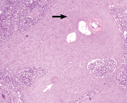
As for the other possibilities:
Cutaneous lymphoma, characterized by atypical proliferation of T or B lymphocytes in the skin, usually presents as a slow-growing indurated nodule or plaque.
Cutaneous mycobacteriosis is caused by infection with Mycobacterium tuberculosis, M leprae, or M marinum. Caseous granulomas are often found on histopathologic study, but special culture and polymerase chain reaction tests are necessary for the diagnosis. Clinically, the lesion is an indurated nodule or plaque that grows slowly, with decreased sensitivity in the case of leprosy, and with a sporotrichoid pattern in the case of M marinum infection.
Our patient underwent a tuberculin skin test, which was negative, as were polymerase chain reaction testing and culture of the biopsy material for mycobacteria.
Sarcoidosis is a systemic disease of unknown cause characterized by noncaseous granulomas in the lungs, mediastinum, lymph nodes, or other sites.1 Cutaneous involvement includes nonspecific clinical forms (eg, nodosum erythema) and other more specific forms (eg, maculopapular, subcutaneous, cicatricial, or lupus pernio variants). This patient has no granulomas on skin biopsy.
Lichen planus is a chronic inflammatory disease of unknown cause that affects the skin, genitalia, mucous membranes, or appendages. It appears as pruritic purple polygonal papules with Wickham striae, usually affecting the wrists and ankles.
A COMMON FORM OF SKIN CANCER
Squamous cell carcinoma and basal cell carcinoma account for 95% of cases of nonmelanomatous skin cancer, and the incidence of squamous cell carcinoma has been increasing.
Long-term exposure to ultraviolet radiation (ie, sunlight), often related to occupation, is the most common cause of squamous cell carcinoma. Other risk factors include thermal injury or burns, ionizing radiation, human papilloma virus infection, immunosuppressive therapy (eg, after organ transplantation2), exposure to chemical carcinogens, and diseases such as xeroderma pigmentosum or oculocutaneous albinism. Unlike basal cell carcinoma, which usually arises de novo, squamous cell carcinoma often arises from precursor lesions, such as actinic keratosis and Bowen disease.
Squamous cell carcinoma commonly manifests as shallow ulcers, often with a keratinous crust and an elevated, indurated border, but also as plaques or nodules, as in this patient. Incipient lesions present as erythematous, firm, keratotic papules, but the lesions on sun-exposed areas such as the head or neck can be pigmented.
The surrounding skin usually shows changes of actinic damage, usually with ulceration, scaling, crusting, or verrucous appearance. When the lesion presents as an erythematous plaque, as in this patient, mycobacterial infection and cutaneous lymphoma should be ruled out. Histologic studies and culture for mycobacteria are necessary for an acute diagnosis. Other conditions in the differential diagnosis are keratoacanthoma, cutaneous metastasis, and sarcoidosis.
The only way to confirm the diagnosis of squamous cell carcinoma is to obtain a specimen by biopsy or surgical excision. Histologic features include atypical keratinocytes extending from the epidermis to the dermis, with dyskeratosis, intercellular bridges, variable central keratinization, and “horn pearls,” depending on how well or how poorly differentiated the tumor.3 Most squamous cell carcinomas arise in solar keratoses, usually at the periphery of the tumor.
Most squamous cell carcinomas are only locally aggressive. Tumors greater than 5 mm in thickness have a 20% risk of metastasis.
TREATMENT OPTIONS
Surgical excision is the most common treatment. Other options are cryotherapy, topical chemotherapy, curettage and electrodesiccation, radiotherapy, and Mohs micrographic surgery. Photodynamic therapy or imiquimod (Aldara) are used only for noninvasive forms.
The possibility of metastasis should be investigated in cases of extensive squamous cell carcinoma, when mucous membranes are affected, and when squamous cell lesions are poorly differentiated.
Our patient’s tumor was removed, with adequate safety margins in surface and depth, and with dermo-epidermal graft closure. The lesion has not recurred at 9 months of follow-up.
- Hunninghake GW, Costabel U, Ando M, et al. ATS/ERS/WASOG statement on sarcoidosis. American Thoracic Society/European Respiratory Society/World Association of Sarcoidosis and other Granulomatous Disorders. Sarcoidosis Vasc Diffuse Lung Dis 1999; 16:149–173.
- Rubel JR, Milford EL, Abdi R. Cutaneous neoplasms in renal transplant recipients. Eur J Dermatol 2002; 12:532–535.
- Cassarino DS, Derienzo DP, Barr RJ. Cutaneous squamous cell carcinoma: a comprehensive clinicopathologic classification—part two. J Cutan Pathol 2006; 33:261–279.
A healthy 68-year-old farmer presents with an asymptomatic skin lesion on his right arm that appeared spontaneously without trauma or bite 5 months ago and has grown progressively since then.
Q: Which is the most likely diagnosis?
- Squamous cell carcinoma
- Cutaneous lymphoma
- Cutaneous mycobacteriosis
- Sarcoidosis
- Lichen planus
As for the other possibilities:
Cutaneous lymphoma, characterized by atypical proliferation of T or B lymphocytes in the skin, usually presents as a slow-growing indurated nodule or plaque.
Cutaneous mycobacteriosis is caused by infection with Mycobacterium tuberculosis, M leprae, or M marinum. Caseous granulomas are often found on histopathologic study, but special culture and polymerase chain reaction tests are necessary for the diagnosis. Clinically, the lesion is an indurated nodule or plaque that grows slowly, with decreased sensitivity in the case of leprosy, and with a sporotrichoid pattern in the case of M marinum infection.
Our patient underwent a tuberculin skin test, which was negative, as were polymerase chain reaction testing and culture of the biopsy material for mycobacteria.
Sarcoidosis is a systemic disease of unknown cause characterized by noncaseous granulomas in the lungs, mediastinum, lymph nodes, or other sites.1 Cutaneous involvement includes nonspecific clinical forms (eg, nodosum erythema) and other more specific forms (eg, maculopapular, subcutaneous, cicatricial, or lupus pernio variants). This patient has no granulomas on skin biopsy.
Lichen planus is a chronic inflammatory disease of unknown cause that affects the skin, genitalia, mucous membranes, or appendages. It appears as pruritic purple polygonal papules with Wickham striae, usually affecting the wrists and ankles.
A COMMON FORM OF SKIN CANCER
Squamous cell carcinoma and basal cell carcinoma account for 95% of cases of nonmelanomatous skin cancer, and the incidence of squamous cell carcinoma has been increasing.
Long-term exposure to ultraviolet radiation (ie, sunlight), often related to occupation, is the most common cause of squamous cell carcinoma. Other risk factors include thermal injury or burns, ionizing radiation, human papilloma virus infection, immunosuppressive therapy (eg, after organ transplantation2), exposure to chemical carcinogens, and diseases such as xeroderma pigmentosum or oculocutaneous albinism. Unlike basal cell carcinoma, which usually arises de novo, squamous cell carcinoma often arises from precursor lesions, such as actinic keratosis and Bowen disease.
Squamous cell carcinoma commonly manifests as shallow ulcers, often with a keratinous crust and an elevated, indurated border, but also as plaques or nodules, as in this patient. Incipient lesions present as erythematous, firm, keratotic papules, but the lesions on sun-exposed areas such as the head or neck can be pigmented.
The surrounding skin usually shows changes of actinic damage, usually with ulceration, scaling, crusting, or verrucous appearance. When the lesion presents as an erythematous plaque, as in this patient, mycobacterial infection and cutaneous lymphoma should be ruled out. Histologic studies and culture for mycobacteria are necessary for an acute diagnosis. Other conditions in the differential diagnosis are keratoacanthoma, cutaneous metastasis, and sarcoidosis.
The only way to confirm the diagnosis of squamous cell carcinoma is to obtain a specimen by biopsy or surgical excision. Histologic features include atypical keratinocytes extending from the epidermis to the dermis, with dyskeratosis, intercellular bridges, variable central keratinization, and “horn pearls,” depending on how well or how poorly differentiated the tumor.3 Most squamous cell carcinomas arise in solar keratoses, usually at the periphery of the tumor.
Most squamous cell carcinomas are only locally aggressive. Tumors greater than 5 mm in thickness have a 20% risk of metastasis.
TREATMENT OPTIONS
Surgical excision is the most common treatment. Other options are cryotherapy, topical chemotherapy, curettage and electrodesiccation, radiotherapy, and Mohs micrographic surgery. Photodynamic therapy or imiquimod (Aldara) are used only for noninvasive forms.
The possibility of metastasis should be investigated in cases of extensive squamous cell carcinoma, when mucous membranes are affected, and when squamous cell lesions are poorly differentiated.
Our patient’s tumor was removed, with adequate safety margins in surface and depth, and with dermo-epidermal graft closure. The lesion has not recurred at 9 months of follow-up.
- Hunninghake GW, Costabel U, Ando M, et al. ATS/ERS/WASOG statement on sarcoidosis. American Thoracic Society/European Respiratory Society/World Association of Sarcoidosis and other Granulomatous Disorders. Sarcoidosis Vasc Diffuse Lung Dis 1999; 16:149–173.
- Rubel JR, Milford EL, Abdi R. Cutaneous neoplasms in renal transplant recipients. Eur J Dermatol 2002; 12:532–535.
- Cassarino DS, Derienzo DP, Barr RJ. Cutaneous squamous cell carcinoma: a comprehensive clinicopathologic classification—part two. J Cutan Pathol 2006; 33:261–279.
Recurrent spontaneous pneumothorax

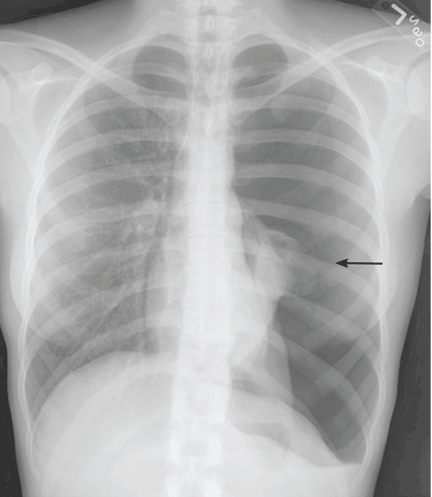
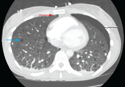
Q: Which is the most likely diagnosis?
- Pulmonary Langerhans cell histiocytosis
- Cystic fibrosis
- Pneumocystis jirovecii pneumonia
- Alpha-1 antitrypsin deficiency
- Tuberous sclerosis complex with lymphangioleiomyomatosis
A: The correct diagnosis is tuberous sclerosis complex with lymphangioleiomyomatosis. The lymphangioleiomyomatosis was suggested by the CT findings, by the recurrence of pneumothorax, and, later, by biopsy results. Lymphangioleiomyomatosis occurs in about 30% of women with the tuberous sclerosis complex.1 However, 10% to 15% of women with lymphangioleiomyomatosis do not have tuberous sclerosis complex, 2 in which case the condition is called sporadic lymphangioleiomyomatosis.
Tuberous sclerosis complex can involve the nerves (seizures, brain tumors), the lungs (lymphangioleiomyomatosis, causing pneumothorax or chylothorax), and the skin; skin lesions include facial angiofibromas (Figure 1), ash-leaf spot (Figure 2), and shagreen patch (Figure 3). It is also associated with abdominal involvement (lymphangiomyomas, renal angiomyolipomas).3
Lymphangioleiomyomatosis usually presents as spontaneous pneumothorax in women of childbearing age. After initial stabilization of pneumothorax with simple aspiration or thoracostomy, the patient should undergo ipsilateral chemical or surgical pleurodesis, as the risk of recurrent pneumothorax is greater than 70%.4 Single or bilateral lung transplantation has been accepted as therapy for end-stage pulmonary lymphangioleiomyomatosis, characterized by recurrent pneumothoraces and chylous pleural fluid collections causing respiratory failure (marked dyspnea, hypoxemia, and reductions in forced expiratory volume in the first second of expiration and in diffusing capacity for carbon monoxide. Recurrence of lymphangioleiomyomatosis in the allograft lung is rare.5 Hormone therapies such as intramuscular progesterone, oral progestins, or gonadotropin-releasing hormone agonists have been used for lymphangioleiomyomatosis (not pneumothorax), but they are no longer recommended.
Pulmonary Langerhans cell histiocytosis, cystic fibrosis, Pneumocystis jirovecii pneumonia, and alpha-1 antitrypsin deficiency have all been associated with spontaneous pneumothorax.
Pulmonary Langerhans cell histiocytosis can affect multiple systems, including lung, skin, bone, and the pituitary gland. More than 90% of patients have a history of smoking.6 Chest radiography reveals a reticulonodular pattern with involvement of the middle and upper lobes. Later, the nodules tend to cavitate and form contiguous cysts that may mimic lymphangioleiomyomatosis on a high-resolution chest CT.
Cystic fibrosis is most often diagnosed before the age of 3.7 In adults, it can present as sinus and pulmonary disease (chronic cough with sputum production, chronic sinusitis with nasal polyposis, radiographic evidence of bronchiectasis and, less commonly, pneumothorax); as a gastrointestinal tract and nutritional abnormality (pancreatic insufficiency, distal intestinal obstruction, focal biliary cirrhosis); and as male infertility.
Pneumocystis jiroveciipneumonia occurs mainly in patients on chronic immunosuppressive drugs or with immune deficiency due to HIV infection. Typical radiographic features are bilateral perihilar interstitial infiltrates that become increasingly homogeneous and diffuse as the disease progresses. Less common findings include solitary or multiple nodules, upper-lobe infiltrates in patients receiving aerosolized pentamidine (NebuPent), pneumatoceles, and pneumothorax.8
Alpha-1 antitrypsin is an inhibitor of neutrophil elastase. Deficiency is associated with severe, early-onset panacinar emphysema with a basilar predominance, with chronic liver disease including cirrhosis, and less commonly with panniculitis and vasculitis associated with antineutrophil cytoplasmic antibody.9 Coalescence of panacinar emphysema leads to the formation of bullae and is important in the development of spontaneous pneumothorax.10
The patient underwent bilateral talc pleurodesis. Lung biopsy at the same time confirmed lymphangioleiomyomatosis. One month later, the right pneumothorax recurred, and she underwent pleurodesis in the right hemithorax with tetracycline. Six months after the second pleurodesis, she was asymptomatic.
- Costello LC, Hartman TE, Ryu JH. High frequency of pulmonary lymphangioleiomyomatosis in women with tuberous sclerosis complex. Mayo Clin Proc 2000; 75:591–594.
- Strizheva GD, Carsillo T, Kruger WD, Sullivan EJ, Ryu JH, Henske EP. The spectrum of mutations in TSC1 and TSC2 in women with tuberous sclerosis and lymphangiomyomatosis. Am J Respir Crit Care Med 2001; 163:253–258.
- McCormack FX. Lymphangioleiomyomatosis: a clinical update. Chest 2008; 133:507–516.
- Almoosa KF, Ryu JH, Mendez J, et al. Management of pneumothorax in lymphangioleiomyomatosis: effects on recurrence and lung transplantation complications. Chest 2006; 129:1274–1281.
- Benden C, Rea F, Behr J, et al. Lung transplantation for lymphangioleiomyomatosis: the European Experience. J Heart Lung Transplant 2009; 28:1–7.
- Vassallo R, Ryu JH, Colby TV, Hartman T, Limper AH. Pulmonary Langerhans’-cell histiocytosis. N Engl J Med 2000; 342:1969–1978.
- Boyle MP. Adult cystic fibrosis. JAMA 2007; 298:1787–1793.
- Thomas CF, Limper AH. Pneumocystis pneumonia. N Engl J Med 2004; 350:2487–2498.
- Silverman EK, Sandhaus RA. Clinical practice. Alpha1-antitrypsin deficiency. N Engl J Med 2009; 360:2749–2757.
- Anderson AE, Furlaneto JA, Foraker AG. Bronchopulmonary derangements in nonsmokers. Am Rev Respir Dis 1970; 101:518–527.
Q: Which is the most likely diagnosis?
- Pulmonary Langerhans cell histiocytosis
- Cystic fibrosis
- Pneumocystis jirovecii pneumonia
- Alpha-1 antitrypsin deficiency
- Tuberous sclerosis complex with lymphangioleiomyomatosis
A: The correct diagnosis is tuberous sclerosis complex with lymphangioleiomyomatosis. The lymphangioleiomyomatosis was suggested by the CT findings, by the recurrence of pneumothorax, and, later, by biopsy results. Lymphangioleiomyomatosis occurs in about 30% of women with the tuberous sclerosis complex.1 However, 10% to 15% of women with lymphangioleiomyomatosis do not have tuberous sclerosis complex, 2 in which case the condition is called sporadic lymphangioleiomyomatosis.
Tuberous sclerosis complex can involve the nerves (seizures, brain tumors), the lungs (lymphangioleiomyomatosis, causing pneumothorax or chylothorax), and the skin; skin lesions include facial angiofibromas (Figure 1), ash-leaf spot (Figure 2), and shagreen patch (Figure 3). It is also associated with abdominal involvement (lymphangiomyomas, renal angiomyolipomas).3
Lymphangioleiomyomatosis usually presents as spontaneous pneumothorax in women of childbearing age. After initial stabilization of pneumothorax with simple aspiration or thoracostomy, the patient should undergo ipsilateral chemical or surgical pleurodesis, as the risk of recurrent pneumothorax is greater than 70%.4 Single or bilateral lung transplantation has been accepted as therapy for end-stage pulmonary lymphangioleiomyomatosis, characterized by recurrent pneumothoraces and chylous pleural fluid collections causing respiratory failure (marked dyspnea, hypoxemia, and reductions in forced expiratory volume in the first second of expiration and in diffusing capacity for carbon monoxide. Recurrence of lymphangioleiomyomatosis in the allograft lung is rare.5 Hormone therapies such as intramuscular progesterone, oral progestins, or gonadotropin-releasing hormone agonists have been used for lymphangioleiomyomatosis (not pneumothorax), but they are no longer recommended.
Pulmonary Langerhans cell histiocytosis, cystic fibrosis, Pneumocystis jirovecii pneumonia, and alpha-1 antitrypsin deficiency have all been associated with spontaneous pneumothorax.
Pulmonary Langerhans cell histiocytosis can affect multiple systems, including lung, skin, bone, and the pituitary gland. More than 90% of patients have a history of smoking.6 Chest radiography reveals a reticulonodular pattern with involvement of the middle and upper lobes. Later, the nodules tend to cavitate and form contiguous cysts that may mimic lymphangioleiomyomatosis on a high-resolution chest CT.
Cystic fibrosis is most often diagnosed before the age of 3.7 In adults, it can present as sinus and pulmonary disease (chronic cough with sputum production, chronic sinusitis with nasal polyposis, radiographic evidence of bronchiectasis and, less commonly, pneumothorax); as a gastrointestinal tract and nutritional abnormality (pancreatic insufficiency, distal intestinal obstruction, focal biliary cirrhosis); and as male infertility.
Pneumocystis jiroveciipneumonia occurs mainly in patients on chronic immunosuppressive drugs or with immune deficiency due to HIV infection. Typical radiographic features are bilateral perihilar interstitial infiltrates that become increasingly homogeneous and diffuse as the disease progresses. Less common findings include solitary or multiple nodules, upper-lobe infiltrates in patients receiving aerosolized pentamidine (NebuPent), pneumatoceles, and pneumothorax.8
Alpha-1 antitrypsin is an inhibitor of neutrophil elastase. Deficiency is associated with severe, early-onset panacinar emphysema with a basilar predominance, with chronic liver disease including cirrhosis, and less commonly with panniculitis and vasculitis associated with antineutrophil cytoplasmic antibody.9 Coalescence of panacinar emphysema leads to the formation of bullae and is important in the development of spontaneous pneumothorax.10
The patient underwent bilateral talc pleurodesis. Lung biopsy at the same time confirmed lymphangioleiomyomatosis. One month later, the right pneumothorax recurred, and she underwent pleurodesis in the right hemithorax with tetracycline. Six months after the second pleurodesis, she was asymptomatic.
- Costello LC, Hartman TE, Ryu JH. High frequency of pulmonary lymphangioleiomyomatosis in women with tuberous sclerosis complex. Mayo Clin Proc 2000; 75:591–594.
- Strizheva GD, Carsillo T, Kruger WD, Sullivan EJ, Ryu JH, Henske EP. The spectrum of mutations in TSC1 and TSC2 in women with tuberous sclerosis and lymphangiomyomatosis. Am J Respir Crit Care Med 2001; 163:253–258.
- McCormack FX. Lymphangioleiomyomatosis: a clinical update. Chest 2008; 133:507–516.
- Almoosa KF, Ryu JH, Mendez J, et al. Management of pneumothorax in lymphangioleiomyomatosis: effects on recurrence and lung transplantation complications. Chest 2006; 129:1274–1281.
- Benden C, Rea F, Behr J, et al. Lung transplantation for lymphangioleiomyomatosis: the European Experience. J Heart Lung Transplant 2009; 28:1–7.
- Vassallo R, Ryu JH, Colby TV, Hartman T, Limper AH. Pulmonary Langerhans’-cell histiocytosis. N Engl J Med 2000; 342:1969–1978.
- Boyle MP. Adult cystic fibrosis. JAMA 2007; 298:1787–1793.
- Thomas CF, Limper AH. Pneumocystis pneumonia. N Engl J Med 2004; 350:2487–2498.
- Silverman EK, Sandhaus RA. Clinical practice. Alpha1-antitrypsin deficiency. N Engl J Med 2009; 360:2749–2757.
- Anderson AE, Furlaneto JA, Foraker AG. Bronchopulmonary derangements in nonsmokers. Am Rev Respir Dis 1970; 101:518–527.
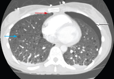
LVH and hypertension: Is treating the pressure not enough?

In patients with “borderline” hypertension or those in whom the duration of blood pressure elevation is hard to ascertain, the finding of end-organ damage has traditionally been used as an argument to institute aggressive antihypertensive therapy. In this setting, retinal hypertensive disease, an S4 gallop, and left ventricular hypertrophy (LVH) are often specifically sought.
LVH and otherwise unexplained chronic kidney disease in patients with hypertension have generally been believed to be products of the elevated arterial pressure, and primary treatment has targeted pressure control. Bauml and Underwood, in this issue of the Journal, emphasize some published clinical trial data indicating that LVH may be an independent risk factor for poorer cardiovascular outcome. Even more provocative is the suggestion that LVH can be reversed, as can the associated increased risk of cardiovascular morbidity, independently of the hypertension.
Given our current understanding that LVH, under some conditions, can be induced by products of the renin-angiotensin system, this would suggest that pharmacologic blockade of this enzyme system should have extra benefit, above that seen from other antihypertensive agents. Conceivably, this may be true only in patients with LVH, and the time course of benefit may not directly parallel that seen with the control of hypertension. That theoretically may explain the lack of uniform advantage of angiotensin blockade over other effective antihypertensive approaches.
Since electrocardiography is a specific but not very sensitive test for LVH, the authors suggest that patients with hypertension be routinely screened for LVH using echocardiography. I am not sure the weight of the evidence supports this approach at present, particularly in the current frenzy of cost containment. Nonetheless, this concept warrants consideration, and at the least, large patient databases might be screened retrospectively to further validate or refute the concept that hypertension-associated LVH is an independent, reversible risk factor for cardiovascular morbidity.
Left ventricular hypertrophy: An overlooked cardiovascular risk factor
Left ventricular hypertrophy (LVH) strongly predicts cardiovascular morbidity and overall mortality in hypertensive patients. 1–7 Antihypertensive treatment that causes LVH to regress decreases the rates of adverse cardiovascular events and improves survival, independent of how much the blood pressure is lowered.8–11 It is clinically important to recognize that LVH is a modifiable risk factor and that management is more complex than just blood pressure control.
This paper reviews the definition of LVH, compares the diagnostic tests for it, and discusses the current evidence-based approach to managing this dangerous risk factor.
A CHRONICALLY ELEVATED CARDIAC WORKLOAD CAUSES LVH
LVH is an abnormal increase in the mass of the left ventricular myocardium caused by a chronically increased workload on the heart.12 This most commonly results from the heart pumping against an elevated afterload, as in hypertension and aortic stenosis. Another notable cause is increased filling of the left ventricle (ie, diastolic overload), which is the underlying mechanism for LVH in patients with aortic or mitral regurgitation and dilated cardiomyopathy. Coronary artery disease can also play a role in the pathogenesis of LVH, as the normal myocardium attempts to compensate for the ischemic or infarcted tissue.13
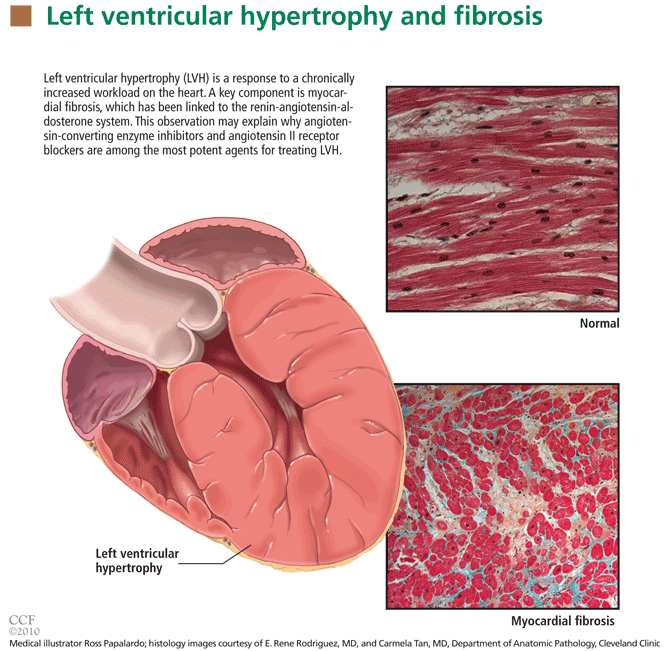
The development of myocardial fibrosis appears to be pathophysiologically linked to the renin-angiotensin-aldosterone system. Specifically, there is evidence that angiotensin II has a profibrotic effect on the myocardium of hypertensive patients.15 This may explain why angiotensin-converting enzyme (ACE) inhibitors and angiotensin II receptor blockers (ARBs) are among the most potent agents for treating LVH, as we will discuss later in this review.
DIAGNOSIS BY ELECTROCARDIOGRAPHY, ECHOCARDIOGRAPHY, OR MRI
Many different criteria for electrocardiographic LVH have been proposed over the years. Most use the voltage in one or more leads, with or without additional factors such as QRS duration, secondary ST-T wave abnormalities, or left atrial abnormalities. The most well known electrocardiographic criteria are the Cornell voltage,21 the Cornell product,22 the Sokolow-Lyon index,23 and the Romhilt-Estes point score system (Table 1).24
- Cornell voltage—median sensitivity 15%, median specificity 96%
- Cornell product—median sensitivity 19.5%, median specificity 91%
- Sokolow-Lyon voltage—median sensitivity 21%, median specificity 89%
- Romhilt-Estes point score—median sensitivity 17%, median specificity 95%.
Of note, the ranges of the published values were extremely broad. For example, the ranges in sensitivity were:
- Cornell voltage—2% to 41%
- Cornell product—8% to 32%
- Sokolow-Lyon voltage—4% to 51%
- Romhilt-Estes point score—0% to 41%.
While the studies with the extreme values may have had issues of small sample size or poor study quality, the wide range in values may primarily be the result of diverse study populations as well as different validation methods and cutoff values to define LVH. Regardless, the overall message of high specificity and low sensitivity is indisputable.
Electrocardiography is insensitive for diagnosing LVH because it relies on measuring the electrical activity of the heart by electrodes on the surface of the skin to predict the left ventricular mass. The intracardiac electrical activity is problematic to measure externally because the measurements are affected by everything between the myocardium and the electrodes, most notably fat, fluid, and air. Because of this effect, electrocardiography underdiagnoses LVH in patients with obesity, pleural effusions, pericardial effusions, anasarca, or chronic obstructive pulmonary disease. In addition, the diagnosis of LVH by electrocardiography is strongly influenced by age and ethnicity.25–26
While electrocardiography is not sensitive and cannot be used to rule out LVH, it still has a role in its diagnosis and management. In the landmark Losartan Intervention for Endpoint Reduction in Hypertension (LIFE) study, regression of LVH (diagnosed electrocardiographically by the Sokolow-Lyon index or the Cornell product criteria) in response to losartan (Cozaar) improved cardiovascular outcomes independent of blood pressure.10 Based on this, it is reasonable that all hypertensive patients and other patients at risk of LVH who undergo electrocardiography be screened with these two criteria.
Echocardiography is the test of choice
Echocardiography, if available, should be the test of choice to assess for LVH. It is much more sensitive than electrocardiography and can also detect other abnormalities such as left ventricular dysfunction and valvular disease.
This test uses transthoracic or transesophageal ultrasonography to measure the left ventricular end-diastolic diameter, posterior wall thickness, and interventricular septum thickness. From these measurements and the patient’s height and weight, the left ventricular mass index can be calculated.27
Several different cutoff values for the left ventricular mass index have been proposed; the LIFE study used values of > 104 g/m2 in women and > 116 g/m2 in men to define LVH.
When using echocardiography to assess for LVH, it is imperative that the left ventricular mass index be used and not just the left ventricular wall thickness, as often happens in clinical practice. This is necessary because diagnosis by wall thickness alone is not a good indicator of LVH, with a concordance between wall thickness and a left ventricular mass index of only 60%.28 In addition, wall thickness tends to underestimate LVH in women and overestimate it in men.
Is echocardiography cost-effective?
Despite its clear advantages, an important consideration about echocardiography as a screening test for all hypertensive patients is its cost.
A suggested way to reduce cost is to measure the left ventricular mass index only.29 A limited echocardiographic examination is much less expensive than a complete two-dimensional echocardiogram ($255 vs $431 per the 2009 Medicare Ambulatory Payment Classification30) and should be the examination performed if the patient has no other clinical indication for echocardiography.
Another way to control cost is to stratify patients by risk and to do echocardiography only in those who would benefit most from it. Based on the prevalence of LVH, one study concluded that echocardiography is most cost-effective in men 50 years or older.31
Further study is necessary to more precisely define the cost-effectiveness of echocardiographic screening for LVH in terms of potentially preventable cardiovascular morbidity and death.
Cardiac MRI: The costly gold standard
Cardiac MRI is the gold standard test for LVH, as it is even more accurate and reproducible than echocardiography.32 It can precisely estimate a patient's left ventricular mass and assess for other structural cardiac abnormalities.
MRI’s use, however, is severely restricted in clinical practice due to its high cost and limited availability. While it may never be used for general screening for LVH, it certainly has a role in clinical research and for assessing cardiac anatomy in special clinical situations.
TREATMENT SHOULD INCLUDE AN ACE INHIBITOR OR ARB
Once LVH has been diagnosed, the next step is to decide on an appropriate treatment plan.
While the choice of therapy will always depend on other comorbidities, a 2003 metaanalysis of antihypertensive medications in the treatment of LVH (controlling for the degree of blood pressure lowering) showed that ARBs were the most efficacious class of agents for reducing the left ventricular mass.33 Specifically, ARBs decreased the mass by 13%, followed by calcium-channel blockers at 11%, ACE inhibitors at 10%, diuretics at 8%, and beta-blockers at 6%. In pairwise comparison, ARBs, calcium-channel blockers, and ACE inhibitors were all significantly more effective in reducing the left ventricular mass than beta-blockers.
As previously discussed, LVH appears to be pathophysiologically linked to myocardial fibrosis and the renin-angiotensin-aldosterone system. For this reason and based on the data presented above regarding the degree of LVH regression, ACE inhibitors or ARBs should be used as the first-line agents for LVH unless they are contraindicated in the individual patient.
The LIFE study
The LIFE study offers the strongest evidence that treating LVH is beneficial. It showed that in hypertensive patients with electrocardiographic LVH by the Cornell product or Sokolow-Lyon criteria, treatment with antihypertensive drugs that resulted in less-severe LVH on electrocardiography was associated with lower rates of cardiovascular morbidity and death, independent of the blood pressure achieved or the drug used.10
The end point in this study was a composite of stroke, myocardial infarction, and cardiovascular death. Regression of electrocardiographic LVH in hypertensive patients has also been shown to decrease the incidence of diabetes mellitus,34 atrial fibrillation,35 and hospitalizations for heart failure.36
The LIFE study also examined the prognostic implications of treating LVH detected by echocardiography. In this prospective cohort substudy, patients who had a lower left ventricular mass index during treatment with antihypertensive drugs had lower rates of cardiovascular morbidity and all-cause mortality, independent of the effects of blood pressure and treatment used.11
These results suggest that there may be a role not only for treating LVH, but also for monitoring for a reduction in the left ventricular mass index as a goal of therapy (similar to the way hemoglobin A1c is used in diabetic patients). If the index is used in this way, one could potentially adjust the dose of current drugs, switch classes, or add an additional drug based on a persistently elevated left ventricular mass index in order to optimize the patient's overall cardiovascular risk. A randomized controlled trial of therapy directed by the mass index vs conventional therapy of LVH would be necessary to assess the clinical utility of this approach.
RECOMMENDATIONS
LVH is a common and potentially modifiable cardiovascular risk factor often overlooked in clinical practice. Ideally, all hypertensive patients should be screened with echocardiography to look for LVH, using the calculated left ventricular mass index rather than wall thickness alone to make the diagnosis. While electrocardiography is specific and also has prognostic implications, it is not sensitive enough to be used alone to screen for LVH.
Once the diagnosis of LVH is made, the initial therapy should be an ARB or an ACE inhibitor. Response to therapy can be assessed by monitoring for a reduction in left ventricular mass index or regression of electrocardiographic LVH.
Treatment-induced regression of LVH decreases adverse cardiovascular events and improves overall survival. When modifying medications in hypertensive patients, it is important to remember that the treatment of LVH is not synonymous with blood pressure control.
- Casale PN, Devereux RB, Milner M, et al. Value of echocardiographic measurement of left ventricular mass in predicting cardiovascular morbid events in hypertensive men. Ann Intern Med 1986; 105:173–178.
- Levy D, Garrison RJ, Savage DD, Kannel WB, Castelli WP. Prognostic implications of echocardiographically determined left ventricular mass in the Framingham Heart Study. N Engl J Med 1990; 322:1561–1566.
- Koren MJ, Devereux RB, Casale PN, Savage DD, Laragh JH. Relation of left ventricular mass and geometry to morbidity and mortality in uncomplicated essential hypertension. Ann Intern Med 1991; 114:345–352.
- Verdecchia P, Carini G, Circo A, et al; MAVI (MAssa Ventricolare sinistra nell’Ipertensione) Study Group. Left ventricular mass and cardiovascular morbidity in essential hypertension: the MAVI study. J Am Coll Cardiol 2001; 38:1829–1835.
- Haider AW, Larson MG, Benjamin EJ, Levy D. Increased left ventricular mass and hypertrophy are associated with increased risk for sudden death. J Am Coll Cardiol 1998; 32:1454–1459.
- Verdecchia P, Porcellati C, Reboldi G, et al. Left ventricular hypertrophy as an independent predictor of acute cerebrovascular events in essential hypertension. Circulation 2001; 104:2039–2044.
- Schillaci G, Verdecchia P, Porcellati C, Cuccurullo O, Cosco C, Perticone F. Continuous relation between left ventricular mass and cardiovascular risk in essential hypertension. Hypertension 2000; 35:580–586.
- Verdecchia P, Schillaci G, Borgioni C, et al. Prognostic significance of serial changes in left ventricular mass in essential hypertension. Circulation 1998; 97:48–54.
- Mathew J, Sleight P, Lonn E, et al; Heart Outcomes Prevention Evaluation (HOPE) Investigators. Reduction of cardiovascular risk by regression of electrocardiographic markers of left ventricular hypertrophy by the angiotensin-converting enzyme inhibitor ramipril. Circulation 2001; 104:1615–1621.
- Okin PM, Devereux RB, Jern S, et al; LIFE Study Investigators. Regression of electrocardiographic left ventricular hypertrophy during antihypertensive treatment and the prediction of major cardiovascular events. JAMA 2004; 292:2343–2349.
- Devereux RB, Wachtell K, Gerdts E, et al. Prognostic significance of left ventricular mass change during treatment of hypertension. JAMA 2004; 292:2350–2356.
- Lorell BH, Carabello BA. Left ventricular hypertrophy: pathogenesis, detection, and prognosis. Circulation 2000; 102:470–479.
- Zabalgoitia M, Berning J, Koren MJ, et al; LIFE Study Investigators. Impact of coronary artery disease on left ventricular systolic function and geometry in hypertensive patients with left ventricular hypertrophy (the LIFE study). Am J Cardiol 2001; 88:646–650.
- Weber KT, Janicki JS, Pick R, Capasso J, Anversa P. Myocardial fibrosis and pathologic hypertrophy in the rat with renovascular hypertension. Am J Cardiol 1990; 65:1G–7G.
- González A, López B, Querejeta R, Díez J. Regulation of myocardial fibrillar collagen by angiotensin II. A role in hypertensive heart disease? J Mol Cell Cardiol 2002; 34:1585–1593.
- Maron BJ. Hypertrophic cardiomyopathy: a systematic review. JAMA 2002; 287:1308–1320.
- Liebson PR, Grandits G, Prineas R, et al. Echocardiographic correlates of left ventricular structure among 844 mildly hypertensive men and women in the Treatment of Mild Hypertension Study (TOMHS). Circulation 1993; 87:476–486.
- Martinez MA, Sancho T, Armada E, et al; Vascular Risk Working Group Grupo Monitorizacíon Ambulatoria de la Presión Arterial (MAPA)-Madrid. Prevalence of left ventricular hypertrophy in patients with mild hypertension in primary care: impact of echocardiography on cardiovascular risk stratification. Am J Hypertens 2003; 16:556–563.
- Pewsner D, Jüni P, Egger M, Battaglia M, Sundström J, Bachmann LM. Accuracy of electrocardiography in diagnosis of left ventricular hypertrophy in arterial hypertension: systematic review. BMJ 2007; 335:711.
- Devereux RB. Is the electrocardiogram still useful for detection of left ventricular hypertrophy? Circulation 1990; 81:1144–1146.
- Casale PN, Devereux RB, Kligfield P, et al. Electrocardiographic detection of left ventricular hypertrophy: development and prospective validation of improved criteria. J Am Coll Cardiol 1985; 6:572–580.
- Molloy TJ, Okin PM, Devereux RB, Kligfield P. Electrocardiographic detection of left ventricular hypertrophy by the simple QRS voltage-duration product. J Am Coll Cardiol 1992; 20:1180–1186.
- Sokolow M, Lyon TP. The ventricular complex in left ventricular hypertrophy as obtained by unipolar precordial and limb leads. Am Heart J 1949; 37:161–186.
- Romhilt DW, Estes EH. A point-score system for the ECG diagnosis of left ventricular hypertrophy. Am Heart J 1968; 75:752–758.
- Levy D, Labib SB, Anderson KM, Christiansen JC, Kannel WB, Castelli WP. Determinants of sensitivity and specificity of electrocardiographic criteria for left ventricular hypertrophy. Circulation 1990; 81:815–820.
- Okin PM, Wright JT, Nieminen MS, et al. Ethnic differences in electrocardiographic criteria for left ventricular hypertrophy: the LIFE study. Losartan Intervention For Endpoint. Am J Hypertens 2002; 15:663–671.
- Devereux RB, Alonso DR, Lutas EM, et al. Echocardiographic assessment of left ventricular hypertrophy: comparison to necropsy findings. Am J Cardiol 1986; 57:450–458.
- Leibowitz D, Planer D, Ben-Ibgi F, Rott D, Weiss AT, Bursztyn M. Measurement of wall thickness alone does not accurately assess the presence of left ventricular hypertrophy. Clin Exp Hypertens 2007; 29:119–125.
- Black HR, Weltin G, Jaffe CC. The limited echocardiogram: a modification of standard echocardiography for use in the routine evaluation of patients with systemic hypertension. Am J Cardiol 1991; 67:1027–1030.
- American Society of Echocardiography Coding and Reimbursement Newsletter, January 2009. http://www.asecho.org/files/public/CodingnewsJan09.pdf. Accessed May 13, 2010.
- Cuspidi C, Meani S, Valerio C, Fusi V, Sala C, Zanchetti A. Left ventricular hypertrophy and cardiovascular risk stratification: impact and cost-effectiveness of echocardiography in recently diagnosed essential hypertensives. J Hypertens 2006; 24:1671–1677.
- Bottini PB, Carr AA, Prisant LM, Flickinger FW, Allison JD, Gottdiener JS. Magnetic resonance imaging compared to echocardiography to assess left ventricular mass in the hypertensive patient. Am J Hypertens 1995; 8:221–228.
- Klingbeil AU, Schneider M, Martus P, Messerli FH, Schmieder RE. A meta-analysis of the effects of treatment on left ventricular mass in essential hypertension. Am J Med 2003; 115:41–46.
- Okin PM, Devereux RB, Harris KE, et al; LIFE Study Investigators. In-treatment resolution or absence of electrocardiographic left ventricular hypertrophy is associated with decreased incidence of new-onset diabetes mellitus in hypertensive patients: the Losartan Intervention for Endpoint Reduction in Hypertension (LIFE) Study. Hypertension 2007; 50:984–990.
- Okin PM, Wachtell K, Devereux RB, et al. Regression of electrocardiographic left ventricular hypertrophy and decreased incidence of new-onset atrial fibrillation in patients with hypertension. JAMA 2006; 296:1242–1248.
- Okin PM, Devereux RB, Harris KE, et al; LIFE Study Investigators. Regression of electrocardiographic left ventricular hypertrophy is associated with less hospitalization for heart failure in hypertensive patients. Ann Intern Med 2007; 147:311–319.
Left ventricular hypertrophy (LVH) strongly predicts cardiovascular morbidity and overall mortality in hypertensive patients. 1–7 Antihypertensive treatment that causes LVH to regress decreases the rates of adverse cardiovascular events and improves survival, independent of how much the blood pressure is lowered.8–11 It is clinically important to recognize that LVH is a modifiable risk factor and that management is more complex than just blood pressure control.
This paper reviews the definition of LVH, compares the diagnostic tests for it, and discusses the current evidence-based approach to managing this dangerous risk factor.
A CHRONICALLY ELEVATED CARDIAC WORKLOAD CAUSES LVH
LVH is an abnormal increase in the mass of the left ventricular myocardium caused by a chronically increased workload on the heart.12 This most commonly results from the heart pumping against an elevated afterload, as in hypertension and aortic stenosis. Another notable cause is increased filling of the left ventricle (ie, diastolic overload), which is the underlying mechanism for LVH in patients with aortic or mitral regurgitation and dilated cardiomyopathy. Coronary artery disease can also play a role in the pathogenesis of LVH, as the normal myocardium attempts to compensate for the ischemic or infarcted tissue.13
The development of myocardial fibrosis appears to be pathophysiologically linked to the renin-angiotensin-aldosterone system. Specifically, there is evidence that angiotensin II has a profibrotic effect on the myocardium of hypertensive patients.15 This may explain why angiotensin-converting enzyme (ACE) inhibitors and angiotensin II receptor blockers (ARBs) are among the most potent agents for treating LVH, as we will discuss later in this review.
DIAGNOSIS BY ELECTROCARDIOGRAPHY, ECHOCARDIOGRAPHY, OR MRI
Many different criteria for electrocardiographic LVH have been proposed over the years. Most use the voltage in one or more leads, with or without additional factors such as QRS duration, secondary ST-T wave abnormalities, or left atrial abnormalities. The most well known electrocardiographic criteria are the Cornell voltage,21 the Cornell product,22 the Sokolow-Lyon index,23 and the Romhilt-Estes point score system (Table 1).24
- Cornell voltage—median sensitivity 15%, median specificity 96%
- Cornell product—median sensitivity 19.5%, median specificity 91%
- Sokolow-Lyon voltage—median sensitivity 21%, median specificity 89%
- Romhilt-Estes point score—median sensitivity 17%, median specificity 95%.
Of note, the ranges of the published values were extremely broad. For example, the ranges in sensitivity were:
- Cornell voltage—2% to 41%
- Cornell product—8% to 32%
- Sokolow-Lyon voltage—4% to 51%
- Romhilt-Estes point score—0% to 41%.
While the studies with the extreme values may have had issues of small sample size or poor study quality, the wide range in values may primarily be the result of diverse study populations as well as different validation methods and cutoff values to define LVH. Regardless, the overall message of high specificity and low sensitivity is indisputable.
Electrocardiography is insensitive for diagnosing LVH because it relies on measuring the electrical activity of the heart by electrodes on the surface of the skin to predict the left ventricular mass. The intracardiac electrical activity is problematic to measure externally because the measurements are affected by everything between the myocardium and the electrodes, most notably fat, fluid, and air. Because of this effect, electrocardiography underdiagnoses LVH in patients with obesity, pleural effusions, pericardial effusions, anasarca, or chronic obstructive pulmonary disease. In addition, the diagnosis of LVH by electrocardiography is strongly influenced by age and ethnicity.25–26
While electrocardiography is not sensitive and cannot be used to rule out LVH, it still has a role in its diagnosis and management. In the landmark Losartan Intervention for Endpoint Reduction in Hypertension (LIFE) study, regression of LVH (diagnosed electrocardiographically by the Sokolow-Lyon index or the Cornell product criteria) in response to losartan (Cozaar) improved cardiovascular outcomes independent of blood pressure.10 Based on this, it is reasonable that all hypertensive patients and other patients at risk of LVH who undergo electrocardiography be screened with these two criteria.
Echocardiography is the test of choice
Echocardiography, if available, should be the test of choice to assess for LVH. It is much more sensitive than electrocardiography and can also detect other abnormalities such as left ventricular dysfunction and valvular disease.
This test uses transthoracic or transesophageal ultrasonography to measure the left ventricular end-diastolic diameter, posterior wall thickness, and interventricular septum thickness. From these measurements and the patient’s height and weight, the left ventricular mass index can be calculated.27
Several different cutoff values for the left ventricular mass index have been proposed; the LIFE study used values of > 104 g/m2 in women and > 116 g/m2 in men to define LVH.
When using echocardiography to assess for LVH, it is imperative that the left ventricular mass index be used and not just the left ventricular wall thickness, as often happens in clinical practice. This is necessary because diagnosis by wall thickness alone is not a good indicator of LVH, with a concordance between wall thickness and a left ventricular mass index of only 60%.28 In addition, wall thickness tends to underestimate LVH in women and overestimate it in men.
Is echocardiography cost-effective?
Despite its clear advantages, an important consideration about echocardiography as a screening test for all hypertensive patients is its cost.
A suggested way to reduce cost is to measure the left ventricular mass index only.29 A limited echocardiographic examination is much less expensive than a complete two-dimensional echocardiogram ($255 vs $431 per the 2009 Medicare Ambulatory Payment Classification30) and should be the examination performed if the patient has no other clinical indication for echocardiography.
Another way to control cost is to stratify patients by risk and to do echocardiography only in those who would benefit most from it. Based on the prevalence of LVH, one study concluded that echocardiography is most cost-effective in men 50 years or older.31
Further study is necessary to more precisely define the cost-effectiveness of echocardiographic screening for LVH in terms of potentially preventable cardiovascular morbidity and death.
Cardiac MRI: The costly gold standard
Cardiac MRI is the gold standard test for LVH, as it is even more accurate and reproducible than echocardiography.32 It can precisely estimate a patient's left ventricular mass and assess for other structural cardiac abnormalities.
MRI’s use, however, is severely restricted in clinical practice due to its high cost and limited availability. While it may never be used for general screening for LVH, it certainly has a role in clinical research and for assessing cardiac anatomy in special clinical situations.
TREATMENT SHOULD INCLUDE AN ACE INHIBITOR OR ARB
Once LVH has been diagnosed, the next step is to decide on an appropriate treatment plan.
While the choice of therapy will always depend on other comorbidities, a 2003 metaanalysis of antihypertensive medications in the treatment of LVH (controlling for the degree of blood pressure lowering) showed that ARBs were the most efficacious class of agents for reducing the left ventricular mass.33 Specifically, ARBs decreased the mass by 13%, followed by calcium-channel blockers at 11%, ACE inhibitors at 10%, diuretics at 8%, and beta-blockers at 6%. In pairwise comparison, ARBs, calcium-channel blockers, and ACE inhibitors were all significantly more effective in reducing the left ventricular mass than beta-blockers.
As previously discussed, LVH appears to be pathophysiologically linked to myocardial fibrosis and the renin-angiotensin-aldosterone system. For this reason and based on the data presented above regarding the degree of LVH regression, ACE inhibitors or ARBs should be used as the first-line agents for LVH unless they are contraindicated in the individual patient.
The LIFE study
The LIFE study offers the strongest evidence that treating LVH is beneficial. It showed that in hypertensive patients with electrocardiographic LVH by the Cornell product or Sokolow-Lyon criteria, treatment with antihypertensive drugs that resulted in less-severe LVH on electrocardiography was associated with lower rates of cardiovascular morbidity and death, independent of the blood pressure achieved or the drug used.10
The end point in this study was a composite of stroke, myocardial infarction, and cardiovascular death. Regression of electrocardiographic LVH in hypertensive patients has also been shown to decrease the incidence of diabetes mellitus,34 atrial fibrillation,35 and hospitalizations for heart failure.36
The LIFE study also examined the prognostic implications of treating LVH detected by echocardiography. In this prospective cohort substudy, patients who had a lower left ventricular mass index during treatment with antihypertensive drugs had lower rates of cardiovascular morbidity and all-cause mortality, independent of the effects of blood pressure and treatment used.11
These results suggest that there may be a role not only for treating LVH, but also for monitoring for a reduction in the left ventricular mass index as a goal of therapy (similar to the way hemoglobin A1c is used in diabetic patients). If the index is used in this way, one could potentially adjust the dose of current drugs, switch classes, or add an additional drug based on a persistently elevated left ventricular mass index in order to optimize the patient's overall cardiovascular risk. A randomized controlled trial of therapy directed by the mass index vs conventional therapy of LVH would be necessary to assess the clinical utility of this approach.
RECOMMENDATIONS
LVH is a common and potentially modifiable cardiovascular risk factor often overlooked in clinical practice. Ideally, all hypertensive patients should be screened with echocardiography to look for LVH, using the calculated left ventricular mass index rather than wall thickness alone to make the diagnosis. While electrocardiography is specific and also has prognostic implications, it is not sensitive enough to be used alone to screen for LVH.
Once the diagnosis of LVH is made, the initial therapy should be an ARB or an ACE inhibitor. Response to therapy can be assessed by monitoring for a reduction in left ventricular mass index or regression of electrocardiographic LVH.
Treatment-induced regression of LVH decreases adverse cardiovascular events and improves overall survival. When modifying medications in hypertensive patients, it is important to remember that the treatment of LVH is not synonymous with blood pressure control.
- Casale PN, Devereux RB, Milner M, et al. Value of echocardiographic measurement of left ventricular mass in predicting cardiovascular morbid events in hypertensive men. Ann Intern Med 1986; 105:173–178.
- Levy D, Garrison RJ, Savage DD, Kannel WB, Castelli WP. Prognostic implications of echocardiographically determined left ventricular mass in the Framingham Heart Study. N Engl J Med 1990; 322:1561–1566.
- Koren MJ, Devereux RB, Casale PN, Savage DD, Laragh JH. Relation of left ventricular mass and geometry to morbidity and mortality in uncomplicated essential hypertension. Ann Intern Med 1991; 114:345–352.
- Verdecchia P, Carini G, Circo A, et al; MAVI (MAssa Ventricolare sinistra nell’Ipertensione) Study Group. Left ventricular mass and cardiovascular morbidity in essential hypertension: the MAVI study. J Am Coll Cardiol 2001; 38:1829–1835.
- Haider AW, Larson MG, Benjamin EJ, Levy D. Increased left ventricular mass and hypertrophy are associated with increased risk for sudden death. J Am Coll Cardiol 1998; 32:1454–1459.
- Verdecchia P, Porcellati C, Reboldi G, et al. Left ventricular hypertrophy as an independent predictor of acute cerebrovascular events in essential hypertension. Circulation 2001; 104:2039–2044.
- Schillaci G, Verdecchia P, Porcellati C, Cuccurullo O, Cosco C, Perticone F. Continuous relation between left ventricular mass and cardiovascular risk in essential hypertension. Hypertension 2000; 35:580–586.
- Verdecchia P, Schillaci G, Borgioni C, et al. Prognostic significance of serial changes in left ventricular mass in essential hypertension. Circulation 1998; 97:48–54.
- Mathew J, Sleight P, Lonn E, et al; Heart Outcomes Prevention Evaluation (HOPE) Investigators. Reduction of cardiovascular risk by regression of electrocardiographic markers of left ventricular hypertrophy by the angiotensin-converting enzyme inhibitor ramipril. Circulation 2001; 104:1615–1621.
- Okin PM, Devereux RB, Jern S, et al; LIFE Study Investigators. Regression of electrocardiographic left ventricular hypertrophy during antihypertensive treatment and the prediction of major cardiovascular events. JAMA 2004; 292:2343–2349.
- Devereux RB, Wachtell K, Gerdts E, et al. Prognostic significance of left ventricular mass change during treatment of hypertension. JAMA 2004; 292:2350–2356.
- Lorell BH, Carabello BA. Left ventricular hypertrophy: pathogenesis, detection, and prognosis. Circulation 2000; 102:470–479.
- Zabalgoitia M, Berning J, Koren MJ, et al; LIFE Study Investigators. Impact of coronary artery disease on left ventricular systolic function and geometry in hypertensive patients with left ventricular hypertrophy (the LIFE study). Am J Cardiol 2001; 88:646–650.
- Weber KT, Janicki JS, Pick R, Capasso J, Anversa P. Myocardial fibrosis and pathologic hypertrophy in the rat with renovascular hypertension. Am J Cardiol 1990; 65:1G–7G.
- González A, López B, Querejeta R, Díez J. Regulation of myocardial fibrillar collagen by angiotensin II. A role in hypertensive heart disease? J Mol Cell Cardiol 2002; 34:1585–1593.
- Maron BJ. Hypertrophic cardiomyopathy: a systematic review. JAMA 2002; 287:1308–1320.
- Liebson PR, Grandits G, Prineas R, et al. Echocardiographic correlates of left ventricular structure among 844 mildly hypertensive men and women in the Treatment of Mild Hypertension Study (TOMHS). Circulation 1993; 87:476–486.
- Martinez MA, Sancho T, Armada E, et al; Vascular Risk Working Group Grupo Monitorizacíon Ambulatoria de la Presión Arterial (MAPA)-Madrid. Prevalence of left ventricular hypertrophy in patients with mild hypertension in primary care: impact of echocardiography on cardiovascular risk stratification. Am J Hypertens 2003; 16:556–563.
- Pewsner D, Jüni P, Egger M, Battaglia M, Sundström J, Bachmann LM. Accuracy of electrocardiography in diagnosis of left ventricular hypertrophy in arterial hypertension: systematic review. BMJ 2007; 335:711.
- Devereux RB. Is the electrocardiogram still useful for detection of left ventricular hypertrophy? Circulation 1990; 81:1144–1146.
- Casale PN, Devereux RB, Kligfield P, et al. Electrocardiographic detection of left ventricular hypertrophy: development and prospective validation of improved criteria. J Am Coll Cardiol 1985; 6:572–580.
- Molloy TJ, Okin PM, Devereux RB, Kligfield P. Electrocardiographic detection of left ventricular hypertrophy by the simple QRS voltage-duration product. J Am Coll Cardiol 1992; 20:1180–1186.
- Sokolow M, Lyon TP. The ventricular complex in left ventricular hypertrophy as obtained by unipolar precordial and limb leads. Am Heart J 1949; 37:161–186.
- Romhilt DW, Estes EH. A point-score system for the ECG diagnosis of left ventricular hypertrophy. Am Heart J 1968; 75:752–758.
- Levy D, Labib SB, Anderson KM, Christiansen JC, Kannel WB, Castelli WP. Determinants of sensitivity and specificity of electrocardiographic criteria for left ventricular hypertrophy. Circulation 1990; 81:815–820.
- Okin PM, Wright JT, Nieminen MS, et al. Ethnic differences in electrocardiographic criteria for left ventricular hypertrophy: the LIFE study. Losartan Intervention For Endpoint. Am J Hypertens 2002; 15:663–671.
- Devereux RB, Alonso DR, Lutas EM, et al. Echocardiographic assessment of left ventricular hypertrophy: comparison to necropsy findings. Am J Cardiol 1986; 57:450–458.
- Leibowitz D, Planer D, Ben-Ibgi F, Rott D, Weiss AT, Bursztyn M. Measurement of wall thickness alone does not accurately assess the presence of left ventricular hypertrophy. Clin Exp Hypertens 2007; 29:119–125.
- Black HR, Weltin G, Jaffe CC. The limited echocardiogram: a modification of standard echocardiography for use in the routine evaluation of patients with systemic hypertension. Am J Cardiol 1991; 67:1027–1030.
- American Society of Echocardiography Coding and Reimbursement Newsletter, January 2009. http://www.asecho.org/files/public/CodingnewsJan09.pdf. Accessed May 13, 2010.
- Cuspidi C, Meani S, Valerio C, Fusi V, Sala C, Zanchetti A. Left ventricular hypertrophy and cardiovascular risk stratification: impact and cost-effectiveness of echocardiography in recently diagnosed essential hypertensives. J Hypertens 2006; 24:1671–1677.
- Bottini PB, Carr AA, Prisant LM, Flickinger FW, Allison JD, Gottdiener JS. Magnetic resonance imaging compared to echocardiography to assess left ventricular mass in the hypertensive patient. Am J Hypertens 1995; 8:221–228.
- Klingbeil AU, Schneider M, Martus P, Messerli FH, Schmieder RE. A meta-analysis of the effects of treatment on left ventricular mass in essential hypertension. Am J Med 2003; 115:41–46.
- Okin PM, Devereux RB, Harris KE, et al; LIFE Study Investigators. In-treatment resolution or absence of electrocardiographic left ventricular hypertrophy is associated with decreased incidence of new-onset diabetes mellitus in hypertensive patients: the Losartan Intervention for Endpoint Reduction in Hypertension (LIFE) Study. Hypertension 2007; 50:984–990.
- Okin PM, Wachtell K, Devereux RB, et al. Regression of electrocardiographic left ventricular hypertrophy and decreased incidence of new-onset atrial fibrillation in patients with hypertension. JAMA 2006; 296:1242–1248.
- Okin PM, Devereux RB, Harris KE, et al; LIFE Study Investigators. Regression of electrocardiographic left ventricular hypertrophy is associated with less hospitalization for heart failure in hypertensive patients. Ann Intern Med 2007; 147:311–319.
KEY POINTS
- LVH is caused by a chronically increased cardiac workload, most commonly from hypertension.
- Ideally, all hypertensive patients should undergo echocardiography to screen for LVH, using the calculated left ventricular mass index.
- Electrocardiography is too insensitive to be used alone to screen for LVH.
- In hypertensive patients, initial therapy of LVH should consist of an angiotensin II receptor blocker or an angiotensin-converting enzyme inhibitor.
- Treatment-induced regression of LVH improves cardiovascular outcomes independent of blood pressure.
- Further study is necessary to examine the utility of following the left ventricular mass index as a treatment goal.