User login
AVAHO
div[contains(@class, 'header__large-screen')]
div[contains(@class, 'read-next-article')]
div[contains(@class, 'nav-primary')]
nav[contains(@class, 'nav-primary')]
section[contains(@class, 'footer-nav-section-wrapper')]
footer[@id='footer']
div[contains(@class, 'main-prefix')]
section[contains(@class, 'nav-hidden')]
div[contains(@class, 'ce-card-content')]
nav[contains(@class, 'nav-ce-stack')]
ESMO helps hematologists assess new cancer drugs
It consists of 11 2- to 3-page forms with checklists to grade treatment trials on the extent to which they meet efficacy and safety thresholds. Each of the 11 forms covers a specific trial scenario, such as a randomized controlled trial with curative intent or a trial of a therapy that is not likely to be curative with a primary endpoint of overall survival.
Treatments with curative intent are graded A, B, or C, while treatments in the noncurative setting are graded on a descending scale from 5 to 1. Scores of A and B in the curative setting and 5 and 4 in the noncurative setting represent substantial benefit.
On the form for RCTs with curative intent, for instance, a survival improvement of 5% or more garners an A but an improvement of less than 3% gets a C. Scores are also annotated for serious acute and/or persistent toxicity if present.
The tool, dubbed the ESMO-MCBS:H (European Society for Medical Oncology Magnitude of Clinical Benefit Scale: Hematology), is explained in an article published in Annals of Oncology. The evaluation forms are available online.
The idea behind the work is to help health care professionals and others to more “accurately assess the value of and prioritise therapies for patients with blood cancers. For clinicians, ESMO-MCBS:H will aid in their clinical decision-making and in the development of evidence-based practice and guidelines,” ESMO said in a press release.
To develop ESMO-MCBS:H, the group tailored its tool for evaluating solid tumor therapies, the ESMO-MCBS, to account for the sometimes different endpoints used in hematologic malignancy trials and the very indolent nature of some blood cancers, such as follicular lymphoma, which hampers development of mature data.
Specific changes include adding a new evaluation form to grade single-arm trials with curative intent, such as those used for CAR-T-cell therapies; incorporating molecular surrogate endpoints used in CML trials; and adding a way to grade outcomes for indolent cancers, among others.
The development process included applying the solid tumor tool to 80 blood cancer studies to identify shortcomings and improve its applicability. The final tool was field tested with 51 international experts from EHA and ESMO who largely agreed on the reasonableness of the trial scores.
ESMO said it expects ESMO-MCBS:H will be useful. The solid tumor tool, first published in 2015, is used by the World Health Organization to screen medications for its essential medicines list as well as by ESMO to generate guidelines and oncology centers across Europe to help with resource allocation decisions.
FROM ANNALS OF ONCOLOGY
Colorectal cancer: Younger patients fare worse
The incidence of metastatic colorectal cancer (mCRC) among adults younger than 50 years has been increasing, and although younger patients are treated with aggressive regimens similarly to older patients, outcomes data, including incidence of toxic effects, across age groups are limited, wrote Lingbin Meng, MD, of H. Lee Moffitt Cancer Center & Research Institute, Tampa, Fla., and colleagues, in their paper on the new research.
“Studies on the age-related disparity ... provided mixed findings,” said corresponding author Hao Xie, MD, of the Mayo Clinic in Rochester, Minn., in an interview.
According to the paper, published in JAMA Network Open, the researchers sought to evaluate the association between age and mCRC treatment-related adverse events and survival.
The study population included 1,223 mCRC patients who underwent first-line treatment with fluorouracil and oxaliplatin therapy in three clinical trials. An additional 736 patients with mCRC from the Moffitt Cancer Center were used to assess genomic alterations and serve as an external validation cohort.
Methods and results
Patients were divided into three age groups: younger than 50 years, 50-65 years, and 65 years and older. Early onset was defined as younger than 50 years. Approximately 58% of the study population was male.
The primary outcomes were treatment-related adverse events and survival rates. Overall survival (OS) and progression-free survival (PFS) were was significantly shorter in the early-onset group, compared with the 50-65 years group (hazard ratios, 1.48 and 1.46, respectively, P < .001 for both) in a multivariate analysis. The shorter OS in the early-onset group was confirmed in the validation cohort.
The early-onset patients had significantly higher incidence of nausea and vomiting, severe abdominal pain, severe anemia, and severe rash, compared with patients in both the 50- to 65-year-old group and the older than 65 years group. In addition, abdominal pain and severe liver toxicity effects were associated with shorter survival in the early-onset patients.
Genomic data from the Moffitt cohort showed a higher prevalence of CTNNB1 mutation among patients younger than 50 years, compared with the 50- to 65-year-old group and the older than 65 years group (6.6%, 3.1%, and 2.3%, respectively; P = .047), as well as ERBB2 amplification (5.1%, 0.6%, and 2.3%, respectively; P = .005), and CREBBP mutation (3.1%, 0.9%, and 0.5%; P = .05), although the prevalence of BRAF mutation was significantly lower in the younger patients, compared with patients in the older groups (7.7%, 8.5%, and 16.7%, respectively; P = .002).
These data suggest that distinct genomic profiles may play a role in the worse outcomes for patients with early-onset mCRC, the researchers said.
The findings were limited by several factors, including the timing of the trials prior to the use of biologics as standard first-line therapy, the researchers noted. Other limitations include a lack of data on treatment adherence and intensity and the location and number of metastases, and potential limited generalizability to other populations given that the majority of the participants were white, they said.
Data support individualized treatment
“We were surprised to find that patients with early-onset metastatic colorectal cancer had worse survival outcome, compared to older patients with metastatic colorectal cancer,” Dr. Xie said, in an interview. “On the other hand, we were not surprised to find unique adverse-event patterns in patients with early-onset metastatic colorectal cancer.”
For clinicians, “The take home message is that we should adopt individualized management approaches [regarding] cancer-direct treatments and treatment-related side effects in patients with early-onset metastatic colorectal cancer,” said Dr. Xie. However, more research is needed in the form of prospective and interventional studies to address treatment-related side effects and to develop novel and personalized therapeutics for patients with early-onset metastatic colorectal cancer, Dr. Xie added.
Early-onset cancer concerns persist
“The increasing shift in early colon cancers demands better understanding, in particular as we attempt a more patient-focused approach to treatments,” said David A. Johnson, MD, chief of gastroenterology at Eastern Virginia Medical School, Norfolk, in an interview. “Clearly, genetic amplifications and oncogene mutations play an essential role in tumorigenesis and tumor progression, but data on specifics are needed.”
With regard to the current study, “it makes sense that the pathways to CRC development and progression at least in part play a role in age-related cancers,” said Dr. Johnson, who was not involved in the study.
The clinical implications from the study are that early-onset CRC “can be aggressive and progressive,” Dr. Johnson said. “Younger patients need to recognize the earlier ages for beginning CRC screening, age 45 years for those at average risk, and certainly report any new sign or symptom to their care provider, in particular blood in the stool.”
As for additional research, “The oncogenetic markers will be helpful in guiding treatment approaches to be more individual specific, rather than just disease focused,” Dr. Johnson said. “The role of the gut microbiome will need evaluation as it relates to these oncogenetic factors,” he noted. Considerations include not only the potential influence of the gut microbiome on the expression of these factors, but also the impact of the gut microbiome on the chemotherapeutic response, as has been evident with checkpoint inhibitors, he added.
The study was supported by a Moffitt Support Grant to Dr. Xie and the University of South Florida Continuing Medical Education Funding. The researchers had no financial conflicts to disclose. Dr. Johnson had no relevant financial conflicts to disclose.
FROM JAMA NETWORK OPEN
Treating Veterans With Small-Cell Lung Cancer
Small-cell lung cancer (SCLC) occurs almost exclusively in cigarette smokers. Veterans are particularly vulnerable to SCLC because of their prevalent smoking history and exposures to carcinogens, including Agent Orange.
SCLC is characterized by the early development of widespread metastases, including liver, bone, and brain.
Unlike, non–-small cell lung cancer, which has seen great improvement in survival from the introduction of immunotherapy and targeted agents, there has been relatively little improvement in SCLC.
Patients generally are classified into limited- and extensive-stage disease, but platinum-based chemotherapy is almost always the standard first-line treatment. Unfortunately, most patients relapse within a year.
In this ReCAP, Dr Shadia Jalal, of Indiana University Melvin and Bren Simon Comprehensive Cancer Center, discusses second-line treatment options for SCLC patients who relapse after chemotherapy. She also discusses four subtypes of SCLC categorized on the basis of specific transcription regulators, which may offer the potential of targeted therapies for this patient population.
--
Shadia Jalal, MD, Associate Professor of Medicine, Physician, Indiana University Melvin and Bren Simon Comprehensive Cancer Center, Indianapolis, Indiana
Shadia Jalal, MD, has disclosed no relevant financial relationships.

Imaging techniques will revolutionize cancer detection, expert predicts
PHOENIX –
In a lecture during a multispecialty roundup of cutting-edge energy-based device applications at the annual conference of the American Society for Laser Medicine and Surgery, Dr. Barton, a biomedical engineer who directs the BIO5 Institute at the University of Arizona, Tucson, said that while no current modality exists to enable physicians in dermatology and other specialties to view internal structures throughout the entire body with cellular resolution, refining existing technologies is a good way to start.
In 2011, renowned cancer researchers Douglas Hanahan, PhD, and Robert A. Weinberg, PhD, proposed six hallmarks of cancer, which include sustaining proliferative signaling, evading growth suppressors, resisting cell death, enabling replicative immortality, inducing angiogenesis, and activating invasion and metastasis. Each hallmark poses unique imaging challenges. For example, enabling replicative immortality “means that the cell nuclei change size and shape; they change their position,” said Dr. Barton, who is also professor of biomedical engineering and optical sciences at the university. “If we want to see that, we’re going to need an imaging modality that’s subcellular in resolution.”
Similarly, if clinicians want to view how proliferative signaling is changing, “that means being able to visualize the cell surface receptors; those are even smaller to actually visualize,” she said. “But we have technologies where we can target those receptors with fluorophores. And then we can look at large areas very quickly.” Meanwhile, the ability of cancer cells to resist cell death and evade growth suppressors often results in thickening of epithelium throughout the body. “So, if we can measure the thickness of the epithelium, we can see that there’s something wrong with that tissue,” she said.
As for cancer’s propensity for invasion and metastasis, “here, we’re looking at how the collagen structure [between the cells] has changed and whether there’s layer breakdown or not. Optical imaging can detect cancer. However, high resolution optical techniques can only image about 1 mm deep, so unless you’re looking at the skin or the eye, you’re going to have to develop an endoscope to be able to view these hallmarks.”
OCT images the tissue microstructure, generally in a resolution of 2-20 microns, at a depth of 1-2 mm, and it measures reflected light. When possible, Dr. Barton combines OCT with laser-induced fluorescence for enhanced accuracy of detection of cancer. Induced fluorescence senses molecular information with the natural fluorophores in the body or with targeted exogenous agents. Then there’s multiphoton microscopy, an advanced imaging technique that enables clinicians to view cellular and subcellular events within living tissue. Early models of this technology “took up entire benches” in physics labs, Dr. Barton said, but she and other investigators are designing smaller devices for use in clinics. “This is exciting, because not only do we [view] subcellular structure with this modality, but it can also be highly sensitive to collagen structure,” she said.
Ovarian cancer model
In a model of ovarian cancer, she and colleagues externalized the ovaries of a mouse, imaged the organs, put them back in, and reassessed them at 8 weeks. “This model develops cancer very quickly,” said Dr. Barton, who once worked for McDonnell Douglas on the Space Station program. At 8 weeks, using fluorescence and targeted agents with a tabletop multiphoton microscopy system, they observed that the proliferation signals of cancer had begun. “So, with an agent targeted to the folate receptor or to other receptors that are implicated in cancer development, we can see that ovaries and fallopian tubes are lighting up,” she said.
With proof of concept established with the mouse study, she and other researchers are drawing from technological advances to create tiny laser systems for use in the clinic to image a variety of structures in the human body. Optics advances include bulk optics and all-fiber designs where engineers can create an imaging probe that’s only 125 microns in diameter, “or maybe even as small as 70 microns in diameter,” she said. “We can do fabrications on the tips of endoscopes to redirect the light and focus it. We can also do 3-D printing and spiral scanning to create miniature devices to make new advances. That means that instead of just white light imaging of the colon or the lung like we have had in the past, we can start moving into smaller structures, such as the eustachian tube, the fallopian tube, the bile ducts, or making miniature devices for brain biopsies, lung biopsies, and maybe being able to get into bronchioles and arterioles.”
According to Dr. Barton, prior research has demonstrated that cerebral vasculature can be imaged with a catheter 400 microns in diameter, the spaces in the lungs can be imaged with a needle that is 310 microns in diameter, and the inner structures of the eustachian tube can be viewed with an endoscope 1 mm in diameter.
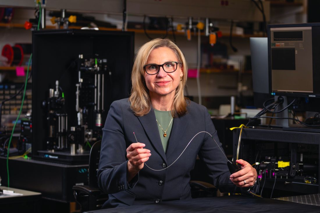
She and her colleagues are developing an OCT/fluorescence imaging falloposcope that is 0.8 mm in diameter, flexible, and steerable, as a tool for early detection of ovarian cancer in humans. “It’s now known that most ovarian cancer starts in the fallopian tubes,” Dr. Barton said. “It’s metastatic disease when those cells break off from the fallopian tubes and go to the ovaries. We wanted to create an imaging system where we created a fiber bundle that we could navigate with white light and with fluorescence so that we can see these early stages of cancer [and] how they fluoresce differently. We also wanted to have an OCT system so that we could image through the wall of the fallopian tube and look for that layer thickening and other precursors to ovarian cancer.”
To date, in vivo testing in healthy women has demonstrated that the miniature endoscope is able to reach the fallopian tubes through the natural orifice of the vagina and uterus. “That is pretty exciting,” she said. “The images may not be of the highest quality, but we are advancing.”
Dr. Barton reported having no relevant financial disclosures.
PHOENIX –
In a lecture during a multispecialty roundup of cutting-edge energy-based device applications at the annual conference of the American Society for Laser Medicine and Surgery, Dr. Barton, a biomedical engineer who directs the BIO5 Institute at the University of Arizona, Tucson, said that while no current modality exists to enable physicians in dermatology and other specialties to view internal structures throughout the entire body with cellular resolution, refining existing technologies is a good way to start.
In 2011, renowned cancer researchers Douglas Hanahan, PhD, and Robert A. Weinberg, PhD, proposed six hallmarks of cancer, which include sustaining proliferative signaling, evading growth suppressors, resisting cell death, enabling replicative immortality, inducing angiogenesis, and activating invasion and metastasis. Each hallmark poses unique imaging challenges. For example, enabling replicative immortality “means that the cell nuclei change size and shape; they change their position,” said Dr. Barton, who is also professor of biomedical engineering and optical sciences at the university. “If we want to see that, we’re going to need an imaging modality that’s subcellular in resolution.”
Similarly, if clinicians want to view how proliferative signaling is changing, “that means being able to visualize the cell surface receptors; those are even smaller to actually visualize,” she said. “But we have technologies where we can target those receptors with fluorophores. And then we can look at large areas very quickly.” Meanwhile, the ability of cancer cells to resist cell death and evade growth suppressors often results in thickening of epithelium throughout the body. “So, if we can measure the thickness of the epithelium, we can see that there’s something wrong with that tissue,” she said.
As for cancer’s propensity for invasion and metastasis, “here, we’re looking at how the collagen structure [between the cells] has changed and whether there’s layer breakdown or not. Optical imaging can detect cancer. However, high resolution optical techniques can only image about 1 mm deep, so unless you’re looking at the skin or the eye, you’re going to have to develop an endoscope to be able to view these hallmarks.”
OCT images the tissue microstructure, generally in a resolution of 2-20 microns, at a depth of 1-2 mm, and it measures reflected light. When possible, Dr. Barton combines OCT with laser-induced fluorescence for enhanced accuracy of detection of cancer. Induced fluorescence senses molecular information with the natural fluorophores in the body or with targeted exogenous agents. Then there’s multiphoton microscopy, an advanced imaging technique that enables clinicians to view cellular and subcellular events within living tissue. Early models of this technology “took up entire benches” in physics labs, Dr. Barton said, but she and other investigators are designing smaller devices for use in clinics. “This is exciting, because not only do we [view] subcellular structure with this modality, but it can also be highly sensitive to collagen structure,” she said.
Ovarian cancer model
In a model of ovarian cancer, she and colleagues externalized the ovaries of a mouse, imaged the organs, put them back in, and reassessed them at 8 weeks. “This model develops cancer very quickly,” said Dr. Barton, who once worked for McDonnell Douglas on the Space Station program. At 8 weeks, using fluorescence and targeted agents with a tabletop multiphoton microscopy system, they observed that the proliferation signals of cancer had begun. “So, with an agent targeted to the folate receptor or to other receptors that are implicated in cancer development, we can see that ovaries and fallopian tubes are lighting up,” she said.
With proof of concept established with the mouse study, she and other researchers are drawing from technological advances to create tiny laser systems for use in the clinic to image a variety of structures in the human body. Optics advances include bulk optics and all-fiber designs where engineers can create an imaging probe that’s only 125 microns in diameter, “or maybe even as small as 70 microns in diameter,” she said. “We can do fabrications on the tips of endoscopes to redirect the light and focus it. We can also do 3-D printing and spiral scanning to create miniature devices to make new advances. That means that instead of just white light imaging of the colon or the lung like we have had in the past, we can start moving into smaller structures, such as the eustachian tube, the fallopian tube, the bile ducts, or making miniature devices for brain biopsies, lung biopsies, and maybe being able to get into bronchioles and arterioles.”
According to Dr. Barton, prior research has demonstrated that cerebral vasculature can be imaged with a catheter 400 microns in diameter, the spaces in the lungs can be imaged with a needle that is 310 microns in diameter, and the inner structures of the eustachian tube can be viewed with an endoscope 1 mm in diameter.
She and her colleagues are developing an OCT/fluorescence imaging falloposcope that is 0.8 mm in diameter, flexible, and steerable, as a tool for early detection of ovarian cancer in humans. “It’s now known that most ovarian cancer starts in the fallopian tubes,” Dr. Barton said. “It’s metastatic disease when those cells break off from the fallopian tubes and go to the ovaries. We wanted to create an imaging system where we created a fiber bundle that we could navigate with white light and with fluorescence so that we can see these early stages of cancer [and] how they fluoresce differently. We also wanted to have an OCT system so that we could image through the wall of the fallopian tube and look for that layer thickening and other precursors to ovarian cancer.”
To date, in vivo testing in healthy women has demonstrated that the miniature endoscope is able to reach the fallopian tubes through the natural orifice of the vagina and uterus. “That is pretty exciting,” she said. “The images may not be of the highest quality, but we are advancing.”
Dr. Barton reported having no relevant financial disclosures.
AT ASLMS 2023
Study supports new NCCN classification for cutaneous squamous cell carcinoma
, according to new findings.
In addition, regardless of the NCCN risk group, the study found that Mohs surgery or peripheral and deep en face margin assessment (PDEMA) conferred a lower risk of developing LR, DM, and disease-related death.
“Although the NCCN included this new high-risk group in the last iteration of the guidelines, there were no studies that identified whether the high-risk group achieved the goal of identifying riskier tumors,” said senior author Emily Ruiz, MD, MPH, associate physician at the Mohs and Dermatologic Surgery Center at Brigham and Women’s Faulkner Hospital, Boston. “Based on the data in our study, the risk groups did risk stratify tumors and so clinicians can utilize the high-risk group risk factors to identify which tumors may require additional surveillance or treatment.”
The study was published online in JAMA Dermatology.
Most patients with CSCC are successfully treated with Mohs micrographic surgery or wide local excision (WLE) alone, but a subset will experience more severe and aggressive disease. While useful for prognostication, current staging systems do not incorporate patient factors or other high-risk tumor features that influence outcomes, which led to the NCCN reclassifying CSCC into low-, high-, and very high-risk groups. The NCCN guidelines also made a new recommendation that Mohs or PDEMA be the preferred method for tissue processing for high- and very-high-risk tumors, based on this new stratification.
However, these changes to the NCCN guidelines have not been validated. The goal of this study was to compare outcomes in very-high-, high-, and low-risk NCCN groups as well as comparing outcomes of CSCCs stratified by Mohs and WLE.
Dr. Ruiz and colleagues conducted a retrospective cohort study using patient data from two tertiary care academic medical centers. Their analysis included 10,196 tumors from 8,727 patients that were then stratified into low-risk (3,054 tumors [30.0%]), high-risk (6,269 tumors [61.5%]), and very-high-risk (873 tumors [8.6%]) groups.
Tumors in the very-high-risk group were more likely to have high-risk tumor and histologic features, such as large-caliber perineural invasion, large diameter, invasion beyond the subcutaneous fat or bone, poor differentiation, and lymphovascular invasion.
The authors found that, compared with the low-risk group, the high- and very-high-risk groups demonstrated a greater risk of LR (high-risk subhazard ratio, 1.99; P = .007; very-high-risk SHR, 12.66; P < .001); NM (high-risk SHR, 4.26; P = .02; very-high-risk SHR, 62.98; P < .001); DM (high-risk SHR, 2.2 × 10⁷; P < .001; very-high-risk SHR, 6.3 × 10⁸; P < .001); and DSD (high-risk SHR, 4.02; P = .03; very-high-risk SHR, 93.87; P < .001).
Adjusted 5-year cumulative incidence was also significantly higher in very-high- vs. high- and low-risk groups for all endpoints.
They next compared the procedures used to treat the tumors. Compared with WLE, patients treated with Mohs or PDEMA had a lower risk of LR (SHR, 0.65; P = .009), DM (SHR, 0.38; P = .02), and DSD (SHR, 0.55; P = .006).
Mohs and PDEMA have already became preferred surgical modalities for high- and very-high-risk tumors, and Dr. Ruiz pointed out that their analysis was for the entire cohort.
“We did not stratify this by risk group,” she said. “So our results do not change anything clinically at this time, but support prior studies that have found Mohs/PDEMA to have improved outcomes, compared to WLE. Further studies are needed evaluating surgical approach by risk-group.”
However, she emphasized, “our studies further validate prior evidence showing Mohs/PDEMA to have the lowest rates of recurrence and in this study, even disease-related death.”
Approached for an independent comment, Jeffrey M. Farma, MD, codirector of the melanoma and skin cancer program, and interim chair, department of surgical oncology, Fox Chase Cancer Center, Philadelphia, noted that this study supports the new reclassification of CSCC tumors by the NCCN, and confirms that the high-risk and very-high-risk tumors surely have a higher propensity for worse outcomes overall.
“That being said, the notion for type of resection and margin assessment is still an area of controversy in the dermatology, surgical oncology, and pathology community,” said Dr. Farma, who is also on the NCCN panel. “I believe we need further studies to truly understand the role of the type of resection and the pathologic evaluation play in this disease process.”
He also pointed out that it is unclear in this dataset if patients initially had any imaging to evaluate for local or regional metastatic disease. “It would be helpful to have a further understanding of which type of provider was performing the excisions, the type of excision decided upon, and if there was a standardized approach to [decide] which patients had MOHS or PDEMA and what was the surveillance for these patients both with imaging and physical examinations,” said Dr. Farma. “This data also evaluated patients over a long time period where practice patterns have evolved.”
Finally, he noted that the number of local and metastatic events subjectively seems low in this cohort. “We also do not know any information about the initial workup of the patients, patterns of recurrence, and adjuvant or palliative treatment after recurrence,” he added. “It is unclear from this manuscript how the type of resection or pathologic evaluation of margins leads to improved outcomes and further prospective studies are warranted.”
Dr. Ruiz reports reported serving as a coinvestigator and principal investigator for Regeneron Pharmaceuticals and as a coinvestigator for Merck and consulting for Checkpoint Therapeutics, BDO, and Genentech outside the submitted work. Dr. Farma has no disclosures other than the NCCN panel. The study was supported by Harvard Catalyst and the Harvard University Clinical and Translational Science Center and by Harvard University and its affiliated academic health care centers and partially supported by the Melvin Markey Discovery Fund at Cleveland Clinic Foundation.
FROM JAMA DERMATOLOGY
Multiprong strategy makes clinical trials less White
CHICAGO – Clinical trials are so White. Only a small percentage of eligible patients participate in clinical trials in the first place, and very few come from racial and ethnic minority groups.
For example, according to the Food and Drug Administration, in trials that resulted in drug approvals from 2017 to 2020, only 2%-5% of participants were Black patients.
When clinical trials lack diverse patient populations, those who are left out have fewer opportunities to get new therapies. Moreover, the scope of the research is limited by smaller phenotypic and genotypic samples, and the trial results are applicable only to more homogeneous patient groups.
There has been a push to include more underrepresented patients in clinical trials. One group reported its success in doing so here at the annual meeting of the American Society of Clinical Oncology.
a period that included a pandemic-induced hiatus in clinical trials in general.
Alliance member Electra D. Paskett, PhD, from the College of Public Health at the Ohio State University in Columbus, presented accrual data from 117 trials led by the Alliance from 2014 to 2022.
During this period, accrual of racial and ethnic minority patients increased from 13.6% to 25.3% for cancer treatment trials and from 13% to 21.5% for cancer control trials.
Overall, the recruitment program resulted in an absolute increase from 13.5 % to 23.6% of underrepresented populations, which translated into a relative 74.8% improvement.
“We’re focusing now on monitoring accrual of women, rural populations, younger AYAs [adolescents and young adults] and older patients, and we’ll see what strategies we need to implement,” Dr. Packett told this news organization.
The Alliance has implemented a real-time accrual dashboard on its website that allows individual sites to review accrual by trial and overall for all of the identified underrepresented populations, she noted.
Program to increase underrepresented patient accrual
The impetus for the program to increase enrollment of underrepresented patients came from the goal set by Monica M. Bertagnolli, MD, group chair of the Alliance from 2011 to 2022 and currently the director of the U.S. National Cancer Institute.
“Our leader, Dr. Bertagnolli, set out a group-wide goal for accrual of underrepresented minorities to our trials of 20%, and that gave us permission to implement a whole host of new strategies,” Dr. Paskett said in an interview.
“These strategies follow the Accrual of Clinical Trials framework, which essentially says that the interaction between the patient and the provider for going on a clinical trial is not just an interaction between the patient and provider but recognizes, for example, that the provider has coworkers and they have norms and beliefs and attitudes, and the patient comes from a family with their own values. And then there are system-level barriers, and there are community barriers that all relate to this interaction about going on a trial,” Dr. Packett said.
What works?
The study was presented as a poster at the meeting. During the poster discussion session, comoderator Victoria S. Blinder, MD, from Memorial Sloan Kettering Cancer Center in New York, asked Dr. Paskett, “If you had a certain amount of money and you really wanted to use that resource to focus on one area, where would you put that resource?”
“I’m going to violate the rules of your question,” Dr. Paskett replied.
“You cannot change this problem by focusing on one thing, and that’s what we showed in our Alliance poster, and what I’ve said is based on over 30 years of work in this area,” she said.
She cited what she considered as the two most important components for improving accrual of underrepresented populations: a commitment by leadership to a recruitment goal, and the development of protocols with specific accrual goals for minority populations.
Still, those are only two components of a comprehensive program that includes the aforementioned accrual goal set by Dr. Bertagnolli, as well as the following:
- Funding of minority junior investigators and research that focuses on issues of concern to underrepresented populations.
- Establishment of work groups that focus on specific populations with the Alliance health disparities committee.
- Translation of informational materials for patients.
- Opening studies at National Cancer Institute Community. Oncology Research Program–designated minority underserved sites.
- Real-time monitoring of accrual demographics by the Alliance and at the trial site.
- Closing protocol enrollment to majority populations.
- Increasing the study sample sizes to enroll additional minority participants and to allow for subgroup analyses.
The study was funded by the National Institutes of Health. Dr. Packett and Dr. Blinder reported no relevant financial relationships.
A version of this article originally appeared on Medscape.com.
AT ASCO 2023
CBSM phone app eases anxiety, depression in cancer patients
CHICAGO – One-third of patients with cancer also experience anxiety or depression, and an estimated 70% of the 18 million patients with cancer and cancer survivors in the US experience emotional symptoms, including fear of recurrence.
Despite many having these symptoms, few patients with cancer have access to psycho-oncologic support.
A digital cognitive-behavioral stress management (CBSM) application may help to ease some of the burden, reported Allison Ramiller, MPH, of Blue Note Therapeutics in San Francisco, which developed the app version of the program.
In addition, patients assigned to the CBSM app were twice as likely as control persons to report that their symptoms were “much” or “very much” improved after using the app for 12 weeks, Ms. Ramiller reported at an oral abstract session at the annual meeting of the American Society of Clinical Oncology (ASCO).
However, the investigators did not report baseline characteristics of patients in each of the study arms, which might have helped to clarify the depth of the effects they saw.
The CBSM program was developed by Michael H. Antoni, PhD, and colleagues in the University of Miami Health System. It is based on cognitive-behavioral therapy but also includes stress management and relaxation techniques to help patients cope with cancer-specific stress.
“”It has been clinically validated and shown to benefit patients with cancer,” Ms. Ramiller said. “However, access is a problem,” she said.
“There aren’t enough qualified, trained providers for the need, and patients with cancer encounter barriers to in-person participation, including things like transportation or financial barriers. So to overcome this, we developed a digitized version of CBSM,” she explained.
Impressive and elegant
“Everything about [the study] I thought was very impressive, very elegant, very nicely done,” said invited discussant Raymond U. Osarogiagbon, MBBS, FACP, chief scientist at Baptist Memorial Health Care Corp in Memphis, Tenn.
“They showed efficacy, they showed safety – very nice – user friendliness – very good. Certainly they look like they’re trying to address a highly important, unmet need in a very elegant way. Certainly, they pointed out it needs longer follow-up to see sustainability. We need to see will this work in other settings. Will this be cost-effective? You’ve gotta believe it probably will be,” he said.
CBSM has previously been shown to help patients with cancer reduce stress, improve general and cancer-specific quality of life at various stages of treatment, reduce symptom burden, and improve coping skills, Ms. Ramiller said.
To see whether these benefits could be conveyed digitally rather than in face-to-face encounters, Ms. Ramiller and colleagues worked with Dr. Antoni to develop the CBSM app.
Patients using the app received therapeutic content over 10 sessions with audio, video, and interactive tools that mimicked the sessions they would have received during in-person interventions.
They then compared the app against the control educational app in the randomized, decentralized RESTORE study.
High-quality control
Ms. Ramiller said that the control app set “a high bar.”
“The control also offered 10 interactive self-guided sessions. Both treatment apps were professionally designed and visually similar in styling, and they were presented as digital therapeutic-specific for cancer patients. And they were also in a match condition, meaning they received the same attention from study staff and cadence of reminders, but importantly, only the intervention app was based on CBSM,” she explained.
A total of 449 patients with cancers of stage I–III who were undergoing active systemic treatment or were planning to undergo such treatment within 6 months were randomly assigned to the CBSM app or the control app.
The CBSM app was superior to the control app for the primary outcome of anxiety reduction over baseline, as measured at 4, 8 and 12 weeks by the Patient-Reported Outcomes Measurement Information System Anxiety Scale (PROMIS-A) (beta = -.03; P = .019).
CBSM was also significantly better than the control app for the secondary endpoints of reducing symptoms of depression, as measured by the PROMIS-D scale (beta = -.02, P = .042), and also at increasing the percentage of patients who reported improvement in anxiety and depression symptoms on the Patient Global Impression of Change instrument (P < .001)
An extension study of the durability of the effects at 3 and 6 months is underway.
The investigators noted that the incremental cost of management of anxiety or depression is greater than $17,000 per patient per year.
“One of the big promises of a digital therapeutic like this is that it could potentially reduce costs,” Ms. Ramiller told the audience, but she acknowledged, “More work is really needed, however, to directly test the potential savings.”
The RESTORE study is funded by Blue Note Therapeutics. Dr. Osarogiagbon owns stock in Gilead, Lilly, and Pfizer, has received honoraria from Biodesix and Medscape, and has a consulting or advisory role for the American Cancer Society AstraZeneca, Genentech/Roche, LUNGevity, National Cancer Institute, and Triptych Health Partners.
A version of this article originally appeared on Medscape.com.
AT ASCO 2023
FDA approves talazoparib for metastatic prostate cancer
Talazoparib is already approved for adults with deleterious or suspected deleterious germline BRCA-mutated HER2-negative locally advanced or metastatic breast cancer. The new approval, granted following priority review, is based on findings from the randomized, placebo-controlled, phase 3 TALAPRO-2 trial, published in The Lancet.
The 399 patients in the study were randomly assigned in a 1:1 ratio to receive either enzalutamide 160 mg daily plus either talazoparib 0.5 mg or placebo daily. Median radiographic progression-free survival (PFS) was not reached in the treatment group; it was 13.8 months in the placebo group (hazard ratio, 0.45). In an exploratory analysis by BRCA mutation status, patients with BRCA-mutated disease who received talazoparib exhibited an even stronger median radiographic PFS (HR, 0.20; not reached vs. 11 months) in comparison with those without BRCA-mutated disease (HR, 0.72; 24.7 vs. 16.7 months).
Serious adverse reactions occurred in 30% of patients who received talazoparib plus enzalutamide. The most common serious adverse reactions, reported in more than 2% of patients, included anemia (9%) and fracture (3%). Discontinuation of talazoparib occurred in 10% of patients, according to a Pfizer statement.
Pfizer also noted that a marketing authorization application for the drug combination has been accepted for review by the European Medicines Agency.
“Despite treatment advancement in metastatic castration-resistant prostate cancer, the disease can progress quickly, and many patients may only receive one line of therapy,” lead investigator Neeraj Agarwal, MD, of the Huntsman Cancer Institute, University of Utah, Salt Lake City, said in a statement. Patients with metastatic castration-resistant prostate cancer harboring HRR genetic alterations have even worse outcomes, and thus the FDA’s approval of the talazoparib and enzalutamide combination “represents a treatment option deserving of excitement and attention.”
A version of this article originally appeared on Medscape.com.
Breast cancer family history linked to better BC survival
TOPLINE:
METHODOLOGY:
- Investigators reviewed 28,649 Swedish women diagnosed with breast cancer from 1991 to 2019.
- Overall, 5,081 patients (17.7%) had at least one female first-degree relative previously diagnosed with breast cancer.
TAKEAWAYS:
- After adjusting for demographics, tumor characteristics, and treatments, a family history of breast cancer was associated with a lower risk of breast cancer–specific death in the full cohort (hazard ratio, 0.78) and in ER-negative women (HR, 0.57) within 5 years of diagnosis, after which point the association was no longer significant.
- The lower risk of death among women with a family history could mean that these women are more motivated and likely to get screened, potentially catching tumors earlier, and may be more likely to adhere to treatment recommendations.
- However, having a family history of early-onset breast cancer (before the age of 40) was associated with a higher risk of breast cancer–specific death (HR, 1.41).
IN PRACTICE:
Although the findings are reassuring for many women with breast cancer, “genetic testing of newly diagnosed patients with early-onset family history may provide useful information to aid treatment and future research,” the researchers concluded.
STUDY DETAILS:
The study was led by Yuqi Zhang, PhD, of the Karolinska Institutet, Stockholm, and published in JAMA Network Open.
LIMITATIONS:
- The main analysis did not include tumor characteristics only available within the last 20 years, including ERBB2 status.
- Relatively wide confidence intervals make the association between a family history of early-onset breast cancer and higher risk of breast cancer death somewhat uncertain.
DISCLOSURES:
- The work was funded by the Swedish Cancer Society and others.
- The investigators report no relevant financial relationships.
A version of this article originally appeared on Medscape.com.
T-DXd active in many solid tumors; ‘shift in thinking’
CHICAGO – Trastuzumab deruxtecan (T-DXd) (Enhertu) already has proven efficacy against HER2-expressing metastatic breast, gastroesophageal, and lung cancers.
including malignancies of the cervix, endometrium, ovaries, bladder, and other sites.
The findings come from the ongoing DESTINY-PanTumor02 trial. Among 267 patients with solid tumors at various organ sites, the investigator-assessed objective response rate among all patients was 37.1%, and ranged from as high as 57.5% for patients with endometrial cancers to as low as 4% for patients with pancreatic cancer, reported Funda Meric-Bernstam, MD, from the University of Texas MD Anderson Cancer Center, Houston.
For patients with tumors that had HER2 immunohistochemistry (IHC) scores of 3+, the highest level of HER2 expression, the overall response rate was 61.3%..
The responses were also durable, with a median duration of 11.8 months among all patients and 22.1 months among patients with IHC 3+ scores.
“Our data to date showed that T-DXd had clinically meaningful activity across a variety of tumor types,” she said in a briefing held prior to her presentation of the data at the annual meeting of the American Society of Clinical Oncology.
“HER2 expression has been around a long time. We think about this all the time in breast cancer and drugs are approved there, but HER2 is expressed in other tumors as well, and that really represents an unmet need, because we have limited options in this situation” commented ASCO expert Bradley Alexander McGregor, MD, from the Dana-Farber Cancer Institute, Boston, an invited discussant at the briefing.
“Aside from pancreatic cancer we saw really, really encouraging results with no new safety signals, so while early I think this really exciting and represents a shift in how we think about cancer care,” he added.
After the presentation, invited discussant Kohei Shitara, MD, of National Cancer Center Hospital East, Kashiwa, Japan, said that he agrees with authors that T-DXd is a potential new treatment option for patients with HER2-expressing solid tumors, and that the evidence suggests the potential for further tumor-agnostic development of the agent.
He cautioned, however, that there is a lack of concordance between local and central assessment of HER2 IHC, and that quality assurance will be required to ensure that the HER2 status of solid tumors is accurately characterized.
At a press briefing, Dr. Meric-Bernstam was asked how she envisioned using T-DXd in therapy for various HER2-expressing tumors.
“I think the activity we’ve seen is quite compelling, and one hopes that eventually this will be a drug that’s accessible for patients that are HER2-expressing across tumor types. Clearly, the activity is very compelling in some of the diseases to think about doing studies for earlier lines,” she said.
“The data indicate that there is tumor-agnostic activity across the board,” she said, but noted that tumors with epithelial components such as ovarian and breast cancers appear to have the highest responses to T-DXd therapy.
Briefing moderator Julie R. Gralow, MD, chief medical officer and executive vice president of ASCO, asked Dr. McGregor whether, in the light of this new data, oncologists should test more patients for HER2 expression.
“We have some cancers where we know HER2 expression is there. I think the good thing about HER2 testing is that it’s an IHC test, so this is something that can be easily done in local pathology [labs],” he said. As more evidence mounts of potential benefit of T-DXd in HER2 expressing tumors, clinicians will need to consider more routine HER2 testing.
A rendezvous with DESTINY
The DESTINY-PanTumor02 trial is a phase 2, open-label, multicenter study looking at T-DXd in patients with advanced solid tumors who are not eligible for therapy with curative intent.
All patients had disease progression after at least two prior lines of therapy, and had tumors with HER2 expression of IHC 3+ or 2+ either by local or central testing. Patients were allowed to have previously received HER2-targeting therapy. Patients also had to have good performance status (Eastern Cooperative Oncology Group/World Health Organization performance status 0 or 1).
The investigators planned to enroll 40 patients in each cohort, including patients with cervical, endometrial, ovarian, biliary tract, pancreatic, or bladder cancers, as well those with other tumors expressing HER2 who were not included in the other cohorts.
Under the protocol, cohorts in which none of the first 15 patients had objective responses would be closed, as happened with the pancreatic cancer cohort.
At a median follow-up of 9.7 months, an objective response was seen in 99 patients out of the 267 in the entire study population (ORR, 37.1%). This ORR consisted of 15 complete responses and 84 partial responses. An additional 123 patients had stable disease.
An analysis of ORR by HER2 expression showed that IHC 3+ expressing tumors had rates ranging from 84.6% in endometrial cancers, 75% in cervical cancer, 63.6% in ovarian cancers, and 56.3% in bladder cancers, down to zero in IHC 3+ expressing pancreatic cancer.
The T-DXd safety profile was consistent with that seen in other clinical trials, with most common adverse events being nausea, fatigue, neutropenia, anemia, diarrhea, and thrombocytopenia. There were 20 cases of interstitial lung disease, one of which was fatal.
The trial is ongoing, and investigators plan to report overall survival and progression-free survival results with additional follow-up.
DESTINY-PanTumor02 is funded by Daiichi Sankyo. Dr. Meric-Bernstam disclosed a consulting/advisory role with multiple pharmaceutical companies, research funding to her institution from Daiichi Sankyo and others, and travel expenses from ESMO and EORTC. Dr. McGregor disclosed a consulting/advisory role and institutional research funding with multiple companies, not including the study’s funder. Dr. Gralow disclosed a consulting or advisory role with Genentech and Roche.
A version of this article first appeared on Medscape.com.
AT ASCO 2023