User login
Bringing you the latest news, research and reviews, exclusive interviews, podcasts, quizzes, and more.
div[contains(@class, 'read-next-article')]
div[contains(@class, 'nav-primary')]
nav[contains(@class, 'nav-primary')]
section[contains(@class, 'footer-nav-section-wrapper')]
nav[contains(@class, 'nav-ce-stack nav-ce-stack__large-screen')]
header[@id='header']
div[contains(@class, 'header__large-screen')]
div[contains(@class, 'read-next-article')]
div[contains(@class, 'main-prefix')]
div[contains(@class, 'nav-primary')]
nav[contains(@class, 'nav-primary')]
section[contains(@class, 'footer-nav-section-wrapper')]
footer[@id='footer']
section[contains(@class, 'nav-hidden')]
div[contains(@class, 'ce-card-content')]
nav[contains(@class, 'nav-ce-stack')]
div[contains(@class, 'view-medstat-quiz-listing-panes')]
div[contains(@class, 'pane-article-sidebar-latest-news')]
Is walking speed following stroke a good predictor of recovery?
, suggests new research backed by imaging data.
In secondary analysis of a previous study, training enabled both “good” and “limited” walkers to increase travel distance during a 2-minute walk. However, for “dual-task” walking, good walkers improved their distance by approximately 10 m after training, whereas limited walkers improved by only 1 m. Brain imaging showed increased brain activity in the limited walkers, which could reduce cognitive resources available for performing a second task while walking.
These findings, which were published online May 30 in Clinical Rehabilitation, may explain the apparent lack of superiority, shown previously, of dual-task training compared with single-task training for patients with stroke and impaired walking ability, researchers noted.
“Imaging data were consistent with our hypothesis that walking automaticity might explain these results,” said lead author Johnny Collett, PhD, senior clinical research fellow at Oxford Brookes University, United Kingdom.
At baseline, participants who walked slowly had increased resting state connectivity between contralesional M1 and cortical areas associated with conscious gait control.
“In response to the intervention, we found increased connectivity with the precuneus in those who walked slowly at baseline, an adaptation that might support walking in more complex situations,” Dr. Collett said.
Benefits questioned
After stroke, many patients have difficulty walking while performing a second task, such as holding a conversation. Training in dual-task walking has provided uncertain benefits, according to clinical research.
In healthy individuals, walking is believed to be a largely automatic process that requires minimal executive resources. Previous studies have suggested that a certain minimum walking speed is required to enable automatic control of walking in the brain.
“We know that those with better walking ability after stroke are better able to cope with additional cognitive loads while walking,” said Dr. Collett. “Here, we proposed that increased automatic gait control may provide a mechanism whereby executive resources are freed up to attend to additional tasks,” he added.
The investigators further hypothesized that greater walking speed is required for automatic gait control. To test these hypotheses, they analyzed data from a previously conducted randomized trial of single- and dual-task walking interventions.
Trial participants were aged 18 years or older, had survived a stroke that had occurred at least 6 months before enrollment, had reduced 2-minute walk distance relative to their peers, and had no comorbid neurologic or psychologic disorders.
Over 10 weeks, participants underwent 20 sessions that included 30 minutes of walking on a treadmill. They were randomly assigned to undergo single-task walking or dual-task walking. The latter incorporated cognitive tasks as distractions.
Good versus limited walkers
In the current study, investigators analyzed various assessments that had been conducted at baseline and after completion of the training sessions, including distance on 2-minute walks with and without a distracting task. In addition, participants underwent imaging with functional near-infrared spectroscopy (fNIRS) and fMRI.
Using previous research as a basis, the researchers defined good walking speed as 0.8 m/sec. They categorized all participants, regardless of their intervention assignments, as having good walking capacity (0.8 m/sec or more) or limited walking capacity (less than 0.79 m/sec).
A total of 50 participants enrolled in the study (mean age, 62 years), and 45 completed the interventions. Of those who completed the interventions, 22 were randomly assigned to undergo single-task training, and 23 were assigned to dual-task training.
The researchers categorized 21 participants as having good walking capacity and 24 as having limited walking capacity. Participants in each category were divided approximately evenly between treatment assignments.
Barthel index score, which assesses functional independence, was higher in the group of good walkers.
Increased travel distance
Results showed that after the interventions, distance traveled during the single-task 2-minute walk increased by 8.9 m for good walkers and by 5.3 m for limited walkers. For the dual-task 2-minute walk, the distance traveled increased by 10.4 m among good walkers and by 1.3 m for limited walkers. Change from baseline on the dual-task walk was not significant for limited walkers.
There was no significant difference between good walkers and limited walkers in their perceptions of participation in community walking. Neither group increased its walking activity significantly following the interventions.
At baseline, limited walkers, in comparison with good walkers, had significantly greater activation in the contralesional hemisphere during dual-task walking, which consisted of incorporating a planning task.
In contrast, for many good walkers, there was a decrease in activation during dual-task walking. Activation in the contralesional hemisphere correlated negatively with dual-task 2-minute walk distance.
The researchers also found a negative correlation between activation and dual-task 2-minute walk distance when the second task was the Stroop task.
Initial step
“The original trial was never designed or powered to compare groups formed by walking speed or test our automaticity hypothesis, and the results need to be viewed within this context,” said Dr. Collett. The small sample size did not allow the researchers to detect small effects of the intervention, especially in the imaging data, he added.
It also prevented the investigators from comparing limited walking and good walking groups according to whether they underwent the single-task or dual-task intervention, “which would be a superior way to investigate our hypotheses,” Dr. Collett said.
“The result of this study should be seen as exploratory, with further investigation needed,” he noted.
Helping stroke survivors to walk in the community is challenging, and new interventions that enable them to navigate complex surroundings need to be designed, said Dr. Collett. “Research is required to better understand the conscious and automatic contribution to gait control, especially with neurological impairment,” he added.
Overall, “our results suggest that improving automatic walking may be an initial step to improve capacity to respond to more complex walking interventions. However, [future] trials are required to test this,” he concluded.
The next frontier?
Commenting on the findings, Louis R. Caplan, MD, professor of neurology at Harvard University and senior neurologist at Beth Israel Deaconess Medical Center, Boston, said that “recovery and rehab are going to be the next frontier in stroke neurology, because there has to be a limitation in the present emphasis on acute care.”
Some patients do not receive acute care on time, and current treatment is not curative, added Dr. Caplan, who was not involved with the research.
Little scientific attention has been paid to how doctors can enhance recovery after stroke, what interventions delay recovery, and what the natural history of recovery is, he said. “This is a very nice study about that.”
Although the study’s methodology was sound, there were some limitations, including that strokes and underlying brain lesions were heterogeneous and that the study population was relatively small, Dr. Caplan said.
He added that “it’s a difficult study to do” and that it is difficult to organize participants into homogeneous groups.
Another limitation cited was lack of long-term follow-up that could indicate whether training provided sustained improvements in walking.
“It would be nice to revisit the same people later and see if their walking has improved, if they’re doing it differently, and if their subjective responses are different,” said Dr. Caplan.
In addition, the study did not examine whether the interventions made it easier for participants to walk with other people or to socialize more. “It may be that it really requires some time for them to gain confidence and for them to integrate that into their social network,” Dr. Caplan said.
“I would call it a proof-of-principle study, not a final study,” he noted. “It’s a study that shows that you can scientifically study rehab” and indicates the possible methodology that could be used.
The study was funded by the Stroke Association. Dr. Collett and Dr. Caplan have reported no relevant financial relationships.
A version of this article first appeared on Medscape.com.
From Clinical Rehabilitation
Female doctors of color say they feel pressure to change their look
It started when a Latina doctor tweeted that she lost points on a practical exam in medical school because of her hoop earrings, with the evaluator writing “earrings, unprofessional.”
That led other female doctors to cite their own experiences, reported The Lily, a Washington Post publication aimed at millennial women. Many women posted photos of themselves wearing hoops, which have long been associated with Latina and African American women, the outlet said.
“There’s a big movement to police women of color and how they present themselves in medical spaces,” said Briana Christophers, an MD-PhD student at the Tri-Institutional MD-PhD Program in New York. “I think in part it’s a way of trying to make people who don’t usually fit the mold, fit the mold.”
Ms. Christophers, who identifies as Latina, said she was urged to wear a black or navy suit when interviewing for doctorate programs. She wore a black suit with a lavender blouse and received comments about that – some positive, some not, she said.
“Sometimes you don’t know how to interpret those sorts of comments,” Ms. Christophers said. “Do you remember because you like the shirt, or because you don’t think I should have done that?”
Doctors of color still stand out in American medicine. The Lily cited the Association of American Medical Colleges as saying that in 2018, Hispanics made up 5.8% of active American doctors and African Americans made up 5%.
Studies show that medical professionals of color often don’t receive the same respect as their White counterparts, with some people questioning whether they’re actually doctors.
“At work, wearing my white coat that has my name pretty big on it with a badge that says doctor on it, I still get asked if I’m the environmental services staff,” Alexandra Sims, MD, a pediatrician in Cincinnati, told The Lily. “I think it just demonstrates how deeply ingrained bias, racism, and sexism are in society and that we have a lot of work to do to disrupt that.”
Dr. Sims said the tweet about hoop earrings led her to wonder about daily decisions she makes about dress.
“Am I too much? Is this too much? Is this earring too big? Is this nail polish color too loud? And how will that be received at work?” she said, noting that she may opt not to wear hoops in certain situations, such as when she’s dealing with a grabby baby.
Monica Verduzco-Gutierrez, MD, professor and chair of the department of rehabilitation medicine at University of Texas Health, San Antonio, said doctors should be judged on the care they provide, not their appearance.
“Judging someone based on their earrings or their jumpsuit or whatever else that they’re noticing about the student is not an appropriate way to judge the student’s ability to take care of a patient,” Dr. Verduzco-Gutierrez said, noting that she was not speaking on behalf of the school.
A version of this article was first published on WebMD.com .
Hearing loss tied to decline in physical functioning
published online in JAMA Network Open.
Hearing loss is associated with slower gait and, in particular, worse balance, the data suggest.
“Because hearing impairment is amenable to prevention and management, it potentially serves as a target for interventions to slow physical decline with aging,” the researchers said.
To examine how hearing impairment relates to physical function in older adults, Pablo Martinez-Amezcua, MD, PhD, MHS, a researcher in the department of epidemiology at Johns Hopkins University, Baltimore, and colleagues analyzed data from the ongoing Atherosclerosis Risk in Communities (ARIC) study.
ARIC initially enrolled more than 15,000 adults in Maryland, Minnesota, Mississippi, and North Carolina between 1987 and 1989. In the present study, the researchers focused on data from 2,956 participants who attended a study visit between 2016 and 2017, during which researchers assessed their hearing using pure tone audiometry.
Hearing-study participants had an average age of 79 years, about 58% were women, and 80% were White. Approximately 33% of the participants had normal hearing, 40% had mild hearing impairment, 23% had moderate hearing impairment, and 4% had severe hearing impairment.
Participants had also undergone assessment of physical functioning at study visits between 2011 and 2019, including a fast-paced 2-minute walk test to measure their walking endurance. Another assessment, the Short Physical Performance Battery (SPPB), tests balance, gait speed, and chair stands (seated participants stand up and sit back down five times as quickly as possible while their arms are crossed).
Dr. Martinez-Amezcua and colleagues found that severe hearing impairment was associated with a lower average SPPB score compared with normal hearing in a regression analysis. Specifically, compared with those with normal hearing, participants with severe hearing impairment were more likely to have low scores on the SPPB (odds ratio, 2.72), balance (OR, 2.72), and gait speed (OR, 2.16).
However, hearing impairment was not significantly associated with the chair stand test results. The researchers note that chair stands may rely more on strength, whereas balance and gait speed may rely more on coordination and movement.
The team also found that people with worse hearing tended to walk a shorter distance during the 2-minute walk test. Compared with participants with normal hearing, participants with moderate hearing impairment walked 2.81 meters less and those with severe hearing impairment walked 5.31 meters less on average, after adjustment for variables including age, sex, and health conditions.
Participants with hearing impairment also tended to have faster declines in physical function over time.
Various mechanisms could explain associations between hearing and physical function, the authors said. For example, an underlying condition such as cardiovascular disease might affect both hearing and physical function. Damage to the inner ear could affect vestibular and auditory systems at the same time. In addition, hearing impairment may relate to cognition, depression, or social isolation, which could influence physical activity.
“Age-related hearing loss is traditionally seen as a barrier for communication,” Dr. Martinez-Amezcua told this news organization. “In the past decade, research on the consequences of hearing loss has identified it as a risk factor for cognitive decline and dementia. Our findings contribute to our understanding of other negative outcomes associated with hearing loss.”
Randomized clinical trials are the best way to assess whether addressing hearing loss might improve physical function, Dr. Martinez-Amezcua said. “Currently there is one clinical trial (ACHIEVE) that will, among other outcomes, study the impact of hearing aids on cognitive and physical function,” he said.
Although interventions may not reverse hearing loss, hearing rehabilitation strategies, including hearing aids and cochlear implants, may help, he added. Educating caregivers and changing a person’s environment can also reduce the effects hearing loss has on daily life, Dr. Martinez-Amezcua said.
“We rely so much in our sense of vision for activities of daily living that we tend to underestimate how important hearing is, and the consequences of hearing loss go beyond having trouble communicating with someone,” he said.
This study and prior research “raise the intriguing idea that hearing may provide essential information to the neural circuits underpinning movement in our environment and that correction for hearing loss may help promote physical well-being,” Willa D. Brenowitz, PhD, MPH, and Margaret I. Wallhagen, PhD, GNP-BC, both at the University of California, San Francisco, wrote in an accompanying commentary. “While this hypothesis is appealing and warrants further investigation, there are multiple other potential explanations of such an association, including potential sources of bias that may affect observational studies such as this one.”
Beyond treating hearing loss, interventions such as physical therapy or tai chi may benefit patients, they suggested.
Because many changes occur during older age, it can be difficult to understand which factor is influencing another, Dr. Brenowitz said in an interview. There are potentially relevant mechanisms through which hearing could affect cognition and physical functioning. Still another explanation could be that some people are “aging in a faster way” than others, Dr. Brenowitz said.
Dr. Martinez-Amezcua and a coauthor disclosed receiving sponsorship from the Cochlear Center for Hearing and Public Health. Another author, Frank R. Lin, MD, PhD, directs the research center, which is partly funded by a philanthropic gift from Cochlear to the Johns Hopkins Bloomberg School of Public Health. Dr. Lin also disclosed personal fees from Frequency Therapeutics and Caption Call. One author serves on a scientific advisory board for Shoebox and Good Machine Studio.
Dr. Wallhagen has served on the board of trustees of the Hearing Loss Association of America and is a member of the board of the Hearing Loss Association of America–California. Dr. Wallhagen also received funding for a pilot project on the impact of hearing loss on communication in the context of chronic serious illness from the National Palliative Care Research Center outside the submitted work.
A version of this article first appeared on Medscape.com.
New details of myocarditis linked to COVID vaccines
Further details from multiple cases of myocarditis linked to the Pfizer and Moderna mRNA COVID vaccines have been described in recent papers in the medical literature.
The cases appear to occur almost exclusively in males and most often in younger age groups. While symptoms and signs of myocarditis mostly resolved with a few days of supportive care, long-term effects are unknown at present.
The authors of all the reports and of two accompanying editorials in JAMA Cardiology are unanimous in their opinion that the benefits of vaccination still outweigh the risks.
The Centers for Disease Control and Prevention’s but committee members delivered a strong endorsement for continuing to vaccinate young people with the mRNA vaccines.
The current case reports are published in two papers in JAMA Cardiology and in three in Circulation.
U.S. military reports 23 cases
In one report in JAMA Cardiology, authors led by Jay Montgomery, MD, from Walter Reed National Military Medical Center in Bethesda, Md., described 23 cases from the U.S. Military Health System of individuals with acute myocarditis who presented within 4 days after mRNA-based COVID-19 vaccination (7 Pfizer and 16 Moderna).
All patients were male, 22 of 23 were on active duty, and the median age was 25 years (range, 20-51); 20 of the 23 cases occurred after receipt of a second dose of an mRNA COVID-19 vaccine.
The patients all presented with acute onset of marked chest pain. All patients had significantly elevated cardiac troponin levels. Among eight patients who underwent cardiac MRI (cMRI), all had findings consistent with the clinical diagnosis of myocarditis.
Additional testing did not identify other possible causes of myocarditis. All patients received brief supportive care and were recovered or recovering.
The authors reported that the military administered more than 2.8 million doses of mRNA COVID-19 vaccine in this period, and while the observed number of myocarditis cases was small, the number was “substantially higher” than expected among male military members after a second vaccine dose.
They noted that, based on historical data, among the 544,000 second doses to military members there may have been 0-10 expected myocarditis cases, but they observed 19 cases.
“All patients in this series reflect substantial similarities in demographic characteristics, proximate vaccine dose, onset interval, and character of vaccine-associated myocarditis. The consistent pattern of clinical presentation, rapid recovery, and absence of evidence of other causes support the diagnosis of hypersensitivity myocarditis,” they stated.
They added that presentation after a second vaccine dose or, in three patients, when vaccination followed SARS-CoV-2 infection, suggests that prior exposure was relevant in the hypersensitivity response.
“The spectrum of clinical presentation and reliance on patients seeking health care and on health care professionals recognizing a rare vaccine-associated adverse event limits determination of the true incidence of this condition,” the authors wrote.
They stressed that recognition of vaccine-associated myocarditis is clinically important because diagnosis impacts management, recommendations for exercise, and monitoring for cardiomyopathy.
But the authors also acknowledged that it is important to frame concerns about potential vaccine-associated myocarditis within the context of the current pandemic.
“Infection with SARS-CoV-2 is a clear cause of serious cardiac injury in many patients. ... Prevalence of cardiac injury may be as high as 60% in seriously ill patients. Notably, nearly 1% of highly fit athletes with mild COVID-19 infection have evidence of myocarditis on cMRI,” they wrote.
“Given that COVID-19 vaccines are remarkably effective at preventing infection, any risk of rare adverse events following immunization must be carefully weighed against the very substantial benefit of vaccination,” they concluded.
Four cases at Duke
In the second paper in JAMA Cardiology, a group led by Han W. Kim, MD, reported four patients with acute myocarditis occurring within days of mRNA COVID-19 vaccination (two Pfizer and two Moderna) in patients treated at Duke University Medical Center, Durham, N.C. The hospital courses of the four patients with myocarditis following COVID-19 vaccination were uneventful, and they were discharged within 2-4 days.
The authors said that, although a causal relationship cannot be established, none of the patients had a viral prodrome or had coincident testing that revealed an alternative explanation.
They stated that these four patients represent the majority of patients with acute myocarditis identified in the past 3 months at their institution, and this led to the highest total number of patients with acute myocarditis, compared with the same 3-month period for the past 5 years.
“Additionally, we identified only those patients with severe unremitting chest pain who sought medical attention. Those with mild or moderate chest pain might not seek medical attention, and it is possible that subclinical myocarditis may occur and could be detected by active surveillance, as has been described with smallpox vaccination,” they wrote.
Further case reports
In one of the papers in Circulation, a group led by Kathryn F. Larson, MD, from the Mayo Clinic in Rochester, Minn., described eight patients hospitalized with chest pain who were diagnosed with myocarditis within 2-4 days of receiving either the Pfizer or Moderna vaccine.
Two of the patients had previously been infected by SARS-CoV-2 without need for hospitalization. All individuals were otherwise healthy males between the ages of 21 and 56 years. All but one patient developed symptoms after their second dose, and the one patient who developed myocarditis after the first vaccine dose had previously been infected with SARS-CoV-2.
Systemic symptoms began within 24 hours after vaccine administration in five of eight patients, with chest pain presenting between 48 and 96 hours later. Troponin values were elevated in all individuals and appeared to peak the day after admission, whereas none had eosinophilia.
Cardiac MRI revealed findings consistent with myocarditis in all patients. All patients had resolution of their chest pain and were discharged from the hospital in stable condition.
“The patients presented here demonstrated typical signs, symptoms, and diagnostic features of acute myocarditis. The temporal association between receiving an mRNA-based COVID-19 vaccine and the development of myocarditis is notable,” the authors said.
They added that they would consider the use of corticosteroids in these patients but cautioned that this could reduce the specific immune response against SARS-COV-2 triggered by the vaccine. “Thus, the duration of corticosteroid administration should be limited to the resolution of the symptoms or ventricular arrhythmias or the recovery of the left ventricular ejection fraction.”
Pending publication of long-term outcome data after SARS-CoV-2 vaccine–related myocarditis, they suggest adherence to the current consensus recommendation to abstain from competitive sports for a period of 3-6 months with reevaluation prior to sports participation.
In another of the Circulation papers, a group led by Carolyn M. Rosner, MSN, presented a case series of seven patients hospitalized for acute myocarditis-like illness following COVID-19 vaccination, from two U.S. medical centers, in Falls Church, Va., and Dallas. All patients were males below the age of 40 years and of White or Hispanic race/ethnicity. Only one patient reported prior history of COVID-19 infection. Six patients received mRNA (Moderna or Pfizer) and one received the adenovirus (Johnson & Johnson) vaccine. All patients presented 3-7 days post vaccination with acute onset chest pain and biochemical evidence of myocardial injury.
Hospital length of stay was 3 days, and all patients’ symptoms resolved by hospital discharge.
And finally, the third paper in Circulation reported a detailed description of one patient – a 52-year-old, previously healthy male who presented with acute myocarditis 3 days after the administration of the second dose of Moderna’s COVID-19 vaccine. The symptoms resolved, and there was a gradual improvement in cMRI findings. Ischemic injury and other potential causes of acute myocardial injury were excluded, as were other potential infectious causes of myocarditis, and there was no evidence of systemic autoimmune disease.
“Clinicians should be aware that myocarditis may be present in patients exhibiting cardiac signs and symptoms 2-4 days after COVID-19 vaccination,” the authors said.
They added that additional surveillance of such adverse events post–COVID-19 vaccination will help identify subgroups at higher risk for this vaccine-related effect, and whether additional precautions are necessary.
‘Benefits outweigh risk’
In an accompanying editorial in JAMA Cardiology, three doctors from the CDC cite several other reports of myocarditis after mRNA COVID vaccination. These include a case report published in Pediatrics of seven male adolescents aged 14-19 years who presented with myocarditis or myopericarditis within 4 days after receipt of a second dose of the Pfizer vaccine.
But the editorialists noted that the most comprehensive data about the risk for myocarditis following immunization with mRNA vaccines comes from Israel.
The Israeli Ministry of Health recently posted data describing 121 myocarditis cases occurring within 30 days of a second dose of mRNA vaccine among 5,049,424 persons, suggesting a crude incidence rate of approximately 24 cases per million.
On the current case reports, the CDC doctors wrote: “The striking clinical similarities in the presentations of these patients, their recent vaccination with an mRNA-based COVID-19 vaccine, and the lack of any alternative etiologies for acute myocarditis suggest an association with immunization.”
They said that acute onset of chest pain 3-5 days after vaccine administration, usually after a second dose, is a typical feature of reported cases and suggests an immune-mediated mechanism.
But SARS-CoV-2 infection also causes cardiac injury which may result in severe outcomes, and based on currently available data, myocarditis following immunization with current mRNA-based vaccines is rare.
“At present, the benefits of immunization in preventing severe morbidity favors continued COVID-19 vaccination, particularly considering the increasing COVID-19 hospitalization rates among adolescents reported during spring 2021,” the editorialists stated.
But they added that many questions remain. These include whether modifications are needed to the vaccine schedule among persons with a history of possible or confirmed myocarditis after COVID vaccine, how should postvaccine myocarditis be managed, how often should follow-up assessments be performed, how might follow-up assessments affect recommendations to avoid vigorous physical activity following the diagnosis of myocarditis, and do all likely cases of acute myocarditis that appear to be uncomplicated require cardiac MRI for more definitive diagnosis?
“While the data needed to answer such questions are being collected, there is an opportunity for researchers with expertise in myocarditis to develop a comprehensive, national assessment of the natural history, pathogenesis, and treatment of acute myocarditis associated with receipt of mRNA-based COVID-19 vaccines,” they concluded.
In a second editorial in JAMA Cardiology, a group of editors from the journal acknowledged that publication of the current case reports may contribute to additional public concern regarding immunization. But they added that clinicians discussing immunization with patients should recognize that these case series suggest that the symptomatic events consistent with myocarditis are still very rare and appear to be self-limiting.
“Given the risks of COVID-19, including the risk of myocarditis from COVID-19 infection, the editors do not believe these case reports are sufficient to interrupt the march toward maximal vaccination against SARS-CoV-2 as expeditiously as possible,” they said.
A version of this article first appeared on Medscape.com.
Magnesium is strongly tied to lower risk for intracranial aneurysm
The effects may be partially mediated by magnesium’s influence on systolic blood pressure, new research suggests.
“The modifiable risk factors for intracranial aneurysm are largely unknown. Our findings provided evidence of a causal association between increased serum magnesium levels and reduced risk of intracranial aneurysm,” said Susanna Larsson, PhD, Karolinska Institutet, Stockholm.
These results suggest that raising serum magnesium levels – through a magnesium-rich diet or magnesium supplementation – “may play a role in the primary prevention of intracranial aneurysm and associated hemorrhage,” Dr. Larsson added.
The study was published online June 22 in Neurology.
Lower risk for rupture
The researchers leveraged randomly allocated genetic variants related to serum magnesium concentrations in a two-sample Mendelian randomization (MR) study to assess whether higher genetically predicted serum magnesium correlates with reduced risk for intracranial aneurysm. They also performed a multivariable MR analysis to assess the role blood pressure might play in this association.
Source data came from a genome-wide association study (GWAS) involving 23,829 individuals that previously identified five single-nucleotide polymorphisms associated with serum magnesium. Genetic association estimates for intracranial aneurysm were derived from a GWAS in 79,429 people (7,495 case patients and 71,934 control patients), and genetic association estimates for systolic blood pressure were derived from a GWAS of 757,601 individuals.
The researchers found that higher genetically predicted serum magnesium concentrations were associated with lower risk for intracranial aneurysm.
The odds ratios per 0.1 mmol/L increment in genetically predicted serum magnesium concentrations were 0.66 (95% confidence interval, 0.49-0.91) for intracranial aneurysm (unruptured and ruptured combined), 0.57 (95% CI, 0.30-1.06) for unruptured intracranial aneurysm, and 0.67 (95% CI, 0.48-0.92) for aneurysmal subarachnoid hemorrhage.
Adjustment for genetically predicted systolic blood pressure partially attenuated the associations of genetically predicted serum magnesium with all three outcomes, suggesting that magnesium’s influence was at least partially mediated by systolic blood pressure.
“In addition to a blood pressure lowering effect, increased magnesium concentrations may reduce the risk of intracranial aneurysm rupture by improving endothelial function and reducing oxidative stress,” the investigators noted.
They caution that the data were derived from people of European ancestry, which limits the generalizability to other populations. “Caution should be taken when extrapolating findings from MR to infer the effect of a clinical intervention, and clinical trials are warranted to guide optimal practice,” they added.
Critical role in vascular health
In an accompanying editorial, Joanna Pera, MD, PhD, of Jagiellonian University Medical College, Krakow, Poland, and Christopher Anderson, MD, of Brigham and Women’s Hospital, Boston, noted that the study “adds to our understanding of the importance of magnesium in vascular health particularly related to cerebral aneurysms.”
There is a need for “both mechanistic and potentially therapeutic investigation into the role that magnesium plays in subarachnoid hemorrhage,” they added.
Further, they wrote, the results “raise interesting new questions about the links between circulating magnesium, intracranial aneurysms, and blood pressure. Arterial hypertension is a well-recognized risk factor for intracranial aneurysm development and rupture. Magnesium supplementation may lower blood pressure values.
“Could this mineral prove useful in developing interventions that could prevent intracranial aneurysm development and/or rupture over and above a simple lowering of blood pressure, perhaps through pleiotropic effects on endothelial function or other mechanisms? With these results in hand, work is clearly needed to learn more about the biology of magnesium in the vascular system and in intracranial aneurysm biology in particular,” Dr. Pera and Dr. Anderson concluded.
This study was supported by the Swedish Research Council for Health, Working Life and Welfare, the British Heart Foundation Research Center of Excellence at Imperial College London, and the National Institute for Health Research Clinical Lectureship at St. George’s, University of London. Dr. Larsson has disclosed no relevant financial relationships. Study coauthor Dipender Gill, PhD, is employed part time by Novo Nordisk. Dr. Pera has disclosed no relevant financial relationships. Dr. Anderson has received research support from the Bayer AG and has consulted for ApoPharma and Invitae.
A version of this article first appeared on Medscape.com.
From Neurology
A less expensive, more convenient treatment option for MS?
Patients with multiple sclerosis (MS) may soon have another less expensive, more convenient treatment option compared with other agents in the same drug class, new research suggests.
The positive results suggest “another strong and reasonably safe medication might be available to increase the repertoire of effective medicines that we can offer MS patients,” said Lawrence Steinman, MD, professor of neurology, Stanford (Calif.) University. “These are delightful data in my opinion,” he added.
The findings were presented at the 2021 Congress of the European Academy of Neurology.
‘Glycoengineered’ antibody
If approved by the U.S. Food and Drug Administration, ublituximab would become the only glycoengineered anti-CD20 monoclonal antibody for MS. Glycoengineering involves changing protein-associated carbohydrates to alter pharmacokinetic properties.
There are currently two approved anti-CD20 agents for MS, but both require 4-hour infusions. For many patients, this means “at least half their day is shot,” Dr. Steinman said. “A lot of people don’t want to or can’t miss a half day of work.” Ublituximab can be infused more rapidly, he noted.
For the study, the investigators analyzed data from the ULTIMATE I and ULTIMATE II studies, which included 1,089 mostly White patients with MS. Almost all participants had the relapsing-remitting form of the disease and were between 18 and 55 years of age (average age, 36 years). Their scores on the Expanded Disability Status Scale (EDSS) were from 0 to 5.5, and they had been neurologically stable for at least 30 days prior to screening.
Participants were required to have experienced two or more relapses within the previous 2 years or one or more relapses in the year prior and/or had one gadolinium-enhancing lesion in the year prior to screening.
The study population was mostly from the Ukraine and Russia. It is more difficult to recruit patients into MS drug studies in the United States and Western Europe because many patients in these countries are already receiving approved drugs, which deters enrollment, explained Dr. Steinman.
Investigators randomly assigned the participants to receive the investigational drug or 14 mg of oral teriflunomide, a drug that blocks the proliferation of immune cells, once daily. The ublituximab group received an initial infusion of 150 mg over 4 hours and then a 1-hour infusion of 450 mg every 6 months over the course of the 96-week study.
Primary outcomes met
For ULTIMATE I, the primary outcome was ARR. Results showed that this rate was 0.076 for the ublituximab group and 0.188 for the teriflunomide group, resulting in a 60% relative reduction (adjusted ARR ratio, 0.406; 95% confidence interval, 0.268-0.615; P < .0001).
In ULTIMATE II, the ARR was 0.091 for ublituximab and 0.178 for teriflunomide, for a relative reduction of 49% (ARR ratio, 0.509; 95% CI, 0.330-0.784; P = .0022).
One way of interpreting these data is that patients are likely to have only one relapse in 10 years, said Dr. Steinman. “So that was very good news.”
It is not clear why relative reductions for ARR differed between the two studies; “probably the real number is somewhere between 60% and 49%,” Dr. Steinman said.
From MRI scans, the total number of relevant lesions was reduced by 97% with ublituximab compared with teriflunomide in ULTIMATE I and by 96% in trial II.
Another “piece of really good news” from the studies is that the drug led to a significant improvement in disability, rather than “just slowing it down,” Dr. Steinman noted.
There was a 116% increased chance of confirmed disability improvement (CDI) with ublituximab versus teriflunomide in the first trial (P = .003) and a 103% increased chance of CDI in the second trial (P = .0026).
The percentage of patients who had no evidence of disease activity was 198% for the patients who received the trial drug in comparison with the control group in trial I and 277% in trial II (P < .0001 for both trials).
A life changer?
Dr. Steinman said the “robust” findings suggest that patients with MS “won’t have a relapse and will improve. Those are two pretty good messages for somebody with this wretched disease.”
The investigational drug was generally well tolerated. The percentage of adverse events (AEs) with the study drug was about the same as with the comparator. About 9.5% of the ublituximab group had a serious AE, compared with 6.2% of the teriflunomide group.
The ublituximab group had more infections (4.0% vs. 2.6%), which Dr. Steinman said is not surprising because the drug is a potent immune suppressant. “It’s an unfortunate consequence of this kind of strong biologic that knocks down a whole arm of the immune system. The wonder to me is that these are still rather infrequent,” he said.
If approved, “it will be interesting to see how regulatory agencies handle this in terms of risk mitigation,” said Dr. Steinman. He added that a warning label might be a consideration.
However, the safety of this drug “is certainly acceptable,” said Dr. Steinman. “In general, this drug is not that different from the other drugs in the class of anti-CD20s.”
Dr. Steinman noted that he understands why some patients prefer an oral drug and may have an “aversion to getting stuck with a needle,” but he pointed out that teriflunomide has some drawbacks. For example, it tends to thin hair.
“For people who have had relapses, people who are unable to do what they want to in life – attend school, hold down jobs, exercise – this new drug could really be life changing,” he said.
He added that he would “strongly urge” his own family and relatives, if they had MS, to take one of the anti-CD20 drugs.
Ublituximab also has a number of advantages over the other agents in the same class. Not only does it work well, have an acceptable safety profile, and require a shorter infusion time, but it could also be less costly, Dr. Steinman noted. “The company has said it intends to come in at a lower price point,” he said.
The company is now planning to prepare a biological license application for use in MS. Interestingly, the drug, in combination with umbralisib (Ukoniq), is already under review by the FDA for use in chronic lymphoctytic leukemia and small lymphocytic lymphoma.
Striking improvement
When session chair Marcello Moccio, MD, Multiple Sclerosis Clinical Care and Research Center, Federico II University, Naples, Italy, asked Dr. Steinman to elaborate on the “very strong effect” of the drug with regard to improving disability, Dr. Steinman said the improvement was “striking.”
Being able to talk to patients about possible improvement rather than about delaying disability “is really gratifying” and provides a “much more constructive and optimistic outlook,” he said.
He noted that as physicians improve their management of patients with MS “and are paying attention to things that we haven’t over the years, like vitamin D and even mental health,” disability progression management “is getting better.”
Dr. Steinman is a consultant for TG Therapeutics.
A version of this article first appeared on Medscape.com.
From EAN 2021
Stimulant reduces ‘sluggish cognitive tempo’ in adults with ADHD
A stimulant used in patients with attention-deficit/hyperactivity disorder might prove useful for other comorbid symptoms, results of a randomized, crossover trial suggest.

In the trial, the investigators reported that lisdexamfetamine (Vyvanse) reduced self-reported symptoms of sluggish cognitive tempo (SCT) by 30%, in addition to lowering ADHD symptoms by more than 40%.
The drug also corrected deficits in executive brain function. Patients had fewer episodes of procrastination, were better able to prioritize, and showed improvements in keeping things in mind.
“These findings highlight the importance of assessing symptoms of sluggish cognitive tempo and executive brain function in patients when they are initially diagnosed with ADHD,” Lenard A. Adler, MD, the lead author, said in a press release. The results were published June 29, 2021, in the Journal of Clinical Psychiatry.
The trial is groundbreaking because it is the first treatment study for ADHD with SCT in adults, Dr. Adler, director of the adult ADHD program at New York University Langone Health, said in an interview. He said that Russell A. Barkley, PhD, a clinical professor of psychiatry at Virginia Commonwealth University, Richmond, defines SCT as having nine cardinal symptoms: prone to daydreaming, easy boredom, trouble staying awake, feeling foggy, spaciness, lethargy, underachieving, less energy, and not processing information quickly or accurately.
Dr. Barkley, who studied more than 1,200 individuals with SCT, discovered that nearly half also had ADHD, Dr. Adler said. Those with the comorbid symptoms also had more impairment.
Whether or not the symptom set of SCT is a distinct disorder or a cotraveling symptom set that goes along with ADHD has been an area of investigation, said Dr. Adler, also a professor in the departments of psychiatry and child and adolescent psychiatry at New York University. Other known comorbid symptoms include executive function deficits and trouble with emotional control.
Stimulants to date have only shown success in children, as far as improving SCT. The goal of this study was to determine the efficacy of lisdexamfetamine on the nature and severity of ADHD symptoms and SCT behavioral indicators in adults with ADHD and SCT.
Two cohorts, alternating regimens
The investigators enrolled 38 adults with DSM-5 ADHD and SCT. Patients were recruited from two academic centers, New York University and the Icahn School of Medicine at Mount Sinai. The randomized 10-week crossover trial included two double-blind treatment periods, each 4 weeks long, with an intervening 2-week, single-blind placebo washout period.
“In crossover design, patients act as their own control, because they receive both treatments,” Dr. Adler said. Recruiting a smaller number of subjects helps to achieve significance in results.
For the first 4 weeks, participants received daily doses of either lisdexamfetamine (30-70 mg/day; mean, 59.1 mg/day) or a placebo sugar pill (mean, 66.6 mg/day). Researchers used standardized tests for SCT signs and symptoms, ADHD, and other measures of brain function to track psychiatric health on a weekly basis. After a month, the two cohorts switched regimens – those taking the placebo started the daily doses of lisdexamfetamine, and the other half stopped the drug and started taking the placebo.
Primary outcomes included the ADHD Rating Scale and Barkley Adult ADHD Rating Scale-IV SCT subscale.
Compared with placebo, adults with ADHD and comorbid SCT showed significant improvement after taking lisdexamfetamine in ratings of SCT and total ADHD symptoms. This was also true of other comorbid symptoms, such as executive function deficits.
In the crossover design, patients who received the drug first hadn’t gone fully back to baseline by the time the investigators crossed them over into the placebo group. “So, we couldn’t combine the two treatment epochs,” Dr. Adler said. However, the effect of the drug versus placebo was comparable in both study arms.
SCT alone was not studied
The trial had some limitations, mainly that it was an initial study with a modest sample size, Dr. Adler said. It also did not examine SCT alone, “so we can’t really say whether the stimulant medicine would improve SCT in patients who don’t have ADHD. What’s notable is when you look at how much of the improvement in SCT was due to improvement in ADHD, it was just 25%.” This means the effects occurring on SCT symptoms were not solely caused by effects on ADHD.
he said.
Dr. Adler would like to see treatment studies of adults with ADHD and SCT in a larger sample, potentially with other stimulants. In addition, future trials could examine the effects of stimulants on adults with SCT that do not have ADHD.
The results of this trial underscore the importance of evaluating adults with ADHD for comorbid symptoms, such as executive function and emotional control, he continued. “Impairing SCT symptoms may very well fall under that umbrella,” Dr. Adler said. “If you don’t identify them, you can’t track them in terms of treatment.”
SCT as a ‘flavor’ of ADHD
The outcome of this study demonstrates that lisdexamfetamine significantly improves both ADHD symptoms and SCT symptoms, said David W. Goodman MD, LFAPA, an assistant professor in the department of psychiatry and behavioral sciences at Johns Hopkins University, Baltimore.
Dr. Goodman, who was not involved in the study, agreed that clinicians should be aware of SCT when assessing adults with ADHD and conceptualize SCT as a “flavor” of ADHD. “SCT is not widely recognized by clinicians outside of the research arena but will likely become an important characteristic of ADHD presentation,” he said in an interview.
“Future studies in adult ADHD should further clarify the prevalence of SCT in the ADHD population and address more specific effective treatment options,” he said.
James M. Swanson, PhD, who also was not involved with the study, agreed in an interview that it documents the clear short-term benefit of stimulants on symptoms of SCT. The study “may be very timely, since adults who were affected by COVID-19 often have residual sequelae manifested as ‘brain fog,’ which resemble SCT,” said Dr. Swanson, professor of pediatrics at the University of California, Irvine.
The study was funded by Takeda Pharmaceutical, manufacturer of lisdexamfetamine. Dr. Adler has received grant/research support and has served as a consultant from Shire/Takeda and other companies. Dr. Goodman is a scientific consultant to Takeda and other pharmaceutical companies in the ADHD arena. Dr. Swanson had no disclosures.
A stimulant used in patients with attention-deficit/hyperactivity disorder might prove useful for other comorbid symptoms, results of a randomized, crossover trial suggest.
In the trial, the investigators reported that lisdexamfetamine (Vyvanse) reduced self-reported symptoms of sluggish cognitive tempo (SCT) by 30%, in addition to lowering ADHD symptoms by more than 40%.
The drug also corrected deficits in executive brain function. Patients had fewer episodes of procrastination, were better able to prioritize, and showed improvements in keeping things in mind.
“These findings highlight the importance of assessing symptoms of sluggish cognitive tempo and executive brain function in patients when they are initially diagnosed with ADHD,” Lenard A. Adler, MD, the lead author, said in a press release. The results were published June 29, 2021, in the Journal of Clinical Psychiatry.
The trial is groundbreaking because it is the first treatment study for ADHD with SCT in adults, Dr. Adler, director of the adult ADHD program at New York University Langone Health, said in an interview. He said that Russell A. Barkley, PhD, a clinical professor of psychiatry at Virginia Commonwealth University, Richmond, defines SCT as having nine cardinal symptoms: prone to daydreaming, easy boredom, trouble staying awake, feeling foggy, spaciness, lethargy, underachieving, less energy, and not processing information quickly or accurately.
Dr. Barkley, who studied more than 1,200 individuals with SCT, discovered that nearly half also had ADHD, Dr. Adler said. Those with the comorbid symptoms also had more impairment.
Whether or not the symptom set of SCT is a distinct disorder or a cotraveling symptom set that goes along with ADHD has been an area of investigation, said Dr. Adler, also a professor in the departments of psychiatry and child and adolescent psychiatry at New York University. Other known comorbid symptoms include executive function deficits and trouble with emotional control.
Stimulants to date have only shown success in children, as far as improving SCT. The goal of this study was to determine the efficacy of lisdexamfetamine on the nature and severity of ADHD symptoms and SCT behavioral indicators in adults with ADHD and SCT.
Two cohorts, alternating regimens
The investigators enrolled 38 adults with DSM-5 ADHD and SCT. Patients were recruited from two academic centers, New York University and the Icahn School of Medicine at Mount Sinai. The randomized 10-week crossover trial included two double-blind treatment periods, each 4 weeks long, with an intervening 2-week, single-blind placebo washout period.
“In crossover design, patients act as their own control, because they receive both treatments,” Dr. Adler said. Recruiting a smaller number of subjects helps to achieve significance in results.
For the first 4 weeks, participants received daily doses of either lisdexamfetamine (30-70 mg/day; mean, 59.1 mg/day) or a placebo sugar pill (mean, 66.6 mg/day). Researchers used standardized tests for SCT signs and symptoms, ADHD, and other measures of brain function to track psychiatric health on a weekly basis. After a month, the two cohorts switched regimens – those taking the placebo started the daily doses of lisdexamfetamine, and the other half stopped the drug and started taking the placebo.
Primary outcomes included the ADHD Rating Scale and Barkley Adult ADHD Rating Scale-IV SCT subscale.
Compared with placebo, adults with ADHD and comorbid SCT showed significant improvement after taking lisdexamfetamine in ratings of SCT and total ADHD symptoms. This was also true of other comorbid symptoms, such as executive function deficits.
In the crossover design, patients who received the drug first hadn’t gone fully back to baseline by the time the investigators crossed them over into the placebo group. “So, we couldn’t combine the two treatment epochs,” Dr. Adler said. However, the effect of the drug versus placebo was comparable in both study arms.
SCT alone was not studied
The trial had some limitations, mainly that it was an initial study with a modest sample size, Dr. Adler said. It also did not examine SCT alone, “so we can’t really say whether the stimulant medicine would improve SCT in patients who don’t have ADHD. What’s notable is when you look at how much of the improvement in SCT was due to improvement in ADHD, it was just 25%.” This means the effects occurring on SCT symptoms were not solely caused by effects on ADHD.
he said.
Dr. Adler would like to see treatment studies of adults with ADHD and SCT in a larger sample, potentially with other stimulants. In addition, future trials could examine the effects of stimulants on adults with SCT that do not have ADHD.
The results of this trial underscore the importance of evaluating adults with ADHD for comorbid symptoms, such as executive function and emotional control, he continued. “Impairing SCT symptoms may very well fall under that umbrella,” Dr. Adler said. “If you don’t identify them, you can’t track them in terms of treatment.”
SCT as a ‘flavor’ of ADHD
The outcome of this study demonstrates that lisdexamfetamine significantly improves both ADHD symptoms and SCT symptoms, said David W. Goodman MD, LFAPA, an assistant professor in the department of psychiatry and behavioral sciences at Johns Hopkins University, Baltimore.
Dr. Goodman, who was not involved in the study, agreed that clinicians should be aware of SCT when assessing adults with ADHD and conceptualize SCT as a “flavor” of ADHD. “SCT is not widely recognized by clinicians outside of the research arena but will likely become an important characteristic of ADHD presentation,” he said in an interview.
“Future studies in adult ADHD should further clarify the prevalence of SCT in the ADHD population and address more specific effective treatment options,” he said.
James M. Swanson, PhD, who also was not involved with the study, agreed in an interview that it documents the clear short-term benefit of stimulants on symptoms of SCT. The study “may be very timely, since adults who were affected by COVID-19 often have residual sequelae manifested as ‘brain fog,’ which resemble SCT,” said Dr. Swanson, professor of pediatrics at the University of California, Irvine.
The study was funded by Takeda Pharmaceutical, manufacturer of lisdexamfetamine. Dr. Adler has received grant/research support and has served as a consultant from Shire/Takeda and other companies. Dr. Goodman is a scientific consultant to Takeda and other pharmaceutical companies in the ADHD arena. Dr. Swanson had no disclosures.
FROM THE JOURNAL OF CLINICAL PSYCHIATRY
A pacemaker that 'just disappears' and a magnetic diet device
Ignore this pacemaker and it will go away
At some point – and now seems to be that point – we have to say enough is enough. The throwaway culture that produces phones, TVs, and computers that get tossed in the trash because they can’t be repaired has gone too far. That’s right, we’re looking at you, medical science!
This time, it’s a pacemaker that just disappears when it’s no longer needed. Some lazy heart surgeon decided that it was way too much trouble to do another surgery to remove the leads when a temporary pacemaker was no longer needed. You know the type: “It sure would be nice if the pacemaker components were biocompatible and were naturally absorbed by the body over the course of a few weeks and wouldn’t need to be surgically extracted.” Slacker.
Well, get a load of this. Researchers at Northwestern and George Washington universities say that they have come up with a transient pacemaker that “harvests energy from an external, remote antenna using near-field communication protocols – the same technology used in smartphones for electronic payments and in RFID tags.”
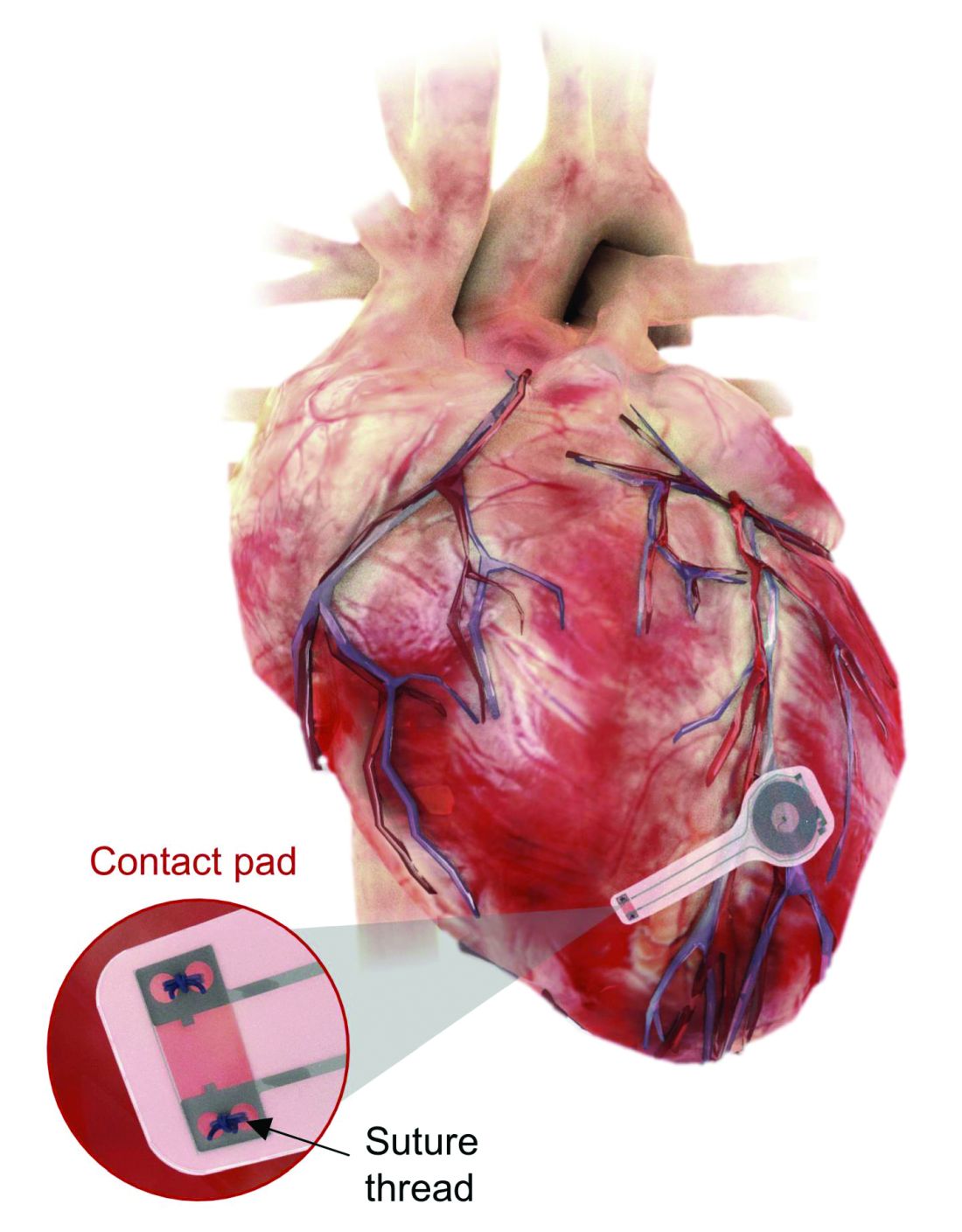
That means no batteries and no wires that have to be removed and can cause infections. Because the infectious disease docs also are too lazy to do their jobs, apparently.
The lack of onboard infrastructure means that the device can be very small – it weighs less than half a gram and is only 250 microns thick. And yes, it is bioresorbable and completely harmless. It fully degrades and disappears in 5-7 weeks through the body’s natural biologic processes, “thereby avoiding the need for physical removal of the pacemaker electrodes. This is potentially a major victory for postoperative patients,” said Dr. Rishi Arora, one of the investigators.
A victory for patients, he says. Not a word about the time and effort saved by the surgeons. Typical.
It’s a mask! No, it’s a COVID-19 test!
Mask wearing has gotten more lax as people get vaccinated for COVID-19, but as wearing masks for virus prevention is becoming more normalized in western society, some saw an opportunity to make them work for diagnosis.
Researchers from the Massachusetts Institute of Technology and the Wyss Institute for Biologically Inspired Engineering at Harvard University have found a way to do just that with their wearable freeze-dried cell-free (wFDCF) technology. A single push of a button releases water from a reservoir in the mask that sequentially activates three different freeze-dried biological reactions, which detect the SARS-CoV-2 virus in the wearer’s breath.
Initially meant as a tool for the Zika outbreak in 2015, the team made a quick pivot in May 2020. But this isn’t just some run-of-the-mill, at-home test. The data prove that the wFDCF mask is comparable to polymerase chain reactions tests, the standard in COVID-19 detection. Plus there aren’t any extra factors to deal with, like room or instrument temperature to ensure accuracy. In just 90 minutes, the mask gives results on a readout in a way similar to that of a pregnancy test. Voilà! To have COVID-19 or not to have COVID-19 is an easily answered question.

At LOTME, we think this is a big improvement from having dogs, or even three-foot rats, sniffing out coronavirus.
But wait, there’s more. “In addition to face masks, our programmable biosensors can be integrated into other garments to provide on-the-go detection of dangerous substances including viruses, bacteria, toxins, and chemical agents,” said Peter Nguyen, PhD, study coauthor and research scientist at the Wyss Institute. The technology can be used on lab coats, scrubs, military uniforms, and uniforms of first responders who may come in contact with hazardous pathogens and toxins. Think of all the lives saved and possible avoidances.
If only it could diagnose bad breath.
Finally, an excuse for the all-beer diet
Weight loss is hard work. Extremely hard work, and, as evidenced by the constant inundation and advertisement of quick fixes, crash diets, and expensive gym memberships, there’s not really a solid, 100% solution to the issue. Until now, thanks to a team of doctors from New Zealand, who’ve decided that the best way to combat obesity is to leave you in constant agony.
The DentalSlim Diet Control device is certainly a radical yet comically logical attempt to combat obesity. The creators say that the biggest problem with dieting is compliance, and, well, it’s difficult to eat too much if you can’t actually open your mouth. The metal contraption is mounted onto your teeth and uses magnetic locks to prevent the user from opening their mouths more than 2 mm. That’s less than a tenth of an inch. Which is not a lot. So not a lot that essentially all you can consume is liquid.
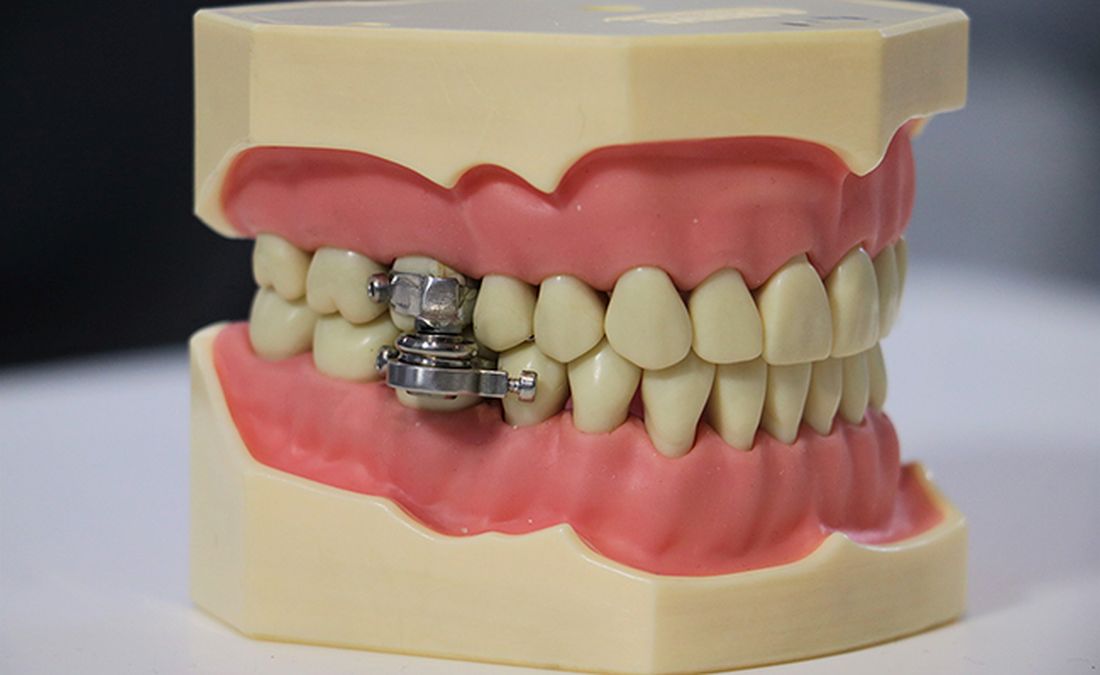
Oh, and they’ve got results to back up their madness. In a small study, seven otherwise healthy obese women lost an average of 5.1% of their body weight after using the DentalSlim for 2 weeks, though they did complain that the device was difficult to use, caused discomfort and difficulty speaking, made them more tense, and in general made life “less satisfying.” And one participant was able to cheat the system and consume nonhealthy food like chocolate by melting it.
So, there you are, if you want a weight-loss solution that tortures you and has far bigger holes than the one it leaves for your mouth, try the DentalSlim. Or, you know, don’t eat that eighth slice of pizza and maybe go for a walk later. Your choice.
Ignore this pacemaker and it will go away
At some point – and now seems to be that point – we have to say enough is enough. The throwaway culture that produces phones, TVs, and computers that get tossed in the trash because they can’t be repaired has gone too far. That’s right, we’re looking at you, medical science!
This time, it’s a pacemaker that just disappears when it’s no longer needed. Some lazy heart surgeon decided that it was way too much trouble to do another surgery to remove the leads when a temporary pacemaker was no longer needed. You know the type: “It sure would be nice if the pacemaker components were biocompatible and were naturally absorbed by the body over the course of a few weeks and wouldn’t need to be surgically extracted.” Slacker.
Well, get a load of this. Researchers at Northwestern and George Washington universities say that they have come up with a transient pacemaker that “harvests energy from an external, remote antenna using near-field communication protocols – the same technology used in smartphones for electronic payments and in RFID tags.”
That means no batteries and no wires that have to be removed and can cause infections. Because the infectious disease docs also are too lazy to do their jobs, apparently.
The lack of onboard infrastructure means that the device can be very small – it weighs less than half a gram and is only 250 microns thick. And yes, it is bioresorbable and completely harmless. It fully degrades and disappears in 5-7 weeks through the body’s natural biologic processes, “thereby avoiding the need for physical removal of the pacemaker electrodes. This is potentially a major victory for postoperative patients,” said Dr. Rishi Arora, one of the investigators.
A victory for patients, he says. Not a word about the time and effort saved by the surgeons. Typical.
It’s a mask! No, it’s a COVID-19 test!
Mask wearing has gotten more lax as people get vaccinated for COVID-19, but as wearing masks for virus prevention is becoming more normalized in western society, some saw an opportunity to make them work for diagnosis.
Researchers from the Massachusetts Institute of Technology and the Wyss Institute for Biologically Inspired Engineering at Harvard University have found a way to do just that with their wearable freeze-dried cell-free (wFDCF) technology. A single push of a button releases water from a reservoir in the mask that sequentially activates three different freeze-dried biological reactions, which detect the SARS-CoV-2 virus in the wearer’s breath.
Initially meant as a tool for the Zika outbreak in 2015, the team made a quick pivot in May 2020. But this isn’t just some run-of-the-mill, at-home test. The data prove that the wFDCF mask is comparable to polymerase chain reactions tests, the standard in COVID-19 detection. Plus there aren’t any extra factors to deal with, like room or instrument temperature to ensure accuracy. In just 90 minutes, the mask gives results on a readout in a way similar to that of a pregnancy test. Voilà! To have COVID-19 or not to have COVID-19 is an easily answered question.
At LOTME, we think this is a big improvement from having dogs, or even three-foot rats, sniffing out coronavirus.
But wait, there’s more. “In addition to face masks, our programmable biosensors can be integrated into other garments to provide on-the-go detection of dangerous substances including viruses, bacteria, toxins, and chemical agents,” said Peter Nguyen, PhD, study coauthor and research scientist at the Wyss Institute. The technology can be used on lab coats, scrubs, military uniforms, and uniforms of first responders who may come in contact with hazardous pathogens and toxins. Think of all the lives saved and possible avoidances.
If only it could diagnose bad breath.
Finally, an excuse for the all-beer diet
Weight loss is hard work. Extremely hard work, and, as evidenced by the constant inundation and advertisement of quick fixes, crash diets, and expensive gym memberships, there’s not really a solid, 100% solution to the issue. Until now, thanks to a team of doctors from New Zealand, who’ve decided that the best way to combat obesity is to leave you in constant agony.
The DentalSlim Diet Control device is certainly a radical yet comically logical attempt to combat obesity. The creators say that the biggest problem with dieting is compliance, and, well, it’s difficult to eat too much if you can’t actually open your mouth. The metal contraption is mounted onto your teeth and uses magnetic locks to prevent the user from opening their mouths more than 2 mm. That’s less than a tenth of an inch. Which is not a lot. So not a lot that essentially all you can consume is liquid.
Oh, and they’ve got results to back up their madness. In a small study, seven otherwise healthy obese women lost an average of 5.1% of their body weight after using the DentalSlim for 2 weeks, though they did complain that the device was difficult to use, caused discomfort and difficulty speaking, made them more tense, and in general made life “less satisfying.” And one participant was able to cheat the system and consume nonhealthy food like chocolate by melting it.
So, there you are, if you want a weight-loss solution that tortures you and has far bigger holes than the one it leaves for your mouth, try the DentalSlim. Or, you know, don’t eat that eighth slice of pizza and maybe go for a walk later. Your choice.
Two case reports identify Guillain-Barré variants after SARS-CoV-2 vaccination
Guillain-Barré syndrome, a rare peripheral nerve disorder that can occur after certain types of viral and bacterial infections, has not to date been definitively linked to infection by SARS-CoV-2 or with vaccination against the virus, despite surveillance searching for such associations.
Spikes in Guillain-Barré syndrome incidence have previously, but rarely, been associated with outbreaks of other viral diseases, including Zika, but not with vaccination, except for a 1976-1977 swine influenza vaccine campaign in the United States that was seen associated with a slight elevation in risk, and was halted when that risk became known. Since then, all sorts of vaccines in the European Union and United States have come with warnings about Guillain-Barré syndrome in their package inserts – a fact that some Guillain-Barré syndrome experts lament as perpetuating the notion that vaccines cause Guillain-Barré syndrome.
Epidemiologic studies in the United Kingdom and Singapore did not detect increases in Guillain-Barré syndrome incidence during the COVID-19 pandemic. And as mass vaccination against COVID-19 got underway early this year, experts cautioned against the temptation to attribute incident Guillain-Barré syndrome cases following vaccination to SARS-CoV-2 without careful statistical and epidemiological analysis. Until now reports of Guillain-Barré syndrome have been scant: clinical trials of a viral vector vaccine developed by Johnson & Johnson saw one in the placebo arm and another in the intervention arm, while another case was reported following administration of a Pfizer mRNA SARS-Cov-2 vaccine.
Recent case reports
None of the patients had evidence of current SARS-CoV-2 infection.
From India, Boby V. Maramattom, MD, of Aster Medcity in Kochi, India, and colleagues reported on seven severe cases of Guillain-Barré syndrome occurring between 10 and 14 days after a first dose of the AstraZeneca vaccine. All but one of the patients were women, all had bilateral facial paresis, all progressed to areflexic quadriplegia, and six required respiratory support. Patients’ ages ranged from 43 to 70. Four developed other cranial neuropathies, including abducens palsy and trigeminal sensory nerve involvement, which are rare in reports of Guillain-Barré syndrome from India, Dr. Maramattom and colleagues noted.
The authors argued that their findings “should prompt all physicians to be vigilant in recognizing Guillain-Barré syndrome in patients who have received the AstraZeneca vaccine. While the risk per patient (5.8 per million) may be relatively low, our observations suggest that this clinically distinct [Guillain-Barré syndrome] variant is more severe than usual and may require mechanical ventilation.”
The U.K. cases, reported by Christopher Martin Allen, MD, and colleagues at Nottingham (England) University Hospitals NHS Trust, describe bifacial weakness and normal facial sensation in four men between 11 and 22 days after their first doses of the Astra-Zeneca vaccine. This type of facial palsy, the authors wrote, was unusual Guillain-Barré syndrome variant that one rapid review found in 3 of 42 European patients diagnosed with Guillain-Barré syndrome following SARS-CoV-2 infection.
Dr. Allen and colleagues acknowledged that causality could not be assumed from the temporal relationship of immunization to onset of bifacial weakness in their report, but argued that their findings argued for “robust postvaccination surveillance” and that “the report of a similar syndrome in the setting of SARS-CoV-2 infection suggests an immunologic response to the spike protein.” If the link is casual, they wrote, “it could be due to a cross-reactive immune response to the SARS-CoV-2 spike protein and components of the peripheral immune system.”
‘The jury is still out’
Asked for comment, neurologist Anthony Amato, MD, of Brigham and Women’s Hospital, Boston, said that he did not see what the two new studies add to what is already known. “Guillain-Barré syndrome has already been reported temporally following COVID-19 along with accompanying editorials that such temporal occurrences do not imply causation and there is a need for surveillance and epidemiological studies.”
Robert Lisak, MD, of Wayne State University, Detroit, and a longtime adviser to the GBS-CIDP Foundation International, commented that “the relationship between vaccines and association with Guillain-Barré syndrome continues to be controversial in part because Guillain-Barré syndrome, a rare disorder, has many reported associated illnesses including infections. Many vaccines have been implicated but with the probable exception of the ‘swine flu’ vaccine in the 1970s, most have not stood up to scrutiny.”
With SARS-Cov-2 infection and vaccines, “the jury is still out,” Dr. Lisak said. “The report from the U.K. is intriguing since they report several cases of an uncommon variant, but the cases from India seem to be more of the usual forms of Guillain-Barré syndrome.”
Dr. Lisak noted that, even if an association turns out to be valid, “we are talking about a very low incidence of Guillain-Barré syndrome associated with COVID-19 vaccines,” one that would not justify avoiding them because of a possible association with Guillain-Barré syndrome.
The GBS-CIDP Foundation, which supports research into Guillain-Barré syndrome and related diseases, has likewise stressed the low risk presented by SARS-CoV-2 vaccines, noting on its website that “the risk of death or long-term complications from COVID in adults still far exceeds the risk of any possible risk of Guillain-Barré syndrome by several orders of magnitude.”
None of the study authors reported financial conflicts of interest related to their research. Dr. Amato is an adviser to the pharmaceutical firms Alexion and Argenx, while Dr. Lisak has received research support or honoraria from Alexion, Novartis, Hoffmann–La Roche, and others.
FROM ANNALS OF NEUROLOGY
Physician fired after slurs, including ‘cannibalism,’ against Israel
Fidaa Wishah, MD, a pediatric radiologist at Phoenix Children’s Hospital in Arizona, has been fired after the hospital reviewed evidence that included her anti-Israel comments on social media, according to the hospital’s statement.
On May 26, Dr. Wishah posted, “We will uncover your thirst to kill our Palestinian children. … We sense your fear. The fear of your collapse. A state based on atrocity, inhumanity, racism and cannibalism never last long! Hey #israel … your end is coming sooner than you think.”
Phoenix Children’s Hospital did not respond to this news organization’s request for comment but said in a statement to the Jewish News Syndicate : “After a thorough review of the facts related to this matter, this individual is no longer providing care at Phoenix Children’s. All children in the care of Phoenix Children’s receive hope, healing and the best possible health care, regardless of race, color, disability, religion, gender, gender identity, sexual orientation or national origin.”
Dr. Wishah’s profile has been removed from the hospital website. Her LinkedIn profile indicates she had been a pediatric radiology fellow at Stanford (Calif.) University, specializing in advanced magnetic resonance imaging and fetal imaging and had been a senior staff pediatric radiologist at Henry Ford Health System in Detroit.
It wasn’t the first time antisemitic comments have led to the firing of a physician. Last year, this news organization wrote about Lara Kollab, DO, a first-year resident fired for her antisemitic tweets. She was subsequently barred from medicine.
In the same post from May 26, Dr. Wishah also wrote: “We will not be #censored anymore! Bomb our media buildings and we have the phones[.] Bribe the mainstream media and we have our small #socialmedia platforms[.] From our windows ... from our streets ... next the rubble we will expose you to the world[.] We will expose the #massacre and #genocide you #zionists are proud of[.]”
Today, CAIR-AZ, a group whose mission is to “enhance understanding of Islam, protect civil rights, promote justice, and empower American Muslims,” according to its website, announced that it, along with three private law firms, will represent Dr. Wishah in what they referred to as “her wrongful termination case against Phoenix Children’s Hospital.”
The announcement, which mentions that Dr. Wishah was born and raised in Gaza, said, “Dr. Wishah has been a medical doctor since 2010 and has spent the vast majority of her career as a pediatric physician. Despite caring for thousands of children, many of whom are Jewish, she has never been accused of discriminating against any of her patients or colleagues.”
The statement added, “PCH’s decision to terminate Dr. Wishah is shameful and an attack on freedom of speech.”
A version of this article first appeared on Medscape.com.