User login
The Official Newspaper of the American Association for Thoracic Surgery
Amplatzer devices outperform oral anticoagulation in atrial fib
PARIS – Percutaneous left atrial appendage closure with an Amplatzer device in patients with nonvalvular atrial fibrillation was associated with significantly lower rates of all-cause and cardiovascular mortality, compared with oral anticoagulation, in a large propensity score–matched observational registry study.
Left atrial appendage closure (LAAC) also bested oral anticoagulation (OAC) with warfarin or a novel oral anticoagulant (NOAC) in terms of net clinical benefit on the basis of the device therapy’s greater protection against stroke and systemic embolism coupled with a trend, albeit not statistically significant, for fewer bleeding events, Steffen Gloekler, MD, reported at the annual congress of the European Association of Percutaneous Cardiovascular Interventions.
The Watchman LAAC device, commercially available both in Europe and the United States, has previously been shown to be superior to OAC in terms of efficacy and noninferior regarding safety. But there have been no randomized trials of an Amplatzer device versus OAC. This lack of data was the impetus for Dr. Gloekler and his coinvestigators to create a meticulously propensity-matched observational registry.
Five hundred consecutive patients with AF who received an Amplatzer Cardiac Plug or its second-generation version, the Amplatzer Amulet, during 2009-2014 were tightly matched to an equal number of AF patients on OAC based on age, sex, body mass index, left ventricular ejection fraction, renal function, coronary artery disease status, hemoglobin level, CHA2DS2-VASc score, and HAS-BLED score. During a mean 2.7 years, or 2,645 patient-years, of follow-up, the composite primary efficacy endpoint, composed of stroke, systemic embolism, and cardiovascular or unexplained death occurred in 5.6% of the LAAC group, compared with 7.8% of controls in the OAC arm, for a statistically significant 30% relative risk reduction. Disabling stroke occurred in 0.7% of Amplatzer patients versus 1.5% of controls. The ischemic stroke rate was 1.5% in the device therapy group and 2% in the OAC arm.
All-cause mortality occurred in 8.3% of Amplatzer patients and 11.6% of the OAC group, for a 28% relative risk reduction. The cardiovascular death rate was 4% in the Amplatzer group, compared with 6.5% of controls, for a 36% risk reduction.
The composite safety endpoint, comprising all major procedural adverse events and major or life-threatening bleeding during follow-up, occurred in 3.6% of the Amplatzer group and 4.6% of the OAC group, for a 20% relative risk reduction that is not significant at this point because of the low number of events. Major, life-threatening, or fatal bleeding occurred in 2% of Amplatzer recipients versus 5.5% of controls, added Dr. Gloekler of University Hospital in Bern, Switzerland.
The net clinical benefit, a composite of death, bleeding, or stroke, occurred in 8.1% of the Amplatzer group, compared with 10.9% of controls, for a significant 24% reduction in relative risk in favor of device therapy.
Of note, at 2.7 years of follow-up only 55% of the OAC group were still taking an anticoagulant: 38% of the original 500 patients were on warfarin, and 17% were taking a NOAC. At that point, 8% of the Amplatzer group were on any anticoagulation therapy.
Discussion of the study focused on that low rate of medication adherence in the OAC arm. Dr. Gloekler’s response was that, after looking at the literature, he was no longer surprised by the finding that only 55% of the control group were on OAC at follow-up.
“If you look in the literature, that’s exactly the real-world adherence for OACs. Even in all four certification trials for the NOACs, the rate of discontinuation was 30% after 2 years – and these were controlled studies. Ours was observational, and it depicts a good deal of the problem with any OAC in my eyes,” Dr. Gloekler said.
Patients on warfarin in the real-world Amplatzer registry study spent on average a mere 30% of time in the therapeutic international normalized ratio range of 2-3.
“That means 70% of the time patients are higher and have an increased bleeding risk or they are lower and don’t have adequate stroke protection,” he noted.
This prompted one observer to comment, “We either have to do a better job in our clinics with OAC or we have to occlude more appendages.”
A large pivotal U.S. trial aimed at winning FDA approval for the Amplatzer Amulet for LAAC is underway. Patients with AF are being randomized to the approved Watchman or investigational Amulet at roughly 100 U.S. and 50 foreign sites.
Dr. Gloekler reported receiving research funds for the registry from the Swiss Heart Foundation and Abbott.
AT EUROPCR
Key clinical point:
Major finding: The primary composite efficacy endpoint of stroke, systemic embolism, or cardiovascular or unexplained death during a mean 2.7 years of follow-up occurred in 5.6% of Amplatzer device recipients, a 30% reduction, compared with the 7.8% rate in the oral anticoagulation group.
Data source: This observational registry included 500 patients with atrial fibrillation who received an Amplatzer left atrial appendage closure device and an equal number of carefully matched AF patients on oral anticoagulation.
Disclosures: The study presenter reported receiving research funds for the registry from the Swiss Heart Foundation and Abbott.
Announcing the 2018 Lifetime Achievement and Scientific Achievement Award Recipients
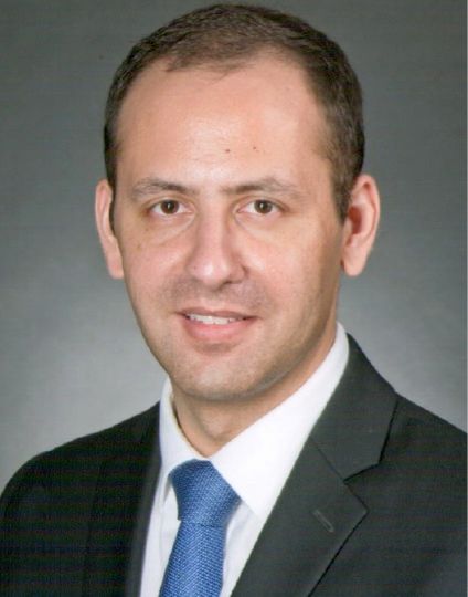
The American Association for Thoracic Surgery is recognizing the contributions of two giants in the field with two of its highest honors. The AATS will present its Lifetime Achievement Award to Aldo R. Castaneda and the Scientific Achievement Award to Magdi Yacoub on April 30, 2018, during the Plenary Session of the AATS Annual Meeting in San Diego, California.
The Lifetime Achievement Award recognizes individuals for their significant contributions to the specialty in the areas of patient care, teaching, research or community service. Dr. Castaneda, who served as the 74th President of the AATS in 1993-94, is the eighth recipient of the award since it was established in 2004.
His humanism has been a part of his work from early on in his career and now in his retirement, he continues to contribute to the well-being of the underserved people of Guatemala through the Aldo Castaneda Foundation.
In 1994, the Association established its Scientific Achievement Award to recognize individuals who have made extraordinary scientific contributions to the field of cardiothoracic surgery. Dr. Yacoub joins 12 other colleagues as a recipient of the highest scientific recognition this Association can bestow upon a surgeon.
As one of the busiest adult, congenital and thoracic transplant surgeons in the world over a very long career, he has always placed basic science and innovation on equal footing with performing operations. His dedication to patients throughout the world continues with his current efforts to develop the Aswan Heart Center to care for the indigent.
Bad news keeps piling up for Absorb coronary scaffold
PARIS – Device thrombosis occurred nearly four times more frequently in recipients of the Absorb everolimus-eluting bioresorbable vascular scaffold than with the Xience everolimus-eluting metallic stent during 2 years of prospective follow-up in the randomized AIDA trial.
AIDA (the Amsterdam Investigator-Initiated Absorb Strategy All-Comers Trial) was the first randomized trial designed to compare the Absorb scaffold to a drug-eluting metallic stent in a broad patient population reflecting routine real-world clinical practice. The disturbing AIDA finding follows upon earlier serious concerns raised regarding an increased risk of scaffold thrombosis – and the particularly worrisome complication of late thrombosis – in the ABSORB Japan and ABSORB II trials, Joanna J. Wykrzykowska, MD, reported at the annual congress of the European Association of Percutaneous Cardiovascular Interventions.
The device was approved by the Food and Drug Administration in July 2016. In March 2017 the agency issued a safety alert regarding the Absorb scaffold after release of the 2-year data from the 2,008-patient ABSORB III trial showing a significantly higher rate of target-lesion failure than with the Xience stent. Both devices are marketed by Abbott Vascular.
AIDA was a single-blind multicenter Dutch trial that randomized 1,845 patients undergoing PCI, 55% of whom presented with acute coronary syndrome and 26% of whom had ST-elevation MI. The primary endpoint was target vessel failure, a composite of cardiac death, target vessel MI, or target vessel revascularization. The 2-year cumulative rate did not differ significantly between the two study arms: 11.7% in the scaffold group and 10.7% in the metallic stent recipients.
However, definite or probable device thrombosis occurred in 3.5% of the scaffold group compared with 0.9% of metallic stent recipients, for a highly significant 3.9-fold increased risk. This was associated with a significantly increased 2-year cumulative risk of MI: 5.5% versus 3.2%.
On the basis of this unsettling finding, coupled with the fact that ABSORB II investigators did not find any instance of very late scaffold thrombosis among 63 patients who remained on dual-antiplatelet therapy (DAPT) continuously for up to 3 years, Dr. Wykrzykowska and her coinvestigators have informed AIDA participants of their treatment assignment. They have also recommended that the Absorb recipients go on extended DAPT, even though there is no high-grade evidence as yet that this will prevent late scaffold thrombosis or that the drug-induced increased bleeding risk of prolonged DAPT might cancel or perhaps even outweigh the potential protection against device thrombosis.
On top of all this, implantation of the scaffold entails a longer procedure time and a greater volume of contrast material.
Discussant Mahmoud Hashemian, MD, observed that while bioresorbable vascular scaffolds are “physiologically ideal” because, unlike metallic stents, theoretically they leave no permanent implant to impede vasomotion and serve as a nidus for neoatherosclerosis, to date they have shown no real-world benefits over current-generation drug-eluting metallic stents, but only disadvantages.
“This doesn’t mean we have to feel hopeless. I’m not hopeless at all,” said Dr. Hashemian, an interventional cardiologist at Day General Hospital in Tehran. “I’m sure this [bioresorbable scaffolds] will be the future of our stents. But it needs more work. The company tells me they are going to launch a newer one, maybe next year, with thinner struts and more expandability.”
Asked about the likely mechanism of prolonged thrombosis risk with Absorb, Dr. Wykrzykowska was quick to say no one really knows at this point.
“Technique [predilation at a 1:1 balloon-to-artery ratio with an appropriately sized balloon] can obviously improve things in the short term for early events, but I don’t think we understand the biology of late events. We don’t understand the interaction between the device and the vessel. It’s extremely complex,” she said.
AIDA was funded by an unrestricted educational grant from Abbott Vascular. Dr. Wykrzykowska reported receiving consulting and lecture fees from the company.
AT EUROPCR
Key clinical point:
Major finding: During 2 years of prospective follow-up, definite or probable device thrombosis occurred in 3.5% of recipients of a bioresorbable vascular scaffold, compared with 0.9% of metallic stent recipients, for a highly significant 3.9-fold increased risk.
Data source: AIDA, a single-blind multicenter Dutch trial that randomized a broadly representative group of 1,845 patients undergoing PCI to the Absorb bioresorbable vascular scaffold or the Xience everolimus-eluting metallic stent.
Disclosures: The AIDA study was funded by an unrestricted educational grant from Abbott Vascular. The presenter reported receiving consulting and lecture fees from the company.
Do sleep interventions prevent atrial fibrillation?
WASHINGTON – If patients have sleep disordered breathing with obstructive sleep apnea, will its treatment have cardiovascular disease benefits, especially in terms of the incidence or severity of atrial fibrillation?
Observational evidence suggests that apnea interventions may help these patients, but no clear case yet exists to prove that a breathing intervention works, experts say, and, as a result, U.S. practice is mixed when it comes to using treatment for obstructive sleep apnea (OSA), specifically continuous positive airway pressure (CPAP), to prevent or treat atrial fibrillation.
“Only a very small number of patients with atrial fibrillation undergo a sleep study,” he said in an interview. “Before I’d send my mother for atrial fibrillation ablation, I would first look for sleep disordered breathing [SDB],” but this generally isn’t happening routinely. Patients with other types of cardiovascular disease who could potentially benefit from sleep disordered breathing diagnosis and treatment are those with hypertension, especially patients who don’t fully respond to three or more antihypertensive drugs and patients with heart failure with preserved ejection fraction, he added.
Dr. Oldenburg also echoed Dr. Mehra in saying that the evidence supporting this approach for managing atrial fibrillation is less than conclusive.
“We need more precise phenotyping of patients” to better focus on patients with cardiovascular disease and sleep disordered breathing who clearly benefit from CPAP intervention, he said.
Results from the Sleep Apnea Cardiovascular Endpoints (SAVE) trial, reported in September 2016, especially tarnished the notion that treating sleep disordered breathing in patients with various cardiovascular diseases can help avoid future cardiovascular events. The multicenter trial enrolled 2,717 adults with moderate to severe obstructive sleep apnea and cardiovascular disease to receive either CPAP plus optimal routine care or optimal routine care only. After an average follow-up of close to 4 years, the patients treated with CPAP showed no benefit in terms of reduced cardiovascular events (N Engl J Med. 2016 Sept 8;375[10]:919-31).
An editorial that ran with this report suggested that the neutral outcome may have occurred because the average nightly duration of CPAP that patients in the trial self administered was just over 3 hours, arguably an inadequate dose. Other possible reasons for the lack of benefit include the time during their sleep cycle when patients administered CPAP (at the start of sleep rather than later) and that CPAP may have a reduced ability to avert new cardiovascular events in patients with established cardiovascular disease (N Engl J Med. 2016 Sept 8;375[8]:994-6).
Regardless of the reasons, the SAVE results, coupled with the neutral results and suggestion of harm from using adaptive servo-ventilation in patients with heart failure with reduced ejection fraction and central sleep apnea in the SERVE-HF trial (N Engl J Med. 2015 Sept 17;373[12]:1095-105), have thrust the management of SDB in patients with cardiovascular disease back to the point where SDB interventions have no well-proven indications for cardiovascular disease patients.
“With the SERVE-HF and SAVE trials not showing benefit, we now have equipoise” for using or not using SDB interventions in these patients, Dr. Mehra said. “It’s not clear that treating OSA improves outcomes. That allows us to randomize patients to a control placebo arm” in future trials.
An important issue in the failure to clearly establish a role for treating OSA in patients with atrial fibrillation or other cardiovascular diseases may have been over reliance on the apnea-hypopnea index (AHI) as the arbiter of OSA severity, Dr. Oldenburg said. “Maybe there are parameters to look at aside from AHI, perhaps hypoxemia burden or desaturation time. AHI is not the whole truth; we need to look at other parameters. AHI may not be the correct metric to look at in patients with various cardiovascular diseases.”
Her analysis also showed that patients with at least 10 minutes of sleep time with an oxygen saturation rate of 90% or less had a 64% increased rate of later atrial fibrillation hospitalizations, compared with those with fewer than 10 minutes spent in this state. Nearly a quarter of the patients studied fell into this category.
“Nocturnal oxygen desaturation may be stronger than AHI for predicting atrial fibrillation development,” Dr. Kendzerska concluded. “The severity of OSA-related intermittent hypoxia may be more important than sleep fragmentation in the development of atrial fibrillation. These findings support a relationship between OSA, chronic nocturnal hypoxemia, and new onset atrial fibrillation.”
However, using oxygen desaturation instead of AHI to gauge the severity of OSA won’t solve all the challenges that sleep researchers currently face in trying to determine the efficacy of breathing interventions to prevent or treat cardiovascular disease. In the neutral SAVE trial, researchers used nocturnal oxygen saturation levels to select patients with clinically meaningful OSA.
Dr. Mehra and Dr. Kendzerska had no disclosures. Dr. Oldenburg has received consultant fees, honoraria, and/or research support from ResMed, Respicardia, and Weinmann.
[email protected]
On Twitter @mitchelzoler
This article was updated on 7/10/17.
EXPERT ANALYSIS FROM ATS 2017
TB meningitis cases in U.S. are fewer but more complicated
BOSTON – The number of cases of meningitis caused by tuberculosis has fallen dramatically in the United States in recent decades as TB itself has become less common, according to findings from a study presented at the annual meeting of the American Academy of Neurology.
However, these findings from patient hospitalizations during 1993-2013 in the Nationwide Inpatient Sample database also indicate that neurologic complications from TB meningitis are on the rise.
The findings suggest that neurologists need to become involved whenever a patient with TB shows signs of neurologic problems, said study lead author Alexander E. Merkler, MD, of Cornell University, New York, in an interview. “They’re at high risk, and some complications can be life threatening.”
According to Dr. Merkler, TB meningitis occurs when a patient’s case of TB invades the meninges surrounding the brain. “It can lead to seizures, stroke, hydrocephalus, and death,” he said at the meeting.
TB meningitis can affect anyone with TB, he said, but those who are immunocompromised and those with diabetes are especially vulnerable.
For their current study, Dr. Merkler and his associates used the Nationwide Inpatient Sample database to track patients hospitalized in the United States with TB meningitis from 1993 to 2013. They found 16,196 new cases over the 20-year period and uncovered a dramatic decrease in the rate of hospitalizations: The incidence fell from 6.2 to 1.9 hospitalizations per million people (rate difference, 4.3; 95% confidence interval, 2.1-6.5; P less than .001), and mortality during index hospitalization fell from 17.6% (95% CI, 12.0%-23.2%) to 7.6%, (95% CI, 2.2%-13.0%).
Dr. Merkler said that mortality appears to have declined as TB itself has become less common. According to the Centers for Disease Control and Prevention, the number of reported TB cases nationally was 9,557 in 2015, a rate of 3.0 cases per 100,000 persons. The total number of annual cases fell each year from 1993 to 2014, the CDC reported, although the rate leveled off at around 3.0/100,000 from 2013 to 2015.
“The fewer people have lung TB, the less they’ll have it going into meningitis and the brain,” Dr. Merkler said. “In terms of mortality, it is going down because we have better supportive care. We’re better at keeping these patients alive and giving them antibiotics sooner.”
However, the study found that the rates of the following complications in hospitalized TB meningitis patients rose over the 20-year period:
• Hydrocephalus, from 2.3% (95% confidence interval, 0.5%-4.2%) to 5.4% (95% CI, 2.3%-10.0%).
• Seizure, from 2.9% (95% CI, 0.3%-5.4%) to 14.1% (95% CI, 7.3%-21.0%).
• Stroke, from 2.9% (95% CI, 0.6%-5.3%) to 13.0% (95% CI, 6.3%-19.8%).
• Vision and hearing impairment, from 8.2% (95% CI, 4.8%-11.6%) to 10.9% (95% CI, 4.1%-17.6%), and from 1.1% (95% CI, 0.0%-2.3%) to 3.3% (95% CI, 0.0%-6.9%), respectively.
Dr. Merkler said it’s not clear why these rates are going up, but it may be because patients have more complications as a result of living longer. Another theory is that a form of drug-resistant TB is boosting the level of these complications, Dr. Merkler said, but he’s skeptical of that idea: “I don’t know why drug resistance would lead to more neurological complications.”
The study was funded by the National Institute of Neurological Disorders and Stroke and the Michael Goldberg Stroke Research Fund. Dr. Merkler reported no relevant financial disclosures.
AT AAN 2017
Key clinical point:
Major finding: The rate of TB meningitis hospitalizations fell from 6.2 to 1.9 per million people (rate difference, 4.3; 95% CI, 2.1-6.5; P less than .001).
Data source: The Nationwide Inpatient Sample database, which revealed 16,196 new cases of TB meningitis from 1993 to 2013.
Disclosures: The study was funded by the National Institute of Neurological Disorders and Stroke and the Michael Goldberg Stroke Research Fund. Dr. Merkler reported no relevant financial disclosures.
Dacomitinib boosts PFS in advanced NSCLC
CHICAGO – The clear advantage goes to the second-generation tyrosine kinase inhibitor in a new trial comparing dacomitinib to gefitinib for advanced non–small cell lung cancer.
In a randomized, open-label phase III trial designed as a head-to-head comparison of the two drugs for the first-line treatment of advanced non–small cell lung cancer (NSCLC), “the blinded, independent review showed that we have a median progression-free survival (PFS) of 14.7 months versus 9.2 months,” said first author Tony Mok, MD, professor and chair of the department of clinical oncology at the Chinese University of Hong Kong. This PFS rate, he said, “is among the highest of randomized phase III trials in the first-line setting.”
Two years into the study, those taking dacomitinib had triple the PFS rate of those on gefitinib (30.6% versus 9.6%). The overall hazard ratio (HR) for PFS with dacomitinib compared to gefitinib was 0.59 (95% confidence interval [CI], 0.47-0.74, P less than .0001).
A previous single-arm phase II trial of the drug, ARCHER 2017, showed a response rate of 75.6% and a median PFS of 18.2 months for patients with NSCLC and an EGFR-activating mutation.
“Based on these data, we thought it was likely that we could have a hypothesis for dacomitinib to be superior to gefitinib, a first-generation TKI [tyrosine kinase inhibitor], in terms of progression-free survival,” Dr. Mok said in a press conference at the annual meeting of the American Society of Clinical Oncology. Dacomitinib is a second-generation TKI.
Patients in the new study, ARCHER 1050, had advanced NSCLC with EGFR-activating mutations and no prior systemic treatment for their advanced disease. In addition, patients had good performance status, could not have had prior TKI exposure, and could not have CNS metastases. This last exclusion, explained Dr. Mok, was because investigators were uncertain about dacomitinib’s CNS penetration at the time of study design, and because gefitinib may also not be the best therapeutic choice for CNS metastases.
Patients were randomized 1:1 to receive either dacomitinib 45 mg orally daily (n = 227), or gefitinib 250 mg orally daily (n = 225). Patients were stratified by whether or not they were ethnically Asian, and by whether they had EGFR mutation of exon 19 or exon 21. Patients were balanced in terms of age, gender, ethnicity, smoking, and performance status between arms. About 75% of the patients were Asian, and 65% were nonsmokers.
The international study enrolled patients from 71 centers in Asia and Europe. At the time of the data cutoff, investigators saw PFS events totaling 59.9% in the dacomitinib arm and 79.6% in the gefitinib arm. Patients were followed for PFS for a median of 22.1 months. “We have relatively mature data,” said Dr. Mok, except for overall survival, with only 36.9% of events occurring at the time of the data cutoff.
The primary endpoint in the open-label trial was PFS in the intention-to-treat population, as assessed by an independent, blinded reviewer. Dr. Mok said that the study was powered to see at least 256 PFS events, and to see an improvement in PFS for dacomitinib that equated to an HR of no more than 0.667. This would translate to median PFS for dacomitinib of 14.3 months versus 9.5 months for gefitinib, values Dr. Mok said were “reasonable.” And, he pointed out, the study results fell almost exactly in line with these predictions, though the actual HR was a bit lower than predicted.
An analysis of PFS by subgroup, also conducted by independent review, found that dacomitinib was favored for all subgroups except for non-Asian patients, for whom the HR was 0.89 but did not reach statistical significance. Since these patients made up about one-fourth of the study population, said Dr. Mok, small sample size was a potential issue. “But we have to ask ourselves the question, do they really perform worse than the Asians, if they have a response?”
To attempt to answer this question, the investigators performed an exploratory analysis of the 72 non-Asian patients who had responded to therapy. Among this group, they saw data similar to that of the overall group, with an HR of 0.547 (95% CI, 0.321-0.933, P less than .0123).
Secondary endpoints included investigator-assessed PFS, overall survival, objective response rate, duration of response, quality of life, and safety assessments.
Objective response rates were similar between arms, at 74.9% for dacomitinib and 71.6% for gefitinib (P = .3883). However, the median duration of response was significantly longer for those on dacomitinib (14.8 versus 8.3 months, P less than .0001).
“This may be best explained by looking into the depth of the response,” said Dr. Mok. Patient-level data showed that dacomitinib patients had a larger reduction in target lesion size; “this may reflect a more potent inhibition of EGFR,” he said.
With the more potent inhibition, however, came more frequent grade 3 adverse events involving diarrhea, dermatitis, stomatitis, and paronychia for those on dacomitinib; however, noted Dr. Mok, serious transaminase elevations were more common in the gefitinib group. “There is no new signal” for concerning toxicity, he said. Dose reductions were more common in dacomitinib than in gefitinib (66.1% versus 18%), but there are two tiers of dose reductions permissible with dacomitinib, giving some flexibility.
Dr. Mok reported financial relationships with multiple pharmaceutical companies, including Pfizer and SFJ Pharmaceuticals, which sponsored the study.
[email protected]
On Twitter @karioakes
AT ASCO 2017
Key clinical point:
Major finding: At 2 years, the dacomitinib arm had triple the PFS rate of the gefitinib arm (30.6% versus 9.6%).
Data source: Randomized, open-label phase III clinical trial of 452 patients who received dacomitinib or gefitinib for first-line therapy for advanced non–small cell lung cancer.
Disclosures: Dr. Mok reported financial relationships with multiple pharmaceutical companies, including Pfizer and SFJ Pharmaceuticals, which funded the study.
Health IT: Cybercrime risks are real
Aging equipment, valuable data, and an improperly trained workforce make health care IT extraordinarily vulnerable to external malfeasance, as demonstrated by the WannaCry virus episode that occurred this spring in the United Kingdom.
Computer hackers used a weakness in the operating system employed by the U.K. National Health Service, allowing the WannaCry virus to spread quickly across connected systems. The ransomware attack locked clinicians out of patient records and diagnostic machines that were connected, bringing patient care to a near standstill.
The attack lasted 3 days until Marcus Hutchins, a 22-year-old security researcher, stumbled onto a way to slow the spread of the virus enough to manage it, but not before nearly 60 million attacks had been conducted, Salim Neino, CEO of Kryptos Logic, testified June 15 at a joint hearing of two subcommittees of the House Science, Space & Technology Committee. Mr. Hutchins is employed by Kryptos Logic.
U.S. officials are keenly aware that a similar attack could happen here. In June, the federally sponsored Health Care Industry Cybersecurity Task Force issued a report on their year-long look at the state of the health care IT in this country. The task force was mandated by the Cybersecurity Act of 2015and formed in March 2016.
Specifically, the task force recommended:
• Defining and streamlining leadership, governance, and expectations for health care industry cybersecurity.
• Increasing the security and resilience of medical devices and health IT.
• Developing the health care workforce capacity necessary to prioritize and ensure cybersecurity awareness and technical capabilities.
• Increasing health care industry readiness through improved cybersecurity awareness and education.
• Identifying mechanisms to protect research and development efforts and intellectual property from attacks or exposure.
• Improving information sharing of industry threats, weaknesses, and mitigations.
Health care cybercrime is a significant problem in the United States. In 2016, 328 U.S. health care firms reported data breaches, up from 268 in 2015, with a total of 16.6 million Americans affected, according to a report conducted by Bitglass (registration required), a security software company. In February 2016, a hospital in California was forced to pay about $17,000 in Bitcoin, an electronic currency that is known to be favored by cybercriminals, to access electronic health records (EHRs) that were held in a similar manner to last month’s attack on the NHS.
For physicians, this may seem like someone else’s problem; however, unsafe day-to-day interactions with connected devices and patient EHRs were among the task force’s primary concerns.
For many, creating a safe password or not giving out critical information may seem like common sense, but many physicians are not able or willing to take the time to make sure they are interacting with systems safely, or they are overconfident in their security system, according to task force member Mark Jarrett, MD, senior vice president and chief quality officer at Northwell Health in New York.
“Most physicians now will try to access medical records of their patients who have been in the hospital because that’s good care,” Dr. Jarrett said in an interview. But they have to recognize that “they cannot give these passwords to other people and they need to make these passwords complex.”
“Phishing” is another concern. In a phishing scam, cybercriminals will pose as a fraudulent institution or individual in order to trick a target into downloading a virus, sending additional valuable information, or even paying money directly to the criminals.
“Physicians checking their emails need to be aware of possible phishing episodes, because they could be infected, and then there is the possibility that infection could be introduced into the system, Dr. Jarrett said. “I think the disconnect is [that physicians] are not used to [cybersecurity]. It’s not part of their daily life and they also, up until recently, thought ‘it’s never going to happen to me.’ ”
While hospitals are not completely incapable of protecting themselves, experts are concerned about an overinflated sense of confidence among health care professionals.
“Health care workers often assume that the IT network and the devices they support function efficiently and that their level of cybersecurity vulnerability is low,” according to the task force report.
This can be a costly assumption, financially, as well for safety; the price per stolen EHR averaged at $380 in 2016-2017, according to the Ponemon Institute’s 2017 Cost of Data Breach Study, released in June. That is nearly triple the average cost of all breaches – $141– and higher than the price of $241 for information stolen from financial industries because, unlike a credit card number, patients’ data are unique and cannot be replaced.
Aging equipment is another concern. Legacy software and machine systems used in medical practices and hospitals are not equipped with the necessary security services needed to handle the growing risks of connectivity, despite being included in the network.
“Every CT machine, every x-ray machine today is connected online, on one consolidated Internet” cybersecurity expert Idan Udi Edry of Trustifi said in an interview. “The more comfortable we are with the digital edge coming into our lives, the more vulnerable we become and the more security we need to implement to protect ourselves.”
Some solutions already have been suggested to help health care professionals replace their outdated equipment, especially private practice physicians or smaller hospitals without much financial wiggle room,
The cybersecurity task force report recommended creating health IT version of Cash for Clunkers, an Obama administration program that offered rebates to consumers who traded in older, less fuel efficient cars when purchasing a new car.
While experts agree that the growing focus on connected health care will continue to create cybersecurity risks, with all members of the health care industry working together, it is possible to keep hospitals and patients safe from would-be criminals.
The next key step is creating regulations that would encourage a cohesive structure of cybersecurity guidelines. According to the task force report, “a priority for regulatory agencies should be to ensure consistency among various federal and state cybersecurity regulations so that health care providers can focus on deploying their resources appropriately between securing patient information and the quality, safety, and accessibility of patient care” rather than having to focus on statutory and regulatory inconsistencies.
[email protected]
On Twitter @eaztweets
When computer hackers took control of the United Kingdom’s National Health Service using a virus known as “WannaCry,” doctors and nurses were left helpless, blocked from the files they would need to treat their patients until they paid to get them those files back.
Doctors were forced to revert to older methods, slowing everything to a snail’s pace.
The media coverage of the event was dramatic, but there is no doubt the effects made it justifiably so.
NHS hospitals had not achieved their goal of being paperless; had they been, the service would have been completely unable to stop the attack.
It was not just software that was affected but medical devices as well. Physicians were unable to perform x-rays, and some hospitals found that the refrigerators used to store blood products were shut down.
While the NHS was particularly vulnerable to the WannaCry because of budget cuts, this cybercrime could have happened to any hospital, and its lessons are applicable far all.
Doctors do understand the value of patients records, but they seem to be unaware of the physical harm that could befall patients from a cyberattack.
This attack needs to serve as a wake-up call for health care professionals who are not invested in their facilities’ cybersecurity practices.
Underfunding left NHS hospitals terribly exposed and, if physicians continue to be complacent with how to handle this issue, the results are sure to be more severe.
Rachel Clarke, MD, is at Oxford (England) University Hospitals NHS Foundation Trust, and Taryn Youngstein, MD, is at Imperial College Healthcare NHS Trust, London. They reported having no relevant financial conflicts of interest. Their remarks were make in a perspective published in the New England Journal of Medicine (doi: 10.1056/NEJMp1706754).
Aging equipment, valuable data, and an improperly trained workforce make health care IT extraordinarily vulnerable to external malfeasance, as demonstrated by the WannaCry virus episode that occurred this spring in the United Kingdom.
Computer hackers used a weakness in the operating system employed by the U.K. National Health Service, allowing the WannaCry virus to spread quickly across connected systems. The ransomware attack locked clinicians out of patient records and diagnostic machines that were connected, bringing patient care to a near standstill.
The attack lasted 3 days until Marcus Hutchins, a 22-year-old security researcher, stumbled onto a way to slow the spread of the virus enough to manage it, but not before nearly 60 million attacks had been conducted, Salim Neino, CEO of Kryptos Logic, testified June 15 at a joint hearing of two subcommittees of the House Science, Space & Technology Committee. Mr. Hutchins is employed by Kryptos Logic.
U.S. officials are keenly aware that a similar attack could happen here. In June, the federally sponsored Health Care Industry Cybersecurity Task Force issued a report on their year-long look at the state of the health care IT in this country. The task force was mandated by the Cybersecurity Act of 2015and formed in March 2016.
Specifically, the task force recommended:
• Defining and streamlining leadership, governance, and expectations for health care industry cybersecurity.
• Increasing the security and resilience of medical devices and health IT.
• Developing the health care workforce capacity necessary to prioritize and ensure cybersecurity awareness and technical capabilities.
• Increasing health care industry readiness through improved cybersecurity awareness and education.
• Identifying mechanisms to protect research and development efforts and intellectual property from attacks or exposure.
• Improving information sharing of industry threats, weaknesses, and mitigations.
Health care cybercrime is a significant problem in the United States. In 2016, 328 U.S. health care firms reported data breaches, up from 268 in 2015, with a total of 16.6 million Americans affected, according to a report conducted by Bitglass (registration required), a security software company. In February 2016, a hospital in California was forced to pay about $17,000 in Bitcoin, an electronic currency that is known to be favored by cybercriminals, to access electronic health records (EHRs) that were held in a similar manner to last month’s attack on the NHS.
For physicians, this may seem like someone else’s problem; however, unsafe day-to-day interactions with connected devices and patient EHRs were among the task force’s primary concerns.
For many, creating a safe password or not giving out critical information may seem like common sense, but many physicians are not able or willing to take the time to make sure they are interacting with systems safely, or they are overconfident in their security system, according to task force member Mark Jarrett, MD, senior vice president and chief quality officer at Northwell Health in New York.
“Most physicians now will try to access medical records of their patients who have been in the hospital because that’s good care,” Dr. Jarrett said in an interview. But they have to recognize that “they cannot give these passwords to other people and they need to make these passwords complex.”
“Phishing” is another concern. In a phishing scam, cybercriminals will pose as a fraudulent institution or individual in order to trick a target into downloading a virus, sending additional valuable information, or even paying money directly to the criminals.
“Physicians checking their emails need to be aware of possible phishing episodes, because they could be infected, and then there is the possibility that infection could be introduced into the system, Dr. Jarrett said. “I think the disconnect is [that physicians] are not used to [cybersecurity]. It’s not part of their daily life and they also, up until recently, thought ‘it’s never going to happen to me.’ ”
While hospitals are not completely incapable of protecting themselves, experts are concerned about an overinflated sense of confidence among health care professionals.
“Health care workers often assume that the IT network and the devices they support function efficiently and that their level of cybersecurity vulnerability is low,” according to the task force report.
This can be a costly assumption, financially, as well for safety; the price per stolen EHR averaged at $380 in 2016-2017, according to the Ponemon Institute’s 2017 Cost of Data Breach Study, released in June. That is nearly triple the average cost of all breaches – $141– and higher than the price of $241 for information stolen from financial industries because, unlike a credit card number, patients’ data are unique and cannot be replaced.
Aging equipment is another concern. Legacy software and machine systems used in medical practices and hospitals are not equipped with the necessary security services needed to handle the growing risks of connectivity, despite being included in the network.
“Every CT machine, every x-ray machine today is connected online, on one consolidated Internet” cybersecurity expert Idan Udi Edry of Trustifi said in an interview. “The more comfortable we are with the digital edge coming into our lives, the more vulnerable we become and the more security we need to implement to protect ourselves.”
Some solutions already have been suggested to help health care professionals replace their outdated equipment, especially private practice physicians or smaller hospitals without much financial wiggle room,
The cybersecurity task force report recommended creating health IT version of Cash for Clunkers, an Obama administration program that offered rebates to consumers who traded in older, less fuel efficient cars when purchasing a new car.
While experts agree that the growing focus on connected health care will continue to create cybersecurity risks, with all members of the health care industry working together, it is possible to keep hospitals and patients safe from would-be criminals.
The next key step is creating regulations that would encourage a cohesive structure of cybersecurity guidelines. According to the task force report, “a priority for regulatory agencies should be to ensure consistency among various federal and state cybersecurity regulations so that health care providers can focus on deploying their resources appropriately between securing patient information and the quality, safety, and accessibility of patient care” rather than having to focus on statutory and regulatory inconsistencies.
[email protected]
On Twitter @eaztweets
Malpractice reform: House passes bill to cap damages
The House of Representatives has passed a bill that would cap damages in medical malpractice cases and impose a tighter time frame for legal challenges against physicians.
The House passed the Protecting Access to Care Act (H.R. 1215) on June 28 by a 218-210 vote. The bill, modeled after California’s Medical Injury Compensation Reform Act (MICRA), would limit noneconomic damages in medical malpractice cases to $250,000, restrict contingency fees charged by attorneys, and enforce a 3-year statute of limitations for liability lawsuits from the date of alleged injury. The legislation also includes a fair share rule in which defendants are liable only for the damages in direct proportion to their percentage of responsibility.
The American College of Physicians (ACP) praised the House for passing the bill, saying the time is ripe to develop and pass common-sense liability reforms.
“The American College of Physicians applauds the House of Representatives for its passage of a multifaceted approach to medical-liability reform,” Jack Ende, MD, ACP president, said in a statement. “ACP believes that any solution to improve the medical liability system in the U.S. should include a multifaceted approach, because no single program or law by itself is likely to achieve the goals of improving patient safety, ensuring fair compensation to patients, strengthening rather than undermining the patient-physician relationship, and reducing the economic costs associated with the current system.”
The American Association for Justice, a lobbying organization for plaintiffs’ attorneys, sent a letter to the House prior to the bill’s passage urging legislators to oppose the bill. More than 75 organizations signed the letter.
“Even if H.R. 1215 applied only to doctors and hospitals, recent studies clearly establish that its provisions would lead to more deaths and injuries, and increased health care costs due to a ‘broad relaxation of care,’ ” the letter stated. “... The latest statistics show that medical errors, most of which are preventable, are the third leading cause of death in America. This intolerable situation is perhaps all the more shocking because we already know about how to fix much of this problem. Congress should focus on improving patient safety and reducing deaths and injuries, not insulating negligent providers from accountability, harming patients, and saddling taxpayers with the cost.”
The legislation would apply to any patient who receives medical care provided via a federal program, such as Medicare or Medicaid, or via a subsidy or tax benefit, such as coverage purchased under the Affordable Care Act or a replacement. Medical care paid for via employer health plans also would fall under the legislation’s umbrella since insurance premiums receive federal tax exemptions. The bill would not preempt state medical malpractice laws that impose damage caps, whether higher or lower than $250,000, nor would the legislation affect the availability of economic damages, according to bill language.
As part of the H.R. 1215, courts could limit how much attorneys receive from a patient’s ultimate award. Specifically, courts would have the power to restrict payments from a plaintiff’s damage recovery to an attorney who claims a financial stake in the outcome by virtue of a contingent fee.
PIAA, a trade association representing medical liability insurers, said the House passage of the bill is a major victory for tort reform advocates. The bill is the first comprehensive medical liability reform legislation to be passed by either chamber of Congress in more than 5 years, according to PIAA.
H.R. 1215 now moves on to the Senate.
[email protected]
On Twitter @legal_med
Outcomes/costs similar for minimally invasive vs. sternotomy-based mitral surgery
NEW YORK – Minimally invasive mitral valve surgery provides outcomes that match those of conventional sternotomy without increasing use of resources, and lower costs after surgery offset potentially higher operation costs, according to a single-center, propensity-matched analysis of almost 500 patients presented at the meeting sponsored by the American Association for Thoracic Surgery.
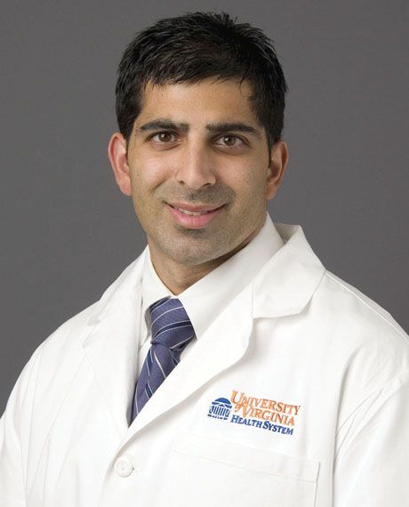
“Minimally invasive mitral surgery has excellent outcomes with fewer transfusions and less time ventilated in this representative cohort,” said Robert Hawkins, MD, of the University of Virginia, Charlottesville, in reporting the results.
“While operative times were longer, surgical costs remained statistically similar, and minimally invasive mitral surgery was associated with similar total costs in more complex mitral cases.”
Dr. Hawkins said this study included higher risk patients to attempt to overcome shortcomings of previously published reports that skewed toward lower-risk, highly selective mitral repairs for degenerative mitral disease. “They’re not really representative of the current state of minimally invasive mitral valve surgery as it currently stands in the higher risk patient population,” he said of previous studies.
Major outcomes were similar in both groups. “The mitral valve repair rate was about 81% for both groups, and the tricuspid valve repair rate was 8.8%,” Dr. Hawkins said. “About 35% had atrial fibrillation surgery, including both ablation and left atrial appendage ligation.”
Dr. Hawkins characterized outcomes in both surgical groups as “excellent,” and added, “The operative mortality rate was 1.3% and the major morbidity rate was 11% and not different between groups.”
Some key operative characteristics differed between the two groups. “As expected the cross clamp times and bypass times for the minimally invasive approaches were longer,” Dr. Hawkins said. Also, those who had minimally invasive mitral surgery had a “dramatic decrease” in transfusion rates.
With regard to resource utilization, minimally invasive surgery had longer operative times – an average of 291 minutes vs. 222 minutes (P less than .0001) – but similar or improved use of postoperative resources. “We see that the minimally invasive approach leads to decreased treatment and ancillary costs without a statistically significant difference in surgical costs despite the longer operative times,” Dr. Hawkins said.
However, he noted the high variability of total hospital costs in higher-risk populations complicate any head-to-head comparisons of resource utilization between the conventional and minimally invasive approaches, so the researchers attempted to drill down to identify predictors of resource use. Using a regression model, they found that minimally invasive approach may actually save money, although this finding was not statistically significant (–$1,524; P = 0.83).
“We see that the major drivers of costs are complications,” Dr. Hawkins said. “Morbidity and mortality led to a $54,000 cost increase, and the addition of tricuspid repair also led to about $60,000 higher costs, which is more likely related to higher risk and thus complications. The costs of higher-acuity cases are driven by the complications and not the approach.”
Dr. Hawkins reported no financial relationships. Dr. Ailawadi disclosed consulting agreements with Edwards Lifesciences, Abbott, Medtronic, and AtriCure.
AT THE 2017 MITRAL VALVE CONCLAVE
Key clinical point: Outcomes and costs of minimally invasive mitral surgery are similar to that of conventional sternotomy for mitral valve surgery.
Major finding: Mortality rates of 1.3% and major morbidity rates of 11% were similar in both groups.
Data source: Propensity-matched analysis of 479 patients who had a primary mitral valve operation from January 2010 to June 2016 at the University of Virginia.
Disclosures: Dr. Hawkins reported no financial disclosures. Coauthor Dr. Gorav Ailawadi disclosed consulting agreements with Edwards Lifesciences, Abbott, Medtronic, and AtriCure.
Concomitant MIMV-TVS no worse than MIMV alone
NEW YORK – Concurrent mitral-tricuspid valve surgery has similar outcomes to isolated minimally invasive mitral valve surgery, according to results of a 12-year review reported at the 2017 Mitral Valve Conclave, sponsored by the American Association for Thoracic Surgery.
Indications for minimally invasive tricuspid valve surgery done at the same time of mitral valve surgery have not been well established, in part because the outcomes of such combined procedures have been underreported.
Dr. Kilic noted that patients who had concomitant TVS were typically higher risk at baseline. “The concomitant group was older, had a higher percentage of female patients, and higher rates of chronic lung disease and cerebrovascular disease as well,” Dr. Kilic said. In comparing the isolated MIMV surgery and MIMV-TVS groups in the unmatched analysis, 9% vs. 14% had chronic lung disease (P = .05), 12% vs. 16% had coronary artery disease (P = .15), 7% vs. 12% had cerebrovascular disease (P = .04), and 93% vs. 90% had elective surgery (P = .18). The majority of tricuspid repairs were for severe tricuspid regurgitation (TR) or moderate TR with a dilated annulus of 40 mm or greater.
The operative characteristics differed significantly between the two groups. “As one might expect, the cardiopulmonary bypass time and aortic occlusion times were longer in the concomitant group; and balloon aortic occlusion was used in more than 70% in each cohort,” Dr. Kilic said. Those differences were similar in the propensity-matched cohort: bypass times were 147.5 minutes for isolated MIMV surgery and 174.6 minutes for MIMV-TVS (P less than .001); and aortic occlusion time 104.8 vs. 128 minutes (P less than .001), respectively.
Operative mortality was 3% for isolated MIMV surgery and 4% for concurrent MIMV-TVS (P = .73), but the isolated MIMV surgery group required fewer permanent pacemakers, 1% vs. 6% (P = .03).
“Aside from permanent pacemaker implantation, the rates of every other complication were similar, including stoke, limb ischemia, atrial fibrillation, gastrointestinal complications, respiratory complications, blood product transfusions as well as discharge to home rates,” Dr. Kilic said. Median hospital length of stays were also similar: 7 days for isolated MIMV surgery vs. 8 days for MIMV-TVS (P = .13).
One limitation of the study Dr. Kilic pointed out was that the decision to perform concomitant MIMV-TVS was surgeon dependent.
Dr. Kilic reported having no financial disclosures.
AT THE 2017 MITRAL VALVE CONCLAVE
Key clinical point: Outcomes of isolated minimally invasive mitral valve surgery (MIMV) and MIMV with concomitant tricuspid valve surgery (TVS) are similar.
Major finding: Operative mortality was 3% for isolated MIMV and 4% for concurrent MIMV-TVS.
Data source: Single-center review of 1,158 patients who underwent either isolated MIMV or MIMV-TVS from 2002 to 2014, including a propensity-matched cohort.
Disclosures: Dr. Kilic reported having no financial disclosures.
