User login
Drug-coated balloon advantage persists in femoral artery disease
NEW YORK – John Laird, MD, of the Adventist Heart Institute, St. Helena, Calif., presented the data at a symposium on vascular and endovascular issues sponsored by the Cleveland Clinic Foundation.
The data were drawn from the IN.PACT trial. In this trial, 331 patients were randomized to a paclitaxel-coated DCB device or standard percutaneous balloon angioplasty (PCBA), Dr. Laird explained.
The 5-year results are consistent with those previously reported at 1, 2, and 3 years. According to Dr. Laird, DCB continues to show an advantage for major outcomes over PCBA, and adverse events remain low.
Three DCB devices now available in the United States for dilatation of narrowed SFA. Although all have been associated with a reduced risk of target lesion revascularization relative to standard PCBA, the long-term follow-up presented from IN.PACT by Dr. Laird are the first to document 5-year outcomes.
In a video interview, Dr. Laird reported that there have been no thrombotic events since the 3-year results were presented.
Overall, he explains that the long-term outcomes provide additional confirmation that DCB is a safe procedure that reduces the need for stenting in SFA occlusions. Although he believes there might be clinically significant differences between available DCB devices, he concludes that DCB can be considered the first-line therapy for treating occluded femoral-popliteal arteries.
REPORTING FROM VEITHSYMPOSIUM

Don’t just work hard; work hard at living
NEW YORK – “A physician falls overboard on a large cruise ship and passengers gather at the guard rail. The first passenger at the guardrail shakes his finger down at the drowning physician and says, ‘You need to learn how to swim!’ Another passenger says, ‘No, man, throw him a life preserver.’ ... Finally, a passenger says, ‘We need better guard rails.’ ”

That’s the analogy Cynthia K. Shortell, MD used to kick off her presentation on physician burnout and the need for resilience at a symposium on vascular and endovascular issues sponsored by the Cleveland Clinic Foundation.
“Which of these is right? Well, of course, the answer is they all are,” said Dr. Shortell, who is a professor of surgery at Duke University Durham, N.C. But certainly, only the life preserver answer was appropriate at that time for the drowning physician, she added.
Continuing the analogy, Dr. Shortell pointed out that surely there is a man overboard, with 1 in 20 surgeons reporting suicidal ideation. That rate jumps threefold if the surgeon has had a recent medical error. In addition, vascular surgeons in particular are within the top tier of specialties at risk for burnout.
“We do need better guardrails,” she said, and described the need to actively engage with the health care system to help solve these issues, including those involving electronic medical records and operating room inefficiency and use. In addition, there is a great need for additional services that are provided to other high-end professionals, including food, concierge service, gym access, and other services that help with tasks of daily life when physicians need to spend most of their time at the hospital.
But, in the end, Dr. Shortell said, “We do need to learn to swim. Ultimately, we do need to take a role in having responsibility to solve this problem on our own. We need to change the way we think about our work, and the way we think about our health, and the way our culture values working hard instead of working hard at living.”
Dr. Shortell also highlighted the need to deal with musculoskeletal issues arising from the way that surgeons operate. This, along with good leadership, are key factors in preventing and remediating burnout.
Dr. Shortell had no disclosures relevant to her talk.
NEW YORK – “A physician falls overboard on a large cruise ship and passengers gather at the guard rail. The first passenger at the guardrail shakes his finger down at the drowning physician and says, ‘You need to learn how to swim!’ Another passenger says, ‘No, man, throw him a life preserver.’ ... Finally, a passenger says, ‘We need better guard rails.’ ”
That’s the analogy Cynthia K. Shortell, MD used to kick off her presentation on physician burnout and the need for resilience at a symposium on vascular and endovascular issues sponsored by the Cleveland Clinic Foundation.
“Which of these is right? Well, of course, the answer is they all are,” said Dr. Shortell, who is a professor of surgery at Duke University Durham, N.C. But certainly, only the life preserver answer was appropriate at that time for the drowning physician, she added.
Continuing the analogy, Dr. Shortell pointed out that surely there is a man overboard, with 1 in 20 surgeons reporting suicidal ideation. That rate jumps threefold if the surgeon has had a recent medical error. In addition, vascular surgeons in particular are within the top tier of specialties at risk for burnout.
“We do need better guardrails,” she said, and described the need to actively engage with the health care system to help solve these issues, including those involving electronic medical records and operating room inefficiency and use. In addition, there is a great need for additional services that are provided to other high-end professionals, including food, concierge service, gym access, and other services that help with tasks of daily life when physicians need to spend most of their time at the hospital.
But, in the end, Dr. Shortell said, “We do need to learn to swim. Ultimately, we do need to take a role in having responsibility to solve this problem on our own. We need to change the way we think about our work, and the way we think about our health, and the way our culture values working hard instead of working hard at living.”
Dr. Shortell also highlighted the need to deal with musculoskeletal issues arising from the way that surgeons operate. This, along with good leadership, are key factors in preventing and remediating burnout.
Dr. Shortell had no disclosures relevant to her talk.
REPORTING FROM THE VEITHSYMPOSIUM
What’s new with the SVS VQI?
NEW YORK – The Vascular Quality Initiative (VQI) is designed to improve the quality, safety, and effectiveness of vascular health care, but also reduce costs through the collection and exchange of information, according to Larry W. Kraiss, MD, professor of surgery, University of Utah, Salt Lake City.

The VQI consists of three major components: a federally accredited patient safety organization (PSO), a registry, and a distributed network of quality groups, which can also serve as a platform for internal quality improvement efforts.
Even though registries are considered less reliable than the venerated randomized clinical trial models are, registries have some unique advantages, according to Dr. Kraiss, who spoke at a symposium on vascular and endovascular issues sponsored by the Cleveland Clinic Foundation. Randomized clinical trials (RCTs) are very expensive to conduct and are criticized for potentially not being generalizable because of their rigidly defined inclusion/exclusion criteria. Despite inherent problems with registry data, especially bias, there are now statistical methods to help account for the biases. Registry-controlled trials are going to be important to answer many questions that cannot be answered by RCTs. Such trials can be too costly or actually unethical to randomize under certain circumstances. Registries, however, provide real-world patient populations in greater numbers than RCTs can manage, including access to rare events that would otherwise not be detected.
The VQI holds an important place in this second tier and is an important source of information for clinical care.
The VQI has grown steadily since its inception, and there are more than 500 centers now participating, including 6 international centers, and 18 regional groups, according to Dr. Kraiss. There are more than 530,000 procedures contained within the VQI’s 12 registries, with more than half of them involving peripheral vascular interventions and carotid endarterectomies. “But there [are] a healthy number of cases in all of the other registries, which provide a very rich resource and evidence base,” he added.
The VQI is becoming very important in the regulatory framework, with the Food and Drug Administration embracing “real-world evidence” as a way to justify some of its decisions, and the VQI has been involved in this process. The VQI has coordinated several post-market surveillance programs, the two most important being in the areas of thoracic endovascular aortic repair (TEVAR) and transcarotid artery revascularization (TCAR), said Dr. Kraiss.
One of the important values of the VQI is the ability to examine benchmarks and to compare performance across centers. For example, looking at aortic abdominal aneurysm (AAA) repair data, the results showed that a number of centers were not following established Society for Vascular Society guidelines and that there were a substantial number of AAAs reported in the VQI that were treated when they were below the threshold for intervention and should have received routine observation only.
So, ultimately two of the greatest values of the VQI are the ability to use it as a means of local quality improvement, and to provide an avenue for important clinical research, Dr. Kraiss concluded.
Dr. Kraiss reported that they had no disclosures.
NEW YORK – The Vascular Quality Initiative (VQI) is designed to improve the quality, safety, and effectiveness of vascular health care, but also reduce costs through the collection and exchange of information, according to Larry W. Kraiss, MD, professor of surgery, University of Utah, Salt Lake City.
The VQI consists of three major components: a federally accredited patient safety organization (PSO), a registry, and a distributed network of quality groups, which can also serve as a platform for internal quality improvement efforts.
Even though registries are considered less reliable than the venerated randomized clinical trial models are, registries have some unique advantages, according to Dr. Kraiss, who spoke at a symposium on vascular and endovascular issues sponsored by the Cleveland Clinic Foundation. Randomized clinical trials (RCTs) are very expensive to conduct and are criticized for potentially not being generalizable because of their rigidly defined inclusion/exclusion criteria. Despite inherent problems with registry data, especially bias, there are now statistical methods to help account for the biases. Registry-controlled trials are going to be important to answer many questions that cannot be answered by RCTs. Such trials can be too costly or actually unethical to randomize under certain circumstances. Registries, however, provide real-world patient populations in greater numbers than RCTs can manage, including access to rare events that would otherwise not be detected.
The VQI holds an important place in this second tier and is an important source of information for clinical care.
The VQI has grown steadily since its inception, and there are more than 500 centers now participating, including 6 international centers, and 18 regional groups, according to Dr. Kraiss. There are more than 530,000 procedures contained within the VQI’s 12 registries, with more than half of them involving peripheral vascular interventions and carotid endarterectomies. “But there [are] a healthy number of cases in all of the other registries, which provide a very rich resource and evidence base,” he added.
The VQI is becoming very important in the regulatory framework, with the Food and Drug Administration embracing “real-world evidence” as a way to justify some of its decisions, and the VQI has been involved in this process. The VQI has coordinated several post-market surveillance programs, the two most important being in the areas of thoracic endovascular aortic repair (TEVAR) and transcarotid artery revascularization (TCAR), said Dr. Kraiss.
One of the important values of the VQI is the ability to examine benchmarks and to compare performance across centers. For example, looking at aortic abdominal aneurysm (AAA) repair data, the results showed that a number of centers were not following established Society for Vascular Society guidelines and that there were a substantial number of AAAs reported in the VQI that were treated when they were below the threshold for intervention and should have received routine observation only.
So, ultimately two of the greatest values of the VQI are the ability to use it as a means of local quality improvement, and to provide an avenue for important clinical research, Dr. Kraiss concluded.
Dr. Kraiss reported that they had no disclosures.
REPORTING FROM THE VEITHSYMPOSIUM
AHA 2018: Part I

Sandy Hook Promise: Four programs help people recognize signs of a threat
ORLANDO – “The caretaker of all living things” – that was the good-natured moniker 7-year-old Daniel Barden had earned from his family – according to his dad, Mark Barden. Daniel would pick up black ants and take them outside “to be with their families,” even when the ant bit his fingers.

A walk down the sidewalk after a rain would take three times longer than it should because Daniel stopped to pick up every worm on the pavement and put it in the grass, lest it dry out in the sun, Mr. Barden said with a chuckle at the annual meeting of the American Academy of Pediatrics.
Daniel was his youngest and full of pure joy, Mr. Barden said, but that ended with his son’s murder during the Sandy Hook Elementary mass shooting Dec. 14, 2012. To honor his son and work to reduce the likelihood of similar mass shootings, Mr. Barden, now the managing director of Sandy Hook Promise in Newtown, Conn., shared with the pediatrician audience the work of the organization formed by Sandy Hook parents to attempt to prevent gun violence before it happens.
“We’re moms and dads and a couple of families who have lost loved ones in that tragedy, and we are growing as an organization,” Mr. Barden said. “Our basic, most fundamental objective is to prevent other families from living with the pain I will live with for the rest of my life.”
Their mission involves “creating a culture engaged and committed to identifying, intervening, and getting help for individuals who may be at risk of hurting themselves or others,” Mr. Barden said.
Sandy Hook Promise accomplishes this goal by educating and empowering communities through their four programs: Start with Hello, SOS Suicide Prevention Program, SaySomething, and Safety Assessment & Intervention. The organization delivers these programs through multiple platforms, including national and local trainers, digital curriculum downloads, interactive online training videos, and using multilingual presenters and English and Spanish materials.
The organization also especially works with schools and student’s clubs to change their culture and feel empowered to speak up and do their part to prevent gun violence too.
These programs resulted from extensive qualitative and quantitative research that Sandy Hook Promise conducted after the shooting with academic researchers, law enforcement, educators, school administrators, mental health professionals, and social movement experts.
“As we see these stories play themselves out over and over again, we start to reveal the story of somebody who didn’t just snap overnight,” Mr. Barden said. Signs that a person may be at risk for committing mass violence include suicidality, preoccupation with weapons, talking about committing violent acts, and general signs of depression and anxiety. “If we can train people how to not only recognize but to look for those signs, we can make a sustainable difference,” he said.
Most mass shootings are planned at least 6 months in advance, he said. About 80% of school shooters tell someone about their plans, and 69% tell multiple people. Similarly, up to 70% of people who die by suicide tell someone they plan to do it or give some other warning sign.
Further, more than a third of violent threats and bullying occurs electronically, so students are well equipped to watch for the signs and report them if they know how and feel comfortable doing so.
Mr. Barden outlined the goals of each of the four Sandy Hook Promise programs.
Start With Hello
This program “teaches youth how to identify and minimize social isolation, marginalization, and rejection in order to create an inclusive, connected community,” Mr. Barden explained. The goals of the program are to reduce bullying, foster socialization, increase engagement, and change a culture from within.
SOS Signs of Suicide
This is Sandy Hook Promise’s newest program and is built on a program developed by the Federal Bureau of Investigation following the Virginia Tech shooting and adapted for school-based applications.
“It also develops a multidisciplinary team within the school who acts as various touch points who know how to recognize a potential warning sign and then triage that information and take steps to get to the root cause of that behavior and not just bandage the wound,” Mr. Barden explained.
SaySomething
The organization’s flagship program does the most to recruit student involvement in recognizing the signs of a potential threat, particularly in social media, and report the individual and their behavior to a trusted adult or through Sandy Hook Promise’s Anonymous Reporting System.
“The kids take this one, and they run with it and do amazing things with it,” Mr. Barden said, noting that it particularly helps students recognize warning signs on social media. “We have growing evidence of kids following this model, and we’ve already prevented mass shootings and numerous suicides with this.”
Safety Assessment & Intervention (SAI) program
This program “trains a multidisciplinary team how to identify, assess, and respond to threats and observed at-risk behaviors,” Mr. Barden said. SAI aims to create a safer, more open school environment with less violence, bullying, and threats. That includes reducing educators’ fear and anxiety, and leading students to have a more positive view of teachers and staff.
Students can report tips to the Anonymous Reporting System through the website, calling the hot line or via a free mobile app. Regardless of the method, the anonymous tips go to a 24/7 multilingual crisis center and, if needed, law enforcement. The crisis center contacts the appropriate school official via text, email, or a phone call, and the case is tracked in real time until it’s addressed, resolved, and closed.
All of these programs are freely available to any school or institution who wants to use them, Mr. Barden said, because the organization does not want cost to get in the way of any school or community that is taking advantage of tools to reduce the risk of violence.
In fact, more than 3.5 million youth and adults in more than 7,000 schools in every state have been trained in these programs, helping hundreds of youth access mental health and wellness help, he said. The program has reduced truancy, bullying, and other forms of violence and victimization, and it has intervened in multiple school shooting plans across the United States.
Mr. Barden wrapped up his address with his gratitude for pediatricians’ willingness to be partners in reducing gun violence.
“I want to tell you how much it means to me that you took the time to come here and listen to my story and the work I’m doing,” he said, “and how proud I am to be able to share it with you, and how proud I am to be able to honor that little kid who truly was the caretaker of all living things and to continue that spirit in his honor and in his absence.”
ORLANDO – “The caretaker of all living things” – that was the good-natured moniker 7-year-old Daniel Barden had earned from his family – according to his dad, Mark Barden. Daniel would pick up black ants and take them outside “to be with their families,” even when the ant bit his fingers.
A walk down the sidewalk after a rain would take three times longer than it should because Daniel stopped to pick up every worm on the pavement and put it in the grass, lest it dry out in the sun, Mr. Barden said with a chuckle at the annual meeting of the American Academy of Pediatrics.
Daniel was his youngest and full of pure joy, Mr. Barden said, but that ended with his son’s murder during the Sandy Hook Elementary mass shooting Dec. 14, 2012. To honor his son and work to reduce the likelihood of similar mass shootings, Mr. Barden, now the managing director of Sandy Hook Promise in Newtown, Conn., shared with the pediatrician audience the work of the organization formed by Sandy Hook parents to attempt to prevent gun violence before it happens.
“We’re moms and dads and a couple of families who have lost loved ones in that tragedy, and we are growing as an organization,” Mr. Barden said. “Our basic, most fundamental objective is to prevent other families from living with the pain I will live with for the rest of my life.”
Their mission involves “creating a culture engaged and committed to identifying, intervening, and getting help for individuals who may be at risk of hurting themselves or others,” Mr. Barden said.
Sandy Hook Promise accomplishes this goal by educating and empowering communities through their four programs: Start with Hello, SOS Suicide Prevention Program, SaySomething, and Safety Assessment & Intervention. The organization delivers these programs through multiple platforms, including national and local trainers, digital curriculum downloads, interactive online training videos, and using multilingual presenters and English and Spanish materials.
The organization also especially works with schools and student’s clubs to change their culture and feel empowered to speak up and do their part to prevent gun violence too.
These programs resulted from extensive qualitative and quantitative research that Sandy Hook Promise conducted after the shooting with academic researchers, law enforcement, educators, school administrators, mental health professionals, and social movement experts.
“As we see these stories play themselves out over and over again, we start to reveal the story of somebody who didn’t just snap overnight,” Mr. Barden said. Signs that a person may be at risk for committing mass violence include suicidality, preoccupation with weapons, talking about committing violent acts, and general signs of depression and anxiety. “If we can train people how to not only recognize but to look for those signs, we can make a sustainable difference,” he said.
Most mass shootings are planned at least 6 months in advance, he said. About 80% of school shooters tell someone about their plans, and 69% tell multiple people. Similarly, up to 70% of people who die by suicide tell someone they plan to do it or give some other warning sign.
Further, more than a third of violent threats and bullying occurs electronically, so students are well equipped to watch for the signs and report them if they know how and feel comfortable doing so.
Mr. Barden outlined the goals of each of the four Sandy Hook Promise programs.
Start With Hello
This program “teaches youth how to identify and minimize social isolation, marginalization, and rejection in order to create an inclusive, connected community,” Mr. Barden explained. The goals of the program are to reduce bullying, foster socialization, increase engagement, and change a culture from within.
SOS Signs of Suicide
This is Sandy Hook Promise’s newest program and is built on a program developed by the Federal Bureau of Investigation following the Virginia Tech shooting and adapted for school-based applications.
“It also develops a multidisciplinary team within the school who acts as various touch points who know how to recognize a potential warning sign and then triage that information and take steps to get to the root cause of that behavior and not just bandage the wound,” Mr. Barden explained.
SaySomething
The organization’s flagship program does the most to recruit student involvement in recognizing the signs of a potential threat, particularly in social media, and report the individual and their behavior to a trusted adult or through Sandy Hook Promise’s Anonymous Reporting System.
“The kids take this one, and they run with it and do amazing things with it,” Mr. Barden said, noting that it particularly helps students recognize warning signs on social media. “We have growing evidence of kids following this model, and we’ve already prevented mass shootings and numerous suicides with this.”
Safety Assessment & Intervention (SAI) program
This program “trains a multidisciplinary team how to identify, assess, and respond to threats and observed at-risk behaviors,” Mr. Barden said. SAI aims to create a safer, more open school environment with less violence, bullying, and threats. That includes reducing educators’ fear and anxiety, and leading students to have a more positive view of teachers and staff.
Students can report tips to the Anonymous Reporting System through the website, calling the hot line or via a free mobile app. Regardless of the method, the anonymous tips go to a 24/7 multilingual crisis center and, if needed, law enforcement. The crisis center contacts the appropriate school official via text, email, or a phone call, and the case is tracked in real time until it’s addressed, resolved, and closed.
All of these programs are freely available to any school or institution who wants to use them, Mr. Barden said, because the organization does not want cost to get in the way of any school or community that is taking advantage of tools to reduce the risk of violence.
In fact, more than 3.5 million youth and adults in more than 7,000 schools in every state have been trained in these programs, helping hundreds of youth access mental health and wellness help, he said. The program has reduced truancy, bullying, and other forms of violence and victimization, and it has intervened in multiple school shooting plans across the United States.
Mr. Barden wrapped up his address with his gratitude for pediatricians’ willingness to be partners in reducing gun violence.
“I want to tell you how much it means to me that you took the time to come here and listen to my story and the work I’m doing,” he said, “and how proud I am to be able to share it with you, and how proud I am to be able to honor that little kid who truly was the caretaker of all living things and to continue that spirit in his honor and in his absence.”
EXPERT ANALYSIS FROM AAP 18
R-CHOP looks viable as first line in follicular lymphoma
A decade of follow-up data suggest that patients with newly diagnosed follicular lymphoma may derive long-term benefit from first-line therapy with the R-CHOP regimen, according to investigators in Japan.

Among patients with untreated follicular and other indolent B-cell lymphomas enrolled in a randomized phase 2/3 trial, the 10-year progression-free survival (PFS) rate for patients assigned to R-CHOP-21 (rituximab plus cyclophosphamide, doxorubicin, vincristine, and prednisone every 3 weeks) was 33%, and the 10-year PFS rate for patients assigned to R-CHOP-14 (R-CHOP every 2 weeks with granulocyte colony-stimulating factor [G-CSF] support] was 39%, and these PFS rates did not differ between the treatment arms, Takashi Watanabe, MD, PhD, from Mie University in Tsu, Japan, and his colleagues reported in the Lancet Haematology.
They also found that, compared with therapeutic regimens prior to the introduction of rituximab, R-CHOP was associated with a reduced likelihood of histological transformation of follicular lymphoma into a poor-prognosis diffuse large B-cell lymphoma (DLBCL), with no apparent increase in risk of secondary malignancies.
“The gold-standard, first-line treatment in patients with advanced-stage follicular lymphoma remains undetermined. However, because of the reduction in incidence of transformation without an increase in either secondary malignancies or fatal infectious events, the R-CHOP regimen should be a candidate for standard treatment, particularly from the viewpoint of long-term follow-up,” the investigators wrote
The JCOG0203 trial, which began enrollment in September 2002, included patients with stage III or IV indolent B-cell lymphomas, including grades 1-3 follicular lymphoma, from 44 Japanese hospitals. The patients were randomly assigned to receive six cycles of either R-CHOP-14 plus G-CSF or R-CHOP-21 administered once daily for 6 days beginning on day 8 of every cycle). Patients did not receive rituximab maintenance in either group.
In the primary analysis of the trial, published in 2011, the investigators reported that in 299 patients there were no significant differences in either PFS or 6-year overall survival (OS).
In the current analysis, the investigators reported that, for 248 patients with grade 1-3a follicular lymphoma, the 8-year PFS rate was 39% and the 10-year PFS rate was 36%.
The cumulative incidence of histological transformation was 3.2% at 5 years, 8.5% at 8 years, and 9.3% 10 years after the last patient was enrolled.
“In our study, survival after histological transformation was still poor; therefore, reducing histological transformation remains a crucial issue for patients with follicular lymphoma,” the investigators wrote.
The cumulative incidence of secondary malignancies at 10 years was 8.1%, and the cumulative incidence of hematological secondary malignancies was 2.9%.
The investigators noted that the actual incidence of secondary solid tumors or hematologic malignancies apart from the setting of autologous stem cell transplants is not known and emphasized that patients should be followed beyond 10 years to ensure that the risk of secondary malignancies is not underestimated.
“Clinicians choosing a first-line treatment for patients with follicular lymphoma should be cautious of secondary malignancies caused by immunochemotherapy and severe complications of infectious diseases in the long-term follow-up – both of which could lead to death,” they advised.
The study was supported by the Ministry of Health, Labour and Welfare of Japan and by the National Cancer Center Research and Development Fund of Japan. Dr. Watanabe has received honoraria from Bristol-Myers Squibb, Takeda, Taisho Toyama, Celgene, Nippon Shinyaku, and Novartis and funding resources from TakaraBio and United Immunity to support the Department of Immuno-Gene Therapy at Mie University. Multiple coauthors reported similar relationships.
SOURCE: Watanabe T et al. Lancet Haematol. 2018 Nov;5(11):e520-31.
A decade of follow-up data suggest that patients with newly diagnosed follicular lymphoma may derive long-term benefit from first-line therapy with the R-CHOP regimen, according to investigators in Japan.
Among patients with untreated follicular and other indolent B-cell lymphomas enrolled in a randomized phase 2/3 trial, the 10-year progression-free survival (PFS) rate for patients assigned to R-CHOP-21 (rituximab plus cyclophosphamide, doxorubicin, vincristine, and prednisone every 3 weeks) was 33%, and the 10-year PFS rate for patients assigned to R-CHOP-14 (R-CHOP every 2 weeks with granulocyte colony-stimulating factor [G-CSF] support] was 39%, and these PFS rates did not differ between the treatment arms, Takashi Watanabe, MD, PhD, from Mie University in Tsu, Japan, and his colleagues reported in the Lancet Haematology.
They also found that, compared with therapeutic regimens prior to the introduction of rituximab, R-CHOP was associated with a reduced likelihood of histological transformation of follicular lymphoma into a poor-prognosis diffuse large B-cell lymphoma (DLBCL), with no apparent increase in risk of secondary malignancies.
“The gold-standard, first-line treatment in patients with advanced-stage follicular lymphoma remains undetermined. However, because of the reduction in incidence of transformation without an increase in either secondary malignancies or fatal infectious events, the R-CHOP regimen should be a candidate for standard treatment, particularly from the viewpoint of long-term follow-up,” the investigators wrote
The JCOG0203 trial, which began enrollment in September 2002, included patients with stage III or IV indolent B-cell lymphomas, including grades 1-3 follicular lymphoma, from 44 Japanese hospitals. The patients were randomly assigned to receive six cycles of either R-CHOP-14 plus G-CSF or R-CHOP-21 administered once daily for 6 days beginning on day 8 of every cycle). Patients did not receive rituximab maintenance in either group.
In the primary analysis of the trial, published in 2011, the investigators reported that in 299 patients there were no significant differences in either PFS or 6-year overall survival (OS).
In the current analysis, the investigators reported that, for 248 patients with grade 1-3a follicular lymphoma, the 8-year PFS rate was 39% and the 10-year PFS rate was 36%.
The cumulative incidence of histological transformation was 3.2% at 5 years, 8.5% at 8 years, and 9.3% 10 years after the last patient was enrolled.
“In our study, survival after histological transformation was still poor; therefore, reducing histological transformation remains a crucial issue for patients with follicular lymphoma,” the investigators wrote.
The cumulative incidence of secondary malignancies at 10 years was 8.1%, and the cumulative incidence of hematological secondary malignancies was 2.9%.
The investigators noted that the actual incidence of secondary solid tumors or hematologic malignancies apart from the setting of autologous stem cell transplants is not known and emphasized that patients should be followed beyond 10 years to ensure that the risk of secondary malignancies is not underestimated.
“Clinicians choosing a first-line treatment for patients with follicular lymphoma should be cautious of secondary malignancies caused by immunochemotherapy and severe complications of infectious diseases in the long-term follow-up – both of which could lead to death,” they advised.
The study was supported by the Ministry of Health, Labour and Welfare of Japan and by the National Cancer Center Research and Development Fund of Japan. Dr. Watanabe has received honoraria from Bristol-Myers Squibb, Takeda, Taisho Toyama, Celgene, Nippon Shinyaku, and Novartis and funding resources from TakaraBio and United Immunity to support the Department of Immuno-Gene Therapy at Mie University. Multiple coauthors reported similar relationships.
SOURCE: Watanabe T et al. Lancet Haematol. 2018 Nov;5(11):e520-31.
FROM LANCET HAEMATOLOGY
Key clinical point:
Major finding: Approximately one-third of patients treated with R-CHOP-14 plus G-CSF or R-CHOP-21 had no disease progression after 10 years of follow-up.
Study details: Long-term analysis of a randomized phase 2/3 trial in 299 patients with indolent B-cell lymphomas, including follicular lymphoma.
Disclosures: The study was supported by the Ministry of Health, Labor and Welfare of Japan and by the National Cancer Center Research and Development Fund of Japan. Dr. Wantanabe has received honoraria from Bristol-Myers Squibb, Takeda, Taisho Toyama, Celgene, Nippon Shinyaku, and Novartis and funding resources from TakaraBio and United Immunity to support the Department of Immuno-Gene Therapy in Mie University. Multiple coauthors reported similar relationships.
Source: Wantanabe T et al. Lancet Haematol. 2018 Nov;5(11):e520-31.
FDA aims to squash youth vaping, smoking
The Food and Drug Administration once again has upped the ante in its war on youth smoking and vaping.

“Today, I’m pursuing actions aimed at addressing the disturbing trend of youth nicotine use and continuing to advance the historic declines we’ve achieved in recent years in the rates of combustible cigarette use among kids,” FDA Commissioner Scott Gottlieb, MD, said in a statement.
First and foremost, the FDA wants to reduce the lure of e-cigarettes by limiting the variety of flavored products for sale in retail outlets. Under the proposal unveiled Nov. 15, only electronic nicotine delivery systems (ENDS) that are unflavored or have tobacco, mint, or menthol flavors would be widely available. Flavored products – think cherry, cotton candy, and mango – would be sold in age-restricted environments, such as stand-alone tobacco retailers like vape shops. The FDA also seeks more stringent enforcement of age verification on ENDS products sold online.
The proposal also would reexamine regulations governing flavored cigars, with the possible aim of banning them.
“These efforts to address flavors and protect youth would dramatically impact the ability of American kids to access tobacco products that we know are both appealing and addicting,” Dr. Gottlieb said in a statement. “This policy framework reflects a redoubling of the FDA’s efforts to protect kids from all nicotine-containing products.”
In a move that seems to be aimed at youth-oriented products like Juul, the FDA will be seeking to remove from the market any ENDS product that is marketed specifically to young people.
Finally, the FDA intends to pursue regulation that would ban menthol from combustible tobacco products.
“I believe these menthol-flavored products represent one of the most common and pernicious routes by which kids initiate on combustible cigarettes,” Dr. Gottlieb said. “The menthol serves to mask some of the unattractive features of smoking that might otherwise discourage a child from smoking. Moreover, I believe that menthol products disproportionately and adversely affect underserved communities. And as a matter of public health, they exacerbate troubling disparities in health related to race and socioeconomic status.”
The policy shift comes as the Centers for Disease Control and Prevention released data from the 2018 National Youth Tobacco Survey showing that use of e-cigarettes among high schoolers is on the rise, growing from 1.5% in 2011 to 20.8% in 2018. Middle schoolers saw use over the same time period increase from 0.6% to 4.9%.
The rise of current use of e-cigarettes was enough to reverse a declining trend in overall tobacco use in recent years between 2015 and 2017.
“FDA’s enforcement efforts and policy framework would restrict access to most flavored e-cigarettes and limit the chances of youth beginning to use these products, while ensuring the products are available to adult smokers as an alternative to combustible cigarettes,” Alex M. Azar II, secretary of the Department of Health & Human Services, said in a statement supporting the FDA’s efforts. “Our obligation at HHS is always to the public health, and we believe FDA’s goals strike the right public health balance in addressing the multifaceted challenge we have before us today.”
Under Dr. Gottlieb, the FDA has been aggressively pursuing ways to reduce tobacco consumption, targeting both ENDS and combustible tobacco regulations in an effort to limit nicotine exposure and reduce the number of people addicted to nicotine and the health issues that come with it.
The American College of Cardiology voiced its support of the FDA’s actions.
“The FDA’s announcement restricting the sale of flavored e-cigarettes and other tobacco products shows they are ready to do their part in making tobacco products less available to our children,” ACC President C. Michael Valentine, MD, said in a statement, adding that the medical community needs to continue to do its part to make sure tobacco use continues to decline, especially in the nonadult population.
The FDA proposals were published as part of an advance notice of proposed rulemaking in the Federal Register. Comments can be made at www.regulations.gov through June 19.
The Food and Drug Administration once again has upped the ante in its war on youth smoking and vaping.
“Today, I’m pursuing actions aimed at addressing the disturbing trend of youth nicotine use and continuing to advance the historic declines we’ve achieved in recent years in the rates of combustible cigarette use among kids,” FDA Commissioner Scott Gottlieb, MD, said in a statement.
First and foremost, the FDA wants to reduce the lure of e-cigarettes by limiting the variety of flavored products for sale in retail outlets. Under the proposal unveiled Nov. 15, only electronic nicotine delivery systems (ENDS) that are unflavored or have tobacco, mint, or menthol flavors would be widely available. Flavored products – think cherry, cotton candy, and mango – would be sold in age-restricted environments, such as stand-alone tobacco retailers like vape shops. The FDA also seeks more stringent enforcement of age verification on ENDS products sold online.
The proposal also would reexamine regulations governing flavored cigars, with the possible aim of banning them.
“These efforts to address flavors and protect youth would dramatically impact the ability of American kids to access tobacco products that we know are both appealing and addicting,” Dr. Gottlieb said in a statement. “This policy framework reflects a redoubling of the FDA’s efforts to protect kids from all nicotine-containing products.”
In a move that seems to be aimed at youth-oriented products like Juul, the FDA will be seeking to remove from the market any ENDS product that is marketed specifically to young people.
Finally, the FDA intends to pursue regulation that would ban menthol from combustible tobacco products.
“I believe these menthol-flavored products represent one of the most common and pernicious routes by which kids initiate on combustible cigarettes,” Dr. Gottlieb said. “The menthol serves to mask some of the unattractive features of smoking that might otherwise discourage a child from smoking. Moreover, I believe that menthol products disproportionately and adversely affect underserved communities. And as a matter of public health, they exacerbate troubling disparities in health related to race and socioeconomic status.”
The policy shift comes as the Centers for Disease Control and Prevention released data from the 2018 National Youth Tobacco Survey showing that use of e-cigarettes among high schoolers is on the rise, growing from 1.5% in 2011 to 20.8% in 2018. Middle schoolers saw use over the same time period increase from 0.6% to 4.9%.
The rise of current use of e-cigarettes was enough to reverse a declining trend in overall tobacco use in recent years between 2015 and 2017.
“FDA’s enforcement efforts and policy framework would restrict access to most flavored e-cigarettes and limit the chances of youth beginning to use these products, while ensuring the products are available to adult smokers as an alternative to combustible cigarettes,” Alex M. Azar II, secretary of the Department of Health & Human Services, said in a statement supporting the FDA’s efforts. “Our obligation at HHS is always to the public health, and we believe FDA’s goals strike the right public health balance in addressing the multifaceted challenge we have before us today.”
Under Dr. Gottlieb, the FDA has been aggressively pursuing ways to reduce tobacco consumption, targeting both ENDS and combustible tobacco regulations in an effort to limit nicotine exposure and reduce the number of people addicted to nicotine and the health issues that come with it.
The American College of Cardiology voiced its support of the FDA’s actions.
“The FDA’s announcement restricting the sale of flavored e-cigarettes and other tobacco products shows they are ready to do their part in making tobacco products less available to our children,” ACC President C. Michael Valentine, MD, said in a statement, adding that the medical community needs to continue to do its part to make sure tobacco use continues to decline, especially in the nonadult population.
The FDA proposals were published as part of an advance notice of proposed rulemaking in the Federal Register. Comments can be made at www.regulations.gov through June 19.
MS disease activity returns in one-third after fingolimod withdrawal
BERLIN – At least one-third of patients being treated with fingolimod (Gilenya) for multiple sclerosis (MS) will relapse after stopping the drug, according to data from two “real-world” studies reported at the annual congress of the European Committee for Treatment and Research in Multiple Sclerosis, one of which was undertaken in pregnant women.

In a retrospective analysis of 110 MS patients who discontinued fingolimod from a 433-patient cohort, around 35% experienced a recurrence of disease activity after fingolimod withdrawal. Study investigator Nuria Cerda, MD, of the Neurologic Clinic and Polyclinic at University Hospital Basel (Switzerland), reported that risk factors for increased disease activity were younger rather than older age at MS diagnosis (P = .01) and at discontinuation (P less than .0001), having had a shorter rather than longer disease duration (P = .01), and evidence of MRI-confirmed disease activity occurring while on treatment with fingolimod (P = .02).
“We aimed to describe the frequency of recurrence of disease activity after fingolimod discontinuation in a real-world cohort of MS patients,” Dr. Cerda said. She noted that there were conflicting data on reappearing disease activity after stopping the drug. Case reports and observational data suggest a frequency of 10%-53%, but post hoc analyses of phase 3 trials with fingolimod have not found any higher risk for recurrence versus placebo.
The study also compared “demographic and clinical characteristics in patients with and without recurrence of disease activity, in order to identify possible risk factors for reactivation of the disease.”
The patients included in the study had been treated with fingolimod for a median of 24 months (interquartile range, 10-43 months) and experienced 118 discontinuation events between them. The majority (n = 81) had relapsing-remitting MS, and the remainder (n = 19) were in transition from RRMS to secondary progressive MS.
The mean age at discontinuation was 40 years and, while 60% of patients switched to another disease-modifying therapy (DMT), 40% did not receive further DMT. The reasons for discontinuation were classified as being from disease activity in 44%, side effects in 21%, pregnancy or childbearing preferences in 16%, noncompliance in 2%, and other reasons in 17%.
Recurrence of disease activity was defined as clinical symptoms, gadolinium-enhancing (Gd+) lesions on MRI, or both, within 6 months of stopping fingolimod. This was seen in 38 patients with 41 events, giving a rate of recurrence of 35%. Of these: “almost 60% had clinical and MRI activity, almost 30% had clinical activity only without Gd+ lesions on MRI, and 12% had MRI activity only without ever having a relapse,” Dr. Cerda said. In those who relapsed, the recurrence took a median 8 weeks to happen after fingolimod discontinuation.
A further definition of severe recurrence of disease activity also was used, defined as either a worsening in the Expanded Disability Status Scale (EDSS) score of more than 2 points (signifying a severe relapse) with or without pronounced MRI activity with at least five cerebral Gd+ lesions within 6 months of fingolimod withdrawal. Thirteen severe events were seen in 11 patients, giving a frequency of approximately 11%. Dr. Cerda reported that patients with severe recurrence were significantly younger at MS diagnosis than were those who did not experience recurrence (P = .003). A trend toward a higher annualized relapse rate (ARR) was seen in patients with severely active versus inactive disease (0.84 vs. 0.54), but this was not statistically significant. In addition, “confirmed disability worsening of 1 EDSS point or more was observed in about 3%.”
Relapse rates coming off fingolimod before or during pregnancy
In another study, which used German registry data on 129 pregnancies that occurred around or after the time of fingolimod withdrawal, up to 56% of women who stopped fingolimod before pregnancy experienced a relapse during pregnancy versus 29% of those who stopped fingolimod upon a positive pregnancy test.
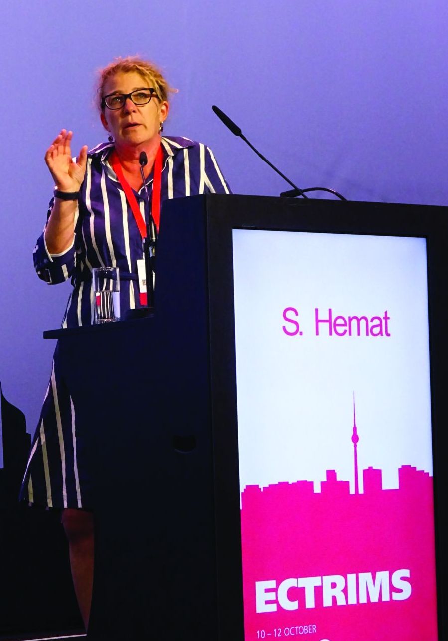
In a comparison of women who stopped fingolimod 1 year to 61 days before their last menstrual period against those who stopped less than 60 days prior to or after their last menstrual period, relapse rates were higher during the first (25.8% for early withdrawal vs. 12.5% with late withdrawal) and second trimesters (32.3% vs. 11.5%) than in the third (9.7% vs. 10.4%), Spalmai Hemat, MD, of the department of neurology at St. Josef Hospital, Ruhr University of Bochum (Germany) reported at the congress.
Relapse rates at 6 months postpartum proved to be similar at 36.7% for women who stopped fingolimod before their pregnancy and 38% in those who stopped later. Difference in the percentage of women experiencing disability progression during pregnancy did not prove to be statistically significant for stopping fingolimod before pregnancy (22.7%) vs. stopping later (11.1%; P = .283), while the 23.8% rate of disability progression after pregnancy among women who stopped the drug later versus 13.6% seen with stopping before was also not statistically significant (P = .31). Relapse during pregnancy was the only significant predictor for relapses postpartum.
“The large majority of women did not experience permanent disability,” Dr. Hemat said, but noted that as many as 10%-20% could experience substantial EDSS worsening (2 or more points) at 6 months postpartum.
Dr. Hemat concluded that more data were needed to see if reintroduction of fingolimod very soon after birth, such as within the first 2 weeks, could reduce the postpartum relapse risk.
Dr. Hemat and Dr. Cerda had no relevant disclosures.
SOURCES: Cerda N et al. Mult Scler. 2018;24(S2):73-4, Abstract 206; Hemat S et al. Mult Scler. 2018;24(S2):74-5, Abstract 207.
BERLIN – At least one-third of patients being treated with fingolimod (Gilenya) for multiple sclerosis (MS) will relapse after stopping the drug, according to data from two “real-world” studies reported at the annual congress of the European Committee for Treatment and Research in Multiple Sclerosis, one of which was undertaken in pregnant women.
In a retrospective analysis of 110 MS patients who discontinued fingolimod from a 433-patient cohort, around 35% experienced a recurrence of disease activity after fingolimod withdrawal. Study investigator Nuria Cerda, MD, of the Neurologic Clinic and Polyclinic at University Hospital Basel (Switzerland), reported that risk factors for increased disease activity were younger rather than older age at MS diagnosis (P = .01) and at discontinuation (P less than .0001), having had a shorter rather than longer disease duration (P = .01), and evidence of MRI-confirmed disease activity occurring while on treatment with fingolimod (P = .02).
“We aimed to describe the frequency of recurrence of disease activity after fingolimod discontinuation in a real-world cohort of MS patients,” Dr. Cerda said. She noted that there were conflicting data on reappearing disease activity after stopping the drug. Case reports and observational data suggest a frequency of 10%-53%, but post hoc analyses of phase 3 trials with fingolimod have not found any higher risk for recurrence versus placebo.
The study also compared “demographic and clinical characteristics in patients with and without recurrence of disease activity, in order to identify possible risk factors for reactivation of the disease.”
The patients included in the study had been treated with fingolimod for a median of 24 months (interquartile range, 10-43 months) and experienced 118 discontinuation events between them. The majority (n = 81) had relapsing-remitting MS, and the remainder (n = 19) were in transition from RRMS to secondary progressive MS.
The mean age at discontinuation was 40 years and, while 60% of patients switched to another disease-modifying therapy (DMT), 40% did not receive further DMT. The reasons for discontinuation were classified as being from disease activity in 44%, side effects in 21%, pregnancy or childbearing preferences in 16%, noncompliance in 2%, and other reasons in 17%.
Recurrence of disease activity was defined as clinical symptoms, gadolinium-enhancing (Gd+) lesions on MRI, or both, within 6 months of stopping fingolimod. This was seen in 38 patients with 41 events, giving a rate of recurrence of 35%. Of these: “almost 60% had clinical and MRI activity, almost 30% had clinical activity only without Gd+ lesions on MRI, and 12% had MRI activity only without ever having a relapse,” Dr. Cerda said. In those who relapsed, the recurrence took a median 8 weeks to happen after fingolimod discontinuation.
A further definition of severe recurrence of disease activity also was used, defined as either a worsening in the Expanded Disability Status Scale (EDSS) score of more than 2 points (signifying a severe relapse) with or without pronounced MRI activity with at least five cerebral Gd+ lesions within 6 months of fingolimod withdrawal. Thirteen severe events were seen in 11 patients, giving a frequency of approximately 11%. Dr. Cerda reported that patients with severe recurrence were significantly younger at MS diagnosis than were those who did not experience recurrence (P = .003). A trend toward a higher annualized relapse rate (ARR) was seen in patients with severely active versus inactive disease (0.84 vs. 0.54), but this was not statistically significant. In addition, “confirmed disability worsening of 1 EDSS point or more was observed in about 3%.”
Relapse rates coming off fingolimod before or during pregnancy
In another study, which used German registry data on 129 pregnancies that occurred around or after the time of fingolimod withdrawal, up to 56% of women who stopped fingolimod before pregnancy experienced a relapse during pregnancy versus 29% of those who stopped fingolimod upon a positive pregnancy test.
In a comparison of women who stopped fingolimod 1 year to 61 days before their last menstrual period against those who stopped less than 60 days prior to or after their last menstrual period, relapse rates were higher during the first (25.8% for early withdrawal vs. 12.5% with late withdrawal) and second trimesters (32.3% vs. 11.5%) than in the third (9.7% vs. 10.4%), Spalmai Hemat, MD, of the department of neurology at St. Josef Hospital, Ruhr University of Bochum (Germany) reported at the congress.
Relapse rates at 6 months postpartum proved to be similar at 36.7% for women who stopped fingolimod before their pregnancy and 38% in those who stopped later. Difference in the percentage of women experiencing disability progression during pregnancy did not prove to be statistically significant for stopping fingolimod before pregnancy (22.7%) vs. stopping later (11.1%; P = .283), while the 23.8% rate of disability progression after pregnancy among women who stopped the drug later versus 13.6% seen with stopping before was also not statistically significant (P = .31). Relapse during pregnancy was the only significant predictor for relapses postpartum.
“The large majority of women did not experience permanent disability,” Dr. Hemat said, but noted that as many as 10%-20% could experience substantial EDSS worsening (2 or more points) at 6 months postpartum.
Dr. Hemat concluded that more data were needed to see if reintroduction of fingolimod very soon after birth, such as within the first 2 weeks, could reduce the postpartum relapse risk.
Dr. Hemat and Dr. Cerda had no relevant disclosures.
SOURCES: Cerda N et al. Mult Scler. 2018;24(S2):73-4, Abstract 206; Hemat S et al. Mult Scler. 2018;24(S2):74-5, Abstract 207.
REPORTING FROM ECTRIMS 2018
Key clinical point:
Major finding: A higher risk of recurrent disease activity was seen in younger MS patients with shorter disease duration and evidence of disease activity while on fingolimod treatment. Pregnant women who stopped more than 2 months before a planned pregnancy also had a higher risk versus those who stopped after a positive pregnancy test.
Study details: A retrospective analysis of 110 MS patients who discontinued fingolimod from a 433-patient cohort, and German registry data on 129 pregnancies that occurred around or after the time of fingolimod withdrawal.
Disclosures: Dr. Spalmai Hemat and Dr. Nuria Cerda had no relevant disclosures.
Sources: Cerda N et al. Mult Scler. 2018;24(S2):73-4, Abstract 206; Hemat S et al. Mult Scler. 2018;24(S2):74-5, Abstract 207.
Bortezomib may unlock resistance in WM with mutations
The use of bortezomib may help overcome treatment resistance in patients with Waldenström macroglobulinemia (WM) with CXCR4 mutations, according to new research.
Romanos Sklavenitis-Pistofidis, MD, of the Dana-Farber Cancer Institute in Boston, and his colleagues compared the effects of treatment with bortezomib/rituximab in patients with WM based on their CXCR4 mutation status. They found no significant difference in progression-free survival (Log-rank, P = .994) or overall survival (Log-rank, P = .407) when comparing patients who have CXCR4 mutations with those who have CXCR4 wild type.
“We report for the first time that a bortezomib-based combination is impervious to the impact of CXCR4 mutations in a cohort of patients with WM,” the researchers wrote in Blood. “Previously, we had shown this to be true in WM cell lines, whereby genetically engineering BCWM.1 and MWCL-1 to overexpress CXCR4 had no impact on bortezomib resistance.”
The researchers noted, however, that the mechanism at work may be different than that seen with bortezomib in other cancers.
“Different experiments have linked CXCR4 expression and bortezomib in a variety of ways in other hematological malignancies, including multiple myeloma. However, despite the complicated association in those cancer types, in WM there seems to be a consistently neutral effect of CXCR4 mutations on bortezomib resistance in both cell line and patient data,” they wrote.
The researchers recommended that the theory be tested in a prospective trial of bortezomib-based therapy in WM patients with CXCR4 mutations. Another question to be investigated, they pointed out, is the role of rituximab in the survival results seen in the current analysis.
The study included 63 patients with WM who were treated with bortezomib/rituximab either as upfront treatment or in the relapsed/refractory setting as part of a phase 2 trial.
Bortezomib was given by IV weekly at 1.6 mg/m2 for six cycles and rituximab was given at 375 mg/m2 during cycles one and four. Patients were taken off therapy after two cycles if they had progressive disease.
The researchers excluded 20 patients from the study because of a lack of material for genotyping. However, they noted that their clinical characteristics were not different from those patients who were included.
Out of 43 patients who were genotyped for CXCR4, 17 patients had a mutation. All patients who carried a CXCR4 mutation also had MYD88 L265P. Ten patients had frameshift mutations, one patient had a nonsense mutation, and six patients had missense mutations. The median follow-up of the analysis was 90.7 months.
The researchers repeated the analysis after excluding six patients with missense mutations and accounting for different treatment settings and found that survival remained unchanged.
The study was supported by the National Institutes of Health, the Leukemia and Lymphoma Society, and the International Waldenström Macroglobulinemia Foundation. One of the authors reported consulting and research funding from Takeda, which markets bortezomib, and other companies.
SOURCE: Sklavenitis-Pistofidis R et al. Blood. 2018 Oct 26. doi: 10.1182/blood-2018-07-863241.
FROM BLOOD
Key clinical point:
Major finding: Progression-free and overall survival were not significantly different between WM patients with CXCR4 mutations and those with CXCR4 wild type (log-rank, P = .994 and P = .407, respectively).
Study details: An analysis of 43 WM patients treated with bortezomib/rituximab and genotyped to determine CXCR4 mutation status.
Disclosures: The study was supported by the National Institutes of Health, the Leukemia and Lymphoma Society, and the International Waldenström Macroglobulinemia Foundation. One of the authors reported consulting and research funding from Takeda, which markets bortezomib, and other companies.
Source: Sklavenitis-Pistofidis R et al. Blood. 2018 Oct 26. doi: 10.1182/blood-2018-07-863241.
CKD, and even dialysis, may not be barriers to pregnancy
SAN DIEGO – Nephrologists are often uncomfortable with the idea of advising women with chronic kidney disease (CKD) about pregnancy, a physician told colleagues. They must do better, she said, with sensitivity and insight into once-extreme possibilities like pregnancy during dialysis.
“For many women, having a child is a life goal, and our women with chronic kidney disease are not different,” said Michelle Hladunewich, MD, of Toronto’s Sunnybrook Health Sciences Center. “When we don’t know what we should do, we tend to over-aggressively counsel our women, and that can traumatize them. It’s our role as nephrologists to help them find the safest window to have their pregnancy,” she said at the meeting sponsored by the American Society of Nephrology.
According to Dr. Hladunewich, there are tens of thousands of women of child-bearing age in the United States who have CKD, end-stage renal disease (ESRD), and kidney transplants. However, she said, research presented at Kidney Week 2018 suggested that many nephrologists do not feel confident about counseling patients regarding issues such as pregnancy outcomes in CKD. “We are not that comfortable with it, but we have to become more comfortable,” she said. “We need to be prepared to talk about contraception if they don’t want to have a child or the plan about how to have a child if they do.”
It’s especially important to understand that while women can fear birth defects and the exacerbation of their disease, they may also feel “they’re not fulfilling a societal norm to have a child like everyone else,” she said.
The risks of pregnancy in CKD can affect the mother (via worse kidney function) and/or the fetus (preeclampsia, poor fetal growth, preterm delivery).
In a 2015 study, Italian researchers compared 504 pregnancies in women with CKD to 836 low-risk pregnancies in women without CKD. They found that the risks of adverse outcomes increased in women at higher stages of CKD, compared with those at lower stages: “Renal function matters, and a stepwise increase in the risk of adverse maternal-fetal outcomes is observed from stage 1 to stages 4-5.”
In addition, the researchers noted that their research suggests “the presence of a baseline risk linked to CKD per se” (J Am Soc Nephrol. 2015 Aug; 26[8]:2011-22).
Dr. Hladunewich recommended focusing on “the safest window of opportunity.” Some patients will progress to end-stage renal disease, and an earlier pregnancy during CKD is a better option, she said. As a result, encouraging an earlier pregnancy can be a wise idea.
In some cases, though, a patient may be far into the stages of CKD. Dr. Hladunewich spoke about the case of a 31-year-old patient with a 29-year history of type 1 diabetes mellitus. She’d had one miscarriage, one preterm birth, and one twin pregnancy that was terminated because of safety concerns including rapid loss of kidney function.
The patient saw Dr. Hladunewich when she had a glomerular filtration rate of 25 mL/min, 3.5 g per 24 hour of proteinuria, and hypertension. The patient had a question: “Dr. Michelle, when can I try again?”
Dr. Hladunewich joked that “I had a small stroke.” But then, she said, “I got to the business of pregnancy counseling.”
She told the woman that her progression to end-stage renal disease was likely inevitable, and “adverse pregnancy outcomes were almost guaranteed.”
The woman responded: “Not now? When?” That, Dr. Hladunewich said, “was when I had my second stroke.”
But there is a possible solution: Pregnancy during dialysis. “Historically, we’ve said absolutely no pregnancy on dialysis,” she said, “but times are changing. We believe aggressive dialysis improves fetal maternal and fetal outcomes.”
Indeed, Dr. Hladunewich led a 2014 study that linked extensive dialysis during pregnancy (compared with less dialysis) to a better likelihood of outcomes such as live birth rate and normal birth weight (JASN May 2014;25[5]:1103-9).
As she noted, “we do offer it as a reproductive option” to patients like the one she mentioned – those who are in ESRD, approaching it, or are nearing the end of their child-bearing years with no transplant in sight. In transplant cases, she said, adequate graft function is linked to good pregnancy outcomes.
Dr. Hladunewich added that it’s important to monitor and adjust treatment of patients during the postpartum period. She said it’s especially important to understand the risks of drugs during breastfeeding. Both dialysis and transplant patients can breastfeed, she said.
Dr. Hladunewich reports no disclosures.
SOURCE: Kidney Week 2018, Abstract FR-OR078.
REPORTING FROM KIDNEY WEEK 2018