User login
Genital psoriasis is the worst: Patients sound off
GENEVA – The great majority of patients with genital psoriasis say their symptoms in the genital area are worse than elsewhere on the body, Kim A. Meeuwis, MD, reported at the annual congress of the European Academy of Dermatology and Venereology.
She presented a qualitative study in which 20 patients with longstanding genital psoriasis sounded off, sharing their perspectives on the disease in one-on-one, semistructured, face-to-face interviews.

Genital psoriasis is common. Epidemiologic studies show 30%-60% of psoriasis patients experience genital involvement at some point in the course of their disease. Yet patients seldom discuss their genital psoriasis with their physicians, and the patient perspective on how the experience of genital psoriasis differs from that of having psoriasis at other locations has been addressed only sparsely in the literature. This lack of attention was the impetus for the current study, she explained.
The 20 participants in the study had an average 18-year history of plaque psoriasis, with an average 7.5-year history of genital involvement. The genital psoriasis was rated moderate or severe in 70% of subjects at the time of the study.
The most commonly reported symptoms of genital psoriasis were itch and discomfort, each of which was cited by all study participants. This was followed by erythema, cited by 95%; stinging and burning, also cited by 95%; pain, cited by 85%; scaling, by 75%; and cracking, by 30%.
Of the patients in the study, 85% reported that their pain and/or discomfort were worse in the genital area than at other sites, and 10% said they were highly self-conscious about their genital psoriasis because others had misidentified them as having a sexually transmitted infection.
Since this was a qualitative study, Dr. Meeuwis provided representative quotes from several patients, including one who asserted, “I really only have discomfort on my psoriasis on the rest of my body ... in my genitals is the only place that actually has pain, or the itching is ... really, really bad.”
Dr. Meeuwis said the study results hold an important lesson for physicians who treat psoriasis: “Due to differences in patient experiences between genital and nongenital skin, it’s really important to make time for the specific evaluation of genital involvement in taking care of patients with psoriasis – and to be sure to ask about it.”
Dr. Meeuwis reported serving as a consultant to Eli Lilly, which sponsored the study, as well as being on an advisory board to Beiersdorf.
GENEVA – The great majority of patients with genital psoriasis say their symptoms in the genital area are worse than elsewhere on the body, Kim A. Meeuwis, MD, reported at the annual congress of the European Academy of Dermatology and Venereology.
She presented a qualitative study in which 20 patients with longstanding genital psoriasis sounded off, sharing their perspectives on the disease in one-on-one, semistructured, face-to-face interviews.
Genital psoriasis is common. Epidemiologic studies show 30%-60% of psoriasis patients experience genital involvement at some point in the course of their disease. Yet patients seldom discuss their genital psoriasis with their physicians, and the patient perspective on how the experience of genital psoriasis differs from that of having psoriasis at other locations has been addressed only sparsely in the literature. This lack of attention was the impetus for the current study, she explained.
The 20 participants in the study had an average 18-year history of plaque psoriasis, with an average 7.5-year history of genital involvement. The genital psoriasis was rated moderate or severe in 70% of subjects at the time of the study.
The most commonly reported symptoms of genital psoriasis were itch and discomfort, each of which was cited by all study participants. This was followed by erythema, cited by 95%; stinging and burning, also cited by 95%; pain, cited by 85%; scaling, by 75%; and cracking, by 30%.
Of the patients in the study, 85% reported that their pain and/or discomfort were worse in the genital area than at other sites, and 10% said they were highly self-conscious about their genital psoriasis because others had misidentified them as having a sexually transmitted infection.
Since this was a qualitative study, Dr. Meeuwis provided representative quotes from several patients, including one who asserted, “I really only have discomfort on my psoriasis on the rest of my body ... in my genitals is the only place that actually has pain, or the itching is ... really, really bad.”
Dr. Meeuwis said the study results hold an important lesson for physicians who treat psoriasis: “Due to differences in patient experiences between genital and nongenital skin, it’s really important to make time for the specific evaluation of genital involvement in taking care of patients with psoriasis – and to be sure to ask about it.”
Dr. Meeuwis reported serving as a consultant to Eli Lilly, which sponsored the study, as well as being on an advisory board to Beiersdorf.
AT THE EADV CONGRESS
Key clinical point:
Major finding: Of the participants in a study of genital psoriasis, 100% reported that a hallmark of their genital disease was itching and discomfort.
Data source: A qualitative study that involved one-on-one interviews with 20 patients with genital psoriasis, who shared their experiences as to how genital involvement differs from their psoriasis elsewhere.
Disclosures: The study was sponsored by Eli Lilly. The presenter reported serving as a consultant to the company.
Advance care planning benefit presents challenges
When Donna Sweet, MD, sees patients for routine exams, death and dying are often the furthest thing from their minds. Regardless of age or health status, however, Dr. Sweet regularly asks patients about end-of-life care and whether they’ve considered their options.
In the past, physicians had to be creative in how they coded for such conversations, but Medicare’s newish advance care planning benefit is changing that.

Staring in 2016, the Centers for Medicare & Medicaid Services began reimbursing physicians for advance care planning discussions with the approval of two new codes: 99497 and 99498. The codes pay about $86 for the first 30-minutes of a face-to-face conversation with a patient, family member, and/or surrogate and about $75 for additional sessions. Services can be furnished in both inpatient and ambulatory settings, and payment is not limited to particular physician specialties.
Dr. Sweet said that she uses these codes a couple times a week when patients visit for reasons such as routine hypertension or diabetes exams or annual Medicare wellness visits. To broach the subject, Dr. Sweet said it helps to have literature about advance care planning in the room that patients can review.
“It’s just a matter of bringing it up,” she said. “Considering some of the other codes, the advance care planning code is really pretty simple.”
However, doctors like Dr. Sweet appear to be in the minority when it comes to providing this service. Of the nearly 57 million beneficiaries enrolled in Medicare at the end of 2016, only about 1% received advance care planning sessions, according to analysis of Medicare data posted by Kaiser Health News. Nationwide, health providers submitted about $93 million in charges, of which $43 million was paid by Medicare.

Challenges deter conversations
During a recent visit with a 72-year-old cancer patient, Bridget Fahy, MD, a surgical oncologist at the University of New Mexico, Albuquerque, spent time discussing advance directives and the importance of naming a surrogate decision maker. Dr. Fahy had treated the patient for two different cancers over the course of 4 years, and he was now diagnosed with a third, she recalled during an interview. Figuring out an advance care plan, though, proved complicated: The man was not married, had no children, and had no family members who lived in the state.
Although Dr. Fahy was aware of the Medicare advance care planning codes, she did not bill the session as such.
“Even in the course of having that conversation, I’m more apt to bill on time than I am specifically to meet the Medicare requirements for the documentation for [the benefit],” she said.

“There are two pieces required to take advantage of the advance care planning benefit code: having the conversation and documenting it,” Dr. Fahy noted. “What I write at the end of a resident note or an advanced practice provider note is going to be more focused on the counseling I had with the patient about their condition, the evaluation, and what the treatment plan is going to be. For surgeons to utilize the advance care planning codes, they have to have knowledge of the code, which many do not; they must know the requirements for documenting the conversation; and they have to have the time needed to have the conversation while also addressing all of the surgery-specific issues that need to be covered during the visit. There are a number of hurdles to overcome.”
Danielle B. Scheurer, MD, a hospitalist and chief quality officer at the Medical University of South Carolina, Charleston, said that she, too, has not used advance care planning codes. The reimbursement tool is a positive step forward, she said, but so far, it’s not an easy insert into a hospitalist’s practice.
“It’s not top of mind as far as a billing practice,” she said. “It’s not built into the typical work flow. Obviously, it’s not every patient, it’s not everyday, so you have to remember to put it into your work flow. That’s probably the biggest barrier for most hospitalists: either not knowing about it at all or not yet figuring out how to weave it into what they already do.”

Overcoming hurdles through experience
Using the advance care planning benefit has been easier said than done in his practice, according to Carl R. Olden, MD, a family physician in Yakima, Wash. The logistics of scheduling and patient reluctance are contributing to low usage of the new codes, said Dr. Olden, a member of the American Academy of Family Physicians board of directors.
Between Sept. 1, 2016, and Aug. 31, 2017, the family medicine, primary care, internal medicine, and pulmonary medicine members of Dr. Olden’s network who provide end-of-life counseling submitted billing for a total of 106,160 Medicare visits. Of those visits, the 99497 code was submitted only 32 times, according to data provided by Dr. Olden.
At Dr. Olden’s 16-physician practice, there are no registered nurses to help set up and start Medicare wellness visits, which the advance care planning session benefit is designed to fit within, he said.
“Most of those Medicare wellness visits are driven by having a registered nurse do most of the work,” he said. “[For us] to schedule a wellness visit, it’s mostly physician work and to do a 30-minute wellness visit, most of us can see three patients in that 30-minute slot, so it ends up not being very cost effective.”

“Most of my Medicare patients are folks that have four to five chronic medical conditions, and for them to make a 30-minute visit to the office and not talk about any of those conditions but to talk about home safety and advance directives and fall prevention, it’s hard for them to understand that,” he said.
Dr. Newman stresses that while the billing approach takes time to learn, the codes can be weaved into regular practice with some preparation and planning. At her practice, she primarily uses the codes for patients with challenging changes in their health status, sometimes setting up meetings in advance and, other times, conducting a spur-of-the-moment conversation.
“It’s a wonderful benefit,” she said. “I’m not surprised it’s taking awhile to take hold. The reason is you have to prepare for these visits. It takes preparation, including a chart review.”
A common misconception is that the visit must be scheduled separately and cannot be added to another visit, she said. Doctors can bill the advance care planning codes on the same day as an evaluation and management service. For instance, if a patient is accompanied by a family member and seen for routine follow-up, the physician can discuss the medical conditions first and later have a discussion about advance care planning. When billing, the physician can then use an evaluation and management code for the part of the visit related to the patient’s medical conditions and also bill for the advance care planning discussion using the new Medicare codes, Dr. Newman said.
“You’re allowed to use a modifier to attach to it to get paid for both on the same day,” she said. She suggested checking local Medicare policy for the use of the appropriate modifier, usually 26. “One thing that’s important to understand is there’s a lot of short discussions about advanced care planning that doesn’t fit the code. So if a patient wants to have a 5-minute conversation – that happens a lot – these will not be billable or counted under this new benefit. Fifteen minutes is the least amount of time that qualifies for 99497.”
Dr. Sweet said that she expects greater use of the codes as more doctors become aware of how they can be used.
“Once people use it a time or two, they will use it a lot more,” Dr. Sweet said. “It takes time to change, and it takes time to make time to do the things we need to do. But especially, as we move into high-value care, something like this hopefully, [doctors] will embrace.”
[email protected]
On Twitter @legal_med
When Donna Sweet, MD, sees patients for routine exams, death and dying are often the furthest thing from their minds. Regardless of age or health status, however, Dr. Sweet regularly asks patients about end-of-life care and whether they’ve considered their options.
In the past, physicians had to be creative in how they coded for such conversations, but Medicare’s newish advance care planning benefit is changing that.
Staring in 2016, the Centers for Medicare & Medicaid Services began reimbursing physicians for advance care planning discussions with the approval of two new codes: 99497 and 99498. The codes pay about $86 for the first 30-minutes of a face-to-face conversation with a patient, family member, and/or surrogate and about $75 for additional sessions. Services can be furnished in both inpatient and ambulatory settings, and payment is not limited to particular physician specialties.
Dr. Sweet said that she uses these codes a couple times a week when patients visit for reasons such as routine hypertension or diabetes exams or annual Medicare wellness visits. To broach the subject, Dr. Sweet said it helps to have literature about advance care planning in the room that patients can review.
“It’s just a matter of bringing it up,” she said. “Considering some of the other codes, the advance care planning code is really pretty simple.”
However, doctors like Dr. Sweet appear to be in the minority when it comes to providing this service. Of the nearly 57 million beneficiaries enrolled in Medicare at the end of 2016, only about 1% received advance care planning sessions, according to analysis of Medicare data posted by Kaiser Health News. Nationwide, health providers submitted about $93 million in charges, of which $43 million was paid by Medicare.
Challenges deter conversations
During a recent visit with a 72-year-old cancer patient, Bridget Fahy, MD, a surgical oncologist at the University of New Mexico, Albuquerque, spent time discussing advance directives and the importance of naming a surrogate decision maker. Dr. Fahy had treated the patient for two different cancers over the course of 4 years, and he was now diagnosed with a third, she recalled during an interview. Figuring out an advance care plan, though, proved complicated: The man was not married, had no children, and had no family members who lived in the state.
Although Dr. Fahy was aware of the Medicare advance care planning codes, she did not bill the session as such.
“Even in the course of having that conversation, I’m more apt to bill on time than I am specifically to meet the Medicare requirements for the documentation for [the benefit],” she said.
“There are two pieces required to take advantage of the advance care planning benefit code: having the conversation and documenting it,” Dr. Fahy noted. “What I write at the end of a resident note or an advanced practice provider note is going to be more focused on the counseling I had with the patient about their condition, the evaluation, and what the treatment plan is going to be. For surgeons to utilize the advance care planning codes, they have to have knowledge of the code, which many do not; they must know the requirements for documenting the conversation; and they have to have the time needed to have the conversation while also addressing all of the surgery-specific issues that need to be covered during the visit. There are a number of hurdles to overcome.”
Danielle B. Scheurer, MD, a hospitalist and chief quality officer at the Medical University of South Carolina, Charleston, said that she, too, has not used advance care planning codes. The reimbursement tool is a positive step forward, she said, but so far, it’s not an easy insert into a hospitalist’s practice.
“It’s not top of mind as far as a billing practice,” she said. “It’s not built into the typical work flow. Obviously, it’s not every patient, it’s not everyday, so you have to remember to put it into your work flow. That’s probably the biggest barrier for most hospitalists: either not knowing about it at all or not yet figuring out how to weave it into what they already do.”
Overcoming hurdles through experience
Using the advance care planning benefit has been easier said than done in his practice, according to Carl R. Olden, MD, a family physician in Yakima, Wash. The logistics of scheduling and patient reluctance are contributing to low usage of the new codes, said Dr. Olden, a member of the American Academy of Family Physicians board of directors.
Between Sept. 1, 2016, and Aug. 31, 2017, the family medicine, primary care, internal medicine, and pulmonary medicine members of Dr. Olden’s network who provide end-of-life counseling submitted billing for a total of 106,160 Medicare visits. Of those visits, the 99497 code was submitted only 32 times, according to data provided by Dr. Olden.
At Dr. Olden’s 16-physician practice, there are no registered nurses to help set up and start Medicare wellness visits, which the advance care planning session benefit is designed to fit within, he said.
“Most of those Medicare wellness visits are driven by having a registered nurse do most of the work,” he said. “[For us] to schedule a wellness visit, it’s mostly physician work and to do a 30-minute wellness visit, most of us can see three patients in that 30-minute slot, so it ends up not being very cost effective.”
“Most of my Medicare patients are folks that have four to five chronic medical conditions, and for them to make a 30-minute visit to the office and not talk about any of those conditions but to talk about home safety and advance directives and fall prevention, it’s hard for them to understand that,” he said.
Dr. Newman stresses that while the billing approach takes time to learn, the codes can be weaved into regular practice with some preparation and planning. At her practice, she primarily uses the codes for patients with challenging changes in their health status, sometimes setting up meetings in advance and, other times, conducting a spur-of-the-moment conversation.
“It’s a wonderful benefit,” she said. “I’m not surprised it’s taking awhile to take hold. The reason is you have to prepare for these visits. It takes preparation, including a chart review.”
A common misconception is that the visit must be scheduled separately and cannot be added to another visit, she said. Doctors can bill the advance care planning codes on the same day as an evaluation and management service. For instance, if a patient is accompanied by a family member and seen for routine follow-up, the physician can discuss the medical conditions first and later have a discussion about advance care planning. When billing, the physician can then use an evaluation and management code for the part of the visit related to the patient’s medical conditions and also bill for the advance care planning discussion using the new Medicare codes, Dr. Newman said.
“You’re allowed to use a modifier to attach to it to get paid for both on the same day,” she said. She suggested checking local Medicare policy for the use of the appropriate modifier, usually 26. “One thing that’s important to understand is there’s a lot of short discussions about advanced care planning that doesn’t fit the code. So if a patient wants to have a 5-minute conversation – that happens a lot – these will not be billable or counted under this new benefit. Fifteen minutes is the least amount of time that qualifies for 99497.”
Dr. Sweet said that she expects greater use of the codes as more doctors become aware of how they can be used.
“Once people use it a time or two, they will use it a lot more,” Dr. Sweet said. “It takes time to change, and it takes time to make time to do the things we need to do. But especially, as we move into high-value care, something like this hopefully, [doctors] will embrace.”
[email protected]
On Twitter @legal_med
Ixekizumab has profound impact on genital psoriasis
GENEVA – The interleukin-17A inhibitor ixekizumab provided rapid clearance of genital psoriasis in a phase 3b clinical trial, with significant improvement seen as early as week 1, Caitriona Ryan, MD, reported at the annual congress of the European Academy of Dermatology and Venereology.
The highly targeted monoclonal antibody also improved the intense itching that’s a particularly prominent feature of genital psoriasis.

“Genital psoriasis is a hidden part of psoriasis. Unfortunately, as dermatologists we do a bad job of evaluating our patients for it. They are ashamed and embarrassed to bring up the topic with their dermatologists. Hopefully, this study will create some awareness around the topic,” she said.
This was the first-ever randomized trial to evaluate the effect of a biologic agent specifically on genital psoriasis. It was also the first study of a biologic in psoriasis patients with less than 10% body surface area involved.
“That’s a very important thing,” according to the dermatologist. “There are lots of patients with genital psoriasis who have less than 10% body surface area involved and therefore don’t qualify for biologic therapy, even though their genital psoriasis can be incredibly debilitating.”
The 12-week, multicenter, double-blind trial included 149 patients with a baseline static Physician’s Global Assessment of Genitalia (sPGA-G) score of at least 3 on a 0-5 scale. All participants had failed to respond to at least one topical therapy for their genital psoriasis, such as a corticosteroid, a calcineurin inhibitor, or a vitamin D analog. The subjects averaged a 16-year history of psoriasis and a 9-year history of genital psoriasis. Thirty-eight percent of participants had an involved body surface area of at least 1% but less than 10%.
Patients were randomized to ixekizumab (Taltz) given in the usual way – a subcutaneous loading dose of 160 mg, followed by repeat 80-mg injections every 2 weeks – or placebo.
The primary study endpoint was achievement of an sPGA-G score of 0 or 1, meaning clear or almost clear, as assessed by blinded investigators. At the 12-week mark, the rate was 73% in the ixekizumab group and 8% in controls. The sPGA-G score already differed significantly between the two study arms at the first assessment, after 1 week. The treatment success rate was closely similar in patients with or without at least 10% total body surface area involved.
A key secondary endpoint concerned sexual health. Among patients who at baseline indicated that in the past week, their genital psoriasis “sometimes,” “often,” or “always” limited the frequency of their sexual activity, at week 12, 78% of those in the ixekizumab group answered the same question on the Sexual Frequency Questionnaire “never” or “rarely,” compared with 21% of controls.
“This is huge. It’s such an important part of our patients’ lives, and there was a big difference by week 1,” Dr. Ryan noted.
On another secondary endpoint, 60% of the ixekizumab group reported at least a 3-point improvement in the 0-10 Genital Itch Numeric Rating Scale at week 12, compared with 8% of controls, with a statistically significant difference apparent at week 2.
“Itch is the most frequently reported symptom in our patients with genital psoriasis, and it seems to be much more impactful than itch from psoriasis elsewhere,” Dr. Ryan commented.
The side effect profile of ixekizumab was the same as has been seen in larger, longer-term studies. There were no serious ixekizumab-related adverse events, and no cases of candidiasis.
The study was sponsored by Eli Lilly. Dr. Ryan reported serving as an advisory board member to and/or receiving honoraria from that company and more than half a dozen other pharmaceutical companies.
GENEVA – The interleukin-17A inhibitor ixekizumab provided rapid clearance of genital psoriasis in a phase 3b clinical trial, with significant improvement seen as early as week 1, Caitriona Ryan, MD, reported at the annual congress of the European Academy of Dermatology and Venereology.
The highly targeted monoclonal antibody also improved the intense itching that’s a particularly prominent feature of genital psoriasis.
“Genital psoriasis is a hidden part of psoriasis. Unfortunately, as dermatologists we do a bad job of evaluating our patients for it. They are ashamed and embarrassed to bring up the topic with their dermatologists. Hopefully, this study will create some awareness around the topic,” she said.
This was the first-ever randomized trial to evaluate the effect of a biologic agent specifically on genital psoriasis. It was also the first study of a biologic in psoriasis patients with less than 10% body surface area involved.
“That’s a very important thing,” according to the dermatologist. “There are lots of patients with genital psoriasis who have less than 10% body surface area involved and therefore don’t qualify for biologic therapy, even though their genital psoriasis can be incredibly debilitating.”
The 12-week, multicenter, double-blind trial included 149 patients with a baseline static Physician’s Global Assessment of Genitalia (sPGA-G) score of at least 3 on a 0-5 scale. All participants had failed to respond to at least one topical therapy for their genital psoriasis, such as a corticosteroid, a calcineurin inhibitor, or a vitamin D analog. The subjects averaged a 16-year history of psoriasis and a 9-year history of genital psoriasis. Thirty-eight percent of participants had an involved body surface area of at least 1% but less than 10%.
Patients were randomized to ixekizumab (Taltz) given in the usual way – a subcutaneous loading dose of 160 mg, followed by repeat 80-mg injections every 2 weeks – or placebo.
The primary study endpoint was achievement of an sPGA-G score of 0 or 1, meaning clear or almost clear, as assessed by blinded investigators. At the 12-week mark, the rate was 73% in the ixekizumab group and 8% in controls. The sPGA-G score already differed significantly between the two study arms at the first assessment, after 1 week. The treatment success rate was closely similar in patients with or without at least 10% total body surface area involved.
A key secondary endpoint concerned sexual health. Among patients who at baseline indicated that in the past week, their genital psoriasis “sometimes,” “often,” or “always” limited the frequency of their sexual activity, at week 12, 78% of those in the ixekizumab group answered the same question on the Sexual Frequency Questionnaire “never” or “rarely,” compared with 21% of controls.
“This is huge. It’s such an important part of our patients’ lives, and there was a big difference by week 1,” Dr. Ryan noted.
On another secondary endpoint, 60% of the ixekizumab group reported at least a 3-point improvement in the 0-10 Genital Itch Numeric Rating Scale at week 12, compared with 8% of controls, with a statistically significant difference apparent at week 2.
“Itch is the most frequently reported symptom in our patients with genital psoriasis, and it seems to be much more impactful than itch from psoriasis elsewhere,” Dr. Ryan commented.
The side effect profile of ixekizumab was the same as has been seen in larger, longer-term studies. There were no serious ixekizumab-related adverse events, and no cases of candidiasis.
The study was sponsored by Eli Lilly. Dr. Ryan reported serving as an advisory board member to and/or receiving honoraria from that company and more than half a dozen other pharmaceutical companies.
AT THE EADV CONGRESS
Key clinical point: First-ever trial of a biologic agent in genital psoriasis shows heartening results.
Major finding: 73% of patients with moderate to severe genital psoriasis were clear or almost clear of their genital disease after 12 weeks of ixekizumab, vs. 8% of controls.
Data source: This was a randomized, double-blind, placebo-controlled, multicenter, 12-week clinical trial in 149 patients with moderate to severe genital psoriasis.
Disclosures: The study was sponsored by Eli Lilly. The presenter reported serving as an advisory board member to and/or receiving honoraria from that company and more than half a dozen other pharmaceutical companies.
Connect with the CHEST Foundation at CHEST 2017
Be sure to check out our ever-growing presence at CHEST 2017, showcasing the numerous ways we support CHEST members, patients, and the community. Have time for a break or interested in networking with leaders in CHEST medicine? Stop by one of our many open invitation activities listed below to learn more about how the CHEST Foundation can support you in your efforts to champion lung health through clinical research grants, community service, and patient education.
SATURDAY OCTOBER 28
2:00 PM – 4:00 PM (Open Invitation)![]()
Nathan Phillips Square
100 Queen St W, Toronto, ON M5H 2N2, Canada
SUNDAY OCTOBER 29
9:00 AM - 5:00 PM
Donor Lounge
Convention Center, 803B
3:15 PM - 4:15 PM
Foundation Session: Severe Asthma Care at Its Best: Shared Decision Making
Convention Center, 716A
4:30 PM - 5:30 PM
Foundation Session: No Money, No Mission: Tips for Getting Your Grant Funded
Convention Center, 716B
MONDAY OCTOBER 30
9:00 AM - 5:00 PM
Convention Center, 803B
8:45 AM – 10:00 AM
Opening Session/CHEST Foundation
Awards Convocation
Convention Center, Hall G, Level 800
6:30 PM - 8:00 PM
Boehringer Ingelheim and CHEST Foundation Patient Engagement Summit
Sheraton, Grand Ballroom Centre
8:00 PM – 10:00 PM
Young Professionals Reception
(RSVP chestfoundation.org/youngprofessionals)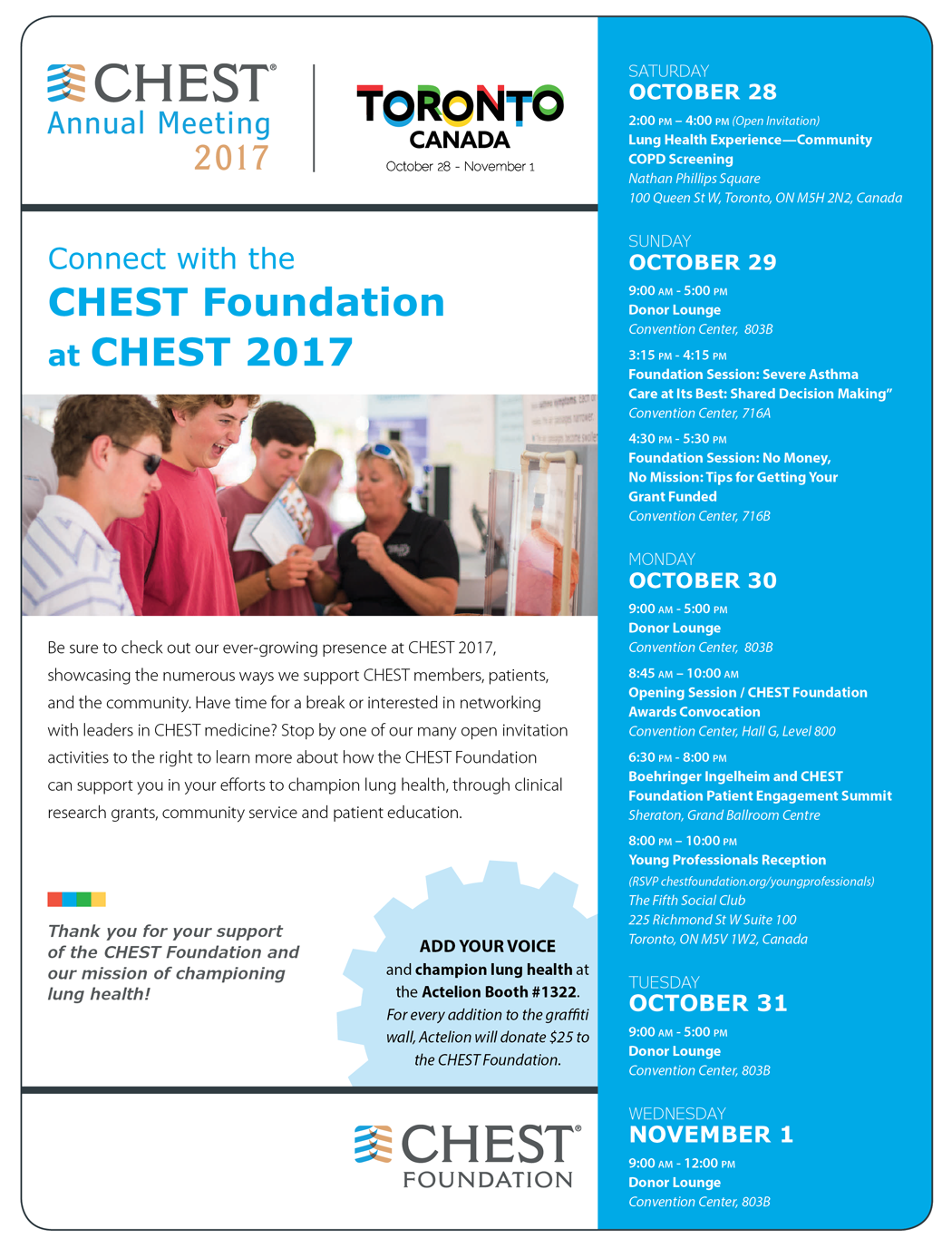
225 Richmond St W Suite 100
Toronto, ON M5V 1W2, Canada
TUESDAY OCTOBER 31
9:00 AM - 5:00 PM
Donor Lounge
Convention Center, 803B
WEDNESDAY NOVEMBER 1
9:00 AM - 12:00 PM
Donor Lounge
Convention Center, 803B
ADD YOUR VOICE
and champion lung health at the Actelion Booth #1322. For every addition to the graffiti wall, Actelion will donate $25 to the CHEST Foundation.
Thank you for your support of the CHEST Foundation and our mission of championing lung health!
Trump order allows end-around ACA rules
An executive order signed by President Donald J. Trump aims to expand health care coverage options, primarily for small businesses.
The order includes three key provisions.
First, it allows small businesses to band together to buy insurance collectively for their workers and allows plans to be sold across state lines, in theory extending the buying power that comes with a larger base of employees to smaller businesses.

Employers participating in association health plans “cannot exclude any employee from joining the plan and cannot develop premiums based on health condition,” according to a White House statement on the executive order.
Second, the executive order allows broader use of limited duration health insurance – short-term policies that are not intended to be comprehensive offerings and are not subject to Affordable Care Act coverage requirements.
These plans “typically feature broad provider networks and high coverage limits,” according to the White House statement. The plans are geared toward “people between jobs, people in counties with only a single insurer offering exchange plans, people with limited coverage networks, and people who missed the open enrollment period but still want insurance,” according to the executive order.
Association health plans and the short-term, limited duration insurance will not need to fully cover the ACA’s essential health benefits package; instead, they are intended to be alternatives to “expensive, mandate-laden [ACA] insurance,” according to the executive order.
Third, the order directs the secretaries of Health & Human Services, Treasury, and Labor to “explore how they can allow more businesses to use tax-free health reimbursement arrangements, or HRAs, to compensate their employees for their health care expenses,” according to the White House statement.
HRAs allow employers to cover select items, such as copayments, deductibles, and other items, that are not covered by the insurance plan offered to employees.
“With these actions, we are moving toward lower costs and more options in the health care market and taking crucial steps toward saving the American people from the nightmare of Obamacare,” President Trump said, adding that the White House will continue to put pressure on Congress to finish repeal and replace activities. He also said that Congress will be pursuing block grants, presumably for Medicaid, which he said he had the votes to accomplish.
The action taken today by the White House is already receiving pushback.
The American College of Physicians “has said repeatedly that any attempts by Congress to ‘repeal and replace’ the ACA must first do no harm to patients,” Susan Thompson Hingle, MD, chair of the American College of Physicians Board of Regents, said in a statement. “This executive order utterly fails that test.”
Under the association health plans “small employers would be allowed to purchase health plans that do not meet the ACA’s requirement to provide essential health benefits,” Dr. Hingle noted. “This means that plans would no longer have to cover medical patients needs; plans could choose not to cover pregnancy, maternity, and newborn care, or even chemotherapy. This would also mean that the ACA’s prohibition on annual and lifetime limits on coverage would no longer apply to any service an employer decides is not essential. These changes would be devastating for patients who need access to the ‘nonessential’ services, leaving them with potentially millions of dollars in out-of-pocket costs despite being insured.”
Association health plans also could further destabilize the individual market if healthy workers – who currently may have purchased coverage on the individual market – shifted to less comprehensive association health plans, leaving even fewer healthy individuals in the individual market. “Insurers will be forced to either leave the markets in droves or charge much higher premiums.”
The rules may not be able to provide the relief that President Trump has promised, according to Julius Hobson, a Washington-based health care lobbyist
“The changes that he is talking about are going to require departments and agencies to promulgate rules. They just simply can’t do that by fiat,” Mr. Hobson said in an interview. “I am not sure that we will see an immediate impact on the executive order. ... When you get down to the nitty-gritty of implementation, we could be a bit away from seeing anything that actually has impact.”
An executive order signed by President Donald J. Trump aims to expand health care coverage options, primarily for small businesses.
The order includes three key provisions.
First, it allows small businesses to band together to buy insurance collectively for their workers and allows plans to be sold across state lines, in theory extending the buying power that comes with a larger base of employees to smaller businesses.
Employers participating in association health plans “cannot exclude any employee from joining the plan and cannot develop premiums based on health condition,” according to a White House statement on the executive order.
Second, the executive order allows broader use of limited duration health insurance – short-term policies that are not intended to be comprehensive offerings and are not subject to Affordable Care Act coverage requirements.
These plans “typically feature broad provider networks and high coverage limits,” according to the White House statement. The plans are geared toward “people between jobs, people in counties with only a single insurer offering exchange plans, people with limited coverage networks, and people who missed the open enrollment period but still want insurance,” according to the executive order.
Association health plans and the short-term, limited duration insurance will not need to fully cover the ACA’s essential health benefits package; instead, they are intended to be alternatives to “expensive, mandate-laden [ACA] insurance,” according to the executive order.
Third, the order directs the secretaries of Health & Human Services, Treasury, and Labor to “explore how they can allow more businesses to use tax-free health reimbursement arrangements, or HRAs, to compensate their employees for their health care expenses,” according to the White House statement.
HRAs allow employers to cover select items, such as copayments, deductibles, and other items, that are not covered by the insurance plan offered to employees.
“With these actions, we are moving toward lower costs and more options in the health care market and taking crucial steps toward saving the American people from the nightmare of Obamacare,” President Trump said, adding that the White House will continue to put pressure on Congress to finish repeal and replace activities. He also said that Congress will be pursuing block grants, presumably for Medicaid, which he said he had the votes to accomplish.
The action taken today by the White House is already receiving pushback.
The American College of Physicians “has said repeatedly that any attempts by Congress to ‘repeal and replace’ the ACA must first do no harm to patients,” Susan Thompson Hingle, MD, chair of the American College of Physicians Board of Regents, said in a statement. “This executive order utterly fails that test.”
Under the association health plans “small employers would be allowed to purchase health plans that do not meet the ACA’s requirement to provide essential health benefits,” Dr. Hingle noted. “This means that plans would no longer have to cover medical patients needs; plans could choose not to cover pregnancy, maternity, and newborn care, or even chemotherapy. This would also mean that the ACA’s prohibition on annual and lifetime limits on coverage would no longer apply to any service an employer decides is not essential. These changes would be devastating for patients who need access to the ‘nonessential’ services, leaving them with potentially millions of dollars in out-of-pocket costs despite being insured.”
Association health plans also could further destabilize the individual market if healthy workers – who currently may have purchased coverage on the individual market – shifted to less comprehensive association health plans, leaving even fewer healthy individuals in the individual market. “Insurers will be forced to either leave the markets in droves or charge much higher premiums.”
The rules may not be able to provide the relief that President Trump has promised, according to Julius Hobson, a Washington-based health care lobbyist
“The changes that he is talking about are going to require departments and agencies to promulgate rules. They just simply can’t do that by fiat,” Mr. Hobson said in an interview. “I am not sure that we will see an immediate impact on the executive order. ... When you get down to the nitty-gritty of implementation, we could be a bit away from seeing anything that actually has impact.”
The cost of conflation: Avoiding loose talk about the duty to warn
The campaign, election, and administration of President Donald Trump have reinvigorated debate over rule 7.3 of the American Psychiatric Association (APA) code of ethics. Known as the Goldwater Rule for its historical roots in a magazine profile and subsequent libel suit by the 1964 Republican presidential nominee,1 this standard deems it unethical for a psychiatrist to offer a professional opinion of a public figure without conducting an examination and obtaining authorization.2 The American Psychological Association similarly provides that assessments must be based on adequate examination of the individual.3
Growing controversy
Shortly after President Trump’s inauguration, a group of 35 mental health professionals penned a letter in the New York Times stating that he was “incapable of serving safely as president.” Importantly, the writers couched their conclusions in professional expertise and specifically criticized the Goldwater Rule as having subjected their colleagues to self-imposed silence.4 A prominent psychiatrist, Allen J. Frances, MD, responded the following day to caution against “psychiatric name-calling” as a substitute for political action.5
Since then, psychiatrists classifying the APA ethics position as a “gag rule” preventing them from performing a public service have garnered considerable press coverage. When the American Psychoanalytic Association (APsaA) reiterated this summer that only APA members are bound by the Goldwater Rule, Boston Globe Media’s STAT news outlet misreported it as a license for psychiatrists to disregard the standard. Amid the ensuing media storm, the APsaA was forced to clarify that it was not countenancing defiance of psychiatry’s flagship organization and that its own longstanding policy remained unchanged.
Among those chafing against the Goldwater Rule in the current political environment, a call to arms has been the profession’s supposed “duty to warn” the public of the president’s mental health. This rationale was made explicit in an eponymous online movement and town hall forum hosted by Bandy X. Lee, MD, MDiv, a member of Yale University’s psychiatry faculty. According to these critics, an inherent tension exists between the Goldwater Rule’s prohibition on volunteering professional opinions from afar and the imperative to warn about the dangers posed by a leader with mental illness.
The duty to warn
Clinicians’ obligation to warn third parties when patients make credible threats or pose a high risk of harm emanates from various state laws, court decisions, and professional ethics rules. In the seminal Tarasoff case, a patient divulged in the course of psychotherapy his plan to murder a fellow student who had rejected his romantic overtures; campus police were alerted, but the intended victim was not. After the plan came to fruition, the California Supreme Court held that therapists must exercise reasonable care to protect “foreseeable victims” where they know or should know that a patient poses a serious danger.6
Although a controversial and massive expansion of tort liability 40 years ago, the basic tenets of Tarasoff have since been adopted by numerous courts, state legislatures, and professional organizations. The American Medical Association (AMA) recognizes an exception to confidentiality to mitigate serious threats of harm to the patient or other identifiable individuals.7 To enable health care professionals to operate in a way that is consistent with these standards, the HIPAA Privacy Rule expressly permits doctors to disclose protected health information, including psychotherapy notes, if the disclosure “is necessary to prevent or lessen a serious and imminent threat to the health or safety of a person or the public.”8
In terms of both professional ethics and privacy law, the duty to warn is framed as a limited and enumerated exception to the general rule that patient communications must be kept in confidence. In the absence of a clinician’s being privy to personal details about a patient via interview and examination, the duty to warn loses all coherence. It is precisely the intimacy of the doctor-patient relationship that gives rise to the fiduciary duty of confidentiality, which in turn must yield to public safety in rare situations where a credible threat is issued against an identifiable victim.
Origins of a misconception
Unlike the duty to warn, the Goldwater Rule is neither premised on nor a departure from the dictates of confidentiality. The rule is codified under the section of the APA ethics standards dealing with community and public health activities, not patient privacy. In nearly all cases where the Goldwater Rule could be invoked, the fundamental issue is that no examination has occurred. If it had, informed consent would be required for treatment, and appropriate authorization would be required for disclosure. Moreover, talking with the media – as opposed to alerting law enforcement, family members, or the subject of a threat – would almost never qualify as an appropriate outlet for discharging a physician’s duty to warn.
Whatever its merits, the Goldwater Rule is intended to distinguish between educational activities – in which psychiatrists share their expertise with the public and shed light on mental illness – and professional opinion wherein psychiatrists offer diagnoses or prognoses unsolicited by the individual.9

The APA has since clarified that the Goldwater Rule does not prohibit “psychologically informed leadership studies” so long as they maintain scholarly standards and do not specify a clinical diagnosis. When appropriately conducted as academic research, including acknowledgment of inherent limitations, psychological profiles do not implicate the Goldwater Rule by drawing clinical conclusions outside clinical practice.

Ultimately, the debate over the Goldwater Rule pits concerns over professional standards and respect for persons against the ability of psychiatrists to apply the expertise and language of their profession according to their own best judgment, without running afoul of an ethical norm. The premise that the Tarasoff principle overrides the Goldwater Rule is a red herring that does a disservice to both. There may be valid reasons to reevaluate the Goldwater Rule, but the duty to warn is not one of them.
Lt. Col. Charles G. Kels practices health and disability law in the U.S. Air Force. Dr. Lori H. Kels teaches and practices psychiatry at the University of the Incarnate Word School of Osteopathic Medicine in San Antonio. Opinions expressed in this article are those of the authors alone and do not necessarily reflect those of the Air Force or Department of Defense.
References
1. Goldwater v. Ginzburg, 414 F2d 324 (2d Cir 1969), cert denied, 396 US 1049 (1970).
2. APA Principles of Medical Ethics, 2013 ed. [7.3].
3. American Psychological Association Ethical Principles of Psychologists and Code of Ethics, 2016 ed. [9.01b].
4. The New York Times. Feb. 14, 2017.
5. The New York Times. Feb. 15, 2017.
6. Tarasoff v. Regents of University of California, 551 P2d 334 (Cal. 1976).
7. AMA Code of Medical Ethics, 2017 ed. [3.2.1(e) Confidentiality].
8. 45 Code of Federal Regulations 164.512(j)
9. JAMA. 2008;300(11):1348-50.
10. Psychiatr Clin North Am. 2002;25(3):A635-A46.
11. APA Opinions of the Ethics Committee, 2017 ed. [Q.7.a].
The campaign, election, and administration of President Donald Trump have reinvigorated debate over rule 7.3 of the American Psychiatric Association (APA) code of ethics. Known as the Goldwater Rule for its historical roots in a magazine profile and subsequent libel suit by the 1964 Republican presidential nominee,1 this standard deems it unethical for a psychiatrist to offer a professional opinion of a public figure without conducting an examination and obtaining authorization.2 The American Psychological Association similarly provides that assessments must be based on adequate examination of the individual.3
Growing controversy
Shortly after President Trump’s inauguration, a group of 35 mental health professionals penned a letter in the New York Times stating that he was “incapable of serving safely as president.” Importantly, the writers couched their conclusions in professional expertise and specifically criticized the Goldwater Rule as having subjected their colleagues to self-imposed silence.4 A prominent psychiatrist, Allen J. Frances, MD, responded the following day to caution against “psychiatric name-calling” as a substitute for political action.5
Since then, psychiatrists classifying the APA ethics position as a “gag rule” preventing them from performing a public service have garnered considerable press coverage. When the American Psychoanalytic Association (APsaA) reiterated this summer that only APA members are bound by the Goldwater Rule, Boston Globe Media’s STAT news outlet misreported it as a license for psychiatrists to disregard the standard. Amid the ensuing media storm, the APsaA was forced to clarify that it was not countenancing defiance of psychiatry’s flagship organization and that its own longstanding policy remained unchanged.
Among those chafing against the Goldwater Rule in the current political environment, a call to arms has been the profession’s supposed “duty to warn” the public of the president’s mental health. This rationale was made explicit in an eponymous online movement and town hall forum hosted by Bandy X. Lee, MD, MDiv, a member of Yale University’s psychiatry faculty. According to these critics, an inherent tension exists between the Goldwater Rule’s prohibition on volunteering professional opinions from afar and the imperative to warn about the dangers posed by a leader with mental illness.
The duty to warn
Clinicians’ obligation to warn third parties when patients make credible threats or pose a high risk of harm emanates from various state laws, court decisions, and professional ethics rules. In the seminal Tarasoff case, a patient divulged in the course of psychotherapy his plan to murder a fellow student who had rejected his romantic overtures; campus police were alerted, but the intended victim was not. After the plan came to fruition, the California Supreme Court held that therapists must exercise reasonable care to protect “foreseeable victims” where they know or should know that a patient poses a serious danger.6
Although a controversial and massive expansion of tort liability 40 years ago, the basic tenets of Tarasoff have since been adopted by numerous courts, state legislatures, and professional organizations. The American Medical Association (AMA) recognizes an exception to confidentiality to mitigate serious threats of harm to the patient or other identifiable individuals.7 To enable health care professionals to operate in a way that is consistent with these standards, the HIPAA Privacy Rule expressly permits doctors to disclose protected health information, including psychotherapy notes, if the disclosure “is necessary to prevent or lessen a serious and imminent threat to the health or safety of a person or the public.”8
In terms of both professional ethics and privacy law, the duty to warn is framed as a limited and enumerated exception to the general rule that patient communications must be kept in confidence. In the absence of a clinician’s being privy to personal details about a patient via interview and examination, the duty to warn loses all coherence. It is precisely the intimacy of the doctor-patient relationship that gives rise to the fiduciary duty of confidentiality, which in turn must yield to public safety in rare situations where a credible threat is issued against an identifiable victim.
Origins of a misconception
Unlike the duty to warn, the Goldwater Rule is neither premised on nor a departure from the dictates of confidentiality. The rule is codified under the section of the APA ethics standards dealing with community and public health activities, not patient privacy. In nearly all cases where the Goldwater Rule could be invoked, the fundamental issue is that no examination has occurred. If it had, informed consent would be required for treatment, and appropriate authorization would be required for disclosure. Moreover, talking with the media – as opposed to alerting law enforcement, family members, or the subject of a threat – would almost never qualify as an appropriate outlet for discharging a physician’s duty to warn.
Whatever its merits, the Goldwater Rule is intended to distinguish between educational activities – in which psychiatrists share their expertise with the public and shed light on mental illness – and professional opinion wherein psychiatrists offer diagnoses or prognoses unsolicited by the individual.9
The APA has since clarified that the Goldwater Rule does not prohibit “psychologically informed leadership studies” so long as they maintain scholarly standards and do not specify a clinical diagnosis. When appropriately conducted as academic research, including acknowledgment of inherent limitations, psychological profiles do not implicate the Goldwater Rule by drawing clinical conclusions outside clinical practice.
Ultimately, the debate over the Goldwater Rule pits concerns over professional standards and respect for persons against the ability of psychiatrists to apply the expertise and language of their profession according to their own best judgment, without running afoul of an ethical norm. The premise that the Tarasoff principle overrides the Goldwater Rule is a red herring that does a disservice to both. There may be valid reasons to reevaluate the Goldwater Rule, but the duty to warn is not one of them.
Lt. Col. Charles G. Kels practices health and disability law in the U.S. Air Force. Dr. Lori H. Kels teaches and practices psychiatry at the University of the Incarnate Word School of Osteopathic Medicine in San Antonio. Opinions expressed in this article are those of the authors alone and do not necessarily reflect those of the Air Force or Department of Defense.
References
1. Goldwater v. Ginzburg, 414 F2d 324 (2d Cir 1969), cert denied, 396 US 1049 (1970).
2. APA Principles of Medical Ethics, 2013 ed. [7.3].
3. American Psychological Association Ethical Principles of Psychologists and Code of Ethics, 2016 ed. [9.01b].
4. The New York Times. Feb. 14, 2017.
5. The New York Times. Feb. 15, 2017.
6. Tarasoff v. Regents of University of California, 551 P2d 334 (Cal. 1976).
7. AMA Code of Medical Ethics, 2017 ed. [3.2.1(e) Confidentiality].
8. 45 Code of Federal Regulations 164.512(j)
9. JAMA. 2008;300(11):1348-50.
10. Psychiatr Clin North Am. 2002;25(3):A635-A46.
11. APA Opinions of the Ethics Committee, 2017 ed. [Q.7.a].

G. lamblia assemblage B more common in HIV-positive people
The intestinal parasite Giardia lamblia assemblage B was more likely in people with HIV than in people without HIV, according to research published in Acta Tropica.
Of the 65 patients with G. lamblia included in a study undertaken by Clarissa Perez Faria, PhD, and her associates at the University of Coimbra (Portugal), 38 were HIV positive, 27 were HIV negative, and 60 patients were microscopy-positive for G. lamblia. In the HIV-positive group, 19 of the 34 microscopy-positive samples were assemblage B, and 15 were assemblage A. In the HIV-negative group, 9 of the 26 microscopy-positive samples were assemblage B, and 17 were assemblage A.
“HIV infection increases the risk of having intestinal parasitic infections, including G. lamblia. The detection and treatment of infections are important measures to improve the quality of life of HIV-infected patients,” the investigators concluded.
Find the full study in Acta Tropica (doi: 10.1016/j.actatropica.2017.04.026).
FROM ACTA TROPICA
Early births stress dads too
BERLIN – The anxiety of a preterm birth affects fathers just as much as it does mothers, significantly increasing depression rates both before and after the baby arrives.
More than one-third of fathers developed depression after their partners were admitted to a hospital with signs of impending preterm labor – similar to the percentage of mothers who experienced depression during that time, Sally Schulze reported at the meeting of the World Psychiatric Association.

The increased prevalence of depression lingered, too, she said. Even at 6 months after the birth, the rate of depression among these men was 2.5 times higher than in the general population.
Ms. Schulze and her colleagues prospectively followed 69 couples in which the woman was admitted to the hospital at high risk of preterm birth. These women had a mean gestational age of 30 weeks and had symptoms of imminent preterm birth: shortening of the cervix, premature rupture of membranes, or active preterm labor. Ms. Schulze compared this group to 49 control couples with no signs of preterm labor, who had come to the hospital to register for a birth at a mean of 35 weeks’ gestation.
The majority of the pregnancies were singletons; there were two twin pregnancies, but no high-order multiples. Couples whose infant died were later excluded from the study.
Both mothers and fathers completed the Edinburg Postnatal Depression Scale at baseline, and at 6 weeks and 6 months after the birth. The survey has been validated for perinatal use. A score of 10 or higher is considered positive for depression.
She divided the preterm birth risk group into two subgroups: couples whose infant was born preterm (26) and couples who made it to term, either by staying in the hospital for treatment and observation, or after being stabilized and released home (27).
Upon admission to the hospital, 35% of the fathers in the preterm birth risk group scored positive for depression, compared with 8% of the fathers in the control group – a significant between-group difference.
“This is especially meaningful when we consider that the background rate of depression among men in Germany is 6%,” Ms. Schulze said. “So our control group fathers were right in line with that, but depression in the preterm birth fathers was significantly elevated.”
At 6 weeks’ postpartum, men in the preterm birth risk group still were experiencing significantly elevated rates of depression, compared with both the control group and the general population. The increase was apparent whether the infant had indeed been born early, or whether it made it to full term (12% and 15%, respectively). Both were significantly higher than the 5% rate among the control group fathers.
“We have to understand that these fathers are now 6 weeks at home with a healthy infant, but they are still experiencing the stress of being exposed to this risk of preterm birth,” Ms. Schulze said.
By 6 months, depression had eased off in fathers whose infants made it to term; at 5%, it was similar to the rate in the control group fathers (4%) and the general population. But many fathers whose babies came early still were experiencing depression (12%).
Ms. Schulze then compared the fathers’ experience to that of the mothers. At baseline, women at risk of preterm birth had exactly the same rate of depression as their partners (35%). However, depression also was elevated among women in the control group (18%). The background rate for depression among German women is 10%, Ms. Schulze said.
At 6 weeks’ postpartum, the timing of birth did not seem to matter as much to the mothers. Depression rates were similarly elevated in those who had a preterm birth and those who did not (25%, 28%). Both were significantly higher than the 17% rate in the control group.
At 6 months, things were leveling out some for these mothers, with depression present in 10% of the preterm birth group, 19% of the full-term birth group, and 13% of the control group.
“Men seem to suffer just as much stress from this experience as women do, although perhaps in a different trajectory,” Ms. Schulze said. “Although there may be different contributing factors, we believe that the psychological care offered to mothers at risk of preterm birth should also be extended to fathers.”
She had no financial disclosures.
[email protected]
On Twitter @alz_gal
BERLIN – The anxiety of a preterm birth affects fathers just as much as it does mothers, significantly increasing depression rates both before and after the baby arrives.
More than one-third of fathers developed depression after their partners were admitted to a hospital with signs of impending preterm labor – similar to the percentage of mothers who experienced depression during that time, Sally Schulze reported at the meeting of the World Psychiatric Association.
The increased prevalence of depression lingered, too, she said. Even at 6 months after the birth, the rate of depression among these men was 2.5 times higher than in the general population.
Ms. Schulze and her colleagues prospectively followed 69 couples in which the woman was admitted to the hospital at high risk of preterm birth. These women had a mean gestational age of 30 weeks and had symptoms of imminent preterm birth: shortening of the cervix, premature rupture of membranes, or active preterm labor. Ms. Schulze compared this group to 49 control couples with no signs of preterm labor, who had come to the hospital to register for a birth at a mean of 35 weeks’ gestation.
The majority of the pregnancies were singletons; there were two twin pregnancies, but no high-order multiples. Couples whose infant died were later excluded from the study.
Both mothers and fathers completed the Edinburg Postnatal Depression Scale at baseline, and at 6 weeks and 6 months after the birth. The survey has been validated for perinatal use. A score of 10 or higher is considered positive for depression.
She divided the preterm birth risk group into two subgroups: couples whose infant was born preterm (26) and couples who made it to term, either by staying in the hospital for treatment and observation, or after being stabilized and released home (27).
Upon admission to the hospital, 35% of the fathers in the preterm birth risk group scored positive for depression, compared with 8% of the fathers in the control group – a significant between-group difference.
“This is especially meaningful when we consider that the background rate of depression among men in Germany is 6%,” Ms. Schulze said. “So our control group fathers were right in line with that, but depression in the preterm birth fathers was significantly elevated.”
At 6 weeks’ postpartum, men in the preterm birth risk group still were experiencing significantly elevated rates of depression, compared with both the control group and the general population. The increase was apparent whether the infant had indeed been born early, or whether it made it to full term (12% and 15%, respectively). Both were significantly higher than the 5% rate among the control group fathers.
“We have to understand that these fathers are now 6 weeks at home with a healthy infant, but they are still experiencing the stress of being exposed to this risk of preterm birth,” Ms. Schulze said.
By 6 months, depression had eased off in fathers whose infants made it to term; at 5%, it was similar to the rate in the control group fathers (4%) and the general population. But many fathers whose babies came early still were experiencing depression (12%).
Ms. Schulze then compared the fathers’ experience to that of the mothers. At baseline, women at risk of preterm birth had exactly the same rate of depression as their partners (35%). However, depression also was elevated among women in the control group (18%). The background rate for depression among German women is 10%, Ms. Schulze said.
At 6 weeks’ postpartum, the timing of birth did not seem to matter as much to the mothers. Depression rates were similarly elevated in those who had a preterm birth and those who did not (25%, 28%). Both were significantly higher than the 17% rate in the control group.
At 6 months, things were leveling out some for these mothers, with depression present in 10% of the preterm birth group, 19% of the full-term birth group, and 13% of the control group.
“Men seem to suffer just as much stress from this experience as women do, although perhaps in a different trajectory,” Ms. Schulze said. “Although there may be different contributing factors, we believe that the psychological care offered to mothers at risk of preterm birth should also be extended to fathers.”
She had no financial disclosures.
[email protected]
On Twitter @alz_gal
AT WPA 2017
Key clinical point:
Major finding: Depression developed in 35% of men whose wives were admitted to the hospital for imminent preterm birth; elevated depression rates lingered for up to 6 months in these fathers.
Data source: The prospective study comprised 69 couples admitted for preterm birth risk, and 49 control couples.
Disclosures: Ms. Schulze had no financial disclosures.
Intensive exercise didn’t improve glycemic control for obese pregnant women
An intensive exercise intervention for obese pregnant women did not improve maternal glycemia, but did attenuate excessive gestational weight gain, according to the a single-center, randomized controlled trial published in Obstetrics & Gynecology.
The Healthy Eating, Exercise and Lifestyle Trial, conducted from November 2013 through April 2016 at the Coombe Women and Infants University Hospital in Dublin, randomized 88 pregnant women with a body mass index of 30 kg/m2 or greater at their first prenatal visit to either a medically supervised exercise program or to the control group. The intervention consisted of 50-60 minutes of exercise at least once per week, including resistance or weights and aerobic exercises (Obstet Gynecol. 2017;130:1001-10).
However, excessive gestational weight gain at 36 weeks’ gestation (greater than 9.1 kg) was lower in the exercise group at 22.2%, compared with 43.2% in the control group (P less than .05), Niamh Daly of Coombe Women and Infants University Hospital and her coauthors reported.
“Our study may have failed to improve maternal glycemia because the ideal time to begin such a program could be before pregnancy,” the researchers wrote. “Pregnant women who are obese, however, should be advised to exercise because it attenuates excessive gestational weight gain.”
The study was partially funded by Friends of the Coombe, a charity organization. The authors reported having no financial disclosures.
FROM OBSTETRICS & GYNECOLOGY
Key clinical point:
Major finding: There was no difference in mean fasting plasma glucose between the control group (90.0 plus or minus 9.0 mg/dL) and the exercise group (93.6 plus or minus 7.2 mg/dL) at 24-28 weeks’ gestation (P = .13).
Data source: A single-center, randomized controlled trial of 88 pregnant women with a BMI of 30 kg/m2 or greater.
Disclosures: The study was partially funded by Friends of the Coombe, a charity organization. The authors reported having no financial disclosures.
MedPAC calls for MIPS repeal
WASHINGTON – In a rare display of a near-immediate consensus, the Medicare Payment Advisory Commission agreed that the Merit-Based Incentive Payment System track of the new Quality Payment Program should be scrapped, although commission members are not yet ready to endorse a replacement plan.
MedPAC staff presented its idea of “repeal and replace” less then 10 months into the first reporting year, with staff member David V. Glass noting during an Oct. 6 meeting that “MIPS will not achieve the goal of identifying and rewarding high-value clinicians.”
“Our most basic concern is that measures used in MIPS have not been proven to be associated with high-value care,” Mr. Glass said. “Many of the MIPS quality measures are process measures, assessing only the care a provider delivers within their four walls.”
MedPAC staff proposed a replacement option affecting all clinicians who are not a part of an advanced Alternative Payment Model program. Under their proposal, Medicare would withhold 2% of each clinician’s Medicare payments. Clinicians could earn back that 2% by joining a large reporting entity (either as part of a formal business structure or something like a virtual group); they could elect to join an advanced APM, earning back the 2% and possibly bonus payments; or they could do nothing and lose the 2%.
Measurements in the proposed value program would be similar to those employed by advanced APMs in that they would be focused on population-based measures assessing clinical quality, patient experience, and value. Potential measures would address avoidable admissions/emergency department visits, mortality, readmissions, ability to obtain care, ability to communicate with clinicians, spending per beneficiary, resource use, and rates of low-value care use. Measures would be calculated based on claims.
MedPAC commissioners were nearly unanimous in their agreement to the idea of repealing MIPS but were not ready to sign off on the proposed replacement.
“I’m really concerned about the burden on physicians, and I’m concerned about some of the outlandish potential rewards for groups under MIPS that can really dissuade them from investing and moving into APMs,” commission member Paul B. Ginsburg, PhD, senior fellow in economic studies at the Brookings Institution said.
“I think we really have to get rid of MIPS and either replace it with this system, which I think has a lot of merit, or just get rid of it,” said commission member Jack Hoadley, PhD, of Georgetown University in Washington, suggesting MIPS would be “even worse” than the old Sustainable Growth Rate formula over time. It is “clear to me that MIPS is not going to ... get us toward high-value care, it is not going to make clinicians’ lives better, it is not going to make patients’ lives better, and there is a lot of money at stake.”
Commission member Dana Gelb Safran, ScD, chief performance measurement and improvement officer at Blue Cross Blue Shield of Massachusetts said she did not believe that there would be any value being gained in return for the money given to clinicians who participate in MIPS.
Not everyone was on board with the proposed replacement.
“I am very much in favor of repealing MIPS, but I don’t get the sense that we’ve gotten the replacement model quite right yet” because the proposed system does not do enough to get physicians into advanced APMs, commission member Craig Samitt, MD, chief clinical officer at Anthem, said. “So if a replacement is a voluntary model that would allow us to keep practicing health care the way we’ve been practicing, then that replacement is not a good replacement.”
The American Medical Association declined to evaluate the proposal that was laid forth by staff.
“The AMA welcomes ideas on how to improve Medicare physician payment policies,” AMA President David O. Barbe said in a statement. “We understand that MedPAC’s proposals are a work in progress, so it’s too early to render any judgment.”
Dr. Barbe noted that the AMA recommends that physicians participate in MIPS, even if it is at the lowest level simply to avoid any penalty and continue investing in the infrastructure to participate. The AMA, AGA, and other medical societies also are asking CMS to allow those who are exempt from MIPS participation to be able to opt into the program.
The American Medical Group Association (AMGA), a trade organization representing multispecialty medical groups, however, has criticized the move by CMS to increase the number of clinicians who are exempt from MIPS.
Under the proposed expansion – which would approximately double the number of clinicians who are MIPS exempt – “MIPS no longer provides really any incentive to get to value and in fact it’s a disincentive,” said Chet Speed, AMGA vice president of public policy. “That is one of the realities that MedPAC was dealing with.”
Mr. Speed emphasized that AMGA has not altered its policy on MIPS, which it wants to see enacted for all and has offered its own resources to help with the transition, but “if AMGA were to entertain a new position on MIPS, I think we probably would go with a more simple route, which is to just get rid of MIPS and repurpose the revenues that were in MIPS to APMs. ... We have not agreed upon that policy but that has been discussed internally.”
He added that “AMGA’s membership does look at MIPS as a tool that has really devolved from a value mechanism to a compliance exercise and nothing more.”
As to whether health care provider associations would come together and support the repeal of MIPS, Mr. Speed was hesitant to predict that, even though many have reservations about it, noting that it could be because the broadening of exclusions, which the AMA and most other associations support, effectively remove a lot of their membership from having to participate anyway, leaving the bigger groups such as Mayo, the Cleveland Clinic and Intermountain Healthcare to fight over a much smaller pot of bonus payments, significantly limiting the returns on investments made to be ready for the MIPS transition.
AT A MEDPAC MEETING