User login
Irregular Erythematous Patch on the Face of an Infant
The Diagnosis: Phakomatosis Pigmentovascularis With Sturge-Weber Syndrome
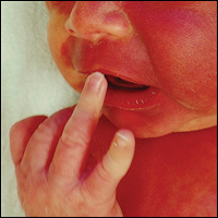
The erythematous patches were identified as capillary malformations (port-wine stains) and the slate gray pigmentary changes as dermal melanocytosis (Mongolian spots)(Figure). In fact, the diagnosis of phakomatosis pigmentovascularis (PPV) type II requires dermal melanocytosis and capillary malformation with and without nevus anemicus.1 In one case series, 46% (7/15) of patients with PPV had nevus anemicus2 but our patient did not.

Phakomatosis pigmentovascularis was divided into 4 types in 1985,3 then later 5 types.4 Subcategories of the 5 types include type A, which denotes a lack of extracutaneous involvement, and type B, which is used when internal manifestations have been exhibited. Since 1947, approximately 222 cases of PPV have been described in the literature.2
A case of PPV associated with Sturge-Weber syndrome (SWS) was reported in 1997.5 Since then, PPV occasionally has been linked with SWS,5-9 though there have been other syndromic associations including Klippel-Trenaunay-Weber syndrome and melanosis oculi.2 The incidence and prevalence of overlap of PPV and SWS is unknown but is likely to be rare. In our case, magnetic resonance imaging of the patient's brain did not reveal the characteristic tram-track appearance of SWS; however, the diagnosis of SWS type II only requires facial angioma with or without glaucoma.9,10 Most cases of PPV originate from Japan, Argentina, and Mexico.2 Interestingly, our patient's parents were both of Mexican ancestry. Phakomatosis pigmentovascularis type IIb is the most common, followed by type IIa.2 Most cases have been described as sporadic, though our patient's mother also exhibited a port-wine stain on the right neck, suggesting a possible genetic association.
The etiology of PPV has been postulated as twin spotting or didymosis (Greek for twin), most commonly seen in plants and animals. A previous review defined twin spotting as 2 mutant tissues situated adjacent to one another and unique from the normal tissue surrounding both of them.2 When the cell loses its heterozygosity, this phenomenon appears. An alternative etiology supplants that a drug or virus toxic to the nervous system causes aberrant angioblasts and melanoblasts.11,12 The etiology of SWS also is unknown, though vasomotor instability has been postulated as a cause.6,13
It is important to exclude associated internal organ involvement with both of these syndromes because approximately 50% of PPV cases have extracutaneous organ involvement.2,14 In fact, PPV is known to involve the brain, skeletal system, and eye, potentially manifesting as deafness, hydrocephalus, extremity overgrowth, scoliosis, cataracts, and more.2 Patients with SWS often exhibit brain and eye symptoms including seizures.1 To screen for extracutaneous involvement, multiple imaging studies should be performed. In our patient, an echocardiogram revealed a patent foramen ovale and normal cardiac anatomy for his age. Brain imaging revealed a hypoplastic left sigmoid and transverse sinus without venous thrombosis and unremarkable appearance of the brain. An ultrasound of the liver, spleen, kidneys, and pancreas revealed no evidence of solid, cystic, or vascular lesions, though the gallbladder exhibited hyperechoic areas.
To manage the skin lesions, some authors recommend Q-switched lasers for pigmented lesions and pulsed dye lasers for capillary malformations.15 Paller and Mancini1 cited evidence that pulsed dye laser treatment before the age of 1 year may offer a psychological advantage, while other views have been offered.16 Some physicians believe that no urgent treatment of capillary malformations is needed unless internal organs are involved.2,15
- Paller AS, Mancini AJ. Hurwitz Clinical Pediatric Dermatology: A Textbook of Skin Disorders of Childhood and Adolescence. 4th ed. New York, NY: Elsevier/Saunders; 2011.
- Fernández-Guarino M, Boixeda P, de Las Heras E, et al. Phakomatosis pigmentovascularis: clinical findings in 15 patients and review of the literature. J Am Acad Dermatol. 2008;58:88-93.
- Hasegawa Y, Yasuhara M. Phakomatosis pigmentovascularis type VIa. Arch Dermatol. 1985;121:651-655.
- Torrelo A, Zambrano A, Happle R. Cutis marmorata telangiectatica congenita and extensive Mongolian spots: type V phacomatosis pigmentovascularis. Br J Dermatol. 2003;148:342-345.
- Teekhasaenee C, Ritch R. Glaucoma in phakomatosis pigmentovascularis. Ophthalmology. 1997;104:150-157.
- Patil B, Sinha G, Nayak B, et al. Bilateral Sturge-Weber and phakomatosis pigmentovascularis with glaucoma, an overlap syndrome [published online May 6, 2015]. Case Rep Ophthalmol Med. 2015;2015:106932.
- Hagiwara K, Uezato H, Nonaka S. Phacomatosis pigmentovascularis type IIb associated with Sturge-Weber syndrome and pyogenic granuloma. J Dermatol. 1998;25:721-729.
- Al Robaee A, Banka N, Alfadley A. Phakomatosis pigmentovascularis type IIb associated with Sturge-Weber syndrome. Pediatr Dermatol. 2004;21:642-645.
- Yang Y, Guo X, Xu J, et al. Phakomatosis pigmentovascularis associated with Sturge-Weber syndrome, ota nevus, and congenital glaucoma. Medicine (Baltimore). 2015;94:E1025.
- Roach ES. Neurocutaneous syndromes. Pediatr Clin North Am. 1992;39:591-620.
- Happle R. Mosaicism in human skin, understanding the patterns and mechanisms. Arch Dermatol. 1993;129:1460-1470.
- Happle R. Loss of heterozygosity in human skin. J Am Acad Dermatol. 1999;85:355-358.
- Comi AM. Pathophysiology of Sturge-Weber syndrome. J Child Neurol. 2003;18:509-516.
- Kim YC, Park HJ, Cinn YW. Phakomatosis pigmentovascularis type IIa with generalized vitiligo. Br J Dermatol. 2002;147:1028-1029.
- Brittain P, Walsh EJ, Smidt AC. Blotchy baby: a case of phakomatosis pigmentovascularis [published online February 1, 2013]. J Pediatr. 2013;162:1293.
- Van der Horst CM, Koster PH, de Borgie CA, et al. Effect of the timing of treatment of port-wine stains with the flash-lamp-pumped pulsed-dye laser. N Engl J Med. 1998;338:1028-1033.
The Diagnosis: Phakomatosis Pigmentovascularis With Sturge-Weber Syndrome
The erythematous patches were identified as capillary malformations (port-wine stains) and the slate gray pigmentary changes as dermal melanocytosis (Mongolian spots)(Figure). In fact, the diagnosis of phakomatosis pigmentovascularis (PPV) type II requires dermal melanocytosis and capillary malformation with and without nevus anemicus.1 In one case series, 46% (7/15) of patients with PPV had nevus anemicus2 but our patient did not.
Phakomatosis pigmentovascularis was divided into 4 types in 1985,3 then later 5 types.4 Subcategories of the 5 types include type A, which denotes a lack of extracutaneous involvement, and type B, which is used when internal manifestations have been exhibited. Since 1947, approximately 222 cases of PPV have been described in the literature.2
A case of PPV associated with Sturge-Weber syndrome (SWS) was reported in 1997.5 Since then, PPV occasionally has been linked with SWS,5-9 though there have been other syndromic associations including Klippel-Trenaunay-Weber syndrome and melanosis oculi.2 The incidence and prevalence of overlap of PPV and SWS is unknown but is likely to be rare. In our case, magnetic resonance imaging of the patient's brain did not reveal the characteristic tram-track appearance of SWS; however, the diagnosis of SWS type II only requires facial angioma with or without glaucoma.9,10 Most cases of PPV originate from Japan, Argentina, and Mexico.2 Interestingly, our patient's parents were both of Mexican ancestry. Phakomatosis pigmentovascularis type IIb is the most common, followed by type IIa.2 Most cases have been described as sporadic, though our patient's mother also exhibited a port-wine stain on the right neck, suggesting a possible genetic association.
The etiology of PPV has been postulated as twin spotting or didymosis (Greek for twin), most commonly seen in plants and animals. A previous review defined twin spotting as 2 mutant tissues situated adjacent to one another and unique from the normal tissue surrounding both of them.2 When the cell loses its heterozygosity, this phenomenon appears. An alternative etiology supplants that a drug or virus toxic to the nervous system causes aberrant angioblasts and melanoblasts.11,12 The etiology of SWS also is unknown, though vasomotor instability has been postulated as a cause.6,13
It is important to exclude associated internal organ involvement with both of these syndromes because approximately 50% of PPV cases have extracutaneous organ involvement.2,14 In fact, PPV is known to involve the brain, skeletal system, and eye, potentially manifesting as deafness, hydrocephalus, extremity overgrowth, scoliosis, cataracts, and more.2 Patients with SWS often exhibit brain and eye symptoms including seizures.1 To screen for extracutaneous involvement, multiple imaging studies should be performed. In our patient, an echocardiogram revealed a patent foramen ovale and normal cardiac anatomy for his age. Brain imaging revealed a hypoplastic left sigmoid and transverse sinus without venous thrombosis and unremarkable appearance of the brain. An ultrasound of the liver, spleen, kidneys, and pancreas revealed no evidence of solid, cystic, or vascular lesions, though the gallbladder exhibited hyperechoic areas.
To manage the skin lesions, some authors recommend Q-switched lasers for pigmented lesions and pulsed dye lasers for capillary malformations.15 Paller and Mancini1 cited evidence that pulsed dye laser treatment before the age of 1 year may offer a psychological advantage, while other views have been offered.16 Some physicians believe that no urgent treatment of capillary malformations is needed unless internal organs are involved.2,15
- Paller AS, Mancini AJ. Hurwitz Clinical Pediatric Dermatology: A Textbook of Skin Disorders of Childhood and Adolescence. 4th ed. New York, NY: Elsevier/Saunders; 2011.
- Fernández-Guarino M, Boixeda P, de Las Heras E, et al. Phakomatosis pigmentovascularis: clinical findings in 15 patients and review of the literature. J Am Acad Dermatol. 2008;58:88-93.
- Hasegawa Y, Yasuhara M. Phakomatosis pigmentovascularis type VIa. Arch Dermatol. 1985;121:651-655.
- Torrelo A, Zambrano A, Happle R. Cutis marmorata telangiectatica congenita and extensive Mongolian spots: type V phacomatosis pigmentovascularis. Br J Dermatol. 2003;148:342-345.
- Teekhasaenee C, Ritch R. Glaucoma in phakomatosis pigmentovascularis. Ophthalmology. 1997;104:150-157.
- Patil B, Sinha G, Nayak B, et al. Bilateral Sturge-Weber and phakomatosis pigmentovascularis with glaucoma, an overlap syndrome [published online May 6, 2015]. Case Rep Ophthalmol Med. 2015;2015:106932.
- Hagiwara K, Uezato H, Nonaka S. Phacomatosis pigmentovascularis type IIb associated with Sturge-Weber syndrome and pyogenic granuloma. J Dermatol. 1998;25:721-729.
- Al Robaee A, Banka N, Alfadley A. Phakomatosis pigmentovascularis type IIb associated with Sturge-Weber syndrome. Pediatr Dermatol. 2004;21:642-645.
- Yang Y, Guo X, Xu J, et al. Phakomatosis pigmentovascularis associated with Sturge-Weber syndrome, ota nevus, and congenital glaucoma. Medicine (Baltimore). 2015;94:E1025.
- Roach ES. Neurocutaneous syndromes. Pediatr Clin North Am. 1992;39:591-620.
- Happle R. Mosaicism in human skin, understanding the patterns and mechanisms. Arch Dermatol. 1993;129:1460-1470.
- Happle R. Loss of heterozygosity in human skin. J Am Acad Dermatol. 1999;85:355-358.
- Comi AM. Pathophysiology of Sturge-Weber syndrome. J Child Neurol. 2003;18:509-516.
- Kim YC, Park HJ, Cinn YW. Phakomatosis pigmentovascularis type IIa with generalized vitiligo. Br J Dermatol. 2002;147:1028-1029.
- Brittain P, Walsh EJ, Smidt AC. Blotchy baby: a case of phakomatosis pigmentovascularis [published online February 1, 2013]. J Pediatr. 2013;162:1293.
- Van der Horst CM, Koster PH, de Borgie CA, et al. Effect of the timing of treatment of port-wine stains with the flash-lamp-pumped pulsed-dye laser. N Engl J Med. 1998;338:1028-1033.
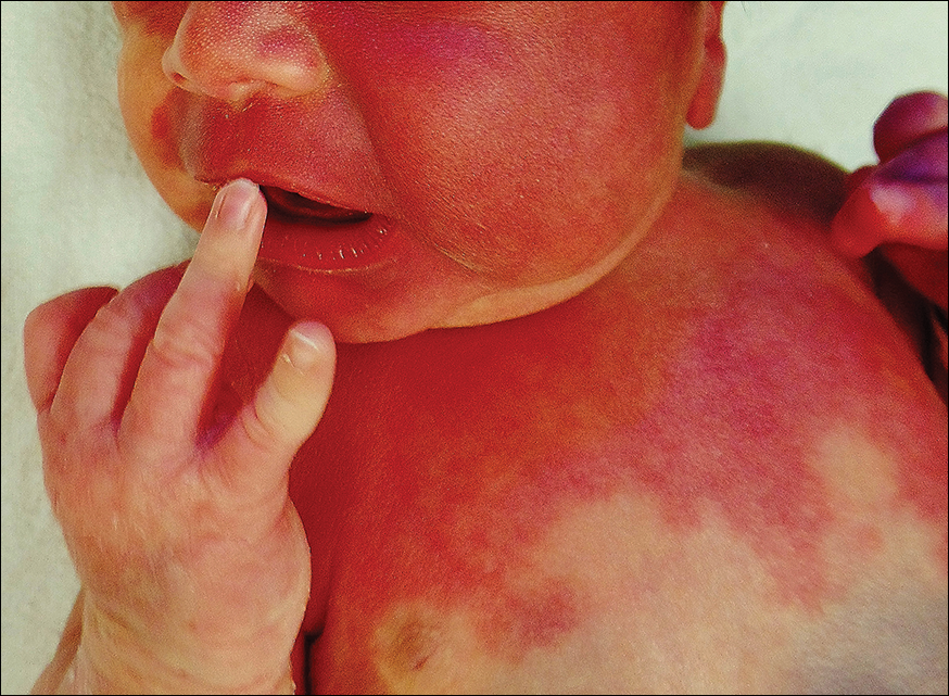
A newborn presented with an irregular and well-demarcated erythematous patch on the face, trunk, buttocks, and toes on the left foot. Another red patch was present on the right side of the face, while a slate gray patch covered the flanks and back. The limbs appeared symmetric and he exhibited no gross deformities. On close physical examination, he was noted to have a cloudy left eye. An ophthalmology consultation revealed a choroidal hemangioma and congenital glaucoma in the left eye.
A Case of Streptococcus pyogenes Sepsis of Possible Oral Origin
Sepsis can be the result of single or multiple factors and sources of infection. Oral sources of sepsis and systemic infection are not commonly considered as the first potential source of infection when evaluating a septic patient. Oral infections of odontogenic or periodontal origin are frequently associated with localized or diffuse cellulitis of the head and neck region.1 The patient’s health status and complicating problems, such as an immunocompromising condition, can further reduce the immune response for controlling chronic sources of infection. This in turn, can lead to acute manifestations such as cellulitis, sepsis, or necrotizing fasciitis. Necrotizing fasciitis is caused by a polymicrobial or mixed aerobic-anaerobic infection from a variety of sources, including Streptococcus pyogenes (S pyogenes).
Case Presentation
A 57-year-old female with a history of major depressive disorder, paroxysmal atrial fibrillation, and opioid dependence that was in remission for more than 3 years was brought to the emergency department (ED) by a family member after the patient developed confusion and lethargy. She was primarily experiencing right breast pain and swelling. The breast pain was associated with high fevers, nausea, vomiting, and chills. On examination the patient was noted to have a fever of 104° F, heart rate of 160 bpm, respirations of 22 breaths per minute, blood pressure (BP) 109/58, and a white blood cell count (WBC) of 8.7 X 10³. There was a noted skin abrasion on her right hand. She was lethargic and confused. Blood cultures were positive for S pyogenes, and a swab of the right breast was negative for bacterial growth.
The patient was admitted to the medical intensive care unit (MICU) and placed on 2 vasopressors for control of low BP and assistance with low urine output. After a 6 L fluid resuscitation, the patient was started on vancomycin and piperacillin/tazobactam for possible cellulitis causing sepsis. An echocardiogram was negative for endocarditis. The patient continued to decline the following day with continuing tachycardia and tachypnea with hypotension and was intubated. Pulmonology was consulted for possible acute respiratory distress syndrome secondary to sepsis. General surgery was consulted for possible necrotizing fasciitis of the chest wall, and cardiology was consulted for low cardiac output.
On day 4 of her hospitalization, the patient was taken to surgery for exploration, drainage, and debridement of the right axilla and breast; cultures with lack of organism growth was noted. While in the MICU, she was followed by the Infectious Disease service as her WBC remained elevated and peaking at 32.6 X 10³, while blood cultures were negative for bacterial growth. The dental service was consulted on day 5 to evaluate for other possible sources of infection.
The patient’s oral condition was noted as having advanced chronic periodontal disease that required full mouth extraction. The patient remained hemodynamically unstable with platelet counts below 50,000 until day 7, at which time she was taken for surgery for full mouth extraction and associated alveoloplasty. On extraction the patient continued to improve and was extubated on day 11 with platelets and WBC returning to normal levels by day 13 of her hospital stay. The patient remained hospitalized for a total MICU stay of 20 days and rehabilitation stay of more than 2 weeks.
Discussion
Oral infections most often present with acute onset and noted oral-facial cellulitis or abscess. Oral source of septicemia often are considered after ruling out most other potential sources. Although it is not certain that this case is directly related to the advanced chronic periodontal disease, S pyogenes has been noted to be a pathogen in periodontal disease progression.
According to the American Dental Association in 2012, dental visits to the ED cost the U.S. health care system $1.6 billion and an average cost of $749 per visit. There are more than 2 million ED visits each year for dental pain and infection, and 39% return due to nonresolution of the dental problem. Patients return to the ED due to lack of access and resources to routine and emergent dental care.2 The average daily cost of an MICU stay with mechanical ventilation was $2,193 in 2002. This particular case consisted of 11 days of mechanical ventilation, 20 MICU days, and an additional 20 days of inpatient rehabilitation which resulted in costs that exceeded $50,000.3
Conclusion
This case demonstrates the successful collaboration of dentistry for the overall medical management of the patient. An integrated approach highlights the need for and the value of integrating dental programs within large tertiary hospital systems. Such integration will likely improve earlier recognition and better management of oral infections resulting in systemic illness and improve patient outcomes, reduced length of hospital stay, and reduction of overall costs.
1. Krishnan V, Johnson JV, Helfric JF. Management of maxillofacial infections: a review of 50 cases. J Oral Maxillofac Surg. 1993; 51(8):868-873.
2. Wall T, Vujicic M. Emergency department use for dental conditions continues to increase. American Dental Association: Health Policy Institute. http://www.ada.org/~/media/ADA/Science%20and%20Research/HPI/Files/HPIBrief_0415_2.ashx. Published April 2015. Accessed September 5, 2017.
3. Dasta JF, McLaughlin TP, Mody SH, Piech CT. Daily cost of an intensive care unit day: the contribution of mechanical ventilation. Crit Care Med. 2005;33(6):1266-1271.
Sepsis can be the result of single or multiple factors and sources of infection. Oral sources of sepsis and systemic infection are not commonly considered as the first potential source of infection when evaluating a septic patient. Oral infections of odontogenic or periodontal origin are frequently associated with localized or diffuse cellulitis of the head and neck region.1 The patient’s health status and complicating problems, such as an immunocompromising condition, can further reduce the immune response for controlling chronic sources of infection. This in turn, can lead to acute manifestations such as cellulitis, sepsis, or necrotizing fasciitis. Necrotizing fasciitis is caused by a polymicrobial or mixed aerobic-anaerobic infection from a variety of sources, including Streptococcus pyogenes (S pyogenes).
Case Presentation
A 57-year-old female with a history of major depressive disorder, paroxysmal atrial fibrillation, and opioid dependence that was in remission for more than 3 years was brought to the emergency department (ED) by a family member after the patient developed confusion and lethargy. She was primarily experiencing right breast pain and swelling. The breast pain was associated with high fevers, nausea, vomiting, and chills. On examination the patient was noted to have a fever of 104° F, heart rate of 160 bpm, respirations of 22 breaths per minute, blood pressure (BP) 109/58, and a white blood cell count (WBC) of 8.7 X 10³. There was a noted skin abrasion on her right hand. She was lethargic and confused. Blood cultures were positive for S pyogenes, and a swab of the right breast was negative for bacterial growth.
The patient was admitted to the medical intensive care unit (MICU) and placed on 2 vasopressors for control of low BP and assistance with low urine output. After a 6 L fluid resuscitation, the patient was started on vancomycin and piperacillin/tazobactam for possible cellulitis causing sepsis. An echocardiogram was negative for endocarditis. The patient continued to decline the following day with continuing tachycardia and tachypnea with hypotension and was intubated. Pulmonology was consulted for possible acute respiratory distress syndrome secondary to sepsis. General surgery was consulted for possible necrotizing fasciitis of the chest wall, and cardiology was consulted for low cardiac output.
On day 4 of her hospitalization, the patient was taken to surgery for exploration, drainage, and debridement of the right axilla and breast; cultures with lack of organism growth was noted. While in the MICU, she was followed by the Infectious Disease service as her WBC remained elevated and peaking at 32.6 X 10³, while blood cultures were negative for bacterial growth. The dental service was consulted on day 5 to evaluate for other possible sources of infection.
The patient’s oral condition was noted as having advanced chronic periodontal disease that required full mouth extraction. The patient remained hemodynamically unstable with platelet counts below 50,000 until day 7, at which time she was taken for surgery for full mouth extraction and associated alveoloplasty. On extraction the patient continued to improve and was extubated on day 11 with platelets and WBC returning to normal levels by day 13 of her hospital stay. The patient remained hospitalized for a total MICU stay of 20 days and rehabilitation stay of more than 2 weeks.
Discussion
Oral infections most often present with acute onset and noted oral-facial cellulitis or abscess. Oral source of septicemia often are considered after ruling out most other potential sources. Although it is not certain that this case is directly related to the advanced chronic periodontal disease, S pyogenes has been noted to be a pathogen in periodontal disease progression.
According to the American Dental Association in 2012, dental visits to the ED cost the U.S. health care system $1.6 billion and an average cost of $749 per visit. There are more than 2 million ED visits each year for dental pain and infection, and 39% return due to nonresolution of the dental problem. Patients return to the ED due to lack of access and resources to routine and emergent dental care.2 The average daily cost of an MICU stay with mechanical ventilation was $2,193 in 2002. This particular case consisted of 11 days of mechanical ventilation, 20 MICU days, and an additional 20 days of inpatient rehabilitation which resulted in costs that exceeded $50,000.3
Conclusion
This case demonstrates the successful collaboration of dentistry for the overall medical management of the patient. An integrated approach highlights the need for and the value of integrating dental programs within large tertiary hospital systems. Such integration will likely improve earlier recognition and better management of oral infections resulting in systemic illness and improve patient outcomes, reduced length of hospital stay, and reduction of overall costs.
1. Krishnan V, Johnson JV, Helfric JF. Management of maxillofacial infections: a review of 50 cases. J Oral Maxillofac Surg. 1993; 51(8):868-873.
2. Wall T, Vujicic M. Emergency department use for dental conditions continues to increase. American Dental Association: Health Policy Institute. http://www.ada.org/~/media/ADA/Science%20and%20Research/HPI/Files/HPIBrief_0415_2.ashx. Published April 2015. Accessed September 5, 2017.
3. Dasta JF, McLaughlin TP, Mody SH, Piech CT. Daily cost of an intensive care unit day: the contribution of mechanical ventilation. Crit Care Med. 2005;33(6):1266-1271.
Natural selection opportunities tied to cancer rates

Countries with the lowest opportunities for natural selection have higher cancer rates than countries with the highest opportunities for natural selection, according to a study published in Evolutionary Applications.
Researchers said this is because modern medicine is enabling people to survive cancers, and their genetic backgrounds are passing from one generation to the next.
The team said the rate of some cancers has doubled and even quadrupled over the past 100 to 150 years, and human evolution has moved away from “survival of the fittest.”
“Modern medicine has enabled the human species to live much longer than would otherwise be expected in the natural world,” said study author Maciej Henneberg, PhD, DSc, of the University of Adelaide in South Australia.
“Besides the obvious benefits that modern medicine gives, it also brings with it an unexpected side-effect—allowing genetic material to be passed from one generation to the next that predisposes people to have poor health, such as type 1 diabetes or cancer.”
“Because of the quality of our healthcare in western society, we have almost removed natural selection as the ‘janitor of the gene pool.’ Unfortunately, the accumulation of genetic mutations over time and across multiple generations is like a delayed death sentence.”
Country comparison
The researchers studied global cancer data from the World Health Organization as well as other health and socioeconomic data from the United Nations and the World Bank of 173 countries. The team compared the top 10 countries with the highest opportunities for natural selection to the 10 countries with the lowest opportunities for natural selection.
“We looked at countries that offered the greatest opportunity to survive cancer compared with those that didn’t,” said study author Wenpeng You, a PhD student at the University of Adelaide. “This does not only take into account factors such as socioeconomic status, urbanization, and quality of medical services but also low mortality and fertility rates, which are the 2 distinguishing features in the ‘better’ world.”
“Countries with low mortality rates may allow more people with cancer genetic background to reproduce and pass cancer genes/mutations to the next generation. Meanwhile, low fertility rates in these countries may not be able to have diverse biological variations to provide the opportunity for selecting a naturally fit population—for example, people without or with less cancer genetic background. Low mortality rate and low fertility rate in the ‘better’ world may have formed a self-reinforcing cycle which has accumulated cancer genetic background at a greater rate than previously thought.”
Based on the researchers’ analysis, the 20 countries are:
| Lowest opportunities for natural selection | Highest opportunities for natural selection |
| Iceland | Burkina Faso |
| Singapore | Chad |
| Japan | Central African Republic |
| Switzerland | Afghanistan |
| Sweden | Somalia |
| Luxembourg | Sierra Leone |
| Germany | Democratic Republic of the Congo |
| Italy | Guinea-Bissau |
| Cyprus | Burundi |
| Andorra | Cameroon |
Cancer incidence
The researchers found the rates of most cancers were higher in the 10 countries with the lowest opportunities for natural selection. The incidence of all cancers was 2.326 times higher in the low-opportunity countries than the high-opportunity ones.
The increased incidences of hematologic malignancies were as follows:
- Non-Hodgkin lymphoma—2.019 times higher in the low-opportunity countries
- Hodgkin lymphoma—3.314 times higher in the low-opportunity countries
- Leukemia—3.574 times higher in the low-opportunity countries
- Multiple myeloma—4.257 times higher in the low-opportunity countries .
Dr Henneberg said that, having removed natural selection as the “janitor of the gene pool,” our modern society is faced with a controversial issue.
“It may be that the only way humankind can be rid of cancer once and for all is through genetic engineering—to repair our genes and take cancer out of the equation,” he said. ![]()
Study supports prophylaxis in kids with ALL

Results of an observational study support targeted antibacterial prophylaxis in children undergoing induction therapy for acute lymphoblastic leukemia (ALL).
Prophylaxis effectively prevented febrile neutropenia and systemic infection in the children studied.
Prophylaxis with the drug levofloxacin reduced the use of treatment antibiotics and the incidence of Clostridium difficile infection.
“This research provides the first major evidence supporting targeted use of antibacterial prophylaxis for at-risk pediatric ALL patients, particularly use of the broad-spectrum antibiotic levofloxacin,” said study author Joshua Wolf, MD, of St. Jude Children’s Research Hospital in Memphis, Tennessee.
“Prophylactic antibiotic therapy with levofloxacin is routine for at-risk adult ALL patients, but it has remained controversial in children. Until this study, evidence supporting the safety and efficacy of prophylactic antibiotic therapy in children with ALL has been sparse.”
Dr Wolf and his colleagues described their study in Clinical Infectious Diseases.
The study included 344 patients newly diagnosed with ALL who were enrolled in the St. Jude Total XVI clinical trial (NCT00549848). Patients were enrolled from 2007 to 2016.
Until July 2014, the patients received prophylactic antibiotic therapy at the discretion of their physicians. Patients typically received cefepime, ciprofloxacin, or vancomycin plus cefepime or ciprofloxacin. And prophylaxis was typically started at the onset of neutropenia after chemotherapy.
Beginning in August 2014, hospital treatment guidelines changed to recommend prophylactic levofloxacin during induction for ALL patients who develop neutropenia expected to last at least 7 days.
Dr Wolf and his colleagues used the change to compare infection rates and other questions in the following patient groups.
| Patient characteristics | No prophylaxis (n=173) | Levofloxacin prophylaxis (n=69) | Other prophylaxis (n=102) |
| Median age in years (range) | 5.8 (3-11.9) | 6.8 (3.9-11.1) | 7 (3.6-11.9) |
| B-ALL | 83% | 78% | 79% |
| Low-risk ALL | 51% | 54% | 50% |
| Standard-risk ALL | 47% | 41% | 42% |
| High-risk ALL | 2% | 6% | 8% |
| Median duration of neutropenia in days (range) | 17 (11-24) | 18 (12-23) | 20 (17-25) |
| Median duration of profound neutropenia in days (range) | 6 (2-13) | 7 (4-12) | 11 (5-16) |
Results
Researchers reported that patients with neutropenia who received any prophylactic therapy were far less likely than those who did not to develop fever, documented or likely infections, or bloodstream infections.
In a multivariate analysis, the adjusted odds ratios in patients who received prophylaxis, compared to those who did not, were as follows.
- Febrile neutropenia—0.23, P<0.001
- Febrile neutropenia with clinically documented infection—0.30, P=0.002
- Febrile neutropenia with microbiologically documented infection—0.25, P<0.001
- Clinically documented infection—0.54, P=0.02
- Microbiologically documented infection—0.40, P<0.001
- Bloodstream infection—0.30, P=0.008
- C difficile infection—0.38, P=0.04
- Likely bacterial infection—0.26, P<0.001
- Any enterocolitis—0.44, P=0.03.
Analysis also revealed that patients who received levofloxacin had a greater reduction in C difficile infection than patients who received other prophylaxis. The adjusted odds ratio was 0.04 (P<0.001).
However, there was no significant difference between the prophylaxis groups when it came to other infections.
Patients who received levofloxacin prophylaxis had significantly less exposure to other antibiotics than patients who received other prophylaxis or no prophylaxis.
This included exposure to cefepime/ceftazidime (P<0.001 for both comparisons), vancomycin (P<0.001 for both), meropenem (P<0.001 for both), and aminoglycosides (P=0.002 for no prophylaxis, P=0.04 for other prophylaxis).
The reduction in exposure to other antibiotics may partly explain why C difficile infections declined in levofloxacin-treated patients, Dr Wolf said.
He also noted that antibiotic resistance did not significantly increase in this study, despite the greater use of levofloxacin to prevent infections.
“We are cautiously optimistic that any impact of levofloxacin on antibacterial resistance will be balanced by the reduction in use of other antibiotics,” Dr Wolf said, “but long-term monitoring of antibiotic resistance patterns in young ALL patients will be needed to prove this.” ![]()
NCCN completes resource on radiation therapy

The National Comprehensive Cancer Network® (NCCN) has announced the release of the newly completed NCCN Radiation Therapy Compendium™.
This resource includes information designed to support clinical decision-making regarding the use of radiation therapy in cancer patients.
The content is based on the NCCN Clinical Practice Guidelines in Oncology and includes information from the 41 guidelines that reference radiation therapy.
“By compiling every recommendation for radiation therapy in one place, we’ve made it significantly easier for specialists . . . to stay up-to-date on the very latest recommendations, regardless of how many different cancer types they treat,” said Robert W. Carlson, MD, chief executive officer of NCCN.
“This targeted content provides radiation oncologists with the specific, cutting-edge information they need, without forcing them to sift through any extraneous information. It’s part of our ongoing effort to always provide the most pertinent data on emerging treatment practices in the clearest, most efficient way possible.”
The NCCN Radiation Therapy Compendium includes a full complement of radiation therapy recommendations found in the current NCCN guidelines, including specific treatment modalities such as 2D/3D conformal external beam radiation therapy, intensity modulated radiation therapy, intra-operative radiation therapy, stereotactic radiosurgery/stereotactic body radiotherapy/stereotactic ablative body radiotherapy, image-guided radiation therapy, low dose-rate/high dose-rate brachytherapy, radioisotope, and particle therapy.
NCCN first announced the launch of the Radiation Therapy Compendium in March at the NCCN Annual Conference: Improving the Quality, Effectiveness, and Efficiency of Cancer Care.
At the time, the NCCN released a preliminary version of the compendium featuring 24 cancer types. The newly completed version now contains all 41 disease sites that are currently being treated using radiation therapy.
The compendium will be updated on a continual basis in conjunction with the library of clinical guidelines.
For more information and to access the NCCN Radiation Therapy Compendium, visit NCCN.org/RTCompendium. The compendium is available free-of-charge through March 2018. ![]()
Multiple myeloma patients with t(11;14) respond best to venetoclax monotherapy
Phase 1 data on venetoclax monotherapy for relapsed/refractory multiple myeloma, initially presented at the 2016 annual meeting of the American Society of Hematology, have been published in Blood.
Of 66 patients enrolled in the study (NCT01794520), 61% were bortezomib and lenalidomide double refractory, and 46% had t(11;14), said Shaji Kumar, MD, of the Mayo Clinic in Rochester, Minn, and his colleagues.
The overall response rate was 21% (14/66), and 15% achieved very good or better partial responses; 12 of the 14 responses occurred in patients with t(11;14).
(Click here to read the initial report and to view a video of Dr. Kumar discussing the study results at ASH 2016.)
Venetoclax is a selective, orally bioavailable BCL-2 inhibitor that is particularly effective against MM cells harboring t(11;14). Biomarker analysis confirmed that response to venetoclax correlated with higher BCL2:BCL2L1 and BCL2:MCL1 mRNA expression ratios.
The study is sponsored by Abbvie, and Dr. Kumar receives research support from Abbvie.
from blood
Chronic passive exposure to cannabis smoke linked to dependence
Secondhand exposure to cannabis smoke appears linked to signs of cannabis dependence, a researcher said at a Drug Enforcement Agency (DEA) Museum forum on marijuana.
The researcher, Adriaan W. Bruijnzeel, PhD, said at the forum that his team’s work with rats raises the prospect of a link between chronic passive exposure and addiction.

“Immediately after the smoke exposure, you can detect high levels of THC [tetrahydrocannabinol], and then there’s a very quick drop,” Dr. Bruijnzeel, of the department of psychiatry at the University of Florida, Gainesville, said at the Oct. 5 forum at the DEA Museum in Arlington, Va. “Negative mood state associated with the cessation of drug intake helps to maintain the drug addiction.”
In an interview, Mark S. Gold, MD, an expert in addiction who serves as the chair of RiverMend Health’s scientific advisory boards, said Dr. Bruijnzeel’s findings offer lessons for clinicians, particularly for those who treat children.
“Like tobacco smoke, marijuana smoke is an environmental toxin that causes brain changes and addiction,” said Dr. Gold, the 17th Distinguished Alumni Professor at the University of Florida who is also with Washington University in St. Louis. “Second and thirdhand effects of cannabis are important new risks to consider when evaluating the children of marijuana smokers.”
Meanwhile, presenters at the forum, which was webcast, also described marijuana misuse as a threat to general mental health and the adolescent brain, as well as a danger to drivers.
Can preventing cannabis use reduce mental illness? “I think the answer is yes, but the pathway is likely to be fairly complicated and not as straightforward as causation,” said Arpana Agrawal, PhD, a professor of psychiatry at Washington University in St. Louis. “Without a doubt, reduction of cannabis, particularly heavy and persistent use, will likely assist in recovery from psychiatric illness.”
In regard to marijuana’s link to psychosis, in particular, the picture is complex, she said. “There does not appear to be much evidence for a straightforward causal model,” she said. “There’s some evidence of risk in genetically vulnerable individuals, there’s overwhelming support for shared biology; factors other than shared biology are likely to be important. And there’s also some support for increased correlation in the context of high potency use.”
Also, evidence suggests that cannabis is a “reverse gateway” drug among U.S. youth, as well as young people in Australia. “The idea is that youth actually initiate their substance use trajectories with marijuana and then work their way back to nicotine,” Dr. Agrawal said.
A stark warning about marijuana use came from Bertha K. Madras, PhD, professor of psychobiology in the department of psychiatry at Harvard Medical School, Boston; a former deputy director of demand reduction at the Office of National Drug Control Policy; and a member of the President’s Commission on Combating Drug Addiction and the Opioid Crisis, chaired by Gov. Chris Christie (R-N.J.). “We’re not waging a war on drugs,” she said. “We are, in fact, defending our brain, which is the repository of our humanity. And supply reduction, which is what the DEA focuses on to some extent, is a form of prevention. ”
She encouraged adopting a prevention message to discourage all drug use by youth.

Robert L. DuPont, MD, who moderated the forum, said heavy, chronic cannabis users show impairment of psychomotor skills linked to driving for as long as 3 weeks after last use. He also pointed to 2010-2014 data from Washington state that showed increases in the percentages of drivers involved in fatal crashes who tested positive for THC, mainly in addition to alcohol and/or other drugs.
There’s hope that a test will be developed to determine the cannabis tissue level that causes the equivalent impairment as the 0.08 g/dL blood alcohol content standard used to define drunken driving because there is no consistent relationship between THC levels and impairment, said Dr. DuPont, who is the first director of the National Institute on Drug Abuse, the second drug czar, and the president of the Institute for Behavior and Health in Rockville, Md.
Still, several states make driving illegal for drivers with any level of THC and/or THC metabolites, according to the Governors Highway Safety Association. THC can stay in the body for days after marijuana use, while metabolites can remain for weeks, according to the Marijuana Policy Project. An additional 16 states outlaw driving with specific THC levels.
For his part, Dr. DuPont said that, although addressing the opioid epidemic is a top national priority, the legalization of marijuana may be the more enduring threat to the nation’s public health; legalization would make it the third legal drug, joining alcohol and tobacco, which are the two leading causes of illness and death in the country.
“This is the third time in the last 45 years that drugs are front and center” in the United States, Dr. DuPont said. “The first one was heroin addiction related to crime in the early 1970s; the second was the crack epidemic in the late 1980s. In many ways, I think [marijuana is] the more important issue for us.”
Dr. Brijnzeel had no disclosures. Dr. Gold serves as chairman of the scientific advisory boards for RiverMend Health. Dr. Agrawal disclosed NIDA grants. Dr. Madras reported serving on the RiverMend advisory board and working with several organizations, such as the U.S. Department of Justice and the American Bar Association. Dr. DuPont also serves on the RiverMend advisory board.
Secondhand exposure to cannabis smoke appears linked to signs of cannabis dependence, a researcher said at a Drug Enforcement Agency (DEA) Museum forum on marijuana.
The researcher, Adriaan W. Bruijnzeel, PhD, said at the forum that his team’s work with rats raises the prospect of a link between chronic passive exposure and addiction.
“Immediately after the smoke exposure, you can detect high levels of THC [tetrahydrocannabinol], and then there’s a very quick drop,” Dr. Bruijnzeel, of the department of psychiatry at the University of Florida, Gainesville, said at the Oct. 5 forum at the DEA Museum in Arlington, Va. “Negative mood state associated with the cessation of drug intake helps to maintain the drug addiction.”
In an interview, Mark S. Gold, MD, an expert in addiction who serves as the chair of RiverMend Health’s scientific advisory boards, said Dr. Bruijnzeel’s findings offer lessons for clinicians, particularly for those who treat children.
“Like tobacco smoke, marijuana smoke is an environmental toxin that causes brain changes and addiction,” said Dr. Gold, the 17th Distinguished Alumni Professor at the University of Florida who is also with Washington University in St. Louis. “Second and thirdhand effects of cannabis are important new risks to consider when evaluating the children of marijuana smokers.”
Meanwhile, presenters at the forum, which was webcast, also described marijuana misuse as a threat to general mental health and the adolescent brain, as well as a danger to drivers.
Can preventing cannabis use reduce mental illness? “I think the answer is yes, but the pathway is likely to be fairly complicated and not as straightforward as causation,” said Arpana Agrawal, PhD, a professor of psychiatry at Washington University in St. Louis. “Without a doubt, reduction of cannabis, particularly heavy and persistent use, will likely assist in recovery from psychiatric illness.”
In regard to marijuana’s link to psychosis, in particular, the picture is complex, she said. “There does not appear to be much evidence for a straightforward causal model,” she said. “There’s some evidence of risk in genetically vulnerable individuals, there’s overwhelming support for shared biology; factors other than shared biology are likely to be important. And there’s also some support for increased correlation in the context of high potency use.”
Also, evidence suggests that cannabis is a “reverse gateway” drug among U.S. youth, as well as young people in Australia. “The idea is that youth actually initiate their substance use trajectories with marijuana and then work their way back to nicotine,” Dr. Agrawal said.
A stark warning about marijuana use came from Bertha K. Madras, PhD, professor of psychobiology in the department of psychiatry at Harvard Medical School, Boston; a former deputy director of demand reduction at the Office of National Drug Control Policy; and a member of the President’s Commission on Combating Drug Addiction and the Opioid Crisis, chaired by Gov. Chris Christie (R-N.J.). “We’re not waging a war on drugs,” she said. “We are, in fact, defending our brain, which is the repository of our humanity. And supply reduction, which is what the DEA focuses on to some extent, is a form of prevention. ”
She encouraged adopting a prevention message to discourage all drug use by youth.
Robert L. DuPont, MD, who moderated the forum, said heavy, chronic cannabis users show impairment of psychomotor skills linked to driving for as long as 3 weeks after last use. He also pointed to 2010-2014 data from Washington state that showed increases in the percentages of drivers involved in fatal crashes who tested positive for THC, mainly in addition to alcohol and/or other drugs.
There’s hope that a test will be developed to determine the cannabis tissue level that causes the equivalent impairment as the 0.08 g/dL blood alcohol content standard used to define drunken driving because there is no consistent relationship between THC levels and impairment, said Dr. DuPont, who is the first director of the National Institute on Drug Abuse, the second drug czar, and the president of the Institute for Behavior and Health in Rockville, Md.
Still, several states make driving illegal for drivers with any level of THC and/or THC metabolites, according to the Governors Highway Safety Association. THC can stay in the body for days after marijuana use, while metabolites can remain for weeks, according to the Marijuana Policy Project. An additional 16 states outlaw driving with specific THC levels.
For his part, Dr. DuPont said that, although addressing the opioid epidemic is a top national priority, the legalization of marijuana may be the more enduring threat to the nation’s public health; legalization would make it the third legal drug, joining alcohol and tobacco, which are the two leading causes of illness and death in the country.
“This is the third time in the last 45 years that drugs are front and center” in the United States, Dr. DuPont said. “The first one was heroin addiction related to crime in the early 1970s; the second was the crack epidemic in the late 1980s. In many ways, I think [marijuana is] the more important issue for us.”
Dr. Brijnzeel had no disclosures. Dr. Gold serves as chairman of the scientific advisory boards for RiverMend Health. Dr. Agrawal disclosed NIDA grants. Dr. Madras reported serving on the RiverMend advisory board and working with several organizations, such as the U.S. Department of Justice and the American Bar Association. Dr. DuPont also serves on the RiverMend advisory board.
Tailored approaches to relapsed/recalcitrant myeloma found within NCCN guidelines
SAN FRANCISCO – The approach to relapsed/refractory multiple myeloma (MM) has to be tailored patient by patient based on the biology of the disease, frailty measures, comorbidities, and prior treatment regimens, Natalie Callander, MD, director of the myeloma clinical program at the University of Wisconsin Carbone Cancer Center, Madison, said at the annual congress on Hematologic Malignancies held by the National Comprehensive Cancer Network.
Despite major treatment breakthroughs in recent years and a wide swath of therapeutic options, multiple myeloma is still an incurable disease that eventually relapses in most patients, Dr. Callander, vice chair of the NCCN Multiple Myeloma Panel, said. “If you are having trouble with your relapsed or refractory patients, you’re not alone.”
As a guide to individualize the treatment approach, the NCCN has put together recommendations for patients with relapsed/recalcitrant MM. “Preferred regimens” include bortezomib-lenalidomide-dexamethasone and other well-established combinations supported by evidence from Phase 1 trials. When relapse occurs more than 6 months after primary induction, one option is simply to repeat the induction therapy.
A long list of “other recommended regimens” includes 21 combination options supported by “very strong Phase 2 data,” she said. The NCCN also has a third list of regimens for the most aggressive disease and certain special circumstances. Two powerful combinations that are used to get the disease under control quickly include high-dose cyclophosphamide or dexamethasone/thalidomide/cisplatin/doxorubicin/cyclophosphamide/etoposide, (DT-PACE) with or without bortezomib.
Dr. Callander underscored the importance of differentiating biochemical relapse, such as increases in serum M protein or other indicators, from clinical relapse, such as new soft tissue plasmacytomas or hypercalcemia.
Patients with clinical relapse typically need a quicker and more vigorous therapeutic response, she said. Studies have found that those in biochemical relapse tend to do better than those in symptomatic relapse.
“This whole concept of biochemical versus symptomatic is really important,” Dr. Callander said.
Among the tips she offered for managing patients with relapse, were to perform a repeat bone marrow biopsy with cytogenetics to gauge the presence of high-risk biology, since this category of patients tends to do far worse and to need a more aggressive treatment approach.
“Getting that kind of information with that follow-up bone marrow biopsy might be very important,” she said.
It’s important to be aware of the phenomenon of “light chain escape,” in which patients with relapsed/refractory disease can stop making intact immunoglobulin G. It’s thought to be due to clonal evolution, in which the clone that was making intact immunoglobulin becomes less dominant. These patients tend to do worse than similar patients who don’t exhibit this phenomenon, Dr. Callander said.
Dr. Callander reported no relevant financial disclosures.
expert analysis AT THE NCCN Hematologic Malignancies Congress
Synbiotic didn’t top placebo for gastrointestinal symptoms in HIV/AIDS
Adding a synbiotic (supplement that contains both pre- and probiotics) to nutritional treatment didn’t significantly beat placebo at reducing gastrointestinal symptoms in patients with HIV/AIDS, a small Brazilian study showed.
The human immunodeficiency virus targets the intestinal immune system, causing gastrointestinal symptoms such as diarrhea, nausea, heartburn, and constipation. Antiretroviral therapy (ART) may exacerbate those symptoms, potentially leading to poor medication adherence and discontinued treatment.
The randomized, double-blind PRECOR-NUT (Treatment With Nutritional Eating Plan and Dietary Fibers in Adult Patients With HIV/AIDS) trial assessed a synbiotic’s ability to reduce gastrointestinal symptoms such as diarrhea and nausea. A total of 64 patients living with HIV/AIDS, taking ART, and experiencing gastrointestinal symptoms were randomized to nutritional treatment paired with either a synbiotic or with placebo. Synbiotic patients took two sachets daily containing fructooligosaccharides, Lactobacillus paracasei, Lactobacillus rhamnosus, Lactobacillus acidophilus, and Bifidobacterium lactis. Both patient groups received nutritional counseling.
Compared with baseline, synbiotic patients saw significant reductions in diarrhea (21.8%), nausea and/or vomiting (28.8%), constipation (13.2%), and dyspepsia (24.5%). Placebo patients saw significant declines from baseline in diarrhea and heartburn (52.8% and 14.8%, respectively).
However, there were no significant differences in symptom reduction between synbiotic and placebo patients for any symptom except heartburn, in which placebo patients experienced a greater decrease.
“Although statistical significance is not observed in some occasions in the present study, the changes in the incidence of gastrointestinal disorders implies substantial benefit for HIV/AIDS patients,” the researchers asserted. The symptom improvements may lead to “better adherence to the pharmacological treatment.”
The Brazilian National Scientific and Technological Development Council and the state of Goias Foundation for Research Support funded the study. SKL Functional Nutrition provided the synbiotic. The study authors had no conflicts of interest.
This story was updated on 10/19/2017.
FROM CLINICAL NUTRITION
Key clinical point:
Major finding: Adding a synbiotic to nutritional treatment didn’t significantly beat placebo at reducing gastrointestinal symptoms in patients with HIV/AIDS.
Data source: A randomized, double-blind trial with 64 patients.
Disclosures: The Brazilian National Scientific and Technological Development Council and the state of Goias Foundation for Research Support funded the study. SKL Functional Nutrition provided the synbiotic. The study authors had no conflicts of interest.
Medicare fee schedule: Proposed pay bump falls short of promise
Physicians will likely see a 0.31% uptick in their Medicare payments in 2018 and not the 0.5% promised in the Medicare Access and CHIP Reauthorization Act.
Officials at the Centers for Medicare & Medicaid Services were not able to find adequate funding in so-called misvalued codes to support the larger increase, as required by law, according to the proposed Medicare physician fee schedule for 2018.
Other provisions in the proposed Medicare physician fee schedule may be more palatable than the petite pay raise.
The proposal would roll back data reporting requirements of the Physician Quality Reporting System (PQRS), to better align them with the new Quality Payment Program (QPP), and will waive half of penalties assessed for not meeting PQRS requirements in 2016.
“We are proposing these changes based on stakeholder feedback and to better align with the MIPS [Merit-based Incentive Payment System track of the QPP] data submission requirements for the quality performance category,” according to a CMS fact sheet on the proposed fee schedule.
“This will allow some physicians who attempted to report for the 2016 performance period to avoid penalties and better align PQRS with MIPS as physicians transition to QPP,” officials from the American College of Physicians said in a statement.
Other physician organizations said they believed the proposal did not go far enough.
“While the reductions in penalties represent a move in the right direction, the [American College of Rheumatology] believes CMS should establish a value modifier adjustment of zero for 2018,” ACR officials said in a statement. “This would align with the agency’s policy to ‘zero out’ the impact of the resource use component of the Merit-based Incentive Payment System in 2019, the successor to the value modifier program. This provides additional time to continue refining the cost measures and gives physicians more time to understand the program.”
The proposed fee schedule also would delay implementation of the appropriate use criteria (AUC) for imaging services, a program that would deny payments for imaging services unless the ordering physician consulted the appropriate use criteria.
The American Medical Association “appreciates CMS’ decision to postpone the implementation of this requirement until 2019 and to make the first year an opportunity for testing and education where consultation would not be required as a condition of payment for imaging services,” according to a statement.
“We also applaud the proposed delay in implementing AUC for diagnostic imaging studies,” ACR said in its statement. “We will be gauging the readiness of our members to use clinical support systems. ... We support simplifying and phasing-in the program requirements. The ACR also strongly supports larger exemptions to the program,” such as physicians in small groups and rural and underserved areas.
The proposed fee schedule also seeks feedback from physicians and organizations on how Medicare Part B pays for biosimilars. Under the 2016 fee schedule, the average sales prices (ASPs) for all biosimilar products assigned to the same reference product are included in the same CPT code, meaning the ASPs for all biosimilars of a common reference product are used to determine a single reimbursement rate.
That CMS is looking deeper at this is being seen as a plus.
Biosimilars “tied to the same reference product may not share all indications with one another or the reference product [and] a blended payment model may cause significant confusion in a multitiered biosimilars market that may include both interchangeable and noninterchangeable products,” the Biosimilars Forum said in a statement.
The current situation “may lead to decreased physician confidence in how they are reimbursed and also dramatically reduce the investment in the development of biosimilars and thereby limit treatment options available to patients.”
Both the Biosimilars Forum and the ACR support unique codes for each biosimilar.
“Physicians can better track and monitor their effectiveness and ensure adequate pharmacovigilance in the area of biosimilars” by employing unique codes, according to ACR officials.
“AGA agrees that there should be separate, unique codes for biosimilars; however, we have additional concerns regarding gastroenterological disorders. Specifically, inflammatory bowel disease (IBD) carries unique risks with regard to immunogenicity and currently there is a paucity of clinical data for biosimilars in people with IBD. Real-world use is often the first experience in IBD for these products.”
The fee schedule proposal also would expand the Medicare Diabetes Prevention Program (DPP), currently a demonstration project, taking it nationwide in 2018. The proposal outlines the payment structure and supplier enrollment requirements and compliance standards, as well as beneficiary engagement incentives.
Physicians would be paid based on performance goals being met by patients, including meeting certain numbers of service and maintenance sessions with the program as well as achieving specific weight loss goals. For beneficiaries who are able to lose at least 5% of body weight, physicians could receive up to $810. If that weight loss goal is not achieved, the most a physician could receive is $125, according to a CMS fact sheet. Currently, DPP can be employed only via office visit; however, the proposal would allow virtual make-up sessions.
“The new proposal provides more flexibility to DPP providers in supporting patient engagement and attendance and by making performance-based payments available if patients meet weight-loss targets over longer periods of time,” according to the AMA.
The fee schedule also proposes more telemedicine coverage, specifically for counseling to discuss the need for lung cancer screening, including eligibility determination and shared decision making, as well psychotherapy for crisis, with codes for the first 60 minutes of intervention and a separate code for each additional 30 minutes. Four add-on codes have been proposed to supplement existing codes that cover interactive complexity, chronic care management services, and health risk assessment.
For clinicians providing behavioral health services, CMS is proposing an increased payment for providing face-to-face office-based services that better reflects overhead expenses.
The final rule is expected in early November.