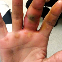
User login
Tools measuring oxygen desaturation produced disparate data
WASHINGTON – Oxygen desaturation index (ODI) scores showed significant variation across two software systems, a study showed.
The researchers assessed the ODI scores of 106 patients using the ResMed ApneaLink Plus system (AL) and the Compumedics Grael Profusion PSG3 system (Comp). “AL ODI values tended to be higher than Comp ODI values, but with significant variability,” they said.
AL showed a bias of an additional 4.4 events per hour (95% limits of agreement, –5.8 to 14.6 events per hour) for ODI scores at 4% desaturation and a bias of an additional 7.1 events per hour (95% limits of agreement, –6.4 to 20.6 events per hour) at 3% desaturation (J Clin Sleep Med. 2017;13[4]:599-605).
This may be problematic for physicians evaluating patients during sleep studies who rely on ODI scores at 3% and 4% desaturations to create accurate apnea severity assessments, the investigators said.
“[The] wide limits of agreement in our study highlight that clinicians cannot be confident that an ODI4% recorded in the AL is the same as that recorded in the Comp,” wrote Yvonne Ng, MBBS, of the department of lung and sleep medicine at Monash Health, Victoria, Australia, and her colleagues. “The differences are large enough to significantly affect diagnostic thresholds for OSA [obstructive sleep apnea] and, in particular, moderate-severe OSA.”
The researchers gathered data from patients undergoing sleep analysis at the Monash Medical Centre, who were, on average, 47 years of age, had a body mass index score of 32 kg/m2, and had an apnea hypopnea index (AHI) of 23.2.
ODI3% scores analyzed through Comp diagnosed 66 patients with OSA (ODI3% greater than or equal to 5 events per hour), while desaturation events analyzed through the AL system diagnosed 90 patients, a 36% increase over Comp (P = .0002).
When researchers tested for moderate to severe OSA (ODI3% greater than or equal to 15 events per hour), 32 patients were diagnosed using the Comp system, compared with 59 patients using the AL system.
Disparities in these measurements create uncertainty among clinicians, who rely on ODI measurements for scores that are accurate and can be easily replicated using an algorithm, the researchers said.
“The current work demonstrates that significantly more patients would receive a diagnosis of OSA, or more particularly, moderate-severe OSA, with the AL ODI, compared to the Comp ODI,” Dr. Ng and her colleagues wrote.
When sensitivity scores for Comp and AL were compared, AL ODI3% scores were significantly more sensitive than Comp, with sensitivity scores of 96% vs. 58%.
Using different fingers for measuring desaturation during the test or differences in algorithms used to assess ODI scores were possible sources of the disparities, the researchers noted.
Differences in internal processing between the two systems were the most likely causes of the discrepancies between the data collected using each system, they added.
Because there is no universal standard for ODI measurements, the researchers were unable to determine which system was more accurate.
Several of the researchers reported receiving financial support, research equipment, or consultancy fees from various entities.
[email protected]
On Twitter @eaztweets
Key clinical point:
Major finding: ODI tests analyzed using the ResMed APneaLink Plus system vs. Compumedics Grael Profusion PSG3 system reported ODI4% bias = 4.4 events per hour (95% limits of agreement, –5.8 to 14.6 events per hour) and ODI3% bias = 7.1 events per hour (95% limits of agreement, –6.4 to 20.6 events per hour).
Data source: ODI test results for 106 participants in a sleep study at Monash Medical Centre.
Disclosures: Several of the researchers reported receiving financial support, research equipment, or consultancy fees from various entities.
No-shows
Of all the headaches inherent in a private medical practice, few are more frustrating than patients who make appointments and then fail to keep them.
No-shows are a problem for all physicians but especially for dermatologists. In one study, the no-show rate in dermatology offices averaged 10% – almost double the average for medical offices as a whole.
Why the higher rate? One reason is a lag between appointment and visit. Many dermatologists are booked well in advance, so, by the time the appointment arrives, some patients’ complaints will have resolved spontaneously, while other patients will have found other offices willing to see them sooner. Another reason is lack of insurance coverage. Studies have shown that the no-show rate is highest when the patient is paying out-of-pocket for the visit.

Whatever the reasons, no-shows are an economic and medicolegal liability. It is worth the considerable effort it often takes to minimize them.
Deal with simple forgetfulness by calling your patients the day before to remind them of their appointments. Reasonably-priced phone software is available from a variety of vendors to automate this process. You could also hire a teenager to do it after school each day.
Whenever possible, use cell phone numbers for reminder calls. Patients often aren’t home during the day, and many don’t listen to their messages when they come in. Patients who have moved will often have a new home phone number, but their cell phone numbers will be the same.
Decrease the wait for new appointments. Keep some slots open each week for new patients, who will often “shop around” for a faster appointment while they’re waiting for an appointment they already have elsewhere.
If your no-shows are incorrigible, consider charging them. One increasingly popular mechanism is a fee ($20 seems to be popular) that must be paid at the time of the next appointment before being seen. Some patients will respond to that by never showing up again. Fine. You need to reserve your appointment slots for patients who plan to keep them. Those more contrite will pay and, hopefully, learn their lesson. Give your receptionists the power to override the charge since there are, obviously, legitimate reasons for missing an appointment.
One physician in my area told me he seldom actually collects the fee, which is okay with him. “After all,” he said, “the purpose is not to generate income. It’s to encourage patients to keep their appointments.”
If you go this route, be sure to post notices in your office and on your website clearly delineating your policy. Emphasize that it is not a service fee and cannot be billed to insurance. Remind patients about it during reminder calls. If you have a high no-show rate for cosmetic and other noninsurance procedures, consider collecting a nonrefundable deposit in advance.
Above all, seek to maximize the strength of your physician-patient relationships. Try not to shuttle patients between different physicians or PAs, and make it clear that you are genuinely concerned about their health. Impress upon them the crucial role they play in their own care, which includes keeping all their appointments.
In our office, significant no-shows (for example, a patient with a melanoma who misses the visit for re-excision) receive a phone call and a certified letter, and their records go into a special file for close follow-up by the nursing staff.
All missed appointments should be documented in the patient’s record – it’s important clinical and medicolegal information. A second missed appointment should prompt a written warning that measures will be taken if it happens again. Make sure to spell out what those measures are, and stick to them. Habitual no-shows should be dismissed from your practice. You cannot afford them.
Dr. Eastern practices dermatology and dermatologic surgery in Belleville, N.J. He is the author of numerous articles and textbook chapters and is a longtime monthly columnist for Dermatology News. Write to him at [email protected].
Of all the headaches inherent in a private medical practice, few are more frustrating than patients who make appointments and then fail to keep them.
No-shows are a problem for all physicians but especially for dermatologists. In one study, the no-show rate in dermatology offices averaged 10% – almost double the average for medical offices as a whole.
Why the higher rate? One reason is a lag between appointment and visit. Many dermatologists are booked well in advance, so, by the time the appointment arrives, some patients’ complaints will have resolved spontaneously, while other patients will have found other offices willing to see them sooner. Another reason is lack of insurance coverage. Studies have shown that the no-show rate is highest when the patient is paying out-of-pocket for the visit.
Whatever the reasons, no-shows are an economic and medicolegal liability. It is worth the considerable effort it often takes to minimize them.
Deal with simple forgetfulness by calling your patients the day before to remind them of their appointments. Reasonably-priced phone software is available from a variety of vendors to automate this process. You could also hire a teenager to do it after school each day.
Whenever possible, use cell phone numbers for reminder calls. Patients often aren’t home during the day, and many don’t listen to their messages when they come in. Patients who have moved will often have a new home phone number, but their cell phone numbers will be the same.
Decrease the wait for new appointments. Keep some slots open each week for new patients, who will often “shop around” for a faster appointment while they’re waiting for an appointment they already have elsewhere.
If your no-shows are incorrigible, consider charging them. One increasingly popular mechanism is a fee ($20 seems to be popular) that must be paid at the time of the next appointment before being seen. Some patients will respond to that by never showing up again. Fine. You need to reserve your appointment slots for patients who plan to keep them. Those more contrite will pay and, hopefully, learn their lesson. Give your receptionists the power to override the charge since there are, obviously, legitimate reasons for missing an appointment.
One physician in my area told me he seldom actually collects the fee, which is okay with him. “After all,” he said, “the purpose is not to generate income. It’s to encourage patients to keep their appointments.”
If you go this route, be sure to post notices in your office and on your website clearly delineating your policy. Emphasize that it is not a service fee and cannot be billed to insurance. Remind patients about it during reminder calls. If you have a high no-show rate for cosmetic and other noninsurance procedures, consider collecting a nonrefundable deposit in advance.
Above all, seek to maximize the strength of your physician-patient relationships. Try not to shuttle patients between different physicians or PAs, and make it clear that you are genuinely concerned about their health. Impress upon them the crucial role they play in their own care, which includes keeping all their appointments.
In our office, significant no-shows (for example, a patient with a melanoma who misses the visit for re-excision) receive a phone call and a certified letter, and their records go into a special file for close follow-up by the nursing staff.
All missed appointments should be documented in the patient’s record – it’s important clinical and medicolegal information. A second missed appointment should prompt a written warning that measures will be taken if it happens again. Make sure to spell out what those measures are, and stick to them. Habitual no-shows should be dismissed from your practice. You cannot afford them.
Dr. Eastern practices dermatology and dermatologic surgery in Belleville, N.J. He is the author of numerous articles and textbook chapters and is a longtime monthly columnist for Dermatology News. Write to him at [email protected].

USPSTF discourages postmenopausal hormone therapy for prevention
Hormone therapy, in the form of estrogen combined with progestin, is not recommended to prevent chronic conditions such as heart disease and diabetes in postmenopausal women, according to updated draft recommendations from the U.S. Preventive Services Task Force. They also recommended against the use of estrogen alone in postmenopausal women who have had a hysterectomy.
The updated recommendations were published online May 16 on the U.S. Preventive Services Task Force website.
After considering new evidence in the last several years, the recommendations are essentially unchanged from the final recommendations published in 2012, according to a Task Force statement published with the recommendations. “The benefits of using menopausal hormone therapy to prevent chronic conditions like heart disease and diabetes do not outweigh the harms in women who have gone through menopause,” Maureen G. Phipps, MD, MPH, a task force member, said in the statement.
The draft recommendations were based on a review of 17 randomized clinical trials published through Aug. 1, 2016, that included data from the Women’s Health Initiative.
Women taking estrogen/progestin reported a significantly lower risk (per 10,000 women approximately 5 years) of colorectal cancer, diabetes, and fractures, compared with women on a placebo, wrote Gerald Gartlehner, MD, and his colleagues at the RTI International–University of North Carolina Evidence-Based Practice Center in Research Triangle Park, NC, in the evidence report accompanying the draft recommendations.
However, the risks for several other conditions were significantly higher among women on hormone therapy, compared with placebo, including invasive breast cancer (52 more cases), coronary heart disease (41 more cases) probable dementia (88 more cases), gallbladder disease (259 more cases), stroke (53 more cases), and venous thromboembolism (120 more cases). Additionally, urinary incontinence rates were higher after a 1-year follow up among women on hormone therapy (876 more cases/10,000 women).
Some evidence suggests that women who began hormone therapy closer to menopause might have a lower risk for developing cardiovascular complications, but the evidence is insufficient for firm conclusions, the researchers wrote.
The recommendations against hormone therapy do not apply to women younger than 50 years who have undergone oophorectomies or premature menopause, or to those considering hormone therapy to manage menopausal symptoms, according to the Task Force.
Public comments on the draft recommendations may be submitted on the Task Force website until June 12. The researchers had no financial conflicts to disclose.
View the recommendations online at uspreventiveservicestaskforce.org.
Despite CVD risk, few internists screen for prior preeclampsia
SAN DIEGO – Women who have preeclampsia are at increased risk for later cardiovascular disease, yet internists performing well-woman exams were unlikely to have asked their patients about a history of preeclampsia, a small study showed.
Just 21 of 89 women were asked about preeclampsia during a well-woman exam, while 88 of 89 were asked about diabetes or smoking history, and all 89 were asked about hypertension (P = .0002 for comparing preeclampsia to each individual comorbidity).
“There is a screening gap leading to missed opportunities to identify women at risk for cardiovascular disease,” Irene Lewnard, MD, said at the annual meeting of the American College of Obstetricians and Gynecologists.
Dr. Lewnard and her colleagues at the Medical College of Wisconsin, Milwaukee, used a retrospective chart review to see whether internal medicine physicians were asking about preeclampsia as well as traditional CVD risk factors during well-woman exams.
The researchers looked at records from 89 women, aged 18-48 years, who had at least one prior delivery to see whether they were asked about preeclampsia. The review also assessed whether physicians had asked about traditional CVD risk factors: smoking, diabetes, and hypertension.
Of the 89 patients, 6 had a confirmed prior history of preeclampsia. The demographic characteristics and obstetric histories of these patients were not significantly different from those of the larger group. The mean patient age was about 35 years, and the average gravidity was three and parity was two.
Dr. Lewnard, an ob.gyn., and her colleagues looked at charts beginning Jan. 1, 2013, and ending May 31, 2016, after both the American Heart Association (AHA) and the American College of Obstetricians and Gynecologists (ACOG) had issued guidelines that recognized the elevated CVD risk for women with a history of preeclampsia.
In 2011, the AHA issued guidelines that preeclampsia should be listed along with gestational diabetes and gestational hypertension as risk factors for CVD. The AHA called for ob.gyns. to refer patients with these conditions to primary care physicians or cardiologists for follow-up, and recommended that providers include questions about pregnancy-related CVD risk factors when taking a history.
In 2013, ACOG recommended early screening for heart disease for women with a history of preterm or recurrent preeclampsia, to include a consideration for early assessment of blood pressure, body mass index, serum lipids, and fasting blood glucose. The group also recommended counseling on modifiable lifestyle factors for these patients.
Data from several large studies support preeclampsia’s status as an independent risk factor for CVD. A 2001 Norwegian study of more than 600,000 births found that, for women who had preeclampsia and were delivered at term, the relative risk for death from cardiovascular disease was 1.65. However, when women with preeclampsia gave birth before 37 weeks’ gestation, the relative risk for later death from CVD rose to 8.12 (BMJ. 2001;323[7323]:1213-7).
A 2007 systematic review and meta-analysis examined data from 3,488,160 women and found a relative risk of 2.16 for ischemic heart disease after an average 11.7 years of follow-up (BMJ 2007;335:974). Finally, a smaller 2010 California study of 14,403 women found a hazard ratio of 2.14 for CVD-related deaths for all women with a history of preeclampsia. For women whose preeclampsia began before 34 weeks’ gestation, that hazard ratio rose to 9.54 (Hypertension. 2010;56:166-71).
When Dr. Lewnard and her colleagues spoke with the internists who had participated in their study, several raised the point that there are not clear guidelines about how to incorporate a history of preeclampsia into risk calculators or treatment recommendations. This knowledge gap, she said, should be addressed, with an ultimate goal of establishing an interdisciplinary set of guidelines for counseling and management of women with prior preeclampsia.
The investigators are assessing whether adding prompts to the electronic medical record could increase the number of primary care physicians who include preeclampsia questions in their history taking.
Dr. Lewnard and her colleagues reported having no outside sources of funding and no conflicts of interest.
[email protected]
On Twitter @karioakes
AT ACOG 2017
Key clinical point:
Major finding: Of 89 women who received well-woman exams, 21 were asked about prior preeclampsia, while 88 were asked about diabetes and smoking, and 89, about hypertension (P = .0002).
Data source: A retrospective record review of 89 women receiving well-woman exams in the year after the American College of Obstetricians and Gynecologists issued CVD screening guidelines for prior preeclampsia.
Disclosures: The study authors reported having no outside sources of funding and no conflicts of interest.
Oral iron of no benefit in heart failure with iron deficiency
High-dose oral iron therapy doesn’t improve exercise capacity in the estimated 50% of patients with symptomatic heart failure who also have iron deficiency, according to a report published online May 16 in JAMA.
Iron deficiency in patients with HF, regardless of their hemoglobin status, is associated with reduced functional capacity, poorer quality of life, and increased mortality. Iron plays a crucial role in the delivery and utilization of oxygen, and “cells with high-energy demands, including skeletal and cardiac myocytes, are particularly sensitive to depleted iron stores,” said Gregory D. Lewis, MD, of the pulmonary critical care unit of Massachusetts General Hospital, Boston, and his associates.
The IRONOUT study was conducted at 23 U.S. medical centers, where outcomes after 16 weeks of oral iron therapy (150 mg twice daily) were compared against matching placebo in 225 patients. The median patient age was 63 years, and the median duration of HF was 5.7 years. Ischemic heart disease was the primary cause of HF in 78% of the study participants.
These patients had low LVEF and poor exercise capacity, despite having high rates of guideline-directed treatment with medications.
The primary endpoint was a change in peak oxygen uptake (peak VO2) at the conclusion of treatment, a measure that “reflects the multiple mechanisms by which iron repletion is expected to improve systemic oxygen delivery and utilization.” Change in peak VO2 was not significantly different between the 111 participants who took oral iron supplements (+23 mL/min) and the 114 who took placebo (–2 mL/min), the investigators wrote (JAMA Pediatr. 2017 May 16. doi: 10.1001/jama.2017.5427).
In subgroup analyses, oral iron also failed to improve peak VO2 in any subgroup of patients: neither men nor women; neither those with decreased hemoglobin nor those with normal hemoglobin levels; nor patients with or without venous congestion at baseline. Oral iron also failed to improve secondary endpoints including 6-minute walk distance, quality of life scores, NT-proBNP levels, and ventilatory efficiency.
In contrast to previous studies of IV iron repletion, oral iron supplementation “produced minimal improvement in iron stores, implicating the route of administration rather than the strategy of iron repletion in the lack of clinical benefit,” Dr. Lewis and his associates said.
This study was funded by the National Heart, Lung, and Blood Institute, which also conceived, designed, and conducted the trial. Dr. Lewis reported ties to Abbott, Novartis, Shape Systems, Stealth Bio Therapeutics, Ironwood, Cheetah Medical, Luitpold, and SoniVie. His associates reported ties to numerous industry sources.
Key clinical point: High-dose oral iron therapy doesn’t improve exercise capacity in the estimated 50% of patients with symptomatic heart failure and iron deficiency.
Major finding: Change in peak VO2 was not significantly different between the 111 participants who took oral iron supplements (+23 mL/min) and the 114 who took placebo (–2 mL/min).
Data source: A multicenter, randomized, double-blind, placebo-controlled phase II trial involving 225 patients treated for 16 weeks.
Disclosures: This study was funded by the National Heart, Lung, and Blood Institute (NCT02188784), which also conceived, designed, and conducted the trial. Dr. Lewis reported ties to Abbott, Novartis, Shape Systems, Stealth Bio Therapeutics, Ironwood, Cheetah Medical, Luitpold, and SoniVie. His associates reported ties to numerous industry sources.
Can prenatal choline lead to prevention of Alzheimer’s?
As psychiatrists, we are the advocates for inserting the biological thread into the tapestry of understanding human behavior. Try as they may, other mental health professionals are not biologists at heart. Accordingly, psychiatrists bring important thoughtfulness to any consideration about mental health and wellness and about the treatment and prevention of problematic thoughts, feelings, and behaviors.
Throughout my career, my main focus has been on identifying strategies and treatments that can prevent mental illness. For example, I wrote a column about prevention for Clinical Psychiatry News from 2004 to 2011, and, as a member of the publication’s Editorial Advisory Board, I continue to try to steer our attention to biological aspects of prevention.

Recently, I have been seeing psychiatric articles on fetal health and mental health, and, because I am excited about the prospect of understanding fetal alcohol exposure, I feel the need to share. A recent article in the American Journal of Psychiatry was provocatively entitled, “Fetal origins of mental health: The developmental origins of health and disease hypothesis (2016. doi: 10.1176/appi.2016.16020138).
Disappointedly, the authors overlooked the biology of fetal alcohol exposure and focused on how psychosocial issues of maternal anxiety, depression, and anxiety could influence neurodevelopment, which could affect mental health outcomes after birth. Of course, I thought, “What about fetal alcohol exposure?” Meanwhile, a commentary in JAMA Psychiatry entitled “Prenatal nutritional deficiency and psychosis: Where do we go from here?” referred to prenatal choline supplementation along with other supplements (2017;74(4):349-50).
When I first stumbled upon the high prevalence of fetal alcohol exposure in low-income African American populations, it occurred to me that, since choline was involved with the psychopathology of fetal alcohol spectrum disorders and acetylcholine seemed to be involved in the psychopathology of Alzheimer’s disease, there might be a relationship between the two (Psychiatric Serv. 2015 May 1. doi: 10.1176/appi.ps.201400162). Such possible links are especially intriguing in light of the Alzheimer’s Association suggestion that Alzheimer’s disease is a “silent epidemic” among African Americans. The association notes that the prevalence among African Americans ranges from 14% to 100% higher than among whites. The problem – how to make the connection, if there were one, between the adults I was seeing and fetal alcohol exposure – proved difficult, because the time between fetal health and adult mental illness was huge. The time from fetal health and geriatric Alzheimer’s disease was even greater.
However, modern biologic science came through again. Maternal choline supplementation has been touted as a potential prenatal treatment for Down syndrome and Alzheimer’s disease (Curr Alzheimer Res. 2016;13[1]:97-106). Using mice that are genetically altered to show the development of Down syndrome and Alzheimer’s disease changes in the brain at 6 months, allowing researchers to seek prevention strategies for this pathophysiology, researchers have found that maternal choline supplementation protects against basal forebrain cholinergic neuron degeneration seen in these animals.
Thus, it would seem the problem of choline deficiency in pregnancy, most exacerbated by fetal alcohol exposure, is preventable by increasing the amount of choline available during pregnancy. So, it makes sense to increase the amount of choline in prenatal vitamins, as it appears that this biotechnical intervention not only would reduce the scourge of fetal alcohol spectrum disorders but also of Alzheimer’s disease (J Fam Med Dis Prev. 2016 Nov 29;2[6]:1-3).
Finally, the Office of Juvenile Justice and Delinquency Prevention has finally released a paper – “Fetal alcohol spectrum disorders listening session report” – from a session held in June 2013 that documents the extent of the problem in juvenile justice facilities.
Unfortunately, many of us have abdicated our role as biologists. We’ve got evidence showing the power of prenatal choline. It is time to stop counting all of the problems that stem from deficiency of choline during pregnancy and start doing something about it.
Dr. Bell is a staff psychiatrist at Jackson Park Hospital Family Medicine Clinic in Chicago, clinical psychiatrist emeritus in the department of psychiatry at the University of Illinois at Chicago, former president/CEO of Community Mental Health Council, and former director of the Institute for Juvenile Research (birthplace of child psychiatry), also in Chicago.
As psychiatrists, we are the advocates for inserting the biological thread into the tapestry of understanding human behavior. Try as they may, other mental health professionals are not biologists at heart. Accordingly, psychiatrists bring important thoughtfulness to any consideration about mental health and wellness and about the treatment and prevention of problematic thoughts, feelings, and behaviors.
Throughout my career, my main focus has been on identifying strategies and treatments that can prevent mental illness. For example, I wrote a column about prevention for Clinical Psychiatry News from 2004 to 2011, and, as a member of the publication’s Editorial Advisory Board, I continue to try to steer our attention to biological aspects of prevention.
Recently, I have been seeing psychiatric articles on fetal health and mental health, and, because I am excited about the prospect of understanding fetal alcohol exposure, I feel the need to share. A recent article in the American Journal of Psychiatry was provocatively entitled, “Fetal origins of mental health: The developmental origins of health and disease hypothesis (2016. doi: 10.1176/appi.2016.16020138).
Disappointedly, the authors overlooked the biology of fetal alcohol exposure and focused on how psychosocial issues of maternal anxiety, depression, and anxiety could influence neurodevelopment, which could affect mental health outcomes after birth. Of course, I thought, “What about fetal alcohol exposure?” Meanwhile, a commentary in JAMA Psychiatry entitled “Prenatal nutritional deficiency and psychosis: Where do we go from here?” referred to prenatal choline supplementation along with other supplements (2017;74(4):349-50).
When I first stumbled upon the high prevalence of fetal alcohol exposure in low-income African American populations, it occurred to me that, since choline was involved with the psychopathology of fetal alcohol spectrum disorders and acetylcholine seemed to be involved in the psychopathology of Alzheimer’s disease, there might be a relationship between the two (Psychiatric Serv. 2015 May 1. doi: 10.1176/appi.ps.201400162). Such possible links are especially intriguing in light of the Alzheimer’s Association suggestion that Alzheimer’s disease is a “silent epidemic” among African Americans. The association notes that the prevalence among African Americans ranges from 14% to 100% higher than among whites. The problem – how to make the connection, if there were one, between the adults I was seeing and fetal alcohol exposure – proved difficult, because the time between fetal health and adult mental illness was huge. The time from fetal health and geriatric Alzheimer’s disease was even greater.
However, modern biologic science came through again. Maternal choline supplementation has been touted as a potential prenatal treatment for Down syndrome and Alzheimer’s disease (Curr Alzheimer Res. 2016;13[1]:97-106). Using mice that are genetically altered to show the development of Down syndrome and Alzheimer’s disease changes in the brain at 6 months, allowing researchers to seek prevention strategies for this pathophysiology, researchers have found that maternal choline supplementation protects against basal forebrain cholinergic neuron degeneration seen in these animals.
Thus, it would seem the problem of choline deficiency in pregnancy, most exacerbated by fetal alcohol exposure, is preventable by increasing the amount of choline available during pregnancy. So, it makes sense to increase the amount of choline in prenatal vitamins, as it appears that this biotechnical intervention not only would reduce the scourge of fetal alcohol spectrum disorders but also of Alzheimer’s disease (J Fam Med Dis Prev. 2016 Nov 29;2[6]:1-3).
Finally, the Office of Juvenile Justice and Delinquency Prevention has finally released a paper – “Fetal alcohol spectrum disorders listening session report” – from a session held in June 2013 that documents the extent of the problem in juvenile justice facilities.
Unfortunately, many of us have abdicated our role as biologists. We’ve got evidence showing the power of prenatal choline. It is time to stop counting all of the problems that stem from deficiency of choline during pregnancy and start doing something about it.
Dr. Bell is a staff psychiatrist at Jackson Park Hospital Family Medicine Clinic in Chicago, clinical psychiatrist emeritus in the department of psychiatry at the University of Illinois at Chicago, former president/CEO of Community Mental Health Council, and former director of the Institute for Juvenile Research (birthplace of child psychiatry), also in Chicago.
Blood test could aid steroid decision in alcoholic hepatitis
AMSTERDAM – Determining the ratio of neutrophils to leukocytes in the blood could help identify patients with alcoholic hepatitis that would and would not benefit from steroid treatment.
Patients who had a neutrophil to lymphocyte ratio (NLR) of between 5 and 8 before being treated with the corticosteroid prednisolone appeared to obtain a benefit versus no-steroid treatment (P = .007) while those with higher and lower NLR values did not, in an analysis presented at the International Liver Congress.
This could potentially help clinicians avoid putting some patients through a futile trial of steroid therapy, study author Ewan H. Forrest, MD, explained in an interview at the meeting, which is sponsored by the European Association for the Study of the Liver (EASL).
“The traditional approach would be to give steroids to patients with severe alcoholic hepatitis, wait 7 days, see if they are getting better, and if so, keep them on the steroids,” Dr. Forrest of the liver unit at Glasgow Royal Infirmary observed. Conversely, if patients are not doing better then steroids should be stopped.
“What we are increasingly aware of is that not only do some people not do well with steroids but also they actually do considerably more badly,” Dr. Forrest cautioned.
Usually, the response to steroid treatment in alcoholic hepatitis is measured by changes in serum bilirubin after a week of treatment, but this, of course, exposes patients to a “futile course of treatment with a risk of complication such as sepsis,” Dr. Forrest and his coauthors noted in a a late-breaking poster.
Determining the NLR has already been shown to help predict the prognosis of patients with several diseases with an underlying inflammatory component, such as cardiovascular diseases and several types of cancer. It also has proven useful in patients with liver disease, although not specifically in alcoholic hepatitis before this study, Dr. Forrest observed.
Data on patients with alcoholic hepatitis who had participated in the multicenter, double-blind, randomized STOPAH trial were used to see if the baseline NLR could help stratify patients who would benefit from steroid therapy.
STOPAH had compared the use of prednisolone or pentoxifylline for the treatment of alcoholic hepatitis but found no benefit for the latter, although there was a possible benefit of steroids for improving overall survival, at least in the short term (N Engl J Med. 2015 Apr;372:1619-28).
Dr. Forrest noted that measurement of the lymphocyte count was not part of the original study design, so data to calculate the NLR were obtained retrospectively. As there had been little or no response to pentoxifylline in the trial, patients who had taken this drug were regarded as having had no treatment in the analysis.
In all, baseline NLR values could be worked out for 630 patients from the STOPAH trial, but 113 were excluded from further analysis as they met the prespecified exclusion criteria of gastrointestinal bleeding or sepsis.
Overall, a NLR of 5 or less, indicating milder liver disease, was associated with significantly better survival at 3 months than if the NLR was 5 or more (85.5% vs. 67.3%; P less than .0001), study findings suggested.
Dr. Forest noted that 29% of patients fell into the “sweet spot” of the NLR of between 5 and 8, where patients did benefit from steroids, but that the 23% of study subjects who had an NLR ratio above 8 did not. These patients may have had disease too severe to benefit from the prednisolone, he suggested, and tended to have a worse prognosis regardless. A baseline NLR greater than 8 was associated with acute kidney infection but not sepsis, the team found.
There was also no great effect of the steroid in the 48% of patients who had an NLR ratio less than 5, suggesting that maybe they had disease that was too mild to warrant such treatment and did well regardless.
Of course, these findings still need further validation, but they are “not far off” from clinical application, Dr. Forrest offered. Calculating the ratio is simple, can be done during a routine whole-blood cell count, and is potentially cost saving because it reduces the standard practice in the United Kingdom of giving “all-comers” 7 days of corticosteroid therapy as a trial to see if they get better, he said.
Dr. Forrest had no conflicts of interest to disclose.
Key clinical point: Calculating the neutrophil to leukocyte ratio (NLR) could save some patients with alcoholic hepatitis from having steroid therapy.
Major finding: 90-day survival was 85.5% vs. 67.3%, (P less than .0001), comparing an NLR less than 5 with an NLR of 5 or greater in all patients.
Data source: 513 patients with alcoholic hepatitis who had participated in the multicenter, double-blind, randomized STOPAH trial.
Disclosures: Dr. Forrest had no conflicts of interest to disclose.
SMILE, Your Myopia Has Just Been Treated Faster!
Fort Belvoir Community Hospital surgeons have performed the first small-incision lenticule extraction (SMILE) in the DoD, according to Health.mil News. The procedure to reduce or eliminate nearsightedness has been performed since 2011; the FDA recently approved it for the U.S.
The very fast and short-pulsed (femtosecond) laser creates a thin disc within the cornea, which is removed through a cut on the corneal surface. Removing the tissue changes the shape of the cornea.
The procedure takes 15 to 20 minutes with the laser activated for about 90 seconds per eye. Both eyes can be treated in the same session. With SMILE, unlike LASIK, no tissue is vaporized, meaning wound recovery time is faster, and with no corneal flap created, there is no risk of flap dislocation.
A clinical study found complications were rare, and by month 12 postsurgery there were only 4 reports of moderate or severe glare and 1 of moderate or severe halos. The most commonly reported effects were starbursts, blurred vision, and difficulty judging distance or depth perception, but at 12 months, more patients reported improvement than worsening. At the 6-month follow-up, 287 of 328 patients were seeing 20/20 or better without glasses.
“We are thrilled to extend this treatment option to active duty service members under the Warfighter Refractive Eye Surgery Program,” said U.S. Army Lt. Col. Bruce Rivers, director of the program at Belvoir Hospital.
The surgery will make a difference for patients. One of the first to have it done, Navy Petty Officer 1st Class Christopher Mahmood, a submarine mechanic, said, “On a submarine we have to be able to put our breathing equipment on in approximately 30 seconds, in case of emergency. Glasses make this difficult. Getting this surgery means I have one less thing to worry about while deployed and can focus 100 percent on the mission.”
The SMILE procedure will be available at Belvoir Hospital, San Diego Naval Medical Center, and Wilford Hall in San Antonio.
5 Points on Pyogenic Flexor Tenosynovitis of the Hand
Pyogenic flexor tenosynovitis (PFT) is a common closed space infection of the flexor tendon sheaths of the hand and remains one of the most challenging problems encountered in orthopedic and hand surgery (Figure 1). PFT also is known as septic flexor tenosynovitis and suppurative flexor tenosynovitis.
Kanavel1 initially described 4 cardinal signs that characterize infection of the flexor tendon sheath: symmetric fusiform swelling of the entire digit, exquisite tenderness to palpation along the course of the tendon sheath, semiflexed posture at rest, and pain with attempted passive extension of the digit. The prevalence of this infection ranges from 2.5% to 9.4%.2 Once the infection is established in a patient, it can cause significant morbidity and disability and produce an economic burden. It can also present a significant treatment dilemma for the treating surgeon, as there is no standardized protocol for managing this common but challenging hand infection. For treatment, many surgeons combine surgical decompression, sheath irrigation, and empiric intravenous (IV) antibiotic administration. However, despite prompt treatment, and regardless of the protocol used, complication rates as high as 38% have been reported.3 Moreover, even after infection eradication, a significant proportion of patients continue to have pain, swelling, stiffness, loss of composite flexion, weakness, and recurrence that potentially requires amputation.
1. What Causes Pyogenic Flexor Tenosynovitis?
PFT can result from hematogenous spread, but local inoculation by a laceration, a puncture, or a bite also is common4-7 (Figure 1). As a consequence of these mechanisms of injury, the most common source of PFT is skin flora. Staphylococcus aureus has been found in up to 75% of positive cultures in several studies.2,5,6,8,9 Methicillin-resistant S aureus (MRSA) has been found in up to 29% of cases, and the incidence continues to increase, particularly in urban areas.2,9-12 Other common bacteria are Staphylococcus epidermidis, β-hemolytic Streptococcus species, and Pseudomonas aeruginosa.5,6,10 Infection by more than 1 species of bacteria is also fairly prevalent. Of 62 patients in a study, 38% had infections with 1 organism, and 62% with 2 or more.6 Twenty-six percent of cultures grew mixed anaerobic and aerobic organisms.6 PFT is seldom caused by Eikenella corrodens from a human bite or Pasteurella multocida from an animal bite.10 Other rare causes of PFT are Listeria monocytogenes13 and Clostridium difficile from a gastrointestinal source.14Neisseria gonorrhea can cause acute tenosynovitis, usually in the setting of disseminated gonococcal infection.15,16 Also reported is mycobacterial tenosynovitis, most commonly caused by Mycobacterium kansasii and Mycobacterium marinum.17
2. Which Antibiotics Are Best Suited to Empirical Management of PFT?
Management of PFT, regardless of the pathogen, includes prompt administration of empiric IV antibiotics, usually followed by surgical drainage.7,18-20 While cultures are being tested, antibiotics should be selected—including antibiotics for empiric coverage against common gram-positive organisms, including Staphylococcus and Streptococcus species.12 The Centers for Disease Control and Prevention recommends empiric coverage for MRSA if the local prevalence exceeds 10% to 15%. Recommended empiric antibiotics are trimethoprim-sulfamethoxazole (TMP-SMX) and clindamycin (both oral) and clindamycin, vancomycin, and daptomycin (all IV).
In addition, institutional and local antibiotic resistance patterns of bacteria should guide treatment and antibiotic selection. First-generation cephalosporins have long been the cornerstone of treatment for infections caused by S aureus, but increasing methicillin resistance has reduced their role in the treatment, particularly the empiric treatment, of MRSA infections. Methicillin resistance first appeared as nosocomial S aureus infections in 1961, only 1 year after the introduction of the semisynthetic penicillin class that includes methicillin. Over the past 2 decades, MRSA has emerged in the community in otherwise young and healthy individuals with no healthcare-associated risk factors. Fortunately, several readily available antibiotics have maintained their efficacy in managing these “community-acquired” MRSA hand infections. TMP-SMX provides adequate coverage for MRSA and is a relatively inexpensive medication, and clindamycin is an equally effective and cost-effective alternative.
Presumptive antibiotics should also cover gram-negative rods and anaerobes, including Clostridium species, especially in immunocompromised patients.7,9 These patients may require additional antibiotics for presumptive coverage of other rarer bacterial causes, especially when unique mechanisms of injury (eg, aquatic injury, farm injury) are involved. Once culture results are ready, antibiotic regimens should be narrowed to cover the specific organisms identified.
3. What Are the Timing and Indications for Surgery?
Nonoperative treatment may be appropriate for PFT patients who present early, typically within 48 hours after penetrating trauma to the hand.21 In a 4-patient series, Neviaser and Gunther19 successfully treated PFT nonoperatively, with IV antibiotics, splinting, and elevation. During nonoperative treatment, the affected hand should be regularly examined. If this treatment is to be successful, clinical symptoms should improve within 48 hours; if they do not, surgical irrigation and débridement should be performed.
Regardless of timing and type of irrigation, surgical treatment remains the treatment of choice for the majority of PFT cases. Michon22 developed a 3-tier PFT classification system that is based on intraoperative findings (Table). ![]()
4. What Are the Surgical Techniques for PFT Drainage?
Several surgical methods have been developed to decompress and irrigate the flexor sheaths of the hand. However, debates about optimal timing of surgical intervention, surgery type (open surgery or closed catheter irrigation only), and irrigation method continue.
Open Irrigation and Débridement
Open irrigation and débridement procedures were originally described for surgical management of PFT.1 Midaxial and palmar (Bruner zigzag) incisions can be used to expose and open the entire sheath for complete drainage and washout. Both incisions afford good access to the flexor sheath, but the midaxial approach may provide more coverage of the sheath after surgery. Open irrigation and débridement is the treatment of choice for the most advanced cases of PFT and for atypical or chronic tenosynovial infections.4,23,24 The Bruner zigzag incision affords ease of surgical dissection, extension, and more exposure of the flexor tendon sheath at the expense of possible difficulty in closure or flap necrosis in the setting of a swollen digit. Alternatively, the midaxial incision has the advantage of a large, more robust skin flap for more reliable closure.
Closed Tendon Sheath Irrigation
In 1943, Dickson-Wright25 first described catheter irrigation of tendon sheath infections. Later, Neviaser4 described this technique in detail. A proximal incision is made over the metacarpal neck. The tendon sheath is cut transversely at the proximal edge of the A1 pulley. An angiocatheter is inserted 1 cm to 2 cm antegrade into the flexor tendon sheath. Then, a distal midaxial incision is made dorsal to the neurovascular bundle at the level of the distal interphalangeal joint on the ulnar aspect of the finger or the radial aspect of the thumb. The distal edge of the flexor sheath is exposed and resected distal to the distal-most pulley. A Penrose drain can be threaded into the tendon sheath beneath the A4 pulley to keep the wound open and allow for fluid drainage. The sheath is flushed gently in the operating room. After surgery, intermittent bedside irrigation can be continued on the floor.
Neviaser4 reported excellent initial results with this technique; 18 of 20 patients regained complete active and passive range of motion (ROM) by 1 week after surgery. Similarly, Juliano and Eglseder,26 using a similar method, reported 100% excellent results for mild PFT and 88.4% excellent results for more severe infection.
Gutowski and colleagues23 reviewed 47 PFT cases to determine if there is a difference in outcomes between PFT treated with open irrigation and débridement and PFT treated with closed catheter irrigation. Between these groups, they found no significant differences in early postoperative outcomes, including resolution of infection, need for additional surgery, and hospital length of stay.
There are also many differing opinions regarding the best irrigation method. Some authors have asserted that normal saline is sufficient,4,5,23 and others that local antibiotics provide added benefit.27-29 Recently, Draeger and colleagues30 reported promising results with local injection of antibiotics into the tendon sheath and the addition of locally administered corticosteroids in the treatment of PFT in an animal model.
Continuous Closed Irrigation
A continuous closed irrigation system with inlet and outlet tubes has yielded successful results.8,31,32 This system consists of 2 fenestrated tubes placed within the infected space, with the tip of the smaller caliber inlet tube positioned just inside the larger outlet tube. Advantages of this system include the patient’s ability to participate in hand therapy with the system in place and avoidance of pain caused by the high pressures involved in intermittent closed irrigation. Duration of this system has ranged from 2 days to 3 weeks, and results have been good.5,8
Postoperative Irrigation
Use of postoperative irrigation on the floor or at home is controversial, as leaving an indwelling catheter in the tendon sheath can lead to complications. Catheters may increase digital stiffness by decreasing the patient’s ability to participate in therapy or may cause additional injury and irritation to the sheath itself if left in place too long. Lille and colleagues6 retrospectively compared the results of intraoperative closed tendon sheath irrigation alone with those of intraoperative and postoperative closed tendon sheath irrigation. There were no significant differences in mean hospital length of stay, follow-up complication rates, or postoperative ROM—which suggests that postoperative intermittent or continuous irrigation is not necessary.
Our Preferred Technique
We recommend a palmar approach that begins with outlining a Bruner zigzag incision along the entire finger. Then, only the distal-most and proximal-most incision lines are opened, thereby exposing the A5 and A1 pulleys, respectively (Figure 2). 
5. What Are the Long-Term Outcomes of PFT?
The principal complication associated with PFT is stiffness with loss of ROM, which can be caused by flexor tendon adhesions, joint capsular thickening, or destruction of the sheath and pulley system.24 In several studies, up to one-fourth of patients with PFT did not obtain full ROM, despite adequate treatment.4-6,27 Therefore, full active ROM exercises should be initiated immediately after surgery to counteract the development of stiffness.
The most severe complication of PFT is amputation of the affected digit (Figures 3A, 3B).
Pang and colleagues2 identified 5 factors associated with increased risk of amputation in patients with PFT: (1) age >43 years; (2) diabetes mellitus, peripheral vascular disease, or renal failure; (3) subcutaneous purulence; (4) signs of digital ischemia at presentation; and (5) growth of more than 1 bacteria species on culture of specimens obtained at time of surgery.
Pang and colleagues2 classified these patients into 3 groups with distinct clinical features and reported each group’s outcomes. The authors based their PFT classification system on increasingly severe clinical presentation, which potentially predicts amputation risk. Patients in stage 1 presented with Kanavel signs of tenosynovitis but no evidence of subcutaneous purulence or ischemia; patients in stage 2 had concurrent localized subcutaneous purulence but no ischemia; and patients in stage 3 had concurrent extensive subcutaneous purulence involving more than 1 phalangeal segment or spreading circumferentially as well as signs of ischemia. These PFT stages were found to correlate with worse patient outcomes. In patients with stage 1 infection, amputation was not required, and average functional return was 80% of total active ROM of the affected digit. In patients with stage 2 infection, the amputation rate was 8%, and return of total active ROM in the remaining digits was 72%. The outcomes for the patients with stage 3 infection were the worst. The amputation rate for patients with all 3 classification criteria (Kanavel signs, subcutaneous purulence, digital ischemia) was 59%, and return of total active ROM in the remaining digits was only 49%. Use of this clinical classification system makes it possible to guide treatment and predict outcome and return to function.
Conclusion
PFT is a common hand infection that can cause significant morbidity. Early treatment is crucial: this requires use of IV antibiotics, or surgical irrigation and débridement in more advanced cases. However, despite prompt and thorough treatment, severe infection can lead to long-term impaired function and even amputation of the affected digit. More research is needed to determine optimal timing and technique for surgical intervention and to elucidate the role of local antibiotics and corticosteroids in treating this infection and potentially preventing the morbid outcomes we currently see.
Am J Orthop. 2017;46(3):E207-E212. Copyright Frontline Medical Communications Inc. 2017. All rights reserved.
1. Kanavel AB. The symptoms, signs, and diagnosis of tenosynovitis and major fascial-space abscesses. In: Kanavel AB, ed. Infections of the Hand. 6th ed. Philadelphia, PA: Lea & Febiger; 1933:364-395.
2. Pang HN, Teoh LC, Yam AK, Lee JY, Puhaindran ME, Tan AB. Factors affecting the prognosis of pyogenic flexor tenosynovitis. J Bone Joint Surg Am. 2007;89(8):1742-1748.
3. Stern PJ, Staneck JL, McDonough JJ, Neale HW, Tyler G. Established hand infections: a controlled, prospective study. J Hand Surg Am. 1983;8(5 pt 1):553-559.
4. Neviaser RJ. Closed tendon sheath irrigation for pyogenic flexor tenosynovitis. J Hand Surg Am. 1978;3(5):462-466.
5. Harris PA, Nanchahal J. Closed continuous irrigation in the treatment of hand infections. J Hand Surg Br. 1999;24(3):328-333.
6. Lille S, Hayakawa T, Neumeister MW, Brown RE, Zook EG, Murray K. Continuous postoperative catheter irrigation is not necessary for the treatment of suppurative flexor tenosynovitis. J Hand Surg Br. 2000;25(3):304-307.
7. Boles SD, Schmidt CC. Pyogenic flexor tenosynovitis. Hand Clin. 1998;14(4):567-578.
8. Nemoto K, Yanagida M, Nemoto T. Closed continuous irrigation as a treatment for infection in the hand. J Hand Surg Br. 1993;18(6):783-789.
9. Dailiana ZH, Rigopoulos N, Varitimidis S, Hantes M, Bargiotas K, Malizos KN. Purulent flexor tenosynovitis: factors influencing the functional outcome. J Hand Surg Eur Vol. 2008;33(3):280-285.
10. Small LN, Ross JJ. Suppurative tenosynovitis and septic bursitis. Infect Dis Clin North Am. 2005;19(4):991-1005, xi.
11. Katsoulis E, Bissell I, Hargreaves DG. MRSA pyogenic flexor tenosynovitis leading to digital ischaemic necrosis and amputation. J Hand Surg Br. 2006;31(3):350-352.
12. Fowler JR Greenhill D, Schaffer AA, Thoder JJ, Ilyas AM. Evolving incidence of MRSA in urban hand infections. Orthopedics. 2013;36(6):796-800.
13. Aubert JP, Stein A, Raoult D, Magalon G. Flexor tenosynovitis in the hand: an unusual aetiology. J Hand Surg Br. 1995;20(4):509-510.
14. Wright TW, Linscheid RL, O’Duffy JD. Acute flexor tenosynovitis in association with Clostridium difficile infection: a case report. J Hand Surg Am. 1996;21(2):304-306.
15. Schaefer RA, Enzenauer RJ, Pruitt A, Corpe RS. Acute gonococcal flexor tenosynovitis in an adolescent male with pharyngitis: a case report and literature review. Clin Orthop Relat Res. 1992;(281):212-215.
16. Mamane W, Falcone MO, Doursounian L, Nourissat G. Isolated gonococcal tenosynovitis. Case report and review of literature [in French]. Chir Main. 2010;29(5):335-337.
17. Regnard PJ, Barry P, Isselin J. Mycobacterial tenosynovitis of the flexor tendons of the hand. A report of five cases. J Hand Surg Br. 1996;21(3):351-354.
18. Abrams RA, Botte MJ. Hand infections: treatment recommendations for specific types. J Am Acad Orthop Surg. 1996;4(4):219-230.
19. Neviaser RJ, Gunther SF. Tenosynovial infections in the hand: diagnosis and management. Instr Course Lect. 1980;29:108-128.
20. Szabo R, Palumbo C. Infections of the hand. In: Chapman M, ed. Chapman’s Orthopedic Surgery. 3rd ed. Philadelphia, PA: Lippincott Williams & Wilkins; 2001:1989-2008.
21. Neviaser R. Acute infections. In: Green D, Hotchkiss R, Pederson W, eds. Green’s Operative Hand Surgery. 4th ed. New York, NY: Churchill Livingstone; 1999:1033-1047.
22. Michon J. Phlegmon of the tendon sheaths [in French]. Ann Chir. 1974;28(4):277-280.
23. Gutowski KA, Ochoa O, Adams WP Jr. Closed-catheter irrigation is as effective as open drainage for treatment of pyogenic flexor tenosynovitis. Ann Plast Surg. 2002;49(4):350-354.
24. Stern PJ. Selected acute infections. Instr Course Lect. 1990;39:539-546.
25. Dickson-Wright A. Tendon sheath infection. Proc R Soc Med. 1943-1944;37:504-505.
26. Juliano PJ, Eglseder WA. Limited open-tendon-sheath irrigation in the treatment of pyogenic flexor tenosynovitis. Orthop Rev. 1991;20(12):1065-1069.
27. Pollen AG. Acute infection of the tendon sheaths. Hand. 1974;6(1):21-25.
28. Besser MI. Digital flexor tendon irrigation. Hand. 1976;8(1):72.
29. Carter SJ, Burman SO, Mersheimer WL. Treatment of digital tenosynovitis by irrigation with peroxide and oxytetracycline: review of nine cases. Ann Surg. 1966;163(4):645-650.
30. Draeger RW, Singh B, Bynum DK, Dahners LE. Corticosteroids as an adjunct to antibiotics and surgical drainage for the treatment of pyogenic flexor tenosynovitis. J Bone Joint Surg Am. 2010;92(16):2653-2662.
31. Delsignore JL, Ritland D, Becker DR, Watson HK. Continuous catheter irrigation for the treatment of suppurative flexor synovitis. Conn Med. 1986;50(8):503-506.
32. Gosain AK, Markisson RE. Catheter irrigation for treatment of pyogenic closed space infections of the hand. Br J Plast Surg. 1991;44(4):270-273.
Pyogenic flexor tenosynovitis (PFT) is a common closed space infection of the flexor tendon sheaths of the hand and remains one of the most challenging problems encountered in orthopedic and hand surgery (Figure 1). PFT also is known as septic flexor tenosynovitis and suppurative flexor tenosynovitis.
Kanavel1 initially described 4 cardinal signs that characterize infection of the flexor tendon sheath: symmetric fusiform swelling of the entire digit, exquisite tenderness to palpation along the course of the tendon sheath, semiflexed posture at rest, and pain with attempted passive extension of the digit. The prevalence of this infection ranges from 2.5% to 9.4%.2 Once the infection is established in a patient, it can cause significant morbidity and disability and produce an economic burden. It can also present a significant treatment dilemma for the treating surgeon, as there is no standardized protocol for managing this common but challenging hand infection. For treatment, many surgeons combine surgical decompression, sheath irrigation, and empiric intravenous (IV) antibiotic administration. However, despite prompt treatment, and regardless of the protocol used, complication rates as high as 38% have been reported.3 Moreover, even after infection eradication, a significant proportion of patients continue to have pain, swelling, stiffness, loss of composite flexion, weakness, and recurrence that potentially requires amputation.
1. What Causes Pyogenic Flexor Tenosynovitis?
PFT can result from hematogenous spread, but local inoculation by a laceration, a puncture, or a bite also is common4-7 (Figure 1). As a consequence of these mechanisms of injury, the most common source of PFT is skin flora. Staphylococcus aureus has been found in up to 75% of positive cultures in several studies.2,5,6,8,9 Methicillin-resistant S aureus (MRSA) has been found in up to 29% of cases, and the incidence continues to increase, particularly in urban areas.2,9-12 Other common bacteria are Staphylococcus epidermidis, β-hemolytic Streptococcus species, and Pseudomonas aeruginosa.5,6,10 Infection by more than 1 species of bacteria is also fairly prevalent. Of 62 patients in a study, 38% had infections with 1 organism, and 62% with 2 or more.6 Twenty-six percent of cultures grew mixed anaerobic and aerobic organisms.6 PFT is seldom caused by Eikenella corrodens from a human bite or Pasteurella multocida from an animal bite.10 Other rare causes of PFT are Listeria monocytogenes13 and Clostridium difficile from a gastrointestinal source.14Neisseria gonorrhea can cause acute tenosynovitis, usually in the setting of disseminated gonococcal infection.15,16 Also reported is mycobacterial tenosynovitis, most commonly caused by Mycobacterium kansasii and Mycobacterium marinum.17
2. Which Antibiotics Are Best Suited to Empirical Management of PFT?
Management of PFT, regardless of the pathogen, includes prompt administration of empiric IV antibiotics, usually followed by surgical drainage.7,18-20 While cultures are being tested, antibiotics should be selected—including antibiotics for empiric coverage against common gram-positive organisms, including Staphylococcus and Streptococcus species.12 The Centers for Disease Control and Prevention recommends empiric coverage for MRSA if the local prevalence exceeds 10% to 15%. Recommended empiric antibiotics are trimethoprim-sulfamethoxazole (TMP-SMX) and clindamycin (both oral) and clindamycin, vancomycin, and daptomycin (all IV).
In addition, institutional and local antibiotic resistance patterns of bacteria should guide treatment and antibiotic selection. First-generation cephalosporins have long been the cornerstone of treatment for infections caused by S aureus, but increasing methicillin resistance has reduced their role in the treatment, particularly the empiric treatment, of MRSA infections. Methicillin resistance first appeared as nosocomial S aureus infections in 1961, only 1 year after the introduction of the semisynthetic penicillin class that includes methicillin. Over the past 2 decades, MRSA has emerged in the community in otherwise young and healthy individuals with no healthcare-associated risk factors. Fortunately, several readily available antibiotics have maintained their efficacy in managing these “community-acquired” MRSA hand infections. TMP-SMX provides adequate coverage for MRSA and is a relatively inexpensive medication, and clindamycin is an equally effective and cost-effective alternative.
Presumptive antibiotics should also cover gram-negative rods and anaerobes, including Clostridium species, especially in immunocompromised patients.7,9 These patients may require additional antibiotics for presumptive coverage of other rarer bacterial causes, especially when unique mechanisms of injury (eg, aquatic injury, farm injury) are involved. Once culture results are ready, antibiotic regimens should be narrowed to cover the specific organisms identified.
3. What Are the Timing and Indications for Surgery?
Nonoperative treatment may be appropriate for PFT patients who present early, typically within 48 hours after penetrating trauma to the hand.21 In a 4-patient series, Neviaser and Gunther19 successfully treated PFT nonoperatively, with IV antibiotics, splinting, and elevation. During nonoperative treatment, the affected hand should be regularly examined. If this treatment is to be successful, clinical symptoms should improve within 48 hours; if they do not, surgical irrigation and débridement should be performed.
Regardless of timing and type of irrigation, surgical treatment remains the treatment of choice for the majority of PFT cases. Michon22 developed a 3-tier PFT classification system that is based on intraoperative findings (Table). ![]()
4. What Are the Surgical Techniques for PFT Drainage?
Several surgical methods have been developed to decompress and irrigate the flexor sheaths of the hand. However, debates about optimal timing of surgical intervention, surgery type (open surgery or closed catheter irrigation only), and irrigation method continue.
Open Irrigation and Débridement
Open irrigation and débridement procedures were originally described for surgical management of PFT.1 Midaxial and palmar (Bruner zigzag) incisions can be used to expose and open the entire sheath for complete drainage and washout. Both incisions afford good access to the flexor sheath, but the midaxial approach may provide more coverage of the sheath after surgery. Open irrigation and débridement is the treatment of choice for the most advanced cases of PFT and for atypical or chronic tenosynovial infections.4,23,24 The Bruner zigzag incision affords ease of surgical dissection, extension, and more exposure of the flexor tendon sheath at the expense of possible difficulty in closure or flap necrosis in the setting of a swollen digit. Alternatively, the midaxial incision has the advantage of a large, more robust skin flap for more reliable closure.
Closed Tendon Sheath Irrigation
In 1943, Dickson-Wright25 first described catheter irrigation of tendon sheath infections. Later, Neviaser4 described this technique in detail. A proximal incision is made over the metacarpal neck. The tendon sheath is cut transversely at the proximal edge of the A1 pulley. An angiocatheter is inserted 1 cm to 2 cm antegrade into the flexor tendon sheath. Then, a distal midaxial incision is made dorsal to the neurovascular bundle at the level of the distal interphalangeal joint on the ulnar aspect of the finger or the radial aspect of the thumb. The distal edge of the flexor sheath is exposed and resected distal to the distal-most pulley. A Penrose drain can be threaded into the tendon sheath beneath the A4 pulley to keep the wound open and allow for fluid drainage. The sheath is flushed gently in the operating room. After surgery, intermittent bedside irrigation can be continued on the floor.
Neviaser4 reported excellent initial results with this technique; 18 of 20 patients regained complete active and passive range of motion (ROM) by 1 week after surgery. Similarly, Juliano and Eglseder,26 using a similar method, reported 100% excellent results for mild PFT and 88.4% excellent results for more severe infection.
Gutowski and colleagues23 reviewed 47 PFT cases to determine if there is a difference in outcomes between PFT treated with open irrigation and débridement and PFT treated with closed catheter irrigation. Between these groups, they found no significant differences in early postoperative outcomes, including resolution of infection, need for additional surgery, and hospital length of stay.
There are also many differing opinions regarding the best irrigation method. Some authors have asserted that normal saline is sufficient,4,5,23 and others that local antibiotics provide added benefit.27-29 Recently, Draeger and colleagues30 reported promising results with local injection of antibiotics into the tendon sheath and the addition of locally administered corticosteroids in the treatment of PFT in an animal model.
Continuous Closed Irrigation
A continuous closed irrigation system with inlet and outlet tubes has yielded successful results.8,31,32 This system consists of 2 fenestrated tubes placed within the infected space, with the tip of the smaller caliber inlet tube positioned just inside the larger outlet tube. Advantages of this system include the patient’s ability to participate in hand therapy with the system in place and avoidance of pain caused by the high pressures involved in intermittent closed irrigation. Duration of this system has ranged from 2 days to 3 weeks, and results have been good.5,8
Postoperative Irrigation
Use of postoperative irrigation on the floor or at home is controversial, as leaving an indwelling catheter in the tendon sheath can lead to complications. Catheters may increase digital stiffness by decreasing the patient’s ability to participate in therapy or may cause additional injury and irritation to the sheath itself if left in place too long. Lille and colleagues6 retrospectively compared the results of intraoperative closed tendon sheath irrigation alone with those of intraoperative and postoperative closed tendon sheath irrigation. There were no significant differences in mean hospital length of stay, follow-up complication rates, or postoperative ROM—which suggests that postoperative intermittent or continuous irrigation is not necessary.
Our Preferred Technique
We recommend a palmar approach that begins with outlining a Bruner zigzag incision along the entire finger. Then, only the distal-most and proximal-most incision lines are opened, thereby exposing the A5 and A1 pulleys, respectively (Figure 2).
5. What Are the Long-Term Outcomes of PFT?
The principal complication associated with PFT is stiffness with loss of ROM, which can be caused by flexor tendon adhesions, joint capsular thickening, or destruction of the sheath and pulley system.24 In several studies, up to one-fourth of patients with PFT did not obtain full ROM, despite adequate treatment.4-6,27 Therefore, full active ROM exercises should be initiated immediately after surgery to counteract the development of stiffness.
The most severe complication of PFT is amputation of the affected digit (Figures 3A, 3B).
Pang and colleagues2 identified 5 factors associated with increased risk of amputation in patients with PFT: (1) age >43 years; (2) diabetes mellitus, peripheral vascular disease, or renal failure; (3) subcutaneous purulence; (4) signs of digital ischemia at presentation; and (5) growth of more than 1 bacteria species on culture of specimens obtained at time of surgery.
Pang and colleagues2 classified these patients into 3 groups with distinct clinical features and reported each group’s outcomes. The authors based their PFT classification system on increasingly severe clinical presentation, which potentially predicts amputation risk. Patients in stage 1 presented with Kanavel signs of tenosynovitis but no evidence of subcutaneous purulence or ischemia; patients in stage 2 had concurrent localized subcutaneous purulence but no ischemia; and patients in stage 3 had concurrent extensive subcutaneous purulence involving more than 1 phalangeal segment or spreading circumferentially as well as signs of ischemia. These PFT stages were found to correlate with worse patient outcomes. In patients with stage 1 infection, amputation was not required, and average functional return was 80% of total active ROM of the affected digit. In patients with stage 2 infection, the amputation rate was 8%, and return of total active ROM in the remaining digits was 72%. The outcomes for the patients with stage 3 infection were the worst. The amputation rate for patients with all 3 classification criteria (Kanavel signs, subcutaneous purulence, digital ischemia) was 59%, and return of total active ROM in the remaining digits was only 49%. Use of this clinical classification system makes it possible to guide treatment and predict outcome and return to function.
Conclusion
PFT is a common hand infection that can cause significant morbidity. Early treatment is crucial: this requires use of IV antibiotics, or surgical irrigation and débridement in more advanced cases. However, despite prompt and thorough treatment, severe infection can lead to long-term impaired function and even amputation of the affected digit. More research is needed to determine optimal timing and technique for surgical intervention and to elucidate the role of local antibiotics and corticosteroids in treating this infection and potentially preventing the morbid outcomes we currently see.
Am J Orthop. 2017;46(3):E207-E212. Copyright Frontline Medical Communications Inc. 2017. All rights reserved.
1. Kanavel AB. The symptoms, signs, and diagnosis of tenosynovitis and major fascial-space abscesses. In: Kanavel AB, ed. Infections of the Hand. 6th ed. Philadelphia, PA: Lea & Febiger; 1933:364-395.
2. Pang HN, Teoh LC, Yam AK, Lee JY, Puhaindran ME, Tan AB. Factors affecting the prognosis of pyogenic flexor tenosynovitis. J Bone Joint Surg Am. 2007;89(8):1742-1748.
3. Stern PJ, Staneck JL, McDonough JJ, Neale HW, Tyler G. Established hand infections: a controlled, prospective study. J Hand Surg Am. 1983;8(5 pt 1):553-559.
4. Neviaser RJ. Closed tendon sheath irrigation for pyogenic flexor tenosynovitis. J Hand Surg Am. 1978;3(5):462-466.
5. Harris PA, Nanchahal J. Closed continuous irrigation in the treatment of hand infections. J Hand Surg Br. 1999;24(3):328-333.
6. Lille S, Hayakawa T, Neumeister MW, Brown RE, Zook EG, Murray K. Continuous postoperative catheter irrigation is not necessary for the treatment of suppurative flexor tenosynovitis. J Hand Surg Br. 2000;25(3):304-307.
7. Boles SD, Schmidt CC. Pyogenic flexor tenosynovitis. Hand Clin. 1998;14(4):567-578.
8. Nemoto K, Yanagida M, Nemoto T. Closed continuous irrigation as a treatment for infection in the hand. J Hand Surg Br. 1993;18(6):783-789.
9. Dailiana ZH, Rigopoulos N, Varitimidis S, Hantes M, Bargiotas K, Malizos KN. Purulent flexor tenosynovitis: factors influencing the functional outcome. J Hand Surg Eur Vol. 2008;33(3):280-285.
10. Small LN, Ross JJ. Suppurative tenosynovitis and septic bursitis. Infect Dis Clin North Am. 2005;19(4):991-1005, xi.
11. Katsoulis E, Bissell I, Hargreaves DG. MRSA pyogenic flexor tenosynovitis leading to digital ischaemic necrosis and amputation. J Hand Surg Br. 2006;31(3):350-352.
12. Fowler JR Greenhill D, Schaffer AA, Thoder JJ, Ilyas AM. Evolving incidence of MRSA in urban hand infections. Orthopedics. 2013;36(6):796-800.
13. Aubert JP, Stein A, Raoult D, Magalon G. Flexor tenosynovitis in the hand: an unusual aetiology. J Hand Surg Br. 1995;20(4):509-510.
14. Wright TW, Linscheid RL, O’Duffy JD. Acute flexor tenosynovitis in association with Clostridium difficile infection: a case report. J Hand Surg Am. 1996;21(2):304-306.
15. Schaefer RA, Enzenauer RJ, Pruitt A, Corpe RS. Acute gonococcal flexor tenosynovitis in an adolescent male with pharyngitis: a case report and literature review. Clin Orthop Relat Res. 1992;(281):212-215.
16. Mamane W, Falcone MO, Doursounian L, Nourissat G. Isolated gonococcal tenosynovitis. Case report and review of literature [in French]. Chir Main. 2010;29(5):335-337.
17. Regnard PJ, Barry P, Isselin J. Mycobacterial tenosynovitis of the flexor tendons of the hand. A report of five cases. J Hand Surg Br. 1996;21(3):351-354.
18. Abrams RA, Botte MJ. Hand infections: treatment recommendations for specific types. J Am Acad Orthop Surg. 1996;4(4):219-230.
19. Neviaser RJ, Gunther SF. Tenosynovial infections in the hand: diagnosis and management. Instr Course Lect. 1980;29:108-128.
20. Szabo R, Palumbo C. Infections of the hand. In: Chapman M, ed. Chapman’s Orthopedic Surgery. 3rd ed. Philadelphia, PA: Lippincott Williams & Wilkins; 2001:1989-2008.
21. Neviaser R. Acute infections. In: Green D, Hotchkiss R, Pederson W, eds. Green’s Operative Hand Surgery. 4th ed. New York, NY: Churchill Livingstone; 1999:1033-1047.
22. Michon J. Phlegmon of the tendon sheaths [in French]. Ann Chir. 1974;28(4):277-280.
23. Gutowski KA, Ochoa O, Adams WP Jr. Closed-catheter irrigation is as effective as open drainage for treatment of pyogenic flexor tenosynovitis. Ann Plast Surg. 2002;49(4):350-354.
24. Stern PJ. Selected acute infections. Instr Course Lect. 1990;39:539-546.
25. Dickson-Wright A. Tendon sheath infection. Proc R Soc Med. 1943-1944;37:504-505.
26. Juliano PJ, Eglseder WA. Limited open-tendon-sheath irrigation in the treatment of pyogenic flexor tenosynovitis. Orthop Rev. 1991;20(12):1065-1069.
27. Pollen AG. Acute infection of the tendon sheaths. Hand. 1974;6(1):21-25.
28. Besser MI. Digital flexor tendon irrigation. Hand. 1976;8(1):72.
29. Carter SJ, Burman SO, Mersheimer WL. Treatment of digital tenosynovitis by irrigation with peroxide and oxytetracycline: review of nine cases. Ann Surg. 1966;163(4):645-650.
30. Draeger RW, Singh B, Bynum DK, Dahners LE. Corticosteroids as an adjunct to antibiotics and surgical drainage for the treatment of pyogenic flexor tenosynovitis. J Bone Joint Surg Am. 2010;92(16):2653-2662.
31. Delsignore JL, Ritland D, Becker DR, Watson HK. Continuous catheter irrigation for the treatment of suppurative flexor synovitis. Conn Med. 1986;50(8):503-506.
32. Gosain AK, Markisson RE. Catheter irrigation for treatment of pyogenic closed space infections of the hand. Br J Plast Surg. 1991;44(4):270-273.