User login
MTX side effects limit patient use
Side effects in conjunction with inadequate disease control lead patients with plaque psoriasis to discontinue methotrexate (MTX) treatment, Dr. Marisol Otero and her colleagues reported.
The investigators identified 85 adult patients with plaque psoriasis from the Continuous Assessment of Psoriasis Treatment Use Registry With Methotrexate (MTX-CAPTURE) who had been treated with MTX for up to of 5.2 years. All had been started on MTX in accordance with Dutch and European guidelines.
Dose adjustments during treatment were made at physicians’ discretion and most patients (84) received folic acid supplements to protect against gastrointestinal side effects. Patients were required to have at least one follow up session with their physician during the study, according to Dr. Otero of the department of dermatology at Radboud University, Nijmegen, the Netherlands, and her colleagues.
At the end of 5 years, 55 patients (64.7%) had discontinued MTX, defined as cessation of MTX for more than 90 days or addition of another systemic psoriasis medication (Br J Dermatol. doi: 10.1111/bjd.15305).
Of the patients who discontinued treatment, 19 (34.5%) did so solely because of side effects, 14 (25.5%) discontinued because of lack of efficacy, and 7 (12.7%) cited the combination of side effects and ineffectiveness. Nine (16.4%) decided to end treatment for other reasons including personal decision, desire for pregnancy, and clinically inactive disease. Six (10.9%) were lost to for follow-up.
Side effects alone were the primary determinant in drug survival, with an overall drug survival rate for MTX of 1.8 years, Dr. Otero noted.
“It was remarkable that discontinuation due to side effects and ineffectiveness, were both common, while our hypothesis was that drug survival of MTX would be mainly limited by side effects,” the investigators said. “Side effects alone or in combination with inadequate disease control were more important in the context of treatment discontinuation than inadequate disease control solely.”
FROM THE BRITISH JOURNAL OF DERMATOLOGY
Key clinical point:
Major finding: More than one-third (34.5%) of patients stopped MTX because of side effects while more than a quarter (25.5%) did so due to lack of efficacy.
Data source: Analysis of 85 patients from a prospective noninterventional daily practice registry.
Disclosures: The study received no external funding. Dr. Otero has worked as a consultant for Eli Lilly. Other investigators reported consultancies and/or clinical trial work with multiple major pharmaceutical companies.
5% dextrose speeds induced-labor delivery
LAS VEGAS – Adding 5% dextrose to the hydrating solution given to women undergoing induced labor safely led to a median 76-minute decrease in labor duration in a single-center randomized study.
Normal saline with 5% dextrose “should be considered the default solute during labor induction in nulliparous women,” Josianne Paré, MD, said at the annual Pregnancy Meeting sponsored by the Society for Maternal-Fetal Medicine. “The uterus is a muscle, and glucose is its main energy substrate.”

During January 2013 to January 2015, women who met these criteria and required induction received either normal saline or normal saline plus 5% dextrose when they began their oxytocin drip, at a rate of 250 mL/hour.
Treatment with the assigned hydrating solutions continued until delivery or C-section. Participants averaged 28 years old, their average body mass index was 26 kg/m2 and average birth weight was about 3,450 g.
The study’s primary outcome was total duration of labor. This was a median of 423 minutes in 96 women who received dextrose and were available for analysis, and a median of 499 minutes in 97 evaluable women who received the saline control, a median difference of 76 minutes that was statistically significant, Dr. Paré reported. Most of the difference in labor duration happened during the first stage, which showed a median 70-minute reduction between the control group and the women receiving dextrose.

“These results support findings from prior studies. Women need glucose during labor,” Elliott Main, MD, commented in an interview. “There is no evidence for a need to exclude glucose from intravenous fluids. Adding some form of glucose is not standard practice today, but the time has come to do it, either by adding glucose to hydrating fluid or have women in labor eat and drink more,” said Dr. Main, medical director of the California Maternal Quality Care Collaborative in Stanford.
Dr. Paré reported having no financial disclosures.
[email protected]
On Twitter @mitchelzoler
LAS VEGAS – Adding 5% dextrose to the hydrating solution given to women undergoing induced labor safely led to a median 76-minute decrease in labor duration in a single-center randomized study.
Normal saline with 5% dextrose “should be considered the default solute during labor induction in nulliparous women,” Josianne Paré, MD, said at the annual Pregnancy Meeting sponsored by the Society for Maternal-Fetal Medicine. “The uterus is a muscle, and glucose is its main energy substrate.”
During January 2013 to January 2015, women who met these criteria and required induction received either normal saline or normal saline plus 5% dextrose when they began their oxytocin drip, at a rate of 250 mL/hour.
Treatment with the assigned hydrating solutions continued until delivery or C-section. Participants averaged 28 years old, their average body mass index was 26 kg/m2 and average birth weight was about 3,450 g.
The study’s primary outcome was total duration of labor. This was a median of 423 minutes in 96 women who received dextrose and were available for analysis, and a median of 499 minutes in 97 evaluable women who received the saline control, a median difference of 76 minutes that was statistically significant, Dr. Paré reported. Most of the difference in labor duration happened during the first stage, which showed a median 70-minute reduction between the control group and the women receiving dextrose.
“These results support findings from prior studies. Women need glucose during labor,” Elliott Main, MD, commented in an interview. “There is no evidence for a need to exclude glucose from intravenous fluids. Adding some form of glucose is not standard practice today, but the time has come to do it, either by adding glucose to hydrating fluid or have women in labor eat and drink more,” said Dr. Main, medical director of the California Maternal Quality Care Collaborative in Stanford.
Dr. Paré reported having no financial disclosures.
[email protected]
On Twitter @mitchelzoler
AT THE PREGNANCY MEETING
Key clinical point:
Major finding: Median labor duration fell by 76 minutes among women who received 5% dextrose, compared with controls.
Data source: DEXTRONS, a single-center, randomized study that enrolled 200 pregnant women.
Disclosures: Dr. Paré reported having no financial disclosures.
Universal cervical length screening reduces preterm birth rate
LAS VEGAS – Universal cervical length screening by transvaginal ultrasound in a low-risk cohort resulted in a significant reduction in spontaneous preterm births.
In a single-center retrospective cohort study of more than 13,000 deliveries, the overall preterm birth rate decreased from 3.8% to 2.4% on implementation of universal cervical length screening (P less than .001).
Screening reduced the numbers of both early and late preterm birth rates. Preterm births occurring earlier than 28 weeks’ gestation dropped from 0.3% to 0.1% (P = .04); preterm births before 34 weeks dropped from 1% to 0.5% (P less than .001); and preterm births before 37 weeks dropped from 2.5% to 1.8% (P = .004).
The data were presented by Alex Argyelan, MD, an ob.gyn. resident at St. Joseph Mercy Hospital–Ann Arbor, Mich., at the Annual Pregnancy Meeting sponsored by the Society for Maternal-Fetal Medicine. Dr. Argyelan noted that the current Society for Maternal-Fetal Medicine (SMFM) position regarding cervical length screening is that it should not yet be universally mandated for singleton pregnancies without a prior history of preterm birth. However, a 2016 SMFM guideline stated, “Nonetheless, implementation of such a screening strategy can be viewed as reasonable and can be considered by individual practitioners.” This statement was made, he said, in recognition of the fact that “a sonographic short cervix is a powerful predictor of spontaneous preterm birth,” and that progesterone administration for gravid women with shortening cervixes can, for many, forestall spontaneous preterm labor.
First author Pooja Mittal Green, MD, a maternal-fetal medicine specialist at St. Joseph Mercy Hospital, and her associates looked at preterm delivery data before and after implementation of a universal cervical length screening protocol at a single tertiary referral center. The data collection period spanned from January 2013 to May 2014, with implementation of universal cervical length screening in October 2014.
A total of 13,396 women were included in the study, with a small minority of just 0.5% having experienced a prior spontaneous preterm birth. The mean patient age was just under 30 years; most patients were white, mirroring the demographics of the county where the study took place. Pre- and postintervention patient characteristics did not vary significantly.
Almost all patients agreed to cervical length screening by ultrasound, with the numbers climbing from 93% during the first year after implementing the universal screening protocol, to 99.2% in 2016.
All of the sonographers participating in the study were Cervical Length Education and Review (CLEAR) certified, and the institution’s lead sonographer carried out a ongoing quality assurance program.
A shortened cervix (25 mm or less) was found in a total of 114 women (1.7%) who underwent cervical length screening. According to the protocol, if maternal cervical length was 25 mm or less, women were offered treatment to attempt to stave off preterm delivery, according to the usual standard of care.
For women with no prior history of preterm delivery, treatment was vaginal progesterone. For women who had a prior preterm birth, cervical cerclage was offered, as well as vaginal progesterone if the patient was not already on 17-alpha-hydroxyprogesterone caproate.
The determination whether spontaneous preterm birth had occurred was made by reviewing labor and delivery birth logs, with a subsequent individual chart review to make sure that the delivery happened after either spontaneous labor or preterm premature rupture of membranes.
Only women who received prenatal care at the study institution were included in the study, and women who had not received any prenatal care were excluded.
Dr. Argyelan and his colleagues did not see any significant differences between the preterm and term delivery groups in maternal age, body mass index, or ethnicity.
“Among spontaneous preterm deliveries in low-risk women, the proportion of deliveries before 34 weeks was decreased” after the intervention, said Dr. Argyelan, who reported seeing a decrease from 28% early (less than 34 weeks’ gestation) preterm births before the intervention to 17% after the intervention.
In response to an audience question, Dr. Argyelan noted that his institution charges $186 for an ultrasound examination that includes assessment of cervical length, saying, “We have not had significant issues being reimbursed.”
Another audience member asked what the institution policy had been before the universal screening program was implemented. “Before the universal screening study, there was a policy of screening those with a history of preterm birth; also, if sonographers saw what looked like a short cervix on transabdominal exam, then they would do a transvaginal scan to further assess the cervix,” Dr. Argyelan said.
Dr. Argyelan reported having no financial disclosures.
[email protected]
On Twitter @karioakes
AT THE ANNUAL PREGNANCY MEETING
Key clinical point:
Major finding: Preterm births dropped from 3.8% to 2.4% (P less than .001) after implementing a universal cervical length screening program.
Data source: Retrospective single-site cohort study of 13,396 singleton pregnancies.
Disclosures: Dr. Argyelan reported having no financial disclosures.
CHMP recommends lenalidomide maintenance

Photo courtesy of Celgene
The European Medicines Agency’s (EMA) Committee for Medicinal Products for Human Use (CHMP) has recommended a new indication for lenalidomide (Revlimid®).
The CHMP advised the European Commission (EC) to approve the use of lenalidomide as maintenance therapy in adults who had newly diagnosed multiple myeloma (MM) prior to receiving an autologous stem cell transplant (ASCT).
If approved by the EC, lenalidomide will be the first licensed maintenance treatment available to this patient population in the European Union.
The EC, which generally follows the CHMP’s recommendations, is expected to make its final decision on this use of lenalidomide in approximately 2 months.
If approval is granted, detailed conditions for the use of lenalidomide will be described in the Summary of Product Characteristics, which will be published in the revised European Public Assessment Report.
Lenalidomide is a product of Celgene.
The CHMP’s recommendation to approve lenalidomide as maintenance in MM was based on the results of 2 cooperative group-led studies, CALGB 10010410 and IFM 2005-0211. Results from both studies were published in NEJM in May 2012.
CALGB 100104 was a phase 3, double-blind study of 460 patients with newly diagnosed MM undergoing ASCT. The patients received continuous daily treatment with lenalidomide or placebo until relapse.
IFM 2005-02 was a phase 3, double-blind study of 614 patients newly diagnosed with MM. The patients were randomized to receive a 2-month consolidation regimen post-ASCT of lenalidomide monotherapy, followed by continuous daily treatment with lenalidomide or placebo until relapse.
“Studies show that maintenance treatment after ASCT with Revlimid may help control residual malignant cells and delay tumor growth by enhancing immune function,” said Michel Attal, MD, of the Institut Universitaire du Cancer Toulouse Oncopole and Institut Claudius Regaud in France.
“Our primary goal is to delay disease progression for as long as possible, and we have seen in several independent studies that Revlimid maintenance after ASCT can halve the risk of disease progression by sustaining the response.” ![]()
Tips for Living With Suspected CTE
Click here to download the PDF. 
Click here to download the PDF.
Click here to download the PDF.

Migraine Increases the Risk of Perioperative Ischemic Stroke
Patients who undergo surgery have an increased risk of perioperative ischemic stroke if they have a history of migraine, according to a prospective study published January 10 in the BMJ. Surgical patients with a history of migraine also have an increased 30-day hospital readmission rate, compared with patients without migraine.
“Understanding this risk period offers unique opportunities to study ischemic stroke in migraine and might result in treatment considerations for patients at risk who are not undergoing surgical intervention,” said Matthias Eikermann, MD, PhD, Associate Professor of Anesthesia at Harvard Medical School in Boston, and colleagues. “An individual perioperative risk assessment for perioperative ischemic stroke in patients with migraine undergoing surgery may be crucial.”

Dr. Eikermann and colleagues conducted a prospective hospital registry study that encompassed 124,558 patients who underwent surgery at Massachusetts General Hospital and two community hospitals in Massachusetts between January 2007 and August 2014. The researchers determined participants’ history of migraine and migraine aura status using ICD-9 diagnosis codes. The primary outcome was perioperative ischemic stroke within 30 days after surgery. The secondary outcome was hospital readmission within 30 days of discharge.
The mean age of participants was 52.6. Approximately 55% of patients were women. The investigators identified 10,179 (8.2%) patients with migraine, of whom 1,278 (12.6%) had migraine with aura. Compared with participants without migraine, migraineurs were more often female, were younger, and had an overall lower frequency of vascular risk factors.
Among the 124,558 patients who underwent surgery, the investigators observed 771 (0.6%) perioperative ischemic strokes. Of all patients with perioperative ischemic stroke, 89 (11.5%) had migraine. Among migraineurs, 18 (2.3%) had migraine with aura, and 71 (9.2%) had migraine without aura. Patients with migraine had an increased risk for perioperative ischemic stroke, compared with patients without migraine (adjusted odds ratio, 1.75). The risk of ischemic stroke was higher for patients with migraine with aura (adjusted odds ratio, 2.61), but also was apparent for migraine without aura (adjusted odds ratio, 1.62), compared with surgical patients without migraine.
Dr. Eikermann and colleagues predicted that 2.4 ischemic strokes would occur for every 1,000 surgical patients. This risk increased to 4.3 strokes for every 1,000 patients with migraine. Stratified by migraine with aura status, these numbers are 3.9 strokes for migraine without aura and 6.3 strokes for migraine with aura.
A total of 10,088 participants were readmitted to the hospital within 30 days. The 30-day hospital readmission rate was higher for patients with migraine than for those with no migraine (adjusted odds ratio, 1.31). Migraine with aura (adjusted odds ratio, 1.59) and migraine without aura (adjusted odds ratio, 1.27) were associated with a higher risk of 30-day hospital readmission, compared with patients without migraine.
“The use of high-dose vasopressors during surgery, as well a history of a possible right-to-left shunt, may represent modifiable risk factors for perioperative ischemic stroke in patients with migraine with aura,” said Dr. Eikermann. “Early detection of symptoms of stroke through close postoperative monitoring of patients at high risk should be crucial to optimize the value of perioperative care in patients with migraine.”
—Erik Greb
Suggested Reading
Timm FP, Houle TT, Grabitz SD, et al. Migraine and risk of perioperative ischemic stroke and hospital readmission: hospital based registry study. BMJ. 2017 Jan 10;356:i6635.
Patients who undergo surgery have an increased risk of perioperative ischemic stroke if they have a history of migraine, according to a prospective study published January 10 in the BMJ. Surgical patients with a history of migraine also have an increased 30-day hospital readmission rate, compared with patients without migraine.
“Understanding this risk period offers unique opportunities to study ischemic stroke in migraine and might result in treatment considerations for patients at risk who are not undergoing surgical intervention,” said Matthias Eikermann, MD, PhD, Associate Professor of Anesthesia at Harvard Medical School in Boston, and colleagues. “An individual perioperative risk assessment for perioperative ischemic stroke in patients with migraine undergoing surgery may be crucial.”
Dr. Eikermann and colleagues conducted a prospective hospital registry study that encompassed 124,558 patients who underwent surgery at Massachusetts General Hospital and two community hospitals in Massachusetts between January 2007 and August 2014. The researchers determined participants’ history of migraine and migraine aura status using ICD-9 diagnosis codes. The primary outcome was perioperative ischemic stroke within 30 days after surgery. The secondary outcome was hospital readmission within 30 days of discharge.
The mean age of participants was 52.6. Approximately 55% of patients were women. The investigators identified 10,179 (8.2%) patients with migraine, of whom 1,278 (12.6%) had migraine with aura. Compared with participants without migraine, migraineurs were more often female, were younger, and had an overall lower frequency of vascular risk factors.
Among the 124,558 patients who underwent surgery, the investigators observed 771 (0.6%) perioperative ischemic strokes. Of all patients with perioperative ischemic stroke, 89 (11.5%) had migraine. Among migraineurs, 18 (2.3%) had migraine with aura, and 71 (9.2%) had migraine without aura. Patients with migraine had an increased risk for perioperative ischemic stroke, compared with patients without migraine (adjusted odds ratio, 1.75). The risk of ischemic stroke was higher for patients with migraine with aura (adjusted odds ratio, 2.61), but also was apparent for migraine without aura (adjusted odds ratio, 1.62), compared with surgical patients without migraine.
Dr. Eikermann and colleagues predicted that 2.4 ischemic strokes would occur for every 1,000 surgical patients. This risk increased to 4.3 strokes for every 1,000 patients with migraine. Stratified by migraine with aura status, these numbers are 3.9 strokes for migraine without aura and 6.3 strokes for migraine with aura.
A total of 10,088 participants were readmitted to the hospital within 30 days. The 30-day hospital readmission rate was higher for patients with migraine than for those with no migraine (adjusted odds ratio, 1.31). Migraine with aura (adjusted odds ratio, 1.59) and migraine without aura (adjusted odds ratio, 1.27) were associated with a higher risk of 30-day hospital readmission, compared with patients without migraine.
“The use of high-dose vasopressors during surgery, as well a history of a possible right-to-left shunt, may represent modifiable risk factors for perioperative ischemic stroke in patients with migraine with aura,” said Dr. Eikermann. “Early detection of symptoms of stroke through close postoperative monitoring of patients at high risk should be crucial to optimize the value of perioperative care in patients with migraine.”
—Erik Greb
Suggested Reading
Timm FP, Houle TT, Grabitz SD, et al. Migraine and risk of perioperative ischemic stroke and hospital readmission: hospital based registry study. BMJ. 2017 Jan 10;356:i6635.

Mobile Devices May Provide Accurate Seizure Detection and Help Prevent SUDEP
HOUSTON—Mobile or wearable devices that observe autonomic and motor changes may provide seizure detection, according to an overview presented at the 70th Annual Meeting of the American Epilepsy Society. Devices that monitor heart rate, electrodermal changes, and electromyogram (EMG) changes also may provide accurate detection of convulsive seizures. Many such devices are in development, and some already are available. They may be convenient for patients to use and could provide opportunities for interventions to prevent sudden unexpected death in epilepsy (SUDEP).
What Do Patients Want?
In 2015, Hoppe and colleagues published the results of their survey of patients’ preferences for automated seizure-detection devices. The authors found that patients want convenient devices such as wearable wristbands, but do not want devices that are conspicuous. Patients want devices that perform automatic seizure registration and make emergency calls to family members or caregivers. They do not, however, want devices that notify their doctors automatically about seizures, preferring to maintain control of their own medical data. On the other hand, patients do want to get help during medical appointments with controlling and using the devices. Finally, patients want devices that predict seizures, but this task “probably requires intracranial recording,” said Gregory Krauss, MD, Professor of Neurology at Johns Hopkins University School of Medicine in Baltimore.

Another survey asked patients about the accuracy that they wanted from these devices. One survey suggested that patients would accept a false-positive or false-negative rate of less than 25%. Seizure-free patients would accept a false-positive rate of less than one per week. Patients with recurring seizures would accept a higher false-positive rate. “Most patients said [that] as long as the ratio of false detections to true detections was not more than 1:1, they would find that acceptable,” said Dr. Krauss.
Patients also want rapid detection of seizures so that they can protect themselves or quickly get help. “The majority of patients would like detection to occur with alerting within one minute” of seizure onset, said Dr. Krauss. Some patients want detection to occur within 30 seconds of seizure onset, and about 14% of patients said that detection within three minutes of onset would be sufficient.
Investigators have pursued various methods for seizure detection. The major methods are physiologic (eg, heart rate, pulse oximetry, and electrodermal activity), motion-related (eg, accelerometers, mattress sensors, EMG, and video motion detection), and responsive testing (eg, testing memory and responsiveness). The best technique may be to combine several of these methods to minimize the rate of false positives, said Dr. Krauss.
SmartWatch
One device that currently is available is SmartWatch, which is manufactured by SmartMonitor. The SmartWatch device is a wrist accelerometer that predominantly detects convulsive seizures. Data from epilepsy monitoring units (EMUs) on the device’s efficacy have been mixed. In a Stanford University study, SmartWatch recorded 62 seizures in 27 patients. Nine patients had 13 tonic–clonic seizures, and the device detected 12 of them. The device had a false positive rate of 87%. In a University of Tennessee study involving 41 patients who had 191 seizures, the SmartWatch device detected 31% of tonic–clonic seizures. These studies suggest that the device has “somewhat limited sensitivity without individualizing settings,” and its accuracy during real-world use is unclear, said Dr. Krauss.
Embrace Watch
The device furthest along in development is Embrace Watch, which is being designed by Empatica. Embrace Watch is a smart watch that performs electrodermal and accelerometer measurements and is connected to a smart phone. It uses an app that graphically displays information about the user’s activity, stress level, and sleep. The watch is intended to detect convulsive seizures and send alerts to caregivers.
One reason that Embrace Watch measures electrodermal activity is that the latter correlates with postictal EEG suppression. Research indicates that postictal generalized EEG suppression corresponds with the risk of SUDEP. “That [finding] raises the possibility that the Empatica device may be useful for intervening in SUDEP,” said Dr. Krauss.
An ongoing study is evaluating Embrace Watch’s efficacy in an EMU. Investigators collected video EEG data for 69 patients and monitored electrodermal activity and movement with the Embrace Watch. The investigators reviewed the detection of 55 convulsive seizures in 22 patients. They tested two classifier models, which were different combinations of electrodermal activity and heart rate changes. One classifier model had 100% sensitivity, but a moderately high rate of false alarms (1.26/day). The other classifier model had a sensitivity of 95% and a rate of 0.2 false alarms per day, which may be acceptable for a patient with uncontrolled seizures, said Dr. Krauss. A separate case study suggests that Embrace Watch accurately detects major seizures in patients with Dravet syndrome.
Brain Sentinel
The FDA is reviewing the Brain Sentinel seizure-monitoring system, which uses EMG recordings to detect convulsive seizures. The device takes advantage of a characteristic firing pattern on EMG that is associated with convulsive seizures. Research has indicated that, when used properly, the system detects seizures with near-perfect accuracy. The system’s accuracy was slightly higher in adults than in a group of adults and children. The average time between seizure onset and alarm was 5.34 seconds in adults and 7.70 seconds in adults and children. The rate of false positives per eight-hour period was 0.51 in adults and 0.48 in adults and children. Most false alarms occurred in a minority of participants. “This device is probably going to be effective for detecting convulsive seizures in patients with uncontrolled epilepsy,” said Dr. Krauss.
EpiWatch App
Dr. Krauss and Nathan Crone, MD, Professor of Neurology at Johns Hopkins University School of Medicine, are developing a seizure-detection app called EpiWatch for use on the Apple Watch. Their objective is to use the Apple Watch’s ability to monitor heart rate, movement, and position to detect seizures. Through the Apple Watch’s user interface, the app asks patients to label seizures and attempts to reduce the number of false detections. During a seizure, the app tests the user’s responsiveness by asking him or her to tap the watch. The test occurs at 20 seconds after seizure onset and at one-minute intervals thereafter. If the user is responsive, the app administers a test of working memory that involves repeating a series of random numbers. At 10 minutes after the seizure ends, the app asks the user whether he or she had had a seizure, whether he or she had had an aura, and whether he or she had lost awareness. These data are logged into a journal that can be transmitted.
Drs. Krauss and Crone are using Apple ResearchKit to study the app’s efficacy. They have enrolled approximately 700 participants in the study, and about 40% of them were able to activate the app during a seizure. The seizure types recorded so far are representative of those of the general US population of patients with epilepsy. Approximately 30% of participants had a 50% increase in heart rate during their seizure. About two-thirds of patients had a 30% increase in heart rate. A significant number of patients with complex partial seizures had decreases in heart rate during their seizure. “Heart rate alone would probably not be a sensitive screening technique for seizures, but it could be one component of an algorithm,” said Dr. Krauss.
Can These Devices Prevent SUDEP?
Many of these devices were designed with the goal of enabling interventions, particularly during severe nocturnal convulsive seizures, to prevent SUDEP. The current understanding of SUDEP provides grounds for optimism that such interventions could be achieved, said Dr. Krauss. Many patients with tonic–clonic seizures have oxygen saturation, postictal generalized EEG suppression, and autonomic alterations. If patients become immobile during a seizure and are lying face down, they may become asphyxiated. “The question is whether we can intervene at that point to prevent the subsequent cardiac arrhythmias which lead to their death,” said Dr. Krauss. A caregiver, for example, could turn a patient on his or her side and stimulate him or her to promote breathing. If the caregiver finds the patient at a later time after seizure onset, he or she could attempt resuscitation. “This is our general concept of perhaps how SUDEP intervention could be performed using mobile device detectors,” Dr. Krauss concluded.
—Erik Greb
Suggested Reading
Hoppe C, Feldmann M, Blachut B, et al. Novel techniques for automated seizure registration: Patients’ wants and needs. Epilepsy Behav. 2015;52(Pt A):1-7.
Lhatoo SD, Nei M, Raghavan M, et al. Nonseizure SUDEP: Sudden unexpected death in epilepsy without preceding epileptic seizures. Epilepsia. 2016;57(7):1161-1168.
Van de Vel A, Smets K, Wouters K, Ceulemans B. Automated non-EEG based seizure detection: Do users have a say? Epilepsy Behav. 2016;62:121-128.
Velez M, Fisher RS, Bartlett V, Le S. Tracking generalized tonic-clonic seizures with a wrist accelerometer linked to an online database. Seizure. 2016;39:13-18.
HOUSTON—Mobile or wearable devices that observe autonomic and motor changes may provide seizure detection, according to an overview presented at the 70th Annual Meeting of the American Epilepsy Society. Devices that monitor heart rate, electrodermal changes, and electromyogram (EMG) changes also may provide accurate detection of convulsive seizures. Many such devices are in development, and some already are available. They may be convenient for patients to use and could provide opportunities for interventions to prevent sudden unexpected death in epilepsy (SUDEP).
What Do Patients Want?
In 2015, Hoppe and colleagues published the results of their survey of patients’ preferences for automated seizure-detection devices. The authors found that patients want convenient devices such as wearable wristbands, but do not want devices that are conspicuous. Patients want devices that perform automatic seizure registration and make emergency calls to family members or caregivers. They do not, however, want devices that notify their doctors automatically about seizures, preferring to maintain control of their own medical data. On the other hand, patients do want to get help during medical appointments with controlling and using the devices. Finally, patients want devices that predict seizures, but this task “probably requires intracranial recording,” said Gregory Krauss, MD, Professor of Neurology at Johns Hopkins University School of Medicine in Baltimore.
Another survey asked patients about the accuracy that they wanted from these devices. One survey suggested that patients would accept a false-positive or false-negative rate of less than 25%. Seizure-free patients would accept a false-positive rate of less than one per week. Patients with recurring seizures would accept a higher false-positive rate. “Most patients said [that] as long as the ratio of false detections to true detections was not more than 1:1, they would find that acceptable,” said Dr. Krauss.
Patients also want rapid detection of seizures so that they can protect themselves or quickly get help. “The majority of patients would like detection to occur with alerting within one minute” of seizure onset, said Dr. Krauss. Some patients want detection to occur within 30 seconds of seizure onset, and about 14% of patients said that detection within three minutes of onset would be sufficient.
Investigators have pursued various methods for seizure detection. The major methods are physiologic (eg, heart rate, pulse oximetry, and electrodermal activity), motion-related (eg, accelerometers, mattress sensors, EMG, and video motion detection), and responsive testing (eg, testing memory and responsiveness). The best technique may be to combine several of these methods to minimize the rate of false positives, said Dr. Krauss.
SmartWatch
One device that currently is available is SmartWatch, which is manufactured by SmartMonitor. The SmartWatch device is a wrist accelerometer that predominantly detects convulsive seizures. Data from epilepsy monitoring units (EMUs) on the device’s efficacy have been mixed. In a Stanford University study, SmartWatch recorded 62 seizures in 27 patients. Nine patients had 13 tonic–clonic seizures, and the device detected 12 of them. The device had a false positive rate of 87%. In a University of Tennessee study involving 41 patients who had 191 seizures, the SmartWatch device detected 31% of tonic–clonic seizures. These studies suggest that the device has “somewhat limited sensitivity without individualizing settings,” and its accuracy during real-world use is unclear, said Dr. Krauss.
Embrace Watch
The device furthest along in development is Embrace Watch, which is being designed by Empatica. Embrace Watch is a smart watch that performs electrodermal and accelerometer measurements and is connected to a smart phone. It uses an app that graphically displays information about the user’s activity, stress level, and sleep. The watch is intended to detect convulsive seizures and send alerts to caregivers.
One reason that Embrace Watch measures electrodermal activity is that the latter correlates with postictal EEG suppression. Research indicates that postictal generalized EEG suppression corresponds with the risk of SUDEP. “That [finding] raises the possibility that the Empatica device may be useful for intervening in SUDEP,” said Dr. Krauss.
An ongoing study is evaluating Embrace Watch’s efficacy in an EMU. Investigators collected video EEG data for 69 patients and monitored electrodermal activity and movement with the Embrace Watch. The investigators reviewed the detection of 55 convulsive seizures in 22 patients. They tested two classifier models, which were different combinations of electrodermal activity and heart rate changes. One classifier model had 100% sensitivity, but a moderately high rate of false alarms (1.26/day). The other classifier model had a sensitivity of 95% and a rate of 0.2 false alarms per day, which may be acceptable for a patient with uncontrolled seizures, said Dr. Krauss. A separate case study suggests that Embrace Watch accurately detects major seizures in patients with Dravet syndrome.
Brain Sentinel
The FDA is reviewing the Brain Sentinel seizure-monitoring system, which uses EMG recordings to detect convulsive seizures. The device takes advantage of a characteristic firing pattern on EMG that is associated with convulsive seizures. Research has indicated that, when used properly, the system detects seizures with near-perfect accuracy. The system’s accuracy was slightly higher in adults than in a group of adults and children. The average time between seizure onset and alarm was 5.34 seconds in adults and 7.70 seconds in adults and children. The rate of false positives per eight-hour period was 0.51 in adults and 0.48 in adults and children. Most false alarms occurred in a minority of participants. “This device is probably going to be effective for detecting convulsive seizures in patients with uncontrolled epilepsy,” said Dr. Krauss.
EpiWatch App
Dr. Krauss and Nathan Crone, MD, Professor of Neurology at Johns Hopkins University School of Medicine, are developing a seizure-detection app called EpiWatch for use on the Apple Watch. Their objective is to use the Apple Watch’s ability to monitor heart rate, movement, and position to detect seizures. Through the Apple Watch’s user interface, the app asks patients to label seizures and attempts to reduce the number of false detections. During a seizure, the app tests the user’s responsiveness by asking him or her to tap the watch. The test occurs at 20 seconds after seizure onset and at one-minute intervals thereafter. If the user is responsive, the app administers a test of working memory that involves repeating a series of random numbers. At 10 minutes after the seizure ends, the app asks the user whether he or she had had a seizure, whether he or she had had an aura, and whether he or she had lost awareness. These data are logged into a journal that can be transmitted.
Drs. Krauss and Crone are using Apple ResearchKit to study the app’s efficacy. They have enrolled approximately 700 participants in the study, and about 40% of them were able to activate the app during a seizure. The seizure types recorded so far are representative of those of the general US population of patients with epilepsy. Approximately 30% of participants had a 50% increase in heart rate during their seizure. About two-thirds of patients had a 30% increase in heart rate. A significant number of patients with complex partial seizures had decreases in heart rate during their seizure. “Heart rate alone would probably not be a sensitive screening technique for seizures, but it could be one component of an algorithm,” said Dr. Krauss.
Can These Devices Prevent SUDEP?
Many of these devices were designed with the goal of enabling interventions, particularly during severe nocturnal convulsive seizures, to prevent SUDEP. The current understanding of SUDEP provides grounds for optimism that such interventions could be achieved, said Dr. Krauss. Many patients with tonic–clonic seizures have oxygen saturation, postictal generalized EEG suppression, and autonomic alterations. If patients become immobile during a seizure and are lying face down, they may become asphyxiated. “The question is whether we can intervene at that point to prevent the subsequent cardiac arrhythmias which lead to their death,” said Dr. Krauss. A caregiver, for example, could turn a patient on his or her side and stimulate him or her to promote breathing. If the caregiver finds the patient at a later time after seizure onset, he or she could attempt resuscitation. “This is our general concept of perhaps how SUDEP intervention could be performed using mobile device detectors,” Dr. Krauss concluded.
—Erik Greb
Suggested Reading
Hoppe C, Feldmann M, Blachut B, et al. Novel techniques for automated seizure registration: Patients’ wants and needs. Epilepsy Behav. 2015;52(Pt A):1-7.
Lhatoo SD, Nei M, Raghavan M, et al. Nonseizure SUDEP: Sudden unexpected death in epilepsy without preceding epileptic seizures. Epilepsia. 2016;57(7):1161-1168.
Van de Vel A, Smets K, Wouters K, Ceulemans B. Automated non-EEG based seizure detection: Do users have a say? Epilepsy Behav. 2016;62:121-128.
Velez M, Fisher RS, Bartlett V, Le S. Tracking generalized tonic-clonic seizures with a wrist accelerometer linked to an online database. Seizure. 2016;39:13-18.

Treatment Reduces Risk of Long-Term Disability in MS
Compared with the natural history of the disease, disease-modifying treatments for multiple sclerosis (MS) significantly reduce the risks of long-term disability worsening and evolution to secondary progressive MS (SPMS), according to research published in the October issue of Annals of Neurology. The data also suggest that patients who have no evidence of disease activity (NEDA) for two years are no more likely to have long-term stability than patients who do not reach this end point.
“These observations challenge the concept that NEDA represents remission. Although NEDA may be a useful measure for assessing relative therapeutic efficacy, many patients who meet NEDA criteria over two years go on to develop clinically significant disability,” said Bruce A. C. Cree, MD, PhD, Associate Professor of Neurology at the University of California, San Francisco School of Medicine. “Worsening in patients who meet the two-year NEDA end point could result from active spinal cord disease not captured with brain MRI, progressive axonal or neuronal degeneration, or an escape from a true but transient remission state.

Researchers had anticipated that between 36% and 50% of patients with relapsing MS (RMS) would develop SPMS, but 11.3% of the cohort transitioned to SPMS during the course of the study. Evolution of sustained disability was also slower than expected, said Dr. Cree.
Examining Long-Term Effects of Treatment
Natural history studies from the pretreatment era suggest that between one-third and one-half of patients with MS experience substantial worsening of neurologic disability approximately 15 years after disease onset. Disease-modifying therapies in MS have been studied during the past two decades and are associated with improvement in the short term. Their effect on long-term outcomes is unknown, however. Furthermore, little is known about the relationship between short-term MRI measurements and long-term disability in MS.
As a result, Dr. Cree and colleagues conducted a prospective study to characterize the accrual of long-term disability in a cohort of actively treated patients with MS. Additionally, they sought to assess whether clinical and MRI data used in clinical trials have long-term prognostic value.
Eligible participants had all clinical subtypes of MS and were first evaluated at the MS Center at the University of California, San Francisco between July 2004 and September 2005. Patients were excluded if they were unable to tolerate MRI scans, had poor venous access, or if they had other significant medical illnesses that might interfere with goals of the study. Investigators followed participants annually for five years. In addition, patients underwent re-evaluation at extended time points for as long as 10 years after baseline.
Researchers defined disability progression as a clinically significant worsening in the Expanded Disability Status Scale (EDSS), the timed 25-foot walk, the nine-hole peg test, and the paced serial auditory addition test.
Two Tiers of Therapy
The first tier of treatment, platform therapy, included interferon (IFN) beta, glatiramer acetate, and off-label therapies such as glucocorticoids, azathioprine, and mycophenolate mofetil. The second tier of treatment, high-potency therapy, included natalizumab, rituximab, mitoxantrone, and cyclophosphamide.
NEDA and Long-Term Disability
Of 517 actively managed patients with MS enrolled in the study, 366 had RMS, 48 had SPMS, 21 had primary progressive MS (PPMS), and 82 had clinically isolated syndrome (CIS). After 10 years of follow-up, neurologic disability was stable or improved, compared with baseline, in 41% of patients. Serum vitamin D levels were inversely associated with short-term MS disease activity, but were not associated with long-term disability. At a median time of 16.8 years after disease onset, 10.7% of patients reached an EDSS score of greater than or equal to 6, and 18.1% of patients evolved from RMS to SPMS.
The investigators concluded that evolution to SPMS in this study was significantly lower than expected, based on natural history studies. They also noted that short-term increases in EDSS did not necessarily predict future accumulation of long-term disability in patients with RMS. In addition, subjects with NEDA by clinical and MRI criteria during the first two years had long-term outcomes that were no different from those of the cohort as a whole.
“Treating to target with two-year NEDA as the goal may not result in protection against long-term disability,” said Dr. Cree. “Long-term studies are urgently needed to determine if high-intensity therapy, initiated at the time of diagnosis or used in patients with seemingly inactive disease, is superior to the escalation approach employed in this cohort.”
—Erica Tricarico
Suggested Reading
University of California, San Francisco MS-EPIC Team, Cree BA, Gourraud PA, et al. Long-term evolution of multiple sclerosis disability in the treatment era. Ann Neurol. 2016;80(4):499-510.
Compared with the natural history of the disease, disease-modifying treatments for multiple sclerosis (MS) significantly reduce the risks of long-term disability worsening and evolution to secondary progressive MS (SPMS), according to research published in the October issue of Annals of Neurology. The data also suggest that patients who have no evidence of disease activity (NEDA) for two years are no more likely to have long-term stability than patients who do not reach this end point.
“These observations challenge the concept that NEDA represents remission. Although NEDA may be a useful measure for assessing relative therapeutic efficacy, many patients who meet NEDA criteria over two years go on to develop clinically significant disability,” said Bruce A. C. Cree, MD, PhD, Associate Professor of Neurology at the University of California, San Francisco School of Medicine. “Worsening in patients who meet the two-year NEDA end point could result from active spinal cord disease not captured with brain MRI, progressive axonal or neuronal degeneration, or an escape from a true but transient remission state.
Researchers had anticipated that between 36% and 50% of patients with relapsing MS (RMS) would develop SPMS, but 11.3% of the cohort transitioned to SPMS during the course of the study. Evolution of sustained disability was also slower than expected, said Dr. Cree.
Examining Long-Term Effects of Treatment
Natural history studies from the pretreatment era suggest that between one-third and one-half of patients with MS experience substantial worsening of neurologic disability approximately 15 years after disease onset. Disease-modifying therapies in MS have been studied during the past two decades and are associated with improvement in the short term. Their effect on long-term outcomes is unknown, however. Furthermore, little is known about the relationship between short-term MRI measurements and long-term disability in MS.
As a result, Dr. Cree and colleagues conducted a prospective study to characterize the accrual of long-term disability in a cohort of actively treated patients with MS. Additionally, they sought to assess whether clinical and MRI data used in clinical trials have long-term prognostic value.
Eligible participants had all clinical subtypes of MS and were first evaluated at the MS Center at the University of California, San Francisco between July 2004 and September 2005. Patients were excluded if they were unable to tolerate MRI scans, had poor venous access, or if they had other significant medical illnesses that might interfere with goals of the study. Investigators followed participants annually for five years. In addition, patients underwent re-evaluation at extended time points for as long as 10 years after baseline.
Researchers defined disability progression as a clinically significant worsening in the Expanded Disability Status Scale (EDSS), the timed 25-foot walk, the nine-hole peg test, and the paced serial auditory addition test.
Two Tiers of Therapy
The first tier of treatment, platform therapy, included interferon (IFN) beta, glatiramer acetate, and off-label therapies such as glucocorticoids, azathioprine, and mycophenolate mofetil. The second tier of treatment, high-potency therapy, included natalizumab, rituximab, mitoxantrone, and cyclophosphamide.
NEDA and Long-Term Disability
Of 517 actively managed patients with MS enrolled in the study, 366 had RMS, 48 had SPMS, 21 had primary progressive MS (PPMS), and 82 had clinically isolated syndrome (CIS). After 10 years of follow-up, neurologic disability was stable or improved, compared with baseline, in 41% of patients. Serum vitamin D levels were inversely associated with short-term MS disease activity, but were not associated with long-term disability. At a median time of 16.8 years after disease onset, 10.7% of patients reached an EDSS score of greater than or equal to 6, and 18.1% of patients evolved from RMS to SPMS.
The investigators concluded that evolution to SPMS in this study was significantly lower than expected, based on natural history studies. They also noted that short-term increases in EDSS did not necessarily predict future accumulation of long-term disability in patients with RMS. In addition, subjects with NEDA by clinical and MRI criteria during the first two years had long-term outcomes that were no different from those of the cohort as a whole.
“Treating to target with two-year NEDA as the goal may not result in protection against long-term disability,” said Dr. Cree. “Long-term studies are urgently needed to determine if high-intensity therapy, initiated at the time of diagnosis or used in patients with seemingly inactive disease, is superior to the escalation approach employed in this cohort.”
—Erica Tricarico
Suggested Reading
University of California, San Francisco MS-EPIC Team, Cree BA, Gourraud PA, et al. Long-term evolution of multiple sclerosis disability in the treatment era. Ann Neurol. 2016;80(4):499-510.

New and Noteworthy Information—February 2017
Concussions may accelerate Alzheimer's disease-related brain atrophy and cognitive decline in people who are at genetic risk for the condition, according to a study published online ahead of print January 11 in Brain. A total of 160 Iraq and Afghanistan war veterans between the ages of 19 and 58, were included in this study. Among males with mild traumatic brain injury, high genetic risk for Alzheimer's disease was associated with cortical thinning as a function of time since injury. A moderated mediation analysis showed that mild traumatic brain injury and high genetic risk indirectly influenced episodic memory performance through cortical thickness. The findings highlight the importance of documenting head injuries, as they may interact with genetic risk to produce negative long-term health consequences, according to the authors.
During pregnancy, Asian women with poor sleep quality or short nocturnal sleep duration exhibit abnormal glucose regulation, according to a study published online ahead of print November 14 in Sleep. In all, 686 women with a singleton pregnancy attended a clinic visit at 26 to 28 weeks of gestation, as part of the Growing Up in Singapore Towards healthy Outcomes mother-offspring cohort study. Self-reported sleep quality and sleep duration were assessed using the Pittsburgh Sleep Quality Index. Two hundred ninety-six women had poor sleep quality, and 77 women were categorized as short sleepers. One hundred thirty-one women were diagnosed with gestational diabetes mellitus. Poor sleep quality and short nocturnal sleep duration were independently associated with increased risk of gestational diabetes mellitus.
Heightened activity in the amygdala is associated with a greater risk of heart disease and stroke, according to a study published online ahead of print January 12 in Lancet. Investigators followed 293 people age 30 or older without known cardiovascular disease or active cancer disorders, who underwent PET/CT scans between January 1, 2005, and December 31, 2008. Twenty-two people had a cardiovascular disease event during mean follow-up of 3.7 years. Amygdalar activity was associated with increased bone-marrow activity, arterial inflammation, and risk of cardiovascular disease events. In a cross-sectional study of 13 patients who underwent psychometric analysis, amygdalar activity was significantly associated with arterial inflammation, and perceived stress was associated with amygdalar activity, arterial inflammation, and C-reactive protein.
Developing hypertension at an older age may protect against dementia, according to a study published online January 16 in Alzheimer's & Dementia. The study included participants from a population-based longitudinal study of people age 90 and older (The 90+ Study) who were survivors of the Leisure World Cohort Study. Researchers estimated hypertension onset age using patient-reported information. A total of 559 participants without dementia were followed every six months for up to 10 years. Two hundred twenty-four participants developed dementia during follow-up. Compared with participants without hypertension, participants whose hypertension onset age was between 80 and 89 had a lower dementia risk, and participants with an onset age of 90 and older had the lowest risk.
Living close to heavy traffic is associated with a higher incidence of dementia, but not Parkinson's disease or multiple sclerosis, according to a study published online ahead of print January 4 in Lancet. Researchers assembled two population-based cohorts that included adults who resided in Ontario, Canada. Between 2001 and 2012, researchers identified 243,611 incident cases of dementia, 31,577 cases of Parkinson's disease, and 9,247 cases of multiple sclerosis. Dementia risk decreased as people lived farther away from a main road. Overall, there was a 7% higher risk of dementia among people living within 50 meters of a main road, a 4% higher risk at 50 to 100 meters, and a 2% higher risk at 101 to 200 meters. There was no increase in risk in those living more than 200 meters away.
The US Preventive Services Task Force (USPSTF) recommends that all women who are planning to become or capable of becoming pregnant take a daily supplement containing 0.4 mg to 0.8 mg of folic acid, according to a recommendation statement published in the January 10 issue of JAMA. In 2009, the USPSTF reviewed the effectiveness of folic acid supplementation in women of childbearing age for the prevention of neural tube defects in infants. The current review assessed new evidence on the benefits and harms of folic acid supplementation. The USPSTF evaluated one randomized clinical trial, two cohort studies, eight case-control studies, and two publications from the previous USPSTF review. The task force found no new substantial evidence about the benefits and harms of folic acid supplementation. The USPSTF reaffirmed its 2009 recommendation.
Low serum 25-hydroxyvitamin D (25[OH]D] concentration is associated with markedly higher risk of frequent headache in men, according to a study published January 3 in Scientific Reports. The study consisted of 2,601 men from the population-based Kuopio Ischaemic Heart Disease Risk Factor Study in eastern Finland. Cross-sectional associations with prevalence of self-reported frequent headache were estimated with adjusted odds ratios. Participants' average serum 25(OH)D concentration was 43.4 nmol/L. A total of 250 men reported frequent (ie, weekly or daily) headache. The average serum 25(OH)D concentration among those with frequent headache was 38.3 nmol/L and 43.9 nmol/L among those without frequent headache, after adjustment for age and year and month of blood draw. After multivariable adjustments, those in the lowest versus the highest serum 25(OH)D quartile had 113% higher odds for frequent headache.
Moderate postlunch napping is associated with better cognition in Chinese older adults, according to a cross-sectional study published online ahead of print December 20, 2016, in the Journal of the American Geriatrics Society. A total of 2,974 people age 65 and older from the China Health and Retirement Longitudinal Study were included in this study. Investigators conducted interview-based cognitive assessments of orientation and attention, episodic memory, and visuospatial abilities. Patients reported their postlunch napping habits. Postlunch napping was reported in 57.7% of participants (mean duration, 63 minutes). Cognitive function was significantly associated with napping. Moderate nappers had better overall cognition than nonnappers or extended nappers. Nonnappers also had significantly poorer cognition than short nappers.
Physical exercise may be effective in the prevention of dementia, according to a study published in the January issue of the Journal of Alzheimer's Disease. Researchers assessed the interactive relationship of APOE genotype and physical exercise on dementia risk over a five-year period in 1,646 older adults from the Canadian Study of Health and Aging who were dementia-free at baseline. Physical exercise moderated the relationship between genotype and dementia. For APOE ε4 noncarriers, the odds of developing dementia were higher in nonexercisers than in exercisers (odds ratio, 1.98). For APOE ε4 carriers, the odds of developing dementia were not significantly different between nonexercisers and exercisers. "Given that most individuals are not at genetic risk, physical exercise may be an effective prevention strategy," the researchers concluded.
Lower adherence to a Mediterranean-type diet is predictive of total brain atrophy over a three-year period, according to a study published online ahead of print January 4 in Neurology. Researchers focused on total brain volume, gray matter volume, and cortical thickness. Investigators gathered information on the eating habits of 967 Scottish people at age 70. In regression models adjusting for relevant demographic and physical health indicators, lower adherence to the Mediterranean-type diet was associated with greater three-year reduction in total brain volume. Cross-sectional associations between Mediterranean-type diet and baseline MRI measures in 562 participants were not significant. Targeted analyses of meat and fish consumption did not replicate previous associations with total brain volume or total gray matter volume.
Children who sustain concussions have a 35% smaller neural response to pitch on average, according to a study published online ahead of print December 22, 2016, in Scientific Reports. Researchers studied 40 children with concussion and a control group. Children with concussion exhibited a signature neural profile. Children had worse representation of the fundamental frequency and smaller, more sluggish neural responses. As the children recovered from their head injuries, their ability to process pitch returned to normal. Neural processing of sound correctly identified 90% of concussion cases and cleared 95% of control cases, suggesting this approach has practical potential as a scalable biologic marker for sports-related concussion and other mild traumatic brain injuries.
People who are homeless sleep less and are more likely to have insomnia and daytime fatigue than people in the general population, according to a study published online ahead of print December 27, 2016, in JAMA Internal Medicine. Investigators analyzed survey responses from 3,453 people who were homeless (2,068 men; mean age, 39.8) and compared them with responses from individuals in the general population. Homeless people reported significantly shorter total sleep time than the general population (6 h 31 min vs 7 h 9 min). Eight percent reported less than four hours of total sleep time over the past 24 hours, compared with 3% of the general population. Homeless women were twice as likely as men to report that they slept less than four hours. Furthermore, insomnia was reported by 41% of homeless people, compared with 19% of controls.
—Kimberly Williams

Torsades de Pointes in Severe Alcohol Withdrawal and Cirrhosis: Implications for Risk Stratification and Management
Torsades de pointes (TdP) is a life-threatening ventricular arrhythmia that is associated with both congenital and acquired QT interval prolongation. QT interval prolongation is commonly observed in acute alcohol withdrawal and cirrhotic cardiomyopathy.1-3 In both conditions, there is a positive correlation between the degree of QT interval prolongation and disease severity.4,5 The precise mechanisms of QT interval prolongation in these conditions are not well understood. One hypothesis is that autonomic hyperexcitability results in altered ventricular repolarization and QT interval prolongation. This mechanism of QT prolongation has been found in acute alcohol withdrawal independent of electrolyte abnormalities, use of QT-prolonging medications, and cirrhosis.1,2,6
The authors report the case of a veteran who was hospitalized for acute alcohol withdrawal and decompensated cirrhosis and was found to have a newly prolonged QT interval. On hospital day 3, the patient developed TdP, which required external defibrillation. Despite correction of electrolyte abnormalities, abstinence from alcohol, avoidance of QT-prolonging medications, and exclusion of cardiac ischemia, there was significant and persistent prolongation of the QT interval—ultimately attributed to cirrhotic cardiomyopathy. Acquired QT interval prolongation is common in both acute alcohol withdrawal and cirrhosis.This case highlights the importance of close monitoring of the QT interval and TdP susceptibility in patients being treated for acute alcohol withdrawal, particularly those with cirrhosis.
Case Report
A 66-year-old male veteran with a 35-year history of alcohol dependence presented for alcohol detoxification. He reported having drunk at least 32 ounces of vodka every day of the preceding 5 years and reported having unsuccessfully attempted self-detoxification several times. Prior detoxification efforts were unsuccessful because of intractable nausea and tremulousness. Additional presenting symptoms included lethargy, anorexia, and a fall with transient right-side hemiparesis (findings on magnetic resonance imaging of the head had been normal).
His medical history included type 2 diabetes, tobacco dependence, and macular degeneration. The only medication being taken was glargine 25 units daily. On admission, the patient was afebrile (98.1°F), normotensive (103/77 mm Hg), and oriented to person, place, and time. Examination also revealed a protuberant abdomen with caput medusae, and no shifting dullness or lower extremity edema. The neurologic examination was nonfocal.
Laboratory test results on admission were significant for elevated serum alcohol level (243.8 mg/dL); elevated levels of aspartate aminotransferase (144 units/L) alanine aminotransferase (25 units/L), and total bilirubin (4.2 mg/L); hypoalbuminemia; normocalemia; hypomagnesemia; normal corrected calcium level; and normal renal function (0.84 mg/dL)(Table). The patient’s admission Child-Pugh score of 10 indicated class C liver disease. Admission electrocardiogram (EKG) revealed normal sinus rhythm, first-degree atrioventricular block, and prolongation of the QTc interval (519 ms). Six years earlier, the patient’s QTc interval had been 409 ms (Figures 1 and 2). As QT interval depends on heart rate, it is most commonly expressed as corrected QT, or QTc, where QTc = QT/(√RR). 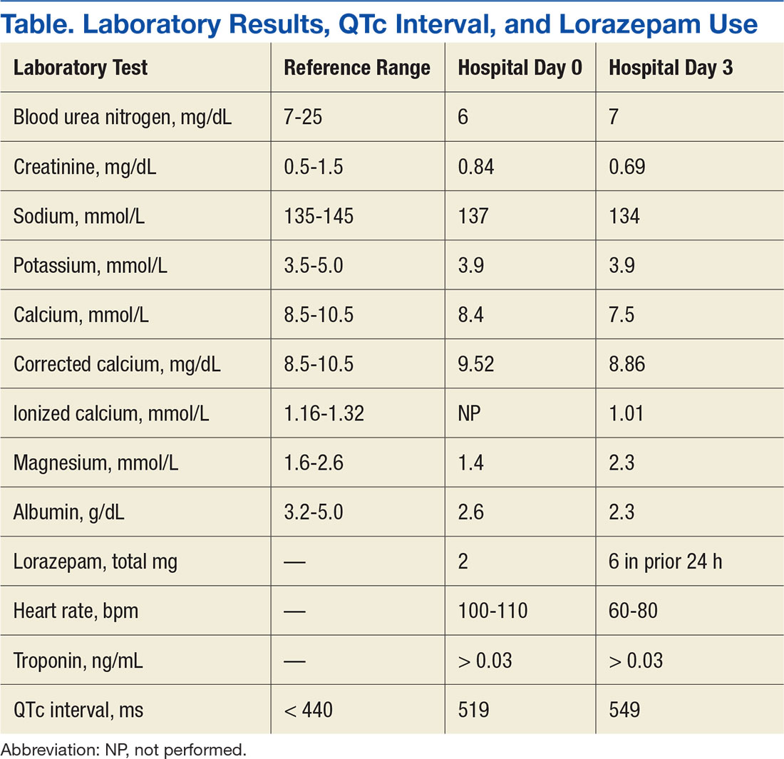
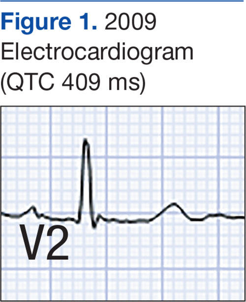
Symptom-triggered therapy for alcohol withdrawal was instituted, and the patient’s electrolyte abnormalities were corrected. Telemetry monitoring demonstrated polymorphic ventricular ectopy, including a 6.8-s run of polymorphic ventricular tachycardia and several shorter runs (4-10 beats) of nonsustained ventricular tachycardia, prompting initiation of a low-dose beta blocker. Based on elevated scores on the symptom-triggered scale for alcohol withdrawal, the Clinical Institute Withdrawal Assessement for Alcohol Withdrawal (CIWA), several doses of oral lorazepam were given for withdrawal symptoms.
The patient became increasingly confused, and new-onset nystagmus was noted. These findings raised concern for Wernicke encephalopathy, so the patient was empirically started on IV high-dose thiamine supplementation. The CIWA scores remained high, and there were frequent episodes of ventricular ectopy during the first 2 hospital days. Interval EKG revealed further prolongation of the QTc interval (549 ms) without evidence of cardiac ischemia (Figure 3). Cardiac enzymes were negative, and electrolyte levels were within normal limits.
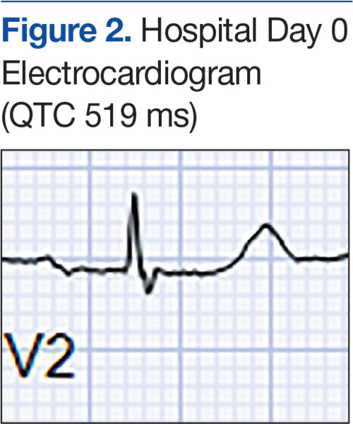
The month-long hospitalization was notable for development of significant ascites, continual electrolyte repletion in the setting of diuresis, formal diagnosis of alcoholic cirrhoisis, cognitive and physical rehabilitation. During the hospitalization, QTc interval remained prolonged (range, 460-500 ms), despite electrolyte repletion, and he was discharged with a wearable cardioverter defibrillator. A month
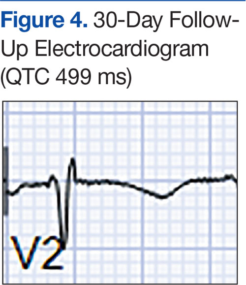
Discussion
Alcohol dependence is a common chronic and relapsing disease that often requires controlled detoxification. Investigators have found a high incidence of QT interval prolongation in alcohol withdrawal and hepatic disease.3,6 Common causes of QT interval prolongation in this setting are poor nutrition, electrolyte abnormalities (particularly hypocalcemia and hypomagnesemia), and use of certain medications.1-3,7,8 In addition, alcohol is directly toxic to the renal tubules, resulting in renal wasting of divalent cations, which may persist up to 30 days after the most recent alcohol exposure.9,10
The patient in this case report was initially thought to have hypomagnesemia-induced long QT syndrome (leading to TdP cardiac arrest), but the authors’ review of laboratory test results revealed the QT interval remained markedly prolonged, despite adequate correction of hypomagnesemia implicating the hyperadrenergic state of acute alcohol withdrawal in QT interval prolongation and TdP cardiac arrest. Interestingly, the QT interval remained prolonged 2 months after TdP arrest, despite sustained normalization of electrolyte levels and the absence of active ischemia or use of QT-prolonging medications.
Given the exclusion of other causes of QT interval prolongation, the authors hypothesized that autonomic hyperactivity of acute alcohol withdrawal and resultant QT interval prolongation were potentiated by underlying cirrhotic cardiomyopathy, a well described cause of QT interval prolongation. Cirrhotic cardiomyopathy is thought to cause QT interval prolongation by delayed repolarization of cardiomyocytes and promotion of sympatho-adrenergic hyperactivity.6 In other case series, TdP development has been associated with severe withdrawal symptoms, particularly delirium tremens.2 In cirrhosis, QT interval prolongation often is described as an early manifestation of cirrhotic cardiomyopathy, irrespective of the underlying etiology, and precedes systolic and diastolic dysfunction.6
The magnitude of QT prolongation has been associated with severity of liver disease as expressed by Child-Pugh score, with reports of QT normalization after liver transplantation.4,5 Patients with higher Child-Pugh scores should be considered to be at elevated risk for malignant ventricular arrhythmias. The authors recommend checking an EKG on admission of any patient who has liver disease or has presented for alcohol withdrawal. Patients with a prolonged QTc interval should be monitored on telemetry. The authors also recommend aggressive repletion of electrolytes, particularly potassium and magnesium, in patients who present with cirrhosis and alcohol withdrawal.
Avoidance of QT-prolonging medications is advisable for all patients with a long QT interval. Beta blockers shorten the QT interval in cirrhotic patients, but the role of beta blockers in preventing malignant arrhythmias in this group of patients is not yet clear.11 The present patient’s QT interval had been normal before he developed cirrhotic liver disease. His presentation was suggestive of acquired long QT syndrome, likely caused by cirrhotic cardiomyopathy given the exhaustive exclusion of other causes of QT interval prolongation.
Conclusion
This case highlights the importance of close monitoring of the QT interval in patients being treated for acute alcohol withdrawal, particularly those with cirrhosis, and suggests that timely and aggressive management of withdrawal, repletion of electrolytes, and telemetry monitoring may prevent life-threatening arrhythmia.
1. Otero-Antón E, González-Quintela A, Saborido J, Torre JA, Virgós A, Barrio E. Prolongation of the QTc interval during alcohol withdrawal syndrome. Acta Cardiol. 1997;52(3):285-294.
2. Cuculi F, Kobza R, Ehmann T, Erne P. ECG changes amongst patients with alcohol withdrawal seizures and delirium tremens. Swiss Med Wkly. 2006;136(13-14):223-227.
3. Mimidis K, Thomopoulos K, Tziakas D, et al. Prolongation of the QTc interval in patients with cirrhosis. Ann Gastroenterol. 2003;16(2):155-158.
4. Bernardi M, Calandra S, Colantoni A, et al. Q-T interval prolongation in cirrhosis: prevalence, relationship with severity, and etiology of the disease and possible pathogenetic factors. Hepatology. 1998;27(1):28-34.
5. Bal JS, Thuluvath PJ. Prolongation of QTc interval: relationship with etiology and severity of liver disease, mortality and liver transplantation. Liver Int. 2003;23(4):243-248.
6. Zardi EM, Abbate A, Zardi DM, et al. Cirrhotic cardiomyopathy. J Am Coll Cardiol. 2010;56(7):539-549.
7. Faigel DO, Metz DC, Kochman ML. Torsade de pointes complicating the treatment of bleeding esophageal varices: association with neuroleptics, vasopressin, and electrolyte imbalance. Am J Gastroenterol. 1995;90(5):822-824.
8. Kotsia AP, Dimitriadis G, Baltogiannis GG, Kolettis TM. Torsade de pointes and persistent QTc prolongation after intravenous amiodarone. Case Rep Med. 2012;2012:673019.
9. Denison H, Jern S, Jagenburg R, Wendestam C, Wallerstedt S. Influence of increased adrenergic activity and magnesium depletion on cardiac rhythm in alcohol withdrawal. Br Heart J. 1994;72(6):554-560.
10. Plaza de los Reyes M, Orozco R, Rosemblitt M, Rendic Y, Espinace M. Renal secretion of magnesium and other electrolytes under the influence of acute ingestion of alcohol, in normal subjects [in Spanish]. Rev Med Chil. 1968;96(3):138-141.
11. Bernardi M, Maggioli C, Dibra V, Zaccherini G. QT interval prolongation in liver cirrhosis: innocent bystander or serious threat? Expert Rev Gastroenterol Hepatol. 2012;6(1):57-66.
Torsades de pointes (TdP) is a life-threatening ventricular arrhythmia that is associated with both congenital and acquired QT interval prolongation. QT interval prolongation is commonly observed in acute alcohol withdrawal and cirrhotic cardiomyopathy.1-3 In both conditions, there is a positive correlation between the degree of QT interval prolongation and disease severity.4,5 The precise mechanisms of QT interval prolongation in these conditions are not well understood. One hypothesis is that autonomic hyperexcitability results in altered ventricular repolarization and QT interval prolongation. This mechanism of QT prolongation has been found in acute alcohol withdrawal independent of electrolyte abnormalities, use of QT-prolonging medications, and cirrhosis.1,2,6
The authors report the case of a veteran who was hospitalized for acute alcohol withdrawal and decompensated cirrhosis and was found to have a newly prolonged QT interval. On hospital day 3, the patient developed TdP, which required external defibrillation. Despite correction of electrolyte abnormalities, abstinence from alcohol, avoidance of QT-prolonging medications, and exclusion of cardiac ischemia, there was significant and persistent prolongation of the QT interval—ultimately attributed to cirrhotic cardiomyopathy. Acquired QT interval prolongation is common in both acute alcohol withdrawal and cirrhosis.This case highlights the importance of close monitoring of the QT interval and TdP susceptibility in patients being treated for acute alcohol withdrawal, particularly those with cirrhosis.
Case Report
A 66-year-old male veteran with a 35-year history of alcohol dependence presented for alcohol detoxification. He reported having drunk at least 32 ounces of vodka every day of the preceding 5 years and reported having unsuccessfully attempted self-detoxification several times. Prior detoxification efforts were unsuccessful because of intractable nausea and tremulousness. Additional presenting symptoms included lethargy, anorexia, and a fall with transient right-side hemiparesis (findings on magnetic resonance imaging of the head had been normal).
His medical history included type 2 diabetes, tobacco dependence, and macular degeneration. The only medication being taken was glargine 25 units daily. On admission, the patient was afebrile (98.1°F), normotensive (103/77 mm Hg), and oriented to person, place, and time. Examination also revealed a protuberant abdomen with caput medusae, and no shifting dullness or lower extremity edema. The neurologic examination was nonfocal.
Laboratory test results on admission were significant for elevated serum alcohol level (243.8 mg/dL); elevated levels of aspartate aminotransferase (144 units/L) alanine aminotransferase (25 units/L), and total bilirubin (4.2 mg/L); hypoalbuminemia; normocalemia; hypomagnesemia; normal corrected calcium level; and normal renal function (0.84 mg/dL)(Table). The patient’s admission Child-Pugh score of 10 indicated class C liver disease. Admission electrocardiogram (EKG) revealed normal sinus rhythm, first-degree atrioventricular block, and prolongation of the QTc interval (519 ms). Six years earlier, the patient’s QTc interval had been 409 ms (Figures 1 and 2). As QT interval depends on heart rate, it is most commonly expressed as corrected QT, or QTc, where QTc = QT/(√RR).
Symptom-triggered therapy for alcohol withdrawal was instituted, and the patient’s electrolyte abnormalities were corrected. Telemetry monitoring demonstrated polymorphic ventricular ectopy, including a 6.8-s run of polymorphic ventricular tachycardia and several shorter runs (4-10 beats) of nonsustained ventricular tachycardia, prompting initiation of a low-dose beta blocker. Based on elevated scores on the symptom-triggered scale for alcohol withdrawal, the Clinical Institute Withdrawal Assessement for Alcohol Withdrawal (CIWA), several doses of oral lorazepam were given for withdrawal symptoms.
The patient became increasingly confused, and new-onset nystagmus was noted. These findings raised concern for Wernicke encephalopathy, so the patient was empirically started on IV high-dose thiamine supplementation. The CIWA scores remained high, and there were frequent episodes of ventricular ectopy during the first 2 hospital days. Interval EKG revealed further prolongation of the QTc interval (549 ms) without evidence of cardiac ischemia (Figure 3). Cardiac enzymes were negative, and electrolyte levels were within normal limits.
The month-long hospitalization was notable for development of significant ascites, continual electrolyte repletion in the setting of diuresis, formal diagnosis of alcoholic cirrhoisis, cognitive and physical rehabilitation. During the hospitalization, QTc interval remained prolonged (range, 460-500 ms), despite electrolyte repletion, and he was discharged with a wearable cardioverter defibrillator. A month
Discussion
Alcohol dependence is a common chronic and relapsing disease that often requires controlled detoxification. Investigators have found a high incidence of QT interval prolongation in alcohol withdrawal and hepatic disease.3,6 Common causes of QT interval prolongation in this setting are poor nutrition, electrolyte abnormalities (particularly hypocalcemia and hypomagnesemia), and use of certain medications.1-3,7,8 In addition, alcohol is directly toxic to the renal tubules, resulting in renal wasting of divalent cations, which may persist up to 30 days after the most recent alcohol exposure.9,10
The patient in this case report was initially thought to have hypomagnesemia-induced long QT syndrome (leading to TdP cardiac arrest), but the authors’ review of laboratory test results revealed the QT interval remained markedly prolonged, despite adequate correction of hypomagnesemia implicating the hyperadrenergic state of acute alcohol withdrawal in QT interval prolongation and TdP cardiac arrest. Interestingly, the QT interval remained prolonged 2 months after TdP arrest, despite sustained normalization of electrolyte levels and the absence of active ischemia or use of QT-prolonging medications.
Given the exclusion of other causes of QT interval prolongation, the authors hypothesized that autonomic hyperactivity of acute alcohol withdrawal and resultant QT interval prolongation were potentiated by underlying cirrhotic cardiomyopathy, a well described cause of QT interval prolongation. Cirrhotic cardiomyopathy is thought to cause QT interval prolongation by delayed repolarization of cardiomyocytes and promotion of sympatho-adrenergic hyperactivity.6 In other case series, TdP development has been associated with severe withdrawal symptoms, particularly delirium tremens.2 In cirrhosis, QT interval prolongation often is described as an early manifestation of cirrhotic cardiomyopathy, irrespective of the underlying etiology, and precedes systolic and diastolic dysfunction.6
The magnitude of QT prolongation has been associated with severity of liver disease as expressed by Child-Pugh score, with reports of QT normalization after liver transplantation.4,5 Patients with higher Child-Pugh scores should be considered to be at elevated risk for malignant ventricular arrhythmias. The authors recommend checking an EKG on admission of any patient who has liver disease or has presented for alcohol withdrawal. Patients with a prolonged QTc interval should be monitored on telemetry. The authors also recommend aggressive repletion of electrolytes, particularly potassium and magnesium, in patients who present with cirrhosis and alcohol withdrawal.
Avoidance of QT-prolonging medications is advisable for all patients with a long QT interval. Beta blockers shorten the QT interval in cirrhotic patients, but the role of beta blockers in preventing malignant arrhythmias in this group of patients is not yet clear.11 The present patient’s QT interval had been normal before he developed cirrhotic liver disease. His presentation was suggestive of acquired long QT syndrome, likely caused by cirrhotic cardiomyopathy given the exhaustive exclusion of other causes of QT interval prolongation.
Conclusion
This case highlights the importance of close monitoring of the QT interval in patients being treated for acute alcohol withdrawal, particularly those with cirrhosis, and suggests that timely and aggressive management of withdrawal, repletion of electrolytes, and telemetry monitoring may prevent life-threatening arrhythmia.
1. Otero-Antón E, González-Quintela A, Saborido J, Torre JA, Virgós A, Barrio E. Prolongation of the QTc interval during alcohol withdrawal syndrome. Acta Cardiol. 1997;52(3):285-294.
2. Cuculi F, Kobza R, Ehmann T, Erne P. ECG changes amongst patients with alcohol withdrawal seizures and delirium tremens. Swiss Med Wkly. 2006;136(13-14):223-227.
3. Mimidis K, Thomopoulos K, Tziakas D, et al. Prolongation of the QTc interval in patients with cirrhosis. Ann Gastroenterol. 2003;16(2):155-158.
4. Bernardi M, Calandra S, Colantoni A, et al. Q-T interval prolongation in cirrhosis: prevalence, relationship with severity, and etiology of the disease and possible pathogenetic factors. Hepatology. 1998;27(1):28-34.
5. Bal JS, Thuluvath PJ. Prolongation of QTc interval: relationship with etiology and severity of liver disease, mortality and liver transplantation. Liver Int. 2003;23(4):243-248.
6. Zardi EM, Abbate A, Zardi DM, et al. Cirrhotic cardiomyopathy. J Am Coll Cardiol. 2010;56(7):539-549.
7. Faigel DO, Metz DC, Kochman ML. Torsade de pointes complicating the treatment of bleeding esophageal varices: association with neuroleptics, vasopressin, and electrolyte imbalance. Am J Gastroenterol. 1995;90(5):822-824.
8. Kotsia AP, Dimitriadis G, Baltogiannis GG, Kolettis TM. Torsade de pointes and persistent QTc prolongation after intravenous amiodarone. Case Rep Med. 2012;2012:673019.
9. Denison H, Jern S, Jagenburg R, Wendestam C, Wallerstedt S. Influence of increased adrenergic activity and magnesium depletion on cardiac rhythm in alcohol withdrawal. Br Heart J. 1994;72(6):554-560.
10. Plaza de los Reyes M, Orozco R, Rosemblitt M, Rendic Y, Espinace M. Renal secretion of magnesium and other electrolytes under the influence of acute ingestion of alcohol, in normal subjects [in Spanish]. Rev Med Chil. 1968;96(3):138-141.
11. Bernardi M, Maggioli C, Dibra V, Zaccherini G. QT interval prolongation in liver cirrhosis: innocent bystander or serious threat? Expert Rev Gastroenterol Hepatol. 2012;6(1):57-66.