User login
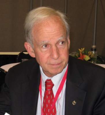
SHM I-PASS Leader Spearheads Fight for Safe Water in Flint

Mona Hanna-Attisha, MD, MPH, the director of the pediatric residency program at Hurley Children’s Hospital and an assistant professor of pediatrics at Michigan State University, has been instrumental in the fight for clean and safe water for residents in Flint, Mich.
Dr. Hanna-Attisha has been a fundamental co-site leader with the SHM I-PASS mentored implementation program and minimized her role in the program as she became the unofficial spokesperson for the Flint water crisis. In January 2016, Dr. Hanna-Attisha took on the lead role for the Pediatric Public Health Initiative with Michigan State University and Hurley Children’s Hospital.
Dr. Hanna-Attisha is a Michigan native and completed her undergraduate degree at the University of Michigan in Ann Arbor. She completed medical school at Michigan State University College of Human Medicine, completed her residency and chief residency at the Children’s Hospital of Michigan, and earned her master’s degree at the University of Michigan School of Public Health.
Dr. Hanna-Attisha’s training and experience has focused heavily on environmental toxins and health disparities, so it’s no surprise that she is deeply involved with addressing the public health emergency in Flint as well as taking measures to ensure continued research and action regarding the impact the contaminated water had on the residents of Flint. Dr. Hanna-Attisha is working with a team of experts to develop evidence-based interventions that will aid in improving the health and development of children and families affected by Flint’s contaminated water. She is educating families on nutrition and diets high in iron, calcium, and vitamin C in order to help manage the effects of contamination.
A report from a Virginia Tech Research Team ignited the investigation into Flint’s water issues and also fueled Dr. Hanna-Attisha’s investigation into the blood-lead levels in the children of Flint. It has been close to two years that the residents of Flint have been exposed to severely toxic levels of lead from the city’s tap water, and Dr. Hanna-Attisha’s analysis of children’s blood-lead levels has been highlighted and published in numerous publications, including the American Journal of Public Health.
The water crisis in Flint hits close to home for Dr. Hanna-Attisha, and the dedication she has to her local community is astounding. It shows in the work she has been doing in her new role with the Pediatric Public Health Initiative.
SHM is proud of all the outstanding work she is doing and appreciates her contributions to the SHM I-PASS program. To keep up to date with Dr. Hanna-Attisha, follow her on Twitter @MonaHannaA.
Mobola Owolabi is senior project manager in SHM’s Center for Hospital Innovation and Improvement.
VIDEO: HOPE-3 trial supports broader role for statins in primary prevention
CHICAGO – The success in the HOPE-3 trial of a two-pronged strategy of a statin plus moderate-dose antihypertensive therapy has expanded the boundaries of primary preventive pharmacotherapy to incorporate many intermediate-cardiovascular-risk patients without cardiovascular disease.
In the Heart Outcomes Evaluation (HOPE)-3 trial, the combination of rosuvastatin (Crestor) at 10 mg/day plus antihypertensive therapy with 16 mg/day of candesartan and 12.5 mg/day of hydrochlorothiazide reduced cardiovascular events in intermediate-risk patients with hypertension, regardless of their baseline LDL-cholesterol and inflammatory biomarker levels. In nonhypertensive study participants, however, blood pressure-lowering therapy provided no added benefit beyond that achieved with the statin alone, which yielded a 25% relative risk reduction in cardiovascular events compared with placebo, Dr. Salim Yusuf reported at the annual meeting of the American College of Cardiology.
The HOPE-3 trial was the first formal study of the polypill concept of fixed-dose, low-dose combination therapy as a public health tool for primary prevention in an intermediate-risk population without cardiovascular disease. It’s a simple, low-cost, safe, pragmatic preventive strategy that doesn’t require baseline laboratory measurements, dose titration visits, or frequent safety monitoring, he noted.
“The original concept of the polypill was to give it to everyone over age 55. We found in HOPE-3 that the polypill concept is not valid for everybody. It demonstrated benefit in hypertensives but not in nonhypertensives, where a statin alone was beneficial,” said Dr. Yusuf, professor of medicine and executive director of the Population Health Research Institute at McMaster University in Hamilton, Ont.
“The clinical implication is that statins should be used much more widely than they currently are. Most guidelines for hypertension don’t say to give a statin. But HOPE-3 is saying hypertensives will benefit. About half of the 40% reduction in the risk of cardiovascular events we saw with combination therapy in patients in the highest third for baseline systolic blood pressure – that is, above 143.5 mm Hg, with a mean of 154 mm Hg – was due to the rosuvastatin and half to the antihypertensive therapy. Our study suggests you can essentially double the benefit of lowering blood pressure in hypertensives if you also lower cholesterol simultaneously,” he noted. Dr. Yusuf discussed the findings in a video interview.
The double-blind trial included a diverse population of 12,705 men age 55 or older and women age 65 or older in 21 countries. All were at intermediate cardiovascular risk by conventional stratification methods; none had a history of cardiovascular disease. They were randomized to rosuvastatin or placebo, dual antihypertensive therapy or placebo, or to all three medications or double placebo. The combined-therapy group experienced a 33.7 mg/dL greater drop in LDL-cholesterol and a 6.2 mm Hg bigger reduction in systolic blood pressure than in patients on dual placebo.
During a median followup of 5.6 years, the composite rate of cardiovascular death, nonfatal MI, or nonfatal stroke was 3.6% in the combined-therapy group and 5.0% with dual placebo, for a relative risk reduction of 29%. The number needed to treat for 5.6 years in order to prevent one event of the composite outcome was 72. However, the separation in the event rate curves for the two groups grew larger over time. With an additional planned followup of 3-5 years in HOPE-3, it’s likely the benefits will become even greater, according to Dr. Yusuf.
Combination therapy proved safe. Muscle aches and weakness as well as lightheadedness were more common in the combined treatment group than with dual placebo. However, permanent discontinuation rates were similar in the two groups.
HOPE-3 coinvestigator Dr. Eva Lonn presented the comparison between patients randomized to antihypertensive therapy without rosuvastatin or to placebo. In a prespecified subgroup analysis, active treatment resulted in a significant 27% reduction in the risk of the composite outcome compared with placebo in patients in the top one-third of baseline systolic blood pressure, no benefit in those with a systolic blood pressure of 131.6-143.5 mm Hg, and a suggestion of possible harm in patients whose systolic pressure at enrollment was 131.5 mm Hg or less.
Thus, the study helps define the minimum blood pressure at which antihypertensive therapy becomes beneficial, noted Dr. Lonn, professor of cardiology at McMaster University.
Dr. Yusuf said enthusiasm for the polypill concept as a means of reducing the growing global burden of cardiovascular disease remains strong among many experts. There is broad interest in a polypill for secondary prevention that will include aspirin, a beta blocker, and an ACE inhibitor of angiotensin receptor blocker as a low-cost means of improving medication adherence. And several large clinical trials of the polypill concept for primary prevention are underway, including a randomized trial conducted by Dr. Yusuf and coworkers in which several thousand high-risk subjects without baseline cardiovascular disease will receive a combination of a statin plus not two but three antihypertensive drugs.
Discussant Dr. Donald M. Lloyd-Jones commented, “What strikes me about HOPE-3 is that this is a population at risk, but not at particularly high risk.” And yet these patients benefited from statin therapy regardless of their baseline LDL, noted Dr. Lloyd-Jones, chair of the department of preventive medicine at Northwestern University, Chicago, and an architect of the risk-based approach to statin use that’s a centerpiece of the current ACC/AHA guidelines on atherosclerotic cardiovascular risk reduction.
In an interview, Dr. Sidney C. Smith, Jr., said the HOPE-3 data are “worthy of consideration” as experts meet at ACC 16 to begin updating guidelines for the treatment of hypertension. In particular, the key findings that moderate-risk hypertensive patients benefited from a statin regardless of their baseline LDL and initiation of blood pressure-lowering therapy was beneficial for patients with a systolic blood pressure in the 140s but not in those with a systolic pressure in the 120s and 130s could be practice changing, according to Dr. Smith, professor of medicine at the University of North Carolina, Chapel Hill.
Drs. Yusuf and Lonn reported receiving institutional research grants from the Canadian Institutes of Health Research and AstraZeneca, which funded the trial.
Simultaneous with the presentation of the HOPE-3 results at ACC 16 in Chicago, the study on cholesterol lowering and the study on blood pressure and cholesterol lowering led by Dr. Yusef as well as the study on blood pressure lowering led by Dr. Lonn were published online at NEJM.org.
The video associated with this article is no longer available on this site. Please view all of our videos on the MDedge YouTube channel
AT ACC 16
Key clinical point: Statin therapy should be used much more widely.
Major finding: The number of moderate-cardiovascular-risk persons needed to treat with a combination of a statin and two antihypertensive drugs for 5.6 years in order to prevent one cardiovascular death, nonfatal MI, or nonfatal stroke was 72.
Data source: The HOPE-3 study was a double-blind, randomized trial including 12,705 men and women at intermediate cardiovascular risk but without cardiovascular disease at enrollment.
Disclosures: The study presenters reported receiving institutional research grants from the Canadian Institutes of Health Research and AstraZeneca, which funded the trial.
Gene discovery could help fight malaria

Photo by James Gathany
Researchers believe they may have discovered a male-determining gene in the malaria-carrying mosquito species Anopheles gambiae.
The discovery of this gene provides scientists with a foundation for studying male mosquito biology.
And this is significant because male mosquitoes offer the potential to develop novel vector control strategies to combat malaria because males do not feed on blood or transmit disease.
One vector control method under development involves genetic modification of the mosquito to bias the population sex ratio toward males.
Modeling has shown the most efficient means for genetic modification of mosquitoes is engineering a driving Y chromosome.
A molecular-level understanding of the Y chromosome of the malaria-carrying mosquito is important to inform and optimize this type of a strategy.
So Omar Akbari, PhD, of the University of California, Riverside, and his colleagues set out to gain such an understanding.
The team used multiple genome sequencing techniques to identify an extensive dataset of Y chromosome sequences and explore their organization and evolution in the Anopheles gambiae complex, a group of at least 7 morphologically indistinguishable species of mosquitoes in the genus Anopheles.
This revealed that only 1 gene, YG2, is exclusive to the Y chromosome across the species complex and may therefore be a male-determining gene.
Dr Akbari and his colleagues described this discovery in PNAS.
Cyclophosphamide nets low rate of chronic GVHD after mobilized blood cell transplantation
High-dose cyclophosphamide is safe and effective when given as prophylaxis for chronic graft-versus-host disease (GVHD) to patients who have undergone transplantation of mobilized blood cells, finds a phase 2 trial reported in Blood.
Investigators led by Dr. Marco Mielcarek, medical director of the Adult Blood and Marrow Transplant Program and an oncologist at the Fred Hutchinson Cancer Research Center in Seattle, Washington, enrolled in the trial 43 patients with high-risk hematologic malignancies.

The patients underwent myeloablative conditioning followed by transplantation with growth factor–mobilized blood cells from related or unrelated donors, and were given high-dose cyclophosphamide on two early posttransplantation days.
Main results showed that the cumulative 1-year incidence of chronic GVHD was 16%, less than half of the roughly 35% seen historically with conventional immunosuppression.
Moreover, cyclophosphamide did not appear to compromise engraftment or control of the underlying malignancy. Only a single patient, one with an HLA-mismatched donor, had failure of primary engraftment; after amendment of the protocol to require HLA matching, there were no additional cases. Just 17% of patients experienced a recurrence of their malignancy by 2 years.
Taken together, the findings suggest that high-dose cyclophosphamide—as combined with two myeloablative conditioning options (to accommodate different malignancies) and with posttransplantation cyclosporine (to reduce the risk of acute GVHD)—may eliminate most of the drawbacks to using mobilized blood cells for transplantation, according to the investigators.
“If these findings are confirmed in future studies, HLA-matched mobilized blood cell transplantation may gain even greater acceptance and further replace marrow as a source of stem cells for most indications,” they maintain.
The patients studied had a median age of 43 years, and slightly more than half were in remission without minimal residual disease.
Blood cells were mobilized with granulocyte colony-stimulating factor (G-CSF). Overall, 28% of patients received grafts from related donors, while 72% received grafts from unrelated donors.
For pretransplant conditioning, patients received fludarabine and targeted busulfan, or total body irradiation with use of a minimum dose of 12 Gy.
The patients were given cyclophosphamide at 50 mg/kg per day on days 3 and 4 after transplantation. This was followed by cyclosporine starting on day 5.
The cumulative 1-year incidence of chronic GVHD as defined by National Institutes of Health criteria (i.e., that requiring systemic immunosuppressive therapy)—the trial’s primary endpoint—was 16%, which fell just short of the goal of 15% the investigators were aiming for (Blood. 2016;127:1502-8). Analyses failed to identify any predictors of this outcome.
Although the estimated cumulative incidence of grade 2 acute GVHD was high, at 77%, none of the patients developed grade 3 or 4 acute GVHD, according to the investigators, who disclosed that they had no competing financial interests.
The single patient who experienced failure of primary engraftment had familial myelodysplastic syndrome and had received a graft from an HLA A-antigen–mismatched unrelated donor.
The 2-year cumulative incidence of nonrelapse mortality was 14%, and the 2-year cumulative incidence of recurrent malignancy was 17%. Projected overall survival was 70%.
Among the 42 patients having at least a year of follow-up, 50% were alive and free of relapse without any systemic immunosuppression at 1 year after transplantation.
High-dose cyclophosphamide is safe and effective when given as prophylaxis for chronic graft-versus-host disease (GVHD) to patients who have undergone transplantation of mobilized blood cells, finds a phase 2 trial reported in Blood.
Investigators led by Dr. Marco Mielcarek, medical director of the Adult Blood and Marrow Transplant Program and an oncologist at the Fred Hutchinson Cancer Research Center in Seattle, Washington, enrolled in the trial 43 patients with high-risk hematologic malignancies.
The patients underwent myeloablative conditioning followed by transplantation with growth factor–mobilized blood cells from related or unrelated donors, and were given high-dose cyclophosphamide on two early posttransplantation days.
Main results showed that the cumulative 1-year incidence of chronic GVHD was 16%, less than half of the roughly 35% seen historically with conventional immunosuppression.
Moreover, cyclophosphamide did not appear to compromise engraftment or control of the underlying malignancy. Only a single patient, one with an HLA-mismatched donor, had failure of primary engraftment; after amendment of the protocol to require HLA matching, there were no additional cases. Just 17% of patients experienced a recurrence of their malignancy by 2 years.
Taken together, the findings suggest that high-dose cyclophosphamide—as combined with two myeloablative conditioning options (to accommodate different malignancies) and with posttransplantation cyclosporine (to reduce the risk of acute GVHD)—may eliminate most of the drawbacks to using mobilized blood cells for transplantation, according to the investigators.
“If these findings are confirmed in future studies, HLA-matched mobilized blood cell transplantation may gain even greater acceptance and further replace marrow as a source of stem cells for most indications,” they maintain.
The patients studied had a median age of 43 years, and slightly more than half were in remission without minimal residual disease.
Blood cells were mobilized with granulocyte colony-stimulating factor (G-CSF). Overall, 28% of patients received grafts from related donors, while 72% received grafts from unrelated donors.
For pretransplant conditioning, patients received fludarabine and targeted busulfan, or total body irradiation with use of a minimum dose of 12 Gy.
The patients were given cyclophosphamide at 50 mg/kg per day on days 3 and 4 after transplantation. This was followed by cyclosporine starting on day 5.
The cumulative 1-year incidence of chronic GVHD as defined by National Institutes of Health criteria (i.e., that requiring systemic immunosuppressive therapy)—the trial’s primary endpoint—was 16%, which fell just short of the goal of 15% the investigators were aiming for (Blood. 2016;127:1502-8). Analyses failed to identify any predictors of this outcome.
Although the estimated cumulative incidence of grade 2 acute GVHD was high, at 77%, none of the patients developed grade 3 or 4 acute GVHD, according to the investigators, who disclosed that they had no competing financial interests.
The single patient who experienced failure of primary engraftment had familial myelodysplastic syndrome and had received a graft from an HLA A-antigen–mismatched unrelated donor.
The 2-year cumulative incidence of nonrelapse mortality was 14%, and the 2-year cumulative incidence of recurrent malignancy was 17%. Projected overall survival was 70%.
Among the 42 patients having at least a year of follow-up, 50% were alive and free of relapse without any systemic immunosuppression at 1 year after transplantation.
FROM BLOOD
Key clinical point: High-dose posttransplant cyclophosphamide is safe and effective for reducing the risk of chronic GVHD after mobilized blood cell transplantation.
Major finding: The cumulative 1-year incidence of chronic GVHD requiring immunosuppressive therapy was 16%.
Data source: A single-arm trial among 43 patients with high-risk hematologic malignancies undergoing growth factor–mobilized blood cell transplantation.
Disclosures: The authors disclosed that they have no competing financial interests.

Heart failure severity at AMI predicts long-term CV death risk
CHICAGO – The severity of heart failure in the setting of acute myocardial infarction predicts long-term cardiovascular death risk, according to a post hoc analysis of data from the IMPROVE IT Trial.
Among 11,185 individuals with MI and known Killip Classification who were part of that randomized, double-blind trial, those with Killip Class II or greater had more than double the risk of long-term cardiovascular death, compared with those with Killip Class I heart failure, Dr Michael G. Silverman, a cardiovascular medicine fellow at Brigham and Women’s Hospital, Boston and a research fellow at the Thrombolysis in MI (TIMI) Study Group reported in a poster at the annual meeting of the American College of Cardiology.

After adjusting for a number of factors, including age, gender, diabetes, hypertension, left ventricular ejection fraction (LVEF), beta blocker and ACE inhibitor/angiotensin receptor blocker use at randomization, and percutaneous coronary intervention at the index event, the 7-year event rate was 14.5% among those with Killip Class II or higher vs. 5.7% those with Killip Class I heart failure (adjusted hazard ratio, 1.9), Dr. Silverman reported on behalf of the TIMI Study Group.
The event rates from 30 days to 6 months were 4.85% and 1.25% in the groups, respectively (adjusted hazard ratio, 1.96), and from 6 months to 7 years they were 1.52% and 0.61%, in the groups, respectively, (adjusted hazard ratio, 1.85).
Further, the increased risk of cardiovascular death associated with Killip Class II or higher was also apparent among important subgroups, including those with ST Segment Elevation MI, those with non-STEMI, those with LVEF of 50 or greater, those with LVEF less than 50, those with diabetes, those without diabetes, men, and women (adjusted hazard ratios ranging from 1.6 to 2.1), Dr Silverman explained in an interview.
The severity of heart failure according to Killip Class is a strong independent predictor of mortality in the setting of acute MI, and the current findings demonstrate that it also predicts cardiovascular death for at least 7 years, suggesting a need for careful attention to the findings of the physical exam in AMI, as it can serve as an important biomarker of long-term cardiovascular death risk, he said.
“AMI patients with Killip Class II or greater warrant continued close medical follow-up and adherence to guideline -directed medial therapy beyond the acute hospitalization to prevent this potentially modifiable outcome,” he concluded.
Dr. Silverman reported having no disclosures.
CHICAGO – The severity of heart failure in the setting of acute myocardial infarction predicts long-term cardiovascular death risk, according to a post hoc analysis of data from the IMPROVE IT Trial.
Among 11,185 individuals with MI and known Killip Classification who were part of that randomized, double-blind trial, those with Killip Class II or greater had more than double the risk of long-term cardiovascular death, compared with those with Killip Class I heart failure, Dr Michael G. Silverman, a cardiovascular medicine fellow at Brigham and Women’s Hospital, Boston and a research fellow at the Thrombolysis in MI (TIMI) Study Group reported in a poster at the annual meeting of the American College of Cardiology.
After adjusting for a number of factors, including age, gender, diabetes, hypertension, left ventricular ejection fraction (LVEF), beta blocker and ACE inhibitor/angiotensin receptor blocker use at randomization, and percutaneous coronary intervention at the index event, the 7-year event rate was 14.5% among those with Killip Class II or higher vs. 5.7% those with Killip Class I heart failure (adjusted hazard ratio, 1.9), Dr. Silverman reported on behalf of the TIMI Study Group.
The event rates from 30 days to 6 months were 4.85% and 1.25% in the groups, respectively (adjusted hazard ratio, 1.96), and from 6 months to 7 years they were 1.52% and 0.61%, in the groups, respectively, (adjusted hazard ratio, 1.85).
Further, the increased risk of cardiovascular death associated with Killip Class II or higher was also apparent among important subgroups, including those with ST Segment Elevation MI, those with non-STEMI, those with LVEF of 50 or greater, those with LVEF less than 50, those with diabetes, those without diabetes, men, and women (adjusted hazard ratios ranging from 1.6 to 2.1), Dr Silverman explained in an interview.
The severity of heart failure according to Killip Class is a strong independent predictor of mortality in the setting of acute MI, and the current findings demonstrate that it also predicts cardiovascular death for at least 7 years, suggesting a need for careful attention to the findings of the physical exam in AMI, as it can serve as an important biomarker of long-term cardiovascular death risk, he said.
“AMI patients with Killip Class II or greater warrant continued close medical follow-up and adherence to guideline -directed medial therapy beyond the acute hospitalization to prevent this potentially modifiable outcome,” he concluded.
Dr. Silverman reported having no disclosures.
AT ACC 16
Key clinical point: The severity of heart failure in the setting of acute myocardial infarction predicts long-term cardiovascular death risk, according to a post hoc analysis of data from the IMPROVE IT Trial.
Major finding: The 7-year event rate was 14.5% among those with Killip Class II or higher vs. 5.7% among those with Killip Class I heart failure (adjusted hazard ratio, 1.9).
Data source: A post-hoc analysis of data from 11,185 subjects from the IMPROVE IT trial.
Disclosures: Dr. Silverman reported having no disclosures.

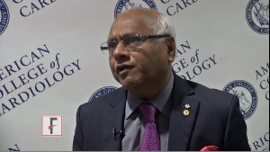
VIDEO: HOPE-3 trial expands scope of primary cardiovascular prevention
CHICAGO – The HOPE-3 trial has brought the primary prevention of cardiovascular disease front-and-center as a hot topic of discussion at the annual meeting of the American College of Cardiology.
The large, randomized trial provides new evidence of a significant reduction in cardiovascular events in an intermediate-risk population that hasn’t previously been the focus of risk reduction via pharmacotherapy.
The video associated with this article is no longer available on this site. Please view all of our videos on the MDedge YouTube channel
The double-blind study randomized nearly 13,000 intermediate-risk men and women with no baseline cardiovascular disease to lipid-lowering with rosuvastatin (Crestor) at 10 mg/day or placebo, dual-antihypertensive therapy with candesartan plus chlorothiazide or placebo regardless of baseline blood pressure, or all three drugs or placebo. After a median of 5.6 years, the combined-therapy group had a 29% reduction in the composite of cardiovascular death or nonfatal MI or stroke compared with placebo-treated controls. Impressively, the benefit was similar regardless of baseline LDL-cholesterol level; however, only subjects with hypertension benefited from the dual-antihypertensive therapy.
In this interview, Dr. B. Hadley Wilson of the Sanger Clinic in Charlotte, N.C., explains why he considers HOPE-3 to be a giant step forward in preventive cardiolo
AT ACC 16
Passing up a product endorsement
Yesterday, my secretary passed a message that someone from television had called with a question. Not knowing what to expect, but trying to be helpful, I returned the call that afternoon.
The fellow was nice enough, and explained he worked for a marketing company. A vitamin company had hired him to promote an over-the-counter supplement to treat Alzheimer’s disease, and he was looking for a neurologist to endorse it in an infomercial. He said I’d be compensated $5,000-$10,000 for the spot.
That’s a pretty decent chunk of change. I could use it. For a few seconds I hemmed and hawed, trying to think of a way to rationalize it. Then I realized ... I just couldn’t. I politely told him “No,” and got off the phone.
I know they’ll find someone to do it. But I just can’t. I’m sure they have some data to back it up, but crappy research papers are a dime a dozen.
I spend a lot of time explaining studies and data to those affected by this terrible disease. I’m trying to help them work their way through a maze of tests, treatments of limited benefit, and reasonable expectations.
Sadly, as with all tragic and incurable diseases, there’s no shortage of hucksters trying to take advantage of the desperate. Part of my job is to help people understand this. They bring in ads from magazines and newspaper promising miracle cures for a host of awful illnesses. I can’t stop them from buying it, but I want to do my best to warn them it’s a scam.
I can’t do that if I switch sides. Once I start plugging non–FDA-approved, non–clinically meaningful junk on late-night TV, I’ve joined the snake-oil salesmen of yesteryear.
I owe my patients better than that. They trust me to help them and to do what’s right.
Like everyone else, I don’t have a perfect reputation, but outside of my online reviews, I think I’m reasonably well thought of in the local community. A decent reputation takes years to build and one crappy decision to lose. I don’t want to do that either.
And under all that, I still have to believe in myself. That everyday I’m trying to do what’s right for people. Because if I’m not doing that, it’s time to hang up my reflex hammer. The first person I have to face every morning is in the mirror. I want to be able to look at him and still believe he’s doing the best he can for those who need his help.
Dr. Block has a solo neurology practice in Scottsdale, Ariz.

Severe Psoriasis, Kidney Disease Linked
WASHINGTON – Another population-based study has found a link between severe psoriasis and kidney disease – this one discovering almost a fivefold increase in the risk of immunoglobulin A nephropathy (IgAN) and a doubling in the risk of glomerular disease.
The findings suggest yet again that psoriasis is a systemic illness, and not something that affects only the skin, Sungat Grewal said at the annual meeting of the American Academy of Dermatology.

“Numerous case reports have generated a hypothesis that psoriasis may be associated,” with an increased risk of IgAN, said Ms. Grewal, of the department of dermatology at the University of Pennsylvania, Philadelphia. “Our study is the first to test this, and it supports the notion that this is no coincidence. Now we need further research to determine if this association is due to causality or to a shared pathophysiology.”
The link between psoriasis and kidney disease has long been noted, but the first study formally investigating this association was published in 2013 (BMJ. 2013 Oct;347:f5961). The study, also conducted by University of Pennsylvania investigators, used a large patient database in the United Kingdom, matched about 143,000 patients with psoriasis with up to five controls without psoriasis each, and found the risk of chronic kidney disease was nearly doubled for those with severe psoriasis (hazard ratio, 1.93).
A similar finding emerged from Taiwan in 2015. Using the national healthcare database, researchers matched about 4,600 patients with psoriasis with about 923,000 controls. They found that having severe psoriasis was associated with almost a doubling in the risk of chronic kidney disease (HR, 1.90) and almost a tripling in the risk of end stage renal disease (HR, 2.97), after adjusting for age, gender, comorbidities, and use of nonsteroidal anti-inflammatory drugs (J Dermatol Sci. 2015 Jun;78[3]:232-8).
Ms. Grewal and her coinvestigators used data from The Health Improvement Network in the United Kingdom – the same database used in the 2013 study. The study group comprised 206,000 patients with psoriasis and about 1 million controls.
In the overall group of patients, the risk of IgAN was not significantly increased. Nor was there a significant overall association with glomerular disease. And when the group was divided by disease severity, there were no significant associations with either IgAN or glomerular disease in the group with mild psoriasis.
Among those with severe psoriasis, however, the risk of IgAN was almost five times higher (HR, 4.75) and the risk of glomerular disease was doubled (HR, 2.05).
But although the hazard ratios look impressive, the clinical reality shouldn’t spark too much concern, Ms. Grewal said. “To keep things in context, it’s very important to remember that the excess risk of nephropathy attributed to severe psoriasis was still quite small – similar to the chance of a spontaneous pregnancy resulting in triplets.”
Still, she said, the link is intriguing, and something clinicians should keep in mind when managing patients with severe psoriasis.
Ms. Grewal had no financial disclosures. She is a medical student at the Commonwealth Medical College (Scranton, Pa.), and is currently spending a year at the Gelfand Clinical Research Lab at the University of Pennsylvania, Philadelphia.
WASHINGTON – Another population-based study has found a link between severe psoriasis and kidney disease – this one discovering almost a fivefold increase in the risk of immunoglobulin A nephropathy (IgAN) and a doubling in the risk of glomerular disease.
The findings suggest yet again that psoriasis is a systemic illness, and not something that affects only the skin, Sungat Grewal said at the annual meeting of the American Academy of Dermatology.
“Numerous case reports have generated a hypothesis that psoriasis may be associated,” with an increased risk of IgAN, said Ms. Grewal, of the department of dermatology at the University of Pennsylvania, Philadelphia. “Our study is the first to test this, and it supports the notion that this is no coincidence. Now we need further research to determine if this association is due to causality or to a shared pathophysiology.”
The link between psoriasis and kidney disease has long been noted, but the first study formally investigating this association was published in 2013 (BMJ. 2013 Oct;347:f5961). The study, also conducted by University of Pennsylvania investigators, used a large patient database in the United Kingdom, matched about 143,000 patients with psoriasis with up to five controls without psoriasis each, and found the risk of chronic kidney disease was nearly doubled for those with severe psoriasis (hazard ratio, 1.93).
A similar finding emerged from Taiwan in 2015. Using the national healthcare database, researchers matched about 4,600 patients with psoriasis with about 923,000 controls. They found that having severe psoriasis was associated with almost a doubling in the risk of chronic kidney disease (HR, 1.90) and almost a tripling in the risk of end stage renal disease (HR, 2.97), after adjusting for age, gender, comorbidities, and use of nonsteroidal anti-inflammatory drugs (J Dermatol Sci. 2015 Jun;78[3]:232-8).
Ms. Grewal and her coinvestigators used data from The Health Improvement Network in the United Kingdom – the same database used in the 2013 study. The study group comprised 206,000 patients with psoriasis and about 1 million controls.
In the overall group of patients, the risk of IgAN was not significantly increased. Nor was there a significant overall association with glomerular disease. And when the group was divided by disease severity, there were no significant associations with either IgAN or glomerular disease in the group with mild psoriasis.
Among those with severe psoriasis, however, the risk of IgAN was almost five times higher (HR, 4.75) and the risk of glomerular disease was doubled (HR, 2.05).
But although the hazard ratios look impressive, the clinical reality shouldn’t spark too much concern, Ms. Grewal said. “To keep things in context, it’s very important to remember that the excess risk of nephropathy attributed to severe psoriasis was still quite small – similar to the chance of a spontaneous pregnancy resulting in triplets.”
Still, she said, the link is intriguing, and something clinicians should keep in mind when managing patients with severe psoriasis.
Ms. Grewal had no financial disclosures. She is a medical student at the Commonwealth Medical College (Scranton, Pa.), and is currently spending a year at the Gelfand Clinical Research Lab at the University of Pennsylvania, Philadelphia.
AT AAD 16

Severe psoriasis, kidney disease linked
WASHINGTON – Another population-based study has found a link between severe psoriasis and kidney disease – this one discovering almost a fivefold increase in the risk of immunoglobulin A nephropathy (IgAN) and a doubling in the risk of glomerular disease.
The findings suggest yet again that psoriasis is a systemic illness, and not something that affects only the skin, Sungat Grewal said at the annual meeting of the American Academy of Dermatology.

“Numerous case reports have generated a hypothesis that psoriasis may be associated,” with an increased risk of IgAN, said Ms. Grewal, of the department of dermatology at the University of Pennsylvania, Philadelphia. “Our study is the first to test this, and it supports the notion that this is no coincidence. Now we need further research to determine if this association is due to causality or to a shared pathophysiology.”
The link between psoriasis and kidney disease has long been noted, but the first study formally investigating this association was published in 2013 (BMJ. 2013 Oct;347:f5961). The study, also conducted by University of Pennsylvania investigators, used a large patient database in the United Kingdom, matched about 143,000 patients with psoriasis with up to five controls without psoriasis each, and found the risk of chronic kidney disease was nearly doubled for those with severe psoriasis (hazard ratio, 1.93).
A similar finding emerged from Taiwan in 2015. Using the national healthcare database, researchers matched about 4,600 patients with psoriasis with about 923,000 controls. They found that having severe psoriasis was associated with almost a doubling in the risk of chronic kidney disease (HR, 1.90) and almost a tripling in the risk of end stage renal disease (HR, 2.97), after adjusting for age, gender, comorbidities, and use of nonsteroidal anti-inflammatory drugs (J Dermatol Sci. 2015 Jun;78[3]:232-8).
Ms. Grewal and her coinvestigators used data from The Health Improvement Network in the United Kingdom – the same database used in the 2013 study. The study group comprised 206,000 patients with psoriasis and about 1 million controls.
In the overall group of patients, the risk of IgAN was not significantly increased. Nor was there a significant overall association with glomerular disease. And when the group was divided by disease severity, there were no significant associations with either IgAN or glomerular disease in the group with mild psoriasis.
Among those with severe psoriasis, however, the risk of IgAN was almost five times higher (HR, 4.75) and the risk of glomerular disease was doubled (HR, 2.05).
But although the hazard ratios look impressive, the clinical reality shouldn’t spark too much concern, Ms. Grewal said. “To keep things in context, it’s very important to remember that the excess risk of nephropathy attributed to severe psoriasis was still quite small – similar to the chance of a spontaneous pregnancy resulting in triplets.”
Still, she said, the link is intriguing, and something clinicians should keep in mind when managing patients with severe psoriasis.
Ms. Grewal had no financial disclosures. She is a medical student at the Commonwealth Medical College (Scranton, Pa.), and is currently spending a year at the Gelfand Clinical Research Lab at the University of Pennsylvania, Philadelphia.
WASHINGTON – Another population-based study has found a link between severe psoriasis and kidney disease – this one discovering almost a fivefold increase in the risk of immunoglobulin A nephropathy (IgAN) and a doubling in the risk of glomerular disease.
The findings suggest yet again that psoriasis is a systemic illness, and not something that affects only the skin, Sungat Grewal said at the annual meeting of the American Academy of Dermatology.
“Numerous case reports have generated a hypothesis that psoriasis may be associated,” with an increased risk of IgAN, said Ms. Grewal, of the department of dermatology at the University of Pennsylvania, Philadelphia. “Our study is the first to test this, and it supports the notion that this is no coincidence. Now we need further research to determine if this association is due to causality or to a shared pathophysiology.”
The link between psoriasis and kidney disease has long been noted, but the first study formally investigating this association was published in 2013 (BMJ. 2013 Oct;347:f5961). The study, also conducted by University of Pennsylvania investigators, used a large patient database in the United Kingdom, matched about 143,000 patients with psoriasis with up to five controls without psoriasis each, and found the risk of chronic kidney disease was nearly doubled for those with severe psoriasis (hazard ratio, 1.93).
A similar finding emerged from Taiwan in 2015. Using the national healthcare database, researchers matched about 4,600 patients with psoriasis with about 923,000 controls. They found that having severe psoriasis was associated with almost a doubling in the risk of chronic kidney disease (HR, 1.90) and almost a tripling in the risk of end stage renal disease (HR, 2.97), after adjusting for age, gender, comorbidities, and use of nonsteroidal anti-inflammatory drugs (J Dermatol Sci. 2015 Jun;78[3]:232-8).
Ms. Grewal and her coinvestigators used data from The Health Improvement Network in the United Kingdom – the same database used in the 2013 study. The study group comprised 206,000 patients with psoriasis and about 1 million controls.
In the overall group of patients, the risk of IgAN was not significantly increased. Nor was there a significant overall association with glomerular disease. And when the group was divided by disease severity, there were no significant associations with either IgAN or glomerular disease in the group with mild psoriasis.
Among those with severe psoriasis, however, the risk of IgAN was almost five times higher (HR, 4.75) and the risk of glomerular disease was doubled (HR, 2.05).
But although the hazard ratios look impressive, the clinical reality shouldn’t spark too much concern, Ms. Grewal said. “To keep things in context, it’s very important to remember that the excess risk of nephropathy attributed to severe psoriasis was still quite small – similar to the chance of a spontaneous pregnancy resulting in triplets.”
Still, she said, the link is intriguing, and something clinicians should keep in mind when managing patients with severe psoriasis.
Ms. Grewal had no financial disclosures. She is a medical student at the Commonwealth Medical College (Scranton, Pa.), and is currently spending a year at the Gelfand Clinical Research Lab at the University of Pennsylvania, Philadelphia.
AT AAD 16
Key clinical point: Severe psoriasis appears to increase the risk of both immunoglobulin A glomerulonephritis and glomerular disease.
Major finding: The risk of glomerulonephritis was five-fold higher and the risk of glomerular disease doubled in those with severe psoriasis.
Data source: A population based cohort study comprised about 1.2 million subjects.
Disclosures: Ms. Sungat Grewal had no financial disclosures.

Prepsychosis links with elevated metabolic syndrome
MADRID – Untreated people at high risk for developing psychosis also showed an increased prevalence of certain components of metabolic syndrome in data collected from 163 German study participants, a finding that gives new insight into the well-documented but poorly delineated link between schizophrenia and metabolic syndrome.
“The findings point out that a high risk for schizophrenia implies a certain risk for patients to develop metabolic syndrome independent of treatment effects,” said Dr. Joachim Cordes, a psychiatrist at the LVR Clinic of the Heinrich-Heine University in Düsseldorf, Germany. He assumed that genetic factors underlie the shared risk some people face for both developing schizophrenia and metabolic syndrome. “I think there is a direct connection between schizophrenia and metabolic syndrome, an inherent factor like a genetic factor,” Dr. Cordes said in an interview. This understanding should influence how patients with newly diagnosed schizophrenia or those at risk for psychosis are managed, he added.

Dr. Cordes’s report was one of several at the meeting sponsored by the European Psychiatric Association that examined different facets of the complex links that tie schizophrenia to metabolic syndrome, an association that already had lots of evidence, including a recent meta-analysis (Schizophr Bull. 2013 March;39[2]:306-18).
He used data collected on 163 people enrolled in the PREVENT study and at high risk for a first psychotic episode. Run at nine German centers, PREVENT primarily tested very early intervention with drug and behavioral therapy to improve outcomes. Dr. Cordes took data collected from these prepsychosis, high-risk patients to assess their prevalence of metabolic syndrome and of the various individual features that define metabolic syndrome, using a definition published by the American Heart Association and the U.S. National Heart, Lung, and Blood Institute (Circulation. 2005 Oct 18;112[17]:2735-52). He compared these metabolic syndrome rates with the general German population, using data from 35,869 randomly selected German adults in more than 1,500 German primary care practices, the German Metabolic and Cardiovascular Risk Project (GEMCAS).
The findings showed a 9.2% prevalence of metabolic syndrome in the prepsychosis group and a 7.4% rate among the general adult population, Dr. Cordes reported. Among men in the prepsychosis group, the metabolic syndrome definers with the largest increments in prevalence were low HDL, in 21% of the prepsychosis people and in 12% of the general population, and elevated blood glucose in 11%, compared with 6%. Among women, the metabolic syndrome definers with the greatest between-group differences were elevated waist circumference, in 30% of those with prepsychosis, compared with 17% in the general population, and low HDL in 19%, compared with 14%.
This apparently inherent link between a tendency toward psychosis and schizophrenia and a tendency to develop features of metabolic syndrome suggests that patients with newly diagnosed schizophrenia need a preventive approach to weight management, Dr. Cordes said. He also suggested prescribing antipsychotic medications that pose the lowest risk for causing further metabolic derangements in patients.
A second report at the meeting came from an assessment of cognitive function and its relationship to metabolic syndrome in 54 women diagnosed with schizophrenia and on stable treatment. The schizophrenia patients with metabolic syndrome, nearly half of the total group, performed significantly worse than those without metabolic syndrome in tests of verbal memory, executive function, and attention and processing speed, findings that support an increased incidence of selective cognitive impairment in patients with schizophrenia and metabolic syndrome, said Dr. Adela C. Botis, a psychiatrist and researcher at the University of Medicine and Pharmacy in Cluj-Napoca, Romania.
Dr. Botis and her associates studied 54 women diagnosed with schizophrenia who had remitted symptoms for at least 6 months on stable antipsychotic treatment. Using the metabolic syndrome definition of the International Diabetes Federation 25 (46%) had metabolic syndrome, and the other 29 (54%) did not. These numbers document the high prevalence of metabolic syndrome in schizophrenia patients.
A multivariate analysis identified demographic and metabolic factors that significantly linked with decrements in several cognitive domains. Economic status and living situation linked with deficits in verbal memory; elevated systolic blood pressure significantly linked with worsened attention and processing speed; high body mass index linked with loss of motor speed; and less education significantly linked with all these increments as well as four other domains,
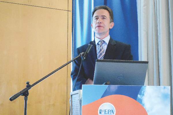
A third report used a post-hoc analysis of data from two separate trials to show that treatment with a relatively new antipsychotic drug, lurasidone (Latuda), produced less metabolic syndrome, compared with risperidone or extended-release quetiapine (Seroquel XR), said Dr. Andrei Pikalov, head of global medical affairs at Sunovion Pharmaceuticals, the company that markets Latuda. Lurasidone received approval for treating schizophrenia in 2010.
He took data from two studies designed to assess lurasidone’s efficacy for treating adults with schizophrenia for 12 months, compared with either risperidone in a study with 621 patients, or with quetiapine XR in a study with 292 patients. He applied the same metabolic syndrome definition used by Dr. Cordes to clinical measurements taken at baseline and after 12 months on treatment.
The results showed that treatment with lurasidone produced less than half the rate of new metabolic syndrome cases, compared with risperidone, a statistically significant difference, and less than two-thirds the rate of quetiapine XR, a difference that did not reach statistical significance.
Dr. Cordes said he has been a speaker for Servier. Dr. Botis had no disclosures. Dr. Pikalov is an employee of Sunovion, which markets lurasidone (Latuda).
On Twitter @mitchelzoler
MADRID – Untreated people at high risk for developing psychosis also showed an increased prevalence of certain components of metabolic syndrome in data collected from 163 German study participants, a finding that gives new insight into the well-documented but poorly delineated link between schizophrenia and metabolic syndrome.
“The findings point out that a high risk for schizophrenia implies a certain risk for patients to develop metabolic syndrome independent of treatment effects,” said Dr. Joachim Cordes, a psychiatrist at the LVR Clinic of the Heinrich-Heine University in Düsseldorf, Germany. He assumed that genetic factors underlie the shared risk some people face for both developing schizophrenia and metabolic syndrome. “I think there is a direct connection between schizophrenia and metabolic syndrome, an inherent factor like a genetic factor,” Dr. Cordes said in an interview. This understanding should influence how patients with newly diagnosed schizophrenia or those at risk for psychosis are managed, he added.
Dr. Cordes’s report was one of several at the meeting sponsored by the European Psychiatric Association that examined different facets of the complex links that tie schizophrenia to metabolic syndrome, an association that already had lots of evidence, including a recent meta-analysis (Schizophr Bull. 2013 March;39[2]:306-18).
He used data collected on 163 people enrolled in the PREVENT study and at high risk for a first psychotic episode. Run at nine German centers, PREVENT primarily tested very early intervention with drug and behavioral therapy to improve outcomes. Dr. Cordes took data collected from these prepsychosis, high-risk patients to assess their prevalence of metabolic syndrome and of the various individual features that define metabolic syndrome, using a definition published by the American Heart Association and the U.S. National Heart, Lung, and Blood Institute (Circulation. 2005 Oct 18;112[17]:2735-52). He compared these metabolic syndrome rates with the general German population, using data from 35,869 randomly selected German adults in more than 1,500 German primary care practices, the German Metabolic and Cardiovascular Risk Project (GEMCAS).
The findings showed a 9.2% prevalence of metabolic syndrome in the prepsychosis group and a 7.4% rate among the general adult population, Dr. Cordes reported. Among men in the prepsychosis group, the metabolic syndrome definers with the largest increments in prevalence were low HDL, in 21% of the prepsychosis people and in 12% of the general population, and elevated blood glucose in 11%, compared with 6%. Among women, the metabolic syndrome definers with the greatest between-group differences were elevated waist circumference, in 30% of those with prepsychosis, compared with 17% in the general population, and low HDL in 19%, compared with 14%.
This apparently inherent link between a tendency toward psychosis and schizophrenia and a tendency to develop features of metabolic syndrome suggests that patients with newly diagnosed schizophrenia need a preventive approach to weight management, Dr. Cordes said. He also suggested prescribing antipsychotic medications that pose the lowest risk for causing further metabolic derangements in patients.
A second report at the meeting came from an assessment of cognitive function and its relationship to metabolic syndrome in 54 women diagnosed with schizophrenia and on stable treatment. The schizophrenia patients with metabolic syndrome, nearly half of the total group, performed significantly worse than those without metabolic syndrome in tests of verbal memory, executive function, and attention and processing speed, findings that support an increased incidence of selective cognitive impairment in patients with schizophrenia and metabolic syndrome, said Dr. Adela C. Botis, a psychiatrist and researcher at the University of Medicine and Pharmacy in Cluj-Napoca, Romania.
Dr. Botis and her associates studied 54 women diagnosed with schizophrenia who had remitted symptoms for at least 6 months on stable antipsychotic treatment. Using the metabolic syndrome definition of the International Diabetes Federation 25 (46%) had metabolic syndrome, and the other 29 (54%) did not. These numbers document the high prevalence of metabolic syndrome in schizophrenia patients.
A multivariate analysis identified demographic and metabolic factors that significantly linked with decrements in several cognitive domains. Economic status and living situation linked with deficits in verbal memory; elevated systolic blood pressure significantly linked with worsened attention and processing speed; high body mass index linked with loss of motor speed; and less education significantly linked with all these increments as well as four other domains,
A third report used a post-hoc analysis of data from two separate trials to show that treatment with a relatively new antipsychotic drug, lurasidone (Latuda), produced less metabolic syndrome, compared with risperidone or extended-release quetiapine (Seroquel XR), said Dr. Andrei Pikalov, head of global medical affairs at Sunovion Pharmaceuticals, the company that markets Latuda. Lurasidone received approval for treating schizophrenia in 2010.
He took data from two studies designed to assess lurasidone’s efficacy for treating adults with schizophrenia for 12 months, compared with either risperidone in a study with 621 patients, or with quetiapine XR in a study with 292 patients. He applied the same metabolic syndrome definition used by Dr. Cordes to clinical measurements taken at baseline and after 12 months on treatment.
The results showed that treatment with lurasidone produced less than half the rate of new metabolic syndrome cases, compared with risperidone, a statistically significant difference, and less than two-thirds the rate of quetiapine XR, a difference that did not reach statistical significance.
Dr. Cordes said he has been a speaker for Servier. Dr. Botis had no disclosures. Dr. Pikalov is an employee of Sunovion, which markets lurasidone (Latuda).
On Twitter @mitchelzoler
AT THE EUROPEAN CONGRESS OF PSYCHIATRY
Key clinical point: People at high risk for a first psychosis episode had an increased prevalence of certain metabolic syndrome components, compared with the general population.
Major finding: Metabolic syndrome was prevalent in 9.2% of the prepsychosis group and in 7.4% of the general population.
Data source: Post-hoc analysis of data from 163 German people with prepsychosis.
Disclosures: Dr. Cordes said he has been a speaker for Servier. Dr. Botis had no disclosures. Dr. Pikalov is an employee of Sunovion, which markets lurasidone (Latuda).