User login
Early estrogen likely prevents bone fractures in Turner syndrome
BOSTON – The longer that estrogen therapy is delayed in girls with Turner syndrome, the lower their bone density will be in subsequent years, based on results of a retrospective, cross-sectional study from Monash University, in Melbourne, Australia.
For every year after age 11 that Turner patients went without estrogen – generally due to delayed initiation, but sometimes noncompliance – there was a significant reduction in bone mineral density in both the lumbar spine (Beta -0.582, P less than 0.001) and femoral neck (Beta -0.383, P = 0.008).

Estrogen deficiency and subsequent suboptimal bone mass accrual are known to contribute to the increased risk of osteoporosis in women with Turner syndrome, and about a doubling of the risk of fragility fractures, mostly of the forearm. About a third of the 76 women in the study had at least one fracture, explained investigator Dr. Amanda Vincent, head of the Midlife Health and Menopause Program at Monash.
“Avoiding estrogen deficiency is important to optimize bone health in Turner syndrome.” It “depends on early diagnosis, age-appropriate pubertal induction, and optimization of compliance,” Dr. Vincent said at the Endocrine Society annual meeting.
The median age of Turner syndrome diagnosis was 11 years, but estrogen treatments didn’t begin until a median age of 15. The women in the study were a median of about 30 years old, which means that they were adolescents at the time when estrogen treatment was often delayed in the mistaken belief that growth hormone therapy would be more effective before puberty was induced.
It’s now known that estrogen replacement works synergistically with, and even potentiates, the effects of growth hormone. Current guidelines recommend pubertal induction by age 13 (J Clin Endocrinol Metab. 2007 Jan;92(1):10-25).
The women had at least one dual-energy x-ray absorptiometry scan at Monash since 1998. Z-scores below - 2, indicating low bone density, were found in the lumbar spines of about a quarter the subjects, and in the femoral necks of about 8%. Primary amenorrhea and premature menopause, followed by vitamin D deficiency, were the most common risk factors for low bone mass. Almost 40% of the women reported non-continuous use of estrogen. About half had undergone growth hormone therapy.
At a median height of 149 cm, the subjects were about 15 cm shorter than age-matched, healthy controls, and also had a slightly higher median body mass index of 25.6 kg/m2. Lumbar spine bone area, bone mineral content, areal bone mineral density, and bone mineral apparent density were significantly lower in Turner syndrome patients. In the femoral neck, areal bone mineral density was significantly lower.
There was no relationship between bone markers and growth hormone use or Turner syndrome karyotype; the predominant karyotype was 45XO, but the study also included mosaic karyotypes.
The investigators had no disclosures.
These are important observations. The bottom line is early recognition and early referral. It’s clear from this study and others that earlier institution of estrogen is beneficial for height, bone density, and fracture risk throughout life. It’s not just an issue of a 20 year old with low bone density; that 20 year old later becomes a 60 year old with low bone density.

|
Dr. Michael Levine |
[However,] we still have a problem with delayed recognition and referral of young girls with Turner syndrome. Most girls with Turner syndrome have some typical phenotypic features, but some do not, so the diagnosis is often made too late. [To get around that problem,] we recommend that all children below the 5th percentile for height – or who flatten out too early on growth curves – be referred to rule out Turner syndrome and other problems.
Dr. Michael Levine is chief of the Division of Endocrinology at The Children’s Hospital of Philadelphia. He made his comments after the study presentation, and was not involved in the work.
These are important observations. The bottom line is early recognition and early referral. It’s clear from this study and others that earlier institution of estrogen is beneficial for height, bone density, and fracture risk throughout life. It’s not just an issue of a 20 year old with low bone density; that 20 year old later becomes a 60 year old with low bone density.
|
|
Dr. Michael Levine |
[However,] we still have a problem with delayed recognition and referral of young girls with Turner syndrome. Most girls with Turner syndrome have some typical phenotypic features, but some do not, so the diagnosis is often made too late. [To get around that problem,] we recommend that all children below the 5th percentile for height – or who flatten out too early on growth curves – be referred to rule out Turner syndrome and other problems.
Dr. Michael Levine is chief of the Division of Endocrinology at The Children’s Hospital of Philadelphia. He made his comments after the study presentation, and was not involved in the work.
BOSTON – The longer that estrogen therapy is delayed in girls with Turner syndrome, the lower their bone density will be in subsequent years, based on results of a retrospective, cross-sectional study from Monash University, in Melbourne, Australia.
For every year after age 11 that Turner patients went without estrogen – generally due to delayed initiation, but sometimes noncompliance – there was a significant reduction in bone mineral density in both the lumbar spine (Beta -0.582, P less than 0.001) and femoral neck (Beta -0.383, P = 0.008).
Estrogen deficiency and subsequent suboptimal bone mass accrual are known to contribute to the increased risk of osteoporosis in women with Turner syndrome, and about a doubling of the risk of fragility fractures, mostly of the forearm. About a third of the 76 women in the study had at least one fracture, explained investigator Dr. Amanda Vincent, head of the Midlife Health and Menopause Program at Monash.
“Avoiding estrogen deficiency is important to optimize bone health in Turner syndrome.” It “depends on early diagnosis, age-appropriate pubertal induction, and optimization of compliance,” Dr. Vincent said at the Endocrine Society annual meeting.
The median age of Turner syndrome diagnosis was 11 years, but estrogen treatments didn’t begin until a median age of 15. The women in the study were a median of about 30 years old, which means that they were adolescents at the time when estrogen treatment was often delayed in the mistaken belief that growth hormone therapy would be more effective before puberty was induced.
It’s now known that estrogen replacement works synergistically with, and even potentiates, the effects of growth hormone. Current guidelines recommend pubertal induction by age 13 (J Clin Endocrinol Metab. 2007 Jan;92(1):10-25).
The women had at least one dual-energy x-ray absorptiometry scan at Monash since 1998. Z-scores below - 2, indicating low bone density, were found in the lumbar spines of about a quarter the subjects, and in the femoral necks of about 8%. Primary amenorrhea and premature menopause, followed by vitamin D deficiency, were the most common risk factors for low bone mass. Almost 40% of the women reported non-continuous use of estrogen. About half had undergone growth hormone therapy.
At a median height of 149 cm, the subjects were about 15 cm shorter than age-matched, healthy controls, and also had a slightly higher median body mass index of 25.6 kg/m2. Lumbar spine bone area, bone mineral content, areal bone mineral density, and bone mineral apparent density were significantly lower in Turner syndrome patients. In the femoral neck, areal bone mineral density was significantly lower.
There was no relationship between bone markers and growth hormone use or Turner syndrome karyotype; the predominant karyotype was 45XO, but the study also included mosaic karyotypes.
The investigators had no disclosures.
AT ENDO 2016
Key clinical point: Induce puberty by age 13 in Turner syndrome.
Major finding: For every year after age 11 that Turner patients went without estrogen – generally due to delayed initiation, but sometimes noncompliance – there was a significant reduction in bone mineral density in both the lubar spine (Beta -0.582, P less than 0.001) and femoral neck (Beta -0.383, P = 0.008).
Data source: Retrospective, cross-section study of 76 Turner syndrome patients
Disclosures: The investigators had no disclosures.

HM16 Speakers Focus on Public Health, Leadership, Future of Hospital Medicine
SAN DIEGO — Hospital medicine’s annual extravaganza nestled into the southwestern corner of the country in March, with a record 4,000 hospitalists and others expanding their knowledge of clinical care, management, leadership, technology, and quality improvement.
They listened, they laughed, they learned.
Read more about the knowledge, experiences hospitalists shared at HM16.
Between the nitty-gritty of the workshops, expert panels, and forums, three high-profile speakers offered broad and insightful perspectives:
- U.S. Surgeon General Vivek Murthy, MD, MBA, a hospitalist by training, on his experiences as a hospitalist and his thoughts on the importance of public health in America;
- New SHM President Brian Harte, MD, SFHM, on the role of hospital medicine in cultivating leadership; and
- Hospitalist pioneer Robert Wachter, MD, MHM, on the future of hospital medicine as it reaches its 20th year since he introduced the term “hospitalist” in a New England Journal of Medicine article.

Dr. Murthy, formerly a hospitalist at Brigham and Women’s Hospital in Boston who in 2009 founded Doctors for America, an organization for healthcare improvement in the U.S., said his career as a hospitalist came as a surprise to him.
“When I was in medical school, I didn’t even know what a hospitalist was,” he said. “When I became a hospitalist, I thought it would be a temporary gig, something I did for a couple of years while I figured out what I really wanted to do. But as it turned out, I really loved what I did as a hospitalist. I love teaching. I love caring for patients. I love being part of a tight-knit team.”
He called good health “the key to opportunity,” explaining health is “intrinsically connected to the American dream.”
Hospitalists can play a role in building a “foundation for health,” he said. Four ingredients to this, he said, are creating a culture in which “healthy is equated with happiness” rather than associated with an attitude of “suck it up and eat your spinach”; changing our environment, such as adding sidewalks to encourage walking, to promote healthy behavior change; focusing on the spirit and mind as well as the body; and cultivating our ability to give and receive kindness, which he called “a source of healing.”
Dr. Harte described hospital medicine as “fertile ground” for leadership development.
“Our day-to-day experiences provide a leadership incubator that really no other specialty can claim,” he said.
He said he hopes that over the next several years, hospitalists and SHM make strides in these areas:
- Continuing and expanding membership;
- Continuing to push members and projects to focus on the Triple Aim, particularly patient- and family-centered care; and
- Better understanding hospitalists’ role in the era of risk.
“We need to clarify our position regarding specialty training and our training programs,” he added.
Dr. Harte recognized that such a discussion can get “difficult and contentious and political,” but that “when we look at what we have to do to be clinically effective, and what our current training programs and family medicine, internal medicine, and pediatrics provide for us, that gap to me only appears to be increasing.”
He said SHM has and will “continue to step up with curricula to fill those gaps.” However, he also said hospitalists “have to question what is the best way to train physicians for the roles of providers in the acute-care setting.”

Dr. Wachter, keeping his tradition of giving the final talk of the four-day conference, retraced the roots and successes of the field over the last 20 years. It was part history lesson, part report card, and part prognostication.
What the field has gotten wrong, so far, amounts to “an amazingly short list,” he said, but it’s not a nonexistent list.
“I think one thing we got wrong was a 7-days-on/7-days-off schedule,” he said, drawing applause. While it might be appealing to a 35-year-old doctor, he added, “I don’t believe this is a viable schedule for a 60-year-old.”
HM modeled itself after its closest cousin, emergency medicine, in which doctors frequently work 10- to 12-hour shifts every other day. Since that every-other-day schedule is not good for continuity, HM essentially strung together shifts for as many consecutive days as possible, leading to the 7-on/7-off. Now, many clinicians won’t consider positions without such a schedule even though it’s not a schedule suitable for everyone.
“I think we’ve shot ourselves in the foot,” he said. “Because what it means is you take all the work that needs to be done and you shove it into a very small amount of space. Therefore, the amount of intensity in that work that you have is, I think, undoable over time. I hope we rethink that.”
He cautioned that SHM is near the age when, all too often, societies begin to be complacent and needs to guard against the instinct to keep doing things as they have always been done.
“We need to instinctively say, ‘Wait a second, am I turning into all of those other societies that have become irrelevant—or less relevant—because of that reflex?’” he questioned.
He predicted that, even though value in care is now becoming an obsession, the digitization of healthcare ultimately will have a deeper impact on medicine.
“You ask me 10 years from now, I’m guessing that the fact that we’ve just gone from analog to digital will have turned out to be a bigger transformation,” he explained. “And the reason I say that is if you look at the history of every other industry that went from analog to digital, eventually the industries got turned upside down.”
Burnout, a prominent topic at the meeting, still doesn’t seem to be worse in hospital medicine than in many other specialties, he said, but it is a concern.
“We need to rethink this. We need to come up with some new practice models using information technology in new ways, collaborating with members of the team in different ways,” he said. “We have to take this issue and figure out a way of solving it.”
Dr. Wachter said hospital medicine needs to keep innovating and finding ways to add value; otherwise, the financial support hospitals give to hospital medicine could begin to shrink.
The field is facing challenges, he said, but he is clearly proud of its accomplishments. He said that before he went out on the stage at an early conference he organized in 1998, during the early days of hospital medicine, his wife asked him, “Are you sure this is a good idea?”
“What I said to her was, ‘It is a good idea, and it will be a good idea if we are successful in recruiting and retaining young people, innovative people who want to change the world,’” he said. “I think we have done that, and I thank all of you for turning this into a good idea.” TH
Thomas R. Collins is a freelance writer in South Florida.
SAN DIEGO — Hospital medicine’s annual extravaganza nestled into the southwestern corner of the country in March, with a record 4,000 hospitalists and others expanding their knowledge of clinical care, management, leadership, technology, and quality improvement.
They listened, they laughed, they learned.
Read more about the knowledge, experiences hospitalists shared at HM16.
Between the nitty-gritty of the workshops, expert panels, and forums, three high-profile speakers offered broad and insightful perspectives:
- U.S. Surgeon General Vivek Murthy, MD, MBA, a hospitalist by training, on his experiences as a hospitalist and his thoughts on the importance of public health in America;
- New SHM President Brian Harte, MD, SFHM, on the role of hospital medicine in cultivating leadership; and
- Hospitalist pioneer Robert Wachter, MD, MHM, on the future of hospital medicine as it reaches its 20th year since he introduced the term “hospitalist” in a New England Journal of Medicine article.
Dr. Murthy, formerly a hospitalist at Brigham and Women’s Hospital in Boston who in 2009 founded Doctors for America, an organization for healthcare improvement in the U.S., said his career as a hospitalist came as a surprise to him.
“When I was in medical school, I didn’t even know what a hospitalist was,” he said. “When I became a hospitalist, I thought it would be a temporary gig, something I did for a couple of years while I figured out what I really wanted to do. But as it turned out, I really loved what I did as a hospitalist. I love teaching. I love caring for patients. I love being part of a tight-knit team.”
He called good health “the key to opportunity,” explaining health is “intrinsically connected to the American dream.”
Hospitalists can play a role in building a “foundation for health,” he said. Four ingredients to this, he said, are creating a culture in which “healthy is equated with happiness” rather than associated with an attitude of “suck it up and eat your spinach”; changing our environment, such as adding sidewalks to encourage walking, to promote healthy behavior change; focusing on the spirit and mind as well as the body; and cultivating our ability to give and receive kindness, which he called “a source of healing.”
Dr. Harte described hospital medicine as “fertile ground” for leadership development.
“Our day-to-day experiences provide a leadership incubator that really no other specialty can claim,” he said.
He said he hopes that over the next several years, hospitalists and SHM make strides in these areas:
- Continuing and expanding membership;
- Continuing to push members and projects to focus on the Triple Aim, particularly patient- and family-centered care; and
- Better understanding hospitalists’ role in the era of risk.
“We need to clarify our position regarding specialty training and our training programs,” he added.
Dr. Harte recognized that such a discussion can get “difficult and contentious and political,” but that “when we look at what we have to do to be clinically effective, and what our current training programs and family medicine, internal medicine, and pediatrics provide for us, that gap to me only appears to be increasing.”
He said SHM has and will “continue to step up with curricula to fill those gaps.” However, he also said hospitalists “have to question what is the best way to train physicians for the roles of providers in the acute-care setting.”
Dr. Wachter, keeping his tradition of giving the final talk of the four-day conference, retraced the roots and successes of the field over the last 20 years. It was part history lesson, part report card, and part prognostication.
What the field has gotten wrong, so far, amounts to “an amazingly short list,” he said, but it’s not a nonexistent list.
“I think one thing we got wrong was a 7-days-on/7-days-off schedule,” he said, drawing applause. While it might be appealing to a 35-year-old doctor, he added, “I don’t believe this is a viable schedule for a 60-year-old.”
HM modeled itself after its closest cousin, emergency medicine, in which doctors frequently work 10- to 12-hour shifts every other day. Since that every-other-day schedule is not good for continuity, HM essentially strung together shifts for as many consecutive days as possible, leading to the 7-on/7-off. Now, many clinicians won’t consider positions without such a schedule even though it’s not a schedule suitable for everyone.
“I think we’ve shot ourselves in the foot,” he said. “Because what it means is you take all the work that needs to be done and you shove it into a very small amount of space. Therefore, the amount of intensity in that work that you have is, I think, undoable over time. I hope we rethink that.”
He cautioned that SHM is near the age when, all too often, societies begin to be complacent and needs to guard against the instinct to keep doing things as they have always been done.
“We need to instinctively say, ‘Wait a second, am I turning into all of those other societies that have become irrelevant—or less relevant—because of that reflex?’” he questioned.
He predicted that, even though value in care is now becoming an obsession, the digitization of healthcare ultimately will have a deeper impact on medicine.
“You ask me 10 years from now, I’m guessing that the fact that we’ve just gone from analog to digital will have turned out to be a bigger transformation,” he explained. “And the reason I say that is if you look at the history of every other industry that went from analog to digital, eventually the industries got turned upside down.”
Burnout, a prominent topic at the meeting, still doesn’t seem to be worse in hospital medicine than in many other specialties, he said, but it is a concern.
“We need to rethink this. We need to come up with some new practice models using information technology in new ways, collaborating with members of the team in different ways,” he said. “We have to take this issue and figure out a way of solving it.”
Dr. Wachter said hospital medicine needs to keep innovating and finding ways to add value; otherwise, the financial support hospitals give to hospital medicine could begin to shrink.
The field is facing challenges, he said, but he is clearly proud of its accomplishments. He said that before he went out on the stage at an early conference he organized in 1998, during the early days of hospital medicine, his wife asked him, “Are you sure this is a good idea?”
“What I said to her was, ‘It is a good idea, and it will be a good idea if we are successful in recruiting and retaining young people, innovative people who want to change the world,’” he said. “I think we have done that, and I thank all of you for turning this into a good idea.” TH
Thomas R. Collins is a freelance writer in South Florida.
CHMP recommends daratumumab for MM

Photo courtesy of Janssen
The European Medicines Agency’s Committee for Medicinal Products for Human Use (CHMP) has recommended conditional marketing authorization for daratumumab (Darzalex), a first-in-class monoclonal antibody targeting CD38.
The recommended indication for daratumumab is as monotherapy for adults with relapsed and refractory multiple myeloma (MM).
The patients must have progressed on their last therapy and have received treatment with both a proteasome inhibitor and an immunomodulatory agent.
The CHMP’s positive opinion will now be reviewed by the European Commission, which has the authority to grant marketing authorization for medicines in the European Economic Area.
The European Commission’s final decision on daratumumab is anticipated in the coming months.
About conditional authorization
A product may receive conditional marketing authorization if the CHMP finds that, although comprehensive clinical data on the safety and efficacy of the product are not available, all of the following requirements are met:
- The risk-benefit balance of the product is positive
- The company developing the product will likely be in a position to provide comprehensive clinical data in the future
- Unmet medical needs will be fulfilled
- The benefit to public health of the immediate availability of the product outweighs the risk inherent in the fact that additional data are still required.
Conditional marketing authorizations are valid for 1 year, on a renewable basis. The holder will be required to complete ongoing studies or to conduct new studies with a view to confirming that the benefit-risk balance of a product is positive. In addition, specific obligations may be imposed in relation to the collection of pharmacovigilance data.
About daratumumab
Daratumumab is the first CD38-directed monoclonal antibody recommended for approval in Europe. It works by binding to CD38, a signaling molecule highly expressed on the surface of MM cells regardless of stage of disease.
In binding to CD38, daratumumab triggers the patient’s own immune system to attack the cancer cells, resulting in rapid tumor cell death through multiple, immune-mediated mechanisms of action and through immunomodulatory effects, in addition to direct tumor cell death via apoptosis.
The CHMP’s positive opinion of daratumumab was based on a review of data from the phase 2 SIRIUS study, the phase 1/2 GEN501 study, and 3 additional supportive studies.
The GEN501 study enrolled 102 patients with relapsed MM or relapsed MM that was refractory to 2 or more prior lines of therapy. The patients received daratumumab at a range of doses and on a number of different schedules.
The results suggested daratumumab is most effective at a dose of 16 mg/kg. At this dose, the overall response rate was 36%. Most adverse events in this study were grade 1 or 2, although serious events did occur.
The SIRIUS study enrolled 124 MM patients who had received 3 or more prior lines of therapy. They received daratumumab at different doses and on different schedules, but 106 patients received the drug at 16 mg/kg.
Twenty-nine percent of the 106 patients responded to treatment, and the median duration of response was 7 months. Thirty percent of patients experienced serious adverse events.
Findings from a combined efficacy analysis of the GEN501 and SIRIUS trials demonstrated that, after a mean follow-up of 14.8 months, the estimated median overall survival for patients who received single-agent daratumumab at 16 mg/kg was 20 months.
Five phase 3 clinical studies with daratumumab in MM patients—in relapsed and frontline settings—are ongoing. Additional studies are ongoing or planned to assess the drug’s potential in other malignant and pre-malignant diseases in which CD38 is expressed.
Janssen has exclusive worldwide rights to the development, manufacturing, and commercialization of daratumumab for all potential indications. Janssen licensed daratumumab from Genmab A/S in August 2012. ![]()
FDA: CT scans safe for patients with electronic medical devices
There’s no need to let fear of electronic interference between computed tomography and electronic medical devices preclude the ordering of such scans for patients with insulin pumps, cardiac implantable electronic devices, or neurostimulators, the Food and Drug Administration said in a written notification.
“The probability of an adverse event being caused by exposing these devices to CT irradiation is extremely low, and it is greatly outweighed by the clinical benefit of a medically indicated CT examination,” according to the new notification, which updates and replaces a preliminary health notification released on July 14, 2008.
The preliminary notification said there was a “possibility that the x-rays used during CT examinations may cause some implanted and external electronic medical devices to malfunction.” It also included recommendations to reduce the potential risk of such events from occurring and cited adverse events experienced by a few patients with medical devices who had undergone CT scanning, including unintended shocks from neurostimulators, malfunctions of insulin infusion pumps, and transient changes in pacemaker output pulse rate.
The new notification says there is an extremely low probability that a CT scanner directly irradiating the circuitry of certain implantable or wearable electronic medical devices can cause sufficient electronic interference to affect the function and operation of the medical device, and this probability is even lower when the radiation dose and the radiation dose rate are reduced. The FDA also notes that the interference is completely avoided when the medical device is outside of the primary x-ray beam of the CT scanner.
The update, which provides additional reports of adverse events by patients with electronic medical devices who had CT scans, states that the number of such events was small, compared with the number of patients with insulin pumps, cardiac implantable electronic devices, and neurostimulators who were scanned without adverse effects.
The FDA encourages health care providers and patients who suspect a problem with a medical imaging device to file a voluntary report through MedWatch, the FDA Safety Information and Adverse Event Reporting Program.
Blisters on face and ear

The FP diagnosed herpes zoster with possible Ramsay Hunt syndrome in this patient. The zoster also appeared superinfected by a bacterial infection, which was causing the extensive erythema, weeping of yellow fluid, and some honey crusting.
Herpes zoster oticus (Ramsay Hunt syndrome) includes the triad of ipsilateral facial paralysis, ear pain, and vesicles in the auditory canal and auricle. Disturbances in taste perception, hearing (tinnitus, hyperacusis), lacrimation, and vestibular function (vertigo) may occur. Fortunately, the child did not have facial paralysis, but did have ear pain and vesicles on the auricle. The child also admitted to some ringing in the right ear.
The FP prescribed oral acyclovir and cultured the weeping fluid to determine if this was truly a bacterial superinfection. The child was started on oral cephalexin 3 times a day to cover Streptococcus pyogenes and Staphylococcus aureus. Oral acetaminophen was prescribed for the pain and fever. The physician called his ear, nose, and throat (ENT) colleague who agreed to see the patient later that day.
The FP and ENT physician decided to follow her without immediate hospitalization since she was taking fluids well and did not appear systemically ill. The following day, the patient was already improving and later that week the culture grew out Staphylococcus aureus that was methicillin sensitive. The child fully recovered from the superinfected herpes zoster without any sequelae. The tinnitus resolved, as well.
Photo courtesy of UTHSCSA Dermatology division. Text for Photo Rounds Friday courtesy of Richard P. Usatine, MD. This case was adapted from: Mayeaux EJ, Usatine R. Zoster. In: Usatine R, Smith M, Mayeaux EJ, et al, eds. Color Atlas of Family Medicine. 2nd ed. New York, NY: McGraw-Hill; 2013:712-717.
To learn more about the Color Atlas of Family Medicine, see: www.amazon.com/Color-Family-Medicine-Richard-Usatine/dp/0071769641/
You can now get the second edition of the Color Atlas of Family Medicine as an app by clicking on this link: usatinemedia.com

Blisters on thigh
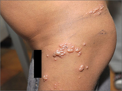
The FP diagnosed the boy with herpes zoster that was found in multiple dermatomes. He did not think it was disseminated because it was still localized to one area and there were not 20 or more lesions outside the primary zoster. However, the FP was very concerned about HIV and the boy was tested. A rapid HIV test came back positive.
The FP discussed the results of his findings with the child's grandmother, who was now caring for the child. An attempt was made to obtain oral or intravenous acyclovir but it was not available in the village or local health center. The child was given oral liquid acetaminophen for the pain and was added to the list for the local HIV clinic. Fortunately, the zoster resolved without antiviral medications and the child began to receive care for his HIV infection.
Photos and text for Photo Rounds Friday courtesy of Richard P. Usatine, MD. This case was adapted from: Mayeaux EJ, Usatine R. Zoster. In: Usatine R, Smith M, Mayeaux EJ, et al, eds. Color Atlas of Family Medicine. 2nd ed. New York, NY: McGraw-Hill; 2013:712-717.
To learn more about the Color Atlas of Family Medicine, see: www.amazon.com/Color-Family-Medicine-Richard-Usatine/dp/0071769641/
You can now get the second edition of the Color Atlas of Family Medicine as an app by clicking on this link: usatinemedia.com

Pruritic rash on 16-year-old girl

Despite the fact that some of the vesicles crossed the midline (which is suggestive of disseminated zoster), the FP felt confident in diagnosing herpes zoster (shingles) in this patient. The FP knew that the varicella-zoster virus (VZV) leaves the dorsal root ganglion to travel down the spinal nerves to the cutaneous nerves of the skin. But she also knew that the vesicles could cross the midline by a few centimeters because the posterior primary ramus of the spinal nerve includes a small cutaneous medial branch that reaches across the midline.1
After a primary infection with either chickenpox or vaccine-type VZV, a latent infection is established in the sensory dorsal root ganglia. Reactivation of this latent VZV infection results in herpes zoster. Both sensory ganglia neurons and satellite cells surrounding the neurons serve as sites of VZV latent infection. Once reactivated, the virus spreads to other cells within the ganglion. The dermatomal distribution of the rash corresponds to the sensory fields of the infected neurons within the specific ganglion.
The pain associated with zoster infections and postherpetic neuralgia (PHN) is thought to result from injury to the peripheral nerves and altered central nervous system processing. PHN occurs more commonly in individuals older than age 60 and in immunosuppressed individuals. The lesions typically crust in approximately a week, with complete resolution within 3 to 4 weeks. If there are more than 20 lesions distributed outside the affected dermatome, the patient has disseminated zoster.
The treatment of herpes zoster includes antiviral agents, such as acyclovir, famciclovir, and valacyclovir. The evidence only supports their use if started within 72 hours of rash onset. Pain can be managed with nonprescription analgesics or narcotics and should be treated aggressively. This may actually prevent or lessen the severity of PHN.
The FP in this case was also concerned about whether the patient might be positive for human immunodeficiency virus (HIV) or have some type of immunosuppression. The patient denied sexual activity and use of intravenous drugs (even when her mom wasn’t in the room). She was otherwise healthy, so no further workup for HIV or immunosuppression was ordered. The FP told the teen to stay home from school until the lesions crusted over. A follow-up visit in one month was scheduled. The zoster resolved and the girl returned to her usual state of good health.
1. Usatine RP, Clemente C. Is herpes zoster unilateral? West J Med. 1999;170:263.
Photos and text for Photo Rounds Friday courtesy of Richard P. Usatine, MD. This case was adapted from: Mayeaux EJ, Usatine R. Zoster. In: Usatine R, Smith M, Mayeaux EJ, et al, eds. Color Atlas of Family Medicine. 2nd ed. New York, NY: McGraw-Hill; 2013:712-717.
To learn more about the Color Atlas of Family Medicine, see: www.amazon.com/Color-Family-Medicine-Richard-Usatine/dp/0071769641/
You can now get the second edition of the Color Atlas of Family Medicine as an app by clicking on this link: usatinemedia.com

Early biopsy predicts levonorgestrel IUD response in endometrial cancer
SAN DIEGO – Endometrial pathology findings at 3 months predicted response to levonorgestrel-releasing IUD treatment for complex atypical hyperplasia or grade 1 endometrial cancer at the MD Anderson Cancer Center in Houston.
Twenty-nine of 32 women (91%) who responded by 12 months showed stromal, glandular, or other endometrial changes indicating an effect at 3 months, vs. only 3 of 9 nonresponders (33%) (P less than .001). There were no differences in responders versus nonresponders in median age (47 vs. 56 years, P = .2) or body mass index (45 vs. 55 kg/m2, P = .16).

The finding addresses an “unmet need” for markers of response to levonorgestrel-releasing IUD therapy. “You can look at [early] pathology” and have an idea how patients will do, Dr. Shannon Westin, a study investigator who is with the department of gynecologic oncology at MD Anderson, said at the annual meeting of the Society of Gynecologic Oncology.
Twenty-seven of 29 women (93%) with complex atypical hyperplasia (CAH) responded completely to the IUD, meaning they had normal endometrium or hyperplasia without atypia at 12 months. The response rate for endometrial cancer was 67%; 7 of 12 women had a complete response, and an 8th was diagnosed at 12 months with CAH, indicating a partial response. The rest of the patients remained stable or progressed.
Endometrial biopsies were performed every 3 months; the team also did molecular testing on tumors from 20 patients. Baseline protein Ki67 – a marker of proliferation – was significantly higher in nonresponders. Expression of several estrogen-induced genes was higher in responders.
Patients opted for the IUD to retain fertility or because obesity or comorbidities precluded surgery. Exclusion criteria included prior treatment for CAH or endometrial cancer, evidence of extrauterine spread, or levonorgestrel IUD contraindications, such as uterine infection.
Adverse events – primarily irregular bleeding and cramping – were mild and tended to resolve by 12 months. Treatment had little effect on measures of social, mental, and physical function. About half of the patients were white, a third were Hispanic, and most of the remaining patients were black.
There was no external funding for the work. Dr. Westin is a consultant for AstraZeneca, Medivation, Roche, Ovation, and Vermillion, and reported receiving research funding from AstraZeneca, Critical Outcomes Technologies, and Novartis.
SAN DIEGO – Endometrial pathology findings at 3 months predicted response to levonorgestrel-releasing IUD treatment for complex atypical hyperplasia or grade 1 endometrial cancer at the MD Anderson Cancer Center in Houston.
Twenty-nine of 32 women (91%) who responded by 12 months showed stromal, glandular, or other endometrial changes indicating an effect at 3 months, vs. only 3 of 9 nonresponders (33%) (P less than .001). There were no differences in responders versus nonresponders in median age (47 vs. 56 years, P = .2) or body mass index (45 vs. 55 kg/m2, P = .16).
The finding addresses an “unmet need” for markers of response to levonorgestrel-releasing IUD therapy. “You can look at [early] pathology” and have an idea how patients will do, Dr. Shannon Westin, a study investigator who is with the department of gynecologic oncology at MD Anderson, said at the annual meeting of the Society of Gynecologic Oncology.
Twenty-seven of 29 women (93%) with complex atypical hyperplasia (CAH) responded completely to the IUD, meaning they had normal endometrium or hyperplasia without atypia at 12 months. The response rate for endometrial cancer was 67%; 7 of 12 women had a complete response, and an 8th was diagnosed at 12 months with CAH, indicating a partial response. The rest of the patients remained stable or progressed.
Endometrial biopsies were performed every 3 months; the team also did molecular testing on tumors from 20 patients. Baseline protein Ki67 – a marker of proliferation – was significantly higher in nonresponders. Expression of several estrogen-induced genes was higher in responders.
Patients opted for the IUD to retain fertility or because obesity or comorbidities precluded surgery. Exclusion criteria included prior treatment for CAH or endometrial cancer, evidence of extrauterine spread, or levonorgestrel IUD contraindications, such as uterine infection.
Adverse events – primarily irregular bleeding and cramping – were mild and tended to resolve by 12 months. Treatment had little effect on measures of social, mental, and physical function. About half of the patients were white, a third were Hispanic, and most of the remaining patients were black.
There was no external funding for the work. Dr. Westin is a consultant for AstraZeneca, Medivation, Roche, Ovation, and Vermillion, and reported receiving research funding from AstraZeneca, Critical Outcomes Technologies, and Novartis.
AT THE ANNUAL MEETING ON WOMEN’S CANCER
Key clinical point: Pathology and molecular findings identify good candidates for levonorgestrel IUD therapy.
Major finding: Twenty-nine of 32 women (91%) who responded by 12 months showed stromal, glandular, or other endometrial changes indicating an effect at 3 months, versus only 3 of 9 nonresponders (33%) (P less than .001).
Data source: A prospective investigation of 41 women.
Disclosures: There was no external funding for the work. Dr. Westin is a consultant for and receives research funding from AstraZeneca and several other companies.

Women with suspected CAD classified as lower risk than men
Women with suspected coronary artery disease had similar symptoms and more heart disease risk factors, compared with men, but were assessed as lower risk by their providers and on all standard risk scores, according to a secondary analysis of the PROMISE trial.
The results “highlight the need for sex-specific approaches to coronary artery disease evaluation and testing,” said Kshipra Hemal at Duke Clinical Research Institute in Durham, N.C., and her associates. The findings will be presented April 3 at the annual meeting of the American College of Cardiology and were published online March 23 in the Journal of the American College of Cardiology: Cardiovascular Imaging.

The PROMISE (Prospective Multicenter Imaging Study for the Evaluation of Chest Pain) trial is one of the largest contemporary trials of symptomatic, nonacute suspected CAD. The study included 10,003 stable outpatients, nearly half of whom were women. The researchers calculated the 2008 Framingham score, 2013 Atherosclerotic Cardiovascular Disease score, 1979 Diamond and Forrester score, modified 2011 Diamond and Forrester score, and 2012 combined Diamond-Forrester and Coronary Artery Surgery Study scores for all patients. Patients also were randomly assigned to either anatomical testing with CT angiography or to functional testing with exercise electrocardiogram, stress nuclear imaging, or stress echocardiogram (J Am Coll Cardiol Img. 2016 Mar 23. doi: 10.1016/j.jcmg.2016.02.001).
Women in the study were an average of 3 years older than the men and were significantly more likely to be hypertensive (67% vs. 63%), dyslipidemic (69% vs. 66%), and to have a family history of premature CAD (35% vs. 29%; P less than .01 for all comparisons), the researchers reported. Nonetheless, all five risk scores characterized women as lower risk than men (P less than .001 for mean differences). Moreover, before testing, providers characterized 41% of women having a low (less than 30%) likelihood of CAD, compared with 34% of men (P less than .001).
Women were more likely than men to be referred for stress echocardiography or nuclear stress test, but only 9.7% had a positive noninvasive test, compared with 15% of men (P less than .001), the researchers also reported. “A number of characteristics predicted positive test results, and many characteristics were similar between the sexes,” they added. “However, in multivariable models, key predictors of test positivity were few and varied by sex.” Body mass index and Framingham risk score predicted a positive test for women, while both the Framingham and modified Diamond-Forrester risk scores predicted a positive test for men.
Chest pain was the most common primary symptom reported by nearly three-quarters of women and men and was described as “crushing/pressure/squeezing/tightness” 53% and 46% of the time, respectively (P less than .001). Dyspnea was the second most frequent primary symptom at 15% for both sexes. Women were more likely than men to describe back pain, neck or jaw pain, or palpitations, but only 0.6% to 2.7% of patients ranked these among their main symptoms.
“Further studies are warranted to examine the underlying pathophysiology and implications for clinical care of the sex-based clinical differences observed along the entire diagnostic pathway of suspected CAD, including risk factor burden, presenting symptoms, and testing results,” the researchers concluded.
The PROMISE study was funded by the National Heart, Lung, and Blood Institute. Ms. Hemal had no disclosures. Senior author Dr. Pamela S. Douglas disclosed grant support from HeartFlow and having served on a data and safety monitoring board for General Electric Healthcare. Two of the other 15 coinvestigators also disclosed relationships with industry; the rest had no disclosures.
Despite symptomatic presentation, greater family history of premature coronary artery disease, and higher risk factor burden, including older age and greater prevalence of hypertension and dyslipidemia, the women in PROMISE were more likely to be characterized as low risk based on standard cardiovascular risk assessment scores and thus, not surprisingly, also were considered to be at lower risk by their providers. These findings add credence to the ongoing concerns that women are preferentially likely to receive less intensive management of CAD than their male counterparts.
The 2014 American Heart Association Consensus Statement on noninvasive diagnostic testing in women with suspected ischemic heart disease highlighted the development of novel diagnostic tools that have an expanded role in the evaluation of symptomatic female patients to detect not only focal epicardial coronary stenosis, but also nonobstructive atherosclerosis as well as the identification of ischemia resulting from microvascular dysfunction. Such methods using advanced imaging are making steady progress in the understanding of microvascular disease and its consequences.
We agree with the PROMISE investigators that focused sex-specific diagnostic strategies are needed to reduce the cardiovascular mortality and morbidity in women. With emerging data on the full pathophysiologic spectrum of ischemic heart disease in women, diagnostic algorithms must include functional and anatomic cardiac tests as well as physiologic assessments of endothelial and microvascular function, for accurately establishing the diagnosis and prognosis of women with suspected IHD.
Dr. Jennifer H. Mieres is with Hofstra University, Hempstead, N.Y. Dr. Robert O. Bonow is with Northwestern University, Chicago. They had no disclosures. These comments are from their editorial (J Am Coll Cardiol Img. 2016 Mar 23. doi: 10.1016/j.jcmg.2016.02.0089).
Women with suspected coronary artery disease had similar symptoms and more heart disease risk factors, compared with men, but were assessed as lower risk by their providers and on all standard risk scores, according to a secondary analysis of the PROMISE trial.
The results “highlight the need for sex-specific approaches to coronary artery disease evaluation and testing,” said Kshipra Hemal at Duke Clinical Research Institute in Durham, N.C., and her associates. The findings will be presented April 3 at the annual meeting of the American College of Cardiology and were published online March 23 in the Journal of the American College of Cardiology: Cardiovascular Imaging.
The PROMISE (Prospective Multicenter Imaging Study for the Evaluation of Chest Pain) trial is one of the largest contemporary trials of symptomatic, nonacute suspected CAD. The study included 10,003 stable outpatients, nearly half of whom were women. The researchers calculated the 2008 Framingham score, 2013 Atherosclerotic Cardiovascular Disease score, 1979 Diamond and Forrester score, modified 2011 Diamond and Forrester score, and 2012 combined Diamond-Forrester and Coronary Artery Surgery Study scores for all patients. Patients also were randomly assigned to either anatomical testing with CT angiography or to functional testing with exercise electrocardiogram, stress nuclear imaging, or stress echocardiogram (J Am Coll Cardiol Img. 2016 Mar 23. doi: 10.1016/j.jcmg.2016.02.001).
Women in the study were an average of 3 years older than the men and were significantly more likely to be hypertensive (67% vs. 63%), dyslipidemic (69% vs. 66%), and to have a family history of premature CAD (35% vs. 29%; P less than .01 for all comparisons), the researchers reported. Nonetheless, all five risk scores characterized women as lower risk than men (P less than .001 for mean differences). Moreover, before testing, providers characterized 41% of women having a low (less than 30%) likelihood of CAD, compared with 34% of men (P less than .001).
Women were more likely than men to be referred for stress echocardiography or nuclear stress test, but only 9.7% had a positive noninvasive test, compared with 15% of men (P less than .001), the researchers also reported. “A number of characteristics predicted positive test results, and many characteristics were similar between the sexes,” they added. “However, in multivariable models, key predictors of test positivity were few and varied by sex.” Body mass index and Framingham risk score predicted a positive test for women, while both the Framingham and modified Diamond-Forrester risk scores predicted a positive test for men.
Chest pain was the most common primary symptom reported by nearly three-quarters of women and men and was described as “crushing/pressure/squeezing/tightness” 53% and 46% of the time, respectively (P less than .001). Dyspnea was the second most frequent primary symptom at 15% for both sexes. Women were more likely than men to describe back pain, neck or jaw pain, or palpitations, but only 0.6% to 2.7% of patients ranked these among their main symptoms.
“Further studies are warranted to examine the underlying pathophysiology and implications for clinical care of the sex-based clinical differences observed along the entire diagnostic pathway of suspected CAD, including risk factor burden, presenting symptoms, and testing results,” the researchers concluded.
The PROMISE study was funded by the National Heart, Lung, and Blood Institute. Ms. Hemal had no disclosures. Senior author Dr. Pamela S. Douglas disclosed grant support from HeartFlow and having served on a data and safety monitoring board for General Electric Healthcare. Two of the other 15 coinvestigators also disclosed relationships with industry; the rest had no disclosures.
FROM ACC 16
Key clinical point: Women with suspected coronary artery disease had similar symptoms and more risk factors for coronary artery disease, compared with men, but were classified as lower risk on risk scores and by providers.
Major finding: All risk scores assessed women as being at lower risk than men. Providers characterized 41% of pretest women and 34% of men as low risk (P less than .001).
Data source: A prospective, multicenter, randomized trial of 10,003 symptomatic outpatients with suspected coronary artery disease.
Disclosures: The PROMISE study was funded by the National Heart, Lung, and Blood Institute. Dr. Hemal had no disclosures. Senior author Dr. Pamela Douglas disclosed grant support from HeartFlow and having served on a data and safety monitoring board for General Electric Healthcare. Two of the other 15 coinvestigators also disclosed relationships with industry; the rest had no disclosures.

Providers need protocols in place to better treat transgender youth
WASHINGTON – Practices and clinics should implement cultural humility training and gender protocols for dealing with transgender youth because many of these youth and their parents find health care experiences to be difficult when procedures for treating and interacting with transgender youth are confusing or nonexistent.
The study is part of an ongoing effort to “provide high-quality, respectful health care for transgender youth [because] these youth are at greatly increased risk of issues including substance abuse, depression, anxiety, homelessness, and suicide,” explained Julia M. Crouch of Seattle Children’s Research Institute.
“A growing body of evidence shows improved health outcomes for transgender youth who received support from family, schools, and providers, and there are now an increasing number of multidisciplinary gender clinics throughout the country [that] have been demonstrated to be a feasible and effective way to provide coordinated care to this population,” Ms. Crouch said at the annual meeting of the Society for Adolescent Health and Medicine.
Ms. Crouch and her coinvestigators recruited transgender youth between the ages of 14 and 22 years, along with parents of transgender youth, all of whom were evaluated and enrolled from the Seattle metropolitan area. Both parents and youth were given the option of participating in either a semistructured interview or a focus group discussion, during which investigators learned about the concerns and experiences of both youth and parents when visiting their health care clinics (J Adolesc Health. 2011 Apr;48[4]:351-7; J Adolesc Health. 2011 Apr;48(4):351-7)..
In total, 13 youth and 16 parents – the latter of whom were not made up of 8 pairs of parents, but rather 16 individual parents of transgender youth who were not necessarily the same 13 youth recruited for the study – were eventually selected for inclusion. The parents split evenly between opting for interviews and focus groups, while four of the youth chose interviews and the remaining nine chose focus groups. Seven youth identified themselves as male, three identified as female, and the remaining three identified as “genderqueer or gender fluid.”
Analysis of interview and focus group conversations identified six key “barriers to care” that Ms. Crouch and her coauthors call necessary to rectify in order to improve the quality of health care provided to transgender youth. These are:
• The dearth of health care providers with sufficient knowledge and interest in working with transgender youth.
• The lack of access to pubertal blockers and cross-sex hormones.
• Doctors and their staff who are unable or unwilling to use the names and pronouns that youths prefer to go by.
• Patients being made to feel uncomfortable or “not normal” by health care providers,
• The lack of a set protocol or treatment methodology for dealing with transgender youth.
• The lack of coordination between health care providers and specialties on treating transgender youth – specifically, a lack of cohesive care between mental and medical health care.
“ ‘It was hard enough to find providers who were accepting new patients, worked with adolescents and my insurance, [and] on top of it, finding someone who was transfriendly made it all but impossible,’ ” Ms. Crouch recalled one transgender youth saying during the study.
Ms. Crouch also recounted that providers’ inability to consistently use the correct pronoun when talking about or to a transgender patient was harmful, and despite most instances being dismissed as unintentional, several were said to be intentional and malicious.
“For example, one parent told us [their] doctor said, ‘her, her, her,’ and [her] son, who was 10 years old, said, ‘him, him, him,’ and the doctor got mad, became dismissive and irritated, and kept saying ‘her,’ ” said Ms. Crouch.
The average age of the youth was 18 years, and of the parents was 49 years. The parents were 88% white and 75% female, with 44% holding at least a college degree. Most of the youth were white (69%), and 77% had either completed high school or some college. Youth and parents were recruited in 2015 from local clinics in the Seattle area, as well as through local and national listservs. Thirty-three percent of the parents were from outside the state of Washington.
The study was funded by the Center for Diversity and Health Equity, and the Clinical and Translational Research Faculty Research Support Fund, at Seattle Children’s Research Institute. Ms. Crouch did not report any relevant financial disclosures.
AT SAHM 16
Key clinical point: Cultural humility training and gender protocols should be adopted for all centers handling transgender youth.
Major finding: A small group of transgender youth and parents of transgender youth outlined six specific protocols that should be adopted by clinicians to improve the treatment of transgender youth.
Data source: A study of 13 transgender youth and 16 parents via interview and focus group discussions.
Disclosures: The study was funded by the Center for Diversity and Health Equity, and the Clinical and Translational Research Faculty Research Support Fund, at Seattle Children’s Research Institute. Ms. Crouch did not report any relevant financial disclosures.