User login
How mAbs produce lasting antitumor effects

Photo by Linda Bartlett
Results of preclinical research help explain how antitumor monoclonal antibodies (mAbs) fight lymphoma.
Researchers uncovered a 2-step process that revolves around 2 antibody-binding receptors found on different types of immune cells.
Experiments suggested that these Fc receptors are needed to eradicate lymphoma and ensure it doesn’t return.
The researchers reported these findings in an article published in Cell.
“These findings suggests ways current anticancer antibody treatments might be improved, as well as combined with other immune-system-stimulating therapies to help cancer patients,” said study author Jeffrey Ravetch, MD, PhD, of The Rockefeller University in New York, New York.
Previous research has shown that antitumor mAbs bind to Fc receptors on activated immune cells, prompting those immune cells to kill tumors.
However, it was unclear which Fc receptors are involved or how the tumor killing led the immune system to generate memory T cells against these same antigens, in case the tumor producing them should return.
Dr Ravetch and David DiLillo, PhD, also of The Rockefeller University, investigated this process by injecting CD20-expressing lymphoma cells into mice with immune systems engineered to contain human Fc receptors, treating the mice with anti-CD20 mAbs, and then re-introducing lymphoma.
Wild-type C57BL/6 mice received syngeneic EL4 lymphoma cells expressing human CD20 (EL4-hCD20 cells). When these mice received treatment with an mIgG2a isotype anti-hCD20 mAb, they all survived.
Ninety days later, after the mAb had been cleared from their systems, the mice were re-challenged with EL4-hCD20 tumor cells, at a dose 10-fold greater than the initial challenge, but they did not receive any additional mAb.
All of these mice survived, but tumor/mAb-primed mice that were re-challenged with EL4-wild-type cells, which don’t express hCD20, had poor survival. Results were similar with a different anti-hCD20 mAb, clone 2B8.
The researchers also re-challenged tumor/mAb-primed mice with B6BL lymphoma cells that expressed either hCD20 or an irrelevant antigen, mCD20. All of the mice re-challenged with B6BL-mCD20 cells had died by day 31, but 80% of the mice re-challenged with B6BL-hCD20 cells survived at least 90 days.
Drs Ravetch and DiLillo said these results suggest an anti-hCD20 immune response is generated after the initial FcγR-mediated clearance of tumor cells by antibody-dependent cellular cytotoxicity.
The researchers then took a closer look at the role of Fc receptors, keeping in mind that different types of immune cells can express different receptors.
Based on the cells the researchers thought were involved, they looked to the Fc receptors expressed by cytotoxic immune cells that carried out the initial attack on tumors, as well as the Fc receptors found on dendritic cells, which are crucial to the formation of memory T cells.
To test the involvement of these receptors, the pair altered the mAbs delivered to the lymphoma-infected mice so as to change their affinity for the receptors. Then, they looked for changes in the survival rate of the mice after the first and second challenges with lymphoma cells.
When they dissected this process, the researchers uncovered 2 steps. The Fc receptor FcγRIIIA, which is found on macrophages, responded to mAbs and prompted the macrophages to engulf and destroy the antibody-laden tumor cells.
These same antibodies, still attached to tumor antigens, activated a second receptor, FcγRIIA, on dendritic cells, which used the antigen to prime T cells. The result was the generation of a T-cell memory response that protected the mice against future lymphoma cells expressing CD20.
“By engineering the antibodies so as to increase their affinity for both FcγRIIIA and FcγRIIA, we were able to optimize both steps in this process,” Dr DiLillo said.
“Current antibody therapies are only engineered to improve the immediate killing of tumor cells but not the formation of immunological memory. We are proposing that an ideal antibody therapy would be engineered to take full advantage of both steps.” ![]()
Corrona Begins
Over the last 15 years the treatment of psoriasis has been transformed with the advent of biologic agents. Now we have a whole new generation of treatments that is emerging. With all of these therapeutic options, the dermatologic community is in need of increasing data to help us further understand both the therapies and the disease state.
A new independent US psoriasis registry has been established. This registry is a joint collaboration with the National Psoriasis Foundation and Corrona, Inc (Consortium of Rheumatology Researchers of North America, Inc). Data will be gathered through comprehensive questionnaires completed by patients and their dermatologists during appointments.
The registry will function to collect and analyze clinical data, and thereby allow investigators to achieve the following: (1) compare the safety and effectiveness of psoriasis treatments, (2) better understand psoriasis comorbidities, and (3) explore the natural history of the disease.
The registry will begin recruiting patients this year. Initially, the registry will track the drug safety reporting for secukinumab. The goal is for the CORRONA psoriasis registry to enroll at least 3000 patients with psoriasis who are taking secukinumab and then follow their treatment for at least 8 years.
In addition to studying safety and effectiveness of therapeutics, the registry also will help identify potential etiologies of psoriasis, study the relationship between psoriasis and other health conditions, and examine the impact of the condition on quality of life, among other outcomes.
To become an investigator in the registry or learn more about it, visit www.psoriasis.org/corrona-registry.
What’s the issue?
This registry is a welcomed addition to the study of psoriasis. It has the potential to add critical information in the years to come.

SAEM: STEMI in the ED: Will lower incidence threaten timely care?
SAN DIEGO – Although cardiovascular disease is on the rise, incidence of ST-elevation myocardial infarction has steadily declined in recent years, with STEMI visits to emergency departments dropping by almost a third between 2006 and 2011, and STEMI-related hospitalizations down as well.
The decline is likely the result of better medical management of known cardiovascular disease, resulting in fewer STEMIs. It may also stem from the bypassing of emergency departments by emergency medical technicians, who can take patients straight to a catheterization lab when they detect STEMI, said Dr. Michael J. Ward, a leading researcher in emergency health care from Vanderbilt University in Nashville, Tenn., at the annual meeting of the Society for Academic Emergency Medicine.

But this trend, while a good thing for most patients, presents potential pitfalls for emergency departments in achieving timely treatment, he said.
In a STEMI incidence study using data on about 1.43 million ED STEMI visits from the Nationwide Emergency Department Sample (NEDS) during 2006-2011, ED STEMI visits per 10,000 U.S. adults declined significantly, from 10.1 in 2006 to 7.3 in 2011. Declines were seen across all age groups and regions during the study period, Dr. Ward and colleagues found in their recently published study (Am. J. Cardiol. 2015;115:167-70).
In a separate analysis of the same data, transfer rates of STEMI patients increased from 15% in 2006 to 20.6% in 2011. Patients without insurance were 60% (adjusted odds ratio, 1.64) more likely to be transferred when presenting to an ED with STEMI than patients with insurance, the investigators found.
Both trends – the decline in presentations to the ED and the increase in transfers – could mean higher risk for patients presenting to EDs with STEMI, Dr. Ward said in an interview.
“You basically have 90 minutes from the time a STEMI patient presents to get the vessel open,” Dr. Ward said. “There’s really very little margin for error. If you’re seeing fewer STEMIs, are you and your staff going to be less practiced? And what if patients present unusually? What if it’s not the older male with chest pain, but a younger female with back pain or just not feeling well?”
The finding of an increase in transfers is problematic as well, he said. “Only about a third of ED facilities have catheterization capabilities. As EDs see fewer and fewer STEMI patients, they may not be able to maintain their ability to recognize and care for them, or develop a lower threshold for transfer.”
Even after adjusting for confounders such as age, presentation at a rural facility, and presentation on a weekend, the likelihood for transfer among self-pay patients, compared with those with any form of insurance, including Medicare and Medicaid, was increased by 64%, Dr. Ward reported.
The findings show that STEMI patients without insurance “are much more likely to be transferred, receiving less timely and therefore lower quality care for the most severe form of heart attack,” Dr. Ward said.
The reasons for this are unknown, he said. “One may be that patients without insurance are presenting to facilities that don’t have the ability to treat them: rural facilities, or facilities without the capability to treat this particular type of emergency. The other possibility is that they’re presenting to one that does have the capability, yet they’re still being transferred.”
Even if a patient with STEMI presents to a facility without the capability to treat a STEMI, and there’s another next door that can, “it still introduces a significant delay,” and with that higher risks, he said.
Dr. Ward’s research was funded by grants from the National Institutes of Health. He disclosed no conflicts of interest.
SAN DIEGO – Although cardiovascular disease is on the rise, incidence of ST-elevation myocardial infarction has steadily declined in recent years, with STEMI visits to emergency departments dropping by almost a third between 2006 and 2011, and STEMI-related hospitalizations down as well.
The decline is likely the result of better medical management of known cardiovascular disease, resulting in fewer STEMIs. It may also stem from the bypassing of emergency departments by emergency medical technicians, who can take patients straight to a catheterization lab when they detect STEMI, said Dr. Michael J. Ward, a leading researcher in emergency health care from Vanderbilt University in Nashville, Tenn., at the annual meeting of the Society for Academic Emergency Medicine.
But this trend, while a good thing for most patients, presents potential pitfalls for emergency departments in achieving timely treatment, he said.
In a STEMI incidence study using data on about 1.43 million ED STEMI visits from the Nationwide Emergency Department Sample (NEDS) during 2006-2011, ED STEMI visits per 10,000 U.S. adults declined significantly, from 10.1 in 2006 to 7.3 in 2011. Declines were seen across all age groups and regions during the study period, Dr. Ward and colleagues found in their recently published study (Am. J. Cardiol. 2015;115:167-70).
In a separate analysis of the same data, transfer rates of STEMI patients increased from 15% in 2006 to 20.6% in 2011. Patients without insurance were 60% (adjusted odds ratio, 1.64) more likely to be transferred when presenting to an ED with STEMI than patients with insurance, the investigators found.
Both trends – the decline in presentations to the ED and the increase in transfers – could mean higher risk for patients presenting to EDs with STEMI, Dr. Ward said in an interview.
“You basically have 90 minutes from the time a STEMI patient presents to get the vessel open,” Dr. Ward said. “There’s really very little margin for error. If you’re seeing fewer STEMIs, are you and your staff going to be less practiced? And what if patients present unusually? What if it’s not the older male with chest pain, but a younger female with back pain or just not feeling well?”
The finding of an increase in transfers is problematic as well, he said. “Only about a third of ED facilities have catheterization capabilities. As EDs see fewer and fewer STEMI patients, they may not be able to maintain their ability to recognize and care for them, or develop a lower threshold for transfer.”
Even after adjusting for confounders such as age, presentation at a rural facility, and presentation on a weekend, the likelihood for transfer among self-pay patients, compared with those with any form of insurance, including Medicare and Medicaid, was increased by 64%, Dr. Ward reported.
The findings show that STEMI patients without insurance “are much more likely to be transferred, receiving less timely and therefore lower quality care for the most severe form of heart attack,” Dr. Ward said.
The reasons for this are unknown, he said. “One may be that patients without insurance are presenting to facilities that don’t have the ability to treat them: rural facilities, or facilities without the capability to treat this particular type of emergency. The other possibility is that they’re presenting to one that does have the capability, yet they’re still being transferred.”
Even if a patient with STEMI presents to a facility without the capability to treat a STEMI, and there’s another next door that can, “it still introduces a significant delay,” and with that higher risks, he said.
Dr. Ward’s research was funded by grants from the National Institutes of Health. He disclosed no conflicts of interest.
AT SAEM 2015
Key clinical point: A decline in STEMI visits to the ED and a rise in transfers of STEMI patients from the ED present challenges to timely catheterization in ED settings.
Major finding: ED visits to emergency departments for STEMI dropped about 30% from 2006 to 2011, a period in which STEMI-related transfers in the ED rose from 15% to more than 20.6%. Risk of transfer was markedly higher (adjusted OR, 1.64) for uninsured patients.
Data source: Records from 1,428,653 ED STEMI visits, including 259,376 (18.2%) transfers, from the Nationwide Emergency Department Sample.
Disclosures: None

Grip strength predicts cardiovascular mortality
Grip strength measurement is a quick and inexpensive way to stratify an individual’s risk of all-cause mortality, and can be a strong predictor of cardiovascular mortality and a moderately strong predictor of incident cardiovascular disease, according to new research in The Lancet.
In a large, longitudinal population study, nearly 140,000 participants identified from the Prospective Urban-Rural Epidemiology (PURE) study were examined during 2003-2009. Grip strength was measured via handgrip dynamometer.
After adjustment, grip strength was inversely associated with all-cause mortality (hazard ratio per 5-kg reduction in grip strength 1.16), cardiovascular mortality (1.17), noncardiovascular mortality (1.17), myocardial infarction (1.07), and stroke (1.09). The results were highly statistically significant and largely similar across various socioeconomic groups, although risk of cancer and grip strength were positively associated more often among patients in higher-income countries than in middle- and lower-income countries, reported Dr. Darryl P. Leong of McMaster University, Hamilton, Ont., and his colleagues.
The researchers also attempted to assess the prognostic value of grip strength on other medical conditions, but “found no significant association” between grip strength and incident diabetes and no association between grip strength and risk of hospital admission for pneumonia or COPD, injury from fall, or fracture.
“Our study suggests that measurement of grip strength is a simple, inexpensive risk-stratifying method to assess risk of death, particularly in individuals who develop a major illness, and that muscle strength is a risk marker for incident cardiovascular disease in a number of countries and populations,” the investigators wrote.
Read the entire article here.
Make the Diagnosis - May 2015
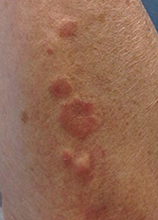
Diagnosis: Granuloma annulare
Granuloma annulare (GA) is a self-limited cutaneous disorder predominantly seen in women that affects children and adults. The cause is unknown. Inciting factors can include herpes zoster infection, sun exposure, medications, and trauma.
Several clinical variants exist. Localized GA is the most common form, often presenting in the first three decades of life as an asymptomatic, erythematous, annular plaque with a firm border and central clearing localized to the wrists, ankles, and dorsal hands or feet. Generalized GA accounts for 15% of reported cases and presents in the fourth to seventh decades of life as multiple asymptomatic or pruritic skin-colored or erythematous papules and plaques on the trunk and extremities. Subcutaneous GA is more common in children and presents as multiple painless nodules on the scalp or extremities. Patch GA can be localized or generalized. Perforating GA presents as asymptomatic erythematous papules that evolve into yellow, umbilicated papules with a clear-to-white discharge.
Histopathologically, an interstitial or palisading pattern is seen with a dermal lymphohistiocytic infiltrate, degenerated collagen, and mucin deposition (visualized with alcian blue or colloidal iron stains). The interstitial pattern presents in the majority of cases.
Diagnosis of GA is predominantly clinically based. When the diagnosis is questionable or the presentation is atypical, biopsy is useful. Granuloma annulare is often self-limiting and resolves within 2 years, although recurrence is possible. First-line therapy for localized GA includes high-potency topical corticosteroids or intralesional corticosteroids. Other treatments include cryotherapy, phototherapy, and topical tacrolimus. For generalized GA, topical or intralesional corticosteroids may be used for select lesions. Topical calcineurin inhibitors, light therapy, hydroxychloroquine, isotretinion, and dapsone also have been reported as treatments in the literature.

This case and photo were submitted by Orli Stern of Ross University and Dr. Donna Bilu Martin of Premier Dermatology, MD. A 60-year-old female with no significant past medical history presented with a 3-month history of asymptomatic, erythematous, firm, annular plaques on her bilateral proximal upper extremities and dorsal hands. The lesions have not been treated in the past.
Tips for Minimizing Recurrence of Seizures

Combo can fight infection, GVHD

Image courtesy of NIAID
NEW ORLEANS—Results of a phase 1 trial suggest that modified T cells can fight infection in patients who have undergone haploidentical hematopoietic
stem cell transplant (haplo-HSCT), and subsequent administration of a bio-inert drug can ameliorate graft-vs-host disease (GVHD) in these patients.
Researchers introduced the suicide gene inducible caspase 9 (iC9) into T cells and infused them into transplant recipients to promote immune reconstitution.
For patients who went on to develop GVHD, the researchers activated the suicide gene by administering a dose of the drug rimiducid (AP1903).
This cleared the patients of GVHD symptoms without jeopardizing the remaining T cells’ ability to fight infection.
The researchers presented these results at the American Society of Gene and Cell Therapy Annual Meeting and reported them in Blood.
The trial was sponsored by Baylor College of Medicine, but Bellicum Pharmaceuticals is the company developing rimiducid and the so-called iC9 “safety switch,” also known as CaspaCIDe.
“We’ve shown that the therapy works, fighting viruses that threaten immune-compromised patients,” said Xiaoou Zhou, PhD, of Baylor College of Medicine in Houston, Texas.
“We have also shown that the switch can turn off the T cells that reproduce out of control, attacking the patient’s graft-vs-host disease. This study was the first to look at any potential effect on the ability of the T cells to fight infection. We found there was no compromise.”
The study included 12 patients with a median age of 10 (range, 2-50) who had undergone haplo-HSCT. They received donor-derived T cells engineered with CaspaCIDe using a dose escalation schedule from 1×10⁴ to 5×10⁶ cells/kg, at a median of 42 days after transplant (range, 31-82 days).
All 12 patients had more rapid immune reconstitution and fewer infections after the infusions, when compared with previously reported results in T-cell-depleted, haplo-HSCT procedures. The CaspaCIDe T cells successfully provided protection from EBV, CMV, VZV, HHV6, and BKV viruses.
The researchers said there were no immediate toxicities related to the T-cell infusions, but 4 patients went on to develop GVHD.
Treatment with rimiducid resolved the patients’ GVHD symptoms within 6 to 48 hours. The researchers found that rimiducid could eliminate the uncontrolled T cells in the central nervous system as well as the peripheral blood.
Even after the problematic T cells were killed, the remaining T cells were able to fight infection without causing further GVHD.
One patient experienced a decrease in cell counts after receiving rimiducid, but counts had normalized by 48 hours. The researchers said there were no other immediate or delayed adverse effects associated with the drug.
One patient developed cytokine release syndrome, but this was resolved in 2 hours with a single dose of rimiducid.
“This study shows that infusing larger numbers of haploidentical donor T cells engineered with CaspaCIDe leads to better infection control,” Dr Zhou said. “We also showed that, if GVHD occurs, it can be rapidly controlled and eliminated by removing alloreactive cells with rimiducid in vivo, and that the productive, antiviral and anticancer cells remain, repopulate, and maintain immunity.”
“This is a significant finding that can lead to broader adoption of curative haploidentical transplants for cancers and genetic blood disorders. It also suggests that CaspaCIDe has great potential with CAR T and TCR therapies, where rapid control of dangerous T-cell-mitigated toxicities, such as cytokine release syndrome, is needed to achieve wide adoption.” ![]()
Group learns how protein promotes AML

A few years ago, researchers discovered that inhibiting the protein BRD4 can treat acute myeloid leukemia (AML). However, the mechanism that explains how the protein works has remained a mystery.
Now, investigators have discovered the larger signaling pathway to which BRD4 belongs. The team said their discovery points to additional therapeutic targets for AML and other malignancies.
The group described this work in Molecular Cell.
BRD4: A retrospective
In 2011, Christopher Vakoc, MD, PhD, of Cold Spring Harbor Laboratory in Cold Spring Harbor, New York, and his colleagues identified potential drug targets for AML using RNA interference. Out of that screen came BRD4 and the realization that leukemia cells were “extremely sensitive” to inhibition of this protein.
In a bit of serendipity, drugs to inhibit BRD4 had just been developed for other purposes. Dr Vakoc and his colleagues tested these drugs and found that one in particular, JQ1, worked well against a mouse model of aggressive AML.
In the past few years, several groups have reported similar therapeutic results in mice using JQ1 and closely related drugs.
“It’s been very satisfying to see that other groups have independently validated our findings,” Dr Vakoc said.
Due to the evidence of their effectiveness in mice, inhibitors of BRD4 moved into clinical trials starting in 2013. Currently, there are 12 active clinical trials targeting BRD4 in leukemia and other cancers, including one sponsored by a company to which Dr Vakoc has licensed JQ1.
“Once we published the first paper in 2011, the main objective in our lab has been to understand why these drugs work,” Dr Vakoc said. “Knowing the mechanism of action of a drug is essential to making the drug better because there will likely be many generations of BRD4 inhibitors.”
JQ1 and BRD4: How they work
In the current study, Dr Vakoc and his colleagues discovered that BRD4 works very closely with transcription factors that bind to DNA and selectively control the activity of certain genes. The transcription factors control blood formation and essentially give blood cells their “identity.”
The researchers found that JQ1 can make leukemia cells shed their leukemia characteristics and differentiate into normal white blood cells. The team also identified an intermediary molecule called p300 between BRD4 and the leukemia-associated transcription factors.
Active areas of research in Dr Vakoc’s lab include exploring other players in the pathway, particularly the molecules that BRD4 controls, and learning more about the transcription factors.
“This new work is leading us to realize that transcription factors are the masters of the biological universe,” Dr Vakoc said. “Clearly, they are driving the cancer phenotype.” ![]()
Genome editing increases hemoglobin production

Scientists have found they can use a genome-editing technique to increase hemoglobin production, and they described this method in Nature Communications.
The team used transcription activator-like effector nucleases (TALENs) to introduce the hereditary persistence of fetal hemoglobin (HPFH)-175T4C point mutation into erythroid cell lines.
This served to switch on the fetal hemoglobin gene and increase hemoglobin production.
“Our laboratory study provides a proof of concept that changing just one letter of DNA in a gene could alleviate the symptoms of sickle cell anemia and thalassemia—inherited diseases in which people have damaged hemoglobin,” said Merlin Crossley, PhD, of the University of New South Wales in Sydney, Australia.
“Because the good genetic variation we introduced already exists in nature, this approach should be effective and safe. However, more research is needed before it can be tested in people as a possible cure for serious blood diseases.”
Dr Crossley and his colleagues introduced the HPFH-175T4C point mutation into erythroid cell lines using TALENs, which can be designed to cut a gene at a specific point, as well as provide the desired piece of donor DNA for insertion.
“Breaks in DNA can be lethal to cells, so they have in-built machinery to repair any nicks as soon as possible, by grabbing any spare DNA that seems to match . . . ,” Dr Crossley explained.
“We exploited this effect. When our genome-editing protein cuts the DNA, the cell quickly replaces it with the donor DNA that we have also provided.”
The researchers pointed out that the HPFH-175T4C point mutation increased fetal globin expression via de novo recruitment of the activator TAL1, which promoted chromatin looping of distal enhancers to the modified γ-globin promoter.
The team also said that, if their editing technique proves safe and effective in hematopoietic stem cells, it could offer significant advantages over other approaches used to treat hemoglobin disorders, such as conventional gene therapy. ![]()
FDA grants drug orphan designation for AML

Image by Lance Liotta
The US Food and Drug Administration (FDA) has granted orphan designation for GMI-1271 to treat acute myeloid leukemia (AML).
GMI-1271, an E-selectin antagonist, has shown promise in preclinical research and proven safe in a phase 1 trial of healthy volunteers, according to GlycoMimetics, Inc., the company developing the drug.
Now, the company is recruiting adults with AML in a phase 1/2 study to test GMI-1271 in combination with chemotherapy.
“Having the FDA designate GMI-1271 as an orphan drug for the treatment of AML is an important accomplishment for GlycoMimetics,” said Helen Thackray, MD, vice president of clinical development and chief medical officer at GlycoMimetics. “This is a significant regulatory milestone for our program.”
The FDA’s orphan drug designation program is designed to promote the development of drugs intended to treat diseases affecting fewer than 200,000 people in the US.
Orphan designation provides a drug’s developer with benefits such as a 7-year period of marketing exclusivity if the drug is approved, protocol assistance, the ability to apply for research funding, tax credits for certain research expenses, and regulatory fee waivers.
Research with GMI-1271
Preclinical research presented at ASH 2013 suggested that GMI-1271 was able to overcome chemotherapy resistance in AML cells in vitro. The drug also reduced the leukemic burden in mouse models of AML when given in combination with daunorubicin and cytarabine.
Murine research presented at ASH 2014 suggested that, by inhibiting E-selectin, GMI-1271 may increase leukemic stem cells’ sensitivity to chemotherapeutic drugs.
At the same meeting, researchers presented preclinical data on GMI-1271 as a treatment for chronic myeloid leukemia, multiple myeloma, and venous thromboembolism.
In November 2014, GlycoMimetics announced results of phase 1 trial of GMI-1271 in healthy volunteers. Twenty-eight adults were enrolled in cohorts to receive the drug at 3 dose levels.
The company said subjects tolerated GMI-1271 well, and pharmacokinetics were as predicted based on preclinical research.
A multicenter, phase 1/2 trial of GMI-1271 in combination with chemotherapy is now recruiting adult patients with AML. ![]()