User login
DDW: Menopausal hormone therapy increases major GI bleed risk
Menopausal hormone therapy is associated with an increased risk of major gastrointestinal bleeding, particularly in the lower gastrointestinal tract, that is associated with duration of use, a study has found.
Analysis of data from 73,863 women enrolled in the Nurses’ Health Study II in 1989 showed that current users of menopausal hormone therapy had a 46% increase in the risk of a major gastrointestinal bleed and a more than twofold increase in the risk of a lower GI bleed or ischemic colitis, compared with never users, said Dr. Prashant Singh of Massachusetts General Hospital, Boston.
Past users showed a much smaller increase risk of bleeding, while increasing duration of hormone therapy was significantly associated with increasing risk of major and low gastrointestinal bleeding.
“Although our findings show that menopausal hormone therapy may increase the risk of major GI bleeding, especially in the lower GI tract, it is important for these patients to know that this therapy is still an effective treatment; however, both clinician and patient should be more cautious in using this therapy in some cases, such as with patients who have a history of ischemic colitis,” Dr. Singh said at the annual Digestive Disease Week.
Dr. Singh does not have any relevant financial or other relationship with any manufacturer or provider of commercial products or services that he discussed during the presentation.
FROM DDW 2015
Key clinical point: Menopausal hormone therapy is associated with an increased risk of major gastrointestinal bleeding, particularly in the lower gastrointestinal tract.
Major finding: Current users of menopausal hormone therapy had a 46% increase in the risk of a major gastrointestinal bleed and a more than twofold increase in the risk of a lower GI bleed or ischemic colitis.
Data source: Analysis of data from 73,863 women enrolled in the Nurses’ Health Study II.
Disclosures: No conflicts of interest were disclosed.
DDW: Urinary enzymes hint at gastric cancer
WASHINGTON – A simple urine test could detect gastric cancer even at an early stage, the test’s developers say.
The test, which looks for the presence of two metalloprotease enzymes labeled ADAM 12 and MMP-9/NGAL had 77.1% sensitivity and 82.9% specificity for gastric cancer when tested in 35 patients with the malignancy and an equal number of healthy controls, reported Dr. Takaya Shimura from the department of surgery at Boston Children’s Hospital and Harvard Medical School in Boston.
“This study represents the first demonstration of the presence of ADAM 12 and MMO-9/NGAL complex in the urine of gastric cancer patients,” he said at the annual Digestive Disease Week.
Dr. Stephen J. Meltzer of Johns Hopkins University, Baltimore, commented in an interview that the findings are convincing but preliminary.
A randomized clinical trial enrolling a larger number of patients and controls would be required before he would consider screening patients for the enzymes, said Dr. Meltzer, who was not involved in the study and comoderated the meeting session where the results were presented.
ADAM 12 (a disintegrin and metalloprotease 12) and MMP-9 (matrix metalloprotease 9) are both members of a family of enzymes involved in cellular adhesion, invasion, growth, and angiogenesis, Dr. Shimura explained. MMP-9, when complexed with NGAL (neutrophil gelatinase associated lipocalin) is protected from autodegradation.
The investigators, from the lab of Dr. Marsha A. Moses at Boston Children’s Hospital, and their collaborators in Japan had previously reported that MMPs in urine were independent predictors of both organ-confined and metastatic cancer.
Urinary assays are noninvasive, using easily accessed tissues that can be handled simply and inexpensively, making them ideal for cancer detection, Dr, Shimura said.
Current tests for gastric cancer, such as carcinoembryonic antigen (CEA) and cancer antigens (CA) 19-9 and 72-4, have poor sensitivity for detecting advanced disease, and are even worse at spotting early disease, he noted.
To see whether they could improve on the current lot of tests, the investigators enrolled 106 patients in a case-control study, settling eventually, after age and sex matching, on a cohort of 70 patients: 35 with primarily early-stage gastric cancer, and 35 healthy controls.
After screening the urine of participants for about 50 different antigenic proteins, they found that the patients with gastric cancer had significantly higher levels in their urine of both ADAM 12 (P < .001) and the MMP-9/NGAL complex (P = .020).
In a multivariate analysis, they showed that both enzymes were strong, independent predictors of gastric cancer, with an odds ratio for urinary MMO-9/NGAL of 6.71 (P = .002), and an OR of 15.4 for ADAM 12 (P = .002). In contrast, Helicobacter pylori infection was associated with a nonsignificant OR of 2.54.
In a receiver operating characteristic (ROC) analysis, they also found that MMP-9/NGAL was associated with an area-under-the curve (AUC) of 0.657 (P = .024), ADAM 12 was associated with an AUC of 0.757 (P < .001), and that the two combined had an AUC of 0.825 (P < .001).
As noted before, the sensitivity of the combined enzymes was 77%, and the specificity was 83%.
Finally, using immunohistochemical analysis, the investigators were able to show that gastric cancer tissues had high levels of coexpression of MMP-9 and NGAL (P <.001) and high expression levels of ADAM 12 (P < .001), compared with adjacent normal tissues.
AT DDW 2015
Key clinical point: Urinary levels of two metalloproteases were significantly elevated in the urine of patients with gastric cancer, compared with controls.
Major finding: High expression of ADAM 12 and MMP-9/NGAL complex had a 77% sensitivity and 83% specificity for gastric cancer.
Data source: Case-control study of 35 patients with gastric cancer and 35 controls.
Disclosures: The study was supported by the Advanced Medical Research Foundation in the United States and the Research Fellowship of the Uehara Memorial Foundation, Japan. Dr. Shimura reported having no conflicts of interest.
DDW: Scheduled for a colonoscopy? Pass the pretzels!
WASHINGTON – A novel, edible colon preparation could make obsolete the fasting and large volume of salty liquid cleansing that keep many a patient from completing their colonoscopies.
A pilot study showed that all 10 patients who ate a series of nutritionally balanced meals, drinks, and snacks such as pretzels and pudding had a successful colon cleansing according to the endoscopist at the time of the procedure, Dr. L. Campbell Levy of Dartmouth-Hitchcock Medical Center in Lebanon, N.H., reported at the annual Digestive Disease Week.
The preparation, which is blended with polyethylene glycol 3350, sorbitol, and ascorbic acid, did not produce any significant changes in electrolytes or creatinine.
There were no adverse events and, equally important, all 10 patients said that they would follow the edible bowel regimen again for a subsequent procedure.
In a video interview, he discussed the small study’s results and the plans for larger, randomized studies.
Dr. Levy reported no relevant conflicts. The inventor of the diet and the founder of Colonary Concepts were involved in the study.
The video associated with this article is no longer available on this site. Please view all of our videos on the MDedge YouTube channel
On Twitter @pwendl
AT DDW® 2015

Wells score not effective at inpatient DVT detection
While effective in outpatient settings, the Wells score was not effective at detecting deep vein thrombosis in an inpatient setting, according to Dr. Patricia Silveira and her associates from Harvard Medical School, Boston.
Of 1,135 patients included in the study, 137 had proximal DVT. DVT incidence in the low, medium, and high pretest probability groups were 5.9%, 9.5%, and 16.4%, respectively. Although statistically significantly different, this is a very narrow range, in contrast to findings in previous studies, where incidence among the three groups was 3.0%, 16.6%, and 74.6% respectively.
The AUC for the accuracy of the Wells score was 0.6, only slightly better than chance. The failure rate in the low pretest group was 5.9%, and the efficiency was 11.9%.
“In inpatients,Wells DVT scores are inflated by comorbidities and nonspecific physical findings common among hospitalized patients, leaving very few patients in the low-probability Wells score category, and many patients without DVT in the moderate- and high-probability categories,” Dr. Erika Leemann Price and Dr. Tracy Minichiello wrote in a related editorial.
Find the full study and editorial in JAMA Internal Medicine (doi:10.1001/jamainternmed.2015.1687; doi:10.1001/jamainternmed.2015.1699).
MRD doesn’t suggest need for more treatment
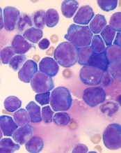
© Hind Medyouf, German
Cancer Research Center
A new study suggests that minimal residual disease (MRD) alone is not predictive of outcomes in children with T-cell acute lymphoblastic leukemia (T-ALL).
Study investigators analyzed a small group of T-ALL patients who received similar treatment regimens, comparing those with and without MRD after induction.
None of the MRD-positive patients relapsed within the follow-up period, despite not receiving intensified treatment to fully eradicate their disease.
These results, published in Pediatric Blood & Cancer, suggest T-ALL patients with MRD may not need intensified treatment and can therefore avoid additional toxicities.
“Until now, the dogma has been that, for patients with leukemia who have minimal residual disease at the end of induction, we need to intensify their treatment, which also increases side effects,” said study author Hisham Abdel-Azim, MD, of Children’s Hospital Los Angeles in California.
“We have found, for T-ALL, patients have excellent outcomes without therapy intensification and its associated toxicities.”
Dr Abdel-Azim and his colleagues studied 33 children (ages 1 to 21) with newly diagnosed T-ALL. Their treatment included induction, augmented consolidation, interim maintenance (high-dose [5 g/m2] or escalating-dose intravenous methotrexate), 1 delayed intensification, and maintenance. Twenty-one patients underwent cranial irradiation, and 1 received a transplant.
After induction, 19 of the 32 patients analyzed had MRD, which was defined as ≥ 0.01% residual leukemia cells. At the end of consolidation, 6 of the 11 patients tested were MRD-positive. And at the end of interim maintenance, 2 of the 4 patients tested were MRD-positive.
The 19 patients who were MRD-positive after induction were in continuous complete remission at a median follow-up of 4 years (range, 1.3-7.1 years). The same was true for 13 of the 14 MRD-negative patients. One of the MRD-negative patients relapsed 18 months after diagnosis but was still alive with refractory disease at last follow-up.
Dr Abdel-Azim and his colleagues noted that there were no significant differences in treatment variables between MRD-positive patients and MRD-negative patients. However, 1 patient did receive a transplant for rising MRD at 5.4 months after diagnosis.
The investigators also said they did not find any associations between MRD positivity after induction and patients’ age, sex, ethnicity, weight, white blood cell count at diagnosis, cytogenetics, immunophenotype, or the type of steroid they received during induction therapy.
The team said these results may be explained by the fact that clearance of leukemia cells from the blood is slower in patients with T-ALL than in those with B-cell ALL. However, the leukemia cells ultimately clear in T-ALL without changes in therapy. ![]()
Team discovers target for antimalarial drugs

a red blood cell
Image courtesy of St. Jude
Children’s Research Hospital
New research indicates that manipulating the permeability of parasitophorous vacuoles could defeat malaria parasites.
The researchers unearthed this finding while studying the way in which the Toxoplasma gondii parasite, which causes toxoplasmosis, and Plasmodium parasites,
which cause malaria, access vital nutrients from their host cells.
The team described this work in Cell Host & Microbe.
Roughly a third of the world’s deadly infectious diseases are caused by pathogens that spend a large portion of their life inside parasitophorous vacuoles. This type of vacuole separates the host cytoplasm and the parasite by a membrane, protecting the parasite from the host cell’s defenses and providing an environment tailored to the parasite’s needs.
However, the membrane of the vacuole also acts as a barrier between the parasite and the host cell. This makes it more difficult for the parasite to release proteins involved in the transformation of the host cell beyond the membrane to spread the disease and for the pathogen to gain access to vital nutrients.
“Ultimately, what defines a parasite is that they require certain key nutrients from their host,” said study author Dan Gold, PhD, of the Massachusetts Institute of Technology in Cambridge.
“So they have had to evolve ways to get around their own barriers to gain access to these nutrients.”
Previous research suggested the vacuoles are selectively permeable to small molecules, allowing certain nutrients to pass through pores in the membrane. But until now, no one has been able to determine the molecular makeup of these pores or how they are formed.
When studying Toxoplasma, Dr Gold and his colleagues discovered 2 proteins secreted by the parasite, known as GRA17 and GRA23, which are responsible for forming these pores in the vacuole. The researchers discovered the proteins’ roles by accident while investigating how the parasites are able to release their own proteins out into the host cell beyond the vacuole membrane.
Similar research into how the related Plasmodium pathogens perform this trick revealed a protein export complex that transports encoded proteins from a parasite into its host red blood cell, which transforms the cell in a way that is vital to the spread of malaria.
“The clinical symptoms of malaria are dependent on this process and this remodeling of the red blood cell that occurs,” Dr Gold said.
The researchers identified proteins secreted by Toxoplasma that appeared to be homologues of this protein export complex in Plasmodium. But when they stopped these proteins from functioning, the team found it made no difference to the export of proteins from the parasite beyond the vacuole.
“We were left wondering what GRA17 and GRA23 actually do if they are not involved in protein export, and so we went back to look at this longstanding phenomenon of nutrient transport,” Dr Gold said.
When they added dyes to the host cell and knocked out the 2 proteins, the researchers found that it prevented the dyes flowing into the vacuole.
“That was our first indication that these proteins actually have a role in small-molecule transfer,” Dr Gold said.
When the researchers expressed a Plasmodium export complex gene in the modified Toxoplasma, they found the dyes were able to flow into the vacuole once again, suggesting this small-molecule transport function had been restored.
Since these proteins are only found in the parasite phylum Apicomplexa, to which both Toxoplasma and Plasmodium belong, they could be used as a drug target against the diseases they cause, the researchers said.
“This very strongly suggests that you could find small-molecule drugs to target these pores, which would be very damaging to these parasites but likely wouldn’t have any interaction with any human molecules,” Dr Gold said. “So I think this is a really strong potential drug target for restricting the access of these parasites to a set of nutrients.”![]()
AAN: Finding ways to improve door-to-needle times in stroke treatment
WASHINGTON – A streamlined emergency care service and a low-cost, tablet-based mobile telestroke system are two examples of shortening the time it takes for acute ischemic stroke patients to receive thrombolytic therapy that were presented at the annual meeting of the American Academy of Neurology.
American Heart Association/American Stroke Association guidelines recommend a door-to-needle (DTN) time of 60 minutes or less and set a goal for participating hospitals to administer tissue plasminogen activator (TPA) to at least 50% of their patients with acute ischemic stroke within 60 minutes of arriving at the hospital.

Dr. Judd Jensen described the efforts of Swedish Medical Center, Englewood, Colo. to streamline the emergency care of patients suspected of having an acute ischemic stroke after a task force determined that their previous “sequential, step-by-step process” wasted time. The median DTN time at the hospital’s stroke center had dropped from 46 minutes in 2010 to 39 minutes in 2013, which was better than the national average, “but we felt we could do better,” said Dr. Jensen, a neurologist at the hospital.
The process was modified so that more of the activities take place simultaneously, which includes immediately sending patients for a CT scan before entering the emergency department and administering IV TPA in the CT area to eligible patients, he explained. Previously, these patients were taken to a bed in the ED on arrival, registered, then examined by the emergency physician and neurologist and transported for a CT scan and then transported back to the ED where TPA was administered, if indicated, after several other steps were completed, including interpreting the CT scan, deciding about treatment, acquiring consent, and contacting the pharmacy to mix the TPA.
This process was improved by increasing pre-hospital notification by emergency medical services (EMS) and establishing a “launchpad” area in the back of the ED where the stroke team meets after EMS notification. On arrival, patients are transferred directly to the CT room where they are examined. The pharmacy is instructed to mix the TPA if an ischemic stroke is suspected, and the TPA is brought to the CT room where a stroke neurologist evaluates the CT scan and TPA is administered if indicated.
The impact of the revised process was evaluated in a prospective study of 262 acute ischemic stroke patients who received IV TPA between January 2010 and December 2014 at the hospital. They had a mean age of 73 years, 44% were male, and 84% were white. Their mean initial National Institutes of Health Stroke Scale (NIHSS) score was 12. The median DTN times dropped to a median of 31 minutes in 2014, Dr. Jensen said.
In 2014, almost 50% of the patients received TPA in 30 minutes or less, compared with about 25% in 2011, 2012, and 2013, he added, noting that 11 minutes was the fastest DTN time in 2014. Patients with an excellent discharge modified Rankin Scale (mRS) score (0 or 1) improved from 31% in 2010 and 30% in 2013 to 46% in 2014. During the time period studied, two patients had a symptomatic intracerebral hemorrhage, one in 2010 and another in 2012.
Dr. Jensen described the process as a multidisciplinary team effort, noting that it is important that emergency room physicians feel comfortable with the administration of TPA in the CT scan area, “because it is still their patient being administered a potentially fatal drug outside of the ED.”
At the meeting, Matthew Padrick, a medical student at the University of Virginia, Charlottesville, presented the results of a pilot study that targeted the EMS transport time as an “untapped treatment window” to improve the time to thrombolytic treatment using a low-cost mobile telestroke system to evaluate patients in the ambulance on their way to the hospital.
Because the catchment area covered by UVA includes a large rural area, transport times to the stroke center can be as long as 30 to 60 minutes, Mr. Padrick said.
In the “Improving Treatment with Rapid Evaluation of Acute Stroke via mobile Telemedicine” (iTREAT) study, he and his associates evaluated the feasibility and reliability of performing acute stroke assessments (with the NIHSS) in the ambulance. The iTREAT system, which includes an Apple iPad with retina display attached to the patient stretcher with an extendable clamp, a secure video conferencing application, a high-speed 4G LTE modem, a magnetic antenna on top of the ambulance, and the regional cellular network, “providing seamless connectivity,” he said. At a total cost of under $2,000, the system is designed so that the neurologist can evaluate the patient remotely, via the iPad.
Acting as patients, three medical students were given two unique stroke scenarios each, with stories and specific instructions; vascular neurologists did a face-to-face assessment and a remote iTREAT assessment from the hospital as the students traveled along the major routes to UVA Medical Center. NIHSS scores in the ambulance with the iTREAT system and with face-to-face assessments correlated well, with an overall intraclass correlation of 0.98, Mr. Padrick reported.
The ratings of audio-video quality during the iTREAT evaluations were judged to be ”good” or “excellent” and the NIHSS correlations and audio-video quality ratings improved with time, he added.
“We currently have IRB approval to move forward with real, live patient encounters and we are currently outfitting and training our local EMS agencies” with the system, Mr. Padrick said in an interview after the meeting.
Mr. Padrick has received research support from the American Heart Association. Dr. Judd had nothing to disclose.
WASHINGTON – A streamlined emergency care service and a low-cost, tablet-based mobile telestroke system are two examples of shortening the time it takes for acute ischemic stroke patients to receive thrombolytic therapy that were presented at the annual meeting of the American Academy of Neurology.
American Heart Association/American Stroke Association guidelines recommend a door-to-needle (DTN) time of 60 minutes or less and set a goal for participating hospitals to administer tissue plasminogen activator (TPA) to at least 50% of their patients with acute ischemic stroke within 60 minutes of arriving at the hospital.
Dr. Judd Jensen described the efforts of Swedish Medical Center, Englewood, Colo. to streamline the emergency care of patients suspected of having an acute ischemic stroke after a task force determined that their previous “sequential, step-by-step process” wasted time. The median DTN time at the hospital’s stroke center had dropped from 46 minutes in 2010 to 39 minutes in 2013, which was better than the national average, “but we felt we could do better,” said Dr. Jensen, a neurologist at the hospital.
The process was modified so that more of the activities take place simultaneously, which includes immediately sending patients for a CT scan before entering the emergency department and administering IV TPA in the CT area to eligible patients, he explained. Previously, these patients were taken to a bed in the ED on arrival, registered, then examined by the emergency physician and neurologist and transported for a CT scan and then transported back to the ED where TPA was administered, if indicated, after several other steps were completed, including interpreting the CT scan, deciding about treatment, acquiring consent, and contacting the pharmacy to mix the TPA.
This process was improved by increasing pre-hospital notification by emergency medical services (EMS) and establishing a “launchpad” area in the back of the ED where the stroke team meets after EMS notification. On arrival, patients are transferred directly to the CT room where they are examined. The pharmacy is instructed to mix the TPA if an ischemic stroke is suspected, and the TPA is brought to the CT room where a stroke neurologist evaluates the CT scan and TPA is administered if indicated.
The impact of the revised process was evaluated in a prospective study of 262 acute ischemic stroke patients who received IV TPA between January 2010 and December 2014 at the hospital. They had a mean age of 73 years, 44% were male, and 84% were white. Their mean initial National Institutes of Health Stroke Scale (NIHSS) score was 12. The median DTN times dropped to a median of 31 minutes in 2014, Dr. Jensen said.
In 2014, almost 50% of the patients received TPA in 30 minutes or less, compared with about 25% in 2011, 2012, and 2013, he added, noting that 11 minutes was the fastest DTN time in 2014. Patients with an excellent discharge modified Rankin Scale (mRS) score (0 or 1) improved from 31% in 2010 and 30% in 2013 to 46% in 2014. During the time period studied, two patients had a symptomatic intracerebral hemorrhage, one in 2010 and another in 2012.
Dr. Jensen described the process as a multidisciplinary team effort, noting that it is important that emergency room physicians feel comfortable with the administration of TPA in the CT scan area, “because it is still their patient being administered a potentially fatal drug outside of the ED.”
At the meeting, Matthew Padrick, a medical student at the University of Virginia, Charlottesville, presented the results of a pilot study that targeted the EMS transport time as an “untapped treatment window” to improve the time to thrombolytic treatment using a low-cost mobile telestroke system to evaluate patients in the ambulance on their way to the hospital.
Because the catchment area covered by UVA includes a large rural area, transport times to the stroke center can be as long as 30 to 60 minutes, Mr. Padrick said.
In the “Improving Treatment with Rapid Evaluation of Acute Stroke via mobile Telemedicine” (iTREAT) study, he and his associates evaluated the feasibility and reliability of performing acute stroke assessments (with the NIHSS) in the ambulance. The iTREAT system, which includes an Apple iPad with retina display attached to the patient stretcher with an extendable clamp, a secure video conferencing application, a high-speed 4G LTE modem, a magnetic antenna on top of the ambulance, and the regional cellular network, “providing seamless connectivity,” he said. At a total cost of under $2,000, the system is designed so that the neurologist can evaluate the patient remotely, via the iPad.
Acting as patients, three medical students were given two unique stroke scenarios each, with stories and specific instructions; vascular neurologists did a face-to-face assessment and a remote iTREAT assessment from the hospital as the students traveled along the major routes to UVA Medical Center. NIHSS scores in the ambulance with the iTREAT system and with face-to-face assessments correlated well, with an overall intraclass correlation of 0.98, Mr. Padrick reported.
The ratings of audio-video quality during the iTREAT evaluations were judged to be ”good” or “excellent” and the NIHSS correlations and audio-video quality ratings improved with time, he added.
“We currently have IRB approval to move forward with real, live patient encounters and we are currently outfitting and training our local EMS agencies” with the system, Mr. Padrick said in an interview after the meeting.
Mr. Padrick has received research support from the American Heart Association. Dr. Judd had nothing to disclose.
AT THE AAN 2015 ANNUAL MEETING

Combo targets AML, BL in the same way

Image by Ed Uthman
Combining a cholesterol-lowering drug and a contraceptive steroid could be a safe, effective treatment for leukemias, lymphomas, and other malignancies, according to researchers.
Their work helps explain how this combination treatment, bezafibrate and medroxyprogesterone acetate (BaP), kills cancer cells.
The team discovered that BaP’s mechanism of action is the same in acute myeloid leukemia (AML) and Burkitt lymphoma (BL) cells.
The findings have been published in Cancer Research.
BaP has been shown to prolong survival in early stage trials of elderly AML patients, when compared to standard palliative care. BaP has also been used alongside chemotherapy to successfully treat children with BL.
However, it was unclear whether BaP’s activity against these 2 very different malignancies was mediated by a common mechanism or by different effects in each cancer.
To gain some insight, Andrew Southam, PhD, of the University of Birmingham in the UK, and his colleagues investigated the drugs’ effects on the metabolism and chemical make-up of AML and BL cells.
They found that, in both cell types, BaP blocks stearoyl CoA desaturase, an enzyme crucial to the production of fatty acids, which cancer cells need to grow and multiply. The team also showed that BaP’s ability to deactivate stearoyl CoA desaturase was what prompted the cancer cells to die.
“Developing drugs to target the fatty-acid building blocks of cancer cells has been a promising area of research in recent years,” Dr Southam said. “It is very exciting we have identified these non-toxic drugs already sitting on pharmacy shelves.”
He and his colleagues believe these findings also open up the possibility that BaP could be used to treat other cancers that rely on high levels of stearoyl CoA desaturase to grow, including chronic lymphocytic leukemia and some types of non-Hodgkin lymphoma, as well as prostate, colon, and esophageal cancers.
“This drug combination shows real promise,” said Chris Bunce, PhD, also of the University of Birmingham.
“Affordable, effective, non-toxic treatments that extend survival, while offering a good quality of life, are in demand for almost all types of cancer.” ![]()
Consider laser ablation therapy for treatment of benign thyroid nodules
NASHVILLE – Ultrasound-guided laser ablation therapy was found to be a clinically safe, effective, and well-tolerated option for the treatment of benign thyroid nodules, both solid and mixed, in a retrospective, multicenter study presented at the annual meeting of the American Association of Clinical Endocrinologists.
“We know that image-guided laser ablation of solid thyroid nodules has demonstrated favorable results in several prospective randomized trials,” said Dr. Enrico Papini of Regina Apostolorum Hospital in Rome. “However, these results were obtained in selected patients, with single treatments and fixed modalities of treatment; so the question is, what happens in a real. clinical practice?”
Dr. Papini explained that the aim of the study was to assess clinical efficacy and side effects of laser ablation therapy (LAT) in a large series of unselected benign thyroid nodules of variable structure and size, using data from centers who use LAT as a standard operating technique. Patients with solid or mixed nodules with up to 40% fluid composition, benign cytological findings, and normal thyroid function were included.
Clinical records of 1,534 thyroid nodules in 1,531 patients, all of whom were treated in the last 10 years, was collected from eight Italian thyroid referral centers. A total of 1,837 LAT procedures were performed on these nodules, of which 1,280 (83% of 1,534) were treated in a single session. All nodules were treated in no more than three consecutive sessions, with a fixed output power of 3 watts. According to Dr. Papini, the laser is only fired for up to 10 minutes.
Mean nodule volume significantly decreased following LAT from 27 ± 24 mL at baseline to 8 ± 8 mL at 12 months after treatment (P < .001), and mean nodule volume reduction was 72% ± 11%, with an overall range of 48%-100%. Mixed nodules experienced significantly larger decreases than solid ones. On average, mixed nodule volume decreased 79% ± 7%, versus 72% ± 11% for solid nodules (P < .001) because of fluid components being drained prior to LAT.
Symptoms decreased from 49% at baseline to 10% at 12 months post-treatment. Similarly robust reductions were also seen in cosmetic signs, which decreased 86% to 8% over 12 months. Only 17 patients experienced a complication, including 8 with a “major” complication of dysphonia, which resolved within 2-84 days, and 9 with “minor” complications, such as skin burn and hematoma.
“Laser ablation was performed in outpatient setting, with no hospital admission after treatment,” said Dr. Papini. “It was well-tolerated, and severe pain – requiring more than 3 days of analgesics – occurred in less than 2% of patients.”
Dr. Papini did not report any relevant financial disclosures.
AT AACE 2015
Key clinical point: Ultrasound-guided laser ablation therapy is a clinically effective and well-tolerated tool for treating benign solid and mixed thyroid nodules.
Major finding: In 1,837 treatments for 1,534 nodules, mean nodule volume decreased from 27 ± 24 mL at baseline to 8 ± 8 mL at 12 months after treatment (P < .001), and mean nodule volume reduction was 72% ± 11% (range 48%-100%).
Data source: Retrospective, multicenter study of 1,534 benign solid and mixed thyroid nodules.
Disclosures: Dr. Papini did not report any relevant financial disclosures.
Botox treatments improve urinary incontinence in neurogenic bladder dysfunction
NEW ORLEANS – Regular injections of onabotulinumtoxinA significantly decreased urinary incontinence in patients with neurogenic detrusor bladder overactivity over 4 years of follow-up in 4-year extension study results of a randomized trial.
Incontinence episodes decreased from an average of four per day to one or less after each treatment, Dr. Eric Rovner said at the annual meeting of the American Urological Association.
Each treatment was effective for about 9 months, and the benefit consistent throughout the 4-year study, said Dr. Rovner of the Medical University of South Carolina, Charleston.

About 90% of patients had at least a 50% reduction in incontinence episodes, and more than half experienced a complete cessation of incontinence.
OnabotulinumtoxinA (Botox) was approved in 2011 as a treatment for neurogenic urinary incontinence. Each treatment consists of 20 injections delivered cystoscopically.
Dr. Rovner reported a post hoc analysis of 227 patients who completed 4 years of treatment – a 1-year placebo-controlled trial, and 3 years of open-label extension with a dosage of 200 units of onabotulinumtoxinA.
Patients were relatively young (mean 45 years); about half were male. Most (53%) had multiple sclerosis. The remainder had a spinal cord injury that affected bladder function. Half were taking an anticholinergic medication, but had not responded to it.
Most patients (71%) were already performing intermittent catheterization. Despite that, they had a mean of four incontinence episodes each day.
Over the entire 4 years, onabotulinumtoxinA was associated with significant and consistent improvements in incontinence, with a mean decrease of up to 3.8 incidents per day each year. Each year, about 90% experienced at least a 50% improvement. About half experienced a complete cessation of incontinence over the period.
Urinary tract infections occurred in 20% of patients in years 1 and 2, and 18% in years 3 and 4, which was not significantly different. Urinary retention was highest in year 1 (12%) and dropped to 2% by years 3 and 4.
In the first year, 39% of those who didn’t need intermittent catheterization at baseline had to begin doing so. By year 2, the de novo catheterization rate was 11%. It was 8% in year 3, and in year 4, there were no new catheterizations.
These changes were not only statistically significant, but clinically important, Dr. Rovner said. On a secondary measure, the Incontinence Quality of Life Questionnaire (I-QOL), patients experienced a mean increase of more than 30 points over each study year. An 11-point change is usually considered clinically meaningful, he said.
“This was making a big difference for these patients.”
Dr. Rovner disclosed relationships with Allergan and a number of other pharmaceutical and medical device companies.
On Twitter @alz_gal
NEW ORLEANS – Regular injections of onabotulinumtoxinA significantly decreased urinary incontinence in patients with neurogenic detrusor bladder overactivity over 4 years of follow-up in 4-year extension study results of a randomized trial.
Incontinence episodes decreased from an average of four per day to one or less after each treatment, Dr. Eric Rovner said at the annual meeting of the American Urological Association.
Each treatment was effective for about 9 months, and the benefit consistent throughout the 4-year study, said Dr. Rovner of the Medical University of South Carolina, Charleston.
About 90% of patients had at least a 50% reduction in incontinence episodes, and more than half experienced a complete cessation of incontinence.
OnabotulinumtoxinA (Botox) was approved in 2011 as a treatment for neurogenic urinary incontinence. Each treatment consists of 20 injections delivered cystoscopically.
Dr. Rovner reported a post hoc analysis of 227 patients who completed 4 years of treatment – a 1-year placebo-controlled trial, and 3 years of open-label extension with a dosage of 200 units of onabotulinumtoxinA.
Patients were relatively young (mean 45 years); about half were male. Most (53%) had multiple sclerosis. The remainder had a spinal cord injury that affected bladder function. Half were taking an anticholinergic medication, but had not responded to it.
Most patients (71%) were already performing intermittent catheterization. Despite that, they had a mean of four incontinence episodes each day.
Over the entire 4 years, onabotulinumtoxinA was associated with significant and consistent improvements in incontinence, with a mean decrease of up to 3.8 incidents per day each year. Each year, about 90% experienced at least a 50% improvement. About half experienced a complete cessation of incontinence over the period.
Urinary tract infections occurred in 20% of patients in years 1 and 2, and 18% in years 3 and 4, which was not significantly different. Urinary retention was highest in year 1 (12%) and dropped to 2% by years 3 and 4.
In the first year, 39% of those who didn’t need intermittent catheterization at baseline had to begin doing so. By year 2, the de novo catheterization rate was 11%. It was 8% in year 3, and in year 4, there were no new catheterizations.
These changes were not only statistically significant, but clinically important, Dr. Rovner said. On a secondary measure, the Incontinence Quality of Life Questionnaire (I-QOL), patients experienced a mean increase of more than 30 points over each study year. An 11-point change is usually considered clinically meaningful, he said.
“This was making a big difference for these patients.”
Dr. Rovner disclosed relationships with Allergan and a number of other pharmaceutical and medical device companies.
On Twitter @alz_gal
AT THE AUA ANNUAL MEETING
Key clinical point: OnabotulinumtoxinA injections produced a consistent and significant improvement in urinary incontinence in patients with neurogenic detrusor bladder overactivity.
Major finding: Nearly 90% of patients had at least a 50% improvement in incontinence episodes, and almost half experienced a complete cessation.
Data source: A post hoc analysis of 4-year results from a 1-year, randomized, placebo-controlled trial with 3 years of open-label extension in 227 patients.
Disclosures: Dr. Rovner disclosed relationships with Allergan and a number of other pharmaceutical and medical device companies.