User login
Colchicine Prevents Postpericardiotomy Syndrome Following Cardiac Surgery
Clinical question
Does perioperative colchicine reduce postpericardiotomy syndrome following cardiac surgery?
Bottom line
The use of colchicine in the perioperative period decreases the incidence of postpericardiotomy syndrome after cardiac surgery with a number needed to treat (NNT) of 10. However, colchicine leads to adverse effects—specifically, gastrointestinal intolerance—and may not be tolerated during the vulnerable postoperative period. Findings in this study also suggest a role for colchicine in the prevention of postoperative atrial fibrillation, but this was not a primary outcome of this study and requires further investigation. (LOE = 1b)
Reference
Study design
Randomized controlled trial (double-blinded)
Funding source
Government
Allocation
Concealed
Setting
Inpatient (any location) with outpatient follow-up
Synopsis
Postpericardiotomy syndrome is a common complication after cardiac surgery and is defined by the presence of 2 of the following 5 criteria: fever without another cause, pleuritic chest pain, friction rub, new or worsening pericardial effusion, or pleural effusion with elevation in C-reactive protein levels.
To test the theory that perioperative colchicine can prevent postpericardiotomy syndrome, these investigators randomized 360 patients undergoing cardiac surgery to receive either colchicine at 0.5 mg twice daily (once daily for those who weighed less than 70 kg) or matching placebo. The study medication was started at 48 hours to 72 hours prior to surgery and continued for 1 month following surgery. Baseline characteristics of the 2 groups were similar, with two thirds of the patients undergoing either heart valve surgery or coronary artery bypass graft surgery.
The primary analysis was by intention to treat, but a prespecified on-treatment analysis was also performed. The overall study drug discontinuation rate for this trial was high (20%). Postpericardiotomy syndrome occurred less frequently in the colchicine group than in the placebo group at 3-month follow-up (19% vs 29%; absolute difference 10%; NNT = 10).
Additionally, although no significant difference was detected in the primary analysis, the on-treatment analysis did show a decrease in postoperative atrial fibrillation in the colchicine group (27% vs 41%; absolute difference 14%; NNT = 7). Adverse events were significantly greater in the colchicine group (20% vs 12%; number needed to treat to harm = 12), mainly due to increased gastrointestinal intolerance, but there was no difference between the 2 groups in the rate of study drug discontinuation.
Dr. Kulkarni is an assistant professor of hospital medicine at Northwestern University in Chicago.
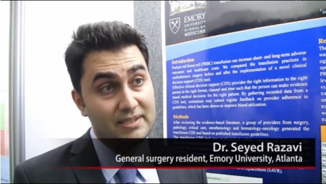
VIDEO: EMR reminder reduces unnecessary transfusions
SAN FRANCISCO – To reduce unnecessary transfusions, all it takes is a reminder in the electronic medical record system that they aren’t generally necessary if surgery patients have hemoglobins greater than 10 g/dL, according to investigators from Emory University in Atlanta.
A reminder in Emory’s EMR dropped transfusion rates in cardiothoracic patients without increasing negative outcomes. It also saved money and reduced the number of surgical site infections. Emory is now rolling it out systemwide (J. Am. Coll. Surg. 2014 June 25 [doi: 10.1016/j.jamcollsurg.2014.06.012]). Lead investigator Dr. Seyed Razavi explained the initiative in a video interview at the annual clinical congress of the American College of Surgeons.
The video associated with this article is no longer available on this site. Please view all of our videos on the MDedge YouTube channel
Dr. Hossein Almassi, FCCP, comments: By their nature, cardiac operations are associated with higher transfusion rates than other surgical procedures. The negative impact of blood transfusion on short term hospital outcomes and mortality is well known. Any effort in reducing the adverse outcomes is a step in the right direction, especially in this era of 'pay per performance". A "reminder in the EMR" is but one such step.
Dr. Almassi specializes in cardiothoracic surgery at the Medical College of Wisconsin in Milwaukee, Wisconsin.
SAN FRANCISCO – To reduce unnecessary transfusions, all it takes is a reminder in the electronic medical record system that they aren’t generally necessary if surgery patients have hemoglobins greater than 10 g/dL, according to investigators from Emory University in Atlanta.
A reminder in Emory’s EMR dropped transfusion rates in cardiothoracic patients without increasing negative outcomes. It also saved money and reduced the number of surgical site infections. Emory is now rolling it out systemwide (J. Am. Coll. Surg. 2014 June 25 [doi: 10.1016/j.jamcollsurg.2014.06.012]). Lead investigator Dr. Seyed Razavi explained the initiative in a video interview at the annual clinical congress of the American College of Surgeons.
The video associated with this article is no longer available on this site. Please view all of our videos on the MDedge YouTube channel
AT THE ACS CLINICAL CONGRESS
Pedunculagin
Pedunculagin is an ellagitannin, a group of polyphenolic hydrolyzable tannins, found in various plants, including Emblica officinalis, Pimenta dioica, and several others (Arch. Pharm. Res. 2014, Feb 7. [Epub ahead of print]; Curr. Drug Targets 2012;13:1900-06). The substance is reported to exhibit anti-inflammatory, anticancer, and antimicrobial activities, and it is considered a potent dietary antioxidant (Z. Naturforsch C. 2007;62:526-36; J. Org. Chem. 1996;61:2606-12; J. Nutr. 2014;144(4 Suppl):555S-60S). Purified from the Manchurian alder (Alnus hirsuta), pedunculagin is also a novel immunomodulating agent (Skin Res. Technol. 2010;16:371-7). Pedunculagin is also one of the hydrolyzable tannins found in Punica granatum (pomegranate), fruit extracts of which have been shown by Afaq et al. to exert photochemopreventive effects against the deleterious effects of ultraviolet B radiation (Photochem. Photobiol. 2005;81:38-45). Pedunculagin was first synthesized (in 2,3- and 4,6-coupled form) in 1996 (J. Org. Chem. 1996;61:2606-12).
Anticancer and antioxidant activity
In a study of 57 tannins and related compounds, Kashiwada et al. noted in a 1992 study that pedunculagin exhibited selective cytotoxicity against melanoma cells (J. Nat. Prod. 1992;55:1033-43).
According to a 2007 report by Marzouk et al., pedunculagin is among one of several tannins identified in the leaves of Pimenta dioica, and it is among the most potent free radical scavengers, as well as one of the most cytotoxic substances against solid tumor cancer cells. Pedunculagin also was found to significantly suppress nitric oxide production and spur the proliferation of T-lymphocytes and macrophages (Z. Naturforsch C. 2007;62:526-36).
In 2012, Kähkönen et al. observed that red raspberry and cloudberry ellagitannins, including pedunculagin, acted as effective radical scavengers, substantially contributing to the antioxidant activity of the berries in lipoprotein and lipid emulsion environments (J. Agric. Food Chem. 2012;60:1167-74).
A 2014 review by Hardman summarized several studies suggesting that potent anticancer properties, including antiproliferative and antiangiogenic activities, have been linked to walnuts. She noted that pedunculagin is one of the key constituents in walnuts to which such characteristics have been attributed (J. Nutr. 2014;144(4 Suppl):555S-60S).
Potential cutaneous applications
In 2010, Lee et al. assessed the effects of pedunculagin on 2,4,6-trinitrochlorobenzene (TNCB)-induced atopic dermatitis-like lesions in NC/Nga mice. Investigators applied a cream containing 0.1% or 0.5% pedunculagin to the positive treatment group; the negative treatment group received the base cream without pedunculagin, with no topical formulations administered to a control group. The investigators found, 4 weeks after treatment, that greater and more rapid improvement in the lesions was experienced by the group that received the higher concentration of pedunculagin (Skin Res. Technol. 2010;16:371-7).
Kim et al., in 2014, isolated pedunculagin and five other phenolic compounds from the leaves of Quercus mongolica (Mongolian oak). They found that pedunculagin exhibited strong in vitro inhibition against the expression of matrix metalloproteinase (MMP)-1 and increased type I procollagen in human fibroblasts exposed to UVB. The Q. mongolica constituent was also found to concentration-dependently exhibit potent scavenging activity against the DPPH (2,2-diphenyl-1-picrylhydrazyl) radical. The investigators suggested that the ellagitannin shows promise for use in preventing and treating cutaneous aging (Arch. Pharm. Res. 2014 Feb. 7. [Epub ahead of print]).
Conclusion
Pedunculagin shows some promise as an agent that can yield dermatologic benefits. However, the body of research on this natural compound is relatively scant. More expansive follow-up work is needed to determine the extent to which pedunculagin can be reasonably incorporated into the dermatologic armamentarium.
Dr. Baumann is chief executive officer of the Baumann Cosmetic & Research Institute in the Design District in Miami. She founded the Cosmetic Dermatology Center at the University of Miami in 1997. Dr. Baumann wrote the textbook “Cosmetic Dermatology: Principles and Practice” (New York: McGraw-Hill, 2002), and a book for consumers, “The Skin Type Solution” (New York: Bantam Dell, 2006). She has contributed to the Cosmeceutical Critique column in Skin & Allergy News since January 2001. Her latest book, “Cosmeceuticals and Cosmetic Ingredients,” was published in November 2014. Dr. Baumann has received funding for clinical grants from Allergan, Aveeno, Avon Products, Evolus, Galderma, GlaxoSmithKline, Kythera, Mary Kay, Medicis Pharmaceuticals, Neutrogena, Philosophy,Topix Pharmaceuticals, and Unilever.
Moderate THST linked to improved survival in thyroid cancer
CORONADO, CALIF.– In an analysis of primary treatments for all stages of differentiated thyroid carcinoma, only thyroid hormone suppressive therapy was associated with both improved overall survival and disease-free survival.
Further, when examining the degree of thyroid hormone suppressive therapy (THST), aggressive THST conferred no additional survival benefit when compared with moderate THST, even when limiting the analysis to patients with distant metastatic disease, results from a long-term analysis of registry showed.

Those are key findings from an updated analysis of data from the National Thyroid Cancer Treatment Cooperative Study Group Registry, which were presented by lead study author Dr. Aubrey Carhill during the annual meeting of the American Thyroid Association.
“To date there are no prospective studies evaluating the longitudinal outcomes of initial long-term therapies in differentiated thyroid carcinoma,” said Dr. Carhill of MD Anderson Cancer Center, Houston. “In the absence of prospective trials, there has been significant reliance on retrospective studies with limited numbers of patients and low event rates as well as significant reliance on expert opinion to guide clinical practice.”
For example, current ATA guidelines for TSH suppression suggest that in long-term follow-up of patients with differentiated thyroid cancer, “those with persistent disease should have TSH levels suppressed to undetectable levels and maintained indefinitely,” Dr. Carhill said, while disease-free, higher-risk patients “should be suppressed to low-moderate levels continued between 5 and 10 years and low-risk patients should be maintained in the low-normal range. Similar levels of evidence exist to support the use of radioactive iodine and the degree of surgical extent.”
The challenge for clinicians, she continued, becomes balancing the potential risks of more aggressive therapies, such as aggressive thyroid hormone suppression, and the risks associated with long-term thyrotoxicosis with the potential benefits of treatment. “This is not always clear, especially in patients who are at very low risk for cancer-specific mortality,” she said. “There remains a need for accurate prognostication in order to identify which patients will benefit from different treatment modalities because current staging systems have limited ability to predict response to treatment.”
Formed in 1987, the National Thyroid Treatment Cooperative Study Group is a multi-institutional effort to assess long-term management of outcomes on patients with differentiated thyroid cancer. The purpose of the present study was to provide a more current analysis of the prospectively collected data, which was last analyzed in 2001. All staging is tracked according to the registry’s staging system, which is very similar to that of the American Joint Committee on Cancer’s TNM Staging System. Therapies analyzed included total/near total thyroidectomy (T/NTT) vs. a lesser extent of surgery; radioactive iodine (RAI) vs. no RAI; and increasing degrees of THST over time.
Dr. Carhill presented findings from an analysis of the effects of initial therapies in 4,941 patients treated at 11 centers in North America between 1987 and 2012. The median length of follow-up was 6 years, which translated to 34,631 person-years of documented follow-up time. The researchers used univariate and multivariate analyses to assess overall and disease-free survival. Moderate THST was defined as TSH maintained in subnormal or normal levels, while aggressive THST was defined as that maintained in undetectable or subnormal levels.
Improved overall survival was observed in stage III patients who received RAI (risk ratio, 0.66; P = .04) and in stage IV patients who received T/NTT and RAI (RR, 0.66 and 0.70, respectively; combined P = .049). Moderate but not aggressive THST was associated with significantly improved overall survival in all stages (RR, 0.13 in stage I, 0.09 in stage II, 0.13 in stage III, and 0.33 in stage IV), as well as with improved disease-free survival (RR, 0.52 in stage I, 0.40 in stage II, 0.18 in stage III, and no RR in stage IV).
In stage I patients, RAI conferred worse disease-free survival (RR, 1.79; P = .0005). “However, further propensity analysis demonstrated that there was no difference in disease-free survival when patients were stratified according to their propensity to receive radioactive iodine,” Dr. Carhill said.
To evaluate the optimal duration of THST, the researchers examined the effect of continuing degrees of suppression beyond 1, 3, and 5 years of follow-up. After 1 and 3 years of follow-up, both initial stage and moderate TSH suppression were independently predictive of improved overall survival (RR, 0.31 and 0.29, respectively). However, after 5 years of follow-up, “although initial stage remained independently predictive, there was no further benefit with any subsequent degree of TSH suppression,” Dr. Carhill said.
The study “confirms prior registry findings of a survival benefit in high-risk groups treated with T/NTT and RAI, and there is no disease-free survival benefit in low-risk groups receiving postoperative RAI,” she concluded. “We also report for the first time that in multivariate analysis of primary treatments for DTC [differentiated thyroid cancer], in all stages, only THST was associated with both improved overall survival and disease-free survival. When examining the degree of THST, aggressive THST confers no additional survival advantage as compared with moderate THST, even in patients with distant metastatic disease, which remains particularly relevant given the risks associated with long-term thyrotoxicosis.” She acknowledged certain limitations of the study, including the potential for institutional bias. “However, we feel that this is somewhat offset due to the size of the registry cohort and the number of sites involved” Dr. Carhill said.
The registry has received support from Genzyme and Pfizer. Dr. Carhill reported having no financial disclosures.
On Twitter @dougbrunk
CORONADO, CALIF.– In an analysis of primary treatments for all stages of differentiated thyroid carcinoma, only thyroid hormone suppressive therapy was associated with both improved overall survival and disease-free survival.
Further, when examining the degree of thyroid hormone suppressive therapy (THST), aggressive THST conferred no additional survival benefit when compared with moderate THST, even when limiting the analysis to patients with distant metastatic disease, results from a long-term analysis of registry showed.
Those are key findings from an updated analysis of data from the National Thyroid Cancer Treatment Cooperative Study Group Registry, which were presented by lead study author Dr. Aubrey Carhill during the annual meeting of the American Thyroid Association.
“To date there are no prospective studies evaluating the longitudinal outcomes of initial long-term therapies in differentiated thyroid carcinoma,” said Dr. Carhill of MD Anderson Cancer Center, Houston. “In the absence of prospective trials, there has been significant reliance on retrospective studies with limited numbers of patients and low event rates as well as significant reliance on expert opinion to guide clinical practice.”
For example, current ATA guidelines for TSH suppression suggest that in long-term follow-up of patients with differentiated thyroid cancer, “those with persistent disease should have TSH levels suppressed to undetectable levels and maintained indefinitely,” Dr. Carhill said, while disease-free, higher-risk patients “should be suppressed to low-moderate levels continued between 5 and 10 years and low-risk patients should be maintained in the low-normal range. Similar levels of evidence exist to support the use of radioactive iodine and the degree of surgical extent.”
The challenge for clinicians, she continued, becomes balancing the potential risks of more aggressive therapies, such as aggressive thyroid hormone suppression, and the risks associated with long-term thyrotoxicosis with the potential benefits of treatment. “This is not always clear, especially in patients who are at very low risk for cancer-specific mortality,” she said. “There remains a need for accurate prognostication in order to identify which patients will benefit from different treatment modalities because current staging systems have limited ability to predict response to treatment.”
Formed in 1987, the National Thyroid Treatment Cooperative Study Group is a multi-institutional effort to assess long-term management of outcomes on patients with differentiated thyroid cancer. The purpose of the present study was to provide a more current analysis of the prospectively collected data, which was last analyzed in 2001. All staging is tracked according to the registry’s staging system, which is very similar to that of the American Joint Committee on Cancer’s TNM Staging System. Therapies analyzed included total/near total thyroidectomy (T/NTT) vs. a lesser extent of surgery; radioactive iodine (RAI) vs. no RAI; and increasing degrees of THST over time.
Dr. Carhill presented findings from an analysis of the effects of initial therapies in 4,941 patients treated at 11 centers in North America between 1987 and 2012. The median length of follow-up was 6 years, which translated to 34,631 person-years of documented follow-up time. The researchers used univariate and multivariate analyses to assess overall and disease-free survival. Moderate THST was defined as TSH maintained in subnormal or normal levels, while aggressive THST was defined as that maintained in undetectable or subnormal levels.
Improved overall survival was observed in stage III patients who received RAI (risk ratio, 0.66; P = .04) and in stage IV patients who received T/NTT and RAI (RR, 0.66 and 0.70, respectively; combined P = .049). Moderate but not aggressive THST was associated with significantly improved overall survival in all stages (RR, 0.13 in stage I, 0.09 in stage II, 0.13 in stage III, and 0.33 in stage IV), as well as with improved disease-free survival (RR, 0.52 in stage I, 0.40 in stage II, 0.18 in stage III, and no RR in stage IV).
In stage I patients, RAI conferred worse disease-free survival (RR, 1.79; P = .0005). “However, further propensity analysis demonstrated that there was no difference in disease-free survival when patients were stratified according to their propensity to receive radioactive iodine,” Dr. Carhill said.
To evaluate the optimal duration of THST, the researchers examined the effect of continuing degrees of suppression beyond 1, 3, and 5 years of follow-up. After 1 and 3 years of follow-up, both initial stage and moderate TSH suppression were independently predictive of improved overall survival (RR, 0.31 and 0.29, respectively). However, after 5 years of follow-up, “although initial stage remained independently predictive, there was no further benefit with any subsequent degree of TSH suppression,” Dr. Carhill said.
The study “confirms prior registry findings of a survival benefit in high-risk groups treated with T/NTT and RAI, and there is no disease-free survival benefit in low-risk groups receiving postoperative RAI,” she concluded. “We also report for the first time that in multivariate analysis of primary treatments for DTC [differentiated thyroid cancer], in all stages, only THST was associated with both improved overall survival and disease-free survival. When examining the degree of THST, aggressive THST confers no additional survival advantage as compared with moderate THST, even in patients with distant metastatic disease, which remains particularly relevant given the risks associated with long-term thyrotoxicosis.” She acknowledged certain limitations of the study, including the potential for institutional bias. “However, we feel that this is somewhat offset due to the size of the registry cohort and the number of sites involved” Dr. Carhill said.
The registry has received support from Genzyme and Pfizer. Dr. Carhill reported having no financial disclosures.
On Twitter @dougbrunk
AT THE ATA ANNUAL MEETING
Key clinical point: Only moderate thyroid hormone suppressive therapy is associated with better outcomes in all stages of differentiated thyroid cancer.
Major finding: Moderate, but not aggressive, THST was linked with significantly improved overall survival in all stages of differentiated thyroid cancer (RR, .13 in stage I, .09 in stage II, .13 in stage III, and .33 in stage IV).
Data source: An analysis of the effects of initial therapies in 4,941 patients from the National Thyroid Cancer Treatment Cooperative Study Group Registry who were treated at 11 centers in North America between 1987 and 2012.
Disclosures:The registry has received support from Genzyme and Pfizer. Dr. Carhill reported having no financial disclosures.

Hospitalists Channel Osler, Pioneer in Bedside Exams
Hands-on workshop helps hospitalists gain confidence in fundamentals, learn to teach physical exam skills better
Hands-on workshop helps hospitalists gain confidence in fundamentals, learn to teach physical exam skills better
Hands-on workshop helps hospitalists gain confidence in fundamentals, learn to teach physical exam skills better
Checklists Improve Outcomes, Require Care-team Buy-in
Dr. Ramiro Jervis and Dr. Umesh Gidwani urge hospitalists to experiment with checklists during the 7th annual Hospital Medicine Symposium in New York City.
Dr. Ramiro Jervis and Dr. Umesh Gidwani urge hospitalists to experiment with checklists during the 7th annual Hospital Medicine Symposium in New York City.
Dr. Ramiro Jervis and Dr. Umesh Gidwani urge hospitalists to experiment with checklists during the 7th annual Hospital Medicine Symposium in New York City.
LISTEN NOW: Dr. Kendall Rogers, MD, SFHM, Encourages Hospitalists to Work as Part of Quality Teams to Achieve Glycemic Control
As SHM's glycemic control lead mentor and a hospitalist at the University of New Mexico in Albuquerque, Kendall Rogers, MD, CPE, FACP, SFHM, offers advice to hospitalists when working as part of a quality team in achieving glycemic control.
As SHM's glycemic control lead mentor and a hospitalist at the University of New Mexico in Albuquerque, Kendall Rogers, MD, CPE, FACP, SFHM, offers advice to hospitalists when working as part of a quality team in achieving glycemic control.
As SHM's glycemic control lead mentor and a hospitalist at the University of New Mexico in Albuquerque, Kendall Rogers, MD, CPE, FACP, SFHM, offers advice to hospitalists when working as part of a quality team in achieving glycemic control.
LISTEN NOW: Kristen Kulasa, MD, Explains How Hospitalists Can Work with Nutritionists and Dieticians
Kristen Kulasa, MD, assistant clinical professor of medicine and director of Inpatient Glycemic Control, Division of Endocrinology, Diabetes, and Metabolism at the University of California in San Diego, provides tips on how hospitalists can work with nutritionists and dieticians for the betterment of diabetic patients. As a mentor for SHM's care coordination program on inpatient diabetes, Dr. Kulasa offers hospitalists advice in treating diabetic patients. She points to SHM’s website, which has a lot of resources to help hospitalists feel comfortable with insulin dosing.
Kristen Kulasa, MD, assistant clinical professor of medicine and director of Inpatient Glycemic Control, Division of Endocrinology, Diabetes, and Metabolism at the University of California in San Diego, provides tips on how hospitalists can work with nutritionists and dieticians for the betterment of diabetic patients. As a mentor for SHM's care coordination program on inpatient diabetes, Dr. Kulasa offers hospitalists advice in treating diabetic patients. She points to SHM’s website, which has a lot of resources to help hospitalists feel comfortable with insulin dosing.
Kristen Kulasa, MD, assistant clinical professor of medicine and director of Inpatient Glycemic Control, Division of Endocrinology, Diabetes, and Metabolism at the University of California in San Diego, provides tips on how hospitalists can work with nutritionists and dieticians for the betterment of diabetic patients. As a mentor for SHM's care coordination program on inpatient diabetes, Dr. Kulasa offers hospitalists advice in treating diabetic patients. She points to SHM’s website, which has a lot of resources to help hospitalists feel comfortable with insulin dosing.
LISTEN NOW: Dr. Carolyn Zelop, MD, Discusses Cardiovascular Emergencies in Pregnant Women
Listen now to excerpts of our interview with Dr. Zelop, a board certified maternal-fetal medicine specialist and director of perinatal ultrasound and research at Valley Hospital in Ridgewood, N.J.
Listen now to excerpts of our interview with Dr. Zelop, a board certified maternal-fetal medicine specialist and director of perinatal ultrasound and research at Valley Hospital in Ridgewood, N.J.
Listen now to excerpts of our interview with Dr. Zelop, a board certified maternal-fetal medicine specialist and director of perinatal ultrasound and research at Valley Hospital in Ridgewood, N.J.
Product News: 11 2014
Effaclar Dermatological Acne System
La Roche-Posay Laboratoire Dermatologique introduces the Effaclar Dermatological Acne System, an over-the-counter system to cleanse, tone, and treat using 3 products: Effaclar Medicated Gel Cleanser, Effaclar Clarifying Solution, and Effaclar Duo. The system is designed to reduce acne with little to no drying or irritation. The Effaclar Dermatological Acne System targets excess sebum, hyperkeratinization, and the main triggers of acne. It can be purchased at select physicians’ offices and pharmacies as well as online. For more information, visit www.laroche-posay.us.
Hydrate Moisturizers
Obagi Medical Products, Inc, introduces 2 moisturizers that provide long-lasting hydration: Hydrate Facial Moisturizer and Hydrate Luxe. Hydrate Facial Moisturizer provides all-day moisture protection for every skin type with immediate and lasting effects on skin barrier function. Hydrate Luxe is engineered with biomimetic peptides to work overnight. It saturates skin with 8-hour moisture protection, and also promotes skin radiance. Both products are dispensed in dermatology, plastic surgery, and other aesthetic physicians’ practices. For more information, visit www.obagi.com.
Stretch Mark Crème
Natural skin care company derma e reveals Stretch Mark Crème to visibly diminish the look of stretch marks, as well as improve texture, color, and overall appearance. Stretch Mark Crème contains argan oil, cocoa butter, coconut oil, and shea butter to intensely condition skin to help increase elasticity and resiliency. It also contains vitamin E to promote self-healing and hyaluronic acid to attract and bind moisture to the skin. For more information, visit www.dermae.com.
Taclonex
LEO Pharma Inc announces a new pediatric indication for Taclonex (calcipotriene 0.005%–betamethasone dipropionate 0.064%) Topical Suspension. Taclonex is a first-line, once-daily combination product indicated for treatment of both scalp and body psoriasis in adults 18 years and older for up to 8 weeks. It is now also indicated for the treatment of plaque psoriasis of the scalp in patients aged 12 to 17 years for the same period. For more information, visit www.taclonex.com.
If you would like your product included in Product News, please e-mail a press release to the Editorial Office at [email protected]
