User login
World Mental Health Day: Preventing suicide
Increased engagement of men in mental health services is needed
Each year, the World Federation of Mental Health chooses a theme for World Mental Health Day, which is Oct. 10. This year’s theme is “Mental Health Promotion and Suicide Prevention.”

About 800,000 people die by suicide every year, according to the World Health Organization. Suicide is the second-leading cause of death among people aged 15-29 years.1
Most suicide occurs in low- and middle-income countries, the WHO reports. In addition, almost two-thirds of those deaths around the world occur in males, a recent study shows.2 The study, conducted by Danah Alothman and Andrew Fogarty, MBBS, of the NIHR Biomedical Research Center at the University of Nottingham (England), looked at sex-specific suicide rates for 182 countries in 2015.
They found that the highest difference between male:female suicide rates were in the Americas (median, 4:1/100,000), and the lowest were in Africa and Asia (median for both continents, 2.7:1/100,000).
“The implication is that as societies become richer and more educated, males have a higher risk of dying as a consequence of suicide relative to females,” they wrote in the Journal of Affective Disorders.
For clinicians who treat patients with mental illness, particularly those of us who practice in the Americas, this sex differential is concerning. We know that women are more likely to be diagnosed the depression.3 But perhaps this has something to do with the way men are socialized around the world. In other words, as John S. Ogrodniczuk, PhD, and John L. Oliffe, PhD, wrote,4 depression in men “often manifests as irritability; anger; hostile, aggressive, abusive behavior; risk taking, substance abuse; and escaping behavior.” They argue that the outward behavior shown by some men with depression might, in fact, “serve as a cover-up mechanism to hide the internal turmoil” they are experiencing. We certainly know that some men adhere to masculine norms such as stoicism, which in turn, heightens self-stigma. Unfortunately, men seek help for depression less often than do women.5 So one key question becomes: What can we as mental health professionals do to better meet the treatment needs of our male patients?
One example of a program that could hold promise in this area is one called Men at Risk. That program, developed by the nonprofit Centre for Suicide Prevention, in Grande Prairie, Alta., helps men who work in the oil, forestry, and agriculture sectors talk about their challenges and encourages them to let go of stigma.6
Factors other than male gender also might increase the likelihood of suicide. It has rightly been said that genetics and environment play a big role on the psyche of the individuals, and the act of suicide is no different when we discuss the etiologic factors that lead to perpetration of such an act. Genetic vulnerability is a factor that cannot be modified or altered in an easy way, hence, control of environmental factors is more pertinent.
Poverty and violence are two major detrimental factors that have reached alarming proportions and can lead people end their lives.
The developing countries, and now to a significant extent, developed countries, face terrorism that affect the human psyche and can lead to depression, psychosis, and substance abuse, and hence, increase the vulnerability toward the act of suicide. In our offices, we psychiatrists come across patients with borderline personality disorder, for example, who present to emergency departments with multiple and repeated suicidal attempts. There is a big role of genetics here – and role of specific interventions, such as dialectical behavior therapy. Pharmacologic treatment can play a vital role.
In order to make the world a safe place, joint global efforts are required. Enhanced security steps, improved immigration screening, and political will are essential to curb this heartbreaking act. Responsible reporting on the part of the media is needed to make suicide contagion less likely.7
Among other important measures are reducing access to guns and other firearms, and increasing health education about consumption of alcohol and other substances. We also need early identification and prompt treatment of mental illnesses; alleviation of poverty; mobilization of community supports; activation of multiple crisis lines; increased availability and affordability of psychotropic medications; reduction of waiting times for seeking treatment of mental illness; enhanced training of crisis workers; and refresher courses for psychiatrists, family physicians, and other allied mental health workers. Above all, strategies are needed to address the stigma associated with seeking help for mental health issues.
Suicide is a global public health issue, and it is of the utmost importance that a collaborative effort be placed in perspective by individual countries within their own health-related policies and parameters.
Good-quality data on suicide prevalence rates would be of the utmost help in understanding the magnitude of this grave problem. The WHO Mental Health Action Plan 2013-2020 indicates the commitment of member states to work toward the global target of reducing the suicide rate in countries by 10% by 2020.
Individual and collective efforts should become the priority to achieve this target going forward.
References
1. World Health Organization. Suicide. 2019 Sep 2.
2. Alothman D and A Fogarty. J Affect Disord. 2020 Jan 1. doi: 10.1016/j.jad.2019.08.093.
3. Albert PR. J Psychiatry Neurosci. 2015 Jul;40(4):219-21.
4. Ogrodniczuk JS and JL Oliffe. Can Fam Physician. 2011;57(2):153-5.
5. Seidler ZE et al. Clin Psychology Rev. 2016;49:106-18.
6. Ellwand O. Men at risk program helping men in Alberta trades, industry, agriculture struggling with mental health issues. Edmonton Sun. 2016 Mar 27.
7. American Association of Suicidology, et al. Recommendations for reporting on suicide.
Dr. Muhammad is clinical professor of psychiatry and consultant psychiatrist at Niagara Health Service, St. Catharines, Ont.
Increased engagement of men in mental health services is needed
Increased engagement of men in mental health services is needed
Each year, the World Federation of Mental Health chooses a theme for World Mental Health Day, which is Oct. 10. This year’s theme is “Mental Health Promotion and Suicide Prevention.”
About 800,000 people die by suicide every year, according to the World Health Organization. Suicide is the second-leading cause of death among people aged 15-29 years.1
Most suicide occurs in low- and middle-income countries, the WHO reports. In addition, almost two-thirds of those deaths around the world occur in males, a recent study shows.2 The study, conducted by Danah Alothman and Andrew Fogarty, MBBS, of the NIHR Biomedical Research Center at the University of Nottingham (England), looked at sex-specific suicide rates for 182 countries in 2015.
They found that the highest difference between male:female suicide rates were in the Americas (median, 4:1/100,000), and the lowest were in Africa and Asia (median for both continents, 2.7:1/100,000).
“The implication is that as societies become richer and more educated, males have a higher risk of dying as a consequence of suicide relative to females,” they wrote in the Journal of Affective Disorders.
For clinicians who treat patients with mental illness, particularly those of us who practice in the Americas, this sex differential is concerning. We know that women are more likely to be diagnosed the depression.3 But perhaps this has something to do with the way men are socialized around the world. In other words, as John S. Ogrodniczuk, PhD, and John L. Oliffe, PhD, wrote,4 depression in men “often manifests as irritability; anger; hostile, aggressive, abusive behavior; risk taking, substance abuse; and escaping behavior.” They argue that the outward behavior shown by some men with depression might, in fact, “serve as a cover-up mechanism to hide the internal turmoil” they are experiencing. We certainly know that some men adhere to masculine norms such as stoicism, which in turn, heightens self-stigma. Unfortunately, men seek help for depression less often than do women.5 So one key question becomes: What can we as mental health professionals do to better meet the treatment needs of our male patients?
One example of a program that could hold promise in this area is one called Men at Risk. That program, developed by the nonprofit Centre for Suicide Prevention, in Grande Prairie, Alta., helps men who work in the oil, forestry, and agriculture sectors talk about their challenges and encourages them to let go of stigma.6
Factors other than male gender also might increase the likelihood of suicide. It has rightly been said that genetics and environment play a big role on the psyche of the individuals, and the act of suicide is no different when we discuss the etiologic factors that lead to perpetration of such an act. Genetic vulnerability is a factor that cannot be modified or altered in an easy way, hence, control of environmental factors is more pertinent.
Poverty and violence are two major detrimental factors that have reached alarming proportions and can lead people end their lives.
The developing countries, and now to a significant extent, developed countries, face terrorism that affect the human psyche and can lead to depression, psychosis, and substance abuse, and hence, increase the vulnerability toward the act of suicide. In our offices, we psychiatrists come across patients with borderline personality disorder, for example, who present to emergency departments with multiple and repeated suicidal attempts. There is a big role of genetics here – and role of specific interventions, such as dialectical behavior therapy. Pharmacologic treatment can play a vital role.
In order to make the world a safe place, joint global efforts are required. Enhanced security steps, improved immigration screening, and political will are essential to curb this heartbreaking act. Responsible reporting on the part of the media is needed to make suicide contagion less likely.7
Among other important measures are reducing access to guns and other firearms, and increasing health education about consumption of alcohol and other substances. We also need early identification and prompt treatment of mental illnesses; alleviation of poverty; mobilization of community supports; activation of multiple crisis lines; increased availability and affordability of psychotropic medications; reduction of waiting times for seeking treatment of mental illness; enhanced training of crisis workers; and refresher courses for psychiatrists, family physicians, and other allied mental health workers. Above all, strategies are needed to address the stigma associated with seeking help for mental health issues.
Suicide is a global public health issue, and it is of the utmost importance that a collaborative effort be placed in perspective by individual countries within their own health-related policies and parameters.
Good-quality data on suicide prevalence rates would be of the utmost help in understanding the magnitude of this grave problem. The WHO Mental Health Action Plan 2013-2020 indicates the commitment of member states to work toward the global target of reducing the suicide rate in countries by 10% by 2020.
Individual and collective efforts should become the priority to achieve this target going forward.
References
1. World Health Organization. Suicide. 2019 Sep 2.
2. Alothman D and A Fogarty. J Affect Disord. 2020 Jan 1. doi: 10.1016/j.jad.2019.08.093.
3. Albert PR. J Psychiatry Neurosci. 2015 Jul;40(4):219-21.
4. Ogrodniczuk JS and JL Oliffe. Can Fam Physician. 2011;57(2):153-5.
5. Seidler ZE et al. Clin Psychology Rev. 2016;49:106-18.
6. Ellwand O. Men at risk program helping men in Alberta trades, industry, agriculture struggling with mental health issues. Edmonton Sun. 2016 Mar 27.
7. American Association of Suicidology, et al. Recommendations for reporting on suicide.
Dr. Muhammad is clinical professor of psychiatry and consultant psychiatrist at Niagara Health Service, St. Catharines, Ont.
Vorinostat demonstrates consistent safety
Berlin, Germany—Vorinostat demonstrates safety and tolerability alone and in combination with other systemic treatments for a wide range of solid and hematologic malignancies, according to a study of collated data from the vorinostat clinical trial program.
Investigators presented the safety data in a poster at the ECCO 15 - 34th ESMO Multidisciplinary Congress. The data suggest that a supratherapeutic single dose (800 mg) of this orally active histone deacetylase inhibitor does not prolong ventricular repolarization to a significant degree. This is reassuring, since cardiac rhythm and EEG changes are thought to be a class effect of HDACs.
Lead author David Siegel, MD, from Hackensack University Medical Center, Hackensack, New Jersey, and his fellow researchers observed that the study data support the overall safety profile of vorinostat use in cancer patients.
They based their analysis on 18 phase 1 and phase 2 vorinostat trials that included 498 patients, 341 who received the agent as monotherapy and 157 treated with the drug in combination with other therapies.
Vorinostat is approved by the US Food and Drug Administration to treat relapsed or refractory cutaneous T-cell lymphoma and was dosed at the approved level of 400 mg/day for 156 of the 341 patients in the monotherapy cohort. In the combination group, vorinostat was given on weekly or 2-weekly schedules instead of continuous dosing.
In the monotherapy group, the most commonly reported treatment-related adverse events were fatigue (61.9%), nausea (55.7%), diarrhea (49.3%), and anorexia (48.1%). The most common grade 3/4 adverse events were fatigue (12.0%), thrombocytopenia (10.6%), dehydration (7.0%), decreased platelet count (5.3%), and anorexia (5.0%).
Seventy-one (20.8%) patients required dose modifications for toxicity and 38 (11.1%) discontinued study medication due to drug-related adverse events. Three drug-related adverse events led to death.
In the combination treatment cohort, nausea (48.4%), diarrhea (40.8%), fatigue (34.4%), and vomiting (31.2%) were the most commonly reported adverse events. The most common grade 3/4 adverse events were fatigue (13.4%), thrombocytopenia (9.6%), neutropenia (8.3%), diarrhea (5.7%), and nausea.
Dose modifications were required in 27 patients (17.2%). Discontinuation due to adverse events was necessary in 31 patients (19.7%), and 1 death was attributed to vorinostat combination treatment.
The QTc phase 1 substudy was randomized, partially blind, and placebo-controlled. None of the 22 evaluable patients included in the analysis experienced a QtcF change greater than 30 msec from their baseline scores. ![]()
Algorithm for Success
The use of a procalcitonin (PCT) algorithm reduced the usage of antibiotics in patients with lower-respiratory-tract infections (LTRI), according to a recent study that may highlight a new way for hospitalists to reduce costs.
The study found the mean duration of antibiotics exposure in the PCT group was lower than in a control group (5.7 days vs. 8.7 days). The researchers, who studied 1,359 patients at six tertiary-care hospitals in Switzerland, also reported less-frequent antibiotic-associated adverse effects, such as nausea, rashes or diarrhea, in the PCT group (JAMA. 2009;302(10):1059-1066).
Scott Flanders, MD, FHM, SHM president and director of the hospitalist program at the University of Michigan Health System in Ann Arbor, says if further review were to show more statistical impacts on costs savings, PCT usage would become more common.
"If you can reduce length-of-stay by half through treatment intervention, then this will easily pay for itself," says Dr. Flanders, who adds, "Hospitalists need to know and have at their fingertips the best avenues of treatment."
Devendra Amin, MD, director of critical-care services at Morton Plant Hospital in Clearwater, Fla., was one of the first physicians to use PCT tests after the Food and Drug Administration (FDA) approved wider usage last year. He says the overuse of antibiotics is a needless cost overrun that hospitalists using PCT tests could better control—and then tout as an example of their ability to reduce costs. Dr. Amin plans to team with a half-dozen of his health system's hospitalists next year to work on a study of the effectiveness of PCT in a community hospital setting.
"If everything else fits, it's another piece of information that's important to the puzzle," Dr. Amin says. "No single test in isolation is going to give you everything you want … but this can help."
Recession? What Recession?
The economy might be in the doldrums, but recruiters are looking for candidates to fill HM positions, says Mark Dotson, senior director of recruitment for Cogent Healthcare, a Brentwood, Tenn.-based company that manages HM programs nationwide. He recently spoke with The Hospitalist eWire about how hospitalists can take advantage of the bullish job market.
Question: What do you look for in HM job candidates?
Answer: We obviously look at their credentials, their training, and the focus of their training in inpatient medicine. We strive to look for physicians who are able to and interested in working in a team environment. They should have a good bedside manner and good communication skills. They should be able to show they have a team-based approach to their work.
Q: What alternative jobs are there in HM that hospitalists might not know about?
A: There are sometimes opportunities to chair a committee that hospitalists aren't aware of. There are also ways to get involved with more specialties by working with physicians on the hospital campus and building relationships with them.
Q: Has the current economic climate affected hospitalist recruiting?
A: Not so much. The demand is still there. But I do think more hospitalists aren’t looking to make a change, because they want stability in their workplace right now. Hospital medicine is a specialty that's growing, so there is stability. Hospitalists have to decide what’s best for their clinical skills and personal interests and not let the economy stop them. There are a hundred more opportunities out there waiting for them.
Budget Checkup

Editor’s note: Part one of a two-part series.
Why does a particular hospitalist practice require more than the typical amount of financial support from a hospital? This is one of the most common questions I am asked. This month and next, I will provide a thorough list of potential answers.
SHM’s “2007-2008 Bi-annual Survey on the State of the Hospital Medicine Movement” showed that hospitals pay an average of $97,400 per year in support per full-time hospitalist. I suspect that amount is higher now. Nevertheless, hospital executives and hospitalists should understand the reasons why the hospital support that is required for their practice might be more or less.
A comprehensive list of potential reasons would include dozens of factors, and my intent is only to highlight some of the most common and significant ones.
Documentation, Coding, Billing, and Collecting
This is an area in which many, if not most, practices have room for improvement. One very simple way to estimate how your group is doing on these things is to think about how you’re performing on the following tasks:
- Do the hospitalists really understand the documentation requirements for each CPT code, and is their performance in selecting CPT codes audited regularly (e.g., annually)?1
- Does the group have a reliable method of charge capture that minimizes problems like lost charges? Is there an established “chain of custody” of this information, from the hospitalist to the biller?
- Is there a rigorous review or audit of the biller’s performance? Does the group monitor metrics, such as days in accounts receivable, collection rate, etc.? Is there a periodic audit of the biller? An audit could be as simple as tracking down five to 10 billed encounters from six months prior for each doctor in the practice, and reviewing the status of each bill (e.g., paid, written off, or perhaps the bill has vanished or never made it into the billing system).
- Is revenue appropriately applied to the hospitalist cost center? For many hospital-employed hospitalists, payors might be including their professional fee payments on the same remittance advice as hospital inpatient payments (due to same tax ID number). The hospital’s business office might be unable or unwilling to break these payments into hospital and professional fee portions and apply them correctly. Hospital-employed hospitalists should know whether their collections are being applied to their revenue center accurately.
Payor Mix
The two factors that govern the amount of professional fee revenues a hospitalist practice will collect are the integrity of the billing process (described above) and the payor mix. The payor mix for most hospitalist practices is roughly 55% to 60% Medicare, 5% to 10% self-pay, 5% to 10% Medicaid, and commercial insurance for the rest.
A hospitalist practice that is significantly different from this example should expect professional fee collections to vary accordingly.
Hospitalist Fee Schedule
My experience is that very few hospitalists know their own fee schedule. The term “fee schedule” is generally used to mean the billed charge for each type of service provided. A hospitalist fee schedule usually fits on a single page, with a list of CPT codes (admits, consults, followups, etc.) down one column and the charge for that service in a second column to the right. It would be reasonable to post the fee schedule in hospitalists’ offices.
Groups that use electronic charge capture, in which the doctor enters into a computer the CPT code to bill for each patient daily, can often see the related charge for each code as it is entered.
Someone connected to the practice, often in the billing office, should review the fee schedule—at least annually—to ensure that services aren’t being billed below the rate allowed by payors.
Negotiated Rates Paid by Commercial Insurance
Some hospitalist groups are able to negotiate higher payments than the typical rates paid by commercial payors. Because commercial insurance is a relatively small portion of most hospitalists’ payor mix, this might not have a large impact on the overall practice finances. So my sense is that most groups don’t pursue this opportunity.
Groups in markets with significant managed care are an exception. They usually are aggressive in negotiations for commercial payor rates.
Some hospital-employed HM groups might end up with lower commercial rates than they could have. Here is how it might happen: A hospital negotiates with Aetna to pay rates for hospital services (the bills submitted by the hospital, not the physician bills) that are attractive to the hospital. To make this proposal more palatable to Aetna, the hospital says it will accept lower rates for its employed physicians, including hospitalists. So the hospitalists’ collections end up lower, and the support paid by the hospital to the hospitalist group is correspondingly higher. The hospital ends up fine in this scenario, because it is being paid an attractive rate by Aetna for hospital services, but the hospitalist practice appears to be underperforming financially.
It is worth knowing if this is an issue at your practice, but in most cases it won’t explain larger problems in the hospitalist budget or amount of support required from the hospital.
Accounting Issues
Budgets and financial statements can be confusing, and revenues and expenses might not be what you expect. For example, in my practice, auditors told our accountants that we needed to accrue an extra month of salary into this year’s budget. So when looking at our fiscal year-end financial statement, the salary expense is for 13 months instead of 12 months. This quirk made it appear that we required more than the budgeted amount of support from our hospital, when in fact we performed better than budget this year.
I certainly can’t explain all the reasons for unusual accounting issues, and I still struggle to understand why accrual accounting is better than cash-basis accounting. My best advice is to have the lead hospitalist in your group get to know the accountant who handles your budget and financial statements. The accountant should explain all of these issues clearly.
In next month’s column, I’ll review how a hospitalist practice’s internal operations, such as staffing and scheduling, can have a major influence on the budget and the amount of support required from the hospital. TH
Dr. Nelson has been a practicing hospitalist since 1988 and is co-founder and past president of SHM. He is a principal in Nelson Flores Hospital Medicine Consultants, a national hospitalist practice management consulting firm (www.nelsonflores.com). He is also course co-director and faculty for SHM’s “Best Practices in Managing a Hospital Medicine Program.” This column represents his views and is not intended to reflect an official position of SHM.
Reference
- Centers for Medicare and Medicaid Services. Improper medicare fee-for-service payments report, November 2006: long report. CMS Web site. Available at: www.cms.hhs.gov/apps/er_report/preview_er_report_print.asp?from=public&which=long&reportID=5. Accessed Sept. 1, 2009.
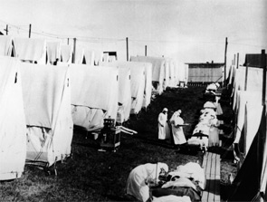
Spanish Flu Redux?

Apocalypse, pestilence, death. As I head back to work after a late-summer vacation, those words are on the tip of my tongue. Now before your mind drifts too far afield, this is not a synopsis of the time spent with family, or even my in-laws—although some have used similar words to describe my mother’s cooking. Rather, these are the descriptors of my vacation reading.
Summer Reading
I started the week relaxing contentedly with Cormac McCarthy’s “The Road.” I chose this book in part because I noticed that it soon will be released as a movie, but mostly because it had won the dustiest-book-in-my-office-reading-pile award. This tale of a young boy and his father traversing a post-apocalyptic America was shocking and surreal. I couldn’t help but interchange images of my 2-year-old son, Greyson, and me out on that road fighting for our existence. In my personal fictional account, I continuously, and heroically, MacGyver my way across a burned-out and treacherous landscape with death-defying adeptness—all the while Grey unknowingly totters, drooling and muttering in tow.
Reality, of course, would paint us in substantially different roles, with mine involving the lion’s share of muttering and drooling, leaving Grey wishing the apocalyptic dealer had dealt him his mother instead.
Next up, “The Last Town on Earth,” by Thomas Mullen. I don’t recall how this book got into my reading pile, but I’m glad it did. The story is set in the fictional city of Commonwealth in 1918. The small, isolated mill town makes the drastic decision to stanch the tide of Spanish flu by cutting itself off from civilization through a self-imposed quarantine.
It is here, on p. 98, that I was sidetracked by a family member’s question—“Do you think this swine flu will be as bad as the Spanish flu?” I was asked shortly after being inquired about my reading choice. “Of course not,” I replied knowingly, moments before realizing I didn’t know. In fact, I didn’t have the faintest idea—not because it’s tough to divine the future, but because I realized I had little more than a passing knowledge of the famous flu that raked the world early last century. And with that, I was off on my final vacation reading session—a quest to slake my thirst for influenza knowledge.
Flu Pandemic
The Spanish flu pandemic of 1918-1920 was the first of three to hit in the 20th century. It took its name not from its site of origin (debated but generally felt to be the U.S., Kansas specifically), but rather from the fact that Spain, a neutral country in World War I, had the most uncensored lines of communication, so the most credible news of the disease came from that country. This provided the false impression that Spain was the only—or at least most dramatically—affected country. Like today’s swine flu, the Spanish flu was an H1N1 influenza. To sate your inner microbiologist, this means the virus exhibits the first of 16 subtypes of hemagglutinin (H) and nine subtypes of neuraminidase (N). Generally, only H1, H2, H3, and N1 and N2 affect humans, and tend to cause mild disease in otherwise healthy populations, killing the immunocompromised, the very young, and the very old. This typically results in a case-fatality rate of about 0.1% and 250,000 to 500,000 deaths worldwide annually.
The Spanish flu, however, was different. For reasons that are not entirely clear, the Spanish flu struck in two waves. The first wave, in the spring of 1918, induced typical flu-like illness with generally mild outcomes, except for the immunocompromised. The second wave was unusual for two reasons. First, it began in the late summer of 1918, rather than the typical winter pattern seen in North America. Second, it was much more deadly, inducing what has been termed a cytokine storm. This immunological avalanche produced more severe disease in the immunopotent young, healthy populations—resulting in its unprecedented mortality in this cohort. In fact, upward of 99% of all Spanish flu deaths were in people younger than 65.
In the end, the pandemic left a broad swath of destruction in its wake. It is estimated that 500 million people—one-third of the world’s population at the time—were infected. The mortality rate was 10% to 20%, resulting in 50 million to 100 million deaths. Put another way, the Spanish flu killed 5% of humanity.
It did so rapidly. Nearly 1 million people died per week in the first 25 weeks of the second wave. To put it in perspective, it took HIV 25 years to reach that number. Thus, historians have termed the Spanish flu “the greatest medical holocaust in history.”
And then as quickly as it commenced, it abated. For example, in Philadelphia, there were about 5,000 flu deaths in one week in October 1918, yet a month later, the virus had nearly disappeared from the city. It’s not clear why this happened, but prevailing theories postulate that either the medical community got better at managing its mortal complications (e.g., bacterial pneumonia), or the bug itself mutated to a less virulent strain.
Is the Swine Flu our Spanish Flu?
On June 11, 2009, the World Health Organization (WHO) declared that the current H1N1 flu virus had reached pandemic status. This novel H1N1 serotype appears to be a direct descendent of the Spanish H1N1 subtype, but the new strain also combines genetic material culled from swine and birds reassorted in a manner that results in limited innate human defenses. And like the Spanish variant, it appears this new strain is hitting earlier in the year than usual and disproportionately affecting the young, with about two-thirds of U.S. deaths coming in the 25- to 64-year-old demographic.
So can we expect hundreds of millions of deaths from swine flu? Probably not. The WHO has been cautious to note that the upgrade to pandemic status was based on the rapidity and ease of spread, not the lethality of the virus. Furthermore, the Centers for Disease Control and Prevention (CDC)—which publishes a wonderful weekly update called FluView (www.cdc.gov/flu/weekly/)—notes that while the number of doctor’s visits for influenza-like illnesses through mid-August is unusually high, the rates of hospitalizations and proportion of deaths attributed to pneumonia and influenza are low and within normal limits for this time of year. Further, the virus continues in its original form, meaning it has not mutated, become more resistant to antiviral drugs, or been altered from the viruses selected for the 2009 vaccine.
So while we certainly must brace for the worst, I feel comfortable in the answer I provided my family member. I also am confident that Grey won’t be quarantined or left to roam the barren Earth anytime soon. TH
Dr. Glasheen is associate professor of medicine at the University of Colorado Denver, where he serves as director of the Hospital Medicine Program and the Hospitalist Training Program, and as associate program director of the Internal Medicine Residency Program.
The Time to Act Is Now

It should come as no surprise to most hospitalists that healthcare-associated infections (HAIs) are among the leading causes of death in the U.S. The Centers for Disease Control and Prevention (CDC) estimates that from 5% to 10% of hospitalized patients develops an HAI, which leads to nearly 100,000 deaths every year.
The big four infection categories are catheter-associated urinary tract infections (CAUTIs), ventilator-associated pneumonia (as well as non-ventilator-associated hospital-acquired pneumonia), central-line-associated bloodstream infections, and surgical-site infections. In addition, Clostridium difficile (C-diff) and methicillin-resistant Staphylococcus aureus (MRSA) infections add to the burden.
While the toll on patients is substantial, the financial burden is equally staggering. It is estimated that HAIs lead to $28 billion to $33 billion in excess healthcare costs each year. So what does this have to do with hospitalists? Everything.
National Efforts to Curb HAIs
The morbidity, mortality, and financial consequences of HAIs have not been lost on patients, payors, and policymakers; each group is demanding action. The Department of Health and Human Services (HHS) is coordinating a national effort addressing HAIs; it aims to bring together many of HHS’ agencies (CDC, Agency for Healthcare Research and Quality, National Institutes of Health, Centers for Medicare and Medicaid Services, etc.) and engage patients, payors, and care providers. As a show of support for these efforts, Congress provided HHS with more than $200 million to target HAIs. After much work, HHS released its action plan to prevent HAIs in January.
SHM was one of the organizations asked to comment on the prevention plan. We did.
- We supported HHS’ focus on process measures (rather than outcomes), which recognizes the inevitability of some HAIs;
- We asked to be more involved in the process of developing and implementing the action plan; and
- We specifically asked for hospitalist representation on the Healthcare Infection Control Practices Advisory Committee (HICPAC), which develops the guidelines and prioritizes national efforts targeting HAI prevention.
Wish granted. In June, I was invited to HHS’ offices in Washington, D.C., along with key stakeholders to hear details of the final plan of action and discuss implementation. The plan (www.hhs.gov/ophs/initiatives/hai/infection.html) addresses key HAIs, establishes baseline rates, and proposes five-year national targets for reductions in infections.
The National Quality Foundation (NQF) has endorsed most of the metrics. The targeted reductions seem reasonable, and they are mostly in line with current evidence on best practices.
Of note, HHS dropped ventilator-associated pneumonia as a target area because of feedback from stakeholders (e.g., SHM) who argued that current definitions of the condition were inadequate to allow accurate measurement of targeted performance improvement efforts. Also of note, SHM was offered a HICPAC seat, which will enhance our ability to further impact the development and evaluation of current and future metrics.
I was invited back to Washington in July to meet with Don Wright, MD, MPH, FAACP, HHS’ principal deputy assistant secretary for health. It’s obvious to me that HHS realizes that any effective campaign to reduce HAI incidence will require engaging hospitalists, and, interestingly, HHS has even heard from other professional societies that hospitalists are a key group to target if you plan to implement hospitalwide interventions that span the ED, hospital wards, ICUs, surgical patients, pediatrics, or any other nook or cranny in the hospital. Hospitalists and SHM appear to be at ground zero in the national effort to combat HAIs.
The Hospitalist’s Role
There are few medical conditions that impact more of SHM’s big tent of membership the way HAIs do. HAIs affect administrators, internists, family practitioners, pediatricians, physician assistants and nurse practitioners, nurses, residents, students, community practitioners, academics, and large management companies … the list goes on. Not surprisingly, efforts to combat HAIs will require teams composed of many of the groups highlighted above working together to create systems-based approaches in their own hospitals—in joint efforts to reduce the rate of preventable HAIs.
Take the most common HAI as an example: catheter-associated urinary tract infections. These infections affect patients in every hospital unit and are familiar to every care provider, regardless of background or practice setting. Administrators should care about CAUTIs in part because CMS no longer pays for a CAUTI when it complicates a hospitalization, but also because these infections adversely affect patient satisfaction. Efforts to reduce CAUTIs will need to address inappropriate catheter insertion, provide alternatives to catheter use (e.g., bladder scans), develop best practices for maintenance of necessary catheters, and facilitate timely removal of catheters no longer needed. Dealing with all of these issues will take a team-based systems approach.
I will not be surprised if hospitalists end up leading these initiatives across the country. Hospitalists will need to share best practices, collaborate in local or national initiatives, provide feedback to SHM and policymakers about what works and what doesn’t, and educate patients about HAIs and prevention. Every hospitalist in the country needs to understand the reasons HAIs develop, know strategies to prevent them, and work to implement these strategies in their hospitals.
Future Directions
Given the urgency, what can you expect next? The action plan is finalized, so HHS is turning its attention to implementation. HHS has reached out to SHM to see how we can get the word out to our members. Dissemination strategies include publication of key messages in The Hospitalist, the Journal of Hospital Medicine, Webinars, e-mail announcements, and presentations at our annual meeting.
And while HHS’ plan of action highlights the metrics, it does not provide detailed strategies to combat HAIs. Prevention tools will need to be developed, tested, and, if effective, disseminated. HHS has asked SHM to help in tool development and dissemination.
HHS will continue to work with CMS to align payment policies that incentivize prevention efforts, and SHM will need to follow these developments closely. In addition, AHRQ is dedicating substantial funds to support the development and dissemination of best practices to prevent HAIs.
HHS acknowledges we still have much to learn about HAIs and their prevention. I expect many hospitalists, as well as SHM, will be at the center of these initiatives. Healthcare-associated infections are a problem that can no longer be ignored. Prevention efforts need to be ramped up. Hospitalists around the country need to prepare to lead and champion these efforts. It is time to act. TH
Dr. Flanders is president of SHM.
Measures of Success
The headlines are harrowing: corporate layoffs; foreclosures on the rise; 401(k) retirement plans halved; government bailouts adding to the national debt. The worst economic downturn since the Great Depression has generated some unexpected outcomes, yet not all of them are bad for hospitalists. Below, four vignettes highlight HM groups that have achieved success despite—or in some cases because of—these troubled times.
A Better Business Agreement
It has taken nearly two years—and sometimes as many as four meetings a week—but Rajeev Alexander, MD, and his colleagues are nearing the finish line of an evolving business arrangement. The new arrangement has come about due to the economic downturn, which forced Oregon Medical Group (OMG), a multispecialty physician group serving hospitals in the Eugene/Springfield area and the HM group’s employer since 1988, to want to divest themselves of the hospitalist group. Now, after a lengthy negotiation, Dr. Alexander’s group of eight hospitalists is busier than ever.
Through what were essentially multiple quasi-buyouts, divestitures, and mergers, Dr. Alexander’s hospitalist group “spun off” from OMG and affiliated with PeaceHealth, a nonprofit health system serving seven hospitals in Oregon, Washington, and Alaska. The new contract means Dr. Alexander’s group is directly employed by Sacred Heart Hospital, a 541-bed PeaceHealth-owned facility in Eugene.
The new contract included a non-compete clause with OMG, which currently employs five hospitalists, yet Dr. Alexander’s group has maintained its patient volume. Compensation held steady and employee benefits improved. During an independent and slow-moving negotiation, Dr. Alexander’s group has merged with another HM service that originally was employed by PeaceHealth. The two HM groups (technically competitors) now practice in the same hospital and are ironing out the terms of the merger. At the moment, the groups have created a mutually respectful joint governance council.
“We’ve tackled the thorniest of problems,” Dr. Alexander says, “first, creating a combined work schedule to distribute patients and divide the work. Those of us on the governance council figured if we could get the docs to actually work together and share patients and communicate with each other as if they were one group, then the momentum for an actual administrative/contractual merger would feel inevitable.”

Although negotiations are expected to last through the end of the year to finalize such details as compensation, recruiting, and a group mission statement, the medical staff at Sacred Heart considers the merger a “done deal” and has thrown its support behind the effort. “Community outpatient docs have been clamoring for our services, and we have been having to hand out numbers and ask them to wait in line, so to speak,” Dr. Alexander says.
Dr. Alexander says he’s learned some lessons through the extensive negotiation process:
- Stay positive. In any business venture, absolutely nothing is impossible, even dodging a noncompete clause.
- Release your preconceptions. Conspiracy theories might abound, but most hospital administrators have the best of intentions. As highly regulated organizations, hospitals might simply be following their own bylaws and fulfilling responsibilities to stakeholders. Seek out at least one administrator whom you can trust, and with whom you can communicate effectively. A mutual understanding of intentions and objectives makes the process more successful for all concerned.
- Look beyond politics. Your trust and respect for administrators and fellow physicians will go a long way toward overcoming obstacles.
- Stick to your plan. Adhere to your goal of remaining independent, if that is important to you. “Our group resisted being funneled into becoming employed by a very large national hospitalist chain,” Dr. Alexander says, “and I would encourage physicians in other parts of the country to stick to their commitments as well.”
- Trust the negotiation process. Even if all goes well, what you’re shooting for at the beginning might not be exactly what you get after negotiations are over. This does not mean you’ve failed, or that hospital administration tricked you or failed to deliver on promises. It simply means you have created a negotiated settlement; both sides have come to a new appreciation for the other’s requirements and have made necessary and respectful accommodations.
Rural Rewards
Based in Traverse City, Hospitalists of Northwest Michigan (HNM) services four community hospitals and continues to witness solid growth. Since 2000, the group has grown from nine to nearly 40 providers, and from 2005 to 2008, patient encounters doubled. “In these hard economic times, hospitals are inviting us in because we provide value to the hospitals through leadership, increased hospital revenues, and improved recruiting and retention of specialists,” says Troy W. Ahlstrom, MD, president of HNM. “We continue to see healthy growth in patient volume as we align patient care goals with the needs of the hospitals and surrounding communities we serve.”
HNM, which established a service at the regional medical center and then assumed management of HM programs at three other rural hospitals, soon will add a fifth service to its ledger. HNM also began a pediatric program at the regional referral center, and the group is exploring the possibility of providing a network of pediatric care throughout the region.
Having grown up in the region, David Friar, MD, CEO of HNM, not only has a better understanding of the needs of rural hospitals, but also a personal investment in his group’s success. “These are our communities. We don’t view the hospitals as just a place to make a profit, but a place where our neighbors work and our families get their care,” he says.
Drs. Ahlstrom and Friar offer the following advice for achieving success in these economic times:
- Optimize receipts. Concern over compliance audits leads many hospitals to sacrifice group receipts by encouraging undercoding. “We’ve found hospitals do a poor job of negotiating the provider portion of third-party payer contracts and frequently lose provider charges because they focus on the much larger facility fees,” says Dr. Ahlstrom. The group’s receipts increased more than 30% when they began using an outside billing firm and adopted productivity incentives to encourage providers to practice better documentation and charge capture. Improving documentation also supports a hospital’s ability to accurately code its patients, which allows a hospital to bill for a more profitable diagnosis-related group (DRG), and improve its case-mix index. With these changes, Hospitalists of Northwest Michigan has increased provider pay and grown their practice while improving the hospitals’ profitability.
- Encourage frugality. The cost-plus model is popular, but it doesn’t incentivize programs to contain costs. In contrast, the fixed-price model encourages hospitalists to find cheaper ways to provide good care. “Because the money we save goes to us, we’ve all found creative ways to provide quality care for a third less money than similar cost-plus programs,” Dr. Ahlstrom says.
- Align incentives. Hospitals live or die on thin margins, Dr. Ahlstrom says. His group trains its employees to ask: What can I do to make the hospital stronger? “What’s good for the hospital is good for us, so we work with the hospitals, not for the hospitals,” Dr. Friar says.

At its smaller hospitals, HNM incentivized orthopedic admissions so that more surgical cases would stay local. Hospitalists were trained to perform stress tests so the hospital can provide testing on weekends. The group pays hospitalists a bonus for each admission, so when the ED calls, the hospitalists say, “Thanks! I’ll be right there.” The group also increased staffing on weekends.
The end result: It improves the hospital’s bottom line by shortening length of stay, and improving quality of care, patient satisfaction, and group morale.
“When we align the incentives, everybody wins,” says Dr. Friar. “The system has more capacity, the patients get better care, and the hospitalists no longer feel that weekend shifts are a huge burden.”
- Build “system-ness.” Sharing providers between hospitals has helped HNM build a cohesive system of quality care. What began as a way to cover shifts has created an interinstitutional camaraderie that allows for the easier flow of patients, improved communication, and widespread use of best-practice models. Sharing such resources as billing, credentialing, benefits, recruiting, and payroll has helped the group stay competitive, Dr. Ahlstrom says.
Growth in a Down Economy

Jude R. Alexander, MD, president of Inpatient Specialists in Rockville, Md., a bedroom community about 12 miles northwest of Washington, D.C., has continued to grow his group despite the down economy. Hospital admissions in the D.C. area decreased sharply in the second half of 2008, and patient volume rebounded slowly in the first half of 2009.
Inpatient Specialists initially downsized its staff, then it used flex physicians to meet demand as volume increased.
Despite national hospital trends of budget shortfalls, downgraded bond ratings, and increases in uninsured patients, two of Inpatient Specialists’ client hospitals chose to invest in the HM program. Dr. Alexander credits the vote of confidence to his group’s track record of optimal resource utilization, which has inherent cost savings in the millions.
Dr. Alexander also recommends HM groups in tough economic circumstances should:
- Maintain good relationships with partner hospitals;
- Run a lean business;
- Focus on excellent customer service to patients, their families, and their PCPs; and
- Build strong alliances with employed physicians by eliciting and giving constructive feedback.
“Following this basic strategy, Inpatient Specialists has experienced 7% growth in patient volume in the past 12 months,” Dr. Alexander says. “We’ve expanded to 40 full-time equivalent hospitalists, and 40 part-time employees.” Inpatient Specialists has its eye on geographic expansion, as well. The group is targeting services throughout the Capitol region—Maryland, Virginia, and the District of Columbia.
Bankruptcy to Profitability in One Year
In 2007, a few months after the 99-bed Auburn Memorial Hospital in Auburn, N.Y., was forced into bankruptcy, James Leyhane, MD, and his hospitalist group were displeased that they weren’t in control of their own program. Physicians had started leaving the hospital; Dr. Leyhane himself had interviewed at another hospital. “Our CEO approached me to ask what would make it right,” Dr. Leyhane recalls. “I said, ‘We’d need to be employed by the hospital.’ ”

The hospital and the private, six-physician internal-medicine group that employed the program entered bids on the HM group. In March 2008, the HM group became contractually employed by the hospital. Dr. Leyhane was given full control as hospitalist director of Auburn Memorial Hospitalists.

As a result of the new alignment, two major shifts took place. First, the hospital CEO more aggressively recruited subspecialists and surgeons. With the HM group now affiliated with the hospital, recruiting surgeons to Auburn Memorial became much easier. Second, more primary-care physicians (PCPs) began sending their patients to Auburn Memorial.
“We were all shocked at how quickly the administration was able to recruit new subspecialists to the area,” Dr. Leyhane says. “That helped get the profitable procedures back to the hospital.”
The biggest surprise came at the end of 2008. Patient volume had risen 11.5% higher than the hospital’s best-case predictions. “As a result of our emerging from under the umbrella of one physician group, the outlying physicians became less fearful that they might lose their patients to that group,” Dr. Leyhane says. “And in good faith, we still maintain a coverage arrangement with that IM group.”
Thus, what was first seen as a bad thing turned into a very good thing for both the hospitalist group and the hospital. Auburn Memorial posted a $3.1 million profit in 2008 (see Table 1).
Dr. Leyhane suggests HM group leaders facing similar financial crunches talk to area subspecialists and find out what it would take to get them affiliated with their institution.
“In our case, a stable hospitalist program was definitely one of their top requests,” Dr. Leyhane says, adding it also would be beneficial to include PCPs in the “what do you want from our hospital?” conversation. TH
Andrea M. Sattinger is a freelance writer based in North Carolina.
Image Source: COLIN ANDERSON/GETTYIMAGES
The headlines are harrowing: corporate layoffs; foreclosures on the rise; 401(k) retirement plans halved; government bailouts adding to the national debt. The worst economic downturn since the Great Depression has generated some unexpected outcomes, yet not all of them are bad for hospitalists. Below, four vignettes highlight HM groups that have achieved success despite—or in some cases because of—these troubled times.
A Better Business Agreement
It has taken nearly two years—and sometimes as many as four meetings a week—but Rajeev Alexander, MD, and his colleagues are nearing the finish line of an evolving business arrangement. The new arrangement has come about due to the economic downturn, which forced Oregon Medical Group (OMG), a multispecialty physician group serving hospitals in the Eugene/Springfield area and the HM group’s employer since 1988, to want to divest themselves of the hospitalist group. Now, after a lengthy negotiation, Dr. Alexander’s group of eight hospitalists is busier than ever.
Through what were essentially multiple quasi-buyouts, divestitures, and mergers, Dr. Alexander’s hospitalist group “spun off” from OMG and affiliated with PeaceHealth, a nonprofit health system serving seven hospitals in Oregon, Washington, and Alaska. The new contract means Dr. Alexander’s group is directly employed by Sacred Heart Hospital, a 541-bed PeaceHealth-owned facility in Eugene.
The new contract included a non-compete clause with OMG, which currently employs five hospitalists, yet Dr. Alexander’s group has maintained its patient volume. Compensation held steady and employee benefits improved. During an independent and slow-moving negotiation, Dr. Alexander’s group has merged with another HM service that originally was employed by PeaceHealth. The two HM groups (technically competitors) now practice in the same hospital and are ironing out the terms of the merger. At the moment, the groups have created a mutually respectful joint governance council.
“We’ve tackled the thorniest of problems,” Dr. Alexander says, “first, creating a combined work schedule to distribute patients and divide the work. Those of us on the governance council figured if we could get the docs to actually work together and share patients and communicate with each other as if they were one group, then the momentum for an actual administrative/contractual merger would feel inevitable.”
Although negotiations are expected to last through the end of the year to finalize such details as compensation, recruiting, and a group mission statement, the medical staff at Sacred Heart considers the merger a “done deal” and has thrown its support behind the effort. “Community outpatient docs have been clamoring for our services, and we have been having to hand out numbers and ask them to wait in line, so to speak,” Dr. Alexander says.
Dr. Alexander says he’s learned some lessons through the extensive negotiation process:
- Stay positive. In any business venture, absolutely nothing is impossible, even dodging a noncompete clause.
- Release your preconceptions. Conspiracy theories might abound, but most hospital administrators have the best of intentions. As highly regulated organizations, hospitals might simply be following their own bylaws and fulfilling responsibilities to stakeholders. Seek out at least one administrator whom you can trust, and with whom you can communicate effectively. A mutual understanding of intentions and objectives makes the process more successful for all concerned.
- Look beyond politics. Your trust and respect for administrators and fellow physicians will go a long way toward overcoming obstacles.
- Stick to your plan. Adhere to your goal of remaining independent, if that is important to you. “Our group resisted being funneled into becoming employed by a very large national hospitalist chain,” Dr. Alexander says, “and I would encourage physicians in other parts of the country to stick to their commitments as well.”
- Trust the negotiation process. Even if all goes well, what you’re shooting for at the beginning might not be exactly what you get after negotiations are over. This does not mean you’ve failed, or that hospital administration tricked you or failed to deliver on promises. It simply means you have created a negotiated settlement; both sides have come to a new appreciation for the other’s requirements and have made necessary and respectful accommodations.
Rural Rewards
Based in Traverse City, Hospitalists of Northwest Michigan (HNM) services four community hospitals and continues to witness solid growth. Since 2000, the group has grown from nine to nearly 40 providers, and from 2005 to 2008, patient encounters doubled. “In these hard economic times, hospitals are inviting us in because we provide value to the hospitals through leadership, increased hospital revenues, and improved recruiting and retention of specialists,” says Troy W. Ahlstrom, MD, president of HNM. “We continue to see healthy growth in patient volume as we align patient care goals with the needs of the hospitals and surrounding communities we serve.”
HNM, which established a service at the regional medical center and then assumed management of HM programs at three other rural hospitals, soon will add a fifth service to its ledger. HNM also began a pediatric program at the regional referral center, and the group is exploring the possibility of providing a network of pediatric care throughout the region.
Having grown up in the region, David Friar, MD, CEO of HNM, not only has a better understanding of the needs of rural hospitals, but also a personal investment in his group’s success. “These are our communities. We don’t view the hospitals as just a place to make a profit, but a place where our neighbors work and our families get their care,” he says.
Drs. Ahlstrom and Friar offer the following advice for achieving success in these economic times:
- Optimize receipts. Concern over compliance audits leads many hospitals to sacrifice group receipts by encouraging undercoding. “We’ve found hospitals do a poor job of negotiating the provider portion of third-party payer contracts and frequently lose provider charges because they focus on the much larger facility fees,” says Dr. Ahlstrom. The group’s receipts increased more than 30% when they began using an outside billing firm and adopted productivity incentives to encourage providers to practice better documentation and charge capture. Improving documentation also supports a hospital’s ability to accurately code its patients, which allows a hospital to bill for a more profitable diagnosis-related group (DRG), and improve its case-mix index. With these changes, Hospitalists of Northwest Michigan has increased provider pay and grown their practice while improving the hospitals’ profitability.
- Encourage frugality. The cost-plus model is popular, but it doesn’t incentivize programs to contain costs. In contrast, the fixed-price model encourages hospitalists to find cheaper ways to provide good care. “Because the money we save goes to us, we’ve all found creative ways to provide quality care for a third less money than similar cost-plus programs,” Dr. Ahlstrom says.
- Align incentives. Hospitals live or die on thin margins, Dr. Ahlstrom says. His group trains its employees to ask: What can I do to make the hospital stronger? “What’s good for the hospital is good for us, so we work with the hospitals, not for the hospitals,” Dr. Friar says.
At its smaller hospitals, HNM incentivized orthopedic admissions so that more surgical cases would stay local. Hospitalists were trained to perform stress tests so the hospital can provide testing on weekends. The group pays hospitalists a bonus for each admission, so when the ED calls, the hospitalists say, “Thanks! I’ll be right there.” The group also increased staffing on weekends.
The end result: It improves the hospital’s bottom line by shortening length of stay, and improving quality of care, patient satisfaction, and group morale.
“When we align the incentives, everybody wins,” says Dr. Friar. “The system has more capacity, the patients get better care, and the hospitalists no longer feel that weekend shifts are a huge burden.”
- Build “system-ness.” Sharing providers between hospitals has helped HNM build a cohesive system of quality care. What began as a way to cover shifts has created an interinstitutional camaraderie that allows for the easier flow of patients, improved communication, and widespread use of best-practice models. Sharing such resources as billing, credentialing, benefits, recruiting, and payroll has helped the group stay competitive, Dr. Ahlstrom says.
Growth in a Down Economy
Jude R. Alexander, MD, president of Inpatient Specialists in Rockville, Md., a bedroom community about 12 miles northwest of Washington, D.C., has continued to grow his group despite the down economy. Hospital admissions in the D.C. area decreased sharply in the second half of 2008, and patient volume rebounded slowly in the first half of 2009.
Inpatient Specialists initially downsized its staff, then it used flex physicians to meet demand as volume increased.
Despite national hospital trends of budget shortfalls, downgraded bond ratings, and increases in uninsured patients, two of Inpatient Specialists’ client hospitals chose to invest in the HM program. Dr. Alexander credits the vote of confidence to his group’s track record of optimal resource utilization, which has inherent cost savings in the millions.
Dr. Alexander also recommends HM groups in tough economic circumstances should:
- Maintain good relationships with partner hospitals;
- Run a lean business;
- Focus on excellent customer service to patients, their families, and their PCPs; and
- Build strong alliances with employed physicians by eliciting and giving constructive feedback.
“Following this basic strategy, Inpatient Specialists has experienced 7% growth in patient volume in the past 12 months,” Dr. Alexander says. “We’ve expanded to 40 full-time equivalent hospitalists, and 40 part-time employees.” Inpatient Specialists has its eye on geographic expansion, as well. The group is targeting services throughout the Capitol region—Maryland, Virginia, and the District of Columbia.
Bankruptcy to Profitability in One Year
In 2007, a few months after the 99-bed Auburn Memorial Hospital in Auburn, N.Y., was forced into bankruptcy, James Leyhane, MD, and his hospitalist group were displeased that they weren’t in control of their own program. Physicians had started leaving the hospital; Dr. Leyhane himself had interviewed at another hospital. “Our CEO approached me to ask what would make it right,” Dr. Leyhane recalls. “I said, ‘We’d need to be employed by the hospital.’ ”
The hospital and the private, six-physician internal-medicine group that employed the program entered bids on the HM group. In March 2008, the HM group became contractually employed by the hospital. Dr. Leyhane was given full control as hospitalist director of Auburn Memorial Hospitalists.
As a result of the new alignment, two major shifts took place. First, the hospital CEO more aggressively recruited subspecialists and surgeons. With the HM group now affiliated with the hospital, recruiting surgeons to Auburn Memorial became much easier. Second, more primary-care physicians (PCPs) began sending their patients to Auburn Memorial.
“We were all shocked at how quickly the administration was able to recruit new subspecialists to the area,” Dr. Leyhane says. “That helped get the profitable procedures back to the hospital.”
The biggest surprise came at the end of 2008. Patient volume had risen 11.5% higher than the hospital’s best-case predictions. “As a result of our emerging from under the umbrella of one physician group, the outlying physicians became less fearful that they might lose their patients to that group,” Dr. Leyhane says. “And in good faith, we still maintain a coverage arrangement with that IM group.”
Thus, what was first seen as a bad thing turned into a very good thing for both the hospitalist group and the hospital. Auburn Memorial posted a $3.1 million profit in 2008 (see Table 1).
Dr. Leyhane suggests HM group leaders facing similar financial crunches talk to area subspecialists and find out what it would take to get them affiliated with their institution.
“In our case, a stable hospitalist program was definitely one of their top requests,” Dr. Leyhane says, adding it also would be beneficial to include PCPs in the “what do you want from our hospital?” conversation. TH
Andrea M. Sattinger is a freelance writer based in North Carolina.
Image Source: COLIN ANDERSON/GETTYIMAGES
How should a patient with a new-onset seizure be managed?
Case
A 42-year-old man is brought to the hospital by his family after a reported seizure. The patient was found on the floor, unresponsive, and suffering convulsions lasting less than a minute. He suffered no apparent trauma before or during the event. He has no history of seizures. His mental status quickly improved; he experienced oriented lucidity with slight drowsiness. His neurological exam is nonfocal, and his vital signs and laboratory values are normal. A noncontrast head computed tomogram (CT) is normal.
What is the appropriate approach to diagnosis and management for this patient with a new-onset seizure?

Overview
A patient with a first seizure presents a dilemma. Underlying causes for seizure are potentially life-threatening, and must be identified if present. A patient whose first seizure is unprovoked is at risk for future seizures (i.e., epilepsy). However, long-term therapy with anticonvulsant medication has morbidity, side effects, and expense. Advising a patient on whether to drive has public safety and legal implications, as well as major lifestyle changes for the patient.
Seizures may be focal (limited to one area of the brain) or generalized (involving both hemispheres). For the most part, focal (also known as partial) seizures do not impair consciousness; generalized seizures do. Approximately 70% of first seizures are partial focal seizures.1 Such provoking causes as head trauma, stroke, alcohol withdrawal, brain tumors, and infections can be identified in about one-third of cases.1
Electroencephalogram (EEG) and computed tomogram (CT) of the brain should be obtained, but insufficient evidence exists to recommend other testing, which should be pursued according to the clinical context.2
Unprovoked seizures recur in about 25% to 50% of patients, resulting in a diagnosis of epilepsy.1,2,4-7
Therapy is unnecessary in patients whose seizures will not recur, but reliably identifying these patients is a challenge. Whether antiepileptic drug (AED) therapy should be initiated in patients with a first unprovoked seizure is controversial and will be reviewed below.
Review of the Data
History: No test or finding can reliably differentiate unwitnessed seizures from other events (e.g., syncope).2 History from a reliable observer often is necessary to determine whether the event actually was a seizure.2 In as many as 50% of patients with a “first” seizure, thorough history will likely reveal previously unrecognized seizures.1 Although most epilepsy syndromes begin in childhood or adolescence, a significant number of patients will experience their first seizure in adulthood.2
A thorough neurologic examination should be performed. In a minority of cases, an exam will suggest a focal lesion. An impaired level of consciousness might represent a post-ictal state or delirium.2
Diagnostic evaluation: If the history suggests a seizure, an EEG should be obtained. Although the EEG will be normal in 50% of patients following a first seizure, an abnormal EEG provides useful information about seizure type and the likelihood of recurrence.2 In nearly a quarter of patients, the EEG will show epileptiform abnormalities that predict future seizures.2
Generally, an EEG should be obtained as soon as feasible, once a seizure is suspected. Some evidence in children suggests that EEG yield is higher in the 24 hours after a first seizure.
A noncontrast head CT or magnetic resonance imaging (MRI) reveals a significant abnormality about 10% of the time.2 A CT or MRI should be obtained. Few studies have compared CT to MRI in terms of yield in determining first seizure etiology, and those that do compare the two suffer from selection bias.2 Although CT or MRI are appropriate in evaluating a patient with a first seizure, the MRI’s greater resolution might provide a higher diagnostic yield in terms of seizure etiology, and, therefore, some experts recommend MRI over CT in nonemergent cases.2
Insufficient data exist to support or refute diagnostic testing beyond brain imaging and EEG. Although electrolyte abnormalities, hypoglycemia, and infections might infrequently cause seizures, such routine blood tests as complete blood count (CBC) and chemistry panels are rarely helpful.
As many as 15% of patients with a seizure will have minor abnormalities on routine lab tests, but the abnormalities do not appear to be the cause of the seizure.2
Lumbar puncture (LP) is categorically recommended only in patients in whom there is a clinical suspicion for infection as a seizure etiology. Reviews suggest that signs and symptoms of infection are typically present in patients with meningitis or another infectious cause for seizure; LP generally has limited utility in other noninfectious causes of seizure.2
The utility of toxicology testing in a first seizure has not been studied widely. Testing urine or blood for the presence of alcohol, cocaine, methamphetamines, benzodiazepines, or drug metabolites could be useful in the appropriate clinical setting.2
It is unclear whether a patient with a first seizure requires hospitalization. If initial testing in the ED rules out serious causes of seizures, the yield for hospitalization is likely to be low. In clinical practice, however, hospitalization is common and often necessary to complete such diagnostic testing as EEG and MRI.
Medical therapy: Patients with suspected epilepsy (e.g., those whose presenting seizure is, in retrospect, not their first seizure) should begin antiepileptic drug therapy (AED).1
Typically, a broad-spectrum AED—one that is effective against both partial and generalized seizures—should be used as initial therapy for epilepsy. These include valproate, lamotrigine, topiramate, zonisamide, and levetiracetam (see Table 1, above). Valproate has the longest history of effectiveness; levetiracetam has fewer drug interactions, and randomized trials support its efficacy.1
Checking blood electrolytes and liver enzymes is recommended before beginning AED treatment. Significant hepatic or renal dysfunction might necessitate dosing adjustments in many AEDs.2
Inpatient consultation with a neurologist might be helpful, although insufficient evidence exists that such consultation improves patient outcomes or makes care more cost-efficient. A neurologist should follow up on patients with a first seizure after hospital discharge.2
Patients with a first seizure that likely was provoked by a reversible condition (e.g., hypotension, hypoglycemia, infection) should generally not begin AED therapy. This also includes patients with multiple seizures in a brief period of time (less than 24 hours), all attributed to the same reversible cause.1
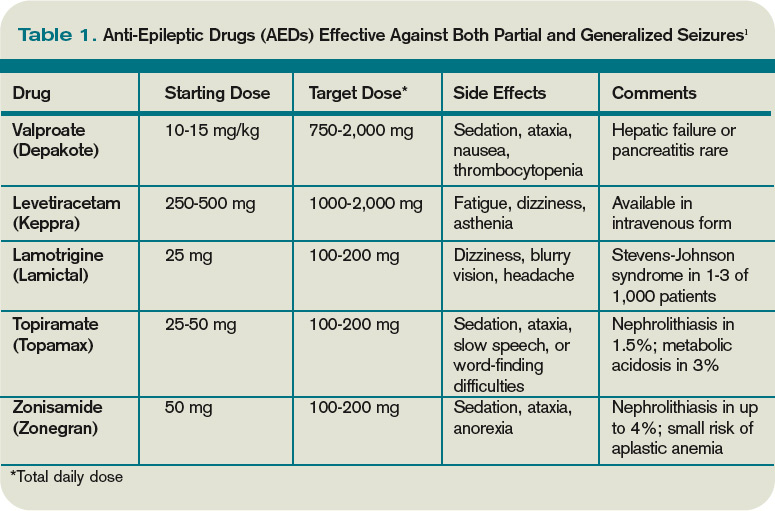
The decision to begin AED therapy after a first unprovoked seizure is controversial. Estimates of the likelihood of seizure recurrence range from 25% at two years to 50% at one year (in the absence of AED therapy).1-2,4-7 The decision to start AED therapy after a first seizure must therefore be individualized for each patient.
Patients at high risk for recurrent seizures should begin AED therapy.1 However, no test or prognostic tool reliably identifies these patients, and initiating therapy carries side effects and places psychological, financial, and social burdens on the patient. The prevailing clinical practice, therefore, has been watchful waiting, with a second seizure constituting proof of high risk for recurrence—and need for AED therapy. Three-quarters of patients with two or more unprovoked seizures likely will go on to have recurrent seizures.6
On the other hand, in patients believed to be at high risk for seizure recurrence, a more aggressive approach of initiating AED therapy after the first seizure is reasonable. A number of risk factors increasing risk for seizure recurrence have been identified (see Table 2, left).1,2 It is justified to initiate AED therapy if any of these factors are present, even after a single seizure. Still, it’s important to note that most people with risk factors will not benefit from AEDs, as only about 40% will have a seizure in the following two years.1
Early initiation of AED therapy might be appropriate for patients with occupations or hobbies in which seizures could be life-threatening (e.g., scuba divers, truck drivers).2
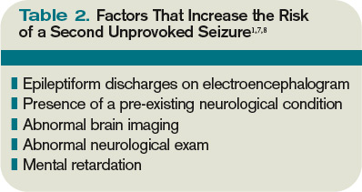
Low-risk patients still have a roughly 20% to 30% risk of seizure recurrence within three years.1 A second seizure that occurs while driving or while engaged in any hazardous activity could lead to serious injury.
Patients should be advised of this small but inescapable risk and instructed to contact their department of motor vehicles for specific legal restrictions, which vary by state. Once three seizure-free years have passed after a patient’s initial seizure, the chance of a recurrence falls to around 10% to 20%.6-7
Back to the Case
Our 42-year-old patient with a first seizure had normal findings on examination, laboratory studies, and brain imaging. An EEG showed epileptiform discharges in a spike and wave pattern. The attending hospitalist counseled him on his elevated risk of future seizures; the patient then elected to begin AED therapy, citing a fear of losing his driving privileges. Levetiracetam was started, which he tolerated despite mild sedation.
A year later, he suffered another seizure at his home. With regular followup and titration of his AED, he remained seizure-free for the next five years.
Bottom Line
Most patients with a single unprovoked seizure can be managed with watchful waiting, counseling, and neurological followup. Initiation of AED therapy is appropriate for patients with a high risk of seizure recurrence, or for whom another seizure could pose personal or social harm. TH
Dr. Hoffman is a hospitalist at Emory University School of Medicine in Atlanta.
References
- French JA, Pedley TA. Clinical practice: Initial management of epilepsy. N Engl J Med. 2008;359:166-176.
- Krumholz A, Wiebe S, Gronseth G, et al. Practice Parameter: evaluating an apparent unprovoked first seizure in adults (an evidence-based review): report of the Quality Standards Subcommittee of the American Academy of Neurology and the American Epilepsy Society. Neurology. 2007;69:1996-2007.
- Schachter SC. Antiepileptic drug therapy: general treatment principles and application for special patient populations. Epilepsia. 1999;40(9):S20-25.
- Hauser WA, Rich SS, Annegers JF, et al. Seizure recurrence after a first unprovoked seizure: an extended follow-up. Neurology. 1990;40:1163-1170.
- Marson A, Jacoby A, Johnson A, et al. Immediate versus deferred antiepileptic drug treatment for epilepsy and single seizures: a randomized controlled trial. Lancet. 2005;365: 2007-2013.
- Hauser WA, Rich SS, Lee JR, Annegers JF, Anderson VE. Risk of recurrent seizures after two unprovoked seizures. N Engl J Med. 1998;338:429-434.
- Berg AT. Risk of recurrence after a first unprovoked seizure. Epilepsia. 2008;49:S13-18.
- Kim LG, Johnson TL, Marson AG, et al. Prediction of risk of seizure recurrence after a single seizure and early epilepsy: further results from the MESS trial. Lancet Neurology. 2006;5(4):317-322.
Case
A 42-year-old man is brought to the hospital by his family after a reported seizure. The patient was found on the floor, unresponsive, and suffering convulsions lasting less than a minute. He suffered no apparent trauma before or during the event. He has no history of seizures. His mental status quickly improved; he experienced oriented lucidity with slight drowsiness. His neurological exam is nonfocal, and his vital signs and laboratory values are normal. A noncontrast head computed tomogram (CT) is normal.
What is the appropriate approach to diagnosis and management for this patient with a new-onset seizure?
Overview
A patient with a first seizure presents a dilemma. Underlying causes for seizure are potentially life-threatening, and must be identified if present. A patient whose first seizure is unprovoked is at risk for future seizures (i.e., epilepsy). However, long-term therapy with anticonvulsant medication has morbidity, side effects, and expense. Advising a patient on whether to drive has public safety and legal implications, as well as major lifestyle changes for the patient.
Seizures may be focal (limited to one area of the brain) or generalized (involving both hemispheres). For the most part, focal (also known as partial) seizures do not impair consciousness; generalized seizures do. Approximately 70% of first seizures are partial focal seizures.1 Such provoking causes as head trauma, stroke, alcohol withdrawal, brain tumors, and infections can be identified in about one-third of cases.1
Electroencephalogram (EEG) and computed tomogram (CT) of the brain should be obtained, but insufficient evidence exists to recommend other testing, which should be pursued according to the clinical context.2
Unprovoked seizures recur in about 25% to 50% of patients, resulting in a diagnosis of epilepsy.1,2,4-7
Therapy is unnecessary in patients whose seizures will not recur, but reliably identifying these patients is a challenge. Whether antiepileptic drug (AED) therapy should be initiated in patients with a first unprovoked seizure is controversial and will be reviewed below.
Review of the Data
History: No test or finding can reliably differentiate unwitnessed seizures from other events (e.g., syncope).2 History from a reliable observer often is necessary to determine whether the event actually was a seizure.2 In as many as 50% of patients with a “first” seizure, thorough history will likely reveal previously unrecognized seizures.1 Although most epilepsy syndromes begin in childhood or adolescence, a significant number of patients will experience their first seizure in adulthood.2
A thorough neurologic examination should be performed. In a minority of cases, an exam will suggest a focal lesion. An impaired level of consciousness might represent a post-ictal state or delirium.2
Diagnostic evaluation: If the history suggests a seizure, an EEG should be obtained. Although the EEG will be normal in 50% of patients following a first seizure, an abnormal EEG provides useful information about seizure type and the likelihood of recurrence.2 In nearly a quarter of patients, the EEG will show epileptiform abnormalities that predict future seizures.2
Generally, an EEG should be obtained as soon as feasible, once a seizure is suspected. Some evidence in children suggests that EEG yield is higher in the 24 hours after a first seizure.
A noncontrast head CT or magnetic resonance imaging (MRI) reveals a significant abnormality about 10% of the time.2 A CT or MRI should be obtained. Few studies have compared CT to MRI in terms of yield in determining first seizure etiology, and those that do compare the two suffer from selection bias.2 Although CT or MRI are appropriate in evaluating a patient with a first seizure, the MRI’s greater resolution might provide a higher diagnostic yield in terms of seizure etiology, and, therefore, some experts recommend MRI over CT in nonemergent cases.2
Insufficient data exist to support or refute diagnostic testing beyond brain imaging and EEG. Although electrolyte abnormalities, hypoglycemia, and infections might infrequently cause seizures, such routine blood tests as complete blood count (CBC) and chemistry panels are rarely helpful.
As many as 15% of patients with a seizure will have minor abnormalities on routine lab tests, but the abnormalities do not appear to be the cause of the seizure.2
Lumbar puncture (LP) is categorically recommended only in patients in whom there is a clinical suspicion for infection as a seizure etiology. Reviews suggest that signs and symptoms of infection are typically present in patients with meningitis or another infectious cause for seizure; LP generally has limited utility in other noninfectious causes of seizure.2
The utility of toxicology testing in a first seizure has not been studied widely. Testing urine or blood for the presence of alcohol, cocaine, methamphetamines, benzodiazepines, or drug metabolites could be useful in the appropriate clinical setting.2
It is unclear whether a patient with a first seizure requires hospitalization. If initial testing in the ED rules out serious causes of seizures, the yield for hospitalization is likely to be low. In clinical practice, however, hospitalization is common and often necessary to complete such diagnostic testing as EEG and MRI.
Medical therapy: Patients with suspected epilepsy (e.g., those whose presenting seizure is, in retrospect, not their first seizure) should begin antiepileptic drug therapy (AED).1
Typically, a broad-spectrum AED—one that is effective against both partial and generalized seizures—should be used as initial therapy for epilepsy. These include valproate, lamotrigine, topiramate, zonisamide, and levetiracetam (see Table 1, above). Valproate has the longest history of effectiveness; levetiracetam has fewer drug interactions, and randomized trials support its efficacy.1
Checking blood electrolytes and liver enzymes is recommended before beginning AED treatment. Significant hepatic or renal dysfunction might necessitate dosing adjustments in many AEDs.2
Inpatient consultation with a neurologist might be helpful, although insufficient evidence exists that such consultation improves patient outcomes or makes care more cost-efficient. A neurologist should follow up on patients with a first seizure after hospital discharge.2
Patients with a first seizure that likely was provoked by a reversible condition (e.g., hypotension, hypoglycemia, infection) should generally not begin AED therapy. This also includes patients with multiple seizures in a brief period of time (less than 24 hours), all attributed to the same reversible cause.1
The decision to begin AED therapy after a first unprovoked seizure is controversial. Estimates of the likelihood of seizure recurrence range from 25% at two years to 50% at one year (in the absence of AED therapy).1-2,4-7 The decision to start AED therapy after a first seizure must therefore be individualized for each patient.
Patients at high risk for recurrent seizures should begin AED therapy.1 However, no test or prognostic tool reliably identifies these patients, and initiating therapy carries side effects and places psychological, financial, and social burdens on the patient. The prevailing clinical practice, therefore, has been watchful waiting, with a second seizure constituting proof of high risk for recurrence—and need for AED therapy. Three-quarters of patients with two or more unprovoked seizures likely will go on to have recurrent seizures.6
On the other hand, in patients believed to be at high risk for seizure recurrence, a more aggressive approach of initiating AED therapy after the first seizure is reasonable. A number of risk factors increasing risk for seizure recurrence have been identified (see Table 2, left).1,2 It is justified to initiate AED therapy if any of these factors are present, even after a single seizure. Still, it’s important to note that most people with risk factors will not benefit from AEDs, as only about 40% will have a seizure in the following two years.1
Early initiation of AED therapy might be appropriate for patients with occupations or hobbies in which seizures could be life-threatening (e.g., scuba divers, truck drivers).2
Low-risk patients still have a roughly 20% to 30% risk of seizure recurrence within three years.1 A second seizure that occurs while driving or while engaged in any hazardous activity could lead to serious injury.
Patients should be advised of this small but inescapable risk and instructed to contact their department of motor vehicles for specific legal restrictions, which vary by state. Once three seizure-free years have passed after a patient’s initial seizure, the chance of a recurrence falls to around 10% to 20%.6-7
Back to the Case
Our 42-year-old patient with a first seizure had normal findings on examination, laboratory studies, and brain imaging. An EEG showed epileptiform discharges in a spike and wave pattern. The attending hospitalist counseled him on his elevated risk of future seizures; the patient then elected to begin AED therapy, citing a fear of losing his driving privileges. Levetiracetam was started, which he tolerated despite mild sedation.
A year later, he suffered another seizure at his home. With regular followup and titration of his AED, he remained seizure-free for the next five years.
Bottom Line
Most patients with a single unprovoked seizure can be managed with watchful waiting, counseling, and neurological followup. Initiation of AED therapy is appropriate for patients with a high risk of seizure recurrence, or for whom another seizure could pose personal or social harm. TH
Dr. Hoffman is a hospitalist at Emory University School of Medicine in Atlanta.
References
- French JA, Pedley TA. Clinical practice: Initial management of epilepsy. N Engl J Med. 2008;359:166-176.
- Krumholz A, Wiebe S, Gronseth G, et al. Practice Parameter: evaluating an apparent unprovoked first seizure in adults (an evidence-based review): report of the Quality Standards Subcommittee of the American Academy of Neurology and the American Epilepsy Society. Neurology. 2007;69:1996-2007.
- Schachter SC. Antiepileptic drug therapy: general treatment principles and application for special patient populations. Epilepsia. 1999;40(9):S20-25.
- Hauser WA, Rich SS, Annegers JF, et al. Seizure recurrence after a first unprovoked seizure: an extended follow-up. Neurology. 1990;40:1163-1170.
- Marson A, Jacoby A, Johnson A, et al. Immediate versus deferred antiepileptic drug treatment for epilepsy and single seizures: a randomized controlled trial. Lancet. 2005;365: 2007-2013.
- Hauser WA, Rich SS, Lee JR, Annegers JF, Anderson VE. Risk of recurrent seizures after two unprovoked seizures. N Engl J Med. 1998;338:429-434.
- Berg AT. Risk of recurrence after a first unprovoked seizure. Epilepsia. 2008;49:S13-18.
- Kim LG, Johnson TL, Marson AG, et al. Prediction of risk of seizure recurrence after a single seizure and early epilepsy: further results from the MESS trial. Lancet Neurology. 2006;5(4):317-322.
Should HM Be in the Comanagement Business?
PRO
Experience, availability make HM the perfect comanagement choice

Hospitalists should be in the business of comanagement, especially the comanagement of surgical patients. Thanks to modern medicine, people are living longer with a higher burden of comorbidity. Comanagement is intended to bring clinical acumen and experience to the bedside of a complex medical patient who is having surgery and needs acute medical care. Hospitalists must bring their extensive clinical experience and on-site availability to ensure the best outcomes.
The most important aspect of comanagement can be summed up in one phrase: right person, right place, and right time.
Right Person
The interaction between pre-existing comorbidities and the physiologic stress of anesthesia, volume shifts, narcotic therapy, sleep deprivation, postoperative anemia, and any number of other stressors can be complex.
Frankly, it is more appropriate for an experienced hospitalist to evaluate and manage these intricacies than a specialty surgeon, although general and critical-care surgeons are likely exceptions. Our internal medicine and geriatric training, not to mention experience, positions us to best meet the needs of these complex patients.
Right Place
Hospitalists are in the hospital and prepared to handle the unexpected. Surgeons cannot be in two places at once; they cannot leave the operating room with another patient on the table. Likewise, a general internist in a consultative role usually cannot leave the clinic on short notice. Hospitalists are physically present and available to go to a patient’s room when there is an immediate clinical need.
Right Time
Early evaluation and treatment are indicated when most postoperative complications occur. One of the defining characteristics of hospitalists is availability. Relying on a surgeon to be at the bedside immediately is inappropriate.
In traditional consultative roles, the medical consultant might also have clinical duties in an outpatient setting. Neither model allows an experienced physician to reliably get to the bedside, then evaluate, monitor, follow up on test results, and manage patient care. Hospitalists are available and can participate in all aspects of care until resolution of the complication.
Additional Opportunity
Another aspect of the comanagement model is the opportunity to extend beyond the postoperative setting to the preoperative evaluation. This brings with it the opportunity to identify possible problems before surgery. In addition, the business model for the preoperative consultation is quite strong.
Meeting patients’ clinical needs is the most important reason for engaging in a comanagement model of care in the perioperative setting. This model brings timely experience, evaluation, and management when and where it is needed. TH
CON
Establish rules of engagement before agreeing to share responsibilities

In the wee hours of a recent busy call night, the ED called me to admit a patient whose automatic implantable cardioverter cefibrillator (AICD) had fired repeatedly. The patient had no other active medical issues. When called, the electrophysiologist, who was on staff, demanded that I admit the patient for “medical comanagement.” The specialist agreed that I probably would have little to add to the care, but his firm expectation was that hospitalists admit his patients and he “consults” … especially at 2 a.m.
Job Description Defined
Comanagement, defined as shared responsibility, authority, and accountability for the management of a hospitalized patient, is an HM mainstay and a primary driver of the explosive growth of our field.1
While it stands to reason that surgical and specialty patients with active medical comorbidities likely fare better if hospitalists are integrated into their care, comanagement has broadened in its application to include scenarios in which the benefits are more dubious. Hospitalist comanagement now encompasses “management” of patients for whom hospitalists have little, if anything, to add.
At the other comanagement extreme, hospitalists, despite little or no formal training, primarily manage patients with acute neurologic, neurosurgical, psychiatric, and orthopedic diagnoses, often with inadequate surgical or specialty involvement.2,3 Although it makes sense for a hospital with only one neurosurgeon to have its hospitalists manage carefully selected neurosurgical patients, the justification for such scenarios becomes harder to reconcile at hospitals where there are no staffing shortages. I suspect the primary justification for hospitalist comanagement in such circumstances is to keep specialists doing lucrative procedures by day and in bed at night, and to ensure that someone manages the paperwork, discharge communication, and patient logistics that are often otherwise ignored.
Rules of Engagement
In well-designed comanagement arrangements, hospitalists and specialists work equitably under clearly defined and mutually agreed upon rules of engagement. They share responsibility for patients, collaborate to improve care, and teach and learn from each other. Unfortunately, in many instances, the power structure has tilted.
Practicing hospitalists frequently complain about their subordinate status and inability to control their working conditions; both are identified risk factors for career dissatisfaction and burnout.4,5
Before entering a comanagement relationship, hospitalists should gain a clear understanding of why they are being asked to comanage, what problems they are expected to fix by doing so, and what the consequences, intended or unintended, might be as a result. There should be mechanisms to ensure that the relationship is equitable and serves the best interests of the patient, rather than the care parties involved. TH
References
- 2007-2008 Bi-annual Survey on the State of the Hospital Medicine Movement. SHM Web site. Available at: www.hospitalmedicine.org/AM/Template.cfm?Section=Surveys2&Template=/CM/HTMLDisplay.cfm&ContentID=18419. Accessed July 26, 2009.
- Plauth WH, Pantilat SZ, Wachter RM, Fenton CL. Hospitalists’ perceptions of their residency training needs: results of a national survey. Am J Med. 2001;111:247-254.
- Glasheen JJ, Epstein KR, Siegal E, Kutner JS, Prochazka AV. The spectrum of community-based hospitalist practice: a call to tailor internal medicine residency training. Arch Intern Med. 2007;167(7):727-728.
- Linzer M , Gerrity M, Douglas JA, McMurray JE, Williams ES, Konrad TR. Physician stress: results from the physician work life study. Stress and Health. 2001;18(1)37-42.
- SHM Career Satisfaction White Paper. SHM Web site. Available at: http://www.hospitalmedicine.org/AM/Template.cfm?Section=Practice_Resources& Template=/CM/ContentDisplay.cfm&ContentID=14631. Accessed July 26, 2009.
The opinions expressed herein are those of the authors and do not necessarily represent those of the Society of Hospital Medicine or The Hospitalist.
PRO
Experience, availability make HM the perfect comanagement choice
Hospitalists should be in the business of comanagement, especially the comanagement of surgical patients. Thanks to modern medicine, people are living longer with a higher burden of comorbidity. Comanagement is intended to bring clinical acumen and experience to the bedside of a complex medical patient who is having surgery and needs acute medical care. Hospitalists must bring their extensive clinical experience and on-site availability to ensure the best outcomes.
The most important aspect of comanagement can be summed up in one phrase: right person, right place, and right time.
Right Person
The interaction between pre-existing comorbidities and the physiologic stress of anesthesia, volume shifts, narcotic therapy, sleep deprivation, postoperative anemia, and any number of other stressors can be complex.
Frankly, it is more appropriate for an experienced hospitalist to evaluate and manage these intricacies than a specialty surgeon, although general and critical-care surgeons are likely exceptions. Our internal medicine and geriatric training, not to mention experience, positions us to best meet the needs of these complex patients.
Right Place
Hospitalists are in the hospital and prepared to handle the unexpected. Surgeons cannot be in two places at once; they cannot leave the operating room with another patient on the table. Likewise, a general internist in a consultative role usually cannot leave the clinic on short notice. Hospitalists are physically present and available to go to a patient’s room when there is an immediate clinical need.
Right Time
Early evaluation and treatment are indicated when most postoperative complications occur. One of the defining characteristics of hospitalists is availability. Relying on a surgeon to be at the bedside immediately is inappropriate.
In traditional consultative roles, the medical consultant might also have clinical duties in an outpatient setting. Neither model allows an experienced physician to reliably get to the bedside, then evaluate, monitor, follow up on test results, and manage patient care. Hospitalists are available and can participate in all aspects of care until resolution of the complication.
Additional Opportunity
Another aspect of the comanagement model is the opportunity to extend beyond the postoperative setting to the preoperative evaluation. This brings with it the opportunity to identify possible problems before surgery. In addition, the business model for the preoperative consultation is quite strong.
Meeting patients’ clinical needs is the most important reason for engaging in a comanagement model of care in the perioperative setting. This model brings timely experience, evaluation, and management when and where it is needed. TH
CON
Establish rules of engagement before agreeing to share responsibilities
In the wee hours of a recent busy call night, the ED called me to admit a patient whose automatic implantable cardioverter cefibrillator (AICD) had fired repeatedly. The patient had no other active medical issues. When called, the electrophysiologist, who was on staff, demanded that I admit the patient for “medical comanagement.” The specialist agreed that I probably would have little to add to the care, but his firm expectation was that hospitalists admit his patients and he “consults” … especially at 2 a.m.
Job Description Defined
Comanagement, defined as shared responsibility, authority, and accountability for the management of a hospitalized patient, is an HM mainstay and a primary driver of the explosive growth of our field.1
While it stands to reason that surgical and specialty patients with active medical comorbidities likely fare better if hospitalists are integrated into their care, comanagement has broadened in its application to include scenarios in which the benefits are more dubious. Hospitalist comanagement now encompasses “management” of patients for whom hospitalists have little, if anything, to add.
At the other comanagement extreme, hospitalists, despite little or no formal training, primarily manage patients with acute neurologic, neurosurgical, psychiatric, and orthopedic diagnoses, often with inadequate surgical or specialty involvement.2,3 Although it makes sense for a hospital with only one neurosurgeon to have its hospitalists manage carefully selected neurosurgical patients, the justification for such scenarios becomes harder to reconcile at hospitals where there are no staffing shortages. I suspect the primary justification for hospitalist comanagement in such circumstances is to keep specialists doing lucrative procedures by day and in bed at night, and to ensure that someone manages the paperwork, discharge communication, and patient logistics that are often otherwise ignored.
Rules of Engagement
In well-designed comanagement arrangements, hospitalists and specialists work equitably under clearly defined and mutually agreed upon rules of engagement. They share responsibility for patients, collaborate to improve care, and teach and learn from each other. Unfortunately, in many instances, the power structure has tilted.
Practicing hospitalists frequently complain about their subordinate status and inability to control their working conditions; both are identified risk factors for career dissatisfaction and burnout.4,5
Before entering a comanagement relationship, hospitalists should gain a clear understanding of why they are being asked to comanage, what problems they are expected to fix by doing so, and what the consequences, intended or unintended, might be as a result. There should be mechanisms to ensure that the relationship is equitable and serves the best interests of the patient, rather than the care parties involved. TH
References
- 2007-2008 Bi-annual Survey on the State of the Hospital Medicine Movement. SHM Web site. Available at: www.hospitalmedicine.org/AM/Template.cfm?Section=Surveys2&Template=/CM/HTMLDisplay.cfm&ContentID=18419. Accessed July 26, 2009.
- Plauth WH, Pantilat SZ, Wachter RM, Fenton CL. Hospitalists’ perceptions of their residency training needs: results of a national survey. Am J Med. 2001;111:247-254.
- Glasheen JJ, Epstein KR, Siegal E, Kutner JS, Prochazka AV. The spectrum of community-based hospitalist practice: a call to tailor internal medicine residency training. Arch Intern Med. 2007;167(7):727-728.
- Linzer M , Gerrity M, Douglas JA, McMurray JE, Williams ES, Konrad TR. Physician stress: results from the physician work life study. Stress and Health. 2001;18(1)37-42.
- SHM Career Satisfaction White Paper. SHM Web site. Available at: http://www.hospitalmedicine.org/AM/Template.cfm?Section=Practice_Resources& Template=/CM/ContentDisplay.cfm&ContentID=14631. Accessed July 26, 2009.
The opinions expressed herein are those of the authors and do not necessarily represent those of the Society of Hospital Medicine or The Hospitalist.