User login
Research Committee Chair Reflects

Before Andy Auerbach, MD, MPH, concludes a four-year term as chair of SHM’s Research Committee, I talked with him about his perspective on hospital medicine research. Dr. Auerbach is an associate professor of medicine at the University of California, San Francisco.
He received a career development award from the National Institutes of Health (NIH) early in his career and is the principal investigator of an R01 research project grant from the NHLBI titled “Improving use of perioperative beta-blockers through a multidimensional QI program.”
He is also a co-author of “Outcomes of Patients Treated by Hospitalists, General Internists, and Family Physicians” in the December 2007 New England Journal of Medicine, which found statistically significant differences in length of stay and cost. He received his medical degree from Dartmouth Medical School in Hanover, N.H., and did his residency training in internal medicine at Yale New Haven Hospital in Connecticut. He completed an MPH in clinical epidemiology at the Harvard School of Public Health in Boston in 1998.
Q: So, is academia as glamorous as it sounds?
Dr. Auerbach: Way more glamorous—you should see my office. And yes, we are in a white tower.
Q: How did you get your start in research?
Dr. Auerbach: I actually started out my research fellowship wanting to be a cardiologist and go into the cath lab while developing the skills to participate in and teach research methods. I found I really enjoyed the work, particularly the creative and entrepreneurial aspects of developing a project or grant and seeing it through to completion.
Q: What are the research options for hospitalists practicing in nonteaching settings?
Dr. Auerbach: I think the most straightforward way to participate in research is to partner with a clinical research organization to help enroll patients in their trials. While you don’t get the opportunity to design the study, you do get to get a feel for consent/enrollment and internal review board [IRB] processes.
The next best way to get involved with research is to partner with a researcher—and this need not be a hospitalist—at your site or very near by. Many QI projects are close to being research-ready and may provide an opportunity to make that work count twice. But it will require you to learn about analytic methods.
I’d also be remiss if I didn’t mention the value of other very useful academic products—rigorous reports of a QI intervention (think of both success and failure stories) and patient case reports. If well referenced and used as teaching documents, these can be very useful ways to advance knowledge.
Q: Are there any particular prerequisites in terms of training that you find especially helpful as you conduct your research?
Dr. Auerbach: It is hard to be a capital-R “Researcher” and compete for career development grants and NIH funding without some advanced [degrees] and a clinical research fellowship. I hesitate to call these prerequisites, but they are nearly so.
Q: What do you like best about your career as a hospitalist?
Dr. Auerbach: I really like acute care medicine, but didn’t want to subspecialize—otherwise I’d be wearing lead in a cath lab now. I also like the questions and processes in the hospital a bit more than the clinic setting.
Q: Who are your mentors and how did you find them?
Dr. Auerbach: I’ve had a remarkable set of mentors from fellowship [Mary Beth Hamel, Roger Davis, Russ Phillips] through my early career [Lee Goldman, Bob Wachter, Ralph Gonzales]. Now that I am early-mid career, I’m trying to pass their teaching on.
Q: Any advice for hospitalists interested in research but daunted by the prospect of starting their own studies?
Dr. Auerbach: If you want to do a scholarly/academic project to round out your personal/career satisfaction, I think the daunting nature of research can be overcome with the right questions and right support—and by defining what these are well before you actually dive into a dataset or implementation project. You also have to decide how much satisfaction you will get from the project in the end compared to the incremental nights/weekends you will spend to plan and execute your project—not to mention publish.
If you are thinking of research as a career, be aware of what makes you happy. If you like to write, enjoy the process of hypothesis generating/testing, and take rejection well you may be happy as a researcher. There are still plenty of nights/weekends to be spent, though.
Making a switch from full-time clinical or administrative work to research means making a very big commitment to going back to get the skills as part of a fellowship.
Q: Do researchers interested in quality improvement questions still have to run their work past the IRB?
Dr. Auerbach: Unfortunately this is now an area of uncertainty for people—unnecessarily so. Until recent events, IRBs have not required approval for QI projects that seek to enhance care according to an evidence-based standard, especially if that standard is endorsed by the institution. If you plan to publish your findings—particularly if you talk to or touch patients, or collect personal health information—I think it is nearly always wise to at least call your local IRB to ask for how you can or should conduct the study. This is best done before you start the project, obviously.
If you want to publish your results using deidentified data after the project is done, our IRB would say that is exempt from review [e.g., no need for approval]. But I think even this case would be worth a phone call to ensure your IRB feels similarly.
Whether or not you get IRB approval, be very aware of how and where you store data. TH
Satisfaction Is Job No. 1
The Career Satisfaction Task Force has focused on two key areas this year to build upon the work that resulted in last year’s white paper “A Challenge for a New Specialty: A White Paper on Hospitalist Career Satisfaction.”
The paper outlined a framework for hospital medicine program leaders and hospitalists to identify important components of matching individuals and programs for the best job fit.
This year, the task force is working to bring the white paper to life and moving it from a conceptual framework to demonstrating how to use it to solve real issues facing programs and individuals.
The first of these projects was a Webinar led by SHM Senior Vice President Joe Miller, Sylvia McKean, MD (course director of Hospital Medicine 2008), and Win Whitcomb, MD (a co-founder of SHM). Each of them has held leadership roles on this task force. About 80 people participated in the December event, and more than three-fourths of attendees rated it highly.
At last year’s Annual Meeting in Dallas, the white paper was presented in a task force workshop. In keeping with our aim to bring the framework to life, this year’s workshop will use real case studies to demonstrate how to use bring the concepts to solutions. The workshop will be facilitated by Chad Whelan, MD, assistant professor of medicine and director of the Hospitalists Scholars Training Program, University of Chicago. Discussing key concepts will be Doug Carlson, MD, associate professor, Pediatrics Division, Washington University School of Medicine in St. Louis, and Tosha Wetterneck, MD, University of Wisconsin Hospital/Clinics, Madison. Drs. Carlson and Wetterneck made significant contributions to the white paper. In this highly interactive workshop, case studies that demonstrate challenges with workload/scheduling and autonomy will be discussed. Drs. Carlson and Wetterneck will lead the participants through discussions aimed at identifying the root causes of struggle and potential solutions for the program.
In the coming months, we hope to develop a series of articles to be published in The Hospitalist addressing the issues of greatest importance for career satisfaction.
The task force realizes there may be opportunities to add knowledge about career satisfaction and provide a valuable service to SHM member. We are in the early stages of developing a survey geared to further clarifying the most important factors in making satisfying career matches as well as providing detailed feedback about programs to their leaders. We are seeking funding to enable us to begin this exciting work.
Hospital Medicine Continues to Make Inroads Overseas
Attendees at this year’s SHM Annual Meeting in San Diego brought with them exciting news of international developments that might broaden the scope of the specialty.
For example, Efren Manjarrez, MD, director of clinical operations for the division of hospital medicine of the Leonard M. Miller School of Medicine at the University of Miami, recently discovered a 500-plus-bed facility at the Universidad de Navarra during his trip to Pamplona, Spain.
Because Dr. Manjarrez is bilingual, past SHM President Mark Williams, MD, professor and chief of the division of hospital medicine at the Northwestern University Feinberg School of Medicine in Chicago, asked him to give a lecture.
“[The Universidad de Navarra] has one of the top two medical schools in Spain and they had the very first symposium on the management of the hospitalized patient,” Dr. Manjarrez says. “They had the very first hospitalist conference in Spain and quite possibly in Europe.”
While in Spain, Dr. Manjarrez realized hospitalists there were “at the grassroots level, where we were about 10 to 12 years ago,” just starting to have a few hospital medical units. The clinic in at the Universidad de Navarra had about five units, and there was a small hospitalist group near Valencia, as well.

Dr. Manjarrez is highly complimentary of the university’s “top-flight medical school,” where he said doctors perform liver transplants.
“They could compete favorably with any city in the United States,” he notes. “They’re interested in organizing hospitalists like Mark Williams and what people ahead of me did for SHM.”
Dr. Manjarrez and Dr. Williams have discussed what will happen with the hospital medicine movement internationally. They forecast that the work begun in the U.S. will globalize fairly soon.
“The Spaniards are very, very on the ball to ask us over there to see what’s going on to get a jump on it,” Dr. Manjarrez says. “SHM needs to start thinking ahead and have an international chapter and plan international meetings abroad to have SHM’s message spread globally. The time is ripe to export what SHM and hospital medicine is doing here”
Dr. Manjarrez notes that Argentina has small pockets of hospitalists. And, Guilherme Brauner Barcellos MD, specialist in internal medicine and intensive care at the Nossa Senhora de Conceicao Hospital in Brazil, is excited about the establishment of a hospital medicine program there. He finds the attempts to develop the specialty in his country “fascinating and challenging.”
“The implementation of hospital medicine, especially those aspects that involve more than just having a general medicine physician dealing with inpatient care, is brand new in Brazil,” Dr. Barcellos says. “We understand there is a long journey ahead.”
Dr. Barcellos is president of the recently formed Brazilian Society of Hospital Medicine and became a member of SHM last year, attending May’s meeting in Dallas. He says the number of hospitalists in Brazil is limited, but “a rapid expansion is predicted.”
“As in the U.S. several years ago, the case for hospitalists in Brazil is still being made,” he notes. “We did a hospital medicine meeting last October, and it led to the formation of the Brazilian Society of Hospital Medicine. We definitely fostered the discussion about the specialty and the model in Brazil.”
He hopes more people will talk about the topic through the new group’s Web site (www.medicinahospitalar.com.br) and in hospitals throughout the country.
“Before the year of 2005, the majority of people here didn’t know about the hospitalists,” he explains. “After that, we had a period in which people were confused, thinking hospitalists were the same as doctors who work in the rapid response teams only. Currently, everybody at least knows about hospital medicine.”
Dr. Williams notes that hospital medicine organizations are forming in Chile, Argentina, Australia, and New Zealand.
Dr. Barcellos believes hospitals across his country “will awake to hospital medicine” when they realize that traditional models aren’t property servicing hospitalized patients anymore. “Much of what is present is wrong, obsolete or out of time, and we should try new attempts to create a different organization,” he says. TH
Molly R. Okeon is a journalist based in California.
Attendees at this year’s SHM Annual Meeting in San Diego brought with them exciting news of international developments that might broaden the scope of the specialty.
For example, Efren Manjarrez, MD, director of clinical operations for the division of hospital medicine of the Leonard M. Miller School of Medicine at the University of Miami, recently discovered a 500-plus-bed facility at the Universidad de Navarra during his trip to Pamplona, Spain.
Because Dr. Manjarrez is bilingual, past SHM President Mark Williams, MD, professor and chief of the division of hospital medicine at the Northwestern University Feinberg School of Medicine in Chicago, asked him to give a lecture.
“[The Universidad de Navarra] has one of the top two medical schools in Spain and they had the very first symposium on the management of the hospitalized patient,” Dr. Manjarrez says. “They had the very first hospitalist conference in Spain and quite possibly in Europe.”
While in Spain, Dr. Manjarrez realized hospitalists there were “at the grassroots level, where we were about 10 to 12 years ago,” just starting to have a few hospital medical units. The clinic in at the Universidad de Navarra had about five units, and there was a small hospitalist group near Valencia, as well.
Dr. Manjarrez is highly complimentary of the university’s “top-flight medical school,” where he said doctors perform liver transplants.
“They could compete favorably with any city in the United States,” he notes. “They’re interested in organizing hospitalists like Mark Williams and what people ahead of me did for SHM.”
Dr. Manjarrez and Dr. Williams have discussed what will happen with the hospital medicine movement internationally. They forecast that the work begun in the U.S. will globalize fairly soon.
“The Spaniards are very, very on the ball to ask us over there to see what’s going on to get a jump on it,” Dr. Manjarrez says. “SHM needs to start thinking ahead and have an international chapter and plan international meetings abroad to have SHM’s message spread globally. The time is ripe to export what SHM and hospital medicine is doing here”
Dr. Manjarrez notes that Argentina has small pockets of hospitalists. And, Guilherme Brauner Barcellos MD, specialist in internal medicine and intensive care at the Nossa Senhora de Conceicao Hospital in Brazil, is excited about the establishment of a hospital medicine program there. He finds the attempts to develop the specialty in his country “fascinating and challenging.”
“The implementation of hospital medicine, especially those aspects that involve more than just having a general medicine physician dealing with inpatient care, is brand new in Brazil,” Dr. Barcellos says. “We understand there is a long journey ahead.”
Dr. Barcellos is president of the recently formed Brazilian Society of Hospital Medicine and became a member of SHM last year, attending May’s meeting in Dallas. He says the number of hospitalists in Brazil is limited, but “a rapid expansion is predicted.”
“As in the U.S. several years ago, the case for hospitalists in Brazil is still being made,” he notes. “We did a hospital medicine meeting last October, and it led to the formation of the Brazilian Society of Hospital Medicine. We definitely fostered the discussion about the specialty and the model in Brazil.”
He hopes more people will talk about the topic through the new group’s Web site (www.medicinahospitalar.com.br) and in hospitals throughout the country.
“Before the year of 2005, the majority of people here didn’t know about the hospitalists,” he explains. “After that, we had a period in which people were confused, thinking hospitalists were the same as doctors who work in the rapid response teams only. Currently, everybody at least knows about hospital medicine.”
Dr. Williams notes that hospital medicine organizations are forming in Chile, Argentina, Australia, and New Zealand.
Dr. Barcellos believes hospitals across his country “will awake to hospital medicine” when they realize that traditional models aren’t property servicing hospitalized patients anymore. “Much of what is present is wrong, obsolete or out of time, and we should try new attempts to create a different organization,” he says. TH
Molly R. Okeon is a journalist based in California.
Research Riddle
The recent uproar over the Office of Human Research Protections (OHRP) ordering a multicenter study of a Michigan ICU checklist to halt data collection has left quality improvement (QI) researchers, ethicists, and legal experts scratching their heads.
Even before the Michigan debacle, there was considerable confusion about how patient privacy rules included in the Health Insurance Portability and Accountability Act of 1996 (HIPAA) affected QI studies. No one was really sure when institutional review board (IRBs) needed to be involved and when patients needed to be officially consented.
The HIPAA contains specific language addressing how patients and patient data should be handled by researchers. And experts say that’s a good thing—in theory.
But a 2007 study in the Journal of the American Medical Association found that many epidemiologists feel the rules have adversely affected research and done little to improve patient privacy.1
The situation for QI researchers is even more confusing. Many see studies examining the effect of QI interventions as fundamentally different from “human subjects research.” And because of this many were shocked when the OHRP halted data collection from the Michigan care-checklist program.
In that case, the OHRP argued that because the study was prospective, it wasn’t simply QI, but rather “human subjects research.” The OHRP demanded that researchers run their plans by the IRBs of every one of the 103 hospitals involved in the research before relenting Feb. 15 and letting the study resume. (Initial results of the study were published in the New England Journal of Medicine in 2006.2)
Many medical experts view the intervention being studied in Michigan—a simple checklist aimed at reminding physicians to follow some common-sense procedures designed to lower the infection rate associated with central lines—as a straightforward attempt at QI. They argued the study should be exempt from some of the rules regarding human research subjects.
Experts say the OHRP’s initial ruling made what was already a confusing subject into an impossibly muddled morass that may have a chilling effect on the publication of QI studies.
Some say the OHRP has extended HIPAA too far. First of all, legal experts say, it must be understood that HIPAA’s rules don’t apply equally to everyone. For example, public health authorities are allowed to gather patient data without consent if they are trying to prevent the spread of disease. And hospitals are allowed to use data to improve the quality of healthcare, says James G. Hodge Jr. an associate professor at the Johns Hopkins School of Public Health in Baltimore, and executive director of the Centers for Law and the Public’s Health at Johns Hopkins.
One key to resolving the issue may be for healthcare experts to come up with a definition of what constitutes “human subjects research.”
“This is where issues keep coming up,” Hodge says. “Is QI just an extension of clinical care or is it research? The answer to that question will tell you whether it implicates the rule or not.”
Researchers, ethicists, and legal experts hotly debate these issues. No one appears to know exactly where to draw the line that divides human subjects research from QI studies.
Even before the checklist controversy arose, HIPAA was already having a deleterious impact on the exchange of QI information, says Mary Ann Baily, PhD, an associate for ethics and health policy at the Hastings Center in Garrison, N.Y.
Loathe to run afoul of the OHRP, at least one of the larger managed-care providers decided against publishing results of its QI studies, Dr. Baily says. “They’re just trying to stay out of the OHRP’s way,” she explains. “At a recent workshop, researchers said, ‘We don’t publish our data. We make our own system work better and keep our heads down. That way we don’t run into any problems. It’s safer that way.’ ”
This means nobody benefits from that company’s research. “What a waste that is,” Dr. Baily asserts.
Not everyone has taken such a defensive position. But there is a wide range of opinions among QI researchers around the country.
“Certainly as someone who is involved in QI research on an operational level and who is also interested in conducting research looking at the effects of QI interventions I struggle with this regularly,” says Peter Lindenauer, MD, MSC, associate professor of medicine at the Tufts University School of Medicine in Boston and associate medical director, Division of Healthcare Quality, at Baystate Medical Center.
“QI officers don’t look at the work they do as being research,” Dr. Lindenauer notes. “They’re often translating research into practice and implementing and developing strategies designed to improve care. So, when you think about it in those terms, it would never dawn on a typical QI officer to seek IRB approval or to get consent from patients to participate.”
Some researchers avoid the issue by looking only at data with patient identifiers stripped. The assumption is that HIPAA rules apply only to medical records with identifiers intact.
Under that assumption, Lakshmi Halasyamani, MD, has performed two heart failure studies without getting into issues of patient consent. But she sees potential problems with future research.
“Because we were looking at the impact of interventions across the whole population of heart failure patients, it wasn’t a problem,” says Dr. Halasyamani, vice chair for the department of medicine at St. Joseph Mercy Hospital in Ann Arbor, Mich., and a member of SHM’s Board of Directors. “So long as you’re looking at global outcomes, the regulations don’t have much impact.”
But Dr. Halasyamani and her colleagues may want to start looking at the benefits of interventions on subgroups. And this is where things can get messy, she says.
Healthcare professionals really need to figure out a working definition for what constitutes research—and they need to do it soon, Dr. Halasyamani says. Without a good definition, Dr. Halasyamani can see a slowdown of QI research and the possibility of researchers using a QI loophole to get around IRBs. “I could see where some researchers might be tempted to call their studies QI to avoid bureaucratic hassles and IRB oversight,” she says.
Some experts think the minute you decide to publish, by definition you’re doing research.
“I believe you have an ethical obligation to share what you learn with other hospitals,” says Dr. Lindenauer. “So, of course, you want to write it up. But once you start to talk about publication and sharing you start to get to the point where you’re crossing the line —where you’re creating generalizable knowledge.”
And that is precisely when government organizations like the OHRP think you’ve crossed over into research, Dr. Lindenauer says. “It’s a tricky question,” he adds.
Ethicist Baily agrees that experts need to work on coming up with a practical definition of research. Right now, the situation is impossible, she says. Take, for example, a medical plan that wants to send postcards to encourage patients to show up for an annual physical. If researchers want to learn whether that technique works, do they need to send a postcard, prior to the reminder postcard, to let patients know that they’re going to be part of a study, she asks.
And even more important, with all the staff cuts at medical institutions around the country, shouldn’t QI officers study whether these cost-cutting measures adversely affect patient care, she asks. TH
Linda Carroll is a medical journalist based in New Jersey.
References
- Ness RB, Joint Policy Committee, Societies of Epidemiology. Influence of the HIPAA Privacy Rule on health research. JAMA. 2007 Nov 14;298(18):2164-2170.
- Pronovost P, Needham D, Berenholtz S, et al. An intervention to decrease catheter-related bloodstream infections in the ICU. N Engl J Med. 2006 Dec 28;355(26):2725-2732. Erratum in: N Engl J Med. 2007 Jun 21;356(25):2660.
SHM Joins D.C. Session on Value-Based Purchasing
SHM had a seat at the table at a high-level roundtable meeting March 6 in Washington, D.C., to discuss Medicare’s value-based purchasing of hospital care.
SHM President Russell Holman, MD, chief operating officer for Cogent Healthcare, Nashville, participated in the roundtable convened by Senate Finance Committee Chairman Max Baucus, D-Mont., and ranking member Chuck Grassley, R-Iowa. The roundtable included representatives from 22 public and private healthcare organizations, including SHM, the American Hospital Association, and National Quality Forum.
“SHM was one of only two physician organizations participating,” points out Laura Allendorf, SHM’s senior adviser for advocacy and government affairs. “That is very significant. I think [our inclusion] is a testament to our growing presence, our advocacy efforts and our willingness to help representatives with healthcare issues like value-based purchasing.”
The Proposed Plan
At the roundtable, representatives from the Centers for Medicare and Medicaid Services (CMS), the Medicare Payment Advisory Commission (MedPAC), and the Government Accountability Office (GAO) presented an overview of the report that CMS submitted to Congress in November, which outlines a hospital value-based purchasing program. The plan is designed to meet CMS’ objective of “transforming Medicare from a passive payer of claims to an active purchaser of care.” It builds on the existing framework of the current pay-for-reporting program, Reporting Hospital Quality Data for Annual Payment Update, but more closely links hospital Medicare payments to performance.
Dr. Holman is well versed in the plan; he and members of SHM’s Public Policy Committee, who were in Washington for annual visits to Capitol Hill, had just met with CMS’s Chief Medical Officer Tom Valuck, MD.
“That same morning, Tom Valuck had spent an hour and a half briefing Public Policy Committee members on Medicare’s value-based purchasing plan,” says Allendorf. “I’d lined up that meeting before the roundtable was set up.”
A transition to CMS’s proposed value-based purchasing plan, or VBP, would probably occur over three years. Under the plan, a percentage of the hospital’s diagnosis related group (DRG) payment would rely on the hospital’s performance on a specific set of measures. Although the report is comprehensive, details on implementation, incentives, and more must be made final.
Public reporting of quality measures remains a key part of the plan; quality of care information would be available to patients through the CMS Hospital Compare site at www.medicare.gov. A PDF of the CMS report to Congress is available at www.cms.hhs.gov/center/hospital.asp.
Support For Harmonization
After the formal presentation on the VBP plan, roundtable moderator John Inglehart, founding editor of Health Affairs, asked a series of questions directed at specific segments of the group, targeting issues surrounding the plan’s quality measures, performance standards, incentives, and plan implementation.
Dr. Holman was asked to comment on how CMS could ensure that hospital measures and physician measures become more aligned.
“The measures that each party are asked to report in terms of Part B of Medicare are somewhat different and can lead to confusion and people working at cross purposes,” Dr. Holman explains. “This adds complexity to a system that’s already too complex. In my statement, I said that what we call harmonization [of reporting measures] is a very important step for CMS to take, so that physicians and hospitals are required to measure and report the same thing. The more we can move toward outcome measures, as well as efficiency and patient experience measures, the more harmonization we’ll have. Focusing on outcomes creates a common goal.”
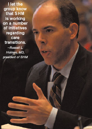
Dr. Holman continued his comments to the roundtable by using an example of harmonization at the heart of hospital medicine: transitions of care. “I let the group know that SHM is working on a number of initiatives regarding care transitions,” he says. “Transitions of care require the whole system to come together; it’s a great way to help galvanize all the stakeholders toward that shared goal.”
In his written statement, Dr. Holman elaborated on SHM’s efforts regarding transitions of care and pushed for an alignment of CMS quality measures and incentives for physicians with those for hospitals.
Air of Excitement
Although they had many concerns about implementation, the roundtable participants were enthusiastic about the plan. “There was a fabulous give-and-take on issues regarding implementation,” says Allendorf.
Dr. Holman stayed after the meeting to discuss the proceedings with other participants. “All in all, the informal comments I heard after the meeting were that this was the most exciting moment they’d had in 30-plus years in healthcare,” he notes. “This marks a substantial change in the payment system, which has always been seen as a barrier to quality.”
What’s Next
The ball is now in the court of Sens. Baucus and Grassley, who will use the roundtable input to draft legislation that would make the necessary statutory changes in time for CMS to implement the plan by fiscal year 2009, or Oct. 1, as mandated by the Deficit Reduction Act (DRA) of 2005.
“I think they’ll be back in touch with [SHM] as they develop the plan,” Allendorf predicts. After the legislation has been submitted, it’s up to Congress to act. “Everything depends on whether any Medicare legislation moves this year, and no one know whether that will happen,” she says.
Whatever final form the CMS plan takes, the future looks bright for value-based purchasing—and for SHM’s continued involvement.
“This roundtable was an excellent opportunity for SHM to develop a relationship with CMS, and to link to the Senate Finance Committee,” Dr. Holman asserts. “My hope is that those relationships will bear fruit over time, and that we can continue to work with those entities, as well as the other roundtable participants, to propose and develop measures over time, and to bring alignment between hospital measures and physician measures.”
Jane Jerrard is a medical writer based in Chicago.
Read More Online
For more information on the roundtable, including Dr. Holman’s written statement and others, as well as a recorded Webcast, visit Sen. Baucus’ Web site at www.senate.gov/~finance/sitepages/VBProundtable030408.htm. TH
SHM had a seat at the table at a high-level roundtable meeting March 6 in Washington, D.C., to discuss Medicare’s value-based purchasing of hospital care.
SHM President Russell Holman, MD, chief operating officer for Cogent Healthcare, Nashville, participated in the roundtable convened by Senate Finance Committee Chairman Max Baucus, D-Mont., and ranking member Chuck Grassley, R-Iowa. The roundtable included representatives from 22 public and private healthcare organizations, including SHM, the American Hospital Association, and National Quality Forum.
“SHM was one of only two physician organizations participating,” points out Laura Allendorf, SHM’s senior adviser for advocacy and government affairs. “That is very significant. I think [our inclusion] is a testament to our growing presence, our advocacy efforts and our willingness to help representatives with healthcare issues like value-based purchasing.”
The Proposed Plan
At the roundtable, representatives from the Centers for Medicare and Medicaid Services (CMS), the Medicare Payment Advisory Commission (MedPAC), and the Government Accountability Office (GAO) presented an overview of the report that CMS submitted to Congress in November, which outlines a hospital value-based purchasing program. The plan is designed to meet CMS’ objective of “transforming Medicare from a passive payer of claims to an active purchaser of care.” It builds on the existing framework of the current pay-for-reporting program, Reporting Hospital Quality Data for Annual Payment Update, but more closely links hospital Medicare payments to performance.
Dr. Holman is well versed in the plan; he and members of SHM’s Public Policy Committee, who were in Washington for annual visits to Capitol Hill, had just met with CMS’s Chief Medical Officer Tom Valuck, MD.
“That same morning, Tom Valuck had spent an hour and a half briefing Public Policy Committee members on Medicare’s value-based purchasing plan,” says Allendorf. “I’d lined up that meeting before the roundtable was set up.”
A transition to CMS’s proposed value-based purchasing plan, or VBP, would probably occur over three years. Under the plan, a percentage of the hospital’s diagnosis related group (DRG) payment would rely on the hospital’s performance on a specific set of measures. Although the report is comprehensive, details on implementation, incentives, and more must be made final.
Public reporting of quality measures remains a key part of the plan; quality of care information would be available to patients through the CMS Hospital Compare site at www.medicare.gov. A PDF of the CMS report to Congress is available at www.cms.hhs.gov/center/hospital.asp.
Support For Harmonization
After the formal presentation on the VBP plan, roundtable moderator John Inglehart, founding editor of Health Affairs, asked a series of questions directed at specific segments of the group, targeting issues surrounding the plan’s quality measures, performance standards, incentives, and plan implementation.
Dr. Holman was asked to comment on how CMS could ensure that hospital measures and physician measures become more aligned.
“The measures that each party are asked to report in terms of Part B of Medicare are somewhat different and can lead to confusion and people working at cross purposes,” Dr. Holman explains. “This adds complexity to a system that’s already too complex. In my statement, I said that what we call harmonization [of reporting measures] is a very important step for CMS to take, so that physicians and hospitals are required to measure and report the same thing. The more we can move toward outcome measures, as well as efficiency and patient experience measures, the more harmonization we’ll have. Focusing on outcomes creates a common goal.”
Dr. Holman continued his comments to the roundtable by using an example of harmonization at the heart of hospital medicine: transitions of care. “I let the group know that SHM is working on a number of initiatives regarding care transitions,” he says. “Transitions of care require the whole system to come together; it’s a great way to help galvanize all the stakeholders toward that shared goal.”
In his written statement, Dr. Holman elaborated on SHM’s efforts regarding transitions of care and pushed for an alignment of CMS quality measures and incentives for physicians with those for hospitals.
Air of Excitement
Although they had many concerns about implementation, the roundtable participants were enthusiastic about the plan. “There was a fabulous give-and-take on issues regarding implementation,” says Allendorf.
Dr. Holman stayed after the meeting to discuss the proceedings with other participants. “All in all, the informal comments I heard after the meeting were that this was the most exciting moment they’d had in 30-plus years in healthcare,” he notes. “This marks a substantial change in the payment system, which has always been seen as a barrier to quality.”
What’s Next
The ball is now in the court of Sens. Baucus and Grassley, who will use the roundtable input to draft legislation that would make the necessary statutory changes in time for CMS to implement the plan by fiscal year 2009, or Oct. 1, as mandated by the Deficit Reduction Act (DRA) of 2005.
“I think they’ll be back in touch with [SHM] as they develop the plan,” Allendorf predicts. After the legislation has been submitted, it’s up to Congress to act. “Everything depends on whether any Medicare legislation moves this year, and no one know whether that will happen,” she says.
Whatever final form the CMS plan takes, the future looks bright for value-based purchasing—and for SHM’s continued involvement.
“This roundtable was an excellent opportunity for SHM to develop a relationship with CMS, and to link to the Senate Finance Committee,” Dr. Holman asserts. “My hope is that those relationships will bear fruit over time, and that we can continue to work with those entities, as well as the other roundtable participants, to propose and develop measures over time, and to bring alignment between hospital measures and physician measures.”
Jane Jerrard is a medical writer based in Chicago.
Read More Online
For more information on the roundtable, including Dr. Holman’s written statement and others, as well as a recorded Webcast, visit Sen. Baucus’ Web site at www.senate.gov/~finance/sitepages/VBProundtable030408.htm. TH
Consult for an HMG

Consult for an HMG
Question: We are an established hospitalist group going on two years. We started with two full-time physicians and now have five. We obviously have room for improvement and growth. Are there any hospitalist physician consultants who would be able to spend a couple of days with us to evaluate our program and review our systems?
Montell Hutchison,
Business Development Specialist,
Marietta Memorial Hospital,
Marietta, Ohio
Dr. Hospitalist responds: Since the term hospitalist was coined in 1996, the field has grown rapidly. The majority of hospitalist programs are less than five years old. Many hospitalist programs, having grown rapidly over the past few years, are planning to evaluate and improve their performance.
There are a number of options available to help programs benchmark themselves to industry standards. Hiring a consultant is certainly a reasonable choice. SHM has been a particularly valuable resource to help leaders understand how to evaluate their program.
On the Practice Resource section of its Web site (www.hospitalmedicine.org), SHM provides the names and descriptions of several practice management consultants. Also on the site is the downloadable dashboard white paper “Measuring Hospitalist Performance: Metrics, Reports, and Dashboards,” developed by the SHM Benchmarks Committee.
The white paper discusses “issues related to developing, reporting and interpreting performance data.” SHM has also developed the practice management course “Best Practices in Managing a Hospital Medicine Program” and published the book Hospitalists: A Guide to Building and Sustaining a Successful Career to assist those interested in evaluating and improving their hospitalist program.
Lastly, SHM surveys hospitalists every other year to gather productivity and compensation date. The results of the latest “Bi-Annual Survey on the State of the Hospital Medicine Movement” will be released at the SHM Annual Meeting in San Diego April 3-5. Survey information will be available on its Web site shortly thereafter.
Lend A Hand
Question: I am a certified diabetes educator with 20 years’ experience in diabetes. I’m about to graduate as an adult nurse practitioner (ANP). My goal is to work with management of inpatient diabetes. Are you aware of any models of ANPs working for or with hospitalists?
Potential Team Member, Ohio
Dr. Hospitalist responds: There are about 20,000 hospitalists in the country. SHM believes that number could grow to 40,000. I am not surprised by the size of that estimate. Just about every hospital I know wants to develop or expand its hospitalist program.
Hospital medicine has filled its ranks over the past decade largely at the expense of primary care. Many physicians have left primary care for hospital medicine. At the same time, many graduates of internal medicine and family medicine training programs have chosen employment in hospital medicine instead of outpatient medicine.
Despite this trend, we are still short of hospitalists. The long-term solution is to expand the numbers of trainees who choose hospital medicine as a career. We are unlikely to see that occur rapidly over the short term. Another potential solution is to increase hospitalist efficiency.
Many in the field are working to redesign systems in the hospital to increase hospitalist efficiency and productivity. Again, these efforts will take time. Adding non-physician providers as care extenders is another viable option many programs are exploring. The results of the latest SHM productivity and compensation survey “Bi-Annual Survey on the State of the Hospital Medicine Movement” being released in April will give a sense of how many midlevel providers (nurse practitioners and physicians’ assistants) are working in hospitalist programs. It will not, however, fully describe their roles and responsibilities. I suspect more hospitalist programs are employing midlevel providers, but the majority of programs do not have midlevel providers among their staff.
For hospitalist programs entertaining the notion of hiring a midlevel provider (and for those midlevel providers interested in joining a hospitalist program), there are barriers to such a relationship. One inherent problem is that most hospitalists do not understand how to work with midlevel providers. Many hospitalists are unaware of the midlevel providers’ education and training.
I am not surprised. Most hospitalists did not work with midlevel providers during their training. There are not only differences in education and training among nurse practitioners (NPs) and physicians’ assistants (PAs), but many state licensing boards also limit their scopes of practice in a different manner. I find that many hospitalists want their midlevel providers to do the scut work hospitalists don’t want to do or feel they don’t have time to do.
I feel this is an expensive way to hire someone to do discharge summaries, put in orders, and make follow-up appointments. One does not need a midlevel education and training to perform those tasks. I suspect most midlevel providers—like hospitalists—find these tasks unsatisfying if it is the majority of their job description. Hospitalist programs would be better served if they involved midlevel providers in the care of patients. But this will require additional training to help hospitalist understand the level of supervision that is necessary for midlevel providers.
For hospitalists who work with midlevel providers, I find too often that many provide too much or too little supervision. Neither is appropriate. Another barrier in some parts of the country relates to the compensation of hospitalists and midlevel providers.
In the Northeast and on the West Coast, for example, we find smaller differences between hospitalist and NP salaries. Since NPs bill at 85% of physician billing, it may not necessarily make financial sense to hire NPs when one can hire a physician at slightly higher cost.
I hope I have not dissuaded you from pursuing a job in hospital medicine. I believe the development of roles for midlevel providers in hospital medicine is critical to the continued expansion of hospital medicine in this country.
I implore hospitalists and midlevel providers to work together to explore and establish models of care that will provide sustainable career opportunities for midlevel providers in hospital medicine. TH
Data Daze

As I write this column, our nation’s economy is looking pretty shaky. In early January, the labor market report showed a significant uptick in the unemployment rate. This led worried investors to sell stocks, and the Federal Reserve lowered the interest rate dramatically in response to the sudden fall in the stock market. Or at least that’s the version of events we’re being fed by most of the press.
Do investors overreact to job numbers? There is a lot of debate about the accuracy of job and unemployment statistics. Clearly they are valuable, but there are all kinds of problems with the way the labor surveys are conducted and the resulting data analyzed.
There may not be a better way to collect the data, so despite their flaws surveys may provide the best information on the labor market that we can get.
The real problem arises when investors—sort of like you and me but a lot richer—look at these data and fail to keep in mind all its strengths and weaknesses. There is a risk people will focus on a single number and overestimate its precision. This has been called the “salience bias.” Writing in The New Yorker, James Surowiecki says this salience bias can lead to “a hard-to-break feedback loop: The fact that traders act as if the jobs report were definitive makes it so. A little information can be a dangerous thing.”1
I discuss hospitalist survey data with people all the time. I’m struck by how often they seem misled by salience bias, among other things. With SHM’s release this month of its latest biannual survey of hospitalist productivity and compensation, now seems like a good time to discuss the strengths and weaknesses in the data—and cautions when interpreting it.
Understand the strengths and limitations of the survey. SHM’s “Bi-Annual Survey on the State of the Hospital Medicine Movement” is a self-reporting survey in which each practice leader (or his/her designee) completes the survey. The responses aren’t verified or audited, so some respondents might submit shoddy data. Perhaps a busy group leader might complete the survey from memory and estimate things like each doctor’s production of work-only relative value units (wRVUs). When I’ve looked at the raw data, I’ve wondered if some respondents are trying to “spin” their numbers higher or lower for a variety of reasons (e.g., to look unusually good or show how hard their doctors can work). And there may be a response bias: Those who think their practice is atypical might not respond to the survey.
This year, SHM worked to “scrub” the data. Outlier metrics were established for each question, and SHM staff followed up with the respondent to ensure he/she understood the question and provided accurate data. In fact, I completed the survey for the group I’m part of and got a call from a survey staffer questioning the productivity I reported for some members of the group (our nocturnists have lower wRVU productivity than others in the group—that is one reason they’re willing to work at night).
Remember that data are historical and should be “aged” to the time period you’re using it. The data in the 2008 SHM survey were collected from October through December.
Review the original questions asked in the survey. To make sense of the survey responses, you will need to clearly understand the question asked. Review the survey instrument and form your own conclusions about ways the questions were posed that might influence the responses. And don’t assume you understand what a particular term means—verify it by looking at the survey instrument. For example, I encounter a wide variety of opinions regarding what constitutes base salary, incentive pay, productivity compensation, bonus, and total compensation. The survey instrument spells these things out clearly.
It can be valuable to trend some data over successive surveys. For example, you may want to know the trend in the average hospitalist’s wRVU productivity over the past two surveys. You should look at the questions asked in both surveys to make sure there hasn’t been a change that could influence the responses. In the case of wRVUs you will need to understand how the January 2007 change in wRVUs values for many services provided by hospitalists was handled by the survey.
Pay attention to how data are lumped or split. Some data are appropriate for analysis of hospitalist groups, other data for individual hospitalists. If half of hospitalist groups use a shift-based schedule, that doesn’t mean half of individual hospitalists work such a schedule. Shift-based schedules are more common in larger groups, so even if half the groups in the country schedule by shifts, there may be 80% of individual hospitalists who use this schedule.
Salary incentives illustrate another way responses are lumped or split. As of the last survey (reported in 2006), most hospitalists had a variable component to their compensation—most often based on productivity or quality. There are relatively few ways hospitalists are paid on productivity (basing it on wRVUs is most common). But there are myriad quality incentives, based on things like Centers for Medicare and Medicaid Services core measures, and patient and referring physician satisfaction. Depending on how you aggregated these different categories in the 2006 survey, you might reach different conclusions about whether more hospitalists have productivity-based incentives or quality-based incentives (productivity was more common in the 2006 survey).
Drill down to respondent populations that most closely match your group. There is a real temptation to overemphasize the “headline” numbers in the survey like the average total salary for a hospitalist. Yet in many cases, it may be more useful to drill down to a population that matches your group. You might be most interested in compensation for hospital-employed hospitalists in non-teaching hospitals in the South (thereby excluding academicians and pediatric hospitalists from your comparison group). Just make sure to look at the resultant sample size (the “n”) reported for that subset of the data to make sure it is large enough to be meaningful.
Remember that the survey is not telling you what is right for your group. The survey simply describes a number of metrics relevant to hospitalist practice. It is not SHM’s position on the right compensation or productivity for a particular practice. It is the best source of national data regarding hospitalists. (See my column “Comp Close-Up” for a comparison between the SHM and Medical Group Management Association surveys [July 2007, p.73]). Things like the two other hospitalist practices in your town probably will have a lot more to do with influencing your group’s productivity and compensation metrics than any national data set.
While it’s tempting to reduce things to a single number (e.g., how much is the average hospitalist paid?) this is falling prey to salience bias. Try to grasp the stories behind the numbers by understanding the survey methods and looking at responses for different subsets of the survey population. And realize that the right or optimal compensation and productivity for a group might be quite different from the survey means and medians. TH
Dr. Nelson has been a practicing hospitalist since 1988 and is co-founder and past president of SHM. He is a principal in Nelson/Flores Associates, a national hospitalist practice management consulting firm. He is also part of the faculty for SHM’s “Best Practices in Managing a Hospital Medicine Program” course. This column represents his views and is not intended to reflect an official position of SHM.
Reference
- Surowiecki J. Running numbers. The New Yorker. January 21, 2008. Available online at www.newyorker.com/talk/financial/2008/01/21/080121ta_talk_surowiecki. Last accessed Feb. 7, 2008.
MySpace For Hospitalists
As I stared down the length of a limp, moderately deflated breakfast burrito, my thoughts meandered. I doubted about the ability of my little chylomicron-infused buddy to dust the cobwebs from a night in the Gaslamp Quarter that overemphasized wine and underemphasized sleep. I pondered the merit of showing up at 6:30 in the morning for this meeting.
I was shaken from my musings by a voice introducing himself as Bob Wachter. Clearly this apparition parading as one of the luminaries of the hospital medicine movement must be another manifestation of a night of overabundant revelry—and not my adviser for this SHM mentorship breakfast.
It turns out it was the real Dr. Wachter, and indeed he was there to provide an hour and a half of his undivided attention to mentor me as an early-career academic hospitalist. The year was 2003, the location San Diego, the event the sixth SHM Annual Meeting. The significance: This was my first SHM meeting.
Descending on San Diego five years later, I’ve had the chance to reflect on my growth as a hospitalist and the enzymatic role the society’s annual meeting has played in my development.
At the time I had been an academic hospitalist at the Denver Veterans Affairs Medical Center for three years. I’d come to San Diego to learn more about this group that was—until a name change announced at this meeting—calling itself the NAIP, or the National Association of Inpatient Physicians. I also came to present, for the first time, a research abstract.
The annual meeting represents many things to many people. For some it is a time to expand their clinical horizons. Indeed, the clinical content is top-notch and ensures that attendees stay current. The meeting also offers opportunities to learn a new skill such as how to build a hospital medicine program, become a top-notch educator, or develop procedural skills.
Others look to the meeting to present their research data to their peers. Still others attend to get away from the grind of their daily job and bathe in the rejuvenating energy of the annual meeting. Some even look to the meeting location as a vehicle to visit distant family or explore a new or favorite city.
I came to my first annual meeting to sample many of these offerings. What I found was something unexpected and powerful—a professional and social network.
This brings me back to my encounter with Bob. I had seen him speak the day prior at the “Building and Improving Hospitalist Programs” pre-course. Bob gave a rousing and edifying discourse about documenting the value of your hospitalist group. Our early morning gathering was part of the SHM mentorship breakfast, where an early-career hospitalist is paired with a more senior hospitalist in his/her area of interest for career counseling.
Meeting the man who coined the word “hospitalist” was, needless to say, a little nerve-wracking. However, Bob was disarming, engaging, and truly interested in helping me advance. We talked about the development of a hospitalist group at the University of Colorado, a new group I was to direct in three short months. Subsequent to our meeting, Bob was instrumental in helping me develop our group’s business plan, connecting frequently via e-mail and phone.
Later that morning as I was setting up my abstract poster, I looked up to find one of my medical school colleagues, Thomas McIlriath, perusing the aisles of posters.
Thomas, who directs the Mercy Medical Group’s hospitalist program in Sacramento, and I had been good friends in medical school but found our friendship sidetracked by the grind of residency training at different institutions. I had not seen or heard from him in seven years. I later stumbled upon another medical school colleague and quite enjoyed the opportunity to reconnect with such important figures from my past.
Over lunch I attended my first SHM research committee meeting. Not knowing what to expect, I found a group of early-career hospitalists interested in developing academic careers and a burgeoning SHM research infrastructure. I also met Andy Auerbach, MD, at the time a slightly younger but no less talented clinician-researcher at the University of California, San Francisco. Over the years I’ve had the chance to get to know Andy much better and have found him an inspiration, a collaborator, and an enjoyable person to reconnect with whenever serendipity offers us the chance.
Toward the end of the first day’s sessions I attended an innovations workshop that featured a presentation of an online billing tool I thought might be the answer to a research question that I had. Unbeknownst to me the presenter was Eric Siegal, MD, who had graduated from my residency the year before I started and had been a passing acquaintance.
In the end, the presentation was not the answer to my question. But connecting with Eric, now a regional director at Cogent Healthcare, was not only pleasurable but has led to a burgeoning professional bond and personal friendship that has resulted in a couple of co-written publications, hikes through the mountains of Montana, and too numerous to count phone calls seeking professional advice.
That evening I presented my data to my peers at the abstract session and found the feedback to be interesting and enlightening. I also found it another opportunity to discover like-minded people with common interests.
One of them was Ken Epstein, MD, with IPC-The Hospitalist Company. It turned out that Ken had access to data that could answer the research question that had brought me to the innovations session earlier in the day.
Since this encounter, Ken and I have published the answer to our research question and formed a friendship and collaboration that continues to grow today.
While I didn’t recognize it at the time, that span of 24 hours in early April 2003 was arguably the most important and productive day of my working career. I reconnected with old friends, developed new professional acquaintances, and, most important, forged several new and lasting friendships.
As I ready myself for another annual meeting in San Diego I excitedly ponder the opportunities that await me—not least of which is another go in the Gaslamp Quarter! TH
Dr. Glasheen is associate professor of medicine at the University of Colorado at Denver, where he serves as director of the Hospital Medicine Program and the Hospitalist Training Program, and as associate program director of the Internal Medicine Residency Program.
See Further
The rate of change within hospital medicine has been astounding over the past 11 years, and we have always tried to position SHM to remain nimble to respond to those changes. Racing legend Mario Andretti once said that if everything seems under control, you aren’t going fast enough.
Rapid responsiveness has become deeply ingrained within the culture of SHM. Every member should be proud because this is the result of the hard work and dedication of the many members who simply refuse to accept the status quo.
A few months ago, I met with a group of young hospitalists. We were discussing a variety of topics when one of the hospitalists asked in a frustrated tone, “When is the change going to stop?” Her program was experiencing rapid growth while still trying to recruit sufficient hospitalists. Yet at the same time, she was enjoying involvement in several quality improvement projects at her hospital.
I began to talk with her about schedules, smart growth, time management—all the management techniques I have picked up over time. But she pushed back: “No, when is it going to stop? I love my job, but all this change and push to see more patients and comply with increasing safety/quality mandates is really stressing me. I simply see no end in sight. Our program is going to end up admitting or consulting on almost every patient in this hospital. We can’t find enough hospitalists! When is it going to stop?”
Before answering her, I thought about all the accomplishments of hospital medicine, particularly those fostered by SHM. The list is numerous. I wondered whether, despite our nimbleness, we were still helping this frustrated hospitalist and her patients. She was asking what it’s going to take to move beyond the frustration despite 11 years of hospital medicine milestones.
My answer: It is not going to stop until we look beyond our current view. We have laid a great foundation at SHM and in hospital medicine, but we must embrace a different method of delivering care to our patients. Our long-term professional satisfaction demands it. Moreover, if we really want to deliver high-quality care to the many patients we are being asked to see, this change is an absolute necessity. It won’t come easily, but hospitalists are not alone in this; many other specialties must face it, as well.
During my 13 years as a hospitalist—half of which were spent in hospital medicine private practice—I have seen patients in 10 hospitals. These hospitals ranged from a 100-bed community hospital to a 600-bed academic medical center. Some have been for profit, some not for profit, and some government sponsored. What is interesting is that in each of these facilities, my ability to see patients was vastly different. In one hospital, I could comfortably see 25 to 30 patients a day. In another, I could barely see eight to 10. In some, the admission process took me 30 minutes, in another 90 minutes.
What is the difference in these hospitals? It usually isn’t much. Maybe it’s the fact the charts are always in one place. Maybe the electronic medical record is simply better, or the ward secretaries more helpful. Or maybe it’s several of these factors, and others. But the point is that it is possible to vastly improve things with some minor tweaks in the system. It usually happens when the hospital and physicians have communicated with each other to make these changes.
But what if the hospital and the hospital medicine group engage in a true partnership—a proactive joint venture in which each fully appreciates the other’s value proposition? What if each seeks to deliver something greater than the mere sum of hospital resources and hospitalist resources? Suppose the hospital embraces the idea of helping build structure and processes to maximize the number of patients seen by a hospitalist. Suppose the hospital medicine program embraces this but also embraces the goal of high-quality care in all its dimensions. What results is true synergy, where both parties benefit tremendously and the real winner is the patient. This won’t happen just by tweaking to make improvements at the edges; only wholesale change will take patient care delivery to a new level.
This will require hospitalists to embrace new methods of patient care. When I speak of this concept to hospitalists, I ask them to imagine doubling the amount of patients they currently see. If your census is 15 patients a day, imagine seeing 30. Most hospitalists immediately push back and say it isn’t possible because they can’t envision a new way of patient care. But what if by engaging in this partnership, the hospital brought to bear all its potential resources to help you—maybe a scribe, or several midlevel providers? What if there were zero barriers to finding data? What if the pharmacist took such a reliable medication history that you could fully depend on it? How about before you see a patient, a history and physical was already done so all you had to do was review the findings?
Many of you are still skeptical. Some of you struggle just getting approval for a part-time administrative person. You can’t ever imagine the hospital providing additional resources. Trust me when I tell you times are changing. One of the chief concerns all hospital executives have is the imperative to change the hospital-physician relationship. Much has been written on this topic, and the prevailing wisdom among hospital leaders is that the old medical staff leadership concept is dead. New types of leadership are necessary if hospitals and physicians are to survive.1
Because I am a hospitalist and a hospital executive, I feel the hospital part is the easy one. As chief medical officer, my main goal is to improve the quality of care delivered. The chief constraint I encounter is the number of engaged physicians. Much of my day is spent working with physicians to engage them in a hospital-physician partnership. The No. 1 thing preventing engagement is clinical workload. From the hospital perspective, I know I need to mobilize hospital resources to help physicians and remove time as the constraint. But for me to take them away from seeing patients may also hurt the hospital.
Now don’t misunderstand me. I am not advocating an improvement in efficiency just for the sake of it or just to improve our recruiting problems. The vision must be the delivery of higher quality care.
Change is not over. SHM’s responsiveness to change will greatly enhance your ability to promote change from within your hospital. Physician leadership is essential.2 We have much more to accomplish, but SHM is well positioned to continue to help you and your patients. We will continue to be nimble. We will continue to respond to the many changes on the road ahead. Even the road we can’t quite see yet. TH
Dr. Cawley is president of SHM.
References
- Petasnick WD. Hospital-physician relationships: imperative for clinical enterprise collaboration. Front Health Serv Manage. 2007;24(1):1:3-10.
- Porter M, Teisberg EO. How physicians can change the future of healthcare. JAMA. 2007;297(10):1103-1111.
Feedback Failures
On the evening of July 30, 1945, a U.S. battleship traveling from Guam to the Philippines was spotted in the Philippine Sea by a Japanese submarine crew. Six torpedoes sped across the black water with devastating effect.
Three hundred sailors died immediately as the stricken ship sank. Another 900 men were left floating in an oil slick in the shark-infested Pacific Ocean for four days.1
The events that led to the sinking represent a classic tale of systems failures. They ring familiar to any physician who has closely examined modern medical error.
The communication breakdown that prevented the ship’s captain from being aware of submarine activity on the same route four days before is no different than communication breakdowns in complex hospital systems. The bureaucratic decision by a remote administrator to withhold the safety measure of an escort to prevent such an attack on the grounds that it “lacked necessity” likely resonates with physicians who have struggled with getting authorization for care.
Why did those sailors remain in the water four days before rescue? Despite being only a two-hour flight from the nearest base, they were not recovered—or even missed—for what must have seemed an endless amount of time.
In the end, slightly more than 300 men were alive when they were spotted by a plane that happened to fly past. The rest succumbed to dehydration, exposure, and sharks. The failure of the Navy to rescue the sailors offers lessons to the clinician trying to improve transitions of care more than a half a century later.
The Feedback Loop
Hospital discharge is a complex process initiated by physician orders on charts, prescription pads, and patient instructions. Most often, the things we assume will be done out of our direct view are carried out satisfactorily. However, any hospitalist can easily recount stories of tests or follow-up that didn’t happen as ordered. Patients who fall through the cracks at discharge—like the stranded sailors of the USS Indianapolis—are, in part, the story of a simple omitted step: the feedback loop.
A feedback loop occurs when the results or consequences of an action are returned as an input loop to the initiating step in order to modify subsequent actions. This fundamental engineering concept can keep complex systems on course. The feedback loop allows the lack of completion of a portion of a process to be recognized—and corrective measures taken—before additional harm occurs.
In Guam, the island base from which the USS Indianapolis departed, the marker indicating the ship on the plotting board was removed when the ship left. Later, the Philippine port of Leyte failed to note that the ship didn’t arrive. Policy at the time was that all ships that left port were presumed to have arrived at their destination unless a call indicating trouble was received. The junior officer who noticed that the ship hadn’t arrived assumed there had been an order to divert to an alternate port. The Navy had no feedback mechanism to communicate between the two ports and raise an alarm when a ship did not arrive. In fact, a Navy directive discouraged communicating the arrival of combat ships as a matter of military secrecy. As a result, no rescue mission was launched—and the sharks began to arrive.
A closer examination of usual hospitalist discharge practice reveals too much similarity for comfort. Similar to how the USS Indianapolis was removed from the plotting board on Guam, discharged patients are removed from the hospitalist’s census list. Follow-up becomes the responsibility of the patient and primary care physician.
Communication with the primary care provider is recognized as a best practice for discharge, but research suggests direct communication occurs less than 20% of the time.2 These dismal statistic suggests the “port of arrival” is unaware our patients are expected in a significant percentage of discharges. Outpatient physicians, like the junior port director in Leyte, may assume that patients who do not call or arrive for appointments have been readmitted, seen by another physician, or otherwise diverted.
Even a superficial review reveals significant deficits in the feedback provided by our current discharge practice. When patients don’t arrive at follow-up appointments, most hospitalists lack any ability to recognize this failure of their transition plan. The assumption in most hospitalist groups is that patients who leave the hospital will achieve follow-up as directed. This “presumption of success” is ill-founded and may expose the patient to potential harm—and the physician to liability.
The case of Shirk v. Kelsey offers parallels to discharge situations hospitalists commonly encounter.3 In this malpractice case, a procedure was unsuccessful in terminating a patient’s pregnancy. The performing physician left follow-up of the pathology results to the patient’s usual outpatient obstetrical provider. The patient’s lack of follow-up was not recognized by the discharging obstetrician. This provider—not the practitioner with whom follow-up was intended—was found liable.
The American Medical Association’s code of ethics states: “Once having undertaken a case, the physician should not neglect the patient.” Hospitalists form physician-patient relationships with hospitalized patients that usually terminate on discharge. Our duty to not abandon or neglect a patient diminishes significantly after discharge, when we are no longer responsible for ongoing hospital care or exchanging information with patients on a daily basis.
But our duty does not disappear. Certainly, the responsibility falls to the hospitalist to be aware of and ensure a follow-up plan for important results such as pathology reports that return after discharge.
Allegations of improper post-discharge communication or failure to pass along critical results that become available once the patient has left the hospital, are common in medical malpractice claims.
Most are settled out of court, and many do not find the physician liable for malpractice. However, legal consequences are far less relevant than the safety and quality of care compromised when patients are failed by a system that lacks feedback loops to ensure safe transitions.
The ultimate goal of medicine is to improve the health and quality of life of our patients. Whether or not a lawsuit results, we need to recognize the commonality of patients who have their care compromised, delayed, or mismanaged because of our inability to recognize a foundering transition plan. Instead of looking at this as a failure of individual physicians to communicate, the problem needs to be addressed by creating effective, reliable systems.
Solutions
Providers discharging patients with follow-up needs should have a mechanism to identify those at highest risk for problems with transition. For these patients, follow-up with a post-discharge telephone call may be an effective feedback step.
One study looking at post-discharge phone contact found that 20% of patients had not filled new discharge prescriptions. Another post-discharge study revealed that a quarter of patients had medication questions that required clarification.4,5 Other research indicates more than one in 10 patients had new or worsening symptoms in the first five days after leaving the hospital.6 Despite these symptoms, 39% of these patients did not have a follow-up appointment established.
An integrated informatics system that prompts hospitalists and primary care physicians when patients do not arrive at expected follow-up or when test results return after discharge would be optimal. But a simple phone call to identify problems can be effective. Some hospitalist groups have incorporated routine post-discharge telephone contact into their practice—but most have not. Research identifying which patients would benefit is needed to allow targeted use of resources.7
While it’s understood that not all patient discharges will go smoothly, just as not all battleships will arrive at port without incident, there is frequently an opportunity to recover when things begin to go awry. A change in the common attitude that hospitalist responsibility ends when the patient leaves the hospital is necessary.
An element of the solution lies in the creation of feedback loops to identify patients who are not obtaining follow-up as expected. This step requires a commitment of resources—something our fragmented medical system, with location-based reimbursement, does not provide incentives for.
Creation of a feedback loop may be as complex as integrated medical informatics systems, or as simple as a follow-up phone call, but it is incumbent on each hospitalist to examine the environment in which they practice and ensure this vital element of a safe and reliable system is being addressed. TH
Drs. Cumbler and Egan are assistant professors in the Section of Hospital Medicine at the University of Colorado at Denver.
References
- Stanton D. In harms way: the sinking of the USS Indianapolis and the extraordinary story of its survivors. New York, NY: Henry Holt and Company LLC; 2001.
- Kripalani S, LeFevre F, Phillips F, Williams M, Basaviah P, Baker D. Deficits in communication and information transfer between hospital-based and primary care physicians: implications for patient safety and continuity of care. JAMA. 2007;297(8):831-841.
- Alpers A. Key legal principles for hospitalists. Am J Med. 1999;111(9):5-9.
- Dudas V, Bookwalter T, Kerr KM, Pantilat SZ. The impact of follow-up telephone calls to patients after hospitalization. Am J Med. 2001;111:26S-30S.
- Boockvar K, LaCorte H, Giambanco V, Fridman B, Siu A. Medication reconciliation for reducing drug
- discrepancy adverse events. Am J Geriatr Pharmacother. 2006;4:236-243.
- Epstein K, Juarez E, Loya K, Gorman MJ, Singer A. Frequency of new or worsening symptoms in the posthospitalization period. JHM. 2007;2:58-68.
- Mistiaen P, Poot E. Telephone follow-up, initiated by a hospital-based health professional, for postdischarge problems in patients discharged from hospital to home. Cochrane Database of Systemic Reviews 2006; 4. Article No.:CD004510. DOI:10.1002/14651858.CD004510.pub3.