User login
Glycemic Control in Medical Inpatients
Diabetes mellitus is a common comorbid condition in hospitalized patients. In 2003, diabetes was listed as a diagnosis in 17.2% of hospital discharges in the United States.1 Because these diagnosis codes do not account for undiagnosed diabetes or hospital‐related hyperglycemia, the true prevalence of diabetes or hyperglycemia in hospitalized patients is likely higher and has been estimated to be as great as 38%.2 Hyperglycemia has been associated with adverse outcomes among hospitalized patients, including infectious complications, increased length of stay, and increased mortality.27 However, because hyperglycemia is not usually the primary reason patients with diabetes are hospitalized, its management is often not a focus in the inpatient setting. Sliding‐scale insulin alone continues to be commonly prescribed despite clinical evidence showing it to be ineffective in achieving glycemic control.8, 9
Recent randomized controlled trials have demonstrated that aggressive treatment of inpatient hyperglycemia improves outcomes in surgical and medical intensive care units10, 11 and in patients admitted for myocardial infarction.12, 13 Based on this clinical evidence and strong observational data linking hyperglycemia to poor patient outcomes in the non‐ICU setting,27 the American Diabetes Association (ADA) now advocates good metabolic control, defined as preprandial glucose levels of 90‐130 mg/dL and peak postprandial glucose levels 180 mg/dL in hospitalized non‐ICU patients with hyperglycemia14 (note that these targets are less aggressive than those for ICU patients, for whom randomized controlled trials showed the benefits of reduced mortality provided by tight glucose control).11 To reach these targets, the ADA and American College of Endocrinology suggest that multidisciplinary teams develop and implement hyperglycemia management guidelines and protocols.15 Protocols should promote the use of continuous intravenous insulin or scheduled subcutaneous insulin as opposed to the use of sliding‐scale insulin alone. Subcutaneous insulin protocols should include target glucose levels; basal, nutritional, and supplemental insulin; and daily adjustments based on previous glucose levels, insulin sensitivity, nutritional intake, illness, and medications.6, 15 To date, few published protocols or algorithms for inpatient subcutaneous insulin have been shown to be effective.16, 17 It is therefore not known how best to design and implement an inpatient diabetes management protocol that is effective, efficient, and self‐perpetuating. The aims of our pilot study were to develop and implement a subcutaneous insulin protocol on a general medicine service, to identify barriers to implementation, and to determine the effect of this protocol on glycemic control.
METHODS
Setting and Participants
This prospective quality‐improvement pilot study was conducted at Brigham and Women's Hospital (BWH) from January 10, 2005, through June 23, 2005. Patients were eligible to participate if they were admitted to either of 2 General Medicine Service (GMS) teams with either a known diagnosis of type 2 diabetes or inpatient hyperglycemia (random laboratory glucose level > 180 mg/dL) and at least 1 fasting point‐of‐care glucose reading > 140 mg/dL. Patients were excluded if they had diabetic ketoacidosis, hyperosmolar hyperglycemic state, another absolute indication for intravenous insulin, or fasting glucose 60 mg/dL on no insulin or if they were pregnant. Each GMS team consisted of a teaching attending, a junior or senior resident, 2 interns, and a clinical pharmacist. Twenty‐six physicians attended on these 2 teams during the study period, 13 of whom were hospitalists. This study was approved by the BWH Institutional Review Board; patient consent to participate in this study was deemed not necessary because of the relatively nonsensitive nature of the data (eg, glucose control, insulin orders), the noninvasive means of data collection (eg, chart review), and the steps taken by research personnel to minimize any breach in patient confidentiality.
Intervention
A multidisciplinary team composed of a diabetologist (M.L.P.), a hospitalist (J.L.S.), and a pharmacist (J.M.T.) developed a subcutaneous insulin protocol that was approved by the BWH Pharmacy and Therapeutics Diabetes Subcommittee. The protocol consisted of a set of treatment recommendations made by a pharmacist to be carried out by the medical team. The primary components are shown in Table 1 (a full description can be found in the Appendix). The main emphasis of the protocol was on discontinuing oral antihyperglycemic agents during hospitalization, initiating basal insulin in most patients, and adjusting basal insulin daily as needed.
|
| Oral agents |
| 1. Stop oral agents in most patients |
| Glucose testing |
| 2. Check bedside blood glucose before meals and at bedtime if eating, or every 6 hours if not eating |
| Insulin |
| 3. Start basal insulin Patient's home dose or NPH 0.1 units/kg before breakfast and at bedtime or insulin glargine 0.2 units/kg at bedtime (max dose 20 units) If NPO, consider half dose unless hyperglycemic |
| 4. Start nutritional insulin Discrete meals: insulin aspart 0.05‐0.1 units/kg per meal or home dose 0‐15 minutes prior to eating Continuous tube feeds: regular insulin every 6 hours or NPH every morning and at bedtime (0.1‐0.2 units/kg per day in addition to basal insulin) Hold if NPO |
| 5. Start correctional insulin Scale provided based on blood glucose and daily scheduled insulin requirements |
| Daily Adjustments |
6. Adjust scheduled insulin daily
|
| Other Considerations |
| 7. Hypoglycemia management (protocols for fruit juice, glucagons, IV dextrose, and when to call physician) |
| 8. Discharge orders (recommendations to discharge most patients on admission medication regimen, avoid sliding scale insulin, simplify dosing for patients requiring new insulin regimens, ensure adequate patient education and prompt outpatient follow‐up) |
All medical residents received general instructions regarding inpatient diabetes control by the research team's diabetologist (M.L.P.) through a 1‐hour department‐wide didactic lecture. The standards of care taught were identical to those in the protocol. In addition, the research team's hospitalist (J.L.S.) contacted each medical resident assigned to the 2 GMS teams electronically to introduce the protocol and describe the purpose and logistics of the pilot study.
A research assistant prospectively identified eligible patients each weekday by screening all patients admitted to the 2 GMS teams using the daily computerized sign‐out system used by all medical residents. Specifically, laboratory random glucose levels, inpatient medications, and medical history were reviewed to determine if each patient met eligibility criteria. Eligibility criteria were confirmed by medical record review. The pharmacist recommended to the primary team that the protocol be initiated for eligible patients. In addition, the pharmacist recommended daily adjustment of the insulin dose according to the protocol as appropriate. A chronologically organized summary of clinical data relevant to glycemic management for each patient, including bedside blood glucose measurements, general dietary intake, use of intravenous dextrose solutions, and administration of systemic steroids, oral diabetes medications, and all insulins, was provided to the team each day by the research assistant.
Measurements
The resident's acceptance of the protocol or reasons for declining it were recorded by the pharmacist on the day the protocol was recommended. Protocol acceptance was categorized as yes, no, or partial. Partial acceptance was defined as resident agreement to use the protocol, but with stated caveats or modifications. Clinical data were collected on each eligible patient for up to 7 days on GMS. Several data sources were used, including physician admission notes, the hospital's computerized clinical data system, vital‐sign sheets, medication administration records, and personal communication with nurses regarding any missing or discrepant data.
All insulin use (prescribed drug, dose, route, schedule and actual administered drug, dose, route, and time) was recorded each day by the research assistant. Use of basal and nutritional insulin and daily dose adjustments if previous hypo‐ or hyperglycemia (categorized as yes, no, or not applicable for each patient each day) were determined by the study pharmacist (J.M.T.) through retrospective review of all orders.
Up to 4 routine bedside blood glucose measurements were recorded each day: for patients eating discrete meals, these were the measurements taken before meals and at bedtime; for patients not eating or receiving continuous nutrition, these were the measurements taken closest to 6 AM, noon, 6 PM, and midnight. Additional measurements were not recorded to avoid ascertainment bias caused by follow‐up testing of abnormal glucose values. Glucose readings on the day of admission were excluded from analysis because these values are not amenable to inpatient ordering practices.
Study outcomes included overall protocol acceptance rate, insulin prescribing practices including use of basal insulin (ie, long‐acting agents such as NPH and insulin glargine), nutritional insulin (ie, scheduled regular, lispro, or aspart insulin given before each meal), daily dose adjustments under the protocol, and mean percentage of glucose readings per person greater than 180 mg/dL (hyperglycemia) and below 60 mg/dL (hypoglycemia). Comparable data from a previous cohort study of 91 GMS patients were used as baseline data for comparisons with the results of the present study.9
Other patient data collected included age, sex, weight, baseline A1C (taken at or within 6 months of admission), diabetic medications used prior to admission (none, oral agents only, or any insulin use); daily inpatient use of oral or intravenous steroids, oral diabetic medications, dextrose‐containing intravenous fluids, tube feeds, total parenteral nutrition, and general nutritional intake (nothing by mouth, clear diet, low carbohydrate diet, house diet).
Statistical Analysis
Characteristics of the study subjects and process and outcome measures were analyzed descriptively using rates, means, and standard deviations or medians with interquartile ranges as appropriate. Comparisons between the pilot study and baseline cohorts were performed using Fisher's exact test for dichotomous outcomes (eg, use of basal insulin). For rates of hyperglycemia (ie, fraction of readings > 180 mg/dL), we used binomial logistic regression, accounting for potential correlation among repeated events by individual patients with a dispersion parameter18 (note that we did not use the same analysis for rates of hypoglycemia because it was such a rare event; for analysis of hypoglycemia, the variables were dichotomized). We also analyzed outcomes by hospital day (through hospital day 5, the limit used in the baseline study) to determine daily trends during the course of hospitalization; for these analyses we used the Mantel‐Haenszel chi‐square test for dichotomous variables and binomial logistic regression with hospital day as the independent variable for rates of hyperglycemia. Two‐sided P values .05 were considered significant. SAS version 9.1 (Cary, NC) was used for all analyses.
RESULTS
After screening all 785 admissions to the 2 medical teams during the study period, we prospectively identified 109 patients (14%) for the pilot study. Twenty patients were subsequently excluded: 7 patients who were discharged the same day they were identified, 4 who did not have a fasting blood glucose value greater than 140 mg/dL, 4 patients who had type 1 diabetes, 2 patients who were admitted with diabetic ketoacidosis, and 3 patients whose data could not be accessed because of repeated unavailability of the medical record. Characteristics of the remaining 89 study subjects are shown in Table 2 and are compared to 91 baseline subjects. The mean age of the study subjects was 68.7 years; 45% were men. Five patients (6%) did not have a previous diagnosis of diabetes, and 51% were taking insulin prior to admission; the median A1C was 6.8%.
| Characteristic | Baseline (n = 91) | Pilot (n = 89) |
|---|---|---|
| ||
| Age (years), mean (SD) | 66.0 (14.5) | 68.7 (14.7) |
| Male | 53/91 (58%) | 40/89 (45%) |
| No diagnosis of diabetes at admission | 7/91 (8%) | 5/89 (6%) |
| Preadmission diabetes regimen | ||
| None | 15/91 (16%) | 14/78 (18%) |
| Oral medications only | 32/91 (35%) | 24/78 (31%) |
| Insulin | 44/91 (48%) | 40/78 (51%) |
| A1C (IQR) | 7.0 (6.0, 8.0) | 6.8 (6.3, 7.8) |
| Hospital length of stay (days), median (IQR) | 5 (3, 7) | 5 (3, 7) |
The medical residents agreed, at least in theory, to follow the subcutaneous insulin protocol for 50 patients (56%), partially accepted it for 8 (9%), and declined for 31 (35%). Reasons for declining the protocol included fear of hypoglycemia, severity of patient's other disease states or overall poor health of patient, concern for the effects of renal insufficiency on insulin clearance, concern for the effect of steroid tapers on glucose levels, desire to titrate oral medications, and anticipation of patient's imminent discharge. Other reasons such as the glucose levels are not that bad and let's watch the glucose levels for one more day suggest that some residents did not view hyperglycemia as an acute problem requiring immediate attention.
Regarding insulin‐ordering practices (Table 3), basal insulin was prescribed for 57 patients (64%) in the pilot group compared to 45 patients (49%) in the baseline group (P = .05). Nutritional insulin was prescribed to 12 patients (13%) in the pilot group compared to no patients in the baseline group (P .001). Oral hypoglycemic agents were prescribed less often in the pilot study than at baseline (20% vs. 38%, P = .01). The use of a standard default sliding scale from the hospital computer order set was high and was not significantly different in the pilot study compared with that at baseline (93% vs. 90%, P = .78). Twenty‐four of the 83 patients in the pilot group (29%) received sliding‐scale insulin without ever receiving basal or nutritional insulin during hospitalization compared to 45 of 91 patients in the baseline group (49%; P = .01 for comparison). Among patients started on basal insulin, 42% (24 of 57) were started after the first full hospital day. The initial basal insulin dose was appropriate according to the protocol (within 20%) in 38 of 57 patients (67%). Only 20 of 61 patients (33%) who had any hypo‐ or hyperglycemia had any change to their insulin regimen made during days 2 through 7 of their hospitalization on GMS, similar to the rate noted at baseline (36%).
| Measure | Baseline | Pilot | P value |
|---|---|---|---|
| |||
| Process | |||
| Any basal insulin during hospitalization | 45/91 (49%) | 57/89 (64%) | 0.05 |
| Any nutritional insulin during hospitalization | 0/91 (0%) | 12/89 (13%) | 0.001 |
| Change in dose to any insulin order during hospitalization | 24/66 (36%) | 20/61 (33%) | 0.71 |
| Standard sliding scale from hospital computer order set | 75/83 (90%) | 76/82 (93%) | 0.78 |
| Any oral antihyperglycemic agents during hospitalization | 35/91 (38%) | 18/89 (20%) | 0.01 |
| Outcome | |||
| Mean percentage of glucose readings > 180 mg/dL (SD) | 33.3% (33.3%) | 31.6% (29.6%) | 0.85 |
| Any hyperglycemia (glucose > 180 mg/dL) | 66/89 (74%) | 59/78 (76%) | 0.86 |
| 1%‐20% of readings | 17/89 (19%) | 15/78 (19%) | 0.85 for trend |
| 20%‐40% | 15/89 (17%) | 15/78 (19%) | |
| 40%‐60% | 15/89 (17%) | 15/78 (19%) | |
| 60%‐80% | 7/89 (8%) | 6/78 (8%) | |
| >80% | 12/89 (13%) | 8/78 (10%) | |
| Any hypoglycemia (glucose 60 mg/dL) | 6/89 (7%) | 10/78 (13%) | 0.20 |
Regarding glucose control (Table 3), the mean percentage of glucose readings per patient greater than 180 mg/dL was not significantly different in the pilot study compared to baseline (31.6% vs. 33.3%, P = .85). Despite implementation of the protocol and increased use of basal and nutritional insulin, 76% of patients had at least 1 routine glucose reading greater than 180 mg/dL, and 37% of patients had at least 40% of their routine glucose readings greater than 180 mg/dL, comparable to baseline (74% and 38%, respectively, P = NS for both comparisons). At least 1 hypoglycemic event (glucose reading below 60 mg/dL) occurred in 7% of patients at baseline and 13% during the pilot study (P = .20). Eleven hypoglycemic events in the pilot study were between 50 and 59 mg/dL (55%), 6 were between 40 and 49 mg/dL (30%), 3 were between 30 and 39 mg/dL (15%), and none were less than 30 mg/dL. Nine occurred before breakfast (45%), 5 before dinner (25%), 3 before lunch (15%), and 3 at bedtime (15%).
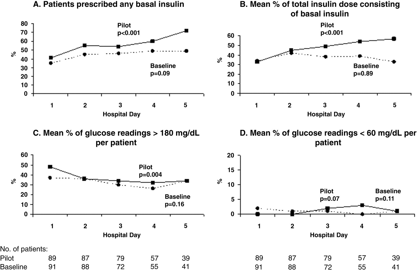
During the pilot study, the use of basal insulin did improve over the first 5 days of hospitalization (Fig. 1), in both the percentage of patients prescribed any basal insulin and the percentage of each patient's total insulin dose (basal, nutritional, and supplemental) given as basal (both P .001 for trend). Hyperglycemia rates also improved during hospitalization (Fig. 1), decreasing from 48% on hospital day 1 to 34% on hospital day 5 (P = .004 for trend). These trends were not observed in the baseline group, with hyperglycemia rates of 37% on hospital day 1 and 34% on hospital day 5 (P = .16 for trend).
Patients for whom the resident accepted or partially accepted the protocol had higher use of basal insulin (91% vs. 13%, P .0001), higher use of nutritional insulin (21% vs. 0%, P = .01), and more frequent dose adjustments (47% vs. 7%, P = .01) compared with patients for whom the resident declined the protocol. However, the rate of hyperglycemia was higher in patients for whom the protocol was accepted or partially accepted than in patients for whom the protocol was declined (37% vs. 20%, P = .02).
DISCUSSION
Our subcutaneous insulin protocol focused on increasing the use of basal and nutritional insulin, avoiding the use of sliding‐scale insulin by itself, and performing daily insulin adjustments in response to the hypo‐ or hyperglycemia of general medical inpatients with diabetes or hyperglycemia.
The most notable finding of our pilot study was that residents were resistant to using the protocol, both in general and in its specific recommendations. Despite receiving education about inpatient diabetes control and protocol recommendations from the team pharmacist, and despite being on a hospitalist‐run medical service, the residents accepted use of the protocol for only half the eligible patients. Patients who were started on basal insulin were often underdosed or started after the first day of hospitalization, and daily dose adjustments were not consistently made despite persistent hypo‐ or hyperglycemia. Although the use of nutritional insulin was greater compared with that in the baseline group, it was still only prescribed for 13% of patients. Use of a standard sliding scale from the hospital computer order set was common in the pilot study and similar to that in the baseline group. These results suggest significant resistance to changing the current standard of practice.
Despite this lack of adherence to the protocol, some modest improvements in processes of care were seen. Basal insulin was ordered more often during the pilot study than at baseline, especially over the course of a hospital stay. Nutritional insulin was also ordered more often during the pilot study than at baseline, but was still infrequent. Oral antihyperglycemic agents were ordered less often during the pilot study than at baseline. This demonstrates that use of the protocol may be able to improve process outcomes. However, the modest improvements in process outcomes could have simply been a result of increased awareness and education, not the protocol itself.
Regarding patient outcomes, the overall hyperglycemia rate did not improve in the pilot study relative to that at baseline. Importantly, hypoglycemia rates did not increase significantly compared with those at baseline. However, because of the small number of hypoglycemia events, the sample size may not have been sufficient to detect a true difference between groups.
The most likely reason that the protocol did not show an effect on glycemic control was that its recommendations were not adhered to. In turn, this may have been a result of incomplete education, training, and implementation measures and/or inherent problems with the protocol that made its recommendations difficult to follow. Another possibility is that the protocol itself may not have been capable of improving glucose control, even when properly used. However, we do know that resident agreement to use the protocol did lead to higher rates of recommended best practices being carried out, such as basal insulin use and daily insulin dose adjustments, and that use of the protocol was associated with improvements in glucose control over the hospital stay. A larger study with a higher degree of protocol adherence would be better able to evaluate the merits of the protocol itself, as would a randomized controlled trial using instrumental variables to measure treatment efficacy. Another possibility explanation for the lack of effect is that glucose control on admission happened to be worse in the pilot group than in the control group: rates of hyperglycemia on day 1 were 48% in the pilot group compared with 37% in the baseline group (Fig. 1). Also, the decreased use of oral agents in the pilot group, a purposeful change to decrease the risk of hypoglycemia, may have counteracted the beneficial effects of more appropriate insulin use. Finally, there were few patients with poorly controlled diabetes at baseline (18 patients with A1C 8.0 in the baseline group and 12 such patients in the pilot group), arguably those most likely to benefit.
There is a pressing need to identify protocols that can improve glucose control in the non‐ICU inpatient setting and successfully implement these protocols with a minimum of resources and effort. To date, most studies that have improved glucose control in the non‐ICU setting have relied on frequent input from diabetologists or nurse‐practitioners.14, 15
The results of this study should be viewed in light of its limitations, including its relatively small sample size (thus limiting our ability to detect possible significant differences between groups) and that it was conducted at a single institution (thus limiting its generalizability). Patients were enrolled on weekdays, so patients admitted and discharged over a weekend or on a holiday may have been missed. Also, because of the nonrandomized design of the study, we cannot exclude the possibility that the improvements noted in the pilot study were a result of the increased education provided or of increased awareness and general improvement in diabetes management over the course of the study. Finally, implementation of the protocol was somewhat labor intensive and required staff support that could be difficult to replicate in other institutions. However, most of the study staff's effort was necessary either to implement the protocol in the absence of an order set or to evaluate barriers to implementation. Widespread implementation of a protocol with an order set, education, and the use of highly reliable tools should be possible with much less effort and resources. The strengths of this study include its prospective data collection methods, which included rigorous inclusion criteria and collection of detailed clinical data.
Our study findings suggest several approaches to improve care in the future. To combat resistance to change, the American Association of Clinical Endocrinologists strongly recommends that each institution ensure that all its clinicians involved agree about general philosophies of diabetes management.19 A more expansive, hospital‐wide educational and promotional plan may increase the initial acceptance of the protocol. Interviews with residents also indicated there was unfamiliarity with diabetes management and significant concerns about the harmful affects of tight glucose control (ie, risk of hypoglycemia), especially in certain patient subgroups. These results confirmed the need for more practical individualized training and sparked the implementation of small‐group, case‐based educational sessions on inpatient diabetes management for all house officers, with a particular focus on patients with multiple comorbidities, on steroid tapers, and/or with renal failure.
The lack of nutritional insulin orders, delays in ordering basal insulin, and use of inadequate doses of insulin may be counteracted by the use of an order set, in our case built into our computer physician order entry (CPOE) system. The use of CPOE also allows reminders to be automatically sent to clinicians if eligible patients are not started on these orders. Clinical inertia (eg, failure to adjust the insulin doses of specific patients despite hyperglycemia) is more difficult to combat but may be addressed through better organization of clinical data, individualized, case‐based education, and CPOE reminders and eventually through culture change.
As a result of our pilot study, additional revisions were made to the protocol in hopes of increasing protocol adherence. For example, for patients eating discrete meals who are not taking insulin at home, the pilot protocol had suggested a starting insulin dose range for basal and nutritional insulin that required 2 separate calculations. The revised protocol was simplified to recommend a total daily insulin dose to be split evenly between basal and nutritional insulin. The daily adjustment instructions were also simplified. The pilot protocol had included a complicated table of adjustment recommendations based on bedside glucose trends. The revised protocol recommends adjusting the new daily dose by adding the total units of insulin given the previous day (including supplemental doses), making minor adjustments for hyper‐ or hypoglycemia and other clinical factors (like renal failure), and splitting this dose evenly between scheduled basal and nutritional insulin. In addition, 3 order sets were built into our computerized physician order entry system to facilitate early and appropriate insulin orders for patients with different diets (discrete meals, continuous tube feeds, and nothing by mouth); 3 different insulin sliding scales were created for patients with different degrees of insulin resistance; a diabetes management page for our electronic medication administration record is being developed to better organize clinical data; and hospital‐wide education and individualized training are ongoing.
In conclusion, the adherence to an inpatient glycemic management protocol that focused on increasing use of basal insulin and performing daily insulin adjustments was only fair. Barriers to successful implementation included clinical inertia regarding individual patients, unfamiliarity with inpatient diabetes management strategies, fear of hypoglycemia, and resistance to changing the current standard of practice. Targeted education, standard order sets, better organization of clinical data, protocol simplification, and institutional culture changes may be necessary for successful protocol implementation and improved inpatient glucose control.
- Agency for Healthcare Research and Quality. HCUPnet, Healthcare Cost and Utilization Project. 8/17/05; http://www.ahrq.gove/HCUPnet/. Accessed 7/17/06,2006.
- ,,,,,.Hyperglycemia: an independent marker of in‐hospital mortality in patients with undiagnosed diabetes.J Clin Endocrinol Metab.2002;87:978–982.
- ,,, et al.Hyperglycaemia is associated with poor outcomes in patients admitted to hospital with acute exacerbations of chronic obstructive pulmonary disease.Thorax2006;61:284–289.
- ,,,,.Stress hyperglycemia and prognosis of stroke in nondiabetic and diabetic patients: a systematic overview.Stroke.2001;32:2426–2432.
- ,,,.Hyperglycemia is associated with adverse outcomes in patients receiving total parenteral nutrition.Diabetes Care.2005;28:2367–2371.
- ,,, et al.Management of diabetes and hyperglycemia in hospitals.Diabetes Care.2004;27:553–597.
- ,,,,,.The relation between hyperglycemia and outcomes in 2,471 patients admitted to the hospital with community‐acquired pneumonia.Diabetes Care.2005;28:810–815.
- ,,.Glycemic control and sliding scale insulin use in medical inpatients with diabetes mellitus.Arch Intern Med.1997;157:545–552.
- ,,,,.Inpatient management of diabetes and hyperglycemia among general medicine patients at a large teaching hospital.J Hosp Med.2006;1:145–150.
- ,,, et al.Intensive insulin therapy in the medical ICU.N Engl J Med.2006;354:449–461.
- ,,, et al.Intensive insulin therapy in the critically ill patients.N Engl J Med.2001;345:1359–1367.
- .Prospective randomised study of intensive insulin treatment on long term survival after acute myocardial infarction in patients with diabetes mellitus.DIGAMI (Diabetes Mellitus, Insulin Glucose Infusion in Acute Myocardial Infarction) Study Group.BMJ.1997;314:1512–1515.
- ,,, et al.Intense metabolic control by means of insulin in patients with diabetes mellitus and acute myocardial infarction (DIGAMI 2): effects on mortality and morbidity.Eur Heart J.2005;26:650–661.
- American Diabetes Association.Standards of Medical Care in Diabetes ‐ 2006.Diabetes Care.2006;29:S4–S42.
- ACE/ADA Task Force on Inpatient Diabetes.American College of Endocrinology and American Diabetes Association consensus statement on inpatient diabetes and glycemic control: A call to action.Diabetes Care.2006;29:1955–1962.
- ,,,.Eliminating inpatient sliding‐scale insulin: a reeducation project with medical house staff.Diabetes Care.2005;28:1008–11.
- ,,, et al.Randomized study of basal‐bolus insulin therapy in the inpatient management of patients with type 2 diabetes (RABBIT 2 Trial).Diabetes Care.2007;30:2181–2186.
- .Extra‐binomial variation in logistic linear models.Appl Stat.1982;31:144–148.
- ,.Hospital management of diabetes.Endocrinol Metab Clin North Am.2005;34:99–116.
Diabetes mellitus is a common comorbid condition in hospitalized patients. In 2003, diabetes was listed as a diagnosis in 17.2% of hospital discharges in the United States.1 Because these diagnosis codes do not account for undiagnosed diabetes or hospital‐related hyperglycemia, the true prevalence of diabetes or hyperglycemia in hospitalized patients is likely higher and has been estimated to be as great as 38%.2 Hyperglycemia has been associated with adverse outcomes among hospitalized patients, including infectious complications, increased length of stay, and increased mortality.27 However, because hyperglycemia is not usually the primary reason patients with diabetes are hospitalized, its management is often not a focus in the inpatient setting. Sliding‐scale insulin alone continues to be commonly prescribed despite clinical evidence showing it to be ineffective in achieving glycemic control.8, 9
Recent randomized controlled trials have demonstrated that aggressive treatment of inpatient hyperglycemia improves outcomes in surgical and medical intensive care units10, 11 and in patients admitted for myocardial infarction.12, 13 Based on this clinical evidence and strong observational data linking hyperglycemia to poor patient outcomes in the non‐ICU setting,27 the American Diabetes Association (ADA) now advocates good metabolic control, defined as preprandial glucose levels of 90‐130 mg/dL and peak postprandial glucose levels 180 mg/dL in hospitalized non‐ICU patients with hyperglycemia14 (note that these targets are less aggressive than those for ICU patients, for whom randomized controlled trials showed the benefits of reduced mortality provided by tight glucose control).11 To reach these targets, the ADA and American College of Endocrinology suggest that multidisciplinary teams develop and implement hyperglycemia management guidelines and protocols.15 Protocols should promote the use of continuous intravenous insulin or scheduled subcutaneous insulin as opposed to the use of sliding‐scale insulin alone. Subcutaneous insulin protocols should include target glucose levels; basal, nutritional, and supplemental insulin; and daily adjustments based on previous glucose levels, insulin sensitivity, nutritional intake, illness, and medications.6, 15 To date, few published protocols or algorithms for inpatient subcutaneous insulin have been shown to be effective.16, 17 It is therefore not known how best to design and implement an inpatient diabetes management protocol that is effective, efficient, and self‐perpetuating. The aims of our pilot study were to develop and implement a subcutaneous insulin protocol on a general medicine service, to identify barriers to implementation, and to determine the effect of this protocol on glycemic control.
METHODS
Setting and Participants
This prospective quality‐improvement pilot study was conducted at Brigham and Women's Hospital (BWH) from January 10, 2005, through June 23, 2005. Patients were eligible to participate if they were admitted to either of 2 General Medicine Service (GMS) teams with either a known diagnosis of type 2 diabetes or inpatient hyperglycemia (random laboratory glucose level > 180 mg/dL) and at least 1 fasting point‐of‐care glucose reading > 140 mg/dL. Patients were excluded if they had diabetic ketoacidosis, hyperosmolar hyperglycemic state, another absolute indication for intravenous insulin, or fasting glucose 60 mg/dL on no insulin or if they were pregnant. Each GMS team consisted of a teaching attending, a junior or senior resident, 2 interns, and a clinical pharmacist. Twenty‐six physicians attended on these 2 teams during the study period, 13 of whom were hospitalists. This study was approved by the BWH Institutional Review Board; patient consent to participate in this study was deemed not necessary because of the relatively nonsensitive nature of the data (eg, glucose control, insulin orders), the noninvasive means of data collection (eg, chart review), and the steps taken by research personnel to minimize any breach in patient confidentiality.
Intervention
A multidisciplinary team composed of a diabetologist (M.L.P.), a hospitalist (J.L.S.), and a pharmacist (J.M.T.) developed a subcutaneous insulin protocol that was approved by the BWH Pharmacy and Therapeutics Diabetes Subcommittee. The protocol consisted of a set of treatment recommendations made by a pharmacist to be carried out by the medical team. The primary components are shown in Table 1 (a full description can be found in the Appendix). The main emphasis of the protocol was on discontinuing oral antihyperglycemic agents during hospitalization, initiating basal insulin in most patients, and adjusting basal insulin daily as needed.
|
| Oral agents |
| 1. Stop oral agents in most patients |
| Glucose testing |
| 2. Check bedside blood glucose before meals and at bedtime if eating, or every 6 hours if not eating |
| Insulin |
| 3. Start basal insulin Patient's home dose or NPH 0.1 units/kg before breakfast and at bedtime or insulin glargine 0.2 units/kg at bedtime (max dose 20 units) If NPO, consider half dose unless hyperglycemic |
| 4. Start nutritional insulin Discrete meals: insulin aspart 0.05‐0.1 units/kg per meal or home dose 0‐15 minutes prior to eating Continuous tube feeds: regular insulin every 6 hours or NPH every morning and at bedtime (0.1‐0.2 units/kg per day in addition to basal insulin) Hold if NPO |
| 5. Start correctional insulin Scale provided based on blood glucose and daily scheduled insulin requirements |
| Daily Adjustments |
6. Adjust scheduled insulin daily
|
| Other Considerations |
| 7. Hypoglycemia management (protocols for fruit juice, glucagons, IV dextrose, and when to call physician) |
| 8. Discharge orders (recommendations to discharge most patients on admission medication regimen, avoid sliding scale insulin, simplify dosing for patients requiring new insulin regimens, ensure adequate patient education and prompt outpatient follow‐up) |
All medical residents received general instructions regarding inpatient diabetes control by the research team's diabetologist (M.L.P.) through a 1‐hour department‐wide didactic lecture. The standards of care taught were identical to those in the protocol. In addition, the research team's hospitalist (J.L.S.) contacted each medical resident assigned to the 2 GMS teams electronically to introduce the protocol and describe the purpose and logistics of the pilot study.
A research assistant prospectively identified eligible patients each weekday by screening all patients admitted to the 2 GMS teams using the daily computerized sign‐out system used by all medical residents. Specifically, laboratory random glucose levels, inpatient medications, and medical history were reviewed to determine if each patient met eligibility criteria. Eligibility criteria were confirmed by medical record review. The pharmacist recommended to the primary team that the protocol be initiated for eligible patients. In addition, the pharmacist recommended daily adjustment of the insulin dose according to the protocol as appropriate. A chronologically organized summary of clinical data relevant to glycemic management for each patient, including bedside blood glucose measurements, general dietary intake, use of intravenous dextrose solutions, and administration of systemic steroids, oral diabetes medications, and all insulins, was provided to the team each day by the research assistant.
Measurements
The resident's acceptance of the protocol or reasons for declining it were recorded by the pharmacist on the day the protocol was recommended. Protocol acceptance was categorized as yes, no, or partial. Partial acceptance was defined as resident agreement to use the protocol, but with stated caveats or modifications. Clinical data were collected on each eligible patient for up to 7 days on GMS. Several data sources were used, including physician admission notes, the hospital's computerized clinical data system, vital‐sign sheets, medication administration records, and personal communication with nurses regarding any missing or discrepant data.
All insulin use (prescribed drug, dose, route, schedule and actual administered drug, dose, route, and time) was recorded each day by the research assistant. Use of basal and nutritional insulin and daily dose adjustments if previous hypo‐ or hyperglycemia (categorized as yes, no, or not applicable for each patient each day) were determined by the study pharmacist (J.M.T.) through retrospective review of all orders.
Up to 4 routine bedside blood glucose measurements were recorded each day: for patients eating discrete meals, these were the measurements taken before meals and at bedtime; for patients not eating or receiving continuous nutrition, these were the measurements taken closest to 6 AM, noon, 6 PM, and midnight. Additional measurements were not recorded to avoid ascertainment bias caused by follow‐up testing of abnormal glucose values. Glucose readings on the day of admission were excluded from analysis because these values are not amenable to inpatient ordering practices.
Study outcomes included overall protocol acceptance rate, insulin prescribing practices including use of basal insulin (ie, long‐acting agents such as NPH and insulin glargine), nutritional insulin (ie, scheduled regular, lispro, or aspart insulin given before each meal), daily dose adjustments under the protocol, and mean percentage of glucose readings per person greater than 180 mg/dL (hyperglycemia) and below 60 mg/dL (hypoglycemia). Comparable data from a previous cohort study of 91 GMS patients were used as baseline data for comparisons with the results of the present study.9
Other patient data collected included age, sex, weight, baseline A1C (taken at or within 6 months of admission), diabetic medications used prior to admission (none, oral agents only, or any insulin use); daily inpatient use of oral or intravenous steroids, oral diabetic medications, dextrose‐containing intravenous fluids, tube feeds, total parenteral nutrition, and general nutritional intake (nothing by mouth, clear diet, low carbohydrate diet, house diet).
Statistical Analysis
Characteristics of the study subjects and process and outcome measures were analyzed descriptively using rates, means, and standard deviations or medians with interquartile ranges as appropriate. Comparisons between the pilot study and baseline cohorts were performed using Fisher's exact test for dichotomous outcomes (eg, use of basal insulin). For rates of hyperglycemia (ie, fraction of readings > 180 mg/dL), we used binomial logistic regression, accounting for potential correlation among repeated events by individual patients with a dispersion parameter18 (note that we did not use the same analysis for rates of hypoglycemia because it was such a rare event; for analysis of hypoglycemia, the variables were dichotomized). We also analyzed outcomes by hospital day (through hospital day 5, the limit used in the baseline study) to determine daily trends during the course of hospitalization; for these analyses we used the Mantel‐Haenszel chi‐square test for dichotomous variables and binomial logistic regression with hospital day as the independent variable for rates of hyperglycemia. Two‐sided P values .05 were considered significant. SAS version 9.1 (Cary, NC) was used for all analyses.
RESULTS
After screening all 785 admissions to the 2 medical teams during the study period, we prospectively identified 109 patients (14%) for the pilot study. Twenty patients were subsequently excluded: 7 patients who were discharged the same day they were identified, 4 who did not have a fasting blood glucose value greater than 140 mg/dL, 4 patients who had type 1 diabetes, 2 patients who were admitted with diabetic ketoacidosis, and 3 patients whose data could not be accessed because of repeated unavailability of the medical record. Characteristics of the remaining 89 study subjects are shown in Table 2 and are compared to 91 baseline subjects. The mean age of the study subjects was 68.7 years; 45% were men. Five patients (6%) did not have a previous diagnosis of diabetes, and 51% were taking insulin prior to admission; the median A1C was 6.8%.
| Characteristic | Baseline (n = 91) | Pilot (n = 89) |
|---|---|---|
| ||
| Age (years), mean (SD) | 66.0 (14.5) | 68.7 (14.7) |
| Male | 53/91 (58%) | 40/89 (45%) |
| No diagnosis of diabetes at admission | 7/91 (8%) | 5/89 (6%) |
| Preadmission diabetes regimen | ||
| None | 15/91 (16%) | 14/78 (18%) |
| Oral medications only | 32/91 (35%) | 24/78 (31%) |
| Insulin | 44/91 (48%) | 40/78 (51%) |
| A1C (IQR) | 7.0 (6.0, 8.0) | 6.8 (6.3, 7.8) |
| Hospital length of stay (days), median (IQR) | 5 (3, 7) | 5 (3, 7) |
The medical residents agreed, at least in theory, to follow the subcutaneous insulin protocol for 50 patients (56%), partially accepted it for 8 (9%), and declined for 31 (35%). Reasons for declining the protocol included fear of hypoglycemia, severity of patient's other disease states or overall poor health of patient, concern for the effects of renal insufficiency on insulin clearance, concern for the effect of steroid tapers on glucose levels, desire to titrate oral medications, and anticipation of patient's imminent discharge. Other reasons such as the glucose levels are not that bad and let's watch the glucose levels for one more day suggest that some residents did not view hyperglycemia as an acute problem requiring immediate attention.
Regarding insulin‐ordering practices (Table 3), basal insulin was prescribed for 57 patients (64%) in the pilot group compared to 45 patients (49%) in the baseline group (P = .05). Nutritional insulin was prescribed to 12 patients (13%) in the pilot group compared to no patients in the baseline group (P .001). Oral hypoglycemic agents were prescribed less often in the pilot study than at baseline (20% vs. 38%, P = .01). The use of a standard default sliding scale from the hospital computer order set was high and was not significantly different in the pilot study compared with that at baseline (93% vs. 90%, P = .78). Twenty‐four of the 83 patients in the pilot group (29%) received sliding‐scale insulin without ever receiving basal or nutritional insulin during hospitalization compared to 45 of 91 patients in the baseline group (49%; P = .01 for comparison). Among patients started on basal insulin, 42% (24 of 57) were started after the first full hospital day. The initial basal insulin dose was appropriate according to the protocol (within 20%) in 38 of 57 patients (67%). Only 20 of 61 patients (33%) who had any hypo‐ or hyperglycemia had any change to their insulin regimen made during days 2 through 7 of their hospitalization on GMS, similar to the rate noted at baseline (36%).
| Measure | Baseline | Pilot | P value |
|---|---|---|---|
| |||
| Process | |||
| Any basal insulin during hospitalization | 45/91 (49%) | 57/89 (64%) | 0.05 |
| Any nutritional insulin during hospitalization | 0/91 (0%) | 12/89 (13%) | 0.001 |
| Change in dose to any insulin order during hospitalization | 24/66 (36%) | 20/61 (33%) | 0.71 |
| Standard sliding scale from hospital computer order set | 75/83 (90%) | 76/82 (93%) | 0.78 |
| Any oral antihyperglycemic agents during hospitalization | 35/91 (38%) | 18/89 (20%) | 0.01 |
| Outcome | |||
| Mean percentage of glucose readings > 180 mg/dL (SD) | 33.3% (33.3%) | 31.6% (29.6%) | 0.85 |
| Any hyperglycemia (glucose > 180 mg/dL) | 66/89 (74%) | 59/78 (76%) | 0.86 |
| 1%‐20% of readings | 17/89 (19%) | 15/78 (19%) | 0.85 for trend |
| 20%‐40% | 15/89 (17%) | 15/78 (19%) | |
| 40%‐60% | 15/89 (17%) | 15/78 (19%) | |
| 60%‐80% | 7/89 (8%) | 6/78 (8%) | |
| >80% | 12/89 (13%) | 8/78 (10%) | |
| Any hypoglycemia (glucose 60 mg/dL) | 6/89 (7%) | 10/78 (13%) | 0.20 |
Regarding glucose control (Table 3), the mean percentage of glucose readings per patient greater than 180 mg/dL was not significantly different in the pilot study compared to baseline (31.6% vs. 33.3%, P = .85). Despite implementation of the protocol and increased use of basal and nutritional insulin, 76% of patients had at least 1 routine glucose reading greater than 180 mg/dL, and 37% of patients had at least 40% of their routine glucose readings greater than 180 mg/dL, comparable to baseline (74% and 38%, respectively, P = NS for both comparisons). At least 1 hypoglycemic event (glucose reading below 60 mg/dL) occurred in 7% of patients at baseline and 13% during the pilot study (P = .20). Eleven hypoglycemic events in the pilot study were between 50 and 59 mg/dL (55%), 6 were between 40 and 49 mg/dL (30%), 3 were between 30 and 39 mg/dL (15%), and none were less than 30 mg/dL. Nine occurred before breakfast (45%), 5 before dinner (25%), 3 before lunch (15%), and 3 at bedtime (15%).
During the pilot study, the use of basal insulin did improve over the first 5 days of hospitalization (Fig. 1), in both the percentage of patients prescribed any basal insulin and the percentage of each patient's total insulin dose (basal, nutritional, and supplemental) given as basal (both P .001 for trend). Hyperglycemia rates also improved during hospitalization (Fig. 1), decreasing from 48% on hospital day 1 to 34% on hospital day 5 (P = .004 for trend). These trends were not observed in the baseline group, with hyperglycemia rates of 37% on hospital day 1 and 34% on hospital day 5 (P = .16 for trend).
Patients for whom the resident accepted or partially accepted the protocol had higher use of basal insulin (91% vs. 13%, P .0001), higher use of nutritional insulin (21% vs. 0%, P = .01), and more frequent dose adjustments (47% vs. 7%, P = .01) compared with patients for whom the resident declined the protocol. However, the rate of hyperglycemia was higher in patients for whom the protocol was accepted or partially accepted than in patients for whom the protocol was declined (37% vs. 20%, P = .02).
DISCUSSION
Our subcutaneous insulin protocol focused on increasing the use of basal and nutritional insulin, avoiding the use of sliding‐scale insulin by itself, and performing daily insulin adjustments in response to the hypo‐ or hyperglycemia of general medical inpatients with diabetes or hyperglycemia.
The most notable finding of our pilot study was that residents were resistant to using the protocol, both in general and in its specific recommendations. Despite receiving education about inpatient diabetes control and protocol recommendations from the team pharmacist, and despite being on a hospitalist‐run medical service, the residents accepted use of the protocol for only half the eligible patients. Patients who were started on basal insulin were often underdosed or started after the first day of hospitalization, and daily dose adjustments were not consistently made despite persistent hypo‐ or hyperglycemia. Although the use of nutritional insulin was greater compared with that in the baseline group, it was still only prescribed for 13% of patients. Use of a standard sliding scale from the hospital computer order set was common in the pilot study and similar to that in the baseline group. These results suggest significant resistance to changing the current standard of practice.
Despite this lack of adherence to the protocol, some modest improvements in processes of care were seen. Basal insulin was ordered more often during the pilot study than at baseline, especially over the course of a hospital stay. Nutritional insulin was also ordered more often during the pilot study than at baseline, but was still infrequent. Oral antihyperglycemic agents were ordered less often during the pilot study than at baseline. This demonstrates that use of the protocol may be able to improve process outcomes. However, the modest improvements in process outcomes could have simply been a result of increased awareness and education, not the protocol itself.
Regarding patient outcomes, the overall hyperglycemia rate did not improve in the pilot study relative to that at baseline. Importantly, hypoglycemia rates did not increase significantly compared with those at baseline. However, because of the small number of hypoglycemia events, the sample size may not have been sufficient to detect a true difference between groups.
The most likely reason that the protocol did not show an effect on glycemic control was that its recommendations were not adhered to. In turn, this may have been a result of incomplete education, training, and implementation measures and/or inherent problems with the protocol that made its recommendations difficult to follow. Another possibility is that the protocol itself may not have been capable of improving glucose control, even when properly used. However, we do know that resident agreement to use the protocol did lead to higher rates of recommended best practices being carried out, such as basal insulin use and daily insulin dose adjustments, and that use of the protocol was associated with improvements in glucose control over the hospital stay. A larger study with a higher degree of protocol adherence would be better able to evaluate the merits of the protocol itself, as would a randomized controlled trial using instrumental variables to measure treatment efficacy. Another possibility explanation for the lack of effect is that glucose control on admission happened to be worse in the pilot group than in the control group: rates of hyperglycemia on day 1 were 48% in the pilot group compared with 37% in the baseline group (Fig. 1). Also, the decreased use of oral agents in the pilot group, a purposeful change to decrease the risk of hypoglycemia, may have counteracted the beneficial effects of more appropriate insulin use. Finally, there were few patients with poorly controlled diabetes at baseline (18 patients with A1C 8.0 in the baseline group and 12 such patients in the pilot group), arguably those most likely to benefit.
There is a pressing need to identify protocols that can improve glucose control in the non‐ICU inpatient setting and successfully implement these protocols with a minimum of resources and effort. To date, most studies that have improved glucose control in the non‐ICU setting have relied on frequent input from diabetologists or nurse‐practitioners.14, 15
The results of this study should be viewed in light of its limitations, including its relatively small sample size (thus limiting our ability to detect possible significant differences between groups) and that it was conducted at a single institution (thus limiting its generalizability). Patients were enrolled on weekdays, so patients admitted and discharged over a weekend or on a holiday may have been missed. Also, because of the nonrandomized design of the study, we cannot exclude the possibility that the improvements noted in the pilot study were a result of the increased education provided or of increased awareness and general improvement in diabetes management over the course of the study. Finally, implementation of the protocol was somewhat labor intensive and required staff support that could be difficult to replicate in other institutions. However, most of the study staff's effort was necessary either to implement the protocol in the absence of an order set or to evaluate barriers to implementation. Widespread implementation of a protocol with an order set, education, and the use of highly reliable tools should be possible with much less effort and resources. The strengths of this study include its prospective data collection methods, which included rigorous inclusion criteria and collection of detailed clinical data.
Our study findings suggest several approaches to improve care in the future. To combat resistance to change, the American Association of Clinical Endocrinologists strongly recommends that each institution ensure that all its clinicians involved agree about general philosophies of diabetes management.19 A more expansive, hospital‐wide educational and promotional plan may increase the initial acceptance of the protocol. Interviews with residents also indicated there was unfamiliarity with diabetes management and significant concerns about the harmful affects of tight glucose control (ie, risk of hypoglycemia), especially in certain patient subgroups. These results confirmed the need for more practical individualized training and sparked the implementation of small‐group, case‐based educational sessions on inpatient diabetes management for all house officers, with a particular focus on patients with multiple comorbidities, on steroid tapers, and/or with renal failure.
The lack of nutritional insulin orders, delays in ordering basal insulin, and use of inadequate doses of insulin may be counteracted by the use of an order set, in our case built into our computer physician order entry (CPOE) system. The use of CPOE also allows reminders to be automatically sent to clinicians if eligible patients are not started on these orders. Clinical inertia (eg, failure to adjust the insulin doses of specific patients despite hyperglycemia) is more difficult to combat but may be addressed through better organization of clinical data, individualized, case‐based education, and CPOE reminders and eventually through culture change.
As a result of our pilot study, additional revisions were made to the protocol in hopes of increasing protocol adherence. For example, for patients eating discrete meals who are not taking insulin at home, the pilot protocol had suggested a starting insulin dose range for basal and nutritional insulin that required 2 separate calculations. The revised protocol was simplified to recommend a total daily insulin dose to be split evenly between basal and nutritional insulin. The daily adjustment instructions were also simplified. The pilot protocol had included a complicated table of adjustment recommendations based on bedside glucose trends. The revised protocol recommends adjusting the new daily dose by adding the total units of insulin given the previous day (including supplemental doses), making minor adjustments for hyper‐ or hypoglycemia and other clinical factors (like renal failure), and splitting this dose evenly between scheduled basal and nutritional insulin. In addition, 3 order sets were built into our computerized physician order entry system to facilitate early and appropriate insulin orders for patients with different diets (discrete meals, continuous tube feeds, and nothing by mouth); 3 different insulin sliding scales were created for patients with different degrees of insulin resistance; a diabetes management page for our electronic medication administration record is being developed to better organize clinical data; and hospital‐wide education and individualized training are ongoing.
In conclusion, the adherence to an inpatient glycemic management protocol that focused on increasing use of basal insulin and performing daily insulin adjustments was only fair. Barriers to successful implementation included clinical inertia regarding individual patients, unfamiliarity with inpatient diabetes management strategies, fear of hypoglycemia, and resistance to changing the current standard of practice. Targeted education, standard order sets, better organization of clinical data, protocol simplification, and institutional culture changes may be necessary for successful protocol implementation and improved inpatient glucose control.
- Agency for Healthcare Research and Quality. HCUPnet, Healthcare Cost and Utilization Project. 8/17/05; http://www.ahrq.gove/HCUPnet/. Accessed 7/17/06,2006.
- ,,,,,.Hyperglycemia: an independent marker of in‐hospital mortality in patients with undiagnosed diabetes.J Clin Endocrinol Metab.2002;87:978–982.
- ,,, et al.Hyperglycaemia is associated with poor outcomes in patients admitted to hospital with acute exacerbations of chronic obstructive pulmonary disease.Thorax2006;61:284–289.
- ,,,,.Stress hyperglycemia and prognosis of stroke in nondiabetic and diabetic patients: a systematic overview.Stroke.2001;32:2426–2432.
- ,,,.Hyperglycemia is associated with adverse outcomes in patients receiving total parenteral nutrition.Diabetes Care.2005;28:2367–2371.
- ,,, et al.Management of diabetes and hyperglycemia in hospitals.Diabetes Care.2004;27:553–597.
- ,,,,,.The relation between hyperglycemia and outcomes in 2,471 patients admitted to the hospital with community‐acquired pneumonia.Diabetes Care.2005;28:810–815.
- ,,.Glycemic control and sliding scale insulin use in medical inpatients with diabetes mellitus.Arch Intern Med.1997;157:545–552.
- ,,,,.Inpatient management of diabetes and hyperglycemia among general medicine patients at a large teaching hospital.J Hosp Med.2006;1:145–150.
- ,,, et al.Intensive insulin therapy in the medical ICU.N Engl J Med.2006;354:449–461.
- ,,, et al.Intensive insulin therapy in the critically ill patients.N Engl J Med.2001;345:1359–1367.
- .Prospective randomised study of intensive insulin treatment on long term survival after acute myocardial infarction in patients with diabetes mellitus.DIGAMI (Diabetes Mellitus, Insulin Glucose Infusion in Acute Myocardial Infarction) Study Group.BMJ.1997;314:1512–1515.
- ,,, et al.Intense metabolic control by means of insulin in patients with diabetes mellitus and acute myocardial infarction (DIGAMI 2): effects on mortality and morbidity.Eur Heart J.2005;26:650–661.
- American Diabetes Association.Standards of Medical Care in Diabetes ‐ 2006.Diabetes Care.2006;29:S4–S42.
- ACE/ADA Task Force on Inpatient Diabetes.American College of Endocrinology and American Diabetes Association consensus statement on inpatient diabetes and glycemic control: A call to action.Diabetes Care.2006;29:1955–1962.
- ,,,.Eliminating inpatient sliding‐scale insulin: a reeducation project with medical house staff.Diabetes Care.2005;28:1008–11.
- ,,, et al.Randomized study of basal‐bolus insulin therapy in the inpatient management of patients with type 2 diabetes (RABBIT 2 Trial).Diabetes Care.2007;30:2181–2186.
- .Extra‐binomial variation in logistic linear models.Appl Stat.1982;31:144–148.
- ,.Hospital management of diabetes.Endocrinol Metab Clin North Am.2005;34:99–116.

Mastery Learning of Procedural Skills
In a supplement to its inaugural issue, the Journal of Hospital Medicine published core competencies for hospitalists covering 3 areas: clinical conditions, systems in health care, and procedures.1 Completion of a traditional internal medicine residency may not provide hospitalists with the skills necessary to safely perform necessary procedures such as thoracentesis. A recent article reported that most internal medicine residents surveyed were uncomfortable performing common procedures, and their discomfort was higher for thoracentesis than for central line insertion, lumbar puncture, or paracentesis.2 This confirmed a previous report that family practice residents had low confidence in performing thoracenteses.3 Thoracentesis also carries the risk of the potentially life‐threatening complication of pneumothorax, which may be increased when performed by physicians‐in‐training.4
One method for improving training and assessment is the use of simulation technology. Simulation has been used to increase knowledge, provide opportunities for deliberate and safe practice, and shape the development of clinical skills.5, 6 Simulation has also been advocated for assessing competence in procedures including carotid angiography,7 emergency airway management,8 basic bronchoscopy,9 and advanced cardiac life support (ACLS).10, 11
Recently, we used simulation technology to help residents reach mastery learning standards for ACLS.11 Mastery learning,12 an extreme form of competency‐based education,13 implies that learners have acquired the clinical knowledge and skill measured against rigorous achievement standards. In mastery learning, educational results are equivalent, whereas educational practice time differs. To demonstrate mastery learning, we first documented a 38% improvement in skill after a simulation‐based educational intervention10 and used a multidisciplinary panel to determine mastery achievement standards for ACLS skills in 6 clinical scenarios.14 These standards were used in a study in which the amount of time needed to achieve skill mastery was allowed to vary while the skill outcomes of the residents were identical clinically.11
The present study had 4 aims. The first was to assess the baseline skill and knowledge of third‐year residents in thoracentesis. The second was to compare the thoracentesis‐related knowledge and skills of residents before and after an educational intervention. The third was to assess the correlation of medical knowledge and clinical experience with performance on a clinical skills examination after simulation training. The last was to document the feasibility of incorporating simulation‐based education into a training program.
METHODS
Objectives and Design
The study, which had a pretestposttest design without a control group,15 was of a simulation‐based, mastery learning educational intervention in thoracentesis. Primary measurements were obtained at baseline (pretest) and after the educational intervention (posttest).
Participants
Study participants were all 40 third‐year residents in the internal medicine residency program at Northwestern University's Chicago campus from January to May 2006. The Northwestern University Institutional Review Board approved the study. Participants provided informed consent before baseline assessment.
This residency program is based at Northwestern Memorial Hospital (NMH) and the Jesse Brown Veteran's Affairs Medical Center. Residents perform thoracenteses under the supervision of second‐ or third‐year residents or faculty members who are credentialed to perform the procedure. A didactic lecture on thoracentesis is part of the annual lecture series.
Procedure
The residents were kept as an intact group during the study period. The research procedure had 2 phases. First, the knowledge and clinical skills of participants at baseline were measured. Second, residents received two 2‐hour education sessions featuring didactic content and deliberate practice using a thoracentesis model. Between 4 and 6 weeks after the pretest, all residents were retested and were expected to meet or exceed a minimum passing score (MPS) on the clinical skills exam. Those who scored below the MPS engaged in more clinical skills practice until the mastery standard was reached. The amount of extra time needed to achieve the MPS was documented.
Educational Intervention
The intervention was designed to help residents acquire the knowledge and skills needed to perform a competent thoracentesis. The necessary components for mastery skill development were contained in the intervention. These included deliberate practice, rigorous skills assessment, and the provision of feedback in a supportive environment.16
The study was conducted in the Northwestern University Center for Advanced Surgical Education (N‐CASE) using the thoracentesis simulator developed by MediSim Inc. (Alton, Ontario) (
Teaching and testing sessions were standardized. In teaching sessions, groups of 2‐4 residents had 4 hours to practice and ask questions, and to receive structured education and feedback from 1 of 2 hospitalist faculty instructors (J.H.B., K.J.O.). One of the 4 hours was devoted to the presentation of didactic material on indications, complications, and interpretation of results and a step‐by‐step demonstration of a thoracentesis. This presentation was videotaped to ensure standardization of content. The remaining 3 hours were devoted to clinical skills exam education, deliberate practice, and feedback.
One resident was present at each pretest and posttest session with 1 of the 2 faculty instructors who gave standardized instructions. The resident was expected to obtain a relevant history; perform a limited physical examination; review PA, lateral, and decubitus chest radiographs; perform a simulated thoracentesis; and order appropriate diagnostic tests. Written examinations were completed at the pretest and posttest sessions.
Measurements
A 25‐item checklist was developed for the thoracentesis procedure using relevant sources17, 18 and rigorous step‐by‐step procedures.19 Each skill or other action was listed in order and given equal weight. Each skill or action was scored dichotomouslyeither 0 = done correctly or 1 = done incorrectly. Checklists were reviewed for completeness and accuracy by 2 authors who frequently perform and supervise thoracenteses (J.H.B., K.J.O.), 2 authors with expertise in checklist design (D.B.W., W.C.M.), and the physician director of the medical intensive care unit at NMH. The checklist was used in a pilot clinical skills examination of 4 chief medical residents to estimate checklist reliability and face validity.
The MPS for the thoracentesis clinical skills examination was determined by 10 clinical experts using the Angoff and Hofstee standard setting methods. The panel was composed of clinical pulmonary critical care medicine faculty (n = 7) and senior fellows (n = 3). Each panel member was given instruction on standard setting and asked to use the Angoff and Hofstee methods to assign pass/fail standards. The Angoff method asks expert judges to estimate the percentage of borderline examinees who would answer each test item correctly. The Hofstee method requires judges to estimate 4 properties of an evaluation's passing scores and failure rates. The panel was asked to repeat their judgments 6 weeks later to assure stability of the MPS. Details about the use of a standard setting exercise to set an MPS for clinical skills examinations have been published previously.14, 20
Evaluation of each resident's skill was recorded on the checklist by 1 of the 2 faculty raters at the pretest and posttest sessions. A random sample of 50% of the pretest sessions was rescored by a third rater with expertise in scoring clinical skills examinations (D.B.W.) to assess interrater reliability. The rescorer was blinded to the results of the first evaluation.
A multiple choice written examination was prepared according to examination development guidelines21 using appropriate reference articles and texts.17, 18, 22 The examination was prepared by 1 author (J.H.B.) and reviewed for accuracy and clarity by 2 others (K.J.O., D.B.W.) and by the director of the medical intensive care unit at NMH. The examination had questions on knowledge and comprehension of the procedure as well as data interpretation and application. It was administered to 9 fourth‐year medical students and 5 pulmonary/critical care fellows to obtain pilot data. Results of the pilot allowed creation of a pretest and a posttest that were equivalent in content and difficulty.23 The Kuder Richardson Formula 20 (KR‐20) reliability coefficients for the 20‐item pretest and the 20‐item posttest were .72 and .74, respectively.
Demographic data were obtained from the participants including age, gender, ethnicity, medical school, and scores on the United States Medical Licensing Examination (USMLE) Steps 1 and 2. Each resident's experience performing the procedure was also collected at pretest.
Primary outcome measures were performance on the posttest written and clinical examinations. Secondary outcome measures were the total training time needed to reach the MPS (minimum = 240 minutes) and a course evaluation questionnaire.
Data Analysis
Checklist score reliability was estimated by calculating interrater reliability, the preferred method for assessments that depend on human judges,24 using the kappa () coefficient25 adjusted using the formula of Brennan and Prediger.26 Within‐group differences from pretest (baseline) to posttest (outcome) were analyzed using paired t tests. Multiple regression analysis was used to assess the correlation of posttest performance on thoracentesis skills with (1) performance on pretest thoracentesis skills, (2) medical knowledge measured by the thoracentesis pretest and posttest and USMLE Steps 1 and 2, (3) clinical experience in performing thoracentesis, (4) clinical self‐confidence about performing thoracentesis, and (5) whether additional training was needed to master the procedure.
RESULTS
All residents consented to participate and completed the entire training protocol. Table 1 presents demographic data about the residents. Most had limited experience performing and supervising thoracenteses.
| Characteristic | PGY‐3 Resident |
|---|---|
| Age (years), mean (SD) | 28.88 (1.57) |
| Male | 23 (57.5%) |
| Female | 17 (42.5%) |
| African American | 1 (2.5%) |
| White | 21 (52.5%) |
| Asian | 14 (35.0%) |
| Other | 4 (10.0%) |
| U.S. medical school graduate | 39 (97.5%) |
| Foreign medical school graduate | 1 (2.5%) |
| Number of thoracentesis procedures | |
| Performed as an intern | |
| 0‐1 | 27.5% |
| 2‐4 | 60.0% |
| 5 | 12.5% |
| Performed as a PGY‐2 and PGY‐3 resident | |
| 0‐1 | 25.0% |
| 2‐4 | 55.0% |
| 5 | 20.0% |
| Supervised others as a PGY‐2 and PGY‐3 resident | |
| 0‐1 | 27.5% |
| 2‐4 | 57.5% |
| 5 | 15.0% |
Interrater reliability for the thoracentesis checklist data was calculated at pretest. Across the 25 checklist items, the mean kappa coefficient was very high (n = .94). The MPS used as the mastery achievement standard was 80% (eg, 20 of 25 checklist items). This was the mean of the Angoff and Hofstee ratings obtained from the first judgment of the expert panel and is displayed in Figure 1.
No resident achieved mastery at pretest. However, 37 of the 40 medicine residents (93%) achieved mastery within the standard 4‐hour thoracentesis curriculum. The remaining 3 residents (7%) needed extra time ranging from 20 to 90 minutes to reach mastery.
Figure 1 is a graphic portrait with descriptive statistics of the residents' pretest and posttest performance on the thoracentesis written and clinical skills exams. For the written exam, the mean score rose from 57.63% to 89.75%, a statistically significant improvement of 56% from pretest to posttest (t[39] = 17.0, P < .0001). The clinical skills exam also showed a highly significant 71% pretest‐to‐posttest gain, as the mean score rose from 51.70% to 88.3% (t[39] = 15.6, P < .0001).
Results from the regression analysis indicate that neither pretest performance, medical knowledge measured by local or USMLE examinations, nor thoracentesis clinical experience was correlated with the posttest measure of thoracentesis clinical skills. However, the need for additional practice to reach the mastery standard on the posttest was a powerful negative predictor of posttest performance: b = .27 (95% CI = .46 to .09; P < .006; r2 = .28). For those residents who required extra practice time, the initial clinical skills posttest score was 20% lower than that of their peers. Although the need for extra deliberate practice was associated with relatively lower initial posttest scores, all residents ultimately met or exceeded the rigorous thoracentesis MPS.
The responses of the 40 residents on a course evaluation questionnaire were uniformly positive. Responses were recorded on a Likert scale where 1 = strongly disagree, 2 = disagree, 3 = uncertain, 4 = agree, and 5 = strongly agree (Table 2). The data show that residents strongly agreed that practice with the medical simulator boosts clinical skills and self‐confidence, that they received useful feedback from the training sessions, and that deliberate practice using the simulator is a valuable educational experience. Residents were uncertain whether practice with the medical simulator has more educational value than patient care.
| Mean | SD | |
|---|---|---|
| Practice with the thoracentesis model boosts my skills to perform this procedure. | 4.3 | 0.8 |
| I receive useful educational feedback from the training sessions. | 4.0 | 0.6 |
| Practice with the thoracentesis model boosts my clinical self‐confidence. | 4.1 | 0.9 |
| Practice with the thoracentesis model has more educational value than patient care experience. | 2.3 | 1.0 |
| The Skills Center staff are competent. | 4.3 | 0.6 |
| Practice sessions in the Skills Center are a good use of my time. | 3.7 | 1.0 |
| Practice sessions using procedural models should be a required component of residency education. | 3.8 | 0.8 |
| Deliberate practice using models is a valuable educational experience. | 4.0 | 0.9 |
| Practice sessions using models are hard work. | 2.1 | 0.7 |
| Increasing the difficulty of simulated clinical problems helps me become a better doctor. | 3.9 | 0.7 |
| The controlled environment in the Skills Center helps me focus on clinical education problems. | 3.9 | 0.8 |
| Practice with the thoracentesis model has helped to prepare me to perform the procedure better than clinical experience alone. | 4.0 | 1.0 |
DISCUSSION
This study demonstrates the use of a mastery learning model to develop the thoracentesis skills of internal medicine residents to a high level. Use of a thoracentesis model in a structured educational program offering an opportunity for deliberate practice with feedback produced large and consistent improvements in residents' skills. An important finding of our study is that despite having completed most of their internal medicine training, residents displayed poor knowledge and clinical skill in thoracentesis procedures at baseline. This is similar to previous studies showing that the procedural skills and knowledge of physicians at all stages of training are often poor. Examples of areas in which significant gaps were found include basic skills such as chest radiography,27 emergency airway management,8 and pulmonary auscultation.28 In contrast, after the mastery learning program, all the residents met or exceeded the MPS for the thoracentesis clinical procedure and scored much higher on the posttest written examination.
Our data also demonstrate that medical knowledge measured by procedure‐specific pretests and posttests and USMLE Steps 1 and 2 scores were not correlated with thoracentesis skill acquisition. This reinforces findings from our previous studies of ACLS skill acquisition10, 11 and supports the difference between professional and academic achievement. Pretest skill performance and clinical experience also were not correlated with posttest outcomes. However, the amount of deliberate practice needed to reach the mastery standard was a powerful negative predictor of posttest thoracentesis skill scores, replicating our research on ACLS.11 We believe that clinical experience was not correlated with posttest outcomes because residents infrequently performed thoracenteses procedures during their training.
This project demonstrates a practical model for outcomes‐based education, certification, and program accreditation. Given the need to move procedural training in internal medicine beyond such historical methods as see one, do one, teach one,29 extension of the mastery model to other invasive procedures deserves further study. At our institution we have been encouraged by the ability of simulation‐based education in ACLS to promote long‐term skill retention30 and improvement in the quality of actual patient care.31 In addition to studying these outcomes for thoracentesis, we plan to incorporate the use of ultrasound when training residents to perform procedures such as thoracentesis and central venous catheter insertion.
Given concerns about the quality of resident preparation to perform invasive procedures, programs such as this should be considered as part of the procedural certification process. As shown by our experience with several classes of residents (n = 158), use of simulation technology to reach high procedural skill levels is effective and feasible in internal medicine residency training. In addition, our residents have consistently enjoyed participating in the simulated training programs. Postcourse questionnaires show that residents agree that deliberate practice with simulation technology complements but does not replace patient care in graduate medical education.5, 10
An important question needing more research is whether performance in a simulated environment transfers to actual clinical settings. Several small studies have demonstrated such a relationship,8, 9, 31, 32 yet the transfer of simulated training to clinical practice requires further study. More work should also be done to assess long‐term retention of skills30 and to determine the utility and benefit of simulation‐based training in procedural certification and credentialing.
This study had several limitations. It was conducted in 1 training program at a single medical center. The sample size (n = 40) was relatively small. The thoracentesis model was used for both education and testing, potentially confounding the events. However, these limitations do not diminish the pronounced impact that the simulation‐based training had on the skills and knowledge of our residents.
In conclusion, this study has demonstrated the ability of deliberate practice using a thoracentesis model to produce high‐level performance of simulated thoracenteses. The project received high ratings from learners and provides reliable assessments of procedural competence. Although internists are performing fewer invasive procedures now than in years past, procedural training is still an important component of internal medicine training.29, 33 Attainment of high procedural skill levels may be especially important for residents who plan to practice hospital medicine. We believe that simulation‐based training using deliberate practice should be a key contributor to future internal medicine residency education, certification, and accreditation.
Acknowledgements
The authors thank Charles Watts, MD, and J. Larry Jameson, MD, PhD, for their support of this work. We recognize and appreciate the Northwestern University internal medicine residents for their dedication to patient care and education.
- ,,,,.Core competencies in hospital medicine: development and methodology.J Hosp Med.2006;1:48–56.
- ,,, et al.Beyond the comfort zone: residents assess their comfort performing inpatient medical procedures.Am J Med.2006;119:71.e17–71.e24.
- ,,.Perception of competency to perform procedures and future practice intent: a national survey of family practice residents.Acad Med.2003;78:926–932.
- ,,,,,.Lower risk and higher yield for thoracentesis when performed by experienced operators.Chest.1993;103:1873–1876.
- ,,, et al.Simulation technology for health care professional skills training and assessment.JAMA.1999;282:861–866.
- ,,, et al.Reliability and validity of a simulation‐based acute care skills assessment for medical students and residents.Anesthesiology.2003;99:1270–1280.
- ,,,.Learning curves and reliability measures for virtual reality simulation in the performance assessment of carotid angiography.J Am Coll Cardiol.2006;47:1796–1802.
- ,,,,.Achieving house staff competence in emergency airway management: results of a teaching program using a computerized patient simulator.Crit Care Med.2004;32:2422–2427.
- ,,.Bronchoscopy simulator effectively prepares junior residents to competently perform basic clinical bronchoscopy.Ann Thorac Surg.2004;78:287–291.
- ,,, et al.Simulation‐based training of internal medicine residents in advanced cardiac life support protocols: a randomized trial.Teach Learn Med.2005;17:210–216.
- ,,, et al.Mastery learning of advanced cardiac life support skills by internal medicine residents using simulation technology and deliberate practice.J Gen Intern Med.2006;21:251–256.
- Block JH, ed.Mastery Learning: Theory and Practice.New York:Holt, Rinehart and Winston;1971.
- ,,,.Competency‐Based Curriculum Development in Medical Education. Public Health Paper No. 68.Geneva, Switzerland:World Health Organization;1978.
- ,,, et al.Comparison of two standard‐setting methods for advanced cardiac life support training.Acad Med.2005;80(10 Suppl):S63–S66.
- ,,.Experimental and Quasi‐Experimental Designs for Generalized Causal Inference.Boston:Houghton Mifflin;2002.
- .Deliberate practice and the acquisition and maintenance of expert performance in medicine and related domains.Acad Med.2004;79(10 Suppl):S70–S81.
- ,,,,.Guidelines for thoracentesis and needle biopsy of the pleura. This position paper of the American Thoracic Society was adopted by the ATS Board of Directors June 1988.Am Rev Resp Dis.1989;140:257–258.
- .Clinical practice. Pleural effusion.N Engl J Med2002;346:1971–1977.
- . The Checklists Development Checklist. Western Michigan University Evaluation Center, July 2000. Available at: http://www.wmich.edu/evalctr/checklists/cdc.htm. Accessed December 15,2005.
- ,,.Procedures for establishing defensible absolute passing scores on performance examinations in health professions education.Teach Learn Med2006;18:50–57.
- ,.Measurement and Assessment in Teaching.8th ed.Upper Saddle River, NJ:Prentice Hall;2000.
- .Pleural Diseases.4th ed.Philadelphia, PA:Lippincott Williams 2000:821–829.
- .Reliability: on the reproducibility of assessment data.Med Educ.2004;38:1006–1012.
- ,,.Statistical Methods for Rates and Proportions.3rd ed.New York:John Wiley 2003.
- ,.Coefficient kappa: some uses, misuses, and alternatives.Educ Psychol Meas.1981;41:687–699.
- ,,,.Competency in chest radiography: a comparison of medical students, residents and fellows.J Gen Intern Med.2006;21:460–465.
- ,.Pulmonary auscultatory skills during training in internal medicine and family practice.Am J Resp Crit Care Med.1999;159:1119–1124.
- ,.What procedures should internists do?Ann Intern Med.2007;146:392–3.
- ,,, et al.A longitudinal study of internal medicine residents' retention of advanced cardiac life support (ACLS) skills.Acad Med.2006;81(10 Suppl):S9–S12.
- ,,,,,.Simulation‐based education improves quality of care during cardiac arrest team responses at an academic teaching hospital: a case‐control study.Chest.2008;[Epub ahead of print].
- ,,, et al.Virtual reality training improves operating room performance: results of a randomized, double‐blinded study.Ann Surg.2002;236:458–464.
- ,.The declining number and variety of procedures done by general internists: a resurvey of members of the American College of Physicians.Ann Intern Med.2007;146:355–360.
In a supplement to its inaugural issue, the Journal of Hospital Medicine published core competencies for hospitalists covering 3 areas: clinical conditions, systems in health care, and procedures.1 Completion of a traditional internal medicine residency may not provide hospitalists with the skills necessary to safely perform necessary procedures such as thoracentesis. A recent article reported that most internal medicine residents surveyed were uncomfortable performing common procedures, and their discomfort was higher for thoracentesis than for central line insertion, lumbar puncture, or paracentesis.2 This confirmed a previous report that family practice residents had low confidence in performing thoracenteses.3 Thoracentesis also carries the risk of the potentially life‐threatening complication of pneumothorax, which may be increased when performed by physicians‐in‐training.4
One method for improving training and assessment is the use of simulation technology. Simulation has been used to increase knowledge, provide opportunities for deliberate and safe practice, and shape the development of clinical skills.5, 6 Simulation has also been advocated for assessing competence in procedures including carotid angiography,7 emergency airway management,8 basic bronchoscopy,9 and advanced cardiac life support (ACLS).10, 11
Recently, we used simulation technology to help residents reach mastery learning standards for ACLS.11 Mastery learning,12 an extreme form of competency‐based education,13 implies that learners have acquired the clinical knowledge and skill measured against rigorous achievement standards. In mastery learning, educational results are equivalent, whereas educational practice time differs. To demonstrate mastery learning, we first documented a 38% improvement in skill after a simulation‐based educational intervention10 and used a multidisciplinary panel to determine mastery achievement standards for ACLS skills in 6 clinical scenarios.14 These standards were used in a study in which the amount of time needed to achieve skill mastery was allowed to vary while the skill outcomes of the residents were identical clinically.11
The present study had 4 aims. The first was to assess the baseline skill and knowledge of third‐year residents in thoracentesis. The second was to compare the thoracentesis‐related knowledge and skills of residents before and after an educational intervention. The third was to assess the correlation of medical knowledge and clinical experience with performance on a clinical skills examination after simulation training. The last was to document the feasibility of incorporating simulation‐based education into a training program.
METHODS
Objectives and Design
The study, which had a pretestposttest design without a control group,15 was of a simulation‐based, mastery learning educational intervention in thoracentesis. Primary measurements were obtained at baseline (pretest) and after the educational intervention (posttest).
Participants
Study participants were all 40 third‐year residents in the internal medicine residency program at Northwestern University's Chicago campus from January to May 2006. The Northwestern University Institutional Review Board approved the study. Participants provided informed consent before baseline assessment.
This residency program is based at Northwestern Memorial Hospital (NMH) and the Jesse Brown Veteran's Affairs Medical Center. Residents perform thoracenteses under the supervision of second‐ or third‐year residents or faculty members who are credentialed to perform the procedure. A didactic lecture on thoracentesis is part of the annual lecture series.
Procedure
The residents were kept as an intact group during the study period. The research procedure had 2 phases. First, the knowledge and clinical skills of participants at baseline were measured. Second, residents received two 2‐hour education sessions featuring didactic content and deliberate practice using a thoracentesis model. Between 4 and 6 weeks after the pretest, all residents were retested and were expected to meet or exceed a minimum passing score (MPS) on the clinical skills exam. Those who scored below the MPS engaged in more clinical skills practice until the mastery standard was reached. The amount of extra time needed to achieve the MPS was documented.
Educational Intervention
The intervention was designed to help residents acquire the knowledge and skills needed to perform a competent thoracentesis. The necessary components for mastery skill development were contained in the intervention. These included deliberate practice, rigorous skills assessment, and the provision of feedback in a supportive environment.16
The study was conducted in the Northwestern University Center for Advanced Surgical Education (N‐CASE) using the thoracentesis simulator developed by MediSim Inc. (Alton, Ontario) (
Teaching and testing sessions were standardized. In teaching sessions, groups of 2‐4 residents had 4 hours to practice and ask questions, and to receive structured education and feedback from 1 of 2 hospitalist faculty instructors (J.H.B., K.J.O.). One of the 4 hours was devoted to the presentation of didactic material on indications, complications, and interpretation of results and a step‐by‐step demonstration of a thoracentesis. This presentation was videotaped to ensure standardization of content. The remaining 3 hours were devoted to clinical skills exam education, deliberate practice, and feedback.
One resident was present at each pretest and posttest session with 1 of the 2 faculty instructors who gave standardized instructions. The resident was expected to obtain a relevant history; perform a limited physical examination; review PA, lateral, and decubitus chest radiographs; perform a simulated thoracentesis; and order appropriate diagnostic tests. Written examinations were completed at the pretest and posttest sessions.
Measurements
A 25‐item checklist was developed for the thoracentesis procedure using relevant sources17, 18 and rigorous step‐by‐step procedures.19 Each skill or other action was listed in order and given equal weight. Each skill or action was scored dichotomouslyeither 0 = done correctly or 1 = done incorrectly. Checklists were reviewed for completeness and accuracy by 2 authors who frequently perform and supervise thoracenteses (J.H.B., K.J.O.), 2 authors with expertise in checklist design (D.B.W., W.C.M.), and the physician director of the medical intensive care unit at NMH. The checklist was used in a pilot clinical skills examination of 4 chief medical residents to estimate checklist reliability and face validity.
The MPS for the thoracentesis clinical skills examination was determined by 10 clinical experts using the Angoff and Hofstee standard setting methods. The panel was composed of clinical pulmonary critical care medicine faculty (n = 7) and senior fellows (n = 3). Each panel member was given instruction on standard setting and asked to use the Angoff and Hofstee methods to assign pass/fail standards. The Angoff method asks expert judges to estimate the percentage of borderline examinees who would answer each test item correctly. The Hofstee method requires judges to estimate 4 properties of an evaluation's passing scores and failure rates. The panel was asked to repeat their judgments 6 weeks later to assure stability of the MPS. Details about the use of a standard setting exercise to set an MPS for clinical skills examinations have been published previously.14, 20
Evaluation of each resident's skill was recorded on the checklist by 1 of the 2 faculty raters at the pretest and posttest sessions. A random sample of 50% of the pretest sessions was rescored by a third rater with expertise in scoring clinical skills examinations (D.B.W.) to assess interrater reliability. The rescorer was blinded to the results of the first evaluation.
A multiple choice written examination was prepared according to examination development guidelines21 using appropriate reference articles and texts.17, 18, 22 The examination was prepared by 1 author (J.H.B.) and reviewed for accuracy and clarity by 2 others (K.J.O., D.B.W.) and by the director of the medical intensive care unit at NMH. The examination had questions on knowledge and comprehension of the procedure as well as data interpretation and application. It was administered to 9 fourth‐year medical students and 5 pulmonary/critical care fellows to obtain pilot data. Results of the pilot allowed creation of a pretest and a posttest that were equivalent in content and difficulty.23 The Kuder Richardson Formula 20 (KR‐20) reliability coefficients for the 20‐item pretest and the 20‐item posttest were .72 and .74, respectively.
Demographic data were obtained from the participants including age, gender, ethnicity, medical school, and scores on the United States Medical Licensing Examination (USMLE) Steps 1 and 2. Each resident's experience performing the procedure was also collected at pretest.
Primary outcome measures were performance on the posttest written and clinical examinations. Secondary outcome measures were the total training time needed to reach the MPS (minimum = 240 minutes) and a course evaluation questionnaire.
Data Analysis
Checklist score reliability was estimated by calculating interrater reliability, the preferred method for assessments that depend on human judges,24 using the kappa () coefficient25 adjusted using the formula of Brennan and Prediger.26 Within‐group differences from pretest (baseline) to posttest (outcome) were analyzed using paired t tests. Multiple regression analysis was used to assess the correlation of posttest performance on thoracentesis skills with (1) performance on pretest thoracentesis skills, (2) medical knowledge measured by the thoracentesis pretest and posttest and USMLE Steps 1 and 2, (3) clinical experience in performing thoracentesis, (4) clinical self‐confidence about performing thoracentesis, and (5) whether additional training was needed to master the procedure.
RESULTS
All residents consented to participate and completed the entire training protocol. Table 1 presents demographic data about the residents. Most had limited experience performing and supervising thoracenteses.
| Characteristic | PGY‐3 Resident |
|---|---|
| Age (years), mean (SD) | 28.88 (1.57) |
| Male | 23 (57.5%) |
| Female | 17 (42.5%) |
| African American | 1 (2.5%) |
| White | 21 (52.5%) |
| Asian | 14 (35.0%) |
| Other | 4 (10.0%) |
| U.S. medical school graduate | 39 (97.5%) |
| Foreign medical school graduate | 1 (2.5%) |
| Number of thoracentesis procedures | |
| Performed as an intern | |
| 0‐1 | 27.5% |
| 2‐4 | 60.0% |
| 5 | 12.5% |
| Performed as a PGY‐2 and PGY‐3 resident | |
| 0‐1 | 25.0% |
| 2‐4 | 55.0% |
| 5 | 20.0% |
| Supervised others as a PGY‐2 and PGY‐3 resident | |
| 0‐1 | 27.5% |
| 2‐4 | 57.5% |
| 5 | 15.0% |
Interrater reliability for the thoracentesis checklist data was calculated at pretest. Across the 25 checklist items, the mean kappa coefficient was very high (n = .94). The MPS used as the mastery achievement standard was 80% (eg, 20 of 25 checklist items). This was the mean of the Angoff and Hofstee ratings obtained from the first judgment of the expert panel and is displayed in Figure 1.
No resident achieved mastery at pretest. However, 37 of the 40 medicine residents (93%) achieved mastery within the standard 4‐hour thoracentesis curriculum. The remaining 3 residents (7%) needed extra time ranging from 20 to 90 minutes to reach mastery.
Figure 1 is a graphic portrait with descriptive statistics of the residents' pretest and posttest performance on the thoracentesis written and clinical skills exams. For the written exam, the mean score rose from 57.63% to 89.75%, a statistically significant improvement of 56% from pretest to posttest (t[39] = 17.0, P < .0001). The clinical skills exam also showed a highly significant 71% pretest‐to‐posttest gain, as the mean score rose from 51.70% to 88.3% (t[39] = 15.6, P < .0001).
Results from the regression analysis indicate that neither pretest performance, medical knowledge measured by local or USMLE examinations, nor thoracentesis clinical experience was correlated with the posttest measure of thoracentesis clinical skills. However, the need for additional practice to reach the mastery standard on the posttest was a powerful negative predictor of posttest performance: b = .27 (95% CI = .46 to .09; P < .006; r2 = .28). For those residents who required extra practice time, the initial clinical skills posttest score was 20% lower than that of their peers. Although the need for extra deliberate practice was associated with relatively lower initial posttest scores, all residents ultimately met or exceeded the rigorous thoracentesis MPS.
The responses of the 40 residents on a course evaluation questionnaire were uniformly positive. Responses were recorded on a Likert scale where 1 = strongly disagree, 2 = disagree, 3 = uncertain, 4 = agree, and 5 = strongly agree (Table 2). The data show that residents strongly agreed that practice with the medical simulator boosts clinical skills and self‐confidence, that they received useful feedback from the training sessions, and that deliberate practice using the simulator is a valuable educational experience. Residents were uncertain whether practice with the medical simulator has more educational value than patient care.
| Mean | SD | |
|---|---|---|
| Practice with the thoracentesis model boosts my skills to perform this procedure. | 4.3 | 0.8 |
| I receive useful educational feedback from the training sessions. | 4.0 | 0.6 |
| Practice with the thoracentesis model boosts my clinical self‐confidence. | 4.1 | 0.9 |
| Practice with the thoracentesis model has more educational value than patient care experience. | 2.3 | 1.0 |
| The Skills Center staff are competent. | 4.3 | 0.6 |
| Practice sessions in the Skills Center are a good use of my time. | 3.7 | 1.0 |
| Practice sessions using procedural models should be a required component of residency education. | 3.8 | 0.8 |
| Deliberate practice using models is a valuable educational experience. | 4.0 | 0.9 |
| Practice sessions using models are hard work. | 2.1 | 0.7 |
| Increasing the difficulty of simulated clinical problems helps me become a better doctor. | 3.9 | 0.7 |
| The controlled environment in the Skills Center helps me focus on clinical education problems. | 3.9 | 0.8 |
| Practice with the thoracentesis model has helped to prepare me to perform the procedure better than clinical experience alone. | 4.0 | 1.0 |
DISCUSSION
This study demonstrates the use of a mastery learning model to develop the thoracentesis skills of internal medicine residents to a high level. Use of a thoracentesis model in a structured educational program offering an opportunity for deliberate practice with feedback produced large and consistent improvements in residents' skills. An important finding of our study is that despite having completed most of their internal medicine training, residents displayed poor knowledge and clinical skill in thoracentesis procedures at baseline. This is similar to previous studies showing that the procedural skills and knowledge of physicians at all stages of training are often poor. Examples of areas in which significant gaps were found include basic skills such as chest radiography,27 emergency airway management,8 and pulmonary auscultation.28 In contrast, after the mastery learning program, all the residents met or exceeded the MPS for the thoracentesis clinical procedure and scored much higher on the posttest written examination.
Our data also demonstrate that medical knowledge measured by procedure‐specific pretests and posttests and USMLE Steps 1 and 2 scores were not correlated with thoracentesis skill acquisition. This reinforces findings from our previous studies of ACLS skill acquisition10, 11 and supports the difference between professional and academic achievement. Pretest skill performance and clinical experience also were not correlated with posttest outcomes. However, the amount of deliberate practice needed to reach the mastery standard was a powerful negative predictor of posttest thoracentesis skill scores, replicating our research on ACLS.11 We believe that clinical experience was not correlated with posttest outcomes because residents infrequently performed thoracenteses procedures during their training.
This project demonstrates a practical model for outcomes‐based education, certification, and program accreditation. Given the need to move procedural training in internal medicine beyond such historical methods as see one, do one, teach one,29 extension of the mastery model to other invasive procedures deserves further study. At our institution we have been encouraged by the ability of simulation‐based education in ACLS to promote long‐term skill retention30 and improvement in the quality of actual patient care.31 In addition to studying these outcomes for thoracentesis, we plan to incorporate the use of ultrasound when training residents to perform procedures such as thoracentesis and central venous catheter insertion.
Given concerns about the quality of resident preparation to perform invasive procedures, programs such as this should be considered as part of the procedural certification process. As shown by our experience with several classes of residents (n = 158), use of simulation technology to reach high procedural skill levels is effective and feasible in internal medicine residency training. In addition, our residents have consistently enjoyed participating in the simulated training programs. Postcourse questionnaires show that residents agree that deliberate practice with simulation technology complements but does not replace patient care in graduate medical education.5, 10
An important question needing more research is whether performance in a simulated environment transfers to actual clinical settings. Several small studies have demonstrated such a relationship,8, 9, 31, 32 yet the transfer of simulated training to clinical practice requires further study. More work should also be done to assess long‐term retention of skills30 and to determine the utility and benefit of simulation‐based training in procedural certification and credentialing.
This study had several limitations. It was conducted in 1 training program at a single medical center. The sample size (n = 40) was relatively small. The thoracentesis model was used for both education and testing, potentially confounding the events. However, these limitations do not diminish the pronounced impact that the simulation‐based training had on the skills and knowledge of our residents.
In conclusion, this study has demonstrated the ability of deliberate practice using a thoracentesis model to produce high‐level performance of simulated thoracenteses. The project received high ratings from learners and provides reliable assessments of procedural competence. Although internists are performing fewer invasive procedures now than in years past, procedural training is still an important component of internal medicine training.29, 33 Attainment of high procedural skill levels may be especially important for residents who plan to practice hospital medicine. We believe that simulation‐based training using deliberate practice should be a key contributor to future internal medicine residency education, certification, and accreditation.
Acknowledgements
The authors thank Charles Watts, MD, and J. Larry Jameson, MD, PhD, for their support of this work. We recognize and appreciate the Northwestern University internal medicine residents for their dedication to patient care and education.
- ,,,,.Core competencies in hospital medicine: development and methodology.J Hosp Med.2006;1:48–56.
- ,,, et al.Beyond the comfort zone: residents assess their comfort performing inpatient medical procedures.Am J Med.2006;119:71.e17–71.e24.
- ,,.Perception of competency to perform procedures and future practice intent: a national survey of family practice residents.Acad Med.2003;78:926–932.
- ,,,,,.Lower risk and higher yield for thoracentesis when performed by experienced operators.Chest.1993;103:1873–1876.
- ,,, et al.Simulation technology for health care professional skills training and assessment.JAMA.1999;282:861–866.
- ,,, et al.Reliability and validity of a simulation‐based acute care skills assessment for medical students and residents.Anesthesiology.2003;99:1270–1280.
- ,,,.Learning curves and reliability measures for virtual reality simulation in the performance assessment of carotid angiography.J Am Coll Cardiol.2006;47:1796–1802.
- ,,,,.Achieving house staff competence in emergency airway management: results of a teaching program using a computerized patient simulator.Crit Care Med.2004;32:2422–2427.
- ,,.Bronchoscopy simulator effectively prepares junior residents to competently perform basic clinical bronchoscopy.Ann Thorac Surg.2004;78:287–291.
- ,,, et al.Simulation‐based training of internal medicine residents in advanced cardiac life support protocols: a randomized trial.Teach Learn Med.2005;17:210–216.
- ,,, et al.Mastery learning of advanced cardiac life support skills by internal medicine residents using simulation technology and deliberate practice.J Gen Intern Med.2006;21:251–256.
- Block JH, ed.Mastery Learning: Theory and Practice.New York:Holt, Rinehart and Winston;1971.
- ,,,.Competency‐Based Curriculum Development in Medical Education. Public Health Paper No. 68.Geneva, Switzerland:World Health Organization;1978.
- ,,, et al.Comparison of two standard‐setting methods for advanced cardiac life support training.Acad Med.2005;80(10 Suppl):S63–S66.
- ,,.Experimental and Quasi‐Experimental Designs for Generalized Causal Inference.Boston:Houghton Mifflin;2002.
- .Deliberate practice and the acquisition and maintenance of expert performance in medicine and related domains.Acad Med.2004;79(10 Suppl):S70–S81.
- ,,,,.Guidelines for thoracentesis and needle biopsy of the pleura. This position paper of the American Thoracic Society was adopted by the ATS Board of Directors June 1988.Am Rev Resp Dis.1989;140:257–258.
- .Clinical practice. Pleural effusion.N Engl J Med2002;346:1971–1977.
- . The Checklists Development Checklist. Western Michigan University Evaluation Center, July 2000. Available at: http://www.wmich.edu/evalctr/checklists/cdc.htm. Accessed December 15,2005.
- ,,.Procedures for establishing defensible absolute passing scores on performance examinations in health professions education.Teach Learn Med2006;18:50–57.
- ,.Measurement and Assessment in Teaching.8th ed.Upper Saddle River, NJ:Prentice Hall;2000.
- .Pleural Diseases.4th ed.Philadelphia, PA:Lippincott Williams 2000:821–829.
- .Reliability: on the reproducibility of assessment data.Med Educ.2004;38:1006–1012.
- ,,.Statistical Methods for Rates and Proportions.3rd ed.New York:John Wiley 2003.
- ,.Coefficient kappa: some uses, misuses, and alternatives.Educ Psychol Meas.1981;41:687–699.
- ,,,.Competency in chest radiography: a comparison of medical students, residents and fellows.J Gen Intern Med.2006;21:460–465.
- ,.Pulmonary auscultatory skills during training in internal medicine and family practice.Am J Resp Crit Care Med.1999;159:1119–1124.
- ,.What procedures should internists do?Ann Intern Med.2007;146:392–3.
- ,,, et al.A longitudinal study of internal medicine residents' retention of advanced cardiac life support (ACLS) skills.Acad Med.2006;81(10 Suppl):S9–S12.
- ,,,,,.Simulation‐based education improves quality of care during cardiac arrest team responses at an academic teaching hospital: a case‐control study.Chest.2008;[Epub ahead of print].
- ,,, et al.Virtual reality training improves operating room performance: results of a randomized, double‐blinded study.Ann Surg.2002;236:458–464.
- ,.The declining number and variety of procedures done by general internists: a resurvey of members of the American College of Physicians.Ann Intern Med.2007;146:355–360.
Copyright © 2008 Society of Hospital Medicine

Postdischarge Follow‐Up Visits for Medical/Pharmacy Students
The increasing burden of chronic illness has prompted concerns about the traditional education model that focuses on management of acute disease.13 Chronic illness has replaced acute disease as the major cause of disability and total national health care expenditures.46 Medical educators have called for improved chronic disease curricula,2, 3 and the Institute of Medicine has asserted that health professions, including medicine and pharmacy, must reexamine how students are educated to manage patients with complex illnesses.7, 8 Despite the rising prevalence of chronic illness, the positive attitudes of medical students toward providing care to such patients decline during training.2, 9 One theory is that the current model of core clerkship training excessively exposes students to highly complex, seriously ill hospitalized patients. Students may become disillusioned and overwhelmed by these encounters, particularly without the opportunity to see improvement or thriving in the outpatient setting.2
There are few curricula on how to transition chronically ill patients from an inpatient to an outpatient setting and the inherent safety risks of this transition. For these patients, the posthospital discharge period is particularly confusing because of the sudden change in health status and new medication regimens.1012 It is very likely that communication among providers and patients will be insufficient during the discharge process,11, 1315 yet physicians tend to overestimate patients' understanding of postdischarge treatment plans and thereby underanticipate problems.16 One intervention to address these concerns is a postdischarge visit. Home visits have been shown to improve students' understanding of continuity of care and of the impact of chronic illness on their patients' medical and psychosocial situations.1719
There is scant structured teaching of third‐year medical students about another critical aspect of transitional care: the role of different health care disciplines. Although research about the impact of undergraduate interdisciplinary education on patient outcomes is limited, training students in interdisciplinary collaboration may improve their ability to provide quality care.2022 Multiple disciplines are critical for a smooth transition of chronically ill patients from an inpatient to an outpatient setting. In particular, pharmacist involvement in a predischarge medication review, patient counseling, and telephone follow‐up has been associated with improved outcomes.11, 12, 23, 24 Early introduction of interdisciplinary team training can improve student attitudes about working within a team.25
To teach the importance of safe discharges and interdisciplinary collaboration in caring for chronically ill patients, we developed an inpatient medicine clerkship curriculum for medical and pharmacy students that included postdischarge visits to students' own team patients. The purpose of the study was to assess the impact of this didactic and experiential curriculum on students' attitudes and self‐assessed skills in the interdisciplinary care and transitional care of chronically ill patients. We hypothesized that the discharge curriculum would improve student attitudes and self‐assessed skills in these domains. Finally, we hypothesized that visiting a patient's home would highlight for students the potential challenges of care transitions for patients.
METHODS
Participants and Setting
Participants were third‐year medical students on an 8‐week internal medicine (IM) clerkship and fourth‐year pharmacy students on a 6‐week pharmacy practice clerkship at a tertiary‐care university‐based hospital between April 2005 and April 2006. The hospital is 1 of 3 IM clerkship sites for medical students and 1 of 9 for pharmacy students. This site was selected because it included both medical and pharmacy students on most inpatient teams.
Clerkship students were assigned to all 7 medical teams, each consisting of an attending physician, a senior IM resident (postgraduate year 2 or 3), 2 IM interns (postgraduate year 1), 1 or 2 medical students, and up to 1 pharmacy student. Hospitalists covered 52% of inpatient months, with the remainder staffed by faculty primary care physicians, specialists, or chief residents. Although only three‐quarters of the medical teams were randomly assigned a pharmacy student at any given point, each team had a pharmacy student for a portion of time that overlapped with the rotation of the medical students. Over the year, 810 medical students rotated on the service during each of 6 blocks, and 46 pharmacy students and 1 pharmacy practice resident rotated during each of 8 blocks. The pharmacy students rotated on a different schedule than the medical students, and thus the curriculum was scheduled around the medical students' clerkship.
The Institutional Review Board of the University of California at San Francisco approved the study.
Intervention (Curriculum Description)
We developed a 3‐part pilot interdisciplinary curriculum (Fig. 1). During the first 2 weeks of the IM clerkship, interdisciplinary faculty, including 3 pharmacists, 2 hospitalists, and occasionally a social worker and geriatric clinical nurse specialist, led a 1‐hour interactive workshop on transitional care. The 3 workshop topics were: roles that various disciplines such as social work and pharmacy play in discharge care; the challenges a patient faces around the time of discharge, using a typical case; and discussion of elements of a postdischarge visit.
Medical and pharmacy students were partnered based on clerkship team assignments in teams of up to 3 student partners (1 or 2 medical students and 1 pharmacy student). Partners were advised to select a consenting patient known to them from the ward team for 1 postdischarge visit. Suggested selection criteria were at least 1 chronic illness, 1 prior hospitalization, and older than age 65 because patients fitting these criteria are most at risk for readmission or adverse outcomes following discharge.15, 26, 27 The student partners scheduled a postdischarge visit by the end of the rotation to the patient's home, nursing home, or subacute care facility. Each patient and the patient's primary care provider (PCP) gave informed consent.
During the postdischarge visit, student partners assessed medication discrepancies, environmental safety, and clinical status using structured data collection protocols developed by the investigators after review of the literature.28, 29 After the visit, students reported back to the ward teams on the patient's status and wrote a visit summary letter to the patient's PCP. The letter described the patient's clinical status and home environment, any medication discrepancies, and follow‐up plans and included a reflection piece. Reflection questions included, How did the visit change your perspective of patient discharge? What were the most critical aspects of this or any discharge? How do you think this experience will affect your future practice? What was the best thing about this experience?
During the last 2 weeks of the rotation, all student participants met with faculty preceptors for an hour‐long group debriefing session on the postdischarge visits.
Survey Instrument and Procedure
Students were asked to complete a presurvey at the beginning of the first workshop and a postsurvey at the end of the second (debriefing) workshop. The surveys contained self‐assessment questions on attitudes and skills in 3 domains: interdisciplinary care, chronic illness management, and transitional care. Questions were developed and tested with IM faculty with experience in student education and with ineligible students on previous rotations, and questions were revised for clarity and comprehensiveness. Students had the option to write a 4‐digit identifier on the pre‐ and postsurveys to allow matched analysis.
The 10‐item presurvey contained 4 items on interdisciplinary care and 3 each on chronic care and follow‐up visits. We reviewed surveys in the literature regarding home care and chronic illness to inform the development of our survey.30, 31 Students rated each item on a 5‐point Likert scale, ranging from 1 (strongly disagree) to 5 (strongly agree). The 22‐item postsurvey included the same 10 items and additional Likert‐scaled questions on satisfaction with the curriculum. Two open‐ended questions solicited opinions about the value of the program and lessons learned for future patient encounters.
Statistical Analysis
We assessed the mean Likert score ( SD) for each presurvey and postsurvey question and compared means ( SD). We evaluated the differences between medical students and for pharmacy students in mean Likert score on the surveys using a dependent‐samples t test and set the level of significance at 0.05.
Change in scores between prepost survey variables were calculated overall and within student type (medicine vs. pharmacy). Because no intercorrelations and possible patterns indicating a structure were found, a factor analysis was not conducted.
Two investigators (C.L., H.N.) read all written responses to the open‐ended questions and independently generated a list of themes. The list was reconciled through discussion and was used to code all comments in order to determine the frequency of each theme. Discrepancies were discussed until consensus was reached.
RESULTS
Participants
Ninety‐seven percent of eligible students (37 of 39 medical students and 22 of 22 pharmacy students) completed the curriculum. Two medical students did not complete the home visit because their patients did not keep the appointment. The presurvey response was 100% for medical students and 91% for pharmacy students. The postsurvey response was 92% for medical students and 86% for pharmacy students; 58% of medical students and 59% of pharmacy students wrote in matching prepost survey identifiers for statistical analysis. Prepost survey responses showed an increase for both student groups in positive attitudes and self‐assessed skill in interdisciplinary collaboration, chronic illness management, and transitional care. Trends over time were highly significant for individual items on matched surveys (P < 0.05; Table 1a,b).
| Question | Medical students (matched respondents n = 23) | Pharmacy students (matched respondents n = 13) | ||||||||
|---|---|---|---|---|---|---|---|---|---|---|
| Presurvey, mean (SD) | Postsurvey, mean (SD) | Mean difference | P value | Effect size | Presurvey, mean (SD) | Postsurvey, mean (SD) | Mean difference | P value | Effect size | |
| ||||||||||
| 1. I am able to state the various roles of the pharmacy students and/or pharmacists (or medical students and/or physicians) in taking care of hospitalized patients. | 2.83 (0.89) | 4.35 (0.57) | 1.52 | < .001* | 1.72 | 3.69 (0.63) | 4.15 (0.38) | 0.46 | .03* | 0.73 |
| 2. I am able to state the various roles of the case manager and/or social worker in taking care of hospitalized patients. | 2.83 (0.78) | 3.91 (0.42) | 1.09 | < .001* | 1.40 | 2.77 (0.83) | 3.54 (0.97) | 0.77 | .01* | 0.92 |
| 3. I am confident in my ability to work with a pharmacy student or pharmacist (or medical student and/or physician) in taking care of inpatients with chronic illness. | 3.22 (1.00) | 4.52 (0.51) | 1.30 | < .001* | 1.31 | 3.62 (0.87) | 4.23 (0.44) | 0.62 | .04* | 0.71 |
| 4. I am confident in my ability to work with a case manager and/or social worker in taking care of inpatients with chronic illness. | 2.96 (0.71) | 3.96 (0.56) | 1.00 | < .001* | 1.42 | 3.08 (0.95) | 3.38 (0.87) | 0.31 | .34 | 0.32 |
| 5. I am confident in my ability to involve patients in making a plan for their care. | 3.74 (0.62) | 4.26 (0.54) | 0.52 | < .001* | 0.84 | 3.23 (0.60) | 4.15 (0.55) | 0.92 | < .001* | 1.54 |
| 6. I am able to assist patients in solving problems they encounter in self‐management of their chronic illness. | 3.30 (0.70) | 3.91 (0.60) | 0.61 | < .001* | 0.87 | 3.75 (0.87) | 3.92 (0.49) | 0.17 | .50 | 0.20 |
| 7. I am confident in my ability to review patients' medications and side effects. | 3.00 (0.85) | 3.70 (0.76) | 0.70 | < .001* | 0.82 | 3.92 (0.76) | 4.46 (0.52) | 0.54 | .03* | 0.71 |
| 8. I am able to review the goals of a follow‐up visit with a patient. | 3.52 (0.95) | 4.43 (0.51) | 0.91 | < .001* | 0.96 | 3.08 (0.76) | 3.62 (0.77) | 0.54 | .05 | 0.71 |
| 9. I can identify factors that may facilitate or impede a patient's transition to an outpatient setting. | 3.48 (0.51) | 4.35 (0.49) | 0.87 | < .001* | 1.70 | 3.00 (0.82) | 3.85 (0.69) | 0.85 | .01* | 1.04 |
| 10. I can identify several topics for review at a follow‐up visit to confirm a safe transition to an outpatient setting. | 3.39 (0.94) | 4.52 (0.59) | 1.13 | < .001* | 1.20 | 3.23 (0.73) | 3.77 (0.73) | 0.54 | .11 | 0.74 |
Twenty‐two student partners of 1 or 2 medical students and 1 pharmacy student visited 22 patients (64% women; mean age 71 years). Most visits (91%) occurred at patients' homes.
Students were satisfied with the curriculum (Table 2). Both the medical and the pharmacy students perceived the 2 most valuable components to be the interdisciplinary collaboration on patient care and the postdischarge visit, followed by the debriefing session. The least useful were the initial workshop on interdisciplinary roles and the write‐up to the PCP. Ninety‐one percent of students agreed that they learned skills valuable for future patient care (medical students 4.4, SD 0.61; pharmacy students 4.1, SD 0.62; Table 3). Most students agreed that the program enhanced their learning about interdisciplinary care (4.3, SD 0.72), discharge planning (4.4, SD 0.70), and humanism (4.4, SD 0.63). Ninety‐three percent agreed that this curriculum was valuable to their education.
| Component | Mean score* (SD) | Rated very good or excellent (%) |
|---|---|---|
| ||
| Joint patient care with medical/pharmacy student | 4.5 (1.04) | 94% |
| Postdischarge visit | 4.3 (0.68) | 91% |
| Debriefing session | 3.9 (1.04) | 75% |
| Team presentation after patient visit | 3.7 (1.32) | 63% |
| Case‐based workshop | 3.6 (1.18) | 54% |
| Write‐up on experience | 3.4 (0.81) | 48% |
| Overall program | 4.1 (1.14) | 86% |
| Medical students (n = 35) | Pharmacy students (n = 18) | All students (n = 53) | ||||
|---|---|---|---|---|---|---|
| Mean score* (SD) | Agree/strongly agree (%) | Mean score (SD) | Agree/strongly agree (%) | Mean score (SD) | Agree/strongly agree (%) | |
| ||||||
| I have learned skills from this program that I plan to apply to future patient care experiences. | 4.4 (0.61) | 94% | 4.1 (0.62) | 84% | 4.3 (0.63) | 91% |
| This program added to my learning about an interdisciplinary approach to patient care beyond the other experiences of this clerkship. | 4.3 (0.74) | 91% | 4.2 (0.71) | 84% | 4.3 (0.72) | 89% |
| This program added to my learning about discharge planning and transitional care beyond the other experiences of this clerkship. | 4.4 (0.66) | 91% | 4.2 (0.79) | 89% | 4.4 (0.70) | 91% |
| This program added to my understanding of a patient as a whole person beyond the other experiences of this clerkship. | 4.3 (0.69) | 89% | 4.5 (0.51) | 100% | 4.4 (0.63) | 93% |
| This program was valuable to my medical education. | 4.3 (0.74) | 91% | 4.4 (0.60) | 95% | 4.4 (0.68) | 93% |
Open‐Ended Comments on Educational Value
Twenty‐nine medical students and 15 pharmacy students wrote responses to the open‐ended questions. Students identified the most valuable component of the curriculum as seeing patients at home in their social context (30 total comments). In the reflection write‐up, one student explained,
I was unaware of the types of living conditions many patients face, especially in the setting of chronic disease. In the future I will try to gain a more detailed understanding of my patients' social situations in order to help identify and anticipate problems in the management of their medical issues.
Thirteen students commented that working as an interdisciplinary team was a valuable experience. Eight students expressed appreciation at learning about transitional care and the components of discharge planning.
I was a little surprised during this home visit to find how much Ms. C had altered her medication regimen. She didn't like how she was feeling on the higher blood pressure medications, so she halved them. She doesn't really like taking pills, in general, so she stopped taking the aspirin, Senna, and Colace. I suppose something that might have made this discharge more successful would have been if we had really elicited her preferences regarding medications while she was in the hospital, such that we could have been more selective in what we prescribed and very clear with her with respect to what exactly we were hoping to accomplish with each.
During group debriefing, students reinforced the themes in their written comments and shared additional reflections. Students observed a shift in dynamics between patient and student provider; the patients appeared more comfortable in familiar settings. Students were also surprised that many of their patients did not have a clear understanding of medication regimens at home. In addition, they discussed the importance of communicating with patients' PCPs about the hospital course and follow‐up.
Also during the debriefing, students expressed the value of the postdischarge visit and interdisciplinary collaboration. Medical students appreciated seeing how the pharmacy students reviewed medications and taught patients how to use their medications. However, the students thought that preparation of paperwork prior to the visit and the write‐up seemed less valuable.
DISCUSSION
A discharge curriculum that included a postdischarge visit to a recently hospitalized patient improved the attitudes and self‐assessed skills of third‐year medical students and fourth‐year pharmacy students about interdisciplinary collaboration and transitions in care. It also deepened their appreciation of the impact of chronic illness on individual patients. To our knowledge, this is the first study to report an interdisciplinary curriculum with postdischarge home visits for students on their inpatient medicine clerkship.
Our curriculum was unique because its activities were linked to patients the students had cared for in the inpatient setting, a relationship that was key to students accepting the curriculum, as was the autonomy they had in selecting one of their patients for a visit. Although home visits are often part of medical school training, they generally occur in the preclinical years5, 19 or during third‐year primary care rotations, during which students are assigned patients at home or in outpatient facilities.17, 32 Home visits have been qualitatively reported to be a valuable aspect of geriatric, primary care, and other ambulatory‐based rotations of medical students.17, 19, 32 Postdischarge visits in graduate medical education have been shown to improve residents' awareness of and skills with transitions in care.28, 33, 34
Another novel aspect of this curriculum was the interdisciplinary collaboration in discharge planning and postdischarge visits. Although educators have implemented conferences on interdisciplinary education in preclinical medical education,3537 patient‐centered curricula in real‐time allow realistic interdisciplinary collaboration between medical and pharmacy students in their core clerkships. In our study, quantitative and qualitative data showed that the student partners valued each other's expertise in the context of a clinically relevant activitydischarge planning and a follow‐up home visit. Students reported confidence in their collaborative abilities after completing the curriculum, and comments supported a broadened understanding of other professionals' roles in patient care. Given that pharmacist involvement in discharge planning has been shown to improve patient outcomes,11, 24 our study supports the idea that medical educators should develop structured curricula on interdisciplinary training in core clerkships.
By evaluating the impact of hospitalization and chronic illness on their patients after discharge, our students developed an appreciation for safe transitions and opportunities to improve patients' health and level of function. We observed that students also appreciated the positive effect of the home environment on patient health and well‐being. From their postdischarge visit, students also became aware of the need for communication with primary care providers, particularly for patients with comorbidities. This type of transitional care experience may help to counter the negative attitudes toward chronic illness that students typically develop during clerkships.2, 9, 38, 39
Of note, although pharmacy students reported improvement in their attitudes and skills with transitional care, the trend toward significance was less than that for medical students. This difference was consistent with the broader rotation goals of each group. At the end of the curriculum, the pharmacy students expressed more comfort with medication review than did medical students, although the latter were better able to conduct transitional care including postdischarge visits and identification of barriers or facilitators to a safe discharge. Another interesting note is that pharmacy students came into the curriculum with a better understanding of the roles of physicians, whereas the medical students had a less clear idea of the pharmacist's role. A possible explanation is that pharmacy students are better trained in their preclinical years to work as a team with medical personnel. The pharmacy school curriculum places an emphasis on independent learning and interdisciplinary collaboration, which may lead to the greater comfort felt by the pharmacy students.
This study had several limitations. The absolute number of visits was small overall; however, nearly all student partners completed their visits. Although the response rate to the postcurriculum survey was high, the response rate to matched prepost surveys was lower. In addition, the survey questions were not validated. Further, although there was significant improvement in students' attitudes and self‐assessed skills after completion of the curriculum, we cannot be certain whether this improvement was a result of the curriculum or of other rotation experiences. We attempted to clarify this effect by asking if the curriculum added to their learning beyond other clerkship experiences, and students perceived that our curriculum was responsible for the positive effect. Also, the curriculum was used at 1 academic site and may not be generalizable to other hospitals, student populations, or team structures. The patients were selected by students, and thus the results may not be reproducible for every population; in some situations, students had to ask several patients until a patient consented to a postdischarge visit.
In implementing this interdisciplinary curriculum, we were challenged by the discordant schedules of the medical and pharmacy students. Initially, it was also difficult to overcome students' concerns about adding an additional expectation to an already busy rotation. The medical students, in particular, voiced concerns about having to leave the hospital during their inpatient rotation. However, this has become much less of an issue with time as the value of the postdischarge visit has become clear to students and team members, with the latter now aware of and supportive of the program.
This discharge curriculum represents a clinically relevant experience that addresses national educational mandates regarding interdisciplinary care and chronic illness across care settings. We are now expanding the curriculum from the original site to our other clerkship sites and are evaluating its impact on patient safety and clinical outcomes. Future research should focus on whether these interdisciplinary postdischarge patient visits lead to improved attitudes and skills during residency training or practice and whether, ultimately, they lead to improved patient outcomes.
Acknowledgements
The authors gratefully acknowledge Deborah Airo for editorial review and Kathleen Kerr for statistical support.
- ,,,,,.Medical students' experiences with and perceptions of chronic illness prior to medical school.Med Educ.1993;27:355–359.
- ,,,,,.Do clerkship experiences affect medical students' attitudes toward chronically ill patients?Acad Med.2001;76:815–820.
- ,,,,.More training needed in chronic care: a survey of US physicians.Acad Med.2004;79:541–548.
- .Chronic disease—the need for a new clinical education.JAMA.2004;292:1057–1059.
- ,,.Medical students as health coaches.Acad Med.2002;77:1164–1166.
- ,,.Persons with chronic conditions. Their prevalence and costs.JAMA.1996;276:1473–1479.
- ,,.To Err Is Human.Washington, DC:National Academy Press;2000.
- Institute of Medicine.Health Professions Education: A Bridge to Quality.Washington DC:National Academy Press;2000.
- ,.The loss of student idealism in the 3rd‐year clinical clerkships.Eval Health Prof.2001:24:61–71.
- ,,,,.Adverse drug events occurring following hospital discharge.J Gen Intern Med.2005;20:317–323.
- ,,, et al.Role of pharmacist counseling in preventing adverse drug events after hospitalization.Arch Intern Med.2006;166:565–571.
- ,,,,.Does the addition of a pharmacist transition coordinator improve evidence‐based medication management and health outcomes in older adults moving from the hospital to a long‐term care facility? Results of a randomized, controlled trial.Am J Geriatr Pharmacother.2004;2:257–264.
- ,,,,.The incidence and severity of adverse events affecting patients after discharge from the hospital.Ann Intern Med.2003;138:161–167.
- ,,,.Posthospital medication discrepancies: Prevalence and contributing factors.Arch Intern Med.2005;165:1842–1847.
- ,,, et al.Adverse events among medical patients after discharge from hospital.CMAJ.2004;170:345–349.
- ,,, et al.Patient‐physician communication at hospital discharge and patients' understanding of the postdischarge treatment plan.Arch Intern Med.1997;157:1026–1030.
- ,,,,,.Reflections of medical students on visiting chronically ill older patients in the home.J Am Geriatr Soc.2006;54:1778–1783.
- ,,.Home care.JAMA.2003;290:1203–1207.
- ,,,,,.Mi casa o su casa? Assessing function and values in the home.J Am Geriatr Soc.2005;53:336–342.
- ,.Interdisciplinary education, and teamwork: a long and winding road.Med Educ.2001;35:867–875.
- ,,.Interdisciplinary education: evaluation of a palliative care training intervention for pre‐professionals.Acad Med.2004;79:769–776.
- ,.Effective interdisciplinary training: Lessons from the University of North Carolina's Student Health Action Coalition.Acad Med.2006;81:749–759.
- ,,,.The impact of follow‐up telephone calls to patients after hospitalization.Am J Med.2001;111(9B):26S–30S.
- ,,,.Clinical pharmacists and inpatient medical care: A systematic review.Arch Intern Med.2006;166:955–964.
- ,,,.Developing an evidence base for interdisciplinary learning: a systematic review.J Adv Nurs.2001;35:228–237.
- ,,,,,.Factors associated with unplanned hospital readmission among patients 65 years of age and older in a Medicare managed care plan.Am J Med.1999;107:13–17.
- ,,.Risk factors for early unplanned hospital readmission in the elderly.J Gen Intern Med.1991;6:223–228.
- ,,, et al.Hospital to home: Improving internal medicine residents' understanding of the needs of older persons after a hospital stay.Acad Med.2003;78:793–797.
- . Care transitions program. University of Colorado at Denver, Health Sciences Center. Available at: http://www.caretransitions.org/index.asp. Accessed April 9,2007.
- ,,,,,.Validation of an instrument designed to assess medical student attitudes toward home care.J Am Geriatr Soc.2001;49:470–473.
- Patient Assessment of Chronic Illness Care (PACIC) from Improving Chronic Illness Care, a national program of the Robert Wood Johnson Foundation. Available at: http://improvingchroniccare.org/tools/pacic.htm. Accessed April 9,2007.
- ,,,,.The determinants of attitudinal change among medical students participating in home care training: A multi‐center study.Acad Med.2002;77:336–343.
- ,,,,.There's no place like home: Evaluating family medicine residents' training in home care.Home Health Care Serv Q.2002;21:1–17.
- ,,.Geriatric follow‐up by home visits after discharge from hospital: a randomized controlled trial.Age Ageing.1992;21:445–450.
- ,,.Multiprofessional learning: the attitudes of medical nursing, and pharmacy students to shared learning.Med Educ.2001;35:876–883.
- ,,.Can participation in a health affairs interdisciplinary case conference improve medical students' knowledge and attitudes?Acad Med.2006;81:257–261.
- ,,, et al.Bringing interdisciplinary and multicultural team building to health care education: the Downstate Team‐Building Initiative.Acad Med.2005;80:74–83.
- , and.“It's always continuing”: First year medical students' perspectives on chronic illness and the care of chronically ill patients.Acad Med.2005;80:183–188.
- ,,,,,.Training U.S. medical students to care for the chronically ill.Acad Med.2004;79:32–40.
The increasing burden of chronic illness has prompted concerns about the traditional education model that focuses on management of acute disease.13 Chronic illness has replaced acute disease as the major cause of disability and total national health care expenditures.46 Medical educators have called for improved chronic disease curricula,2, 3 and the Institute of Medicine has asserted that health professions, including medicine and pharmacy, must reexamine how students are educated to manage patients with complex illnesses.7, 8 Despite the rising prevalence of chronic illness, the positive attitudes of medical students toward providing care to such patients decline during training.2, 9 One theory is that the current model of core clerkship training excessively exposes students to highly complex, seriously ill hospitalized patients. Students may become disillusioned and overwhelmed by these encounters, particularly without the opportunity to see improvement or thriving in the outpatient setting.2
There are few curricula on how to transition chronically ill patients from an inpatient to an outpatient setting and the inherent safety risks of this transition. For these patients, the posthospital discharge period is particularly confusing because of the sudden change in health status and new medication regimens.1012 It is very likely that communication among providers and patients will be insufficient during the discharge process,11, 1315 yet physicians tend to overestimate patients' understanding of postdischarge treatment plans and thereby underanticipate problems.16 One intervention to address these concerns is a postdischarge visit. Home visits have been shown to improve students' understanding of continuity of care and of the impact of chronic illness on their patients' medical and psychosocial situations.1719
There is scant structured teaching of third‐year medical students about another critical aspect of transitional care: the role of different health care disciplines. Although research about the impact of undergraduate interdisciplinary education on patient outcomes is limited, training students in interdisciplinary collaboration may improve their ability to provide quality care.2022 Multiple disciplines are critical for a smooth transition of chronically ill patients from an inpatient to an outpatient setting. In particular, pharmacist involvement in a predischarge medication review, patient counseling, and telephone follow‐up has been associated with improved outcomes.11, 12, 23, 24 Early introduction of interdisciplinary team training can improve student attitudes about working within a team.25
To teach the importance of safe discharges and interdisciplinary collaboration in caring for chronically ill patients, we developed an inpatient medicine clerkship curriculum for medical and pharmacy students that included postdischarge visits to students' own team patients. The purpose of the study was to assess the impact of this didactic and experiential curriculum on students' attitudes and self‐assessed skills in the interdisciplinary care and transitional care of chronically ill patients. We hypothesized that the discharge curriculum would improve student attitudes and self‐assessed skills in these domains. Finally, we hypothesized that visiting a patient's home would highlight for students the potential challenges of care transitions for patients.
METHODS
Participants and Setting
Participants were third‐year medical students on an 8‐week internal medicine (IM) clerkship and fourth‐year pharmacy students on a 6‐week pharmacy practice clerkship at a tertiary‐care university‐based hospital between April 2005 and April 2006. The hospital is 1 of 3 IM clerkship sites for medical students and 1 of 9 for pharmacy students. This site was selected because it included both medical and pharmacy students on most inpatient teams.
Clerkship students were assigned to all 7 medical teams, each consisting of an attending physician, a senior IM resident (postgraduate year 2 or 3), 2 IM interns (postgraduate year 1), 1 or 2 medical students, and up to 1 pharmacy student. Hospitalists covered 52% of inpatient months, with the remainder staffed by faculty primary care physicians, specialists, or chief residents. Although only three‐quarters of the medical teams were randomly assigned a pharmacy student at any given point, each team had a pharmacy student for a portion of time that overlapped with the rotation of the medical students. Over the year, 810 medical students rotated on the service during each of 6 blocks, and 46 pharmacy students and 1 pharmacy practice resident rotated during each of 8 blocks. The pharmacy students rotated on a different schedule than the medical students, and thus the curriculum was scheduled around the medical students' clerkship.
The Institutional Review Board of the University of California at San Francisco approved the study.
Intervention (Curriculum Description)
We developed a 3‐part pilot interdisciplinary curriculum (Fig. 1). During the first 2 weeks of the IM clerkship, interdisciplinary faculty, including 3 pharmacists, 2 hospitalists, and occasionally a social worker and geriatric clinical nurse specialist, led a 1‐hour interactive workshop on transitional care. The 3 workshop topics were: roles that various disciplines such as social work and pharmacy play in discharge care; the challenges a patient faces around the time of discharge, using a typical case; and discussion of elements of a postdischarge visit.
Medical and pharmacy students were partnered based on clerkship team assignments in teams of up to 3 student partners (1 or 2 medical students and 1 pharmacy student). Partners were advised to select a consenting patient known to them from the ward team for 1 postdischarge visit. Suggested selection criteria were at least 1 chronic illness, 1 prior hospitalization, and older than age 65 because patients fitting these criteria are most at risk for readmission or adverse outcomes following discharge.15, 26, 27 The student partners scheduled a postdischarge visit by the end of the rotation to the patient's home, nursing home, or subacute care facility. Each patient and the patient's primary care provider (PCP) gave informed consent.
During the postdischarge visit, student partners assessed medication discrepancies, environmental safety, and clinical status using structured data collection protocols developed by the investigators after review of the literature.28, 29 After the visit, students reported back to the ward teams on the patient's status and wrote a visit summary letter to the patient's PCP. The letter described the patient's clinical status and home environment, any medication discrepancies, and follow‐up plans and included a reflection piece. Reflection questions included, How did the visit change your perspective of patient discharge? What were the most critical aspects of this or any discharge? How do you think this experience will affect your future practice? What was the best thing about this experience?
During the last 2 weeks of the rotation, all student participants met with faculty preceptors for an hour‐long group debriefing session on the postdischarge visits.
Survey Instrument and Procedure
Students were asked to complete a presurvey at the beginning of the first workshop and a postsurvey at the end of the second (debriefing) workshop. The surveys contained self‐assessment questions on attitudes and skills in 3 domains: interdisciplinary care, chronic illness management, and transitional care. Questions were developed and tested with IM faculty with experience in student education and with ineligible students on previous rotations, and questions were revised for clarity and comprehensiveness. Students had the option to write a 4‐digit identifier on the pre‐ and postsurveys to allow matched analysis.
The 10‐item presurvey contained 4 items on interdisciplinary care and 3 each on chronic care and follow‐up visits. We reviewed surveys in the literature regarding home care and chronic illness to inform the development of our survey.30, 31 Students rated each item on a 5‐point Likert scale, ranging from 1 (strongly disagree) to 5 (strongly agree). The 22‐item postsurvey included the same 10 items and additional Likert‐scaled questions on satisfaction with the curriculum. Two open‐ended questions solicited opinions about the value of the program and lessons learned for future patient encounters.
Statistical Analysis
We assessed the mean Likert score ( SD) for each presurvey and postsurvey question and compared means ( SD). We evaluated the differences between medical students and for pharmacy students in mean Likert score on the surveys using a dependent‐samples t test and set the level of significance at 0.05.
Change in scores between prepost survey variables were calculated overall and within student type (medicine vs. pharmacy). Because no intercorrelations and possible patterns indicating a structure were found, a factor analysis was not conducted.
Two investigators (C.L., H.N.) read all written responses to the open‐ended questions and independently generated a list of themes. The list was reconciled through discussion and was used to code all comments in order to determine the frequency of each theme. Discrepancies were discussed until consensus was reached.
RESULTS
Participants
Ninety‐seven percent of eligible students (37 of 39 medical students and 22 of 22 pharmacy students) completed the curriculum. Two medical students did not complete the home visit because their patients did not keep the appointment. The presurvey response was 100% for medical students and 91% for pharmacy students. The postsurvey response was 92% for medical students and 86% for pharmacy students; 58% of medical students and 59% of pharmacy students wrote in matching prepost survey identifiers for statistical analysis. Prepost survey responses showed an increase for both student groups in positive attitudes and self‐assessed skill in interdisciplinary collaboration, chronic illness management, and transitional care. Trends over time were highly significant for individual items on matched surveys (P < 0.05; Table 1a,b).
| Question | Medical students (matched respondents n = 23) | Pharmacy students (matched respondents n = 13) | ||||||||
|---|---|---|---|---|---|---|---|---|---|---|
| Presurvey, mean (SD) | Postsurvey, mean (SD) | Mean difference | P value | Effect size | Presurvey, mean (SD) | Postsurvey, mean (SD) | Mean difference | P value | Effect size | |
| ||||||||||
| 1. I am able to state the various roles of the pharmacy students and/or pharmacists (or medical students and/or physicians) in taking care of hospitalized patients. | 2.83 (0.89) | 4.35 (0.57) | 1.52 | < .001* | 1.72 | 3.69 (0.63) | 4.15 (0.38) | 0.46 | .03* | 0.73 |
| 2. I am able to state the various roles of the case manager and/or social worker in taking care of hospitalized patients. | 2.83 (0.78) | 3.91 (0.42) | 1.09 | < .001* | 1.40 | 2.77 (0.83) | 3.54 (0.97) | 0.77 | .01* | 0.92 |
| 3. I am confident in my ability to work with a pharmacy student or pharmacist (or medical student and/or physician) in taking care of inpatients with chronic illness. | 3.22 (1.00) | 4.52 (0.51) | 1.30 | < .001* | 1.31 | 3.62 (0.87) | 4.23 (0.44) | 0.62 | .04* | 0.71 |
| 4. I am confident in my ability to work with a case manager and/or social worker in taking care of inpatients with chronic illness. | 2.96 (0.71) | 3.96 (0.56) | 1.00 | < .001* | 1.42 | 3.08 (0.95) | 3.38 (0.87) | 0.31 | .34 | 0.32 |
| 5. I am confident in my ability to involve patients in making a plan for their care. | 3.74 (0.62) | 4.26 (0.54) | 0.52 | < .001* | 0.84 | 3.23 (0.60) | 4.15 (0.55) | 0.92 | < .001* | 1.54 |
| 6. I am able to assist patients in solving problems they encounter in self‐management of their chronic illness. | 3.30 (0.70) | 3.91 (0.60) | 0.61 | < .001* | 0.87 | 3.75 (0.87) | 3.92 (0.49) | 0.17 | .50 | 0.20 |
| 7. I am confident in my ability to review patients' medications and side effects. | 3.00 (0.85) | 3.70 (0.76) | 0.70 | < .001* | 0.82 | 3.92 (0.76) | 4.46 (0.52) | 0.54 | .03* | 0.71 |
| 8. I am able to review the goals of a follow‐up visit with a patient. | 3.52 (0.95) | 4.43 (0.51) | 0.91 | < .001* | 0.96 | 3.08 (0.76) | 3.62 (0.77) | 0.54 | .05 | 0.71 |
| 9. I can identify factors that may facilitate or impede a patient's transition to an outpatient setting. | 3.48 (0.51) | 4.35 (0.49) | 0.87 | < .001* | 1.70 | 3.00 (0.82) | 3.85 (0.69) | 0.85 | .01* | 1.04 |
| 10. I can identify several topics for review at a follow‐up visit to confirm a safe transition to an outpatient setting. | 3.39 (0.94) | 4.52 (0.59) | 1.13 | < .001* | 1.20 | 3.23 (0.73) | 3.77 (0.73) | 0.54 | .11 | 0.74 |
Twenty‐two student partners of 1 or 2 medical students and 1 pharmacy student visited 22 patients (64% women; mean age 71 years). Most visits (91%) occurred at patients' homes.
Students were satisfied with the curriculum (Table 2). Both the medical and the pharmacy students perceived the 2 most valuable components to be the interdisciplinary collaboration on patient care and the postdischarge visit, followed by the debriefing session. The least useful were the initial workshop on interdisciplinary roles and the write‐up to the PCP. Ninety‐one percent of students agreed that they learned skills valuable for future patient care (medical students 4.4, SD 0.61; pharmacy students 4.1, SD 0.62; Table 3). Most students agreed that the program enhanced their learning about interdisciplinary care (4.3, SD 0.72), discharge planning (4.4, SD 0.70), and humanism (4.4, SD 0.63). Ninety‐three percent agreed that this curriculum was valuable to their education.
| Component | Mean score* (SD) | Rated very good or excellent (%) |
|---|---|---|
| ||
| Joint patient care with medical/pharmacy student | 4.5 (1.04) | 94% |
| Postdischarge visit | 4.3 (0.68) | 91% |
| Debriefing session | 3.9 (1.04) | 75% |
| Team presentation after patient visit | 3.7 (1.32) | 63% |
| Case‐based workshop | 3.6 (1.18) | 54% |
| Write‐up on experience | 3.4 (0.81) | 48% |
| Overall program | 4.1 (1.14) | 86% |
| Medical students (n = 35) | Pharmacy students (n = 18) | All students (n = 53) | ||||
|---|---|---|---|---|---|---|
| Mean score* (SD) | Agree/strongly agree (%) | Mean score (SD) | Agree/strongly agree (%) | Mean score (SD) | Agree/strongly agree (%) | |
| ||||||
| I have learned skills from this program that I plan to apply to future patient care experiences. | 4.4 (0.61) | 94% | 4.1 (0.62) | 84% | 4.3 (0.63) | 91% |
| This program added to my learning about an interdisciplinary approach to patient care beyond the other experiences of this clerkship. | 4.3 (0.74) | 91% | 4.2 (0.71) | 84% | 4.3 (0.72) | 89% |
| This program added to my learning about discharge planning and transitional care beyond the other experiences of this clerkship. | 4.4 (0.66) | 91% | 4.2 (0.79) | 89% | 4.4 (0.70) | 91% |
| This program added to my understanding of a patient as a whole person beyond the other experiences of this clerkship. | 4.3 (0.69) | 89% | 4.5 (0.51) | 100% | 4.4 (0.63) | 93% |
| This program was valuable to my medical education. | 4.3 (0.74) | 91% | 4.4 (0.60) | 95% | 4.4 (0.68) | 93% |
Open‐Ended Comments on Educational Value
Twenty‐nine medical students and 15 pharmacy students wrote responses to the open‐ended questions. Students identified the most valuable component of the curriculum as seeing patients at home in their social context (30 total comments). In the reflection write‐up, one student explained,
I was unaware of the types of living conditions many patients face, especially in the setting of chronic disease. In the future I will try to gain a more detailed understanding of my patients' social situations in order to help identify and anticipate problems in the management of their medical issues.
Thirteen students commented that working as an interdisciplinary team was a valuable experience. Eight students expressed appreciation at learning about transitional care and the components of discharge planning.
I was a little surprised during this home visit to find how much Ms. C had altered her medication regimen. She didn't like how she was feeling on the higher blood pressure medications, so she halved them. She doesn't really like taking pills, in general, so she stopped taking the aspirin, Senna, and Colace. I suppose something that might have made this discharge more successful would have been if we had really elicited her preferences regarding medications while she was in the hospital, such that we could have been more selective in what we prescribed and very clear with her with respect to what exactly we were hoping to accomplish with each.
During group debriefing, students reinforced the themes in their written comments and shared additional reflections. Students observed a shift in dynamics between patient and student provider; the patients appeared more comfortable in familiar settings. Students were also surprised that many of their patients did not have a clear understanding of medication regimens at home. In addition, they discussed the importance of communicating with patients' PCPs about the hospital course and follow‐up.
Also during the debriefing, students expressed the value of the postdischarge visit and interdisciplinary collaboration. Medical students appreciated seeing how the pharmacy students reviewed medications and taught patients how to use their medications. However, the students thought that preparation of paperwork prior to the visit and the write‐up seemed less valuable.
DISCUSSION
A discharge curriculum that included a postdischarge visit to a recently hospitalized patient improved the attitudes and self‐assessed skills of third‐year medical students and fourth‐year pharmacy students about interdisciplinary collaboration and transitions in care. It also deepened their appreciation of the impact of chronic illness on individual patients. To our knowledge, this is the first study to report an interdisciplinary curriculum with postdischarge home visits for students on their inpatient medicine clerkship.
Our curriculum was unique because its activities were linked to patients the students had cared for in the inpatient setting, a relationship that was key to students accepting the curriculum, as was the autonomy they had in selecting one of their patients for a visit. Although home visits are often part of medical school training, they generally occur in the preclinical years5, 19 or during third‐year primary care rotations, during which students are assigned patients at home or in outpatient facilities.17, 32 Home visits have been qualitatively reported to be a valuable aspect of geriatric, primary care, and other ambulatory‐based rotations of medical students.17, 19, 32 Postdischarge visits in graduate medical education have been shown to improve residents' awareness of and skills with transitions in care.28, 33, 34
Another novel aspect of this curriculum was the interdisciplinary collaboration in discharge planning and postdischarge visits. Although educators have implemented conferences on interdisciplinary education in preclinical medical education,3537 patient‐centered curricula in real‐time allow realistic interdisciplinary collaboration between medical and pharmacy students in their core clerkships. In our study, quantitative and qualitative data showed that the student partners valued each other's expertise in the context of a clinically relevant activitydischarge planning and a follow‐up home visit. Students reported confidence in their collaborative abilities after completing the curriculum, and comments supported a broadened understanding of other professionals' roles in patient care. Given that pharmacist involvement in discharge planning has been shown to improve patient outcomes,11, 24 our study supports the idea that medical educators should develop structured curricula on interdisciplinary training in core clerkships.
By evaluating the impact of hospitalization and chronic illness on their patients after discharge, our students developed an appreciation for safe transitions and opportunities to improve patients' health and level of function. We observed that students also appreciated the positive effect of the home environment on patient health and well‐being. From their postdischarge visit, students also became aware of the need for communication with primary care providers, particularly for patients with comorbidities. This type of transitional care experience may help to counter the negative attitudes toward chronic illness that students typically develop during clerkships.2, 9, 38, 39
Of note, although pharmacy students reported improvement in their attitudes and skills with transitional care, the trend toward significance was less than that for medical students. This difference was consistent with the broader rotation goals of each group. At the end of the curriculum, the pharmacy students expressed more comfort with medication review than did medical students, although the latter were better able to conduct transitional care including postdischarge visits and identification of barriers or facilitators to a safe discharge. Another interesting note is that pharmacy students came into the curriculum with a better understanding of the roles of physicians, whereas the medical students had a less clear idea of the pharmacist's role. A possible explanation is that pharmacy students are better trained in their preclinical years to work as a team with medical personnel. The pharmacy school curriculum places an emphasis on independent learning and interdisciplinary collaboration, which may lead to the greater comfort felt by the pharmacy students.
This study had several limitations. The absolute number of visits was small overall; however, nearly all student partners completed their visits. Although the response rate to the postcurriculum survey was high, the response rate to matched prepost surveys was lower. In addition, the survey questions were not validated. Further, although there was significant improvement in students' attitudes and self‐assessed skills after completion of the curriculum, we cannot be certain whether this improvement was a result of the curriculum or of other rotation experiences. We attempted to clarify this effect by asking if the curriculum added to their learning beyond other clerkship experiences, and students perceived that our curriculum was responsible for the positive effect. Also, the curriculum was used at 1 academic site and may not be generalizable to other hospitals, student populations, or team structures. The patients were selected by students, and thus the results may not be reproducible for every population; in some situations, students had to ask several patients until a patient consented to a postdischarge visit.
In implementing this interdisciplinary curriculum, we were challenged by the discordant schedules of the medical and pharmacy students. Initially, it was also difficult to overcome students' concerns about adding an additional expectation to an already busy rotation. The medical students, in particular, voiced concerns about having to leave the hospital during their inpatient rotation. However, this has become much less of an issue with time as the value of the postdischarge visit has become clear to students and team members, with the latter now aware of and supportive of the program.
This discharge curriculum represents a clinically relevant experience that addresses national educational mandates regarding interdisciplinary care and chronic illness across care settings. We are now expanding the curriculum from the original site to our other clerkship sites and are evaluating its impact on patient safety and clinical outcomes. Future research should focus on whether these interdisciplinary postdischarge patient visits lead to improved attitudes and skills during residency training or practice and whether, ultimately, they lead to improved patient outcomes.
Acknowledgements
The authors gratefully acknowledge Deborah Airo for editorial review and Kathleen Kerr for statistical support.
- ,,,,,.Medical students' experiences with and perceptions of chronic illness prior to medical school.Med Educ.1993;27:355–359.
- ,,,,,.Do clerkship experiences affect medical students' attitudes toward chronically ill patients?Acad Med.2001;76:815–820.
- ,,,,.More training needed in chronic care: a survey of US physicians.Acad Med.2004;79:541–548.
- .Chronic disease—the need for a new clinical education.JAMA.2004;292:1057–1059.
- ,,.Medical students as health coaches.Acad Med.2002;77:1164–1166.
- ,,.Persons with chronic conditions. Their prevalence and costs.JAMA.1996;276:1473–1479.
- ,,.To Err Is Human.Washington, DC:National Academy Press;2000.
- Institute of Medicine.Health Professions Education: A Bridge to Quality.Washington DC:National Academy Press;2000.
- ,.The loss of student idealism in the 3rd‐year clinical clerkships.Eval Health Prof.2001:24:61–71.
- ,,,,.Adverse drug events occurring following hospital discharge.J Gen Intern Med.2005;20:317–323.
- ,,, et al.Role of pharmacist counseling in preventing adverse drug events after hospitalization.Arch Intern Med.2006;166:565–571.
- ,,,,.Does the addition of a pharmacist transition coordinator improve evidence‐based medication management and health outcomes in older adults moving from the hospital to a long‐term care facility? Results of a randomized, controlled trial.Am J Geriatr Pharmacother.2004;2:257–264.
- ,,,,.The incidence and severity of adverse events affecting patients after discharge from the hospital.Ann Intern Med.2003;138:161–167.
- ,,,.Posthospital medication discrepancies: Prevalence and contributing factors.Arch Intern Med.2005;165:1842–1847.
- ,,, et al.Adverse events among medical patients after discharge from hospital.CMAJ.2004;170:345–349.
- ,,, et al.Patient‐physician communication at hospital discharge and patients' understanding of the postdischarge treatment plan.Arch Intern Med.1997;157:1026–1030.
- ,,,,,.Reflections of medical students on visiting chronically ill older patients in the home.J Am Geriatr Soc.2006;54:1778–1783.
- ,,.Home care.JAMA.2003;290:1203–1207.
- ,,,,,.Mi casa o su casa? Assessing function and values in the home.J Am Geriatr Soc.2005;53:336–342.
- ,.Interdisciplinary education, and teamwork: a long and winding road.Med Educ.2001;35:867–875.
- ,,.Interdisciplinary education: evaluation of a palliative care training intervention for pre‐professionals.Acad Med.2004;79:769–776.
- ,.Effective interdisciplinary training: Lessons from the University of North Carolina's Student Health Action Coalition.Acad Med.2006;81:749–759.
- ,,,.The impact of follow‐up telephone calls to patients after hospitalization.Am J Med.2001;111(9B):26S–30S.
- ,,,.Clinical pharmacists and inpatient medical care: A systematic review.Arch Intern Med.2006;166:955–964.
- ,,,.Developing an evidence base for interdisciplinary learning: a systematic review.J Adv Nurs.2001;35:228–237.
- ,,,,,.Factors associated with unplanned hospital readmission among patients 65 years of age and older in a Medicare managed care plan.Am J Med.1999;107:13–17.
- ,,.Risk factors for early unplanned hospital readmission in the elderly.J Gen Intern Med.1991;6:223–228.
- ,,, et al.Hospital to home: Improving internal medicine residents' understanding of the needs of older persons after a hospital stay.Acad Med.2003;78:793–797.
- . Care transitions program. University of Colorado at Denver, Health Sciences Center. Available at: http://www.caretransitions.org/index.asp. Accessed April 9,2007.
- ,,,,,.Validation of an instrument designed to assess medical student attitudes toward home care.J Am Geriatr Soc.2001;49:470–473.
- Patient Assessment of Chronic Illness Care (PACIC) from Improving Chronic Illness Care, a national program of the Robert Wood Johnson Foundation. Available at: http://improvingchroniccare.org/tools/pacic.htm. Accessed April 9,2007.
- ,,,,.The determinants of attitudinal change among medical students participating in home care training: A multi‐center study.Acad Med.2002;77:336–343.
- ,,,,.There's no place like home: Evaluating family medicine residents' training in home care.Home Health Care Serv Q.2002;21:1–17.
- ,,.Geriatric follow‐up by home visits after discharge from hospital: a randomized controlled trial.Age Ageing.1992;21:445–450.
- ,,.Multiprofessional learning: the attitudes of medical nursing, and pharmacy students to shared learning.Med Educ.2001;35:876–883.
- ,,.Can participation in a health affairs interdisciplinary case conference improve medical students' knowledge and attitudes?Acad Med.2006;81:257–261.
- ,,, et al.Bringing interdisciplinary and multicultural team building to health care education: the Downstate Team‐Building Initiative.Acad Med.2005;80:74–83.
- , and.“It's always continuing”: First year medical students' perspectives on chronic illness and the care of chronically ill patients.Acad Med.2005;80:183–188.
- ,,,,,.Training U.S. medical students to care for the chronically ill.Acad Med.2004;79:32–40.
Copyright © 2008 Society of Hospital Medicine

Camplyobacter Empyema
A 72‐year‐old man had been suffering from low‐grade fever, minimally productive cough, and shortness of breath for 1 week when he experienced sudden, moderately severe right‐sided chest pain. His local primary care physician found no abnormalities on physical exam and laboratory testing. A chest x‐ray, however, did reveal a small right‐sided pleural effusion. The patient was empirically started on levofloxacin but noticed no improvement. Two weeks into his illness, he was referred to our institution for further management. By this time, he reported a rapid 10‐pound weight loss and a daily low‐grade fever. Chest examination revealed dullness to percussion along with decreased breath sounds in the right posterior lung fields. A complete blood count showed an elevated white count of 17,000/mL with 14,000 neutrophils. Hemoglobin was 13.5 g/dL. A repeat chest x‐ ray and then a CT scan showed a multiloculated pleural effusion in the right lower hemithorax. Ultrasound‐guided tap of this effusion showed cloudy fluid consistent with pus, with a protein of 4.8 g/dL and total nucleated cells of 6000/mL. A gram stain on this fluid was negative.
The patient had a history remarkable for severe underlying chronic obstructive pulmonary disease (COPD). His forced expiratory volume in 1 second (FEV1) was 21%, and his diffusing capacity of carbon monoxide (DLCO) was 27%. Therefore, decortication under general anesthesia was not an option. So the largest pus pocket was drained under CT guidance, and the patient was dismissed home on levofloxacin.
He returned for follow‐up after 3 weeks and reported daily low‐grade fever, night sweats, and an additional weight loss of 14 pounds. His white count had risen to 18,300/mL with a neutrophil count of 16,600. Hemoglobin had fallen to 11.9 g/dL. A repeat CT scan showed that although the previously drained fluid pocket had resolved, a moderate amount of fluid had reaccumulated in other pockets. Delayed anaerobic culture results from the hospitalization 3 weeks earlier were now available and, interestingly, showed 2+ growth of Campylobacter jejuni, broadly sensitive to all antibiotics including penicillin. Piperacillin/tazobactam was started intravenously, and CT‐guided drainage of the largest pus pocket was again performed.
We carefully reexamined the patient's CT scan, and there appeared to be a lesion in the right main‐stem bronchus. We decided to perform a bronchoscopy, which revealed a foreign body in the right main‐stem bronchus. The foreign body turned out to be a piece of chicken and a peanut. On specific questioning of the patient again, he admitted that at times he coughed after eating too quickly. Specifically, he remembered that a few days before falling sick he was at a village fair, where he had had chicken, and he thought he might have coughed after eating it. He denied any diarrheal illness in the recent past. We obtained a swallow study and upper gastrointestinal endoscopy, both of which were unremarkable.
He improved remarkably after removal of the foreign body and was sent home on amoxicillin‐clavulanic acid for 3 weeks.
DISCUSSION
Campylobacter is one of the most common zoonoses in the world.1 Commercially raised poultry is nearly always colonized with Campylobacter jejuni, and therefore, not surprisingly, 50% to 70% of C. jejuni infection in humans is caused by undercooked poultry.2 The most common presentation of C. jejuni in humans is acute enteritis or colitis, but it can have numerous extraintestinal manifestations.3 Bacteremia occurs in fewer than 1% of patients, but C. jejuni meningitis and endocarditis have been reported. Hepatitis, interstitial nephritis, hemolytic‐uremic syndrome, and IgA nephropathy are other reported complications. Our patient probably aspirated a piece of undercooked chicken that likely was the source of the C. jejuni, causing a persistent empyema.
Most patients fully recover from C. jejuni infections without medications, but if illness is severe or prolonged, antibiotics are recommended. Macrolides are usually the first‐line treatment, but their increasing veterinary use is leading to their being resistant to these drugs.4 Most isolates are not susceptible to cephalosporins or penicillins, except amoxicillin or ticarcillin plus clavulanic acid. The C. jejuni isolated in culture in our lab from this patient was unusual in being broadly sensitive.
Our patient aspirated a foreign body in the form of chicken and a peanut without even realizing it. This is extremely uncommon, although foreign‐body aspiration in otherwise healthy and alert adults sometimes does occur. The most common presentation is sudden choking, coughing, and vomiting, followed by wheezing and breathlessness. Patients may also present with persistent cough, hemoptysis, fever, breathlessness, or wheezing. Children may present with cyanosis.
Inorganic foreign bodies tend to be from dental accidents, and organic aspirated foreign bodies tend to depend on the types of food eaten in a particular population, with bones, nuts, and apple pips the most common. In adults, all foreign bodies tend to lodge in the right bronchial tree. Aspiration of organic material is usually diagnosed later than aspiration of nonorganic material.5 In either case, airway foreign‐body aspiration is a common cause of recurrent bacterial pneumonia, and long delays in diagnosis are quite typical.6
Plain x‐rays may be entirely normal. A CT may demonstrate an aspirated foreign body in the lumen of the tracheobronchial tree. Other common findings are atelectasis, hyperlucency, bronchiectasis, lobar consolidation, ipsilateral pleural effusion, and lymphadenopathy and a thickened bronchial wall adjacent to the foreign body.7 Newer methods such as CT virtual bronchoscopy are being evaluated for use in selected cases when clinical suspicion is high.8 0
- ,,,.Campylobacter jejuni—an emerging foodborne pathogen.Emerg Infect Dis.1999;5(1):28–35.
- ,.Sources of Campylobacter colonization in broiler chickens.Appl Environ Microbiol.2003;69:4343–4351.
- .Clinical aspects of Campylobacter jejuni infections in adults.West J Med.1994;161(2):148–152.
- ,.Macrolide resistance in Campylobacter jejuni and Campylobacter coli.J Antimicrob Chemother.2006;58:243–255.
- ,,,.Bronchoscopic removal of foreign bodies in adults: experience with 62 patients from 1974–1998.Eur Respir J.1999;14:792–795.
- ,,,,.Foreign body aspiration into the lower airway in Chinese adults.Chest.1997;112(1):129–133.
- ,,,,,.CT findings of the chest in adults with aspirated foreign bodies.Eur Radiol.2001;11:606–611.
- ,,, et al.CT virtual bronchoscopy in the evaluation of children with suspected foreign body aspiration.Eur J Radiol.2003;48:188–192.
A 72‐year‐old man had been suffering from low‐grade fever, minimally productive cough, and shortness of breath for 1 week when he experienced sudden, moderately severe right‐sided chest pain. His local primary care physician found no abnormalities on physical exam and laboratory testing. A chest x‐ray, however, did reveal a small right‐sided pleural effusion. The patient was empirically started on levofloxacin but noticed no improvement. Two weeks into his illness, he was referred to our institution for further management. By this time, he reported a rapid 10‐pound weight loss and a daily low‐grade fever. Chest examination revealed dullness to percussion along with decreased breath sounds in the right posterior lung fields. A complete blood count showed an elevated white count of 17,000/mL with 14,000 neutrophils. Hemoglobin was 13.5 g/dL. A repeat chest x‐ ray and then a CT scan showed a multiloculated pleural effusion in the right lower hemithorax. Ultrasound‐guided tap of this effusion showed cloudy fluid consistent with pus, with a protein of 4.8 g/dL and total nucleated cells of 6000/mL. A gram stain on this fluid was negative.
The patient had a history remarkable for severe underlying chronic obstructive pulmonary disease (COPD). His forced expiratory volume in 1 second (FEV1) was 21%, and his diffusing capacity of carbon monoxide (DLCO) was 27%. Therefore, decortication under general anesthesia was not an option. So the largest pus pocket was drained under CT guidance, and the patient was dismissed home on levofloxacin.
He returned for follow‐up after 3 weeks and reported daily low‐grade fever, night sweats, and an additional weight loss of 14 pounds. His white count had risen to 18,300/mL with a neutrophil count of 16,600. Hemoglobin had fallen to 11.9 g/dL. A repeat CT scan showed that although the previously drained fluid pocket had resolved, a moderate amount of fluid had reaccumulated in other pockets. Delayed anaerobic culture results from the hospitalization 3 weeks earlier were now available and, interestingly, showed 2+ growth of Campylobacter jejuni, broadly sensitive to all antibiotics including penicillin. Piperacillin/tazobactam was started intravenously, and CT‐guided drainage of the largest pus pocket was again performed.
We carefully reexamined the patient's CT scan, and there appeared to be a lesion in the right main‐stem bronchus. We decided to perform a bronchoscopy, which revealed a foreign body in the right main‐stem bronchus. The foreign body turned out to be a piece of chicken and a peanut. On specific questioning of the patient again, he admitted that at times he coughed after eating too quickly. Specifically, he remembered that a few days before falling sick he was at a village fair, where he had had chicken, and he thought he might have coughed after eating it. He denied any diarrheal illness in the recent past. We obtained a swallow study and upper gastrointestinal endoscopy, both of which were unremarkable.
He improved remarkably after removal of the foreign body and was sent home on amoxicillin‐clavulanic acid for 3 weeks.
DISCUSSION
Campylobacter is one of the most common zoonoses in the world.1 Commercially raised poultry is nearly always colonized with Campylobacter jejuni, and therefore, not surprisingly, 50% to 70% of C. jejuni infection in humans is caused by undercooked poultry.2 The most common presentation of C. jejuni in humans is acute enteritis or colitis, but it can have numerous extraintestinal manifestations.3 Bacteremia occurs in fewer than 1% of patients, but C. jejuni meningitis and endocarditis have been reported. Hepatitis, interstitial nephritis, hemolytic‐uremic syndrome, and IgA nephropathy are other reported complications. Our patient probably aspirated a piece of undercooked chicken that likely was the source of the C. jejuni, causing a persistent empyema.
Most patients fully recover from C. jejuni infections without medications, but if illness is severe or prolonged, antibiotics are recommended. Macrolides are usually the first‐line treatment, but their increasing veterinary use is leading to their being resistant to these drugs.4 Most isolates are not susceptible to cephalosporins or penicillins, except amoxicillin or ticarcillin plus clavulanic acid. The C. jejuni isolated in culture in our lab from this patient was unusual in being broadly sensitive.
Our patient aspirated a foreign body in the form of chicken and a peanut without even realizing it. This is extremely uncommon, although foreign‐body aspiration in otherwise healthy and alert adults sometimes does occur. The most common presentation is sudden choking, coughing, and vomiting, followed by wheezing and breathlessness. Patients may also present with persistent cough, hemoptysis, fever, breathlessness, or wheezing. Children may present with cyanosis.
Inorganic foreign bodies tend to be from dental accidents, and organic aspirated foreign bodies tend to depend on the types of food eaten in a particular population, with bones, nuts, and apple pips the most common. In adults, all foreign bodies tend to lodge in the right bronchial tree. Aspiration of organic material is usually diagnosed later than aspiration of nonorganic material.5 In either case, airway foreign‐body aspiration is a common cause of recurrent bacterial pneumonia, and long delays in diagnosis are quite typical.6
Plain x‐rays may be entirely normal. A CT may demonstrate an aspirated foreign body in the lumen of the tracheobronchial tree. Other common findings are atelectasis, hyperlucency, bronchiectasis, lobar consolidation, ipsilateral pleural effusion, and lymphadenopathy and a thickened bronchial wall adjacent to the foreign body.7 Newer methods such as CT virtual bronchoscopy are being evaluated for use in selected cases when clinical suspicion is high.8 0
- ,,,.Campylobacter jejuni—an emerging foodborne pathogen.Emerg Infect Dis.1999;5(1):28–35.
- ,.Sources of Campylobacter colonization in broiler chickens.Appl Environ Microbiol.2003;69:4343–4351.
- .Clinical aspects of Campylobacter jejuni infections in adults.West J Med.1994;161(2):148–152.
- ,.Macrolide resistance in Campylobacter jejuni and Campylobacter coli.J Antimicrob Chemother.2006;58:243–255.
- ,,,.Bronchoscopic removal of foreign bodies in adults: experience with 62 patients from 1974–1998.Eur Respir J.1999;14:792–795.
- ,,,,.Foreign body aspiration into the lower airway in Chinese adults.Chest.1997;112(1):129–133.
- ,,,,,.CT findings of the chest in adults with aspirated foreign bodies.Eur Radiol.2001;11:606–611.
- ,,, et al.CT virtual bronchoscopy in the evaluation of children with suspected foreign body aspiration.Eur J Radiol.2003;48:188–192.

Editorial
Autonomy is one of the most familiar principles in Western bioethics, whereas informed consent is probably its most practical expression.1 Autonomy's modern formulation was particularly shaped by political philosophers like John Locke (1632‐1704), who worried about the coercive powers of the state.2 As Lockean‐inspired governments evolved over the last 3 centuries, their legislatures became increasingly disposed to granting citizens an ever‐increasing number of individual rights and freedoms. In American medicine, that sensibility began to take a determinate shape early in the 20th century, such as in Judge Benjamin Cardozo's famous declaration in 1914 that:
Every human being of adult years and sound mind has a right to determine what shall be done with his body, and a surgeon who performs an operation without his patient's consent commits an assault for which he is liable in damages.3
Another half century would be required, however, to agree on the informational content, or scope of disclosure, that would reasonably educate patients on what they would be consenting to. Precedent‐setting decisions in the 1960s and 1970s, such as in Natanson v. Kline4 and Canterbury v. Spence,5 ultimately held that informing a patient about a proposed clinical intervention must include an explanation as to why the intervention is recommended and what particular benefits might accrue from it. Most important, however, is informing the patient about any significant risks the intervention poses. Not associated with or pertaining to error or negligence, but rather understood as foreseeable complications or adverse events that could occur even if the standard of care was scrupulously followed, risk information must be imparted to decisionally able patients or their surrogates to honor their autonomy, or right of bodily ownership.6
The problem with determining whether a risk should be disclosed is that it is often reduced to a judgment call about a risk's severity and frequency. The common understanding is that risks whose severity and frequency are both extremely low need not be discussed. Risk disclosure becomes complex when either of these variables begins to increase, but even then, a significant likelihood of temporary headache or gastrointestinal upset associated with some treatment might not be mentioned. On the other hand, courts have awarded damages to plaintiffs who experienced the materialization of a 1 in 2500 chance of a serious but undisclosed risk.7 The ethical challenge in judging whether a particular risk needs to be disclosed involves the difficulty inherent in determining at what point in the comingling of risk severity and likelihood of materialization does disclosure become required.8
The article by Upadhyay et al. investigates a related facet about risk disclosure.9 For a long time, hospitals have exhibited inconsistent policies for securing informed consent for certain common but nevertheless risky procedures or treatments, especially those involving medications. Many hospitals, for example, would have staff members simply tell patients that they needed diuretics or thrombolytics, even though in certain instances, and especially with thrombolytic agents, the risk of a significant adverse event could well exceed some reasonable disclosure threshold (which is often set at 1%).8
The article by Upadhyay et al. suggests at least 3 issues meriting serious ethical consideration. The first is that the risk scenario primarily discussed in the articlea serious cerebral bleed from thrombolysis with a frequency of from 1% to 20%would most certainly require formal informed consent from patients. To the extent that hospitals recognize such risk scenarios but fail to secure informed consent, they are violating their patients' autonomous rights. The article by Upadhyay et al. is therefore a clarion call to these institutions to become more aggressive and conscientious in honoring their informed consent duties to patients.
A second issue is that the patients surveyed in the study overwhelmingly desired risk disclosure. Notice that if a treatment's risk magnitude is such that it would normally obligate disclosure, the only factors that would preclude disclosure in nonemergent cases would be (1) if the patient was deemed judgmentally or psychologically impaired (and even then, next of kin or the patient's proxy would need to be contacted and informed) or (2) if the patient refused to hear a recitation of the risks (perhaps because it would cause him or her excessive anxiety).10 Otherwise, and as implied by the empirical findings reported in the article, disclosure in an instance like thrombolysis would not only be consistent with (and therefore obligated by) more familiar instances of disclosure such as occur in surgical interventions, it would also be consistent with patient centeredness, as indicated by the responses of the research participants themselves.
But a third issue raises a serious ethical complication. Many patients interviewed in this study also wanted informed consent (or at least wanted to provide permission) for seemingly banal medical interventions. Although respecting patient autonomy is an enduring tenet of medical ethics, it can be argued that it could be limited by other ethical constraints. If respecting a patient's autonomy becomes synonymous with an ethical obligation to disclose all potential risks of every possible treatment regardless of their likelihood or severity, the physician's time might be unreasonably compromised.11 For example, it seems fair to say that many physicians would think it ethically excessive or unreasonable to demand that busy hospitalists discuss the risks, benefits, alternatives, and likelihood of success before ordering intravenous furosemide, potassium supplementation, or routine phlebotomy.
In the general care of hospitalized patients, virtually all physicians will obtain specific, written informed consent prior to invasive procedures, but many might assume that consent for routine medical care has been secured during the consent documentation process of the patient's admission to hospital. Upadhyay et al.'s findings, however, make us question the extent to which consent on admission is ethically sufficient. If it is not, then we must ask what other opportunities exist for effecting patient‐centered explanations of proposed interventions without unduly compromising a health professional's duties and commitments during the workday.
A solution may consist in the way that artful communication skills are key to the physicianpatient relationship. The Accreditation Council on Graduate Medical Education outlines 6 core competencies that all resident physicians should attain during training. One core measure is communication skills: Residents must be able to demonstrate interpersonal and communication skills that result in effective information exchange and teaming with patients, their patients' families, and professional associates.12
Perhaps the individuals surveyed in this study would not require explicit informed consent from a physician if they enjoyed an appropriate number of informational exchanges with all their treating professionals. Their daily treatment plan with its attendant risks and benefits could be discussed in reasonable detail, their comprehension could be elicited through teach back, and their remaining concerns could be explored through empathic communication techniques. This process, which would fold informed consent into a more elaborate, transparent, and humanistically oriented sharing of information, might ease the tension over autonomy versus time constraints by spreading informational responsibilities throughout the health care system. Achieving that quality of informational exchange, however, will require a serious institutional and especially educational commitment in our undergraduate and graduate training programs because it is unlikely that most physicians or other health professionals would seek such skill development on their own.
- ,,.Clinical Ethics: a Practical Approach to Ethical Decisions in Clinical Medicine.6th ed.New York:McGraw‐Hill;2006.
- .Two Treatises of Government.Cambridge, UK:Cambridge University Press;1988.
- . Society of New York Hospital, 105 N.E. 92 (1914).
- ,350 P.2d1093 (1960).
- ,464 F.2d772 (1972).
- ,.Principles of Biomedical Ethics.5th ed.Oxford, UK:Oxford University Press;2001.
- ,286 A.2d647 (1971).
- .Informed Consent: A Guide for Health Care Providers.Rockville, MD:Aspen Systems Corporation;1981.
- ,,,,.Patients' predilections regarding informed consent for hospital treatments.J Hosp Med.2008;3:6–11.
- Council on Ethical and Judicial Affairs.Code of Medical Ethics: Current Opinions with Annotations.2002–2003 ed.Chicago, IL:AMA Press;2002:8.08.
- ,.Physicians' silent decisions: Because patient autonomy does not always come first.Am J Bioeth.2007;7:33–38.
- Available at http://www.acgme.org/outcome/comp/compFull.asp#4 (emphasis added). Accessed on November 6,2007.
Autonomy is one of the most familiar principles in Western bioethics, whereas informed consent is probably its most practical expression.1 Autonomy's modern formulation was particularly shaped by political philosophers like John Locke (1632‐1704), who worried about the coercive powers of the state.2 As Lockean‐inspired governments evolved over the last 3 centuries, their legislatures became increasingly disposed to granting citizens an ever‐increasing number of individual rights and freedoms. In American medicine, that sensibility began to take a determinate shape early in the 20th century, such as in Judge Benjamin Cardozo's famous declaration in 1914 that:
Every human being of adult years and sound mind has a right to determine what shall be done with his body, and a surgeon who performs an operation without his patient's consent commits an assault for which he is liable in damages.3
Another half century would be required, however, to agree on the informational content, or scope of disclosure, that would reasonably educate patients on what they would be consenting to. Precedent‐setting decisions in the 1960s and 1970s, such as in Natanson v. Kline4 and Canterbury v. Spence,5 ultimately held that informing a patient about a proposed clinical intervention must include an explanation as to why the intervention is recommended and what particular benefits might accrue from it. Most important, however, is informing the patient about any significant risks the intervention poses. Not associated with or pertaining to error or negligence, but rather understood as foreseeable complications or adverse events that could occur even if the standard of care was scrupulously followed, risk information must be imparted to decisionally able patients or their surrogates to honor their autonomy, or right of bodily ownership.6
The problem with determining whether a risk should be disclosed is that it is often reduced to a judgment call about a risk's severity and frequency. The common understanding is that risks whose severity and frequency are both extremely low need not be discussed. Risk disclosure becomes complex when either of these variables begins to increase, but even then, a significant likelihood of temporary headache or gastrointestinal upset associated with some treatment might not be mentioned. On the other hand, courts have awarded damages to plaintiffs who experienced the materialization of a 1 in 2500 chance of a serious but undisclosed risk.7 The ethical challenge in judging whether a particular risk needs to be disclosed involves the difficulty inherent in determining at what point in the comingling of risk severity and likelihood of materialization does disclosure become required.8
The article by Upadhyay et al. investigates a related facet about risk disclosure.9 For a long time, hospitals have exhibited inconsistent policies for securing informed consent for certain common but nevertheless risky procedures or treatments, especially those involving medications. Many hospitals, for example, would have staff members simply tell patients that they needed diuretics or thrombolytics, even though in certain instances, and especially with thrombolytic agents, the risk of a significant adverse event could well exceed some reasonable disclosure threshold (which is often set at 1%).8
The article by Upadhyay et al. suggests at least 3 issues meriting serious ethical consideration. The first is that the risk scenario primarily discussed in the articlea serious cerebral bleed from thrombolysis with a frequency of from 1% to 20%would most certainly require formal informed consent from patients. To the extent that hospitals recognize such risk scenarios but fail to secure informed consent, they are violating their patients' autonomous rights. The article by Upadhyay et al. is therefore a clarion call to these institutions to become more aggressive and conscientious in honoring their informed consent duties to patients.
A second issue is that the patients surveyed in the study overwhelmingly desired risk disclosure. Notice that if a treatment's risk magnitude is such that it would normally obligate disclosure, the only factors that would preclude disclosure in nonemergent cases would be (1) if the patient was deemed judgmentally or psychologically impaired (and even then, next of kin or the patient's proxy would need to be contacted and informed) or (2) if the patient refused to hear a recitation of the risks (perhaps because it would cause him or her excessive anxiety).10 Otherwise, and as implied by the empirical findings reported in the article, disclosure in an instance like thrombolysis would not only be consistent with (and therefore obligated by) more familiar instances of disclosure such as occur in surgical interventions, it would also be consistent with patient centeredness, as indicated by the responses of the research participants themselves.
But a third issue raises a serious ethical complication. Many patients interviewed in this study also wanted informed consent (or at least wanted to provide permission) for seemingly banal medical interventions. Although respecting patient autonomy is an enduring tenet of medical ethics, it can be argued that it could be limited by other ethical constraints. If respecting a patient's autonomy becomes synonymous with an ethical obligation to disclose all potential risks of every possible treatment regardless of their likelihood or severity, the physician's time might be unreasonably compromised.11 For example, it seems fair to say that many physicians would think it ethically excessive or unreasonable to demand that busy hospitalists discuss the risks, benefits, alternatives, and likelihood of success before ordering intravenous furosemide, potassium supplementation, or routine phlebotomy.
In the general care of hospitalized patients, virtually all physicians will obtain specific, written informed consent prior to invasive procedures, but many might assume that consent for routine medical care has been secured during the consent documentation process of the patient's admission to hospital. Upadhyay et al.'s findings, however, make us question the extent to which consent on admission is ethically sufficient. If it is not, then we must ask what other opportunities exist for effecting patient‐centered explanations of proposed interventions without unduly compromising a health professional's duties and commitments during the workday.
A solution may consist in the way that artful communication skills are key to the physicianpatient relationship. The Accreditation Council on Graduate Medical Education outlines 6 core competencies that all resident physicians should attain during training. One core measure is communication skills: Residents must be able to demonstrate interpersonal and communication skills that result in effective information exchange and teaming with patients, their patients' families, and professional associates.12
Perhaps the individuals surveyed in this study would not require explicit informed consent from a physician if they enjoyed an appropriate number of informational exchanges with all their treating professionals. Their daily treatment plan with its attendant risks and benefits could be discussed in reasonable detail, their comprehension could be elicited through teach back, and their remaining concerns could be explored through empathic communication techniques. This process, which would fold informed consent into a more elaborate, transparent, and humanistically oriented sharing of information, might ease the tension over autonomy versus time constraints by spreading informational responsibilities throughout the health care system. Achieving that quality of informational exchange, however, will require a serious institutional and especially educational commitment in our undergraduate and graduate training programs because it is unlikely that most physicians or other health professionals would seek such skill development on their own.
- ,,.Clinical Ethics: a Practical Approach to Ethical Decisions in Clinical Medicine.6th ed.New York:McGraw‐Hill;2006.
- .Two Treatises of Government.Cambridge, UK:Cambridge University Press;1988.
- . Society of New York Hospital, 105 N.E. 92 (1914).
- ,350 P.2d1093 (1960).
- ,464 F.2d772 (1972).
- ,.Principles of Biomedical Ethics.5th ed.Oxford, UK:Oxford University Press;2001.
- ,286 A.2d647 (1971).
- .Informed Consent: A Guide for Health Care Providers.Rockville, MD:Aspen Systems Corporation;1981.
- ,,,,.Patients' predilections regarding informed consent for hospital treatments.J Hosp Med.2008;3:6–11.
- Council on Ethical and Judicial Affairs.Code of Medical Ethics: Current Opinions with Annotations.2002–2003 ed.Chicago, IL:AMA Press;2002:8.08.
- ,.Physicians' silent decisions: Because patient autonomy does not always come first.Am J Bioeth.2007;7:33–38.
- Available at http://www.acgme.org/outcome/comp/compFull.asp#4 (emphasis added). Accessed on November 6,2007.
Prescription Issues after Hospital Discharge
The period immediately following hospital discharge is a vulnerable time for patients, who must assume responsibilities for their own care as they return home.1 The process of hospital discharge may be a rushed event, and patients often have difficulty understanding and following their postdischarge treatment plan.2, 3 Medication‐related problems after hospital discharge, which include patients not filling or refilling prescriptions,46 not understanding how to take medications,2, 3 showing discrepancies between what they are and what they should be taking,79 and having adverse drug events,1012 are a major cause of morbidity and mortality.13
According to prior studies, elderly patients and patients taking more than 5 medications are more likely to experience problems with their medications.5, 14 Adverse drug events are more common with certain high‐risk drugs, including cardiovascular agents, anticoagulants, insulin, antibiotics, and steroids.11, 14, 15 Beyond this, however, patient management of prescription medications after hospital discharge has not been well described. In particular, studies in the community setting and in the immediate postdischarge period are needed.
We conducted a large observational study of patients at 170 community hospitals in order to examine the frequency of prescription‐related issues 4872 hours after hospital discharge. These issues included problems with filling or taking medications prescribed at discharge. We hypothesized that age and number of medications would be independently associated with prescription‐related problems. We also examined the effects of other factors, including insurance type, length of stay, severity of illness (SOI), clinical diagnosis, and use of certain high‐risk drugs.
METHODS
Setting and Population
Information for the present analysis consisted of deidentified clinical, administrative, and survey data provided by a national hospitalist management group, IPCThe Hospitalist Company. At the time of the study, IPC employed more than 300 physicians working at 170 community hospitals in 18 regions across the United States. As part of their daily patient management, physicians entered clinical and administrative data into a proprietary Web‐based program. At discharge, physicians completed discharge summaries in the same program. These summaries were faxed to the outpatient physicians scheduled to see the patients and were transmitted electronically to a call center. The call center attempted to contact all patients at home to assess their clinical status and satisfaction and to assist with any postdischarge needs. The call center staff made up to 2 attempts to reach each patient by telephone within 3 days of discharge. Patients who were reached were interviewed using a scripted survey. Any identified medical needs were addressed in a separate follow‐up call by a nurse.
Patients were included in this analysis if they were at least 18 years old, were treated by an IPC hospitalist, had been discharged between January 1, 2005, and December 31, 2005, and were successfully surveyed by telephone 4872 hours after hospital discharge. If patients had more than 1 discharge during the study period, only the first survey and its corresponding hospital stay were included. The analytic plan was approved by the Emory University Institutional Review Board.
Data Collection
Hospitalists recorded the age, sex, and insurance coverage of each hospitalized patient and noted the discharge diagnoses and medications on the discharge summary. Primary diagnosis and length of stay were determined from hospitalist billing data, which were entered daily into the Web‐based program. Each patient's severity of illness was classified as minor, moderate, major, or extreme using a commercially available program (3M Health Information Systems) that considered patient age, primary diagnosis, diagnosis‐related group (DRG), and nonoperating room procedures.
A common patient identifier code linked these data with patient‐reported information obtained from the call center. Patients indicated whether they picked up their prescribed medications and if they had any trouble understanding how to take their medications. For the present analysis, patients were considered to have a prescription‐related issue if they had problems filling or taking medications prescribed at discharge, a composite variable defined as including not picking up discharge medications, not knowing whether discharge medications had been picked up, not taking discharge medications, or not understanding how to take discharge medications.
Statistical Analysis
Initial analyses included construction of frequency tables to estimate the distribution of prescription‐related issues across patient demographic characteristics, insurance type, clinical diagnosis, and number of medications, as well as among users of certain high‐risk classes of medication. Some continuous variables, such as age and number of medications, were categorized (age into clinically relevant categories, number of medications into tertiles). Separate variables were created for clinical diagnoses by mapping DRGs to 26 major diagnostic categories (MDCs) so that comparisons could be made based on the frequency of prescription‐related issues for those with a primary diagnosis pertaining to a particular organ system versus those with a primary diagnosis outside that organ system. The 10 most common MDCs were circulatory, digestive, respiratory, nervous, skin‐subcutaneous‐breast, kidney‐urinary, musculoskeletal‐connective, hepatobiliary‐pancreas, endocrine‐nutrition‐metabolic, and infectious. The categories of the severity of illness variable were reduced to 3 by combining major and extreme because there were so few in the extreme category.
Unadjusted odds ratios were calculated based on a logistic regression model relating any prescription‐related issues to each possible covariate 1 at a time (ie, not adjusted for any of the other covariates). Adjusted odds ratios were obtained through stepwise building of a logistic regression model. Initially, all possible covariates were entered into the model, and the model was then reduced using Wald test results to assess the significance of dropped parameters. All analyses were conducted using SAS version 9.1 (Cary, NC) and a significance level of 0.05.
RESULTS
In 2005, there were 104,506 eligible adult hospital discharges, corresponding to 96,179 patients. Excluding discharged patients who could not be contacted by the call center or who refused to complete the survey (n = 67,084), multiple surveys of the same patient (n = 3156), and surveys with insufficient data to determine whether there were prescription‐related issues (n = 3067) left 31,199 patients available for analysis (effective response rate 32.4%).
More than half the participants (57.0%) were women, and the mean age was 61.1 years (SD 17.8 years). The median number of discharge medications was 4 (range 128). The most frequently prescribed drugs were antibiotics and analgesics, followed by several cardiovascular drug classes (Table 1). About 60% of the primary diagnoses were of circulatory, digestive, and respiratory disorders (Table 1). Compared with nonparticipants, the study sample was more likely to be female, older, and covered by Medicare. Study patients also had greater comorbidity, as indicated by greater severity of illness rating and number of discharge medications.
| Characteristic | Study sample (n = 31,199) | Excluded patients (n = 65,060) |
|---|---|---|
| ||
| Age (years), n (%) | ||
| 34 | 2712 (8.7%) | 9064 (13.9%) |
| 3549 | 5645 (18.1%) | 16,327 (25.1%) |
| 5064 | 8359 (26.8%) | 17,583 (27.0%) |
| 6579 | 9192 (29.5%) | 13,660 (21.0%) |
| 80 | 5291 (17.0%) | 8426 (13.0%) |
| Sex, n (% female) | 17,450 (57.0%) | 34,298 (52.7%) |
| Insurance type, n (%) | ||
| Medicare | 12,455 (39.9%) | 21,255 (32.7%) |
| Medicare HMO | 966 (3.1%) | 1462 (2.2%) |
| HMO (non‐Medicare) | 11,076 (35.5%) | 22,666 (34.8%) |
| Medicaid | 1599 (5.1%) | 4315 (6.6%) |
| Self‐pay/uninsured | 2151 (6.9%) | 7717 (11.9%) |
| Commercial | 2952 (9.5%) | 7645 (11.8%) |
| Severity of illness, n (%) | ||
| Minor | 12,097 (39.1%) | 27,958 (43.6%) |
| Moderate | 14,800 (47.9%) | 29,020 (45.1%) |
| Major/extreme | 4020 (13.0%) | 7353 (11.4%) |
| Length of stay (days), mean (SD) | 3.9 (3.9) | 3.6 (4.0) |
| Discharge medications, n (%) | ||
| 12 | 8100 (26.0%) | 22,060 (33.9%) |
| 35 | 13,299 (42.6%) | 26,654 (41.0%) |
| 6 | 9800 (31.4%) | 16,346 (25.1%) |
| Medication class, n (%) | ||
| Antibiotics | 9927 (31.8%) | 17,721 (27.2%) |
| Analgesics | 9153 (29.3%) | 18,660 (28.7%) |
| Beta‐blockers | 8398 (26.9%) | 14,733 (22.6%) |
| Aspirin | 7028 (22.5%) | 13,040 (20.0%) |
| ACE inhibitors | 6493 (20.8%) | 11,640 (17.9%) |
| Lipid‐lowering agents | 5661 (18.1%) | 9421 (14.5%) |
| Diuretics | 5100 (16.3%) | 8393 (12.9%) |
| Inhalers | 4352 (13.9%) | 7297 (11.2%) |
| Oral hypoglycemics | 3705 (11.9%) | 6819 (10.5%) |
| Steroids | 3521 (11.3%) | 6056 (9.3%) |
| Anticoagulants | 3152 (10.1%) | 4820 (7.4%) |
| Insulins | 2236 (7.2%) | 4589 (7.1%) |
| Angiotensin II receptor blockers | 2034 (6.5%) | 3285 (5.0%) |
| Major diagnostic category, n (%) | ||
| Circulatory | 7971 (25.7%) | 16,963 (26.3%) |
| Digestive | 4990 (16.1%) | 10,211 (15.8%) |
| Respiratory | 4955 (16.0%) | 8482 (13.1%) |
| Nervous | 2469 (8.0%) | 5505 (8.5%) |
| Skin‐subcutaneous‐breast | 1738 (5.6%) | 3664 (5.7%) |
| Renal | 1610 (5.2%) | 3216 (5.0%) |
| Musculoskeletal‐connective | 1357 (4.4%) | 2592 (4.0%) |
| Hepatobiliary‐pancreas | 1273 (4.1%) | 2844 (4.4%) |
| Endocrine‐nutrition‐metabolic | 1195 (3.9%) | 2666 (4.1%) |
| Infectious | 771 (2.5%) | 1429 (2.2%) |
Overall, 7.2% of patients (n = 2253) had prescription‐related issues 4872 hours after hospital discharge. This included not picking up prescribed discharge medications (n = 1797, or 79.8% of issues), not knowing if they were picked up (n = 55 or 2.4%), and admitting to not taking (n = 154, or 6.8%) or not understanding how to take (n = 247, or 11%) medications.
In unadjusted analyses, prescription‐related issues were significantly associated with age, sex, insurance type, severity of illness rating, length of stay, number of discharge medications, certain medication types, and major diagnostic category (Table 2). Except for the youngest patients (age < 35 years), having prescription‐related issues appeared to be inversely related to patient age. Adults 3549 years old had the highest frequency of problems filling or taking medications (9.3%), whereas patients 80 years or older had the lowest frequency (5.6%). Analysis by insurance status showed that patients with Medicaid (12.6%) or self‐pay/uninsured status (11.9%) had significantly higher rates of prescription issues and patients with non‐Medicare HMO or commercial insurance had significantly lower rates (6.1% and 4.9%, respectively). Being prescribed at least 6 medications or taking ACE inhibitors, inhalers, oral hypoglycemics, or insulins was also associated with a higher frequency of prescription‐related problems in unadjusted analyses. Patients prescribed antibiotics or anticoagulants were less likely to report problems in unadjusted analyses.
| Characteristic | Prescription‐related issues, n (%) | Unadjusted OR (95% CI) | P value |
|---|---|---|---|
| |||
| Age | < .0001 | ||
| 34 | 195 (7.2%) | ||
| 3549 | 523 (9.3%) | 1.32 (1.111.56) | |
| 5064 | 646 (7.7%) | 1.08 (0.921.28) | |
| 6579 | 592 (6.4%) | 0.89 (0.751.05) | |
| 80 | 297 (5.6%) | 0.77 (0.640.93) | |
| Sex | .035 | ||
| Male | 906 (6.9%) | ||
| Female | 1310 (7.5%) | 1.10 (1.011.20) | |
| Insurance type | < .0001 | ||
| Medicare | 891 (7.2%) | ||
| Medicare HMO | 86 (8.9%) | 1.27 (1.011.60) | |
| HMO (non‐Medicare) | 674 (6.1%) | 0.84 (0.760.93) | |
| Medicaid | 201 (12.6%) | 1.87 (1.592.20) | |
| Self‐pay/uninsured | 256 (11.9%) | 1.75 (1.512.03) | |
| Commercial | 145 (4.9%) | 0.66 (0.550.79) | |
| Severity of illness | .0007 | ||
| Minor | 794 (6.6%) | ||
| Moderate | 1107 (7.5%) | 1.15 (1.051.27) | |
| Major/extreme | 328 (8.2%) | 1.27 (1.111.45) | |
| Length of stay (days) | 1.01 (1.001.02) | .014 | |
| Discharge medications | < .0001 | ||
| 12 | 526 (6.5%) | ||
| 35 | 928 (7.0%) | 1.08 (0.971.21) | |
| 6 | 799 (8.2%) | 1.28 (1.141.43) | |
| Medication class | |||
| Antibiotics | 639 (6.4%) | 0.84 (0.760.92) | .0003 |
| Analgesics | 656 (7.2%) | 0.99 (0.901.09) | .8 |
| Beta‐blockers | 608 (7.2%) | 1.00 (0.911.11) | .9 |
| Aspirin | 539 (7.7%) | 1.09 (0.981.20) | .1 |
| ACE inhibitors | 520 (8.0%) | 1.15 (1.041.28) | .006 |
| Lipid‐lowering agents | 438 (7.7%) | 1.10 (0.981.22) | .1 |
| Diuretics | 370 (7.3%) | 1.00 (0.901.13) | .9 |
| Inhalers | 373 (8.6%) | 1.25 (1.111.40) | .0002 |
| Oral hypoglycemics | 295 (8.0%) | 1.23 (0.991.28) | .06 |
| Steroids | 261 (7.4%) | 1.03 (0.901.18) | .64 |
| Anticoagulants | 189 (6.0%) | 0.80 (0.690.94) | .005 |
| Insulins | 210 (9.4%) | 1.37 (1.181.59) | < .0001 |
| Angiotensin II receptor blockers | 126 (6.2%) | 0.84 (0.701.01) | .06 |
| Major diagnostic category | |||
| Circulatory | 619 (7.8%) | 1.12 (1.011.23) | .025 |
| Digestive | 398 (8.0%) | 1.14 (1.021.28) | .02 |
| Respiratory | 354 (7.1%) | 0.99 (0.881.11) | .86 |
| Nervous | 188 (7.6%) | 1.07 (0.911.25) | .41 |
| Skin‐subcutaneous‐breast | 71 (4.1%) | 0.53 (0.420.68) | < .0001 |
| Renal | 98 (6.1%) | 0.83 (0.671.02) | .075 |
| Musculoskeletal‐connective | 74 (5.5%) | 0.73 (0.580.93) | .01 |
| Hepatobiliary‐pancreas | 105 (8.3%) | 1.17 (0.951.43) | .14 |
| Endocrine‐nutrition‐metabolic | 93 (7.8%) | 1.09 (0.881.35) | .43 |
| Infectious | 45 (5.8%) | 0.79 (0.591.08) | .14 |
In multivariable models, age, sex, insurance type, severity of illness, number of medications, and certain medication types were independently associated with prescription‐related issues after discharge (Table 3). Seniors reported significantly fewer problems than the youngest patients (6579 years, OR 0.69; 80 years, OR 0.59). Those with Medicare HMOs, Medicaid, or no insurance had more difficulty obtaining and taking prescription medications (OR 1.29, 1.33, and 1.31, respectively), whereas patients with HMO or commercial insurance plans had less difficulty (OR 0.68 and 0.51, respectively). Prescription‐related problems were also more common among women (OR 1.11), patients with higher severity of illness (moderate SOI, OR 1.12; major/extreme SOI, OR 1.23), and those with 6 or more discharge medications (OR 1.35). In adjusted analyses, inhalers were the only type of medication associated with a significantly higher frequency of problems (OR 1.14).
| Characteristic | Adjusted OR (95% CI) | P value |
|---|---|---|
| ||
| Age | < .0001 | |
| 34 | ||
| 3549 | 1.27 (1.071.51) | |
| 5064 | 1.02 (0.861.21) | |
| 6579 | 0.69 (0.570.84) | |
| 80 | 0.59 (0.480.73) | |
| Sex | .03 | |
| Male | ||
| Female | 1.11 (1.011.21) | |
| Insurance type | < .0001 | |
| Medicare | ||
| Medicare HMO | 1.29 (1.021.63) | |
| HMO (non‐Medicare) | 0.68 (0.600.76) | |
| Medicaid | 1.33 (1.111.60) | |
| Self‐pay/uninsured | 1.31 (1.101.56) | |
| Commercial | 0.51 (0.420.62) | |
| Severity of illness | .008 | |
| Minor | ||
| Moderate | 1.12 (1.021.24) | |
| Major/extreme | 1.23 (1.071.42) | |
| Discharge medications | < .0001 | |
| 12 | ||
| 35 | 1.11 (0.991.24) | |
| 6 | 1.35 (1.191.54) | |
| Medication class | ||
| Antibiotic | 0.78 (0.710.86) | < .0001 |
| Inhalers | 1.14 (1.011.29) | .04 |
| Anticoagulants | 0.81 (0.690.95) | .009 |
| Angiotensin II receptor blockers | 0.81 (0.670.98) | .03 |
| Major diagnostic category | ||
| Skin‐subcutaneous‐breast | 0.52 (0.410.67) | < .0001 |
| Musculoskeletal‐connective | 0.74 (0.580.94) | .01 |
Analyses were repeated using only failure to pick up medications as the dependent variable, and results were similar (not shown).
DISCUSSION
In this large multicenter study, 7.2% of patients reported problems obtaining or taking prescribed medications in the 4872 hours following hospital discharge. In about 80% of cases, the problem was failure to pick up discharge medications. Multivariable analyses showed adults 3549 years old; women; patients with Medicare HMO insurance, Medicaid, or no coverage (self‐pay); adults with high severity of illness rating; and those prescribed more than 5 medications or an inhaler had significantly greater odds of prescription‐related issues. Other factors were protective including age 65 or older; HMO or commercial insurance; prescription of antibiotics, anticoagulants, or angiotensin II receptor blockers; and major diagnosis in the skin or musculoskeletal category.
Among all the groups studied, patients with Medicaid or no insurance had the highest frequency of problems filling and taking discharge medications (12.6% and 11.9%, respectively). This was likely related to their having less prescription drug coverage or experiencing other financial constraints. In previous studies, patients have expressed concern over the rising cost of medications and have admitted to not filling prescriptions or stretching out the use of medications to make them last longer because of high out‐of‐pocket costs.5, 16 Prescriptions given at hospital discharge may pose a significant unexpected expense for patients who have a fixed monthly income, rely on samples from outpatient physicians for their medications, or need time to research cost‐saving measures such as discount plans. Greater attention by physicians to knowing the cost of discharge medications, to prescribing only those drugs that are truly necessary, and to discussing cost‐saving strategies with patients may help to minimize financial concerns and improve the ability of patients to fill discharge prescriptions.17
The finding that polypharmacy is associated with greater odds of prescription‐related issues is consistent with research that found that other medication problems such as adverse drug events and nonadherence were more prevalent among patients prescribed more than 5 medications.5, 14 Polypharmacy may have contributed to prescription‐related difficulties in this study by increasing medication costs or by increasing the chance that patients had a problem with at least 1 medication.
The higher frequency of prescription‐related issues among patients prescribed inhalers indicates that this category of medication may be associated with lower fill rates or greater confusion after discharge. This would be concerning, given that repeat exacerbations of obstructive lung disease may lead to rehospitalization. Other medications, including anticoagulants and antibiotics, were associated with a lower frequency of problems. This may have been the result of better education at discharge about the importance of promptly filling prescriptions for these agents in order to avoid a lapse in therapy following acute treatment for thromboembolic disorders or acute infections. It is hoped that a similar educational effort about filling prescriptions for inhalers also would have occurred. These effects have not been noted in prior research and require further substantiation.11, 14 Also, the observed relationships may be related to the size of the data set and the number of variables considered, rather than to a true effect.
The main strength of this study was that the data from which conclusions were drawn came from a large and geographically diverse patient population. However, the study also had several limitations. First, the response rate was relatively low, primarily because this study was a retrospective analysis performed using data collected for clinical and administrative reasons. Patient contact number was missing or incorrect in 16% of cases. Also, because of the narrow window of time during which the survey was administered, the call center, which was following up an average of 370 discharged patients per day, was only able to make 1 or 2 attempts to reach each patient. This contrasts with prospective research on postdischarge medication use such as the study by Forster and colleagues, in which the investigator made up to 20 attempts to reach patients at different times and on different days.11 Despite these efforts, the follow‐up rate was only 69%, underscoring the challenge of data collection in this setting.
The low response rate raises the possibility that the estimated prevalence of prescription‐related issues may be inaccurate. Although highly unlikely, if all the nonresponders had problems with their prescriptions, the true event rate would be 69.9%. Conversely, if none of the nonresponders had problems, the true event rate would be 2.3%. Given the characteristics of responders and nonresponders, however, we expect that a higher survey completion rate would have yielded similar results. Nonresponders had certain characteristics that would be expected to be associated with a higher frequency of prescription‐related issues (younger age, uninsured, covered by Medicaid), but these were balanced by others that would be expected to be associated with a lower frequency of problems (higher percentage of men, lower severity of illness, fewer medications).
Another study limitation concerns the self‐reported nature of the composite outcome variable. After reviewing the structure of the call center data, we chose this composite measure because it conceptually represented difficulties in obtaining or taking prescribed discharge medications. When we analyzed results using only the most prevalent component of this composite variable, the results were similar. However, all these findings could have been influenced by social desirability bias. Patients may have underreported not filling their discharge prescriptions and also may not acknowledged difficulties in understanding how to take the medications. We would therefore expect the true prevalence of prescription‐related concerns after hospital discharge to be higher than that found in this study.
These limitations notwithstanding, the findings from this large, multicenter study show that prescription‐related issues are common after hospital discharge and, further, that they usually take the form of not filling discharge prescriptions. The highest‐risk patients appear to be those without insurance and those covered by Medicaid or Medicare HMOs, as well as adults age 3549, patients prescribed 6 or more medications, and patients with a higher severity of illness. When preparing patients to leave the hospital, physicians and other health care providers should strive to identify financial, behavioral, and other barriers to proper medication use so that appropriate assistance or counseling may be offered prior to discharge.18, 19 Close follow‐up of patients by telephone may also be a helpful approach to promptly identifying prescription‐related issues and other problems so that providers can intervene before more serious complications arise.
- ,.Lost in transition: challenges and opportunities for improving the quality of transitional care.Ann Intern Med.2004;141:533–536.
- ,,, et al.Patient‐physician communication at hospital discharge and patients' understanding of the postdischarge treatment plan.Arch Intern Med.1997;157:1026–1030.
- ,.Patients' understanding of their treatment plans and diagnosis at discharge.Mayo Clin Proc2005;80:991–994.
- ,,.Medication adherence in elderly patients receiving home health services following hospital discharge.Ann Pharmacother.2001;35:539–545.
- ,.Uncovering a multitude of sins: medication management in the home post acute hospitalisation among the chronically ill.Aust N Z J Med.1999;29(2):220–227.
- ,,, et al.Impact of medication therapy discontinuation on mortality after myocardial infarction.Arch Intern Med.2006;166:1842–1847.
- ,,.A new tool for identifying discrepancies in postacute medications for community‐dwelling older adults.Am J Geriatr Pharmacother.2004;2(2):141–147.
- ,,,.Posthospital medication discrepancies: prevalence and contributing factors.Arch Intern Med.2005;165:1842–1847.
- ,,, et al.Role of pharmacist counseling in preventing adverse drug events after hospitalization.Arch Intern Med.2006;166:565–571.
- ,,, et al.Adverse events among medical patients after discharge from hospital.Can Med Assoc J.2004;170:345–349.
- ,,,,.The incidence and severity of adverse events affecting patients after discharge from the hospital.Ann Intern Med.2003;138:161–167.
- ,,,,.Adverse drug events occurring following hospital discharge.J Gen Intern Med.2005;20:317–323.
- ,,,.Medical errors related to discontinuity of care from an inpatient to an outpatient setting.J Gen Intern Med2003;18:646–651.
- ,,, et al.Adverse drug events in ambulatory care.N Engl J Med.2003;348:1556–1564.
- MA Coalition for the Prevention of Medical Errors. Reconciling medications. Recommended practices. Available at: http://www.macoalition.org/documents/RecMedPractices.pdf. Accessed July 27,2005.
- ,,.Cost‐related medication underuse: do patients with chronic illnesses tell their doctors?Arch Intern Med.2004;164:1749–1755.
- ,,,,,.Physician communication about the cost and acquisition of newly prescribed medications.Am J Manag Care.2006;12:657–664.
- ,,.Medication education of acutely hospitalized older patients.J Gen Intern Med.1999;14:610–616.
- ,,.Assessing the quality of preparation for posthospital care from the patient's perspective: the care transitions measure.Med Care.2005;43:246–255.
The period immediately following hospital discharge is a vulnerable time for patients, who must assume responsibilities for their own care as they return home.1 The process of hospital discharge may be a rushed event, and patients often have difficulty understanding and following their postdischarge treatment plan.2, 3 Medication‐related problems after hospital discharge, which include patients not filling or refilling prescriptions,46 not understanding how to take medications,2, 3 showing discrepancies between what they are and what they should be taking,79 and having adverse drug events,1012 are a major cause of morbidity and mortality.13
According to prior studies, elderly patients and patients taking more than 5 medications are more likely to experience problems with their medications.5, 14 Adverse drug events are more common with certain high‐risk drugs, including cardiovascular agents, anticoagulants, insulin, antibiotics, and steroids.11, 14, 15 Beyond this, however, patient management of prescription medications after hospital discharge has not been well described. In particular, studies in the community setting and in the immediate postdischarge period are needed.
We conducted a large observational study of patients at 170 community hospitals in order to examine the frequency of prescription‐related issues 4872 hours after hospital discharge. These issues included problems with filling or taking medications prescribed at discharge. We hypothesized that age and number of medications would be independently associated with prescription‐related problems. We also examined the effects of other factors, including insurance type, length of stay, severity of illness (SOI), clinical diagnosis, and use of certain high‐risk drugs.
METHODS
Setting and Population
Information for the present analysis consisted of deidentified clinical, administrative, and survey data provided by a national hospitalist management group, IPCThe Hospitalist Company. At the time of the study, IPC employed more than 300 physicians working at 170 community hospitals in 18 regions across the United States. As part of their daily patient management, physicians entered clinical and administrative data into a proprietary Web‐based program. At discharge, physicians completed discharge summaries in the same program. These summaries were faxed to the outpatient physicians scheduled to see the patients and were transmitted electronically to a call center. The call center attempted to contact all patients at home to assess their clinical status and satisfaction and to assist with any postdischarge needs. The call center staff made up to 2 attempts to reach each patient by telephone within 3 days of discharge. Patients who were reached were interviewed using a scripted survey. Any identified medical needs were addressed in a separate follow‐up call by a nurse.
Patients were included in this analysis if they were at least 18 years old, were treated by an IPC hospitalist, had been discharged between January 1, 2005, and December 31, 2005, and were successfully surveyed by telephone 4872 hours after hospital discharge. If patients had more than 1 discharge during the study period, only the first survey and its corresponding hospital stay were included. The analytic plan was approved by the Emory University Institutional Review Board.
Data Collection
Hospitalists recorded the age, sex, and insurance coverage of each hospitalized patient and noted the discharge diagnoses and medications on the discharge summary. Primary diagnosis and length of stay were determined from hospitalist billing data, which were entered daily into the Web‐based program. Each patient's severity of illness was classified as minor, moderate, major, or extreme using a commercially available program (3M Health Information Systems) that considered patient age, primary diagnosis, diagnosis‐related group (DRG), and nonoperating room procedures.
A common patient identifier code linked these data with patient‐reported information obtained from the call center. Patients indicated whether they picked up their prescribed medications and if they had any trouble understanding how to take their medications. For the present analysis, patients were considered to have a prescription‐related issue if they had problems filling or taking medications prescribed at discharge, a composite variable defined as including not picking up discharge medications, not knowing whether discharge medications had been picked up, not taking discharge medications, or not understanding how to take discharge medications.
Statistical Analysis
Initial analyses included construction of frequency tables to estimate the distribution of prescription‐related issues across patient demographic characteristics, insurance type, clinical diagnosis, and number of medications, as well as among users of certain high‐risk classes of medication. Some continuous variables, such as age and number of medications, were categorized (age into clinically relevant categories, number of medications into tertiles). Separate variables were created for clinical diagnoses by mapping DRGs to 26 major diagnostic categories (MDCs) so that comparisons could be made based on the frequency of prescription‐related issues for those with a primary diagnosis pertaining to a particular organ system versus those with a primary diagnosis outside that organ system. The 10 most common MDCs were circulatory, digestive, respiratory, nervous, skin‐subcutaneous‐breast, kidney‐urinary, musculoskeletal‐connective, hepatobiliary‐pancreas, endocrine‐nutrition‐metabolic, and infectious. The categories of the severity of illness variable were reduced to 3 by combining major and extreme because there were so few in the extreme category.
Unadjusted odds ratios were calculated based on a logistic regression model relating any prescription‐related issues to each possible covariate 1 at a time (ie, not adjusted for any of the other covariates). Adjusted odds ratios were obtained through stepwise building of a logistic regression model. Initially, all possible covariates were entered into the model, and the model was then reduced using Wald test results to assess the significance of dropped parameters. All analyses were conducted using SAS version 9.1 (Cary, NC) and a significance level of 0.05.
RESULTS
In 2005, there were 104,506 eligible adult hospital discharges, corresponding to 96,179 patients. Excluding discharged patients who could not be contacted by the call center or who refused to complete the survey (n = 67,084), multiple surveys of the same patient (n = 3156), and surveys with insufficient data to determine whether there were prescription‐related issues (n = 3067) left 31,199 patients available for analysis (effective response rate 32.4%).
More than half the participants (57.0%) were women, and the mean age was 61.1 years (SD 17.8 years). The median number of discharge medications was 4 (range 128). The most frequently prescribed drugs were antibiotics and analgesics, followed by several cardiovascular drug classes (Table 1). About 60% of the primary diagnoses were of circulatory, digestive, and respiratory disorders (Table 1). Compared with nonparticipants, the study sample was more likely to be female, older, and covered by Medicare. Study patients also had greater comorbidity, as indicated by greater severity of illness rating and number of discharge medications.
| Characteristic | Study sample (n = 31,199) | Excluded patients (n = 65,060) |
|---|---|---|
| ||
| Age (years), n (%) | ||
| 34 | 2712 (8.7%) | 9064 (13.9%) |
| 3549 | 5645 (18.1%) | 16,327 (25.1%) |
| 5064 | 8359 (26.8%) | 17,583 (27.0%) |
| 6579 | 9192 (29.5%) | 13,660 (21.0%) |
| 80 | 5291 (17.0%) | 8426 (13.0%) |
| Sex, n (% female) | 17,450 (57.0%) | 34,298 (52.7%) |
| Insurance type, n (%) | ||
| Medicare | 12,455 (39.9%) | 21,255 (32.7%) |
| Medicare HMO | 966 (3.1%) | 1462 (2.2%) |
| HMO (non‐Medicare) | 11,076 (35.5%) | 22,666 (34.8%) |
| Medicaid | 1599 (5.1%) | 4315 (6.6%) |
| Self‐pay/uninsured | 2151 (6.9%) | 7717 (11.9%) |
| Commercial | 2952 (9.5%) | 7645 (11.8%) |
| Severity of illness, n (%) | ||
| Minor | 12,097 (39.1%) | 27,958 (43.6%) |
| Moderate | 14,800 (47.9%) | 29,020 (45.1%) |
| Major/extreme | 4020 (13.0%) | 7353 (11.4%) |
| Length of stay (days), mean (SD) | 3.9 (3.9) | 3.6 (4.0) |
| Discharge medications, n (%) | ||
| 12 | 8100 (26.0%) | 22,060 (33.9%) |
| 35 | 13,299 (42.6%) | 26,654 (41.0%) |
| 6 | 9800 (31.4%) | 16,346 (25.1%) |
| Medication class, n (%) | ||
| Antibiotics | 9927 (31.8%) | 17,721 (27.2%) |
| Analgesics | 9153 (29.3%) | 18,660 (28.7%) |
| Beta‐blockers | 8398 (26.9%) | 14,733 (22.6%) |
| Aspirin | 7028 (22.5%) | 13,040 (20.0%) |
| ACE inhibitors | 6493 (20.8%) | 11,640 (17.9%) |
| Lipid‐lowering agents | 5661 (18.1%) | 9421 (14.5%) |
| Diuretics | 5100 (16.3%) | 8393 (12.9%) |
| Inhalers | 4352 (13.9%) | 7297 (11.2%) |
| Oral hypoglycemics | 3705 (11.9%) | 6819 (10.5%) |
| Steroids | 3521 (11.3%) | 6056 (9.3%) |
| Anticoagulants | 3152 (10.1%) | 4820 (7.4%) |
| Insulins | 2236 (7.2%) | 4589 (7.1%) |
| Angiotensin II receptor blockers | 2034 (6.5%) | 3285 (5.0%) |
| Major diagnostic category, n (%) | ||
| Circulatory | 7971 (25.7%) | 16,963 (26.3%) |
| Digestive | 4990 (16.1%) | 10,211 (15.8%) |
| Respiratory | 4955 (16.0%) | 8482 (13.1%) |
| Nervous | 2469 (8.0%) | 5505 (8.5%) |
| Skin‐subcutaneous‐breast | 1738 (5.6%) | 3664 (5.7%) |
| Renal | 1610 (5.2%) | 3216 (5.0%) |
| Musculoskeletal‐connective | 1357 (4.4%) | 2592 (4.0%) |
| Hepatobiliary‐pancreas | 1273 (4.1%) | 2844 (4.4%) |
| Endocrine‐nutrition‐metabolic | 1195 (3.9%) | 2666 (4.1%) |
| Infectious | 771 (2.5%) | 1429 (2.2%) |
Overall, 7.2% of patients (n = 2253) had prescription‐related issues 4872 hours after hospital discharge. This included not picking up prescribed discharge medications (n = 1797, or 79.8% of issues), not knowing if they were picked up (n = 55 or 2.4%), and admitting to not taking (n = 154, or 6.8%) or not understanding how to take (n = 247, or 11%) medications.
In unadjusted analyses, prescription‐related issues were significantly associated with age, sex, insurance type, severity of illness rating, length of stay, number of discharge medications, certain medication types, and major diagnostic category (Table 2). Except for the youngest patients (age < 35 years), having prescription‐related issues appeared to be inversely related to patient age. Adults 3549 years old had the highest frequency of problems filling or taking medications (9.3%), whereas patients 80 years or older had the lowest frequency (5.6%). Analysis by insurance status showed that patients with Medicaid (12.6%) or self‐pay/uninsured status (11.9%) had significantly higher rates of prescription issues and patients with non‐Medicare HMO or commercial insurance had significantly lower rates (6.1% and 4.9%, respectively). Being prescribed at least 6 medications or taking ACE inhibitors, inhalers, oral hypoglycemics, or insulins was also associated with a higher frequency of prescription‐related problems in unadjusted analyses. Patients prescribed antibiotics or anticoagulants were less likely to report problems in unadjusted analyses.
| Characteristic | Prescription‐related issues, n (%) | Unadjusted OR (95% CI) | P value |
|---|---|---|---|
| |||
| Age | < .0001 | ||
| 34 | 195 (7.2%) | ||
| 3549 | 523 (9.3%) | 1.32 (1.111.56) | |
| 5064 | 646 (7.7%) | 1.08 (0.921.28) | |
| 6579 | 592 (6.4%) | 0.89 (0.751.05) | |
| 80 | 297 (5.6%) | 0.77 (0.640.93) | |
| Sex | .035 | ||
| Male | 906 (6.9%) | ||
| Female | 1310 (7.5%) | 1.10 (1.011.20) | |
| Insurance type | < .0001 | ||
| Medicare | 891 (7.2%) | ||
| Medicare HMO | 86 (8.9%) | 1.27 (1.011.60) | |
| HMO (non‐Medicare) | 674 (6.1%) | 0.84 (0.760.93) | |
| Medicaid | 201 (12.6%) | 1.87 (1.592.20) | |
| Self‐pay/uninsured | 256 (11.9%) | 1.75 (1.512.03) | |
| Commercial | 145 (4.9%) | 0.66 (0.550.79) | |
| Severity of illness | .0007 | ||
| Minor | 794 (6.6%) | ||
| Moderate | 1107 (7.5%) | 1.15 (1.051.27) | |
| Major/extreme | 328 (8.2%) | 1.27 (1.111.45) | |
| Length of stay (days) | 1.01 (1.001.02) | .014 | |
| Discharge medications | < .0001 | ||
| 12 | 526 (6.5%) | ||
| 35 | 928 (7.0%) | 1.08 (0.971.21) | |
| 6 | 799 (8.2%) | 1.28 (1.141.43) | |
| Medication class | |||
| Antibiotics | 639 (6.4%) | 0.84 (0.760.92) | .0003 |
| Analgesics | 656 (7.2%) | 0.99 (0.901.09) | .8 |
| Beta‐blockers | 608 (7.2%) | 1.00 (0.911.11) | .9 |
| Aspirin | 539 (7.7%) | 1.09 (0.981.20) | .1 |
| ACE inhibitors | 520 (8.0%) | 1.15 (1.041.28) | .006 |
| Lipid‐lowering agents | 438 (7.7%) | 1.10 (0.981.22) | .1 |
| Diuretics | 370 (7.3%) | 1.00 (0.901.13) | .9 |
| Inhalers | 373 (8.6%) | 1.25 (1.111.40) | .0002 |
| Oral hypoglycemics | 295 (8.0%) | 1.23 (0.991.28) | .06 |
| Steroids | 261 (7.4%) | 1.03 (0.901.18) | .64 |
| Anticoagulants | 189 (6.0%) | 0.80 (0.690.94) | .005 |
| Insulins | 210 (9.4%) | 1.37 (1.181.59) | < .0001 |
| Angiotensin II receptor blockers | 126 (6.2%) | 0.84 (0.701.01) | .06 |
| Major diagnostic category | |||
| Circulatory | 619 (7.8%) | 1.12 (1.011.23) | .025 |
| Digestive | 398 (8.0%) | 1.14 (1.021.28) | .02 |
| Respiratory | 354 (7.1%) | 0.99 (0.881.11) | .86 |
| Nervous | 188 (7.6%) | 1.07 (0.911.25) | .41 |
| Skin‐subcutaneous‐breast | 71 (4.1%) | 0.53 (0.420.68) | < .0001 |
| Renal | 98 (6.1%) | 0.83 (0.671.02) | .075 |
| Musculoskeletal‐connective | 74 (5.5%) | 0.73 (0.580.93) | .01 |
| Hepatobiliary‐pancreas | 105 (8.3%) | 1.17 (0.951.43) | .14 |
| Endocrine‐nutrition‐metabolic | 93 (7.8%) | 1.09 (0.881.35) | .43 |
| Infectious | 45 (5.8%) | 0.79 (0.591.08) | .14 |
In multivariable models, age, sex, insurance type, severity of illness, number of medications, and certain medication types were independently associated with prescription‐related issues after discharge (Table 3). Seniors reported significantly fewer problems than the youngest patients (6579 years, OR 0.69; 80 years, OR 0.59). Those with Medicare HMOs, Medicaid, or no insurance had more difficulty obtaining and taking prescription medications (OR 1.29, 1.33, and 1.31, respectively), whereas patients with HMO or commercial insurance plans had less difficulty (OR 0.68 and 0.51, respectively). Prescription‐related problems were also more common among women (OR 1.11), patients with higher severity of illness (moderate SOI, OR 1.12; major/extreme SOI, OR 1.23), and those with 6 or more discharge medications (OR 1.35). In adjusted analyses, inhalers were the only type of medication associated with a significantly higher frequency of problems (OR 1.14).
| Characteristic | Adjusted OR (95% CI) | P value |
|---|---|---|
| ||
| Age | < .0001 | |
| 34 | ||
| 3549 | 1.27 (1.071.51) | |
| 5064 | 1.02 (0.861.21) | |
| 6579 | 0.69 (0.570.84) | |
| 80 | 0.59 (0.480.73) | |
| Sex | .03 | |
| Male | ||
| Female | 1.11 (1.011.21) | |
| Insurance type | < .0001 | |
| Medicare | ||
| Medicare HMO | 1.29 (1.021.63) | |
| HMO (non‐Medicare) | 0.68 (0.600.76) | |
| Medicaid | 1.33 (1.111.60) | |
| Self‐pay/uninsured | 1.31 (1.101.56) | |
| Commercial | 0.51 (0.420.62) | |
| Severity of illness | .008 | |
| Minor | ||
| Moderate | 1.12 (1.021.24) | |
| Major/extreme | 1.23 (1.071.42) | |
| Discharge medications | < .0001 | |
| 12 | ||
| 35 | 1.11 (0.991.24) | |
| 6 | 1.35 (1.191.54) | |
| Medication class | ||
| Antibiotic | 0.78 (0.710.86) | < .0001 |
| Inhalers | 1.14 (1.011.29) | .04 |
| Anticoagulants | 0.81 (0.690.95) | .009 |
| Angiotensin II receptor blockers | 0.81 (0.670.98) | .03 |
| Major diagnostic category | ||
| Skin‐subcutaneous‐breast | 0.52 (0.410.67) | < .0001 |
| Musculoskeletal‐connective | 0.74 (0.580.94) | .01 |
Analyses were repeated using only failure to pick up medications as the dependent variable, and results were similar (not shown).
DISCUSSION
In this large multicenter study, 7.2% of patients reported problems obtaining or taking prescribed medications in the 4872 hours following hospital discharge. In about 80% of cases, the problem was failure to pick up discharge medications. Multivariable analyses showed adults 3549 years old; women; patients with Medicare HMO insurance, Medicaid, or no coverage (self‐pay); adults with high severity of illness rating; and those prescribed more than 5 medications or an inhaler had significantly greater odds of prescription‐related issues. Other factors were protective including age 65 or older; HMO or commercial insurance; prescription of antibiotics, anticoagulants, or angiotensin II receptor blockers; and major diagnosis in the skin or musculoskeletal category.
Among all the groups studied, patients with Medicaid or no insurance had the highest frequency of problems filling and taking discharge medications (12.6% and 11.9%, respectively). This was likely related to their having less prescription drug coverage or experiencing other financial constraints. In previous studies, patients have expressed concern over the rising cost of medications and have admitted to not filling prescriptions or stretching out the use of medications to make them last longer because of high out‐of‐pocket costs.5, 16 Prescriptions given at hospital discharge may pose a significant unexpected expense for patients who have a fixed monthly income, rely on samples from outpatient physicians for their medications, or need time to research cost‐saving measures such as discount plans. Greater attention by physicians to knowing the cost of discharge medications, to prescribing only those drugs that are truly necessary, and to discussing cost‐saving strategies with patients may help to minimize financial concerns and improve the ability of patients to fill discharge prescriptions.17
The finding that polypharmacy is associated with greater odds of prescription‐related issues is consistent with research that found that other medication problems such as adverse drug events and nonadherence were more prevalent among patients prescribed more than 5 medications.5, 14 Polypharmacy may have contributed to prescription‐related difficulties in this study by increasing medication costs or by increasing the chance that patients had a problem with at least 1 medication.
The higher frequency of prescription‐related issues among patients prescribed inhalers indicates that this category of medication may be associated with lower fill rates or greater confusion after discharge. This would be concerning, given that repeat exacerbations of obstructive lung disease may lead to rehospitalization. Other medications, including anticoagulants and antibiotics, were associated with a lower frequency of problems. This may have been the result of better education at discharge about the importance of promptly filling prescriptions for these agents in order to avoid a lapse in therapy following acute treatment for thromboembolic disorders or acute infections. It is hoped that a similar educational effort about filling prescriptions for inhalers also would have occurred. These effects have not been noted in prior research and require further substantiation.11, 14 Also, the observed relationships may be related to the size of the data set and the number of variables considered, rather than to a true effect.
The main strength of this study was that the data from which conclusions were drawn came from a large and geographically diverse patient population. However, the study also had several limitations. First, the response rate was relatively low, primarily because this study was a retrospective analysis performed using data collected for clinical and administrative reasons. Patient contact number was missing or incorrect in 16% of cases. Also, because of the narrow window of time during which the survey was administered, the call center, which was following up an average of 370 discharged patients per day, was only able to make 1 or 2 attempts to reach each patient. This contrasts with prospective research on postdischarge medication use such as the study by Forster and colleagues, in which the investigator made up to 20 attempts to reach patients at different times and on different days.11 Despite these efforts, the follow‐up rate was only 69%, underscoring the challenge of data collection in this setting.
The low response rate raises the possibility that the estimated prevalence of prescription‐related issues may be inaccurate. Although highly unlikely, if all the nonresponders had problems with their prescriptions, the true event rate would be 69.9%. Conversely, if none of the nonresponders had problems, the true event rate would be 2.3%. Given the characteristics of responders and nonresponders, however, we expect that a higher survey completion rate would have yielded similar results. Nonresponders had certain characteristics that would be expected to be associated with a higher frequency of prescription‐related issues (younger age, uninsured, covered by Medicaid), but these were balanced by others that would be expected to be associated with a lower frequency of problems (higher percentage of men, lower severity of illness, fewer medications).
Another study limitation concerns the self‐reported nature of the composite outcome variable. After reviewing the structure of the call center data, we chose this composite measure because it conceptually represented difficulties in obtaining or taking prescribed discharge medications. When we analyzed results using only the most prevalent component of this composite variable, the results were similar. However, all these findings could have been influenced by social desirability bias. Patients may have underreported not filling their discharge prescriptions and also may not acknowledged difficulties in understanding how to take the medications. We would therefore expect the true prevalence of prescription‐related concerns after hospital discharge to be higher than that found in this study.
These limitations notwithstanding, the findings from this large, multicenter study show that prescription‐related issues are common after hospital discharge and, further, that they usually take the form of not filling discharge prescriptions. The highest‐risk patients appear to be those without insurance and those covered by Medicaid or Medicare HMOs, as well as adults age 3549, patients prescribed 6 or more medications, and patients with a higher severity of illness. When preparing patients to leave the hospital, physicians and other health care providers should strive to identify financial, behavioral, and other barriers to proper medication use so that appropriate assistance or counseling may be offered prior to discharge.18, 19 Close follow‐up of patients by telephone may also be a helpful approach to promptly identifying prescription‐related issues and other problems so that providers can intervene before more serious complications arise.
- ,.Lost in transition: challenges and opportunities for improving the quality of transitional care.Ann Intern Med.2004;141:533–536.
- ,,, et al.Patient‐physician communication at hospital discharge and patients' understanding of the postdischarge treatment plan.Arch Intern Med.1997;157:1026–1030.
- ,.Patients' understanding of their treatment plans and diagnosis at discharge.Mayo Clin Proc2005;80:991–994.
- ,,.Medication adherence in elderly patients receiving home health services following hospital discharge.Ann Pharmacother.2001;35:539–545.
- ,.Uncovering a multitude of sins: medication management in the home post acute hospitalisation among the chronically ill.Aust N Z J Med.1999;29(2):220–227.
- ,,, et al.Impact of medication therapy discontinuation on mortality after myocardial infarction.Arch Intern Med.2006;166:1842–1847.
- ,,.A new tool for identifying discrepancies in postacute medications for community‐dwelling older adults.Am J Geriatr Pharmacother.2004;2(2):141–147.
- ,,,.Posthospital medication discrepancies: prevalence and contributing factors.Arch Intern Med.2005;165:1842–1847.
- ,,, et al.Role of pharmacist counseling in preventing adverse drug events after hospitalization.Arch Intern Med.2006;166:565–571.
- ,,, et al.Adverse events among medical patients after discharge from hospital.Can Med Assoc J.2004;170:345–349.
- ,,,,.The incidence and severity of adverse events affecting patients after discharge from the hospital.Ann Intern Med.2003;138:161–167.
- ,,,,.Adverse drug events occurring following hospital discharge.J Gen Intern Med.2005;20:317–323.
- ,,,.Medical errors related to discontinuity of care from an inpatient to an outpatient setting.J Gen Intern Med2003;18:646–651.
- ,,, et al.Adverse drug events in ambulatory care.N Engl J Med.2003;348:1556–1564.
- MA Coalition for the Prevention of Medical Errors. Reconciling medications. Recommended practices. Available at: http://www.macoalition.org/documents/RecMedPractices.pdf. Accessed July 27,2005.
- ,,.Cost‐related medication underuse: do patients with chronic illnesses tell their doctors?Arch Intern Med.2004;164:1749–1755.
- ,,,,,.Physician communication about the cost and acquisition of newly prescribed medications.Am J Manag Care.2006;12:657–664.
- ,,.Medication education of acutely hospitalized older patients.J Gen Intern Med.1999;14:610–616.
- ,,.Assessing the quality of preparation for posthospital care from the patient's perspective: the care transitions measure.Med Care.2005;43:246–255.
Copyright © 2008 Society of Hospital Medicine
In the Literature
In This Edition
- Statin and beta-blocker use reduces probability of long-term mortality after vascular surgery.
- Low-probability clinical assessment does not exclude pulmonary embolism.
- Upper-extremity DVT is increasing and warrants more aggressive treatment.
- Intensive clinical case management reduces length of hospital stay in CAP.
- Daily chest radiographs have low diagnostic and therapeutic value in the ICU.
- CDAD is associated with high 30-day mortality rate in the ICU setting.
- Frequent nocturnal hemodialysis reduces left ventricular mass and blood pressure.
- Glycemic control in hospitalized patients remains suboptimal.
- Carvedilol fails to show benefit for children with heart failure.
Does Ambulatory Use of Statins, Beta-blockers Reduce Mortality After Vascular Surgery?
Background: Mortality for vascular surgery remains high. Considering promising new data on use of perioperative statins, the question is, does use of statins and/or beta-blockers within 30 days of surgery reduce long-term mortality? Long-term post-operative mortality has not commonly been reported.
Study design: A retrospective observational cohort study.
Setting: Five Veterans Affairs (VA) medical centers in four western states.
Synopsis: Data were gathered from the regional Department of Veterans Affairs administrative and relational database for the 3,062 patients who had vascular surgery at five VA medical centers from January 1998 to March 2005. All had decreased long-term mortality after vascular surgery when they started taking beta-blockers or statins or both within 30 days before or after surgery, compared with patients taking neither drug. Higher-risk patients benefited the most from combination therapy with statins and beta-blockers, with a 33% reduction in mortality after two years.
Study results were limited by several factors, most related to the study’s retrospective nature. There were differences between users and non-users of statins and beta-blockers. Use of the medications was not random, only 1% of study participants were women, and perhaps most importantly, information regarding tobacco use was available for only 47% of the patients.
Bottom Line: The use of statins and beta-blockers in combination should be considered for all patients undergoing vascular surgery.
Citation: Barrett TW, Mori M, DeBoer D. Association of ambulatory use of statins and beta-blockers with long-term mortality after vascular surgery. J Hosp Med. 2007; 2(4):241-252.
What Are the Presenting Characteristics of Patients with PE?
Background: The identification of patients who should undergo diagnostic testing for pulmonary embolism (PE) rests on the identification of clinical signs and symptoms. Because these findings are frequently subtle, diagnosis of PE is often delayed or missed.
Study design: Prospective multicenter study.
Setting: Eight academic centers, using a study focusing on inpatients and outpatients.
Synopsis: The most common clinical symptoms associated with PE were the hemoptysis/pleuritic chest pain syndrome (44%) and uncomplicated dyspnea (36%). Circulatory collapse was uncommon (8%). The most common presenting signs were tachypnea (57%), orthopnea (36%), tachycardia (26%), decreased breath sounds (21%), and crackles (21%). Neither oxygen saturation nor the A-a gradient provides useful diagnostic value in excluding PE.
Compared with segmental pulmonary artery embolism, proximal pulmonary emboli more often presented with typical signs and symptoms. Dyspnea, tachypnea, or pleuritic chest pain occurred in 77% of patients with segmental artery embolism.
Bottom Line: Because symptoms may be mild or even absent, a high level of clinical suspicion is critical for identifying patients in whom further diagnostic testing for pulmonary embolism is warranted.
Citation: Stein PD, Afzal B, Fadi M, et al. Clinical characteristics of patients with acute pulmonary embolism: Data from PIOPED II. Am J Med. 2007;120:871-879.
What Incidence, Risk Factors, and Outcomes Are Associated with Upper-Extremity DVT?
Background: The incidence of upper-extremity deep-vein thrombosis (DVT) is increasing although the risk factors and clinical outcomes are not as well established as for lower-extremity DVT.
Study design: Retrospective observational study.
Setting: Twelve hospitals serving the community of Worchester, Mass.
Synopsis: In this study of 483 people with DVT, the incidence of lower-extremity DVT was six times as common as upper-extremity DVT. The risk factor most strongly associated with upper-extremity DVT was a history of a recent indwelling central venous catheter. In this study, patients with upper-extremity DVT (69) were less likely to receive long-term anticoagulation with warfarin (Coumadin) than patients with lower-extremity DVT, although there were no differences in observed outcomes.
Recurrent upper-extremity DVT occurred in 10 of the 69 patients. Only one patient (1.5%) with an upper-extremity DVT suffered a PE, compared with 15% of patients with lower-extremity DVT.
There was not a significant incidence of PE associated with upper-extremity DVT in this study because of the low number of cases of upper-extremity DVTs (n=69). But hospitalists should not use the data to infer that upper-extremity DVT is a benign condition not requiring aggressive treatment.
Bottom Line: Upper-extremity DVT is strongly associated with central venous catheters. Further study is needed to define its appropriate treatment, possible prophylaxis, and associated morbidity.
Citation: Spencer FA, Emery C, Lessard D, et al. Upper extremity deep vein thrombosis: a community-based perspective. Am J Med. 2007;120:678-684.
What Are Hospital Mortality Risk Factors among Critically Ill CDAD Patients?
Background: C. difficile-associated disease (CDAD) is an important hospital-acquired infection among critically ill patients. Risk factors for hospital mortality in critically ill patients with CDAD have not previously been identified.
Study design: A retrospective, single-center, observational, cohort study.
Setting: A 1,200-bed urban teaching facility.
Synopsis: During a two-year period, all patients in the ICU setting with a diagnosis of CDAD were evaluated. CDAD was defined by the presence of diarrhea or pseudomembranous colitis and a positive assay finding for C. difficile toxin A, toxin B, or both.
A crude 30-day mortality rate of 36.7% was found for patients with CDAD in the ICU setting. Significant risk factors for 30-day mortality included greater severity of illness, the presence of septic shock, and having CDAD develop on the hospital ward prior to ICU transfer. Mortality attributable to CDAD was relatively low (6.1%). CDAD was associated with an excess LOS in the ICU (2.2 days) and hospital LOS (4.5 days).
Bottom Line: CDAD is associated with high 30-day mortality rate but no less attributable mortality. Preventing horizontal transmission in the hospital may reduce mortality.
Citation: Kenneally C, Rosini JM, Skrupky LP, et al. Analysis of 30-day mortality for C. difficile-associated disease in the ICU setting. Chest. 2007;132:418-424.
Do Standardized Order Sets, Intensive Case Management Reduce LOS in CAP Patients?
Background: Community-acquired pneumonia (CAP) results in significant costs to the healthcare system. Length of stay (LOS) affects cost as well as risk for hospital-acquired medical complications. CAP studies have found that guideline adherence improves outcomes such as mortality but does not reduce LOS.
Study design: Sequential course of study with three consecutive blocks of patients.
Setting: Single-institution teaching hospital.
Synopsis: Three consecutive blocks of approximately 110 patients were enrolled. Block 1 patients underwent treatment not guided by order sets or case management. For block 2 patients, clinicians were reminded to use the order sets. If the care processes were not completed, case managers (trained medical residents) would intervene. Emphasis was placed on prompting for timely conversion to oral antibiotics and discharge.
For block 3 patients, clinicians were reminded to use order sets, but no case management was involved. Among the groups, no difference in pneumonia severity or time to clinical stability was found. The mean LOS was 8.8 days in block 1, 5.3 days in block 2, and 7.3 days in block 3.
Order sets (block 3) reduced LOS by 1.5 days (p=0.01) over conventional therapy (block 1). Order sets combined with case management (block 2) reduced LOS by 3.5 days (p<0.001) over conventional therapy.
Bottom Line: Standardized order sets combined with intensive case management reduce LOS in CAP. However, the cost effectiveness and long-term application of this approach are uncertain.
Citation: Fishbane S, Niederman MS, Daly C, et al. The impact of standardized order sets and intensive clinical case management on outcomes in community-acquired pneumonia. Arch Intern Med. 2007;167:1664-1669.
Do Daily Chest Radiographs Have Diagnostic, Therapeutic Value in Medical-Surgical ICUs?
Background: The American College of Radiology recommends daily chest radiographs (CXR) on patients in the intensive care unit (ICU), regardless of the patient’s clinical status. Previous non-blinded studies suggested CXR should be obtained in the ICU only when clinically indicated but did not address the utility of routine daily CXR in finding unsuspected pathology.
Study design: Prospective controlled study.
Setting: University-affiliated hospital ICU in the Netherlands.
Synopsis: For one year, 1,780 daily routine CXR on 559 ICU admissions were reviewed by a radiologist and blinded to the attending physician, who could view radiographs ordered with a clinical indication.
Daily CXR assisted in a diagnosis in 4.4% of cases, most frequently detecting infiltrates or tracheal tube malposition. These findings resulted in a change in clinical management in only 1.9% of the total. For the following six months, daily CXR was abandoned and data were collected on ICU length of stay, readmission, mortality, and cost. The study was not powered to detect differences between the two groups.
This is an observational study that does not provide outcome data on routine daily CXR in either specific disease states or on general ICU patients. Also, the mixed medical-surgical ICU setting may be difficult to generalize to some hospitalists’ practices.
Bottom Line: Routine daily CXR in the medical-surgical ICU has a low diagnostic and therapeutic value.
Citation: Hendrikse KA, Gratama JW, Jove W, et al. Low value of routine chest radiographs in a mixed medical-surgical ICU. Chest. 2007;132:823-828.
Does Frequent Nocturnal Hemodialysis Reduce LV Mass in Patients with ESRD?
Background: Left ventricle (LV) hypertrophy, heart failure, and sudden cardiac death are responsible for significant morbidity and mortality in patients with end-stage renal disease (ESRD). In the general population, reduction of LV mass lowers risk of major cardiovascular events. Some evidence suggests that nocturnal hemodialysis reduces LV mass and blood pressure, and improves mineral metabolism.
Study design: Small randomized controlled trial.
Setting: Two university medical centers in Alberta, Canada.
Synopsis: Fifty-two hemodialysis patients were randomized to receive nocturnal hemodialysis six times weekly or conventional hemodialysis three times weekly. Cardiovascular magnetic resonance imaging assessed LV mass at the beginning and end of six months. Secondary outcomes included health-related quality of life, predialysis systolic blood pressure, and calcium-phosphate product.
LV mass decreased with nocturnal hemodialysis (p=.04). Average systolic blood pressure dropped 7 mm Hg despite antihypertensive medication reductions or discontinuation in many patients receiving nocturnal hemodialysis. The calcium-phosphate product decreased, thus reducing the need for phosphate binders and calcium supplementation. No significant effect on health-related quality of life was found in the primary analysis; however, a small improvement was seen in the nocturnal hemodialysis arm when comparing values from the time of randomization and six months.
The outcomes measured were not validated in patients with ESRD. The dose of dialysis was not compared between the two groups. Confidence intervals were wide and the duration of follow-up limited. The study was underpowered for differences in mortality, quality of life, or adverse event rates.
Bottom Line: Frequent nocturnal hemodialysis may improve cardiovascular outcomes, reduce the need for medications, and enhance quality of life for patients with ESRD having the physical and mental capacity to perform it safely.
Citation: Culleton BF, Walsh M, Klarenbach SW, et al. Effect of frequent nocturnal hemodialysis vs. conventional hemodialysis on left ventricular mass and quality of life. JAMA. 2007;298(11):1291-1299.
Is Glycemic Control in Non-critically Ill Hospitalized Patients Adequate?
Background: In-hospital hyperglycemia is associated with adverse outcomes. Recent guidelines support tight glycemic control for most hospitalized patient populations. Little is known about the current practice of glycemic control in non-critically ill patients.
Study design: Retrospective cohort analysis.
Setting: A 200-bed tertiary-care U.S. teaching hospital.
Synopsis: Hospital databases were reviewed for 2,916 non-critically ill patients discharged after three days with a diagnosis of diabetes or hyperglycemia. Glycemic control was assessed by blood glucose (BG) measurement during the first 24 hours, BG prior to discharge, and overall hospital stay.
Hyperglycemia (BG more than 200 mg/dL) occurred in 20% to 25% of patients throughout the hospital stay or during the first or final 24 hours. The same percentage had at least one hypoglycemic episode (BG less than 70 mg/dL). Most patients received insulin, either alone or in combination with oral agents. Of those, 58% received short-acting bolus insulin, while only 42% were treated with basal-bolus insulin regimens. Insulin administered during the first and the final 24 hours increased in 54% of patients, decreased in 39%, and remained unchanged in 7%. Almost one-third had reductions in insulin therapy despite persistent hyperglycemia.
This single-site study did not distinguish between pre-existing diabetes, unrecognized diabetes, or stress-induced hyperglycemia. The electronic databases did not permit analysis of clinical decision-making behavior or the nutritional support utilized to explain the findings.
Bottom Line: Glycemic control in non-critically ill hospitalized patients appears limited by failure to change treatment when indicated (clinical inertia) and diminution of treatment despite ongoing hyperglycemia (negative therapeutic momentum).
Citation: Cook CV, Castro JC, Schmidt RE, et al. Diabetes care in hospitalized noncritically ill patients: More evidence for clinical inertia and negative therapeutic momentum. J Hosp Med. 2007;2:203-211.
Does Carvedilol Significantly Improve Outcomes in Youths with Symptomatic Systolic Heart Failure?
Background: Although beta-blockers improve symptoms and survival in adults with heart failure, little is known about these medications in children and adolescents. Treatment recommendations in children and adolescents with heart failure usually must be extrapolated from the results of clinical trials conducted in adults.
Study design: A multicenter, randomized, double-blind placebo controlled study.
Setting: 26 U.S. hospitals.
Synopsis: 161 children and adolescents with symptomatic systolic heart failure on conventional heart failure medications were randomized in a 1:1:1 ratio to twice-daily dosing with placebo, low-dose carvedilol (Coreg) or high-dose carvedilol for eight months. Patients were determined to have a response of worsened, improved, or unchanged, based on variables involving a change in New York Heart Association class, hospitalization requiring IV medications, or withdrawal from the study for treatment failure or lack of therapeutic response.
Carvedilol had no significant effect on the primary end points above, although there may have been some difference in benefit based on ventricular morphology. Because fewer patients overall experienced worsening of their heart failure than expected and because of the high rate of spontaneous improvement seen, the study may have been underpowered. Randomized clinical trials in pediatrics are exceedingly rare, and trials that are done routinely have study populations far smaller than this one.
Bottom Line: Carvedilol has not been shown to benefit children and adolescents with symptomatic systolic heart failure.
Citation: Shaddy RE, Boucek MM, Hsu DT, et al. Carvedilol for children and adolescents with heart failure: a randomized controlled trial. JAMA. 2007; 298(10):1171-1179. TH
SHM Forms Hospitalist IT Task Force

Do you speak geek? If you haven’t already, you may hear that phrase or something similar in the halls of your hospital or institution.
As hospitals face the challenge of implementing computerized physician order entry (CPOE) and electronic medical records (EMRs), many hospitals are turning to hospitalists to help guide them through the complex and daunting task of translating a critical initiative into an information technology (IT) success story. More and more, hospitalists are asked to play any number of roles in leading their institution to the IT Promised Land. Are you one of these people? Do you want to be? Not sure how to get started or where to turn for help? Look no further—SHM is here to help.
Late last year, SHM convened a small group of hospitalists with extensive IT experience. The meeting led to the formation of SHM’s new Hospitalist IT Task Force and a list of initiatives to help those of you interested in bridging the gap between the hospital and IT. In addition to this laundry list of ideas, the group described a set of roles a hospitalist can play in facilitating a CPOE or other IT project. Hospitalists involved in IT can act as:
Communicators: There are gaps in knowledge and understanding between physicians and IT staff. Medical staff members might not understand the IT vocabulary/processes, while the IT staff might not be familiar with medical vocabulary/processes. Hospitalists must translate the clinical needs of the hospital for the IT community when implementing programs like CPOE.
Champions: Every project needs a champion to have a chance at success. Knowledgeable hospitalists can communicate the value of IT initiatives to the hospital and drive these projects to a positive conclusion. Hospitalists understand the implications of transitioning from a paper to electronic environment and can engage the right people and resources to support these initiatives.
Experienced leaders (power users): There is a growing community of hospitalists who have implemented CPOE/EMR and other IT initiatives. They have been in the trenches. They know what works and what doesn’t, and they understand the pros and cons of different solutions. They are power users of medical IT and possess significant knowledge that can help others.
Reviewers: Each hospital has to select a technical solution that fits its administrative and clinical needs. The hospital will evaluate multiple options and selecting the appropriate solution. Hospitalists who play the roles of communicator, champion, and/or experienced leader can be valuable when solutions are being reviewed and evaluated.
Have you served in one of these roles? Would you like to get more involved in IT? SHM’s Hospitalist IT Task Force is exploring different ways to assist our members. Potential initiatives include:
- Developing an online resource of articles, reference material, and Web sites that provide guidance and support related to IT in a hospital setting;
- Holding an open forum at Hospital Medicine 2008, SHM’s Annual Meeting from April 3-5 in San Diego, to discuss the roles, challenges, successes, and pitfalls encountered in IT initiatives; and
- Creating other educational vehicles for hospitalists working with IT in their hospital.
The success of an IT project depends on having the right people at the table. They are committed to success, they make open and honest contributions, and they work to align the needs of the organization with the capabilities of the technical solution by taking users’ needs into full consideration.
SHM’s Hospitalist IT Task Force is working to develop the right solutions to help you improve your hospital or project. If you are one of our hospitalist IT users and have an opinion, idea, or experience you would like to share, we would like to hear from you. Contact the Hospitalist IT Task Force at [email protected]. TH
Inside SHM Quality Summit

In October, SHM embarked on the exciting endeavor of gathering leaders in education, research, standards, and clinical practice to begin developing ideas for furthering quality improvement initiatives in hospital medicine.
At the one-day Quality Summit in Chicago, participants were asked to consider and discuss their “big-picture” vision for improving quality care in hospitals. The meeting was led by Janet Nagamine, MD, chair of SHM’s Hospital Quality Patient Safety (HQPS) Committee, and Larry Wellikson, MD, the CEO of SHM.
As Dr. Nagamine opened the meeting, she expressed both the great excitement and angst that comes with undertaking such a huge initiative as creating a quality road map for SHM. Explaining that the day was to be devoted to determining vision, Dr. Wellikson further clarified that the goal of the summit was to set priorities and create strategies for moving forward.
Russell Holman, MD, SHM’s president, expressed appreciation for the wealth of experience and background of the attendees and encouraged participants to think as visionaries. Dr. Holman remarked on SHM’s devotion to a higher calling centered on looking at patient care as being inclusive and collaborative. The group was urged to put forth their best thinking to advance the quality and safety agenda.
Pre-work for the summit focused on bringing attendees up to speed with all SHM’s initiatives related to quality improvement. To understand the scope and breadth of work undertaken by SHM, each participant was asked to thoroughly examine the most updated Resource Rooms (Web-based, interactive learning tools) and to look at a comprehensive list of organizations with whom SHM is involved. Armed with a complete picture of what SHM has done, the group was expected to think about plans for progress.
Participants worked in large and small groups to generate themes to pursue in quality endeavors.
The group agreed on the benefit of expanding SHM’s resources in education and implementation.
A generally supported theme was that training in quality improvement should be offered in medical schools and residency and fellowship programs. Additionally, those who have experience with quality improvement can benefit from additional support with implementing projects. Discussions focused on SHM’s success with educational opportunities by creating multidisciplinary teams and focusing on putting principles into practice (e.g., the Venous Thromboembolism Prevention Collaborative).
Additionally, small groups identified the potential for SHM to further the national hospital quality and patient-safety agenda by expanding research efforts into national networks. SHM’s relationships with national organizations and leaders in the quality arena were a focal point of discussion. One small group was devoted entirely to developing an innovative care collaborative comprising national leaders in nursing, pharmacy, quality, and patient care.
One noteworthy conclusion attendees could draw at the end of the summit was that SHM functions with great excitement and initiative. From leadership to members, volunteers, and staff, SHM is not an organization that rests on accomplishments but one that uses progress as a launch pad for continued improvement.
The people making decisions about quality endeavors to pursue have front-line experience and are in touch with what will improve patient care.
It was evident that while no one person or organization has all the answers, SHM is willing to do what it takes in terms of trying new things and forging new relationships.

Hospital Medicine Fast Facts 10 Key Metrics for Monitoring Hospitalist Performance
- Volume data: Measurements indicating “volume of services” provided by a hospitalist group or by individual hospitalists. Volume data, in general terms, are counts of services performed by hospitalists.
- Case mix: A tool used to characterize the clinical complexity of the patients treated by the hospital medicine group (and comparison groups). The goal of case mix is to allow “apples to apples” comparisons.
- Patient satisfaction: A survey-based measure often considered an element of quality outcomes. Surveys, often designed and administered by vendors, are typically designed to measure a patient’s perception of his or her overall hospital experience.
- Length of stay: The number of days of inpatient care utilized by a patient or a group of patients.
- Hospital cost: Measures the money expended by a hospital to care for its patients, most often expressed as cost per unit of service (e.g., cost per patient day or cost per discharge).
- Productivity measures: Objective qualifications of physician productivity (e.g., encounters, Relative Value Units).
- Provider satisfaction: The most common metric addresses referring-physician satisfaction and uses a survey to measure perceptions of their overall experience with the hospital medicine program (e.g., the care of their patient and interactions with the hospitalists). Other providers could be monitored for satisfaction, including specialists and nurses.
- Mortality: A measure of the number of patient deaths over a defined time period. Typically, the observed mortality metric is compared with expected mortality.
- Readmission rate: Describes how often patients admitted to the hospital by a physician or practice are admitted again, within a defined period following discharge.
- Joint Commission Core Measures: These are evidence-based, standardized “core” measures to track the performance of hospitals in providing quality healthcare. Four diagnoses are included: acute myocardial infarction, congestive heart failure, community-acquired pneumonia, and pregnancy and related conditions.
To download “Measuring Hospitalist Performance: Metrics, Reports, and Dashboards.” Visit the “SHM Initiatives” section at www.hospitalmedicine.org.
In October, SHM embarked on the exciting endeavor of gathering leaders in education, research, standards, and clinical practice to begin developing ideas for furthering quality improvement initiatives in hospital medicine.
At the one-day Quality Summit in Chicago, participants were asked to consider and discuss their “big-picture” vision for improving quality care in hospitals. The meeting was led by Janet Nagamine, MD, chair of SHM’s Hospital Quality Patient Safety (HQPS) Committee, and Larry Wellikson, MD, the CEO of SHM.
As Dr. Nagamine opened the meeting, she expressed both the great excitement and angst that comes with undertaking such a huge initiative as creating a quality road map for SHM. Explaining that the day was to be devoted to determining vision, Dr. Wellikson further clarified that the goal of the summit was to set priorities and create strategies for moving forward.
Russell Holman, MD, SHM’s president, expressed appreciation for the wealth of experience and background of the attendees and encouraged participants to think as visionaries. Dr. Holman remarked on SHM’s devotion to a higher calling centered on looking at patient care as being inclusive and collaborative. The group was urged to put forth their best thinking to advance the quality and safety agenda.
Pre-work for the summit focused on bringing attendees up to speed with all SHM’s initiatives related to quality improvement. To understand the scope and breadth of work undertaken by SHM, each participant was asked to thoroughly examine the most updated Resource Rooms (Web-based, interactive learning tools) and to look at a comprehensive list of organizations with whom SHM is involved. Armed with a complete picture of what SHM has done, the group was expected to think about plans for progress.
Participants worked in large and small groups to generate themes to pursue in quality endeavors.
The group agreed on the benefit of expanding SHM’s resources in education and implementation.
A generally supported theme was that training in quality improvement should be offered in medical schools and residency and fellowship programs. Additionally, those who have experience with quality improvement can benefit from additional support with implementing projects. Discussions focused on SHM’s success with educational opportunities by creating multidisciplinary teams and focusing on putting principles into practice (e.g., the Venous Thromboembolism Prevention Collaborative).
Additionally, small groups identified the potential for SHM to further the national hospital quality and patient-safety agenda by expanding research efforts into national networks. SHM’s relationships with national organizations and leaders in the quality arena were a focal point of discussion. One small group was devoted entirely to developing an innovative care collaborative comprising national leaders in nursing, pharmacy, quality, and patient care.
One noteworthy conclusion attendees could draw at the end of the summit was that SHM functions with great excitement and initiative. From leadership to members, volunteers, and staff, SHM is not an organization that rests on accomplishments but one that uses progress as a launch pad for continued improvement.
The people making decisions about quality endeavors to pursue have front-line experience and are in touch with what will improve patient care.
It was evident that while no one person or organization has all the answers, SHM is willing to do what it takes in terms of trying new things and forging new relationships.
Hospital Medicine Fast Facts 10 Key Metrics for Monitoring Hospitalist Performance
- Volume data: Measurements indicating “volume of services” provided by a hospitalist group or by individual hospitalists. Volume data, in general terms, are counts of services performed by hospitalists.
- Case mix: A tool used to characterize the clinical complexity of the patients treated by the hospital medicine group (and comparison groups). The goal of case mix is to allow “apples to apples” comparisons.
- Patient satisfaction: A survey-based measure often considered an element of quality outcomes. Surveys, often designed and administered by vendors, are typically designed to measure a patient’s perception of his or her overall hospital experience.
- Length of stay: The number of days of inpatient care utilized by a patient or a group of patients.
- Hospital cost: Measures the money expended by a hospital to care for its patients, most often expressed as cost per unit of service (e.g., cost per patient day or cost per discharge).
- Productivity measures: Objective qualifications of physician productivity (e.g., encounters, Relative Value Units).
- Provider satisfaction: The most common metric addresses referring-physician satisfaction and uses a survey to measure perceptions of their overall experience with the hospital medicine program (e.g., the care of their patient and interactions with the hospitalists). Other providers could be monitored for satisfaction, including specialists and nurses.
- Mortality: A measure of the number of patient deaths over a defined time period. Typically, the observed mortality metric is compared with expected mortality.
- Readmission rate: Describes how often patients admitted to the hospital by a physician or practice are admitted again, within a defined period following discharge.
- Joint Commission Core Measures: These are evidence-based, standardized “core” measures to track the performance of hospitals in providing quality healthcare. Four diagnoses are included: acute myocardial infarction, congestive heart failure, community-acquired pneumonia, and pregnancy and related conditions.
To download “Measuring Hospitalist Performance: Metrics, Reports, and Dashboards.” Visit the “SHM Initiatives” section at www.hospitalmedicine.org.
Those Who Do
Hospitalist Bradley Rosen, MD, has become something of a celebrity lately. Dr. Rosen, assistant director of the Procedures Center at Cedars-Sinai Medical Center in Los Angeles, is making news as the prime example of physicians carving new turf by becoming experts in performing medical procedures.
But it’s his center’s eye-popping statistics that are generating interest from patient safety groups and hospitals around the country. Dr. Rosen has documented a complication rate of less than 1% for procedures performed at the center. Published data for similar procedures done elsewhere sets the rate at between 3% and 5%.
The statistics don’t surprise Dr. Rosen. “The more you do something, the better you are going to be at it, and the better you are able to deal with the unexpected,” he explains.
Stories on proceduralists have also generated interest from hospitalists, who wonder if becoming experts in procedures can make them a more valuable part of the healthcare team and make their jobs more varied.
—Bradley Rosen, MD, assistant director, Procedures Center, Cedars-Sinai Medical Center, Los Angeles
Safety Advantages
The evolution of proceduralists is first and foremost a patient safety measure. Many internists have given up doing procedures, concerned that they don’t do enough of them to stay proficient. In a study published in The Annals of Internal Medicine, internists reported that they do 50% fewer procedures today than they did 18 years ago. And the American Board of Internal Medicine has reduced the number of procedures required for certification, saying internists should focus on core procedures they are likely to do frequently. Proceduralists are moving in to fill the void.
Also driving the proceduralist movement is concern that residents don’t get enough experience in doing today’s more complicated procedures and are being trained by other residents.
“Unfortunately, training in procedures hasn’t progressed much from when I was a resident,” says Joseph Li, MD, director of the hospital medicine program at Beth Israel Deaconess Medical Center in Boston and assistant professor of medicine at the Harvard Medical School. “When I had to do a thoracentesis, for example, a junior resident was teaching me, and we would get three or four kits because I knew that I would screw up. We had no notion of cost, and although I felt bad sticking a patient a bunch of times, it was the way it worked in the teaching hospital. Unfortunately that is still the way it’s done in the overwhelming majority of medical schools today.”
The Trend Spreads
Simply put, proceduralists perform procedures. They may perform them all or part of the time and may teach others how to do them. Depending on where they work and how they’ve been trained, they perform thoracentesis, paracentesis, lumbar punctures, central line and arterial line placement, difficult IVs, percutaneous tracheostomy, chest tube insertion, skin biopsy, intubations, and conscious sedation.
Cedars-Sinai is the only hospital to establish a dedicated proceduralist center. Four proceduralists, with the help of a nurse practitioner and 14 nurses, perform about 24 medical procedures, according to Dr. Rosen.
The center was created in 1991 by Mark Ault, MD, FACEP, director of the division of general internal medicine at Cedars-Sinai, whom Dr. Rosen calls “the godfather of proceduralists.” Dr. Ault started the center after he found patients stayed in the hospital longer than necessary while waiting for procedures, Dr. Rosen says.
The early proceduralists came from critical and pulmonary care, and later from the academic hospitalists ranks. Proceduralists spend between 50% and 75% their time performing procedures and the rest on academic hospitalist duties such as supervising and teaching procedures to residents, working in clinics, rounding, and research.
In addition to working in the center, physicians perform procedures at the bedside using a mobile cart stocked with everything they need. “The advent of the portable ultrasound has really transformed vascular access and allows us to do procedures at the bedside, without having to move a patient,” Dr. Rosen says.
At Beth Israel Deaconess, 20 of the hospital’s 24 hospitalists have received advanced training and feel comfortable doing procedures. They also teach and supervise residents, according to Dr. Li.
“When a patient needs a procedure, the resident or physician pages 9-4-TAP, and we arrange a time to supervise the resident doing the procedure,” Dr. Li explains. “In about 80% of the cases, the resident does the procedure without my intervention. About 20% of the time I need to step in and do the procedure.”
The University of Chicago Pritzker School of Medicine started a procedures service five years ago, which is run by the critical care faculty and intensivists from 8 a.m. to 5 p.m. on weekdays. Hospitalists work as proceduralists to fill in the gaps at other times of the day and night and on weekends, according to Nilam Soni, MD, instructor of medicine in the school’s section of hospital medicine.
Dr. Soni received advanced training in procedures and says he enjoys doing procedures for the patients he sees as a hospitalist. “Being able to do procedures gives you a sense of confidence that you can take care of your patients without having to worry about finding someone to do a procedure,” Dr. Soni says.
Northwestern University Feinberg School of Medicine in Chicago is focusing on developing procedure-training programs for residents using advanced simulation, according to Jeffrey Barsuk, MD, FACP, assistant professor of medicine in the division of hospital medicine.
Small But Growing
The proceduralist movement makes up in enthusiasm what it lacks in numbers. There may be only 20 to 30 physicians in the country calling themselves proceduralists. However, countless physicians do procedures without the title. Interventional radiologists, intensivists, critical care physicians, pulmonologists, and surgeons to do procedures in larger hospitals. At small community hospitals, “Everyone does everything,” Dr. Soni says.
Fueled by patient safety concerns and the need for advanced training, there is a growing demand for experts to do procedures. Because hospitalists staff hospitals round the clock, they are the obvious physicians to move into the field. “Hospitalists are in the best position to take ownership of procedures because we are in the hospital 24/7,” Dr. Soni says. “We can zip down to the patients’ rooms and take care of a problem before it becomes serious.”
Another advantage is that a hospitalist is likely to have seen a patient before a procedure is needed. Dr. Soni believes it’s not as frightening for a patient to have a procedure done at bedside by someone they have met. “And we can educate patients about the procedure and answer follow-up questions because we are there,” he notes.
However, physicians doing procedures may not agree that hospitalists should take over the service. In some institutions the idea of establishing a proceduralist service or center has met roadblocks from physicians who see proceduralists an interlopers.
At Cedars-Sinai this hasn’t been a problem. “Our interventional radiologists and surgeons have been supportive because they have as much as they can handle,” Dr. Rosen explains. “They are content to focus on the more complicated procedures.”
Hospitalists specializing in procedures say it adds variety to their usual routines. “It takes a different mentality and different skills,” Dr. Rosen explains. “It’s much like surgery. You get a feeling of accomplishment when you’re done and then you go on to something else. It’s very satisfying,”
From a revenue standpoint, hospitalists can bill for the procedures they perform, although reimbursement for the typical procedure is not “jaw-dropping,” Dr. Rosen says.
For hospitalists, developing procedure skills may lead to career advancement. “The more you have to offer, the more valuable you are,” Dr. Soni advises. “By becoming a proceduralist you generate money for the hospital instead of being just an expense.”
Training and Standards
Whether hospitalists or other physicians do procedures, most of them agree there is a need for training and certifying of proceduralists. “Currently there are no standards for mastery in performing procedures,” Dr. Li says. “We measure mastery by personal belief. You ask me if I feel comfortable doing a certain procedure, and I say ‘Yes’ or ‘No.’ ”
SHM has identified performing procedures as one of the skills all hospitalists should be able to demonstrate, according to Dr. Li. To that end, an advanced procedures training course will be held at Hospital Medicine 2008, SHM’s Annual Meeting in April. For the first time, procedure experts will train hospitalists using different simulators, portable ultrasound, and other equipment.
“The future growth of proceduralist services and centers will come from being closely associated with and staffed by hospitalists,” Dr. Rosen says. He believes it’s an opportunity for hospitalists to supply another value-added service and have more variety in their work. TH
Barbara Dillard is a medical journalist based in Chicago.