User login
Bringing you the latest news, research and reviews, exclusive interviews, podcasts, quizzes, and more.
div[contains(@class, 'header__large-screen')]
div[contains(@class, 'read-next-article')]
div[contains(@class, 'nav-primary')]
nav[contains(@class, 'nav-primary')]
section[contains(@class, 'footer-nav-section-wrapper')]
footer[@id='footer']
div[contains(@class, 'main-prefix')]
section[contains(@class, 'nav-hidden')]
div[contains(@class, 'ce-card-content')]
nav[contains(@class, 'nav-ce-stack')]
Patients Want the Facts Delivered in a Personal Story
Poor communication between physician and patient can cause a lot of harm, according to Joseph N. Cappella, PhD, Gerald R. Miller Professor Emeritus of Communication at the University of Pennsylvania in Philadelphia, and Richard N. Street Jr, PhD, professor of communication and media science at Texas A&M University in Houston, Texas. When a physician and patient talk past each other, it may impair the patient’s compliance with preventive measures, screening, and treatment; undermine the physician-patient relationship; exacerbate fears and concerns; and possibly lead patients to rely on misleading, incomplete, or simply incorrect information, turning away from evidence-based medicine.
Drs. Cappella and Street made these points in an essay recently published in JAMA. The essay marks the beginning of the JAMA series Communicating Medicine.
“Helping clinicians deliver accurate information more effectively can lead to better-informed patients,” wrote Anne R. Cappola, MD, professor of endocrinology, diabetes, and metabolism at the University of Pennsylvania, and Kirsten Bibbins-Domingo, MD, PhD, professor of medicine at the University of California, San Francisco, in an accompanying editorial. Drs. Cappola and Bibbins-Domingo also are editors of JAMA.
To establish a common understanding between physician and patient, Drs. Cappella and Street identified the following four responsibilities of the physician:
- Discover what the patient understands and why
- Provide accurate information in an understandable manner
- Promote the credibility of the information
- Verify whether the patient has understood.
“Research has shown that although medical facts need to be the basis for the clinician’s core message, those facts are more effectively communicated in a patient-clinician relationship characterized by trust and cooperation and when the information is presented in a manner that fosters patient understanding,” wrote Drs. Cappella and Street. This approach includes using interpreters for patients who do not fluently speak the physician’s language and supplementing explanations with simple written information, images, and videos.
Patients generally believe their physician’s information, and most patients view their physicians as a trustworthy source. Trust is based on the belief that the physician has the patient’s best interests at heart.
However, patients may be distrustful of their physician’s information if it contradicts their own belief system or personal experiences or because they inherently distrust the medical profession.
In addition, patients are less willing to accept explanations and recommendations if they feel misunderstood, judged, discriminated against, or rushed by the physician. The basis for effective communication is a relationship with patients that is built on trust and respect. Empirically supported strategies for expressing respect and building trust include the following:
- Affirming the patient’s values
- Anticipating and addressing false or misleading information
- Using simple, jargon-free language
- Embedding facts into a story, rather than presenting the scientific evidence dryly.
“Conveying factual material using these techniques makes facts more engaging and memorable,” wrote Drs. Cappella and Street. It is crucial to inquire about and consider the patient’s perspective, health beliefs, assumptions, concerns, needs, and stories in the conversation.
This story was translated from the Medscape German edition using several editorial tools, including AI, as part of the process. Human editors reviewed this content before publication. A version of this article appeared on Medscape.com.
Eat Earlier and More Often to Prevent Obesity
TOPLINE:
(BMI) and reduced obesity risk.
METHODOLOGY:
- According to recent research in the field of “chrononutrition,” which refers to the circadian pattern of eating behaviors, the timing of eating can affect an individual’s health and obesity.
- This exploratory, population-based study looked at the association between the timing of the largest meal of the day and the number of meals per day with BMI and obesity in 2050 nonpregnant adults in Brazil (ages 18-65 years; 15% with BMI ≥ 30; 73% women).
- In an online survey, participants reported their weight and height for BMI calculation and filled in questionnaires related to meal timing and frequency as well as diet quality and lifestyle traits.
- The 24-hour clock time (hh:mm) averages for the first eating event, lunch, and evening eating event were 8:27, 12:47, and 20:57, respectively, among all the participants.
- The median time of the largest meal was 12:38 and was the dividing line to classify people as early-eaters or late-eaters. Overall, lunch was the largest meal for 75% of people, and 75% ate more than three meals a day.
TAKEAWAY:
- Compared with participants who had up to three meals a day, those who reported more than three meals a day had a 0.48 lower BMI (P = .04) and lower odds of obesity (odds ratio [OR], 0.68; P = .005).
- Eating the largest meal later was associated with higher BMI values (0.07 for each additional hour; P = .03) and higher odds of obesity (OR, 1.04; P = .01).
- The group that reported dinner as the largest meal of the day had a 0.85 higher BMI (P = .02) and greater odds of obesity (OR, 1.67; P = .004) than the group that did not have dinner as the largest meal.
- On the other hand, having lunch as the main meal appeared to serve as a protective factor with lower odds of obesity (OR, 0.71; P = .01).
IN PRACTICE:
“Late-eaters (individuals who ate their largest meal after 12:38) exhibited several obesogenic and unhealthy behaviors (such as lower diet quality, shorter sleep duration, sedentary lifestyle, and prolonged screen time) that could potentially contribute to long-term weight gain and obesity,” the authors wrote.
SOURCE:
Giovana Longo-Silva, Faculty of Nutrition, Federal University of Alagoas, Maceió, Alagoas, Brazil, led this study, which was published online in Clinical Nutrition ESPEN.
LIMITATIONS:
The study used self-reported questionnaires, which are susceptible to underreporting. The participants included a greater number of highly educated women. The study used food scoring to evaluate the overall quality of each person’s dietary intake and may have missed variations in the distribution of nutrients in meals and in the total amount of energy and nutrients consumed, which could affect the BMI of participants. Despite adjustments for sociodemographic, diet-related, and lifestyle traits, a cross-sectional study cannot distinguish between cause and effect.
DISCLOSURES:
This work was supported by Fundação de Amparo à Pesquisa do Estado de Alagoas. The authors declared no conflicts of interest.
A version of this article appeared on Medscape.com.
Most Sudden Infant Deaths Occur in Shared Sleep Space
, according to new data published online in Pediatrics.
SUID occur in infants less than 1 year old. The deaths happen without an obvious cause before investigation and account for 3,400 deaths per year in the United States.
Alexa B. Erck Lambert, MPH, Maternal and Infant Health Branch of the Centers for Disease Control and Prevention (CDC), led the study that examined 7,595 such deaths in 23 US jurisdictions from 2011 to 2020, using data from the CDC’s SUID Case Registry.
The researchers reported that the prevalence of surface sharing ranges from 34% to 64% among living infants and about 50% among SUID.
Common Factors
They found common factors when infants share sleep space compared with infants who did not. Those who shared space, for example, were often 0-3 months old; publicly insured; non-Hispanic Black; found in an adult bed, couch, or chair; exposed to maternal cigarette smoking prenatally; and supervised by a parent when they died or had a supervisor who was impaired by drugs or alcohol at the time of death.
Having a supervisor who was impaired by drugs or alcohol was much more common among sharing (16.3%) than nonsharing infants (4.7%).
The American Academy of Pediatrics (AAP) guidance says a safe sleep environment for infants includes a place to sleep on a nonshared sleep surface (in a crib or bassinet) without soft bedding, and lying on the back facing up.
Most Who Died had Multiple Unsafe Sleep Factors
At least 76% of all SUID had multiple unsafe sleep factors present, regardless of whether the infants shared sleep space. Unsafe sleep factors include an inclined or soft sleep surface, sleeping on the side or stomach, sleeping with soft or loose bedding or objects, not breastfeeding, prenatal or environmental exposure to cigarette smoke, and overheating.
Sharing sleep space combined with parents’ smoking and maternal alcohol or drug use greatly increases risk of sudden infant death, the authors noted.
Sharing More Common With Multiples
Among SUID, surface sharing was more common among multiples than singletons and more common in an adult bed than in the same crib. The authors noted that parents often cite financial reasons for such arrangements.
However, AAP recommends multiples sleep on separate surfaces. The authors say pediatricians and other healthcare providers should be aware of free crib distribution programs. A study by Hauck et al. found “crib distribution and safe sleep education positively influence knowledge and practices about safe sleep.”
Robin Haynes, PhD, who studies causes underlying the pathology of SIDS at Boston Children’s Hospital, pointed to the Cribs for Kids website as a place for parents and clinicians to start for help with providing separate sleeping surfaces.
Dr. Haynes said the large number of infants included is a strength of the study. The findings help confirm the risk of sharing a sleep surface, she said, but the details on characteristics of sleep-sharing environments provide “novel insight into this problem,” she said.
“For basic researchers,” Dr. Haynes said, “it reiterates that most cases of sudden and unexpected infant deaths are exposed to multiple risk factors. It also highlights the role that young infant age and maternal smoking have as risk factors that contribute to biological vulnerabilities in infants.”
The results also give clinicians more information on characteristics of bedsharing families and some of the factors related to bedsharing, including socioeconomic and behavioral factors, she said. She highlighted the higher risk of SUID when drug or alcohol impairment is involved while bedsharing.
“All of this information is really important and helps clinicians shape the safe sleep messages to families,” she said.
The study authors and Dr. Haynes report no relevant financial relationships.
FROM PEDIATRICS
What Skin Manifestations Are Associated With Pediatric IBD?
TOPLINE:
Skin conditions burden many children with inflammatory bowel disease (IBD), according to the authors of a single-center study.
METHODOLOGY:
- Researchers retrospectively reviewed the medical charts of 425 children and adolescents with (CD) or ulcerative (UC) at one or more dermatologic diagnoses who were seen at Mayo Clinic, Rochester, Minnesota, between 1999 and 2017.
- Of the children studied, 53% were male, 64.9% had CD, and 42.8% had one or more cutaneous infections.
- They used the chi-square/Fischer’s exact test to compare categorical outcomes between patients with CD and UC and to detect differences in IBD/CD/UC disease severity and skin conditions.
- Researchers retrospectively reviewed the medical charts of 425 children and adolescents with Crohn’s disease (CD) or ulcerative colitis (UC) at one or more dermatologic diagnoses who were seen at Mayo Clinic, Rochester, Minnesota, between 1999 and 2017.
- Of the children studied, 53% were male, 64.9% had CD, and 42.8% had one or more cutaneous infections.
- They used the chi-square/Fischer’s exact test to compare categorical outcomes between patients with CD and UC and to detect differences in IBD/CD/UC disease severity and skin conditions.
TAKEAWAY:
- The most common noninfectious dermatologic condition among the 425 children and adolescents was (30.8%), followed by eczema (15.8%) and perianal skin tags (14.6%).
- Angular cheilitis was more common among those with CD than those with UC (7.2% vs 2%, respectively; P = .024) as was keratosis pilaris (6.9% vs 0.7%; P = .003), and perianal skin complications such as skin tags (20.3% vs 4%), fistulas (13.4% vs 2.7%), and abscesses (13.4% vs 2%; P < .001 for all associations).
- Fungal skin infections were more frequently diagnosed in children with UC than those with CD (15.4% vs 8%; P = .017).
- The researchers observed that the severity of IBD correlated with a higher prevalence of perianal fistula (P = .003), perianal region abscess (P = .041), psoriasis (P < .001), and pyoderma gangrenosum (P = .003).
IN PRACTICE:
“Early identification of common dermatologic conditions in children and adolescents with IBD and recognizing their characteristic associations may alter management and improve skin-related outcomes in this patient population,” the authors wrote.
SOURCE:
Corresponding author Megha M. Tollefson, MD, of the Department of Dermatology at Mayo Clinic, Rochester, Minnesota, and colleagues conducted the research, which was published in Pediatric Dermatology.
LIMITATIONS:
The single-center design and the fact that database studies are subject to extraction error. There was no age- and sex-matched cohort to determine whether the prevalence of cutaneous infections, acne, eczema, and other inflammatory disorders was truly increased in IBD.
DISCLOSURES:
The researchers reported having no disclosures.
A version of this article appeared on Medscape.com.
Company Announces Regulatory Filing for Nemolizumab for Two Indications
On February 14, 2024, Galderma announced that .
A first-in-class investigational monoclonal antibody specifically designed to inhibit interleukin (IL) IL-31 signaling, nemolizumab has also been granted FDA Priority Review for prurigo nodularis, according to a press release from the company. The European Medicines Agency has also accepted Galderma’s Marketing Authorization Applications for nemolizumab for both prurigo nodularis and atopic dermatitis.
The regulatory developments follow data from the phase III OLYMPIA clinical trial program, which evaluated the efficacy and safety of nemolizumab administered subcutaneously every 4 weeks in patients with prurigo nodularis (NCT04501679 and NCT04501666). According to the press release, in OLYMPIA 1 and 2, 58% and 56% of patients, respectively, achieved at least a least four-point reduction in itch intensity as measured by the peak-pruritus numerical rating scale (PP-NRS), compared with 17% and 21% in the placebo groups (P < .0001). At the same time, 26% and 38% of nemolizumab-treated patients reached clearance or almost-clearance of skin lesions on the investigator’s global assessment (IGA) score, compared with 7% and 11% in the placebo groups (P < .0001).
FDA Approves Drug to Reduce Accidental Food Allergies
The US Food and Drug Administration (FDA) has approved omalizumab (Xolair, Genentech) for reducing allergic reactions to foods in adults and most children. The drug is meant to be taken regularly by patients with food allergies to reduce the risk for reactions, including anaphylaxis, in case of accidental exposure to one or more allergens. The injection is not approved for emergency treatment of an allergic reaction.
Omalizumab first was approved for persistent allergic asthma in 2003. It also is approved for chronic spontaneous urticaria and chronic rhinosinusitis with nasal polyps.
, the FDA said. Peanut-allergen powder (Palforzia) can reduce reactions to peanut, but its benefits are limited to that allergy.
“While it will not eliminate food allergies or allow patients to consume food allergens freely, its repeated use will help reduce the health impact if accidental exposure occurs,” said Kelly Stone, MD, PhD, associate director of the division of pulmonology, allergy, and critical care in the FDA’s Center for Drug Evaluation and Research, in a news release.
The safety and efficacy of the monoclonal antibody in reducing allergic reactions was studied in a double-blind, placebo-controlled study of 168 children and adults who were allergic to peanut and at least two other foods, including milk, egg, wheat, cashew, hazelnut, or walnut. Patients received omalizumab or placebo for 16-20 weeks. At the end of the study, patients consumed peanut protein (equivalent to 2.5 peanuts). Of those who received the drug, 68% were able to consume peanut without moderate or severe allergic symptoms, versus 6% in the placebo group.
More patients who received the medication also avoided moderate or severe reactions to cashews (42% vs 3%), milk (66% vs 11%), and eggs (67% vs 0%).
The most common side effects of omalizumab included injection site reactions and fever. The drug’s label includes warnings and precautions about anaphylaxis, cancer, fever, joint pain, rash, parasitic (worm) infection, and abnormal laboratory tests. Omalizumab comes with a boxed warning for anaphylaxis and should be started only in a healthcare setting equipped to manage anaphylaxis, according to the FDA.
A version of this article appeared on Medscape.com.
An 8-Year-Old Male With Asymptomatic Brown Rough Plaques on the Dorsum of the Right Hand and Fingers, Accompanied by Widening of the Knuckles
During examination, the patient was observed repetitively cracking his knuckles, making a fist with the right hand, placing the left hand on top, and rubbing the hand, a behavior he routinely did multiple times daily. The observed pattern of finger involvement on the dorsum of the right hand corresponded to areas subjected to significant pressure during the described activity. Consequently, a diagnosis of lichen simplex chronicus (LSC) secondary to mechanical rubbing, along with associated pachydermodactyly on the fingers of the right hand, was established.

Lichen simplex chronicus and pachydermodactyly are both attributed to microtrauma inflicted upon the skin. Lichen simplex chronicus often constitutes a diagnosis of exclusion and is characterized by repetitive trauma-induced keratinocyte proliferation and melanocyte activation, resulting in hyperpigmentation and skin thickening. Although typically observed in women between the fourth and fifth decades of life, LSC is rarely reported in children. In adults, LSC-related rubbing or scratching frequently arises from chronic pruritic dermatitis such as eczema or psoriasis, neurodermatitis from dysesthesia, or habitual movements, as exhibited by this young patient. While generally benign, LSC may become infected. In rare instances, malignant transformation may occur.
The association with pachydermodactyly implicates microtrauma, necessitating careful observation and questioning to elucidate the cause, as demonstrated in this case. Lesions are typically hyperpigmented, though cases of associated hypopigmentation or depigmentation have been documented. Affected areas typically fall within the patient’s hand and finger reach, with lesion improvement over several months achievable through trigger avoidance.
Pachydermodactyly, a rare but benign fibromatosis around the proximal interphalangeal joints, is often misdiagnosed as juvenile idiopathic arthritis, potentially leading to unnecessary treatments and patient anxiety. Microtrauma history due to digit manipulation is prevalent among affected individuals, with most also exhibiting neuropsychiatric disorders. Histological examination of pachydermodactyly reveals hypergranulosis and dermal thickening, accompanied by increased fibroblasts and collagen types I, III, and V, differing from the epidermal changes seen in LSC.
The differential diagnosis also included phytophotodermatitis, a phototoxic dermatologic reaction following exposure to ultraviolet light subsequent to contact with furocoumarin-containing plant chemicals. However, the persistence of the patient’s lesions for over a year precluded this diagnosis. Secondary hyperpigmentation was also contemplated but excluded due to the absence of preceding inflammatory dermatitis.

Treatment of LSC primarily involves identifying and addressing any underlying conditions, repairing the skin barrier, reducing inflammation, and modifying behaviors contributing to chronic microtrauma, as observed in this patient. Topical corticosteroids may aid in decreasing epidermal thickening and discoloration, though lesion resolution necessitates behavior cessation.
It’s important to identify these types of skin changes in children to avoid unnecessary medical treatments for these benign conditions.
Dr. Matiz is a pediatric dermatologist at Southern California Permanente Medical Group, San Diego.
Suggested Reading
Seier JA, Dissemond J. Lichen Simplex Chronicus Due to Mechanical Irritation. Dtsch Arztebl Int. 2022 Nov 18;119(46):802. doi: 10.3238/arztebl.m2022.0213.
Small S et al. A 12-Year-Old Boy Presenting With Unilateral Proximal Interphalangeal Joint Swelling. BMJ Case Rep. 2011 Apr 13:2011:bcr0120113719. doi: 10.1136/bcr.01.2011.3719.
Voicu C et al Lichen Simplex Chronicus as an Essential Part of the Dermatologic Masquerade. Open Access Maced J Med Sci. 2017 Jul 24;5(4):556-557. doi: 10.3889/oamjms.2017.133.
During examination, the patient was observed repetitively cracking his knuckles, making a fist with the right hand, placing the left hand on top, and rubbing the hand, a behavior he routinely did multiple times daily. The observed pattern of finger involvement on the dorsum of the right hand corresponded to areas subjected to significant pressure during the described activity. Consequently, a diagnosis of lichen simplex chronicus (LSC) secondary to mechanical rubbing, along with associated pachydermodactyly on the fingers of the right hand, was established.
Lichen simplex chronicus and pachydermodactyly are both attributed to microtrauma inflicted upon the skin. Lichen simplex chronicus often constitutes a diagnosis of exclusion and is characterized by repetitive trauma-induced keratinocyte proliferation and melanocyte activation, resulting in hyperpigmentation and skin thickening. Although typically observed in women between the fourth and fifth decades of life, LSC is rarely reported in children. In adults, LSC-related rubbing or scratching frequently arises from chronic pruritic dermatitis such as eczema or psoriasis, neurodermatitis from dysesthesia, or habitual movements, as exhibited by this young patient. While generally benign, LSC may become infected. In rare instances, malignant transformation may occur.
The association with pachydermodactyly implicates microtrauma, necessitating careful observation and questioning to elucidate the cause, as demonstrated in this case. Lesions are typically hyperpigmented, though cases of associated hypopigmentation or depigmentation have been documented. Affected areas typically fall within the patient’s hand and finger reach, with lesion improvement over several months achievable through trigger avoidance.
Pachydermodactyly, a rare but benign fibromatosis around the proximal interphalangeal joints, is often misdiagnosed as juvenile idiopathic arthritis, potentially leading to unnecessary treatments and patient anxiety. Microtrauma history due to digit manipulation is prevalent among affected individuals, with most also exhibiting neuropsychiatric disorders. Histological examination of pachydermodactyly reveals hypergranulosis and dermal thickening, accompanied by increased fibroblasts and collagen types I, III, and V, differing from the epidermal changes seen in LSC.
The differential diagnosis also included phytophotodermatitis, a phototoxic dermatologic reaction following exposure to ultraviolet light subsequent to contact with furocoumarin-containing plant chemicals. However, the persistence of the patient’s lesions for over a year precluded this diagnosis. Secondary hyperpigmentation was also contemplated but excluded due to the absence of preceding inflammatory dermatitis.
Treatment of LSC primarily involves identifying and addressing any underlying conditions, repairing the skin barrier, reducing inflammation, and modifying behaviors contributing to chronic microtrauma, as observed in this patient. Topical corticosteroids may aid in decreasing epidermal thickening and discoloration, though lesion resolution necessitates behavior cessation.
It’s important to identify these types of skin changes in children to avoid unnecessary medical treatments for these benign conditions.
Dr. Matiz is a pediatric dermatologist at Southern California Permanente Medical Group, San Diego.
Suggested Reading
Seier JA, Dissemond J. Lichen Simplex Chronicus Due to Mechanical Irritation. Dtsch Arztebl Int. 2022 Nov 18;119(46):802. doi: 10.3238/arztebl.m2022.0213.
Small S et al. A 12-Year-Old Boy Presenting With Unilateral Proximal Interphalangeal Joint Swelling. BMJ Case Rep. 2011 Apr 13:2011:bcr0120113719. doi: 10.1136/bcr.01.2011.3719.
Voicu C et al Lichen Simplex Chronicus as an Essential Part of the Dermatologic Masquerade. Open Access Maced J Med Sci. 2017 Jul 24;5(4):556-557. doi: 10.3889/oamjms.2017.133.
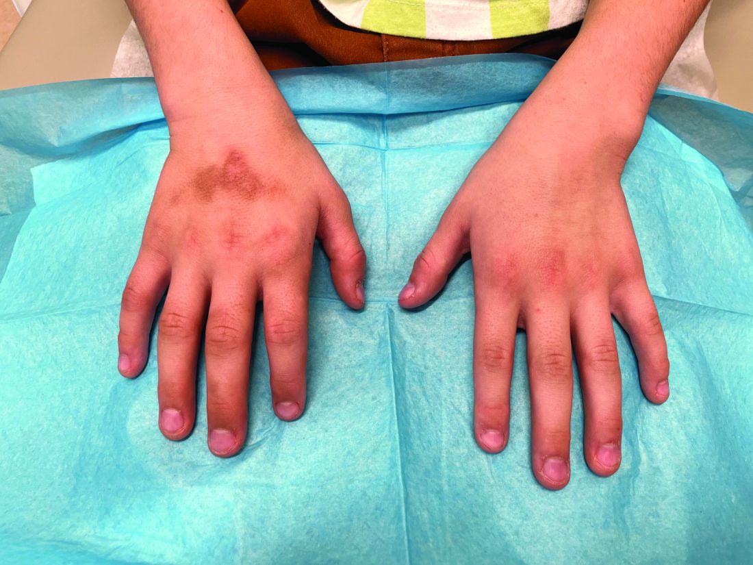
The patient was otherwise healthy, with no current medication intake, and he engaged in baseball and soccer activities. Upon physical examination, a hyperpigmented lichenified irregular plaque was observed on the dorsum of the right hand, along with irregular hyperpigmented macules and plaques on the fingers. Fusiform widening of the interphalangeal joints on the second, third, and fourth fingers of the right hand was noted, without associated pain, edema, or erythema.
Parent-Led Digital CBT Effective for Childhood Anxiety
while substantially reducing cost and therapist time, new research showed.
In a randomized controlled trial, children participating in the program Online Support and Intervention (OSI) for Child Anxiety showed similar reductions in anxiety and improvements in daily functioning as peers receiving standard CBT.
“This study shows that by making the most of digital tools, we can deliver effective treatments more efficiently, helping services to better meet the growing demand for mental health services for children in ways that can also be more accessible for many families,” lead investigator Cathy Creswell, PhD, Departments of Experimental Psychology and Psychiatry, Oxford University, Oxford, England, told this news organization.
“I believe by incorporating this approach into standard care, we could address some of the major challenges faced by services and families,” Dr. Creswell added. “We are now moving the work out of the research environment into routine practice.”
The study was published online in The Lancet Psychiatry
Care Gaps for Common Problem
Anxiety is common in children, yet gaps exist between needed and available care, which investigators say could be filled by digitally augmented psychological treatments.
OSI, the digital platform used in the current study, was designed with therapists and families to aid parents in helping their children overcome problems with anxiety with remote therapist support.
The program provides parents with the core CBT content in accessible forms, including information in text, audio, and video and exercises supported by worksheets and quizzes.
There is also an optional child game app to help motivate the child to engage with the intervention. Parents are supported with weekly brief telephone or video call sessions with the therapist.
The two-arm randomized controlled non-inferiority trial included 444 families from 34 participating Child and Adolescent Mental Health Services (CAMHS) sites in England and Northern Ireland. Half received OSI plus therapist support and half CAMHS treatment as usual. The children were between 5 and 12 years old.
A total of 176 (79%) participants in the OSI plus therapist support group and 164 (74%) in the treatment as usual group completed the 26-week assessment.
‘Compelling’ Evidence
The primary clinical outcome was parent-reported interference caused by child anxiety at 26 weeks, using the Child Anxiety Impact Scale-Parent report.
On this measure, OSI plus therapist support was non-inferior to usual treatment, with a standardized mean difference of only 0.01 (95% CI, −0.15 to 0.17; P < .0001).
The intervention was also significantly non-inferior to usual treatment across all secondary outcomes, including total anxiety and depression scores, overall functioning, peer relationship problems, and prosocial behavior.
In addition, OSI plus therapist support was associated with nearly 60% less therapist time (182 min on average vs 307 min) and with lower costs than standard treatment. The OSI program was “likely to be cost-effective under several scenarios,” the researchers reported. Qualitative interviews showed “good” acceptability of the online program.
“This trial presents compelling clinical evidence and promising cost-effectiveness evidence that digitally augmented psychological therapies with therapist support can increase efficiencies in and access to child mental health services without compromising patient outcomes,” Dr. Creswell and colleagues concluded.
“Efforts are now needed to take full advantage of the opportunity that digitally augmented psychological treatments can bring to drive a step change in children’s mental health services, learning from successful examples of digital implementation elsewhere in health services,” they added.
‘Call to Action’
“We desperately need more trials” like this one, which showed the “clear value of a digitally augmented intervention over the usual face-to-face treatment” for child anxiety, wrote the authors of an accompanying editorial.
“Moreover, with the intervention delivered across 34 CAMHS settings in England and Northern Ireland, this study gives us confidence that the new intervention is effective in a range of clinical contexts at least in high-income countries and offers invaluable information about barriers and facilitators to future implementation,” wrote Sam Cartwright-Hatton, PhD, with the University of Sussex, Brighton and Hove, and Abby Dunn, PhD, with the University of Surrey, Guilford, England. “The potential benefits to overburdened services are clear.”
“That regular CAMHS clinicians, with minimal training and support from researchers, delivered the intervention within their standard caseload shows that it can be embedded within routine practice without a requirement for highly prepared and supervised clinicians,” they added.
However, more information is needed on the content and quality of the traditional CBT provided in the control group. It’s also important to determine if the program would be as effective with even less clinical support and in all types of childhood anxiety.
In addition, most clinicians in the OSI group only treated one patient with the new program and didn’t take advantage of the additional support offered by the research team, “which means we have not seen the true effectiveness of this intervention in the hands of well-practiced and well-trained staff,” Drs. Cartwright-Hatton and Dunn wrote.
Analyses included recruitment at the lower target amount, and one fifth of children were not offered treatment within the 12-week window recommended in the trial, they added.
“Although these issues place limits on the conclusions that can be drawn, they should also be seen as a call to action,” they wrote, adding that real-world clinical trials with greater clinician participation are needed. “All credit to this exceptional team for making this trial happen and for making it work as well as it did.”
The study was funded by the Department for Health and Social Care, UK Research and Innovation Research Grant, National Institute for Health and Care (NIHR) Research Policy Research Programme, Oxford and Thames Valley NIHR Applied Research Collaboration, and Oxford Health NIHR Biomedical Research Centre. Dr. Creswell is co-developer of the OSI platform and the author of a book for parents that is used in many of the participating clinical teams to augment treatment as usual for child anxiety problems and receives royalties from sales. Dr. Cartwright-Hatton and Dr. Dunn had no disclosures.
A version of this article appeared on Medscape.com.
FROM THE LANCET PSYCHIATRY
Lichen Sclerosus: The Silent Genital Health Concern Often Missed
Ashley Winter, MD, remembers the first time she Googled the skin condition lichen sclerosus. Most of the websites listed the autoimmune condition as a rare disease.
In the realm of genital health, some conditions remain shrouded in silence and consequently are more likely to go undercounted and underdiagnosed, said Dr. Winter, a urologist based in Los Angeles.
“I truly believe that we just miss the diagnosis a vast majority of the time because there isn’t enough training on [detecting] it,” said Dr. Winter.
, according to the US National Institutes of Health. The condition also more commonly occurs among women, and symptoms include hypopigmentation, itching, pain, changes in skin appearance, and skin atrophy.
“Most cases [affect the] genital [area] only, so often patients don’t bring it up because they don’t want to be examined,” said Sarah Lonowski, MD, assistant professor of dermatology and codirector of the Multidisciplinary Autoimmune Skin Disease/Derm-Rheum Program at the University of Nebraska–Lincoln. “It’s a sensitive area, it’s an uncomfortable area to have examined, so it comes with a lot of emotional burden,” for patients, Dr. Lonowski said.
Receiving a lichen sclerosis diagnosis can take 5 years or longer, in part because the condition’s symptoms can lead clinicians to first make a diagnosis of a yeast infection or bacterial vaginosis, according to Christina Kraus, MD, assistant professor of dermatology at UCI Health in Irvine, California.
“There is still limited information on this condition in medical education, and it is not uncommon for clinicians who are not in dermatology or gynecology to be unfamiliar with this diagnosis,” Dr. Kraus said.
Because no medical tests are available to confirm lichen sclerosus, clinicians diagnose the condition based on the skin’s appearance and symptoms. In some cases, a skin biopsy may help differentiate it from similar rashes that occur in the genital area.
Prepubescent children and postmenopausal women are most likely to develop genital lichen sclerosis, so pediatricians and primary care physicians may be the first to see possible cases, Dr. Lonowski said.
Patients “may not mention it unless they’re asked,” Dr. Lonowski said, adding clinicians can inquire with patients about genital health, examine bothersome areas, “and refer if you’re not sure.”
Clinicians may also miss the condition during physical exams if they do not examine the vulvar skin, she said. The exact cause also remains elusive, but researchers believe genetic and hormonal factors, as well as an overactive immune response, may contribute to development of the condition.
Watch Out for Presentation
While lichen sclerosus more frequently occurs in women, men are also affected by the condition. Benjamin N. Breyer, MD, professor and chair of urology at the University of California San Francisco, said lichen sclerosus is one of the most common skin conditions he treats in his male patients.
“Advanced cases can cause urethral narrowing, which is a condition I treat commonly,” said Dr. Breyer. “Lichen sclerosus is often an underrecognized cause of pain or tearing with erections and sex in men.”
Similar to women, lichen sclerosus presents as white color changes on the skin. For men, the condition can also result in fusion of the shaft skin to the head of the penis and burying or concealment of the penis, Dr. Breyer said.
“This leads to challenges with intimacy and urination and can have extensive impacts on quality of life,” said Dr. Breyer.
For women, the skin changes often extend to the perianal area and can cause scarring, said Dr. Kraus.
“Early scarring may present as adherence of the labia minora to the labia majora or inability to fully retract the clitoral hood from the clitoris,” said Dr. Kraus.
In both men and women, lichen sclerosus and another autoimmune condition known as morphea, characterized by skin hardening and discoloration, often present together, said Dr. Lonowski.
“If you have a patient with known morphea, it’s important to ask about genital symptoms,” said Dr. Lonowski. “The association between the two is fairly strong.”
Circumcision is often the first step to help prevent chronic inflammation among male patients, said Dr. Breyer. Because lichen sclerosus is associated with an increased risk for penile cancer, “it is important to biopsy suspicious lesions,” Dr. Breyer added.
Increasing awareness of lichen sclerosus is crucial for early detection and timely intervention, said Dr. Lonowski. The first-line treatment of genital lichen sclerosus is strong topical steroid ointments to reduce inflammation. Clinicians might prescribe this treatment for use twice daily for 2-3 months and then assesses the patient on their response.
“It is fairly responsive to treatment in most cases,” said Dr. Lonowski.
Once symptoms have improved, Dr. Lonowski transitions patients to a maintenance regimen, which might include using the same steroid but only three times a week, switching to a topical with a less potent steroid dosage, or using a combination of a topical steroid and a nonsteroidal anti-inflammatory cream. Despite the prolonged use of the drug, she said patients with lichen sclerosus usually do not present with side effects like discoloration or thinning of skin.
“You may achieve control or remission, but we don’t stop treatment completely because we know the natural history of the disease is to have flares and recurrence.”
If left untreated, the condition can lead to atrophy, scarring, and distortion of the genital anatomy and, in some cases, develop into squamous cell carcinoma.
“The fact that you can do a topical cream intervention and prevent cancer is huge,” said Dr. Winter.
She said open discussions surrounding genital health led by primary care providers can destigmatize conditions like lichen sclerosus and promote early detection and management.
“We need to foster an environment where individuals feel comfortable discussing their symptoms openly,” Dr. Winter said. “Increased awareness can pave the way for early detection, which is crucial for managing the condition effectively.”
The experts included in the story reported no relevant disclosures.
A version of this article appeared on Medscape.com.
Urologist Sues Health System Over Noncompete Clause
The lawsuit brings renewed attention to the ongoing public discourse around restrictive covenants for physicians as more state and federal legislators signal plans to limit or ban the practice.
According to a civil suit filed on January 30 in the Court of Common Pleas, Scranton, Pennsylvania, Eric Rottenberg, MD, signed a 3-year employment agreement with Commonwealth Health Physician Network (CPN) in November 2022. He worked for the health system from May to November 2023, seeing patients at several of its locations, including Wilkes-Barre General Hospital and other facilities throughout northeast and central Pennsylvania.
Although Dr. Rottenberg previously practiced in Albany, New York, court records state he did not bring a significant referral or patient base to the new role, receive any specialized training, or have knowledge of CPN’s trade secrets during his tenure.
Instead, he was a “9-to-5 practitioner,” or a physician-employee like a “locum tenens whose replacement would not cost the employer more than his traditional compensation,” the complaint said. Dr. Rottenberg only treated patients assigned to him by CPN and its parent company, Commonwealth Health Systems, and did not take a patient base with him upon his departure from CPN.
Commonwealth Health spokesperson Annmarie Poslock declined to comment on pending litigation.
After becoming frustrated by “restrictions on his ability to practice medicine” at CPN, Dr. Rottenberg submitted the required 90-day written notice to terminate the employment agreement. He subsequently received a letter from Simon Ratliff, CPN’s chief executive officer, confirming that his last day of employment would be February 11, 2024. Ratliff also reiterated that the noncompete clause would be enforced, essentially banning Dr. Rottenberg from practicing within a 20-mile radius of his previous CPN locations for the next 2 years, court documents said.
Dr. Rottenberg was recruited by Lehigh Valley Physician Group (LVPG), part of Lehigh Valley Health Network, in December 2023 for a urology position at its Dickson City and Scranton locations — some of which are within 20 miles of CPN facilities, the complaint said.
Employers often include noncompete terms in physician contracts because they want to keep the departing physician’s patients from following them to a competitor. However, about a dozen states and the District of Columbia have passed legislation that allows physicians and other clinicians to more easily exit contracts and change jobs.
For example, an Indiana law took effect on July 1 that prohibits employers from entering a noncompete agreement with primary care physicians. Minnesota legislators also banned new noncompete agreements for all employees effective July 1.
“There’s actually been a long-standing push for bans on physician noncompetes going back to some of the first states to pass them, like Colorado, Delaware, and Massachusetts, in the late 1970s and early 1980s,” said Evan Starr, PhD, associate professor of management and organization at the Robert H. Smith School of Business at the University of Maryland.
Although New York Governor Kathy Hochul recently vetoed a bill that would have outlawed restrictive covenants, more states may consider passing laws that limit or ban noncompetes amid increasing patient equity and care access concerns. Dr. Starr told this news organization that one reason to eliminate restrictive covenants is because they can cause “third-party harm” to patients. “The patient doesn’t get the choice to sign a noncompete, but they’re going to be impacted by that agreement if the physician has to leave the area,” he said.
Interestingly, one profession — lawyers — is the only occupation in the US for which noncompete agreements are banned, says Dr. Starr. “Basically, the American Medical Association (AMA) and other physician governing bodies haven’t made the same policies to exempt themselves that the lawyers have.”
That may be changing. In June, the AMA’s House of Delegates adopted policies to support the prohibition of noncompete contracts for employed physicians. The change came several months after the Federal Trade Commission (FTC) proposed a new rule that could more broadly ban companies from enforcing noncompete clauses.
Despite Rottenberg’s attorney, Ryan Campbell, Esq, claiming that the noncompete is unenforceable without a protectable business interest, CPN would not release him from the agreement and opted to move forward with litigation, court records said. The suit cites several other cases where Pennsylvania judges have released physicians from similar restrictive covenants.
Mr. Campbell told this news organization that he and his client are “working diligently with CPN and its counsel to resolve the matter amicably and without further litigation.”
As employers await the FTC’s final rule, Dr. Starr says they could take steps to eliminate noncompete agreements altogether in favor of other stipulations. Contract terms prohibiting physicians from soliciting former patients could protect business interests and still allow patients to seek their preferred physician on their own accord.
A version of this article appeared on Medscape.com .