User login
Before surgery: Have you done enough to mitigate risk?
• Identify cardiac, pulmonary, renal, infectious, and hematologic risk factors, and steps that can be taken to minimize risk. C
• Check serum albumin levels of all patients at risk for hypoalbuminemia; levels <35 g/L are strongly associated with postoperative pulmonary complications. B
• Help patients with diabetes achieve optimal glycemic control prior to surgery to minimize the risk of infection. B
• Avoid routine use of ancillary testing; evidence supports the use of such tests in only a small minority of surgical candidates. C
Strength of recommendation (SOR)
A Good-quality patient-oriented evidence
B Inconsistent or limited-quality patient-oriented evidence
C Consensus, usual practice, opinion, disease-oriented evidence, case series
CASE Charlie H, an elderly man who has been your patient for more than 10 years, is scheduled for inguinal hernia repair, and has come in for a preoperative evaluation. Based on his medical history and a physical examination, you identify several risk factors for surgical complications: a low functional capacity (<4 METS), obesity (BMI=39), advanced age (70 years), and type 2 diabetes (well controlled). What should you write in your consultation note about Charlie’s perioperative risks, and what interventions should you institute—or recommend—to mitigate his risk?
A preoperative consult, a service that family physicians are well positioned to provide, requires a thorough and systematic approach. But because of time pressures—as well as a dearth of perioperative templates, guidelines, and checklists—a cursory history and physical exam often takes the place of a comprehensive evaluation.
A thorough medical history is the most valuable tool of a physician doing a preop consult, but a comprehensive evaluation also involves the assessment of perioperative risk factors, ancillary tests to consider, and interventions to recommend to mitigate risks. Although various published guidelines address specific systems, there are few places where family physicians can find a complete toolkit. The text and tables that follow, which form the core of a comprehensive resource initially compiled to help our residents conduct clear and effective preoperative consults, will help you safeguard your patients.
A system-by-system review starts with the heart
The vast majority of perioperative problems fall into a handful of categories: cardiac, pulmonary, renal, infectious, and hematologic complications (TABLE 1). When a surgeon requests a preoperative evaluation, however, the patient’s cardiac status is generally the primary concern. This is also the portion of the preop consult with the most formally structured guidelines; those issued by the American College of Cardiology and American Heart Association (ACC/AHA) are the most widely used.1 Initially based primarily on expert opinion, the ACC/AHA guidelines are increasingly evidence-based (http://circ.ahajournals.org/cgi/reprint/CIRCULATIONAHA.109.192690).1,2 These guidelines address the evaluation of patients for noncardiac surgery. Both cardiac surgery and emergent operations are beyond the scope of the guidelines, and are not addressed here.
Patients with unstable coronary syndromes—eg, unstable angina or myocardial infarction (MI) within the past 30 days, decompensated heart failure (HF), significant arrhythmias, or severe valvular disease—face an increased risk of perioperative morbidity and mortality. To reduce the risk, such patients require optimization of the underlying condition before undergoing elective surgery.1

Stable ischemic heart disease, compensated HF, diabetes, chronic renal failure/insufficiency (CRF), cerebrovascular disease, and poor functional capacity (defined as <4 metabolic equivalents [METS]) in an asymptomatic patient also increase the risk of complications, but to a lesser degree. If a patient has coronary artery disease, evidence of ischemia on preoperative testing, or more than 1 of these clinical risk factors, surgery may proceed. Keep in mind, though, that the ACC/AHA guidelines suggest that the use of a beta-blocker, titrated to control heart rate and blood pressure, is reasonable in intermediate- or high-risk procedures (TABLE 1).1,3,4
Is additional cardiac testing necessary? Whether you’re assessing for cardiac status or other risks, for that matter, evidence supports the use of ancillary testing in only a small minority of surgical patients. A general rule of thumb—regardless of the system you’re assessing—is to consider adjunctive testing only if the outcome has the potential to alter patient management. Thus, exercise stress testing or resting electrocardiography (EKG), among other tests, may be considered on an individual basis (TABLE 2), but studies have failed to demonstrate improved outcomes with added testing of cardiac status on a routine basis.5,6
Evidence is insufficient to make a firm recommendation regarding additional cardiac testing, even for patients with more than 3 clinical risk factors. Nonetheless, the ACC/AHA guidelines favor the use of adjunctive testing in such cases, especially for patients who are candidates for high-risk procedures, such as vascular surgery.1
What’s the local standard of care? Studies to determine when further testing is beneficial and which tests would benefit which patients are ongoing. In the absence of definitive findings, it behooves primary care physicians to familiarize themselves with the practices and preferences of the cardiologists and anesthesiologists at the facility where the surgery will be performed and to follow the local standard of care.
TABLE 1
Identifying—and minimizing—perioperative risk
| Patient-specific risk factors | Procedure-specific risk factors | Risk reduction recommendations |
|---|---|---|
| Cardiac | ||
| Major risks • Decompensated HF • Severe valve disease • Significant arrhythmia • Unstable coronary syndrome Other cardiovascular risks • Cerebrovascular disease • CRF or AKI • Compensated/prior HF • Diabetes • Functional capacity <4 METS • Ischemic heart disease | Vascular surgery | • Optimize treatment of underlying conditions • Consider beta-blockers perioperatively1,3,4 • Consider adjunctive testing if results could alter patient management |
| Pulmonary | ||
| • Acute URI • Requiring assistance with ADLs • Age >60 years • Elevated BUN (>21 mg/dL) • COPD • HF • Hypoalbuminemia (<35 g/L) • Presence of any systemic disease | • Emergency surgery • General anesthesia • Surgery >3 h • Abdominal, head or neck, thoracic, or vascular surgery • Neurosurgery | • Postop incentive spirometry • Postop nasogastric tube • Consider intraoperative use of LMA • Smoking cessation (30 days preoperatively)28 |
| Renal | ||
| • Age >60 years • CRF (especially with creatinine >2.1 mg/dL) • Diabetes (especially insulin-dependent) • HF • Jaundice | • Aortic or cardiovascular surgery • Liver transplantation | • Ensure preoperative euvolemia and good osmolar status • Minimize exposure to nephrotoxins • Avoid perioperative hypotension (maintain MAP >65 mm Hg) • Consider preoperative dialysis if GFR <15 mL/min14 |
| Infectious | ||
| • Advanced age • Corticosteroid use • Hyperglycemia • Hypoalbuminemia • Immunocompromised • Malnutrition/obesity • Peripheral vascular disease • Postoperative incontinence • Preexisting infection • Prior radiation therapy • Smoking | • Blood transfusion • Surgery >3 h • Perioperative hypothermia • Perioperative hypoxia • Preoperative shaving • Prolonged preoperative hospital stay | • Optimize diabetes management (HbA1c <7); tight perioperative glycemic control • Treat preexisting infections • Provide nutritional supplementation (7-14 days preoperatively) • Smoking cessation (30 days preoperatively)28 |
| Hematologic: Perioperative bleeding | ||
| • Collagen vascular disease • GI or urogenital blood loss • Heavy or prolonged menses • Hematologic disease • Hemophilia or other inherited disorder • History of easy bruising or bleeding • Hypersplenism • Liver or renal disease • Severe bleeding after dental extraction, other surgery, or childbirth • Physical findings suggestive of purpura, hematoma, jaundice, or cirrhosis • Use of medications that affect hemostasis | • Minimal risk/JHSRCS 1 (eg, breast biopsy, carpal tunnel procedure, cataract surgery) • Mild risk/JHSRCS 2 (eg, laparoscopy, arthroscopy, inquinal hernia repair) • Moderate risk/JHSRCS 3 (eg, open abdominal procedure, arthroplasty) • Significant risk/JHSRCS 4 (eg, open thoracic surgery, major vascular/skeletal procedure) | • Optimize treatment of preexisting conditions • Discontinue antihemostatic medications, if medically feasible • Consider autologous blood banking |
| Hematologic: Perioperative anemia | ||
| • Hemoglobinopathies • Preexisting iron deficiency anemia • Preexisting pernicious anemia | • Risk of bleeding based on type of surgery (see Perioperative bleeding, above) | • Correct anemia prior to surgery • Consider preoperative erythropoietin • Avoid preoperative transfusion |
| Hematologic: Venous thromboembolism | ||
| • Acute medical illness • Age (older) • Cancer (active or occult); cancer therapy • Estrogen/SERMs • Erythropoiesis-stimulating agents • Immobility • IBD • Lower-extremity paresis • Myeloproliferative disorders • Nephrotic syndrome • Obesity • Paroxysmal nocturnal hemoglobinuria • Pregnancy/postpartum • Previous VTE • Smoking • Thrombophilia • Venous compression | • Cardiothoracic surgery • Central venous catheterization • Major surgery (general, gynecologic, orthopedic, peripheral vascular, or urologic) • Neurosurgery • Trauma | • Ensure early, aggressive mobilization • Provide mechanical prophylaxis • Consider chemoprophylaxis |
| ADL, activities of daily living; AKI, acute kidney injury; BUN, blood urea nitrogen; COPD, chronic obstructive pulmonary disease; CRF, chronic renal failure/insufficiency; GFR, glomerular filtration rate; GI, gastrointestinal; HbA1c, hemoglobin A1c; HF, heart failure; IBD, inflammatory bowel disease; JHSRCS, Johns Hopkins Surgical Risk Classification System; LMA, laryngeal mask airway; MAP, mean arterial pressure; METS, metabolic equivalents; SERMs, selective estrogen receptor modulators; URI, upper respiratory infection; VTE, venous thromboembolism. | ||
TABLE 2
When should you order these ancillary tests?*
| Albumin† For at-risk populations9 |
| BUN, creatinine, electrolytes For at-risk subpopulations21 |
| Chest x-ray It depends. It is not used routinely for predicting risk but may be appropriate for patients with previous diagnosis of COPD or asthma.9 |
| CBC, platelets Do not order routinely; check hemoglobin if procedure increases risk for bleeding. |
| Coagulation studies Do not order routinely.35,36 |
| Echocardiogram It is reasonable to order for patients with dyspnea of unknown origin, history of HF and worsening dyspnea, or other change in clinical status and may be considered for patients with previously documented cardiomyopathy.1 |
| EKG Vascular surgery: Order for patients with ≥1 clinical risk factors; it is also reasonable for patients with no clinical risk factors.1Intermediate-risk procedure: Order for patients with CHD, PAD, or CVD and consider for patients with ≥1 clinical risk factors.1 |
| Exercise stress-testing Order for patients with active cardiac conditions; it is reasonable for vascular surgery candidates with ≥3 clinical risk factors and poor functional capacity and may be considered for patients undergoing vascular or intermediate-risk procedure who have 1-2 clinical risk factors and poor functional capacity.1 |
| Spirometry, pulmonary-function testing Do not order routinely for predicting risk, but may be appropriate for patients with previous diagnosis of COPD or asthma.9 |
| Urinalysis Order routinely.20 |
| BUN, blood urea nitrogen; CBC, complete blood count; CHD, coronary heart disease; COPD, chronic obstructive pulmonary disease; CVD, cardiovascular disease; EKG, electrocardiography; HF, heart failure; PAD, peripheral artery disease. |
| *Most commonly recommended ancillary tests for which there are at least minimal data to suggest the validity of the opinion-based recommendation. Answers are opinion-based, not evidence-based. With the exception of albumin testing, the tests listed here are lacking in patient-oriented evidence of benefit from routine use. |
| † Routine testing of albumin levels is evidence-based. |
Identify pulmonary risks with help from ACP
Postoperative pulmonary complications are as prevalent as cardiac complications, and contribute equally to morbidity, mortality, and length of stay. But pulmonary complications are better predictors of long-term mortality after surgery.7
There are several well-validated risk factors for increased perioperative pulmonary morbidity and mortality—HF, chronic obstructive pulmonary disease (COPD), advanced age, and the need for assistance with activities of daily living among them. In addition to identifying patient-specific risk factors, knowledge of the type of surgery planned will provide insight into procedure-specific risk factors (TABLE 1). The approach to the surgical pulmonary patient is addressed in an American College of Physicians (ACP) guideline published in 2006 and available at http://www.annals.org/content/144/8/575.full.pdf+html.7
What tests to consider? The ACP guideline is notable not only for its recommendations, but for the things that are not recommended but may nevertheless be considered the standard of care in some locales. Chest radiography and spirometry are 2 such examples. Although these tests may be appropriate on an individual basis for patients with a previous diagnosis of COPD or asthma, their routine use is of little value—and the ACP does not recommend them as part of a standard preop evaluation.7 Some laboratory tests may aid in risk stratification, however.
A serum albumin level <35 g/L is strongly associated with postop pulmonary complications.8 Checking levels in all patients suspected of hypoalbuminemia, including any patient with 1 or more pulmonary risk factors, is reasonable for a physician performing a preoperative evaluation. Consider checking blood urea nitrogen (BUN) levels, as well. Uremia (BUN >21 mg/dL) is also associated with increased pulmonary complications, although not as strongly as hypoalbuminemia.
Postpone or proceed? Acute conditions are another key consideration. An upper respiratory infection (URI) increases the risk of postoperative pulmonary complications, especially in children.9,10 A simple algorithm offers guidance in deciding when to postpone surgery in pediatric patients with a URI:9
Recommend that it be delayed if the procedure involves general anesthesia and 1 or more of the following risk factors is present: asthma, a history of prematurity, copious secretions, a parent who smokes, planned use of an endotracheal tube, or a procedure involving the airway.
Surgery can proceed if symptoms of the infection are mild, general anesthesia is not required, or a risk/benefit analysis supports it. Considerations include the urgency of the procedure, whether the surgery has previously been postponed, the comfort level of the clinicians involved, and the distance the family must travel for the procedure.11
If you recommend that surgery proceed as planned, suggest perioperative interventions to mitigate risk. Recommend that a laryngeal mask airway be used, if needed, in place of an endotracheal tube; that pulse oximetry monitoring occur; that good hydration and humidification of air be provided; and that the patient receive anticholinergic agents for secretions.
Other measures that have been shown to be effective in reducing perioperative pulmonary complications include deep breathing exercises (incentive spirometry) and the use of a nasogastric tube for those with postoperative emesis, intolerance of oral intake, or symptomatic abdominal distension.7 If your patient has risk factors for pulmonary complications, include a recommendation for a postop nasogastric tube in your preop consultation note. However, newer data indicating that patients had fewer pulmonary complications, a more rapid return of normal bowel function, no increased discomfort, and no increase in anastomotic leaks without a nasogastric tube12,13 may lead to guideline revision.
A scoring system helps evaluate renal risk
Patients with CRF face increased risk of perioperative morbidity and mortality. But as long as the glomerular filtration rate (GFR) is >25 mL/min—which is only 25% of normal—surgery is generally well tolerated. As GFR drops to 10 to 15 mL/min, the rate of surgical complications rises rapidly, reaching 55% to 60%. For such patients, preoperative dialysis is worth considering.14
Postoperative acute kidney injury (AKI), as acute renal failure is now known,15 is associated with a 58% mortality rate.16 Fortunately, this complication develops in only about 1% of surgical patients.17 Both patient-specific risk factors (CRF, with creatinine >2.1 mg/dL; HF; diabetes, particularly being insulin dependent; age >60 years; jaundice) and procedure-specific risks (aortic, cardiovascular, or liver transplant surgery) help predict which surgical candidates face the highest risk.16,18,19 Thakar et al have developed a scoring system to identify those at greatest risk for AKI.20 (See “Cardiovascular surgery and acute kidney injury: Scoring the risk” at www.jfponline.com by clicking on “Before surgery: Have you done enough to mitigate risk?” and scrolling to the end.)
Minimize renal complications. Helping patients achieve good intravascular volume and osmolar status preoperatively will reduce their risk of renal complications. Other prophylactic measures: Minimize exposure to nephrotoxins (eg, nonsteroidal anti-inflammatory drugs or contrast media) to the extent possible. Consider evaluating the serum electrolyte and creatinine levels of patients with multiple risk factors to determine whether they can safely undergo surgery; some experts suggest preoperative urinalysis, as well.18
Patients with end-stage renal disease have very high perioperative morbidity.21 They are at increased risk for hyperkalemia, infection, hyper- and hypotension, bleeding, arrhythmias, and clotted fistulas, in descending order of incidence.18 Preoperative planning, including the need for dialysis before surgery, is necessary to manage these risks.
A scoring system developed by Thakar et al20 is a valuable tool in assessing the likelihood that a patient requiring cardiovascular surgery will develop acute kidney injury (AKI).
To identify those at greatest risk, add 1 point for each of the following:
- female sex
- heart failure
- ejection fraction <35%
- chronic obstructive pulmonary disease (COPD)
- insulin-dependent diabetes
- history of prior cardiac surgery
- valve-only cardiac procedure scheduled
Add 2 points for each of the following:
- preoperative intra-aortic balloon pump (IABP)
- emergency surgery
- combined coronary artery bypass graft (CABG)/valve surgery scheduled
- other cardiac surgery (except CABG) scheduled
- creatinine level from 1.2 to 2.1 mg/dL
And add 5 points for a creatinine level >2.1 mg/dL.
Patients with a total score ≤5 have less than a 2% risk of developing AKI; those with scores between 6 and 8 have an 8% to 10% risk, and patients with scores >8 have more than a 20% risk for developing postoperative AKI.
Risk of postop infection: Focusing on the foreseeable
Postoperative infections, both at the surgical site and remote from the incision, are a significant cause of morbidity and mortality. Pneumonia is among the most prominent remote infections associated with surgery,22 and early ambulation, deep breathing exercises, and tight glycemic control can greatly decrease the risk.
Surgical site infection (SSI) remains an important concern, occurring in 37% of cases.23 Risk factors include hyperglycemia, malnutrition, perioperative steroid use, preexisting infections, tobacco smoking, peripheral vascular disease, advanced age, radiation therapy, blood transfusions, prolonged preoperative stay, preoperative shaving, hypothermia, hypoxia, length of operation, and postoperative incontinence.24 While many of these risk factors are dependent on interventions in the operating room and recovery room or during subsequent hospitalization, it is important to address foreseeable risks as part of the preoperative evaluation.
Glycemic control is crucial. Perhaps the most well-documented risk for SSI is hyperglycemia—a common problem among hospitalized patients.16 Hyperglycemia impairs leukocyte and complement function,25,26 thereby increasing risk of infectious complications. Tight glycemic control in the surgical patient, especially on the surgical intensive care unit, has been associated with improved outcomes.27
Identify the presence of diabetes in the preoperative consult note, and adjust the patient’s medication regimen as needed, to help him or her achieve optimal glycemic control. In some cases, it may be necessary to delay nonurgent surgery until the patient achieves adequate control.
Malnutrition is another risk factor for SSI. For patients who are undernourished or morbidly obese, checking serum albumin levels may be beneficial. Supplementation for 1 to 2 weeks prior to surgery may decrease the risk of infection for patients who are undernourished;28 for obese patients, weight loss is beneficial. Although significant preoperative weight loss may not be possible, it is important to list an elevated body mass index as a risk factor in the consultation note.
Corticosteroids, used to treat conditions such as COPD, inflammatory bowel disease, allergies, and autoimmune disorders, are another risk factor for perioperative infection. In addition to their effect on glycemic control, corticosteroids directly suppress the immune system. Whenever possible, they should be discontinued preoperatively. If this is not possible, call attention to the patient’s use of corticosteroids in the consultation note.
Preexisting infection presents the possibility of the spread of organisms to the surgical site and, whenever possible, surgery should be postponed until the infection resolves. If the patient has a history of prior infection or colonization with methicillin-resistant Staphylococcus aureus, be sure to include that in the consultation note, as well.
Leukocyte-containing blood product transfusions are associated with a 2-fold increase in some postop infectious complications.29-31 This is in addition to the well-known risk of bloodborne pathogens associated with transfusions, and is yet another reason to avoid perioperative transfusions whenever possible.
Smoking impairs tissue oxygenation, which delays healing and increases risk of infection. Smoking cessation should be strongly encouraged at every preoperative consultation. Recommend nicotine replacement therapy even for patients who aren’t willing to quit altogether; point out that giving up cigarettes for just 30 days (or more) before surgery can decrease the likelihood of complications.28
In addition to these identified risk factors, anything that compromises the immune status increases the risk of infection. Alcohol or drug abusers, chronic pain patients, transplant recipients, cancer patients taking immunosuppressants, postsplenectomy patients, and patients with HIV are all at increased risk. Identify any such conditions during your preoperative evaluation, and be sure to include them in your communication with the surgical team.
A common request in preop consults relates to bacterial endocarditis prophylaxis. Only an extremely small number of cases of infective endocarditis occur with dental procedures, however, so the benefits of antibiotic prophylaxis would be minimal, even if the prophylactic therapy were 100% effective.32 As a result, the ACC/AHA guidelines recommend prophylaxis for dental procedures only for patients with underlying cardiac conditions associated with the highest risk of adverse outcome from infective endocarditis. Administration of antibiotics solely to prevent endocarditis is not recommended for patients undergoing genitourinary or gastrointestinal tract procedures.32
Hematologic evaluation: Address risks of bleeding, clotting
Historically, a variety of tests have been employed in an effort to identify patients at risk for bleeding complications prior to surgery—including prothrombin time, partial thromboplastin time, platelet count, and bleeding time, or platelet function. While highly reproducible, automated, and inexpensive when considered individually, the cumulative cost of routine use of these tests is high.33
A recent review of the literature indicates that, for surgical patients without synthetic liver dysfunction or a history of oral anticoagulant use, routine testing is of little value in the assessment of bleeding risk.34 Patients with a negative bleeding history do not require routine coagulation screening prior to surgery.35
Instead, use the medical history to identify risk factors for bleeding. These include excessive bruising, nosebleeds, prolonged bleeding after cuts, bleeding >3 minutes after brushing teeth, and heavy or prolonged menses (TABLE 1). Patients with a past medical history of liver disease; renal failure; hypersplenism; hematologic disease; collagen vascular disease; hemophilia or other inherited hemorrhagic disorder; gastrointestinal or urogenital blood loss; and severe bleeding after dental extraction, other surgery, or childbirth are also at heightened risk, as are those who take medications that affect hemostasis. Physical findings suggestive of risk include purpura, hematoma, jaundice, and signs of cirrhosis.34
While laboratory testing is only appropriate to confirm those at risk in the subpopulation selected by the history and physical, here, as with other adjunctive testing, it is important to consider local standards and the preferences of the surgeon who requested the preop consult.
Treat anemia. Preoperative anemia is linked to adverse outcomes in surgical patients,36 although it is not clear whether the anemia itself or the perioperative transfusions associated with the condition are at the root of the problem.37 Macrocytic anemia may require treatment with vitamin B12 and folate; iron deficiency anemia is treated with iron. Some physicians also recommend the use of erythropoietin starting 3 weeks prior to surgery for patients with normocytic anemia with hemoglobin <13 g/dL.38,39
Thromboembolism risk. Excessive clotting is responsible for more perioperative complications than excessive bleeding. There is a high prevalence of venous thromboembolism (VTE) among surgical patients, with both patient- and procedure-specific risk factors. Although a variety of coagulopathies increase the baseline risk for VTE, routine laboratory screening of the general surgical population for thrombophilia is not recommended.34,35
When risk factors are present based on both the patient’s medical history and the type of procedure, prophylactic measures may be needed (TABLE 3). Options include mechanical prophylaxis (graduated compression stockings and intermittent pneumatic devices) and chemoprophylaxis. Recommended for high-risk cases, such as patients undergoing orthopedic surgery that precludes early mobilization, chemoprophylaxis options include low-molecular-weight heparin, low-dose unfractionated heparin, fondaparinux (a synthetic factor Xa inhibitor), and vitamin K antagonists such as warfarin.40 Aspirin alone is not recommended, as it has not been found to be an effective prophylaxis for VTE.
CASE After following this system-by-system review of your patient, Charlie H, you identify and explicitly communicate the following risk factors in your consultation note:
- Cardiovascular: type 2 diabetes, low functional capacity
- Pulmonary: advanced age
- Renal: advanced age
- Infectious: type 2 diabetes, advanced age, BMI=39
- Hematologic: advanced age, obesity
Based on these findings, you develop the following plan for Charlie H, detailed in the consultation note you submit to the surgical team:
- Continue home diabetes medications perioperatively, supplemented with a basal-bolus insulin regimen.
- Initiate incentive spirometry postoperatively; use an NG tube if postoperative nausea and vomiting occur.
- Maintain MAP >65 mm Hg.
- Institute aggressive early ambulation and use of graduated compression stockings for DVT prophylaxis.
Submitted by ___________ on ________.
TABLE 3
Perioperative thromboembolism: Risk and prophylaxis
| DVT/VTE risk40 | Prophylaxis recommendations |
|---|---|
| Low (<10%) • Mobile patients40 • Minimal patient-specific risk factors • Surgery <30 min41 | • Early mobilization |
| Medium (10%-40%) • Most general, gynecologic, or urologic procedures40 • Surgery >30 min41 • Additional patient-specific risk factors • Moderate-risk procedure with high risk of bleeding35 | • Chemoprophylaxis (LMWH, LDUH, Fpx) • Mechanical prophylaxis GCS; may consider adding IPD) |
| High (40%-80%) • Trauma, major surgery40 • Either patient or procedure is high risk for VTE and patient is at high risk of bleeding | • Chemoprophylaxis (LMWH, Fpx, VKA) • Mechanical prophylaxis (GCS and IPD) |
| DVT, deep vein thrombosis; Fpx, fondaparinux; GCS, graduated compression stockings; IPD, intermittent pneumatic device; LDUH, low-dose unfractionated heparin; LMWH, low-molecular-weight heparin; VKA, vitamin K antagonists; VTE, venous thromboembolism. | |
CORRESPONDENCE Mark K. Huntington, MD, PhD, FAAFP, Center for Family Medicine, 1115 East Twentieth Street, Sioux Falls, SD 57105; [email protected]
1. Fleisher LA, Beckman JA, Brown KA, et al. ACC/AHA 2007 guidelines on perioperative cardiovascular evaluation and care for noncardiac surgery: a report of the American College of Cardiology/American Heart Association Task Force on Practice Guidelines. J Am Coll Cardiol. 2007;50:e159-e241.
2. Tricoci P, Allen JM, Kramer JM, et al. Scientific evidence underlying the ACC/AHA clinical practice guidelines. JAMA. 2009;301:831-841.
3. Devereaux PJ, Yang H, Yusuf S, et al. Effects of extended-release metoprolol succinate in patients undergoing non-cardiac surgery (POISE trial): a randomised controlled trial. Lancet. 2008;371:1839-1847.
4. Fleisher LA, Beckman JA, Brown KA, et al. 2009 ACCF/AHA focused update on perioperative beta blockade incorporated into the ACC/AHA 2007 guidelines on perioperative cardiovascular evaluation and care for noncardiac surgery: a report of the American College of Cardiology Foundation/American Heart Association Task Force on Practice Guidelines. Circulation. 2009;120:e169-e276.
5. Falcone RA, Nass C, Jermyn R, et al. The value of preoperative pharmacologic stress testing before vascular surgery using ACC/AHA guidelines: a prospective, randomized trial. J Cardiothorac Vasc Anesth. 2003;17:694-698.
6. McFalls EO, Ward HB, Moritz TE, et al. Coronary-artery revascularization before elective major vascular surgery. N Engl J Med. 2004;351:2795-2804.
7. Qaseem A, Snow V, Fitterman N, et al. Risk assessment for and strategies to reduce perioperative pulmonary complications for patients undergoing noncardiothoracic surgery: a guideline from the American College of Physicians. Ann Intern Med. 2006;144:575-580.
8. Gibbs J, Cull W, Henderson W, et al. Preoperative serum albumin level as a predictor of operative mortality and morbidity: results from the National VA Surgical Risk Study. Arch Surg. 1999;134:36-42.
9. Tait AR, Malviya S, Voepel-Lewis T, et al. Risk factors for perioperative adverse respiratory events in children with upper respiratory tract infections. Anesthesiology. 2001;95:299-306.
10. Parnis SJ, Barker DS, Van Der Walt JH. Clinical predictors of anaesthetic complications in children with respiratory tract infections. Paediatr Anaesth. 2001;11:29-40.
11. Tait AR, Malviya S. Anesthesia for the child with an upper respiratory tract infection: still a dilemma? Anesth Analg. 2005;100:59-65.
12. Tanguy M, Seguin P, Malledant Y. Bench-to-bedside review: Routine postoperative use of the nasogastric tube - utility or futility? Crit Care. 2007;11:201.-
13. Nelson R, Tse B, Edwards S. Systematic review of prophylactic nasogastric decompression after abdominal operations. Br J Surg. 2005;92:673-680.
14. Gilbert PL, Stein R. Preoperative evaluation of the patient with chronic renal disease. Mt Sinai J Med. 1991;58:69-74.
15. Himmelfarb J, Ikizler TA. Acute kidney injury: changing lexicography, definitions, and epidemiology. Kidney Int. 2007;71:971-976.
16. Weldon BC, Monk TG. The patient at risk for acute renal failure. Recognition, prevention, and preoperative optimization. Anesthesiol Clin North Am. 2000;18:705-717.
17. Carmichael P, Carmichael AR. Acute renal failure in the surgical setting. ANZ J Surg. 2003;73:144-153.
18. Kellerman PS. Perioperative care of the renal patient. Arch Intern Med. 1994;154:1674-1688.
19. Jones DR, Lee HT. Perioperative renal protection. Best Pract Res Clin Anaesthesiol. 2008;22:193-208.
20. Thakar CV, Arrigain S, Worley S, et al. A clinical score to predict acute renal failure after cardiac surgery. J Am Soc Nephrol. 2005;16:162-168.
21. Deutsch E, Bernstein RC, Addonizio P, et al. Coronary artery bypass surgery in patients on chronic hemodialysis. A case-control study. Ann Intern Med. 1989;110:369-372.
22. Ebell MH. Predicting postoperative pulmonary complications. Am Fam Physician. 2007;75:1837-1838.
23. Hedrick TL, Anastacio MM, Sawyer RG. Prevention of surgical site infections. Expert Rev Anti Infect Ther. 2006;4:223-233.
24. Spear M. Risk factors for surgical site infections. Plast Surg Nurs. 2008;28:201-204.
25. Blondet JJ, Beilman GJ. Glycemic control and prevention of perioperative infection. Curr Opin Crit Care. 2007;13:421-427.
26. Lipshutz AK, Gropper MA. Perioperative glycemic control: an evidence-based review. Anesthesiology. 2009;110:408-421.
27. vandenBerge G, Wouters P, Weekers F, et al. Intensive insulin therapy in critically ill patients. N Engl J Med. 2001;345:1359-1367.
28. Kirby JP, Mazuski JE. Prevention of surgical site infection. Surg Clin North Am. 2009;89:365-389,?.
29. Vamvakas EC. White-blood-cell-containing allogeneic blood transfusion and postoperative infection or mortality: an updated meta-analysis. Vox Sang. 2007;92:224-232.
30. Vamvakas EC, Carven JH. Transfusion of white-cell containing allogeneic blood components and postoperative wound infection: effect of confounding factors. Transfus Med. 1998;8:29-36.
31. Bierbaum BE, Callaghan JJ, Galante JO, et al. An analysis of blood management in patients having a total hip or knee arthroplasty. J Bone Joint Surg Am. 1999;81:2-10.
32. Wilson W, Taubert KA, Gewitz M, et al. Prevention of infective endocarditis: guidelines from the American Heart Association. Circulation. 2007;116:1736-1754.
33. Owen CA, Jr. Historical account of tests of hemostasis. Am J Clin Pathol. 1990;93(4 suppl 1):S3-S8.
34. Eckman MH, Erban JK, Singh SK, et al. Screening for the risk for bleeding or thrombosis. Ann Intern Med. 2003;138:W15-W24.
35. Chee YL, Crawford JC, Watson HG, et al. Guidelines on the assessment of bleeding risk prior to surgery or invasive procedures. British Committee for Standards in Haematology. Br J Haematol. 2008;140:496-504.
36. Carson JL, Poses RM, Spence RK, et al. Severity of anaemia and operative mortality and morbidity. Lancet. 1988;331:727-729.
37. Benoist S, Panis Y, Pannegeon V, et al. Predictive factors for perioperative blood transfusions in rectal resection for cancer: A multivariate analysis of a group of 212 patients. Surgery. 2001;129:433-439.
38. Kumar A, Dimov V. Why treat anemia in the preoperative period of joint replacement surgery with erythropoietin? IMPACT consults. Proceedings of the 2nd Annual Cleveland Clinic Perioperative Medicine Summit. Cleve Clin J Med. 2006;73 (suppl 1):S13-S15.
39. Laupacis A, Fergusson D. Erythropoietin to minimize perioperative blood transfusion: a systematic review of randomized trials. The International Study of Peri-operative Transfusion (ISPOT) Investigators. Transfus Med. 1998;8:309-317.
40. Geerts WH, Bergqvist D, Pineo GF, et al. Prevention of venous thromboembolism: American College of Chest Physicians Evidence-Based Clinical Practice Guidelines (8th Edition). Chest. 2008;133(suppl 6):S381-S453.
41. Ryan K, Johnson S. Preventing DVT: a perioperative perspective. J Periop Pract. 2009;19:55-59.
• Identify cardiac, pulmonary, renal, infectious, and hematologic risk factors, and steps that can be taken to minimize risk. C
• Check serum albumin levels of all patients at risk for hypoalbuminemia; levels <35 g/L are strongly associated with postoperative pulmonary complications. B
• Help patients with diabetes achieve optimal glycemic control prior to surgery to minimize the risk of infection. B
• Avoid routine use of ancillary testing; evidence supports the use of such tests in only a small minority of surgical candidates. C
Strength of recommendation (SOR)
A Good-quality patient-oriented evidence
B Inconsistent or limited-quality patient-oriented evidence
C Consensus, usual practice, opinion, disease-oriented evidence, case series
CASE Charlie H, an elderly man who has been your patient for more than 10 years, is scheduled for inguinal hernia repair, and has come in for a preoperative evaluation. Based on his medical history and a physical examination, you identify several risk factors for surgical complications: a low functional capacity (<4 METS), obesity (BMI=39), advanced age (70 years), and type 2 diabetes (well controlled). What should you write in your consultation note about Charlie’s perioperative risks, and what interventions should you institute—or recommend—to mitigate his risk?
A preoperative consult, a service that family physicians are well positioned to provide, requires a thorough and systematic approach. But because of time pressures—as well as a dearth of perioperative templates, guidelines, and checklists—a cursory history and physical exam often takes the place of a comprehensive evaluation.
A thorough medical history is the most valuable tool of a physician doing a preop consult, but a comprehensive evaluation also involves the assessment of perioperative risk factors, ancillary tests to consider, and interventions to recommend to mitigate risks. Although various published guidelines address specific systems, there are few places where family physicians can find a complete toolkit. The text and tables that follow, which form the core of a comprehensive resource initially compiled to help our residents conduct clear and effective preoperative consults, will help you safeguard your patients.
A system-by-system review starts with the heart
The vast majority of perioperative problems fall into a handful of categories: cardiac, pulmonary, renal, infectious, and hematologic complications (TABLE 1). When a surgeon requests a preoperative evaluation, however, the patient’s cardiac status is generally the primary concern. This is also the portion of the preop consult with the most formally structured guidelines; those issued by the American College of Cardiology and American Heart Association (ACC/AHA) are the most widely used.1 Initially based primarily on expert opinion, the ACC/AHA guidelines are increasingly evidence-based (http://circ.ahajournals.org/cgi/reprint/CIRCULATIONAHA.109.192690).1,2 These guidelines address the evaluation of patients for noncardiac surgery. Both cardiac surgery and emergent operations are beyond the scope of the guidelines, and are not addressed here.
Patients with unstable coronary syndromes—eg, unstable angina or myocardial infarction (MI) within the past 30 days, decompensated heart failure (HF), significant arrhythmias, or severe valvular disease—face an increased risk of perioperative morbidity and mortality. To reduce the risk, such patients require optimization of the underlying condition before undergoing elective surgery.1
Stable ischemic heart disease, compensated HF, diabetes, chronic renal failure/insufficiency (CRF), cerebrovascular disease, and poor functional capacity (defined as <4 metabolic equivalents [METS]) in an asymptomatic patient also increase the risk of complications, but to a lesser degree. If a patient has coronary artery disease, evidence of ischemia on preoperative testing, or more than 1 of these clinical risk factors, surgery may proceed. Keep in mind, though, that the ACC/AHA guidelines suggest that the use of a beta-blocker, titrated to control heart rate and blood pressure, is reasonable in intermediate- or high-risk procedures (TABLE 1).1,3,4
Is additional cardiac testing necessary? Whether you’re assessing for cardiac status or other risks, for that matter, evidence supports the use of ancillary testing in only a small minority of surgical patients. A general rule of thumb—regardless of the system you’re assessing—is to consider adjunctive testing only if the outcome has the potential to alter patient management. Thus, exercise stress testing or resting electrocardiography (EKG), among other tests, may be considered on an individual basis (TABLE 2), but studies have failed to demonstrate improved outcomes with added testing of cardiac status on a routine basis.5,6
Evidence is insufficient to make a firm recommendation regarding additional cardiac testing, even for patients with more than 3 clinical risk factors. Nonetheless, the ACC/AHA guidelines favor the use of adjunctive testing in such cases, especially for patients who are candidates for high-risk procedures, such as vascular surgery.1
What’s the local standard of care? Studies to determine when further testing is beneficial and which tests would benefit which patients are ongoing. In the absence of definitive findings, it behooves primary care physicians to familiarize themselves with the practices and preferences of the cardiologists and anesthesiologists at the facility where the surgery will be performed and to follow the local standard of care.
TABLE 1
Identifying—and minimizing—perioperative risk
| Patient-specific risk factors | Procedure-specific risk factors | Risk reduction recommendations |
|---|---|---|
| Cardiac | ||
| Major risks • Decompensated HF • Severe valve disease • Significant arrhythmia • Unstable coronary syndrome Other cardiovascular risks • Cerebrovascular disease • CRF or AKI • Compensated/prior HF • Diabetes • Functional capacity <4 METS • Ischemic heart disease | Vascular surgery | • Optimize treatment of underlying conditions • Consider beta-blockers perioperatively1,3,4 • Consider adjunctive testing if results could alter patient management |
| Pulmonary | ||
| • Acute URI • Requiring assistance with ADLs • Age >60 years • Elevated BUN (>21 mg/dL) • COPD • HF • Hypoalbuminemia (<35 g/L) • Presence of any systemic disease | • Emergency surgery • General anesthesia • Surgery >3 h • Abdominal, head or neck, thoracic, or vascular surgery • Neurosurgery | • Postop incentive spirometry • Postop nasogastric tube • Consider intraoperative use of LMA • Smoking cessation (30 days preoperatively)28 |
| Renal | ||
| • Age >60 years • CRF (especially with creatinine >2.1 mg/dL) • Diabetes (especially insulin-dependent) • HF • Jaundice | • Aortic or cardiovascular surgery • Liver transplantation | • Ensure preoperative euvolemia and good osmolar status • Minimize exposure to nephrotoxins • Avoid perioperative hypotension (maintain MAP >65 mm Hg) • Consider preoperative dialysis if GFR <15 mL/min14 |
| Infectious | ||
| • Advanced age • Corticosteroid use • Hyperglycemia • Hypoalbuminemia • Immunocompromised • Malnutrition/obesity • Peripheral vascular disease • Postoperative incontinence • Preexisting infection • Prior radiation therapy • Smoking | • Blood transfusion • Surgery >3 h • Perioperative hypothermia • Perioperative hypoxia • Preoperative shaving • Prolonged preoperative hospital stay | • Optimize diabetes management (HbA1c <7); tight perioperative glycemic control • Treat preexisting infections • Provide nutritional supplementation (7-14 days preoperatively) • Smoking cessation (30 days preoperatively)28 |
| Hematologic: Perioperative bleeding | ||
| • Collagen vascular disease • GI or urogenital blood loss • Heavy or prolonged menses • Hematologic disease • Hemophilia or other inherited disorder • History of easy bruising or bleeding • Hypersplenism • Liver or renal disease • Severe bleeding after dental extraction, other surgery, or childbirth • Physical findings suggestive of purpura, hematoma, jaundice, or cirrhosis • Use of medications that affect hemostasis | • Minimal risk/JHSRCS 1 (eg, breast biopsy, carpal tunnel procedure, cataract surgery) • Mild risk/JHSRCS 2 (eg, laparoscopy, arthroscopy, inquinal hernia repair) • Moderate risk/JHSRCS 3 (eg, open abdominal procedure, arthroplasty) • Significant risk/JHSRCS 4 (eg, open thoracic surgery, major vascular/skeletal procedure) | • Optimize treatment of preexisting conditions • Discontinue antihemostatic medications, if medically feasible • Consider autologous blood banking |
| Hematologic: Perioperative anemia | ||
| • Hemoglobinopathies • Preexisting iron deficiency anemia • Preexisting pernicious anemia | • Risk of bleeding based on type of surgery (see Perioperative bleeding, above) | • Correct anemia prior to surgery • Consider preoperative erythropoietin • Avoid preoperative transfusion |
| Hematologic: Venous thromboembolism | ||
| • Acute medical illness • Age (older) • Cancer (active or occult); cancer therapy • Estrogen/SERMs • Erythropoiesis-stimulating agents • Immobility • IBD • Lower-extremity paresis • Myeloproliferative disorders • Nephrotic syndrome • Obesity • Paroxysmal nocturnal hemoglobinuria • Pregnancy/postpartum • Previous VTE • Smoking • Thrombophilia • Venous compression | • Cardiothoracic surgery • Central venous catheterization • Major surgery (general, gynecologic, orthopedic, peripheral vascular, or urologic) • Neurosurgery • Trauma | • Ensure early, aggressive mobilization • Provide mechanical prophylaxis • Consider chemoprophylaxis |
| ADL, activities of daily living; AKI, acute kidney injury; BUN, blood urea nitrogen; COPD, chronic obstructive pulmonary disease; CRF, chronic renal failure/insufficiency; GFR, glomerular filtration rate; GI, gastrointestinal; HbA1c, hemoglobin A1c; HF, heart failure; IBD, inflammatory bowel disease; JHSRCS, Johns Hopkins Surgical Risk Classification System; LMA, laryngeal mask airway; MAP, mean arterial pressure; METS, metabolic equivalents; SERMs, selective estrogen receptor modulators; URI, upper respiratory infection; VTE, venous thromboembolism. | ||
TABLE 2
When should you order these ancillary tests?*
| Albumin† For at-risk populations9 |
| BUN, creatinine, electrolytes For at-risk subpopulations21 |
| Chest x-ray It depends. It is not used routinely for predicting risk but may be appropriate for patients with previous diagnosis of COPD or asthma.9 |
| CBC, platelets Do not order routinely; check hemoglobin if procedure increases risk for bleeding. |
| Coagulation studies Do not order routinely.35,36 |
| Echocardiogram It is reasonable to order for patients with dyspnea of unknown origin, history of HF and worsening dyspnea, or other change in clinical status and may be considered for patients with previously documented cardiomyopathy.1 |
| EKG Vascular surgery: Order for patients with ≥1 clinical risk factors; it is also reasonable for patients with no clinical risk factors.1Intermediate-risk procedure: Order for patients with CHD, PAD, or CVD and consider for patients with ≥1 clinical risk factors.1 |
| Exercise stress-testing Order for patients with active cardiac conditions; it is reasonable for vascular surgery candidates with ≥3 clinical risk factors and poor functional capacity and may be considered for patients undergoing vascular or intermediate-risk procedure who have 1-2 clinical risk factors and poor functional capacity.1 |
| Spirometry, pulmonary-function testing Do not order routinely for predicting risk, but may be appropriate for patients with previous diagnosis of COPD or asthma.9 |
| Urinalysis Order routinely.20 |
| BUN, blood urea nitrogen; CBC, complete blood count; CHD, coronary heart disease; COPD, chronic obstructive pulmonary disease; CVD, cardiovascular disease; EKG, electrocardiography; HF, heart failure; PAD, peripheral artery disease. |
| *Most commonly recommended ancillary tests for which there are at least minimal data to suggest the validity of the opinion-based recommendation. Answers are opinion-based, not evidence-based. With the exception of albumin testing, the tests listed here are lacking in patient-oriented evidence of benefit from routine use. |
| † Routine testing of albumin levels is evidence-based. |
Identify pulmonary risks with help from ACP
Postoperative pulmonary complications are as prevalent as cardiac complications, and contribute equally to morbidity, mortality, and length of stay. But pulmonary complications are better predictors of long-term mortality after surgery.7
There are several well-validated risk factors for increased perioperative pulmonary morbidity and mortality—HF, chronic obstructive pulmonary disease (COPD), advanced age, and the need for assistance with activities of daily living among them. In addition to identifying patient-specific risk factors, knowledge of the type of surgery planned will provide insight into procedure-specific risk factors (TABLE 1). The approach to the surgical pulmonary patient is addressed in an American College of Physicians (ACP) guideline published in 2006 and available at http://www.annals.org/content/144/8/575.full.pdf+html.7
What tests to consider? The ACP guideline is notable not only for its recommendations, but for the things that are not recommended but may nevertheless be considered the standard of care in some locales. Chest radiography and spirometry are 2 such examples. Although these tests may be appropriate on an individual basis for patients with a previous diagnosis of COPD or asthma, their routine use is of little value—and the ACP does not recommend them as part of a standard preop evaluation.7 Some laboratory tests may aid in risk stratification, however.
A serum albumin level <35 g/L is strongly associated with postop pulmonary complications.8 Checking levels in all patients suspected of hypoalbuminemia, including any patient with 1 or more pulmonary risk factors, is reasonable for a physician performing a preoperative evaluation. Consider checking blood urea nitrogen (BUN) levels, as well. Uremia (BUN >21 mg/dL) is also associated with increased pulmonary complications, although not as strongly as hypoalbuminemia.
Postpone or proceed? Acute conditions are another key consideration. An upper respiratory infection (URI) increases the risk of postoperative pulmonary complications, especially in children.9,10 A simple algorithm offers guidance in deciding when to postpone surgery in pediatric patients with a URI:9
Recommend that it be delayed if the procedure involves general anesthesia and 1 or more of the following risk factors is present: asthma, a history of prematurity, copious secretions, a parent who smokes, planned use of an endotracheal tube, or a procedure involving the airway.
Surgery can proceed if symptoms of the infection are mild, general anesthesia is not required, or a risk/benefit analysis supports it. Considerations include the urgency of the procedure, whether the surgery has previously been postponed, the comfort level of the clinicians involved, and the distance the family must travel for the procedure.11
If you recommend that surgery proceed as planned, suggest perioperative interventions to mitigate risk. Recommend that a laryngeal mask airway be used, if needed, in place of an endotracheal tube; that pulse oximetry monitoring occur; that good hydration and humidification of air be provided; and that the patient receive anticholinergic agents for secretions.
Other measures that have been shown to be effective in reducing perioperative pulmonary complications include deep breathing exercises (incentive spirometry) and the use of a nasogastric tube for those with postoperative emesis, intolerance of oral intake, or symptomatic abdominal distension.7 If your patient has risk factors for pulmonary complications, include a recommendation for a postop nasogastric tube in your preop consultation note. However, newer data indicating that patients had fewer pulmonary complications, a more rapid return of normal bowel function, no increased discomfort, and no increase in anastomotic leaks without a nasogastric tube12,13 may lead to guideline revision.
A scoring system helps evaluate renal risk
Patients with CRF face increased risk of perioperative morbidity and mortality. But as long as the glomerular filtration rate (GFR) is >25 mL/min—which is only 25% of normal—surgery is generally well tolerated. As GFR drops to 10 to 15 mL/min, the rate of surgical complications rises rapidly, reaching 55% to 60%. For such patients, preoperative dialysis is worth considering.14
Postoperative acute kidney injury (AKI), as acute renal failure is now known,15 is associated with a 58% mortality rate.16 Fortunately, this complication develops in only about 1% of surgical patients.17 Both patient-specific risk factors (CRF, with creatinine >2.1 mg/dL; HF; diabetes, particularly being insulin dependent; age >60 years; jaundice) and procedure-specific risks (aortic, cardiovascular, or liver transplant surgery) help predict which surgical candidates face the highest risk.16,18,19 Thakar et al have developed a scoring system to identify those at greatest risk for AKI.20 (See “Cardiovascular surgery and acute kidney injury: Scoring the risk” at www.jfponline.com by clicking on “Before surgery: Have you done enough to mitigate risk?” and scrolling to the end.)
Minimize renal complications. Helping patients achieve good intravascular volume and osmolar status preoperatively will reduce their risk of renal complications. Other prophylactic measures: Minimize exposure to nephrotoxins (eg, nonsteroidal anti-inflammatory drugs or contrast media) to the extent possible. Consider evaluating the serum electrolyte and creatinine levels of patients with multiple risk factors to determine whether they can safely undergo surgery; some experts suggest preoperative urinalysis, as well.18
Patients with end-stage renal disease have very high perioperative morbidity.21 They are at increased risk for hyperkalemia, infection, hyper- and hypotension, bleeding, arrhythmias, and clotted fistulas, in descending order of incidence.18 Preoperative planning, including the need for dialysis before surgery, is necessary to manage these risks.
A scoring system developed by Thakar et al20 is a valuable tool in assessing the likelihood that a patient requiring cardiovascular surgery will develop acute kidney injury (AKI).
To identify those at greatest risk, add 1 point for each of the following:
- female sex
- heart failure
- ejection fraction <35%
- chronic obstructive pulmonary disease (COPD)
- insulin-dependent diabetes
- history of prior cardiac surgery
- valve-only cardiac procedure scheduled
Add 2 points for each of the following:
- preoperative intra-aortic balloon pump (IABP)
- emergency surgery
- combined coronary artery bypass graft (CABG)/valve surgery scheduled
- other cardiac surgery (except CABG) scheduled
- creatinine level from 1.2 to 2.1 mg/dL
And add 5 points for a creatinine level >2.1 mg/dL.
Patients with a total score ≤5 have less than a 2% risk of developing AKI; those with scores between 6 and 8 have an 8% to 10% risk, and patients with scores >8 have more than a 20% risk for developing postoperative AKI.
Risk of postop infection: Focusing on the foreseeable
Postoperative infections, both at the surgical site and remote from the incision, are a significant cause of morbidity and mortality. Pneumonia is among the most prominent remote infections associated with surgery,22 and early ambulation, deep breathing exercises, and tight glycemic control can greatly decrease the risk.
Surgical site infection (SSI) remains an important concern, occurring in 37% of cases.23 Risk factors include hyperglycemia, malnutrition, perioperative steroid use, preexisting infections, tobacco smoking, peripheral vascular disease, advanced age, radiation therapy, blood transfusions, prolonged preoperative stay, preoperative shaving, hypothermia, hypoxia, length of operation, and postoperative incontinence.24 While many of these risk factors are dependent on interventions in the operating room and recovery room or during subsequent hospitalization, it is important to address foreseeable risks as part of the preoperative evaluation.
Glycemic control is crucial. Perhaps the most well-documented risk for SSI is hyperglycemia—a common problem among hospitalized patients.16 Hyperglycemia impairs leukocyte and complement function,25,26 thereby increasing risk of infectious complications. Tight glycemic control in the surgical patient, especially on the surgical intensive care unit, has been associated with improved outcomes.27
Identify the presence of diabetes in the preoperative consult note, and adjust the patient’s medication regimen as needed, to help him or her achieve optimal glycemic control. In some cases, it may be necessary to delay nonurgent surgery until the patient achieves adequate control.
Malnutrition is another risk factor for SSI. For patients who are undernourished or morbidly obese, checking serum albumin levels may be beneficial. Supplementation for 1 to 2 weeks prior to surgery may decrease the risk of infection for patients who are undernourished;28 for obese patients, weight loss is beneficial. Although significant preoperative weight loss may not be possible, it is important to list an elevated body mass index as a risk factor in the consultation note.
Corticosteroids, used to treat conditions such as COPD, inflammatory bowel disease, allergies, and autoimmune disorders, are another risk factor for perioperative infection. In addition to their effect on glycemic control, corticosteroids directly suppress the immune system. Whenever possible, they should be discontinued preoperatively. If this is not possible, call attention to the patient’s use of corticosteroids in the consultation note.
Preexisting infection presents the possibility of the spread of organisms to the surgical site and, whenever possible, surgery should be postponed until the infection resolves. If the patient has a history of prior infection or colonization with methicillin-resistant Staphylococcus aureus, be sure to include that in the consultation note, as well.
Leukocyte-containing blood product transfusions are associated with a 2-fold increase in some postop infectious complications.29-31 This is in addition to the well-known risk of bloodborne pathogens associated with transfusions, and is yet another reason to avoid perioperative transfusions whenever possible.
Smoking impairs tissue oxygenation, which delays healing and increases risk of infection. Smoking cessation should be strongly encouraged at every preoperative consultation. Recommend nicotine replacement therapy even for patients who aren’t willing to quit altogether; point out that giving up cigarettes for just 30 days (or more) before surgery can decrease the likelihood of complications.28
In addition to these identified risk factors, anything that compromises the immune status increases the risk of infection. Alcohol or drug abusers, chronic pain patients, transplant recipients, cancer patients taking immunosuppressants, postsplenectomy patients, and patients with HIV are all at increased risk. Identify any such conditions during your preoperative evaluation, and be sure to include them in your communication with the surgical team.
A common request in preop consults relates to bacterial endocarditis prophylaxis. Only an extremely small number of cases of infective endocarditis occur with dental procedures, however, so the benefits of antibiotic prophylaxis would be minimal, even if the prophylactic therapy were 100% effective.32 As a result, the ACC/AHA guidelines recommend prophylaxis for dental procedures only for patients with underlying cardiac conditions associated with the highest risk of adverse outcome from infective endocarditis. Administration of antibiotics solely to prevent endocarditis is not recommended for patients undergoing genitourinary or gastrointestinal tract procedures.32
Hematologic evaluation: Address risks of bleeding, clotting
Historically, a variety of tests have been employed in an effort to identify patients at risk for bleeding complications prior to surgery—including prothrombin time, partial thromboplastin time, platelet count, and bleeding time, or platelet function. While highly reproducible, automated, and inexpensive when considered individually, the cumulative cost of routine use of these tests is high.33
A recent review of the literature indicates that, for surgical patients without synthetic liver dysfunction or a history of oral anticoagulant use, routine testing is of little value in the assessment of bleeding risk.34 Patients with a negative bleeding history do not require routine coagulation screening prior to surgery.35
Instead, use the medical history to identify risk factors for bleeding. These include excessive bruising, nosebleeds, prolonged bleeding after cuts, bleeding >3 minutes after brushing teeth, and heavy or prolonged menses (TABLE 1). Patients with a past medical history of liver disease; renal failure; hypersplenism; hematologic disease; collagen vascular disease; hemophilia or other inherited hemorrhagic disorder; gastrointestinal or urogenital blood loss; and severe bleeding after dental extraction, other surgery, or childbirth are also at heightened risk, as are those who take medications that affect hemostasis. Physical findings suggestive of risk include purpura, hematoma, jaundice, and signs of cirrhosis.34
While laboratory testing is only appropriate to confirm those at risk in the subpopulation selected by the history and physical, here, as with other adjunctive testing, it is important to consider local standards and the preferences of the surgeon who requested the preop consult.
Treat anemia. Preoperative anemia is linked to adverse outcomes in surgical patients,36 although it is not clear whether the anemia itself or the perioperative transfusions associated with the condition are at the root of the problem.37 Macrocytic anemia may require treatment with vitamin B12 and folate; iron deficiency anemia is treated with iron. Some physicians also recommend the use of erythropoietin starting 3 weeks prior to surgery for patients with normocytic anemia with hemoglobin <13 g/dL.38,39
Thromboembolism risk. Excessive clotting is responsible for more perioperative complications than excessive bleeding. There is a high prevalence of venous thromboembolism (VTE) among surgical patients, with both patient- and procedure-specific risk factors. Although a variety of coagulopathies increase the baseline risk for VTE, routine laboratory screening of the general surgical population for thrombophilia is not recommended.34,35
When risk factors are present based on both the patient’s medical history and the type of procedure, prophylactic measures may be needed (TABLE 3). Options include mechanical prophylaxis (graduated compression stockings and intermittent pneumatic devices) and chemoprophylaxis. Recommended for high-risk cases, such as patients undergoing orthopedic surgery that precludes early mobilization, chemoprophylaxis options include low-molecular-weight heparin, low-dose unfractionated heparin, fondaparinux (a synthetic factor Xa inhibitor), and vitamin K antagonists such as warfarin.40 Aspirin alone is not recommended, as it has not been found to be an effective prophylaxis for VTE.
CASE After following this system-by-system review of your patient, Charlie H, you identify and explicitly communicate the following risk factors in your consultation note:
- Cardiovascular: type 2 diabetes, low functional capacity
- Pulmonary: advanced age
- Renal: advanced age
- Infectious: type 2 diabetes, advanced age, BMI=39
- Hematologic: advanced age, obesity
Based on these findings, you develop the following plan for Charlie H, detailed in the consultation note you submit to the surgical team:
- Continue home diabetes medications perioperatively, supplemented with a basal-bolus insulin regimen.
- Initiate incentive spirometry postoperatively; use an NG tube if postoperative nausea and vomiting occur.
- Maintain MAP >65 mm Hg.
- Institute aggressive early ambulation and use of graduated compression stockings for DVT prophylaxis.
Submitted by ___________ on ________.
TABLE 3
Perioperative thromboembolism: Risk and prophylaxis
| DVT/VTE risk40 | Prophylaxis recommendations |
|---|---|
| Low (<10%) • Mobile patients40 • Minimal patient-specific risk factors • Surgery <30 min41 | • Early mobilization |
| Medium (10%-40%) • Most general, gynecologic, or urologic procedures40 • Surgery >30 min41 • Additional patient-specific risk factors • Moderate-risk procedure with high risk of bleeding35 | • Chemoprophylaxis (LMWH, LDUH, Fpx) • Mechanical prophylaxis GCS; may consider adding IPD) |
| High (40%-80%) • Trauma, major surgery40 • Either patient or procedure is high risk for VTE and patient is at high risk of bleeding | • Chemoprophylaxis (LMWH, Fpx, VKA) • Mechanical prophylaxis (GCS and IPD) |
| DVT, deep vein thrombosis; Fpx, fondaparinux; GCS, graduated compression stockings; IPD, intermittent pneumatic device; LDUH, low-dose unfractionated heparin; LMWH, low-molecular-weight heparin; VKA, vitamin K antagonists; VTE, venous thromboembolism. | |
CORRESPONDENCE Mark K. Huntington, MD, PhD, FAAFP, Center for Family Medicine, 1115 East Twentieth Street, Sioux Falls, SD 57105; [email protected]
1. Fleisher LA, Beckman JA, Brown KA, et al. ACC/AHA 2007 guidelines on perioperative cardiovascular evaluation and care for noncardiac surgery: a report of the American College of Cardiology/American Heart Association Task Force on Practice Guidelines. J Am Coll Cardiol. 2007;50:e159-e241.
2. Tricoci P, Allen JM, Kramer JM, et al. Scientific evidence underlying the ACC/AHA clinical practice guidelines. JAMA. 2009;301:831-841.
3. Devereaux PJ, Yang H, Yusuf S, et al. Effects of extended-release metoprolol succinate in patients undergoing non-cardiac surgery (POISE trial): a randomised controlled trial. Lancet. 2008;371:1839-1847.
4. Fleisher LA, Beckman JA, Brown KA, et al. 2009 ACCF/AHA focused update on perioperative beta blockade incorporated into the ACC/AHA 2007 guidelines on perioperative cardiovascular evaluation and care for noncardiac surgery: a report of the American College of Cardiology Foundation/American Heart Association Task Force on Practice Guidelines. Circulation. 2009;120:e169-e276.
5. Falcone RA, Nass C, Jermyn R, et al. The value of preoperative pharmacologic stress testing before vascular surgery using ACC/AHA guidelines: a prospective, randomized trial. J Cardiothorac Vasc Anesth. 2003;17:694-698.
6. McFalls EO, Ward HB, Moritz TE, et al. Coronary-artery revascularization before elective major vascular surgery. N Engl J Med. 2004;351:2795-2804.
7. Qaseem A, Snow V, Fitterman N, et al. Risk assessment for and strategies to reduce perioperative pulmonary complications for patients undergoing noncardiothoracic surgery: a guideline from the American College of Physicians. Ann Intern Med. 2006;144:575-580.
8. Gibbs J, Cull W, Henderson W, et al. Preoperative serum albumin level as a predictor of operative mortality and morbidity: results from the National VA Surgical Risk Study. Arch Surg. 1999;134:36-42.
9. Tait AR, Malviya S, Voepel-Lewis T, et al. Risk factors for perioperative adverse respiratory events in children with upper respiratory tract infections. Anesthesiology. 2001;95:299-306.
10. Parnis SJ, Barker DS, Van Der Walt JH. Clinical predictors of anaesthetic complications in children with respiratory tract infections. Paediatr Anaesth. 2001;11:29-40.
11. Tait AR, Malviya S. Anesthesia for the child with an upper respiratory tract infection: still a dilemma? Anesth Analg. 2005;100:59-65.
12. Tanguy M, Seguin P, Malledant Y. Bench-to-bedside review: Routine postoperative use of the nasogastric tube - utility or futility? Crit Care. 2007;11:201.-
13. Nelson R, Tse B, Edwards S. Systematic review of prophylactic nasogastric decompression after abdominal operations. Br J Surg. 2005;92:673-680.
14. Gilbert PL, Stein R. Preoperative evaluation of the patient with chronic renal disease. Mt Sinai J Med. 1991;58:69-74.
15. Himmelfarb J, Ikizler TA. Acute kidney injury: changing lexicography, definitions, and epidemiology. Kidney Int. 2007;71:971-976.
16. Weldon BC, Monk TG. The patient at risk for acute renal failure. Recognition, prevention, and preoperative optimization. Anesthesiol Clin North Am. 2000;18:705-717.
17. Carmichael P, Carmichael AR. Acute renal failure in the surgical setting. ANZ J Surg. 2003;73:144-153.
18. Kellerman PS. Perioperative care of the renal patient. Arch Intern Med. 1994;154:1674-1688.
19. Jones DR, Lee HT. Perioperative renal protection. Best Pract Res Clin Anaesthesiol. 2008;22:193-208.
20. Thakar CV, Arrigain S, Worley S, et al. A clinical score to predict acute renal failure after cardiac surgery. J Am Soc Nephrol. 2005;16:162-168.
21. Deutsch E, Bernstein RC, Addonizio P, et al. Coronary artery bypass surgery in patients on chronic hemodialysis. A case-control study. Ann Intern Med. 1989;110:369-372.
22. Ebell MH. Predicting postoperative pulmonary complications. Am Fam Physician. 2007;75:1837-1838.
23. Hedrick TL, Anastacio MM, Sawyer RG. Prevention of surgical site infections. Expert Rev Anti Infect Ther. 2006;4:223-233.
24. Spear M. Risk factors for surgical site infections. Plast Surg Nurs. 2008;28:201-204.
25. Blondet JJ, Beilman GJ. Glycemic control and prevention of perioperative infection. Curr Opin Crit Care. 2007;13:421-427.
26. Lipshutz AK, Gropper MA. Perioperative glycemic control: an evidence-based review. Anesthesiology. 2009;110:408-421.
27. vandenBerge G, Wouters P, Weekers F, et al. Intensive insulin therapy in critically ill patients. N Engl J Med. 2001;345:1359-1367.
28. Kirby JP, Mazuski JE. Prevention of surgical site infection. Surg Clin North Am. 2009;89:365-389,?.
29. Vamvakas EC. White-blood-cell-containing allogeneic blood transfusion and postoperative infection or mortality: an updated meta-analysis. Vox Sang. 2007;92:224-232.
30. Vamvakas EC, Carven JH. Transfusion of white-cell containing allogeneic blood components and postoperative wound infection: effect of confounding factors. Transfus Med. 1998;8:29-36.
31. Bierbaum BE, Callaghan JJ, Galante JO, et al. An analysis of blood management in patients having a total hip or knee arthroplasty. J Bone Joint Surg Am. 1999;81:2-10.
32. Wilson W, Taubert KA, Gewitz M, et al. Prevention of infective endocarditis: guidelines from the American Heart Association. Circulation. 2007;116:1736-1754.
33. Owen CA, Jr. Historical account of tests of hemostasis. Am J Clin Pathol. 1990;93(4 suppl 1):S3-S8.
34. Eckman MH, Erban JK, Singh SK, et al. Screening for the risk for bleeding or thrombosis. Ann Intern Med. 2003;138:W15-W24.
35. Chee YL, Crawford JC, Watson HG, et al. Guidelines on the assessment of bleeding risk prior to surgery or invasive procedures. British Committee for Standards in Haematology. Br J Haematol. 2008;140:496-504.
36. Carson JL, Poses RM, Spence RK, et al. Severity of anaemia and operative mortality and morbidity. Lancet. 1988;331:727-729.
37. Benoist S, Panis Y, Pannegeon V, et al. Predictive factors for perioperative blood transfusions in rectal resection for cancer: A multivariate analysis of a group of 212 patients. Surgery. 2001;129:433-439.
38. Kumar A, Dimov V. Why treat anemia in the preoperative period of joint replacement surgery with erythropoietin? IMPACT consults. Proceedings of the 2nd Annual Cleveland Clinic Perioperative Medicine Summit. Cleve Clin J Med. 2006;73 (suppl 1):S13-S15.
39. Laupacis A, Fergusson D. Erythropoietin to minimize perioperative blood transfusion: a systematic review of randomized trials. The International Study of Peri-operative Transfusion (ISPOT) Investigators. Transfus Med. 1998;8:309-317.
40. Geerts WH, Bergqvist D, Pineo GF, et al. Prevention of venous thromboembolism: American College of Chest Physicians Evidence-Based Clinical Practice Guidelines (8th Edition). Chest. 2008;133(suppl 6):S381-S453.
41. Ryan K, Johnson S. Preventing DVT: a perioperative perspective. J Periop Pract. 2009;19:55-59.
The Journal of Family Practice ©2010 Dowden Health Media
Preventing perinatal transmission of HIV: Your vigilance can pay off
• Approach perinatal HIV transmission comprehensively, from prevention of unintended pregnancies among women with HIV to providing follow-up care to mothers with HIV and to their children. C
• All HIV-infected pregnant women should be offered highly active antiretroviral therapy (HAART). A
Strength of recommendation (SOR)
A Good-quality patient-oriented evidence
B Inconsistent or limited-quality patient-oriented evidence
C Consensus, usual practice, opinion, disease-oriented evidence, case series
A long-standing 25-year-old patient of yours reports that a home-pregnancy test proved positive. You confirm the pregnancy. This will be her first child. On routine prenatal testing, her human immunodeficiency virus (HIV) test result is also positive (enzyme-linked immunosorbent assay [ELISA], with confirmatory Western blot). She is concerned that the infection may be transmitted to her child and asks what can be done to prevent it.
With the use of appropriate interventions, the likelihood of her child becoming infected is less than 2%. In the United States today, most infants born to HIV-positive women will remain uninfected. Interventions recommended by the Public Health Service Task Force (US guidelines) include giving antiretroviral medications to the woman during pregnancy; managing the delivery, with the option of vaginal delivery vs cesarean section (to be determined closer to the time of delivery, based on her response to antiretroviral agents); giving a 6-week course of zidovudine (ZDV) to her infant; and avoiding breastfeeding.1,2
What heightens the risk?
Perinatal transmission of HIV from mother to child can occur during pregnancy, labor and delivery, or breastfeeding.3 Risk of transmission is heightened if a mother has a high viral load and low CD4+ cell count or has advanced HIV illness or AIDS; rupture of membranes is prolonged, exceeding 4 hours; or invasive obstetrical procedures are required.3
Without intervention, the risk of transmission is 15% to 30%.4 Approximately 70% of transmission is believed to occur before delivery (20% before 36 weeks’ gestation, 50% from 36 weeks through labor), with roughly 30% of transmission occurring during delivery and the infant’s passage through the birth canal.5 Breastfeeding adds 5% to 20% to baseline risk, raising the total modifiable risk to as high as 50%.5
A comprehensive approach from WHO
The World Health Organization (WHO) has described a comprehensive approach to preventing perinatal transmission of HIV,4 which takes into consideration several factors across the spectrum of women’s and children’s health and assumes the involvement of a range of health care providers, including family physicians. The WHO approach focuses on 4 main areas and their respective interventions:
- Preventing HIV infection among women of childbearing age
- Preventing unintended pregnancies among women with HIV
- Preventing HIV transmission from a mother to her infant
- Providing appropriate care, treatment, and support to mothers with HIV and their children and families.
Preventing HIV infection among women of childbearing age
In 2007, HIV/AIDS was diagnosed in nearly 11,000 American women.6 African American women are 22 times more likely than white women to become infected, and Hispanic women are 5 times more likely.6
More than 80% of women become infected with HIV through high-risk heterosexual contact, including unprotected sex with multiple partners (eg, sex in exchange for money or narcotics), sex with men who have sex with men, and sex with injection drug users.7
Injection drug use is associated with 1 in 6 new HIV/AIDS diagnoses in women.6,7 Young women of childbearing age have a higher risk of becoming HIV infected than older women,6,7 and pregnancy itself increases a woman’s vulnerability to infection.6,7 In addition, women may not appreciate their male partner’s risk factors for HIV infection.7
The presence of other sexually transmitted infections (STIs) greatly increases the risk of acquiring and transmitting HIV infection.8 Poverty, too, is a risk factor for acquiring HIV infection. A study of African American women in North Carolina found that unemployment, receipt of public assistance, and the exchange of sex for money or housing were significantly more likely among HIV-infected women than among uninfected women.9
Awareness of these risk factors is important for those who care for minority and disadvantaged populations. In treating at-risk women, consider early referral to social services and reinforce HIV prevention strategies, such as condom use with each sexual contact, to help reduce new HIV infections.7,10 Screening of young women for gonorrhea and chlamydia and prompt treatment of these and other STIs may also have an impact on HIV transmission.8,11
Preventing unintended pregnancies among women with HIV
Many HIV-infected women do not receive regular health care,12,13 including family planning services. And many do not know they are infected.12
The Centers for Disease Control and Prevention (CDC) recommends routine voluntary screening for HIV as a standard part of basic medical care.14 This is particularly important among higher-risk populations. Family physicians could offer a full range of family planning options for women who choose to undergo screening or otherwise test positive for HIV.
Preventing HIV transmission from mother to infant
As many as 40% of HIV-infected infants in the United States are born to mothers unaware of their HIV status at delivery.15 The CDC emphasizes that it is never too late for pregnant women to be tested, and recommends an “opt out” approach, thereby establishing HIV testing as a routine part of prenatal care.14 Recommendations also include repeating the HIV screen in the third trimester for women who meet certain criteria (TABLE 1);14 antiretrovirals given to a mother during labor and to the infant after birth can still significantly reduce the risk of perinatal transmission.16 Accordingly, the use of rapid HIV tests in delivery rooms is recommended for women with unclear HIV status.15 US guidelines also cover scenarios in which maternal HIV infection is first realized during labor or after a child has been born.1
Women known to be living with HIV who wish to become pregnant can be assured that, if current US guidelines are followed, the risk of HIV transmission to their infant is less than 2%.1 These guidelines include detailed recommendations for antiretroviral drug use by pregnant HIV-infected women that vary by clinical scenario. The guidelines are updated frequently and are available at http://aidsinfo.nih.gov/contentfiles/PerinatalGL.pdf.1 The full package of interventions also includes obstetric measures and a course of ZDV administered to the infant after birth.
Antiretrovirals during pregnancy. Pediatric AIDS Clinical Trials Group Protocol 076 (PACTG 076) was the first major study of antiretroviral use for perinatal transmission prevention.17 This randomized, placebo-controlled study evaluated antiretroviral prophylaxis with ZDV monotherapy. ZDV was given to mothers orally, beginning at 14 to 34 weeks of pregnancy, and intravenously during labor and delivery, and to newborns orally for the first 6 weeks of life. Infants were formula fed. At 18 months, HIV transmission occurred with 25.5% of women receiving placebo and 8.3% of women receiving ZDV—a 67.5% relative risk reduction.17
Subsequently, it was realized that antiretroviral regimens that use 3 medications are superior to those that use only 1 or 2.18 Current standard of care in the United States is use of a 3-drug combination during pregnancy (known as highly active antiretroviral therapy, or HAART).1 HAART generally comprises 2 nucleoside reverse transcriptase inhibitors (ZDV, lamivudine) and either a non-nucleoside reverse transcriptase inhibitor (nevirapine) or a protease inhibitor (lopinavir/ritonavir).
Some women meet criteria for HAART based on their clinical or immunologic status (history of an AIDS-defining condition or severe HIV-associated symptoms, or a CD4+ count <350 cells/mm3, respectively). However, all pregnant women should be offered HAART, regardless of their immunologic or virologic status, as perinatal transmission may occur even at very low or undetectable viral loads.1 HIV antiretroviral resistance should be assessed before initiating HAART.1
A woman’s primary care and obstetric providers are important to the success of antepartum antiretroviral therapy, even though they may not directly manage the regimen. The goal of therapy is an undetectable maternal viral load at delivery,1 an achievement that depends in large part on adherence to antiretroviral therapy,19 which may be influenced by the degree to which coordinated care is delivered.20
Obstetric interventions. Compared with vaginal delivery, cesarean delivery reduces perinatal HIV transmission.21,22 To be most effective, a cesarean section must be performed electively, before membranes rupture.12 For most HIV-infected women, cesarean section is as safe as it is for HIV-negative women. For women with advanced disease or AIDS, cesarean section may carry a higher risk of maternal complications.23
For women with unknown HIV RNA levels (viral load) or a viral load >1000 copies/mL near the time of delivery, US guidelines recommend that scheduled cesarean delivery be performed at 38 weeks’ gestation, whether or not they are receiving antepartum antiretroviral drugs.12 For women taking antiretrovirals who have a viral load of <1000 copies/mL, guidelines advise that data are insufficient to evaluate the potential benefit of cesarean delivery in preventing perinatal transmission.12 When viral load is <1000 copies/mL, administration of intravenous ZDV followed by vaginal delivery (as in PACTG 076–2 mg/kg intravenous load over 1 hour, then 1 mg/kg per hour until delivery) is an option, and is commonly used in the United States.1,23
Antiretroviral administration to infants. All HIV-exposed newborns receive ZDV as a standard part of perinatal transmission prevention,1 initiated as close to delivery as possible. Infants whose gestation was 35 weeks at birth receive an oral dose of 2 mg/kg (or 1.5 mg/kg intravenously if unable to take oral medications) within the first 6 to 12 hours after birth, then every 6 hours until 6 weeks of age. Infants less than 35 weeks’ gestation at birth receive 2 mg/kg orally every 12 hours (or 1.5 mg/kg intravenously), advancing to every 8 hours at 2 weeks of age if they were 30 weeks’ gestation at birth, or at 4 weeks of age if less than 30 weeks’ gestation at birth.
In unusual circumstances, such as when a mother is known to have a high viral load at delivery or an antiretroviral-resistant virus, other antiretroviral agents may be added to ZDV for infant prophylaxis.1 The decision to use additional antiretrovirals necessitates consultation with a pediatric HIV specialist, preferably before delivery.
With the exception of efavirenz—thought to have potential teratogenicity when administered in the first trimester of pregnancy1—antiretrovirals are generally considered safe in pregnancy and for newborns;7 rarely, significant organ system pathology due to mitochondrial toxicity has been observed in infants exposed to antiretrovirals.24 Prophylactic ZDV use may be associated with anemia in infants, but this is generally mild and resolves by 12 weeks of age without treatment.25 Follow-up of the data from PACTG 076 has shown no long-term adverse effects associated with ZDV.26
TABLE 1
Repeated HIV screening is recommended for pregnant women in their third trimester who meet these criteria14
| Women who receive health care in jurisdictions with elevated incidence of HIV or AIDS among women ages 15 to 45 years |
| Women who receive health care in facilities in which prenatal screening identifies at least 1 HIV-infected pregnant woman per 1000 women screened |
| Women who are known to be at high risk for acquiring HIV |
| Women who have signs or symptoms consistent with acute HIV infection |
| NOTE: A second HIV test during the third trimester, preferably <36 weeks of gestation, is cost effective even in areas of low HIV prevalence and may be considered for all pregnant women. |
| AIDS, acquired immune deficiency syndrome; HIV, human immunodeficiency virus. |
Providing care, Tx, support to mothers with HIV and their newborns
Prevention of perinatal HIV transmission does not end when an infant completes 6 weeks of ZDV therapy. The mother must have postpartum care and ongoing management of HIV infection; the infant must undergo assessment of HIV status and monitoring while receiving ZDV, and receive general infant care. The longitudinal, family-centered approach with family medicine offers an opportunity to optimize the overall wellness of a woman with HIV, including mental health, contraceptive counseling, cervical screening, and a decision on whether to continue antiretroviral therapy, based on clinical status (HIV disease stage) and immunologic (CD4) factors. 1
In the United States and other developed countries where safe and feasible formula feeding is possible, breastfeeding is not recommended for HIV-infected mothers, due to the risk of virus transmission via breast milk.1
Given that maternal antibodies to HIV cross the placenta and are detectable in HIV-exposed infants up to 18 months of age, antibody tests such as HIV ELISA and rapid HIV tests are not suitable for the diagnosis of HIV in infants.12 Rather, a virologic polymerase chain reaction (PCR) test must be used.1 While HIV DNA PCR is the gold standard for infant diagnosis, HIV RNA PCR is also sensitive and specific.1 HIV-exposed infants should receive routine childhood immunizations according to the usual schedule.1,27 HIV-infected infants should be referred to a pediatric HIV specialist.
Untreated HIV infection in infants may have a variable course, including rapid progression to AIDS and death.28 Often the first manifestation of rapid progression is Pneumocystis jiroveci pneumonia (formerly Pneumocystis carinii pneumonia), which may be seen even before HIV diagnosis is realized in an infant who becomes HIV-infected despite prophylaxis.1,29 Unless there is adequate proof to presumptively exclude HIV infection (negative results on 2 virologic tests conducted ≥2 weeks postpartum, 1 of which must be at least 4 weeks postpartum),1,29 all HIV-exposed infants should be started at age 6 weeks (after completion of ZDV prophylaxis) on trimethoprim-sulfamethoxazole (TMP-SMX, Bactrim, Septra) prophylaxis against Pneumocystis pneumonia. TMP-SMX prophylaxis should be continued until 2 virologic tests for HIV yield negative results.1 If TMP-SMX toxicity develops, dapsone and atovaquone are alternatives.29 TABLE 2 outlines the follow-up of HIV-exposed infants, including testing for HIV.
TABLE 2
Follow-up measures for infants exposed to maternal HIV1,27
| ZDV prophylaxis From birth to age 6 weeks |
| Diagnosis of HIV Virologic test (required for infants <18 months of age; HIV DNA PCR preferred) At birth (optional), 14 to 21 days, 1 to 2 months, 4 to 6 months
|
| HIV ELISA 18 months. Confirmatory test if HIV DNA PCR tests performed as above are negative |
| Complete blood count At birth, when initiating ZDV prophylaxis, and at time of 1st HIV DNA PCR (2-3 weeks) |
| Prophylaxis against Pneumocystis pneumonia Trimethoprim-sulfamethoxazole beginning at 6 weeks, with discontinuation of ZDV prophylaxis, unless HIV presumptively excluded (see above) |
| Routine Immunizations As per general US pediatric immunization schedule |
| DNA, deoxyribose nucleic acid; ELISA, enzyme-linked immunosorbent assay; HIV, human immunodeficiency virus; PCR, polymerase chain reaction; ZDV, zidovudine. |
CORRESPONDENCE
Michael A. Tolle, MD, Baylor College of Medicine, 6701 Fannin Street, CC1210, Houston, TX 77030; [email protected]
Acknowledgements
The author thanks Heidi Schwarzwald, MD, Gordon Schutze, MD, and Mark Kline, MD, for their comments on this manuscript.
1. Force–Perinatal HIV Guidelines Working Group. Public Health Service Task Force recommendations for use of antiretroviral drugs in pregnant HIV-infected women for maternal health and interventions to reduce perinatal HIV transmission in the United States. April 29, 2009. Available at: http://aidsinfo.nih.gov/contentfiles/PerinatalGL.pdf. Accessed January 25, 2010.
2. Kline MW. Perspectives on the pediatric HIV/AIDS pandemic: catalyzing access of children to care and treatment. Pediatrics. 2006;117:1388-1393.
3. Tolle M, Dewey D. Prevention of mother-to-child transmission of HIV infection. In: HIV Curriculum for the Health Professional. 4th ed. Houston, TX: BCM; 2009:90-119. Available at: http://bayloraids.org/curriculum/files/6.pdf. Accessed January 25, 2010.
4. World Health Organization. Antiretroviral drugs for treating pregnant women and preventing HIV infection in infants: toward universal access–recommendations for a public health approach. August 2006. Available at: http://www.who.int/hiv/pub/mtct/arv_guidelines_mtct.pdf. Accessed February 3, 2010.
5. Kourtis AP, Lee FK, Abrams EJ, et al. Mother-to-child transmission of HIV-1: timing and implications for prevention. Lancet Infect Dis. 2006;6:726-732.
6. Centers for Disease Control and Prevention. HIV/AIDS Surveillance in Women. Available at: http://www.cdc.gov/hiv/topics/surveillance/resources/slides/women/slides/Women.pdf. Accessed June 17, 2008.
7. Centers for Disease Control and Prevention. Cases of HIV Infection and AIDS in the United States and Dependent Areas, 2007. Available at http://www.cdc.gov/hiv/topics/surveillance/resources/reports/2007report/default.htm. Accessed June 17, 2008.
8. Cohen MS. HIV and sexually transmitted diseases: lethal synergy. Top HIV Med. 2004;12:104-107.
9. Centers for Disease Control and Prevention. HIV transmission among black women—North Carolina, 2004. MMWR Morbid Mortal Wkly Rep. 2005;54:89-94.
10. Latka M. Drug-using women need comprehensive sexual risk reduction interventions. Clin Infect Dis. 2003;37(suppl S):S445-S450.
11. Kimani J, Kaul R, Nagelkerke NJ, et al. Reduced rates of HIV acquisition during unprotected sex by Kenyan female sex workers predating population declines in HIV prevalence. AIDS. 2008;22:131-137.
12. Centers for Disease Control and Prevention. Achievements in public health: reduction in perinatal transmission of HIV infection—United States, 1985-2005. MMWR Morbid Mortal Wkly Rep. 2006;55:592-597.
13. Lindau ST, Jerome J, Miller K, et al. Mothers on the margins: implications for eradicating perinatal HIV. Soc Sci Med. 2006;62:59-69.
14. Branson BM, Handsfield HH, Lampe MA, et al. Centers for Disease Control and Prevention. Revised recommendations for the HIV testing of adults, adolescents, and pregnant women in health-care settings. MMWR Recomm Rep. 2006;55(RR14):1-17.
15. Lampe MA. Rapid HIV testing in labor and delivery: a safety net to prevent perinatal transmission. Available at: http://www.cdc.gov/hiv/topics/testing/rapid/pdf/LampePlenary2.pdf. Accessed February 3, 2010.
16. Centers for Disease Control and Prevention One test. Two lives. HIV screening for prenatal care. Available at: http://www.cdc.gov/Features/1Test2Lives. Accessed February 3, 2010.
17. Conner EM, Sperling RS, Gelber R, et al. Reduction of maternal-infant transmission of HIV type 1 with zidovudine treatment. N Engl J Med. 1994;33:1173-1180.
18. Cooper ER, Charurat M, Mofenson L, et al. Combination antiretroviral strategies for the treatment of pregnant HIV-1 infected women and prevention of perinatal HIV-1 transmission. J Acquir Immune Defic Syndr. 2002;29:484-494.
19. Maggiolo F, Ripamonti D, Arici C, et al. Simpler regimens may enhance adherence to antiretrovirals in HIV-infected patients. HIV Clin Trials. 2002;3:371-378.
20. Nwokike J, Torpey K, Amenyah R, et al. Health systems-level strategic framework for improving adherence to antiretroviral therapy. AIDS 2006 - XVI International AIDS Conference: Abstract CDB1245.
21. Kind C, Rudin C, Siegrist CA, et al. Prevention of vertical HIV transmission: additive protective effect of elective cesarean section and zidovudine prophylaxis. AIDS. 1998;12:205-210.
22. The European Mode of Delivery Collaboration. Elective caesarean-section versus vaginal delivery in prevention of vertical HIV-1 transmission: a randomised clinical trial. Lancet. 1999;353:1035-1039.
23. Zorrilla CD. HIV infection in women: perinatal issues and cervical cancer surveillance. Top HIV Med. 2007;15:1-5.
24. Brogly SB, Ylitalo N, Mofenson LM, et al. In utero nucleoside reverse transcriptase inhibitor exposure and signs of possible mitochondrial dysfunction in HIV-uninfected children. AIDS. 2007;21:929-938.
25. Pacheco SE, McIntosh K, Lu M, et al. Effect of perinatal antiretroviral drug exposure on hematologic values in HIV-uninfected children: an analysis of the women and infants transmission study. J Infect Dis. 2006;194:1089-1097.
26. Culnane M, Fowler MG, Lee SS, et al. Lack of long-term effects of in utero exposure to zidovudine among uninfected children born to HIV-infected women. JAMA. 1999;281:151-157.
27. Centers for Disease Control and Prevention. Recommended immunization schedules for persons aged 0 through 18 years—United States, 2010. MMWR Morbid Mortal Wkly Rep. 2010;58(51&52):1-4.
28. Anabwani GM, Woldetsadik EA, Kline MW. Treatment of human immunodeficiency virus (HIV) in children using antiretroviral drugs. Semin Pediatr Infect Dis. 2005;16:116-124.
29. Centers for Disease Control and Prevention. Guidelines for the prevention and treatment of opportunistic infections among HIV-exposed and HIV-infected children. MMWR Recomm Rep. 2009;58(RR-11):1-176.
• Approach perinatal HIV transmission comprehensively, from prevention of unintended pregnancies among women with HIV to providing follow-up care to mothers with HIV and to their children. C
• All HIV-infected pregnant women should be offered highly active antiretroviral therapy (HAART). A
Strength of recommendation (SOR)
A Good-quality patient-oriented evidence
B Inconsistent or limited-quality patient-oriented evidence
C Consensus, usual practice, opinion, disease-oriented evidence, case series
A long-standing 25-year-old patient of yours reports that a home-pregnancy test proved positive. You confirm the pregnancy. This will be her first child. On routine prenatal testing, her human immunodeficiency virus (HIV) test result is also positive (enzyme-linked immunosorbent assay [ELISA], with confirmatory Western blot). She is concerned that the infection may be transmitted to her child and asks what can be done to prevent it.
With the use of appropriate interventions, the likelihood of her child becoming infected is less than 2%. In the United States today, most infants born to HIV-positive women will remain uninfected. Interventions recommended by the Public Health Service Task Force (US guidelines) include giving antiretroviral medications to the woman during pregnancy; managing the delivery, with the option of vaginal delivery vs cesarean section (to be determined closer to the time of delivery, based on her response to antiretroviral agents); giving a 6-week course of zidovudine (ZDV) to her infant; and avoiding breastfeeding.1,2
What heightens the risk?
Perinatal transmission of HIV from mother to child can occur during pregnancy, labor and delivery, or breastfeeding.3 Risk of transmission is heightened if a mother has a high viral load and low CD4+ cell count or has advanced HIV illness or AIDS; rupture of membranes is prolonged, exceeding 4 hours; or invasive obstetrical procedures are required.3
Without intervention, the risk of transmission is 15% to 30%.4 Approximately 70% of transmission is believed to occur before delivery (20% before 36 weeks’ gestation, 50% from 36 weeks through labor), with roughly 30% of transmission occurring during delivery and the infant’s passage through the birth canal.5 Breastfeeding adds 5% to 20% to baseline risk, raising the total modifiable risk to as high as 50%.5
A comprehensive approach from WHO
The World Health Organization (WHO) has described a comprehensive approach to preventing perinatal transmission of HIV,4 which takes into consideration several factors across the spectrum of women’s and children’s health and assumes the involvement of a range of health care providers, including family physicians. The WHO approach focuses on 4 main areas and their respective interventions:
- Preventing HIV infection among women of childbearing age
- Preventing unintended pregnancies among women with HIV
- Preventing HIV transmission from a mother to her infant
- Providing appropriate care, treatment, and support to mothers with HIV and their children and families.
Preventing HIV infection among women of childbearing age
In 2007, HIV/AIDS was diagnosed in nearly 11,000 American women.6 African American women are 22 times more likely than white women to become infected, and Hispanic women are 5 times more likely.6
More than 80% of women become infected with HIV through high-risk heterosexual contact, including unprotected sex with multiple partners (eg, sex in exchange for money or narcotics), sex with men who have sex with men, and sex with injection drug users.7
Injection drug use is associated with 1 in 6 new HIV/AIDS diagnoses in women.6,7 Young women of childbearing age have a higher risk of becoming HIV infected than older women,6,7 and pregnancy itself increases a woman’s vulnerability to infection.6,7 In addition, women may not appreciate their male partner’s risk factors for HIV infection.7
The presence of other sexually transmitted infections (STIs) greatly increases the risk of acquiring and transmitting HIV infection.8 Poverty, too, is a risk factor for acquiring HIV infection. A study of African American women in North Carolina found that unemployment, receipt of public assistance, and the exchange of sex for money or housing were significantly more likely among HIV-infected women than among uninfected women.9
Awareness of these risk factors is important for those who care for minority and disadvantaged populations. In treating at-risk women, consider early referral to social services and reinforce HIV prevention strategies, such as condom use with each sexual contact, to help reduce new HIV infections.7,10 Screening of young women for gonorrhea and chlamydia and prompt treatment of these and other STIs may also have an impact on HIV transmission.8,11
Preventing unintended pregnancies among women with HIV
Many HIV-infected women do not receive regular health care,12,13 including family planning services. And many do not know they are infected.12
The Centers for Disease Control and Prevention (CDC) recommends routine voluntary screening for HIV as a standard part of basic medical care.14 This is particularly important among higher-risk populations. Family physicians could offer a full range of family planning options for women who choose to undergo screening or otherwise test positive for HIV.
Preventing HIV transmission from mother to infant
As many as 40% of HIV-infected infants in the United States are born to mothers unaware of their HIV status at delivery.15 The CDC emphasizes that it is never too late for pregnant women to be tested, and recommends an “opt out” approach, thereby establishing HIV testing as a routine part of prenatal care.14 Recommendations also include repeating the HIV screen in the third trimester for women who meet certain criteria (TABLE 1);14 antiretrovirals given to a mother during labor and to the infant after birth can still significantly reduce the risk of perinatal transmission.16 Accordingly, the use of rapid HIV tests in delivery rooms is recommended for women with unclear HIV status.15 US guidelines also cover scenarios in which maternal HIV infection is first realized during labor or after a child has been born.1
Women known to be living with HIV who wish to become pregnant can be assured that, if current US guidelines are followed, the risk of HIV transmission to their infant is less than 2%.1 These guidelines include detailed recommendations for antiretroviral drug use by pregnant HIV-infected women that vary by clinical scenario. The guidelines are updated frequently and are available at http://aidsinfo.nih.gov/contentfiles/PerinatalGL.pdf.1 The full package of interventions also includes obstetric measures and a course of ZDV administered to the infant after birth.
Antiretrovirals during pregnancy. Pediatric AIDS Clinical Trials Group Protocol 076 (PACTG 076) was the first major study of antiretroviral use for perinatal transmission prevention.17 This randomized, placebo-controlled study evaluated antiretroviral prophylaxis with ZDV monotherapy. ZDV was given to mothers orally, beginning at 14 to 34 weeks of pregnancy, and intravenously during labor and delivery, and to newborns orally for the first 6 weeks of life. Infants were formula fed. At 18 months, HIV transmission occurred with 25.5% of women receiving placebo and 8.3% of women receiving ZDV—a 67.5% relative risk reduction.17
Subsequently, it was realized that antiretroviral regimens that use 3 medications are superior to those that use only 1 or 2.18 Current standard of care in the United States is use of a 3-drug combination during pregnancy (known as highly active antiretroviral therapy, or HAART).1 HAART generally comprises 2 nucleoside reverse transcriptase inhibitors (ZDV, lamivudine) and either a non-nucleoside reverse transcriptase inhibitor (nevirapine) or a protease inhibitor (lopinavir/ritonavir).
Some women meet criteria for HAART based on their clinical or immunologic status (history of an AIDS-defining condition or severe HIV-associated symptoms, or a CD4+ count <350 cells/mm3, respectively). However, all pregnant women should be offered HAART, regardless of their immunologic or virologic status, as perinatal transmission may occur even at very low or undetectable viral loads.1 HIV antiretroviral resistance should be assessed before initiating HAART.1
A woman’s primary care and obstetric providers are important to the success of antepartum antiretroviral therapy, even though they may not directly manage the regimen. The goal of therapy is an undetectable maternal viral load at delivery,1 an achievement that depends in large part on adherence to antiretroviral therapy,19 which may be influenced by the degree to which coordinated care is delivered.20
Obstetric interventions. Compared with vaginal delivery, cesarean delivery reduces perinatal HIV transmission.21,22 To be most effective, a cesarean section must be performed electively, before membranes rupture.12 For most HIV-infected women, cesarean section is as safe as it is for HIV-negative women. For women with advanced disease or AIDS, cesarean section may carry a higher risk of maternal complications.23
For women with unknown HIV RNA levels (viral load) or a viral load >1000 copies/mL near the time of delivery, US guidelines recommend that scheduled cesarean delivery be performed at 38 weeks’ gestation, whether or not they are receiving antepartum antiretroviral drugs.12 For women taking antiretrovirals who have a viral load of <1000 copies/mL, guidelines advise that data are insufficient to evaluate the potential benefit of cesarean delivery in preventing perinatal transmission.12 When viral load is <1000 copies/mL, administration of intravenous ZDV followed by vaginal delivery (as in PACTG 076–2 mg/kg intravenous load over 1 hour, then 1 mg/kg per hour until delivery) is an option, and is commonly used in the United States.1,23
Antiretroviral administration to infants. All HIV-exposed newborns receive ZDV as a standard part of perinatal transmission prevention,1 initiated as close to delivery as possible. Infants whose gestation was 35 weeks at birth receive an oral dose of 2 mg/kg (or 1.5 mg/kg intravenously if unable to take oral medications) within the first 6 to 12 hours after birth, then every 6 hours until 6 weeks of age. Infants less than 35 weeks’ gestation at birth receive 2 mg/kg orally every 12 hours (or 1.5 mg/kg intravenously), advancing to every 8 hours at 2 weeks of age if they were 30 weeks’ gestation at birth, or at 4 weeks of age if less than 30 weeks’ gestation at birth.
In unusual circumstances, such as when a mother is known to have a high viral load at delivery or an antiretroviral-resistant virus, other antiretroviral agents may be added to ZDV for infant prophylaxis.1 The decision to use additional antiretrovirals necessitates consultation with a pediatric HIV specialist, preferably before delivery.
With the exception of efavirenz—thought to have potential teratogenicity when administered in the first trimester of pregnancy1—antiretrovirals are generally considered safe in pregnancy and for newborns;7 rarely, significant organ system pathology due to mitochondrial toxicity has been observed in infants exposed to antiretrovirals.24 Prophylactic ZDV use may be associated with anemia in infants, but this is generally mild and resolves by 12 weeks of age without treatment.25 Follow-up of the data from PACTG 076 has shown no long-term adverse effects associated with ZDV.26
TABLE 1
Repeated HIV screening is recommended for pregnant women in their third trimester who meet these criteria14
| Women who receive health care in jurisdictions with elevated incidence of HIV or AIDS among women ages 15 to 45 years |
| Women who receive health care in facilities in which prenatal screening identifies at least 1 HIV-infected pregnant woman per 1000 women screened |
| Women who are known to be at high risk for acquiring HIV |
| Women who have signs or symptoms consistent with acute HIV infection |
| NOTE: A second HIV test during the third trimester, preferably <36 weeks of gestation, is cost effective even in areas of low HIV prevalence and may be considered for all pregnant women. |
| AIDS, acquired immune deficiency syndrome; HIV, human immunodeficiency virus. |
Providing care, Tx, support to mothers with HIV and their newborns
Prevention of perinatal HIV transmission does not end when an infant completes 6 weeks of ZDV therapy. The mother must have postpartum care and ongoing management of HIV infection; the infant must undergo assessment of HIV status and monitoring while receiving ZDV, and receive general infant care. The longitudinal, family-centered approach with family medicine offers an opportunity to optimize the overall wellness of a woman with HIV, including mental health, contraceptive counseling, cervical screening, and a decision on whether to continue antiretroviral therapy, based on clinical status (HIV disease stage) and immunologic (CD4) factors. 1
In the United States and other developed countries where safe and feasible formula feeding is possible, breastfeeding is not recommended for HIV-infected mothers, due to the risk of virus transmission via breast milk.1
Given that maternal antibodies to HIV cross the placenta and are detectable in HIV-exposed infants up to 18 months of age, antibody tests such as HIV ELISA and rapid HIV tests are not suitable for the diagnosis of HIV in infants.12 Rather, a virologic polymerase chain reaction (PCR) test must be used.1 While HIV DNA PCR is the gold standard for infant diagnosis, HIV RNA PCR is also sensitive and specific.1 HIV-exposed infants should receive routine childhood immunizations according to the usual schedule.1,27 HIV-infected infants should be referred to a pediatric HIV specialist.
Untreated HIV infection in infants may have a variable course, including rapid progression to AIDS and death.28 Often the first manifestation of rapid progression is Pneumocystis jiroveci pneumonia (formerly Pneumocystis carinii pneumonia), which may be seen even before HIV diagnosis is realized in an infant who becomes HIV-infected despite prophylaxis.1,29 Unless there is adequate proof to presumptively exclude HIV infection (negative results on 2 virologic tests conducted ≥2 weeks postpartum, 1 of which must be at least 4 weeks postpartum),1,29 all HIV-exposed infants should be started at age 6 weeks (after completion of ZDV prophylaxis) on trimethoprim-sulfamethoxazole (TMP-SMX, Bactrim, Septra) prophylaxis against Pneumocystis pneumonia. TMP-SMX prophylaxis should be continued until 2 virologic tests for HIV yield negative results.1 If TMP-SMX toxicity develops, dapsone and atovaquone are alternatives.29 TABLE 2 outlines the follow-up of HIV-exposed infants, including testing for HIV.
TABLE 2
Follow-up measures for infants exposed to maternal HIV1,27
| ZDV prophylaxis From birth to age 6 weeks |
| Diagnosis of HIV Virologic test (required for infants <18 months of age; HIV DNA PCR preferred) At birth (optional), 14 to 21 days, 1 to 2 months, 4 to 6 months
|
| HIV ELISA 18 months. Confirmatory test if HIV DNA PCR tests performed as above are negative |
| Complete blood count At birth, when initiating ZDV prophylaxis, and at time of 1st HIV DNA PCR (2-3 weeks) |
| Prophylaxis against Pneumocystis pneumonia Trimethoprim-sulfamethoxazole beginning at 6 weeks, with discontinuation of ZDV prophylaxis, unless HIV presumptively excluded (see above) |
| Routine Immunizations As per general US pediatric immunization schedule |
| DNA, deoxyribose nucleic acid; ELISA, enzyme-linked immunosorbent assay; HIV, human immunodeficiency virus; PCR, polymerase chain reaction; ZDV, zidovudine. |
CORRESPONDENCE
Michael A. Tolle, MD, Baylor College of Medicine, 6701 Fannin Street, CC1210, Houston, TX 77030; [email protected]
Acknowledgements
The author thanks Heidi Schwarzwald, MD, Gordon Schutze, MD, and Mark Kline, MD, for their comments on this manuscript.
1. Force–Perinatal HIV Guidelines Working Group. Public Health Service Task Force recommendations for use of antiretroviral drugs in pregnant HIV-infected women for maternal health and interventions to reduce perinatal HIV transmission in the United States. April 29, 2009. Available at: http://aidsinfo.nih.gov/contentfiles/PerinatalGL.pdf. Accessed January 25, 2010.
2. Kline MW. Perspectives on the pediatric HIV/AIDS pandemic: catalyzing access of children to care and treatment. Pediatrics. 2006;117:1388-1393.
3. Tolle M, Dewey D. Prevention of mother-to-child transmission of HIV infection. In: HIV Curriculum for the Health Professional. 4th ed. Houston, TX: BCM; 2009:90-119. Available at: http://bayloraids.org/curriculum/files/6.pdf. Accessed January 25, 2010.
4. World Health Organization. Antiretroviral drugs for treating pregnant women and preventing HIV infection in infants: toward universal access–recommendations for a public health approach. August 2006. Available at: http://www.who.int/hiv/pub/mtct/arv_guidelines_mtct.pdf. Accessed February 3, 2010.
5. Kourtis AP, Lee FK, Abrams EJ, et al. Mother-to-child transmission of HIV-1: timing and implications for prevention. Lancet Infect Dis. 2006;6:726-732.
6. Centers for Disease Control and Prevention. HIV/AIDS Surveillance in Women. Available at: http://www.cdc.gov/hiv/topics/surveillance/resources/slides/women/slides/Women.pdf. Accessed June 17, 2008.
7. Centers for Disease Control and Prevention. Cases of HIV Infection and AIDS in the United States and Dependent Areas, 2007. Available at http://www.cdc.gov/hiv/topics/surveillance/resources/reports/2007report/default.htm. Accessed June 17, 2008.
8. Cohen MS. HIV and sexually transmitted diseases: lethal synergy. Top HIV Med. 2004;12:104-107.
9. Centers for Disease Control and Prevention. HIV transmission among black women—North Carolina, 2004. MMWR Morbid Mortal Wkly Rep. 2005;54:89-94.
10. Latka M. Drug-using women need comprehensive sexual risk reduction interventions. Clin Infect Dis. 2003;37(suppl S):S445-S450.
11. Kimani J, Kaul R, Nagelkerke NJ, et al. Reduced rates of HIV acquisition during unprotected sex by Kenyan female sex workers predating population declines in HIV prevalence. AIDS. 2008;22:131-137.
12. Centers for Disease Control and Prevention. Achievements in public health: reduction in perinatal transmission of HIV infection—United States, 1985-2005. MMWR Morbid Mortal Wkly Rep. 2006;55:592-597.
13. Lindau ST, Jerome J, Miller K, et al. Mothers on the margins: implications for eradicating perinatal HIV. Soc Sci Med. 2006;62:59-69.
14. Branson BM, Handsfield HH, Lampe MA, et al. Centers for Disease Control and Prevention. Revised recommendations for the HIV testing of adults, adolescents, and pregnant women in health-care settings. MMWR Recomm Rep. 2006;55(RR14):1-17.
15. Lampe MA. Rapid HIV testing in labor and delivery: a safety net to prevent perinatal transmission. Available at: http://www.cdc.gov/hiv/topics/testing/rapid/pdf/LampePlenary2.pdf. Accessed February 3, 2010.
16. Centers for Disease Control and Prevention One test. Two lives. HIV screening for prenatal care. Available at: http://www.cdc.gov/Features/1Test2Lives. Accessed February 3, 2010.
17. Conner EM, Sperling RS, Gelber R, et al. Reduction of maternal-infant transmission of HIV type 1 with zidovudine treatment. N Engl J Med. 1994;33:1173-1180.
18. Cooper ER, Charurat M, Mofenson L, et al. Combination antiretroviral strategies for the treatment of pregnant HIV-1 infected women and prevention of perinatal HIV-1 transmission. J Acquir Immune Defic Syndr. 2002;29:484-494.
19. Maggiolo F, Ripamonti D, Arici C, et al. Simpler regimens may enhance adherence to antiretrovirals in HIV-infected patients. HIV Clin Trials. 2002;3:371-378.
20. Nwokike J, Torpey K, Amenyah R, et al. Health systems-level strategic framework for improving adherence to antiretroviral therapy. AIDS 2006 - XVI International AIDS Conference: Abstract CDB1245.
21. Kind C, Rudin C, Siegrist CA, et al. Prevention of vertical HIV transmission: additive protective effect of elective cesarean section and zidovudine prophylaxis. AIDS. 1998;12:205-210.
22. The European Mode of Delivery Collaboration. Elective caesarean-section versus vaginal delivery in prevention of vertical HIV-1 transmission: a randomised clinical trial. Lancet. 1999;353:1035-1039.
23. Zorrilla CD. HIV infection in women: perinatal issues and cervical cancer surveillance. Top HIV Med. 2007;15:1-5.
24. Brogly SB, Ylitalo N, Mofenson LM, et al. In utero nucleoside reverse transcriptase inhibitor exposure and signs of possible mitochondrial dysfunction in HIV-uninfected children. AIDS. 2007;21:929-938.
25. Pacheco SE, McIntosh K, Lu M, et al. Effect of perinatal antiretroviral drug exposure on hematologic values in HIV-uninfected children: an analysis of the women and infants transmission study. J Infect Dis. 2006;194:1089-1097.
26. Culnane M, Fowler MG, Lee SS, et al. Lack of long-term effects of in utero exposure to zidovudine among uninfected children born to HIV-infected women. JAMA. 1999;281:151-157.
27. Centers for Disease Control and Prevention. Recommended immunization schedules for persons aged 0 through 18 years—United States, 2010. MMWR Morbid Mortal Wkly Rep. 2010;58(51&52):1-4.
28. Anabwani GM, Woldetsadik EA, Kline MW. Treatment of human immunodeficiency virus (HIV) in children using antiretroviral drugs. Semin Pediatr Infect Dis. 2005;16:116-124.
29. Centers for Disease Control and Prevention. Guidelines for the prevention and treatment of opportunistic infections among HIV-exposed and HIV-infected children. MMWR Recomm Rep. 2009;58(RR-11):1-176.
Treating anxiety without SSRIs
• Prescribe selective serotonin reuptake inhibitors (SSRIs), serotonin-norepinephrine reuptake inhibitors (SNRIs), or selected anticonvulsants as first-line treatment for generalized anxiety disorder (GAD). A
• Avoid the use of benzodiazepines for long-term treatment of GAD. C
• Do not recommend valerian, kava extract, or St. John’s wort for the treatment of GAD; tell patients there is insufficient evidence of their efficacy. C
Strength of recommendation (SOR)
A Good-quality patient-oriented evidence
B Inconsistent or limited-quality patient-oriented evidence
C Consensus, usual practice, opinion, disease-oriented evidence, case series
CASE 1 Brad S, a 39-year-old lawyer, is in your office for a follow-up visit. A month ago, you diagnosed him with generalized anxiety disorder and prescribed paroxetine. Brad reports that the medication worked “like a miracle,” rapidly resolving his constant worry and rumination. Unfortunately, though, he is experiencing a bothersome side effect—sexual dysfunction. He’s having difficulty achieving ejaculation during intercourse and wants to know if you can give him “something else that works just as well.” What would you recommend?
In any given year, about 6.8 million Americans—roughly 3.1% of people ages 18 and older—suffer from generalized anxiety disorder (GAD), according to the most recent national survey of psychiatric illness.1,2 GAD is associated with overuse of medical services. In addition, patients with GAD frequently present with somatic illness, typically in primary care settings.3-5 Women are twice as likely as men to be affected,6 and the onset of GAD more commonly occurs at or around midlife, rather than at earlier ages.4
Identifying and treating GAD promptly is a high priority, as it exacts a high burden of suffering. Physical and mental comorbidities are extremely common (TABLE). In fact, 66% of those with GAD have at least 1 additional psychiatric condition—most frequently, major depression.7
Further evidence of the toll GAD takes comes from the National Comorbidity Survey, a congressionally mandated study of more than 8000 US residents conducted in 1994. Among the respondents, 82% of those who had ever been diagnosed with GAD said they had sought professional help for the disorder, taken medication for it, or found that it interfered with their life or activities “a lot.” 7
Patients with GAD, like Brad, often start their search for help in primary care. And GAD can usually be treated successfully in such a setting.5 Thus, it is crucial for family physicians to not only be on the lookout for signs and symptoms of GAD (See “Is it GAD?”), but to familiarize themselves with the most effective pharmacological treatments.
While paroxetine, like other selective serotonin reuptake inhibitors (SSRIs), is well established as a safe and effective treatment for GAD,8,9 1 or more of the most common side effects are often bothersome to patients. These include nausea, reported by 22% of patients; headache, reported by 12% of patients, and abnormal ejaculation/sexual dysfunction, reported by 11% of patients in a study of longterm use of paroxetine for GAD.10 This review describes the other options you may want to consider—and the ones you’ll want to avoid.
TABLE
GAD: Common comorbidities7,44
| Psychiatric | Hazard ratio | 95% CI |
|---|---|---|
| Agoraphobia | 19.2 | 9.1-40.8 |
| Dysthymia | 24.8 | 12.4-49.5 |
| Major depression | 13.9 | 7.9-24.2 |
| Mania | 19.6 | 7.24-53.27 |
| Panic disorder | 26.1 | 9.8-68.2 |
| Substance abuse | 1.99 | 0.68-11.41 |
| Medical | Hazard ratio | 95% CI |
| Atherosclerosis | 1.99 | 0.93-4.13 |
| Cerebrovascular disease | 2.95 | 1.09-3.65 |
| GI | 1.40 | 1.13-1.73 |
| Hypertension | 1.33 | 1.05-1.66 |
| Ischemic heart disease | 1.50 | 1.10-2.04 |
| Respiratory disease | 1.28 | 1.08-1.47 |
| CI, confidence interval; GAD, generalized anxiety disorder; GI, gastrointestinal. | ||
Options are numerous, but which is best for your patient?
Many other drug classes and medications are used to treat GAD: tricyclic antidepressants, atypical antipsychotics, serotoninnorepinephrine reuptake inhibitors (SNRIs), anticonvulsants, benzodiazepines, and bupropion, among them. But a number of these options present problems of their own.
Tricyclics, for example, have demonstrated efficacy in treating GAD,11 but are associated with sedation and anticholinergic side effects and are typically not as well tolerated as some other choices. Atypical antipsychotics also have troublesome side effects—primarily, somnolence and weight gain. In addition, atypical antipsychotics require monitoring for rare but life-threatening adverse reactions, such as agranulocytosis, which limits their practicality in a primary care setting.
Benzodiazepines, while rapidly alleviating feelings of anxiety, have significant withdrawal effects after long-term use. However, they are often used successfully as a short-term treatment for GAD. For that reason, we will include benzodiazepines—along with SNRIs, anticonvulsants, and bupropion—in our discussion of SSRI alternatives for GAD.
SNRIs have high efficacy
Of the 3 SNRIs on the market—desvenlafaxine, duloxetine, and venlafaxine—the latter 2 are approved for the treatment of GAD. Venlafaxine, in particular, has shown great efficacy as both a short- and long-term treatment.12-14
A meta-analysis by Meoni et al demonstrated that venlafaxine ER (extended release) provided significantly higher response rates than placebo for the relief of both the psychological and somatic symptoms of GAD.13 By week 24 of treatment, the rates of improvement in the treatment group were 66% for psychological symptoms and 67% for somatic symptoms, vs 35% and 47%, respectively, for those in the placebo group. A randomized controlled trial (RCT) by Montgomery et al also found venlafaxine to be well tolerated, with no significant difference in rates of discontinuation due to adverse effects between the SNRI and placebo.12 (In another study, duloxetine was found to be an effective treatment for GAD, but had a significantly higher dropout rate than placebo.15)
Venlafaxine has some of the same adverse effects as the SSRIs, however; commonly reported side effects include nausea, dizziness, and somnolence,12,16 as well as a significant incidence of sexual side effects.14,17 For this reason, venlafaxine would not be the best choice for Brad. It might, however, be an option for a patient who is bothered by sedation or weight gain caused by SSRIs.
Anticonvulsants: A newer option for anxiety
Anticonvulsants have been used only recently to treat anxiety disorders—an indication for which this class of drugs has not received approval. The mechanism of action appears to involve suppression of neuronally activated “fear circuits” in the amygdala and hippocampus. These circuits are part of the autonomic output that occurs when someone initially experiences fear, and when he or she reexperiences the fear in a nonthreatening setting. Periodic or chronic overactivation of these circuits may lead to panic attacks, GAD, and other anxiety disorders.18
Anticonvulsants suppress neuronal activation through a variety of mechanisms, including gamma-aminobutyric acid stimulation (valproate), sodium channel blockade (carbamazepine, phenytoin), and calcium channel blockade (pregabalin). Although a variety of anticonvulsants have been tested as a treatment for GAD, pregabalin is the only 1 that multiple RCTs have found to be effective.19-22 In an RCT comparing pregabalin, lorazepam, and placebo, both pregabalin and lorazepam decreased anxiety scores significantly more than placebo. Pregabalin was better tolerated than lorazepam and had the added benefit of not being associated with withdrawal effects upon discontinuation of the drug. In addition, pregabalin showed efficacy compared with placebo in as little as 1 week.22
In another RCT comparing pregabalin, venlafaxine, and placebo, Montgomery et al showed that both pregabalin and venlafaxine were superior to placebo.23 Pregabalin had a faster onset of action than venlafaxine (1 vs 2 weeks) and was better tolerated.
Doses of pregabalin used in clinical trials ranged from 150 mg to 600 mg daily, given in divided doses.24 In clinical practice, physicians are advised to start at the lower dose and titrate upward until either an effective dosage is reached or side effects become bothersome.
Common side effects of pregabalin include dizziness (8%-45%), somnolence (4%-28%), weight gain (up to 16%), and edema (up to 16%.) Thrombocytopenia occurs in 3% of patients; other blood dyscrasias are rare.25 Laboratory monitoring is not routinely indicated, and neither sexual side effects nor gastrointestinal disturbances are commonly reported.25
With evidence indicating that GAD is a chronic, recurrent disease,26 long-term efficacy is important. One RCT found that longterm use of pregabalin (24 weeks) maintained remission of symptoms more effectively than placebo.27
The combination of fast onset of action, high efficacy, and lack of sexual side effects makes pregabalin an attractive drug for the treatment of GAD, especially for patients who cannot tolerate SSRIs. However, cost may be a consideration. Unlike SSRIs, pregabalin is not available as a generic. At a discount retailer such as Costco, the per-pill cost of pregabalin 150 mg (typically taken twice a day) is $2.45; in contrast, paroxetine 20 mg (generally taken only once daily) is 40 cents per pill. In addition, pregabalin is a class V controlled substance, and little is known about its long-term effects.
CASE 1 Pregabalin makes sense for Brad. After discussing SSRI alternatives with Brad, he decides to switch to pregabalin, despite the higher cost. He makes an appointment for the following month. At that visit, you’re pleased to see that Brad is feeling better and happy with the treatment choice he has made. Your next GAD patient, however, is a more difficult case.
CASE 2 Janet W, a 60-year-old patient whom you “inherited” from a former partner, has been taking alprazolam 1 mg tid for many years for “excessive nervousness.” She frequently complains about lack of energy and weight gain, but resists any suggestion that she discontinue alprazolam. “I can’t function without something to calm my nerves,” Janet says.
Benzodiazepines as a “bridge”
Benzodiazepines have long been established as effective in treating anxiety symptoms. Because of their fast onset of action, drugs in this class are often used as a “bridging strategy,” to give rapid relief from symptoms while another medication, typically an SSRI, is started and titrated.
A 2005 meta-analysis comparing benzodiazepines with placebo for short-term treatment of GAD showed the drugs to be superior to placebo in reducing anxiety symptoms. Patient satisfaction with benzodiazepines was high, as evidenced by a significantly lower dropout rate among those in the benzodiazepine group (20.5%), compared with the placebo group (30.2%).28 Typical side effects are somnolence and weight gain.
There is ample evidence that long-term benzodiazepine use produces tolerance and severe withdrawal effects. Discontinuing benzodiazepines after long-term use (>3 months) can be challenging, with patients reporting irritability, insomnia, and anxiety. Several strategies to ease the withdrawal process have been explored.
A 2000 RCT compared the overlapping use of imipramine or buspirone vs placebo when tapering patients off their long-term benzodiazepine regimen.29 The study found that patients who took imipramine before and during their benzodiazepine taper were significantly more likely to discontinue their benzodiazepines compared with those using placebo. Successful discontinuation for those using buspirone—a 5-HT1A receptor agonist—approached statistical significance.29 (Buspirone has also been studied as a primary treatment for GAD, and found to have a relatively small effect and a side effect profile that includes dizziness, nausea, and asthenia.30 And there is preliminary evidence that bupropion XL may be as efficacious as escitalopram in treating GAD, with both drugs being well tolerated.31)
Another study focusing on the discontinuation of benzodiazepines found that adding cognitive behavioral therapy (CBT) to a gradual taper regimen significantly improved patients’ chances of complete cessation. Seventy-five percent of patients receiving CBT stopped taking benzodiazepines, compared with 37% of patients in the placebo-plus-taper group.32
CASE 2 After educating Janet about the risks of continued long-term use of benzodiazepines, you propose a plan to enable her to decrease her dose of alprazolam over many weeks. It involves a referral to CBT to give Janet the opportunity to find nonpharmacologic ways of managing her anxiety, and a prescription for imipramine 75 mg daily, which she would take while she tapers her benzodiazepine use at a rate of 25% per week. Reluctantly, Janet agrees.
At her 1-month follow-up, Janet reports that she has followed the tapering schedule, but that she frequently feels nervous and is having trouble sleeping. She states that she does not want to be dependent on drugs, and asks if there is a natural treatment to calm her nerves.
Signs and symptoms of generalized anxiety disorder (GAD) include excessive, and largely uncontrollable, worry; tenseness or restlessness; fatigue; difficulty concentrating; irritability; muscle tension; and sleep disturbances, lasting for at least 6 months.45 But GAD is associated with a wide range of physical and psychiatric comorbidities, and patients frequently present with somatic complaints, as well.
A medical history and physical exam to look for causes, comorbidities, and conditions that mimic GAD is an important first step when you suspect that a patient has an anxiety disorder. The differential diagnosis for GAD includes hyperthyroidism and Cushing’s disease, arrhythmias, anginal symptoms, pheochromocytoma, mitral valve prolapse, and excessive caffeine intake.46
Screening for major depression, the most common psychiatric comorbidity, and for alcohol and drug use is indicated, as is a medication history. Prescription medications or illicit drugs (and even some over-the-counter products) may be the cause of anxiety symptoms, or be surreptitiously used to alleviate them.
What to tell patients about “natural” alternatives
Patients may express a preference for “natural” treatments for GAD, and ask about valerian, kava extract, or St. John’s wort (hypericum). All 3 are available in the United States and marketed for the treatment of anxiety and insomnia (valerian), depression (hypericum), and as a euphoric (kava).
Valerian. A Cochrane review found insufficient evidence to draw any conclusion about the efficacy of valerian for the treatment of GAD because of the paucity of RCTs available for review.33 The single RCT found to be acceptable for review had a small sample size (N=36) and showed no significant difference in symptom reduction among the valerian, diazepam, and placebo groups.34
Kava extract. Cochrane published a systematic review of kava extract in 2002, including a meta-analysis of 5 RCTs.35 The meta-analysis showed a significant reduction in the Hamilton Anxiety Scale (HAMA) score for kava users vs placebo, although the effect size was small. The authors of this meta-analysis chose to exclude a 2002 RCT by Connor et al36 because that study used a different kava preparation than the others. However, Connor found that kava extract was not superior to placebo in reducing anxiety symptoms as measured by the HAMA, and inclusion of this study would have reduced the meta-analysis conclusion to borderline significance.
In addition, there are serious concerns about the association of kava with hepatotoxicity, including liver failure.37 According to the National Center for Complementary and Alternative Medicine (NCCAM), there is some evidence that kava may be beneficial in treating anxiety.38 However, NCCAM-funded studies of kava were suspended after the US Food and Drug Administration issued a warning in 2002 about a link between kava supplements and the risk of severe liver damage.38,39
St. John’s wort. There are case reports of the efficacy of St. John’s wort—commonly used as an alternative treatment for depression—in the treatment of GAD.40,41 NCCAM is conducting studies of this supplement for a broader spectrum of mood disorders.42 Because of the lack of robust evidence of its effectiveness in treating GAD, however, the authors of a review article urged physicians not to recommend St. John’s wort as a treatment for anxiety.43
CASE 2 You discuss “natural” alternatives with Janet, explaining that they are lacking in evidence of efficacy, and slow her tapering schedule, which minimizes her rebound symptoms. Eventually, Janet is able to reduce her benzodiazepine use to occasional prn dosing, and to discontinue her use of imipramine. At a follow-up visit 6 months later, she reports that she feels more energetic and mentally alert since she discontinued regular use of the benzodiazepine.
CASE 1 Brad stays on pregabalin with minimal sedation, no sexual dysfunction, and marked improvement in his GAD. “I hate the idea of taking medication every day, but this really works,” he says. At a follow-up visit about a year later, you ask Brad whether he would like to continue treatment. He reports that the drug is working well, and he is reluctant to stop taking it. “This has made a big improvement in my life,” Brad says.
CORRESPONDENCE
Kimberly Zoberi, MD, 2325 Dougherty Ferry Road, Suite 100, St. Louis, MO 63122; [email protected]
1. National Institute of Mental Health. The numbers count: mental disorders in America. 2008. Available at: http://www.nimh.nih.gov/health/publications/the-numbers-count-mental-disorders-in-america/index.shtml. Accessed February 2, 2010.
2. Kessler RC, Chiu WT, Demler O, et al. Prevalence, severity, and comorbidity of twelve-month DSM-IV disorders in the National Comorbidity Survey Replication (NCS-R). Arch Gen Psychiatry. 2005;62:617-627.
3. Stein MB, Heimberg RG. Well-being and life satisfaction in generalized anxiety disorder: comparison to major depressive disorder in a community sample. J Affect Disord. 2004;79:161-166.
4. Wittchen HU. Generalized anxiety disorder: prevalence, burden, and cost to society. Depress Anxiety. 2002;16:162-171.
5. Roy-Byrne PP, Wagner A. Primary care perspectives on generalized anxiety disorder. J Clin Psychiatry 2004;65(suppl 13):S20-S26.
6. Ballenger JC, Davidson JR, Lecrubier Y, et al. Consensus statement on generalized anxiety disorder from the International Consensus Group on Depression and Anxiety. J Clin Psychiatry. 2001;62 (suppl 11):S53-S58.
7. Wittchen HU, Zhao S, Kessler RC, et al. DSM-III-R generalized anxiety disorder in the National Comorbidity Survey. Arch Gen Psychiatry. 1994;51:355-364.
8. Sheehan DV, Mao CG. Paroxetine treatment of generalized anxiety disorder. Psychopharmacol Bull. 2003;37(suppl 1):S64-S75.
9. Hidalgo RB, Tupler LA, Davidson JR. An effect-size analysis of pharmacologic treatments for generalized anxiety disorder. J Psychopharmacol. 2007;21:864-872.
10. Stocchi F, Nnordera G, Jokinen RH, et al. Efficacy and tolerability of paroxetine for the long-term treatment of generalized anxiety disorder. J Clin Psychiatry. 2003;6:250-258.
11. Rickels K, Downing R, Schweizer E, et al. Antidepressants for the treatment of generalized anxiety disorder. A placebo-controlled comparison of imipramine, trazodone, and diazepam. Arch Gen Psychiatry. 1993;50:884-895.
12. Montgomery SA, Mahe H, Haudiquet V, et al. Effectiveness of venlafaxine, extended release formulation, in the short-term and long-term treatment of generalized anxiety disorder: results of a survival analysis. J Clin Psychopharmacol. 2002;22:561-567.
13. Meoni P, Hackett D, Lader M. Pooled analysis of venlafaxine XR efficacy on somatic and psychic symptoms of anxiety in patients with generalized anxiety disorder. Depress Anxiety. 2004;19:127-132.
14. Gelenberg AJ, Lydiard RB, Rudolph RL, et al. Efficacy of venlafaxine extended-release capsules in nondepressed outpatients with generalized anxiety disorder: a 6-month randomized controlled trial. JAMA. 2000;283:3082-3088.
15. Rynn M, Russell J, Erickson J, et al. Efficacy and safety of duloxetine in the treatment of generalized anxiety disorder: a flexible-dose, progressive-titration, placebo-controlled trial. Depress Anxiety. 2008;25:182-189.
16. Kelsey JE. Efficacy, safety, and tolerability of venlafaxine XR in generalized anxiety disorder. Depress Anxiety. 2000;12 (suppl 1):S81-S84.
17. Kim TS, Pae CU, Yoon SJ, et al. Comparison of venlafaxine extended release versus paroxetine for treatment of patients with generalized anxiety disorder. Psychiatry Clin Neurosci. 2006;60:347-351.
18. Stahl SM. The ups and downs of novel antiemetic drugs, part 2: an illustration. J Clin Psychiatry. 2003;64:626-627.
19. Mula M, Pini S, Cassano GB. The role of anticonvulsant drugs in anxiety disorders: a critical review of the evidence. J Clin Psychopharmacol. 2007;27:263-272.
20. Rickels K, Pollack MH, Feltner DE, et al. Pregabalin for treatment of generalized anxiety disorder: a 4-week, multicenter, double-blind, placebo-controlled trial of pregabalin and alprazolam. Arch Gen Psychiatry. 2005;62:1022-1030.
21. Feltner DE, Crockatt JG, Dubovsky SJ, et al. A randomized, double-blind, placebo-controlled, fixed-dose, multicenter study of pregabalin in patients with generalized anxiety disorder. J Clin Psychopharmacol. 2003;2:240-249.
22. Pande AC, Crockatt JG, Feltner DE, et al. Pregabalin in generalized anxiety disorder: a placebo-controlled trial. Am J Psychiatry. 2003;160:533-540.
23. Montgomery SA, Tobias K, Zornberg GL, et al. Efficacy and safety of pregabalin in the treatment of generalized anxiety disorder: a 6-week, multicenter, randomized, double-blind, placebo-controlled comparison of pregabalin and venlafaxine. J Clin Psychiatry. 2006;67:771-782.
24. Bandelow B, Wedekind D, Leon T. Pregabalin for the treatment of generalized anxiety disorder: a novel pharmacologic intervention. Expert Rev Neurother. 2007;7:769-781.
25. Davidson JR. First-line pharmacotherapy approaches for generalized anxiety disorder. J Clin Psychiatry. 2009;70(suppl 2):S25-S31.
26. Rickels K, Schweizer E. The clinical course and long-term management of generalized anxiety disorder. J Clin Psychopharmacol. 1990;10(suppl 3):S101-S110.
27. Feltner D, Wittchen HU, Kovoussi R, et al. Long-term efficacy of pregabalin in generalized anxiety disorder. Int Clin Psychopharmacol. 2008;23:18-28.
28. Mitte K, Noack P, Steil R, et al. A meta-analytic review of the efficacy of drug treatment in generalized anxiety disorder. J Clin Psychopharmacol. 2005;25:141-150.
29. Rickels K, DeMartinis N, Garcia-Espana F, et al. Imipramine and buspirone in treatment of patients with generalized anxiety disorder who are discontinuing long-term benzodiazepine therapy. Am J Psychiatry. 2000;157:1973-1979.
30. Davidson JR, DuPont RL, Hedges D, et al. Efficacy, safety, and tolerability of venlafaxine extended release and buspirone in outpatients with generalized anxiety disorder. J Clin Psychiatry. 1999;60:528-535.
31. Bystritsky A, Kerwin L, Feusner JD, et al. A pilot controlled trial of bupropion XL versus escitalopram in generalized anxiety disorder. Psychopharmacol Bull. 2008;41:48-51.
32. Gosselin P, Ladouceur R, Morin CM, et al. Benzodiazepine discontinuation among adults with GAD: a randomized trial of cognitive-behavioral therapy. J Consult Clin Psychol. 2006;74:908-919.
33. Miyasaka LS, Atallah AN, Soares BG. Valerian for anxiety disorders. Cochrane Database Syst Rev. 2006;(4):CD004515.-
34. Andreatini R, Sartori VA, Seabra ML, et al. Effect of valepotriates (valerian extract) in generalized anxiety disorder: a randomized placebo-controlled pilot study. Phytother Res. 2002;16:650-654.
35. Pittler MH, Ernst E. Kava extract for treating anxiety. Cochrane Database Syst Rev. 2002;(2):CD003383.-
36. Connor KM, Davidson JR. A placebo-controlled study of kava kava in generalized anxiety disorder. Int Clin Psychopharmacol. 2002;17:185-188.
37. Stickel F, Baumuller HM, Steitz K, et al. Hepatitis induced by kava (Piper methysticum rhizoma). J Hepatol. 2003;39:62-67.
38. National Center for Complementary and Alternative Medicine. Kava. Available at: http://nccam.nih.gov/health/kava/ataglance.htm. Accessed February 1, 2010.
39. US Food and Drug Administration. Consumer advisory: kava-containing dietary supplements may be associated with severe liver injury. March 25, 2002. Available at: http://www.fda.gov/Food/ResourcesForYou/Consumers/ucm085482.htm. Accessed February 5, 2010.
40. Davidson JR, Connor KM. St. John’s wort in generalized anxiety disorder: three case reports. J Clin Psychopharmacol. 2001;21:635-636.
41. Kobak KA, Taylor L, Futterer R, et al. St. John’s wort in generalized anxiety disorder: three more case reports. J Clin Psychopharmacol. 2003;23:531-532.
42. National Center for Complementary and Alternative Medicine. St. John’s wort. Available at: http://ncaam.nih.gov/health/stjohnswort/ataglance.htm. Accessed February 1, 2010.
43. Saeed SA, Bloch RM, Antonacci DJ. Herbal and dietary supplements for treatment of anxiety disorders. Am Fam Physician. 2007;76:549-556.
44. Bowen RC, Senthilsevan A, Barale A. Physical illness as an outcome of chronic anxiety disorders. Can J Psychiatry. 2000;45:459-464.
45. American Psychiatric Association. Quick reference to the Diagnostic Criteria from DSM-IV-TR. Washington, DC: American Psychiatric Association; 2000.
46. Gliatto MF. Generalized anxiety disorder. Am Fam Physician. 2000;62:1591-1600, 1602.Available at: http://www.aafp.org/afp/20001001/1591.html. Accessed February 1, 2010.
• Prescribe selective serotonin reuptake inhibitors (SSRIs), serotonin-norepinephrine reuptake inhibitors (SNRIs), or selected anticonvulsants as first-line treatment for generalized anxiety disorder (GAD). A
• Avoid the use of benzodiazepines for long-term treatment of GAD. C
• Do not recommend valerian, kava extract, or St. John’s wort for the treatment of GAD; tell patients there is insufficient evidence of their efficacy. C
Strength of recommendation (SOR)
A Good-quality patient-oriented evidence
B Inconsistent or limited-quality patient-oriented evidence
C Consensus, usual practice, opinion, disease-oriented evidence, case series
CASE 1 Brad S, a 39-year-old lawyer, is in your office for a follow-up visit. A month ago, you diagnosed him with generalized anxiety disorder and prescribed paroxetine. Brad reports that the medication worked “like a miracle,” rapidly resolving his constant worry and rumination. Unfortunately, though, he is experiencing a bothersome side effect—sexual dysfunction. He’s having difficulty achieving ejaculation during intercourse and wants to know if you can give him “something else that works just as well.” What would you recommend?
In any given year, about 6.8 million Americans—roughly 3.1% of people ages 18 and older—suffer from generalized anxiety disorder (GAD), according to the most recent national survey of psychiatric illness.1,2 GAD is associated with overuse of medical services. In addition, patients with GAD frequently present with somatic illness, typically in primary care settings.3-5 Women are twice as likely as men to be affected,6 and the onset of GAD more commonly occurs at or around midlife, rather than at earlier ages.4
Identifying and treating GAD promptly is a high priority, as it exacts a high burden of suffering. Physical and mental comorbidities are extremely common (TABLE). In fact, 66% of those with GAD have at least 1 additional psychiatric condition—most frequently, major depression.7
Further evidence of the toll GAD takes comes from the National Comorbidity Survey, a congressionally mandated study of more than 8000 US residents conducted in 1994. Among the respondents, 82% of those who had ever been diagnosed with GAD said they had sought professional help for the disorder, taken medication for it, or found that it interfered with their life or activities “a lot.” 7
Patients with GAD, like Brad, often start their search for help in primary care. And GAD can usually be treated successfully in such a setting.5 Thus, it is crucial for family physicians to not only be on the lookout for signs and symptoms of GAD (See “Is it GAD?”), but to familiarize themselves with the most effective pharmacological treatments.
While paroxetine, like other selective serotonin reuptake inhibitors (SSRIs), is well established as a safe and effective treatment for GAD,8,9 1 or more of the most common side effects are often bothersome to patients. These include nausea, reported by 22% of patients; headache, reported by 12% of patients, and abnormal ejaculation/sexual dysfunction, reported by 11% of patients in a study of longterm use of paroxetine for GAD.10 This review describes the other options you may want to consider—and the ones you’ll want to avoid.
TABLE
GAD: Common comorbidities7,44
| Psychiatric | Hazard ratio | 95% CI |
|---|---|---|
| Agoraphobia | 19.2 | 9.1-40.8 |
| Dysthymia | 24.8 | 12.4-49.5 |
| Major depression | 13.9 | 7.9-24.2 |
| Mania | 19.6 | 7.24-53.27 |
| Panic disorder | 26.1 | 9.8-68.2 |
| Substance abuse | 1.99 | 0.68-11.41 |
| Medical | Hazard ratio | 95% CI |
| Atherosclerosis | 1.99 | 0.93-4.13 |
| Cerebrovascular disease | 2.95 | 1.09-3.65 |
| GI | 1.40 | 1.13-1.73 |
| Hypertension | 1.33 | 1.05-1.66 |
| Ischemic heart disease | 1.50 | 1.10-2.04 |
| Respiratory disease | 1.28 | 1.08-1.47 |
| CI, confidence interval; GAD, generalized anxiety disorder; GI, gastrointestinal. | ||
Options are numerous, but which is best for your patient?
Many other drug classes and medications are used to treat GAD: tricyclic antidepressants, atypical antipsychotics, serotoninnorepinephrine reuptake inhibitors (SNRIs), anticonvulsants, benzodiazepines, and bupropion, among them. But a number of these options present problems of their own.
Tricyclics, for example, have demonstrated efficacy in treating GAD,11 but are associated with sedation and anticholinergic side effects and are typically not as well tolerated as some other choices. Atypical antipsychotics also have troublesome side effects—primarily, somnolence and weight gain. In addition, atypical antipsychotics require monitoring for rare but life-threatening adverse reactions, such as agranulocytosis, which limits their practicality in a primary care setting.
Benzodiazepines, while rapidly alleviating feelings of anxiety, have significant withdrawal effects after long-term use. However, they are often used successfully as a short-term treatment for GAD. For that reason, we will include benzodiazepines—along with SNRIs, anticonvulsants, and bupropion—in our discussion of SSRI alternatives for GAD.
SNRIs have high efficacy
Of the 3 SNRIs on the market—desvenlafaxine, duloxetine, and venlafaxine—the latter 2 are approved for the treatment of GAD. Venlafaxine, in particular, has shown great efficacy as both a short- and long-term treatment.12-14
A meta-analysis by Meoni et al demonstrated that venlafaxine ER (extended release) provided significantly higher response rates than placebo for the relief of both the psychological and somatic symptoms of GAD.13 By week 24 of treatment, the rates of improvement in the treatment group were 66% for psychological symptoms and 67% for somatic symptoms, vs 35% and 47%, respectively, for those in the placebo group. A randomized controlled trial (RCT) by Montgomery et al also found venlafaxine to be well tolerated, with no significant difference in rates of discontinuation due to adverse effects between the SNRI and placebo.12 (In another study, duloxetine was found to be an effective treatment for GAD, but had a significantly higher dropout rate than placebo.15)
Venlafaxine has some of the same adverse effects as the SSRIs, however; commonly reported side effects include nausea, dizziness, and somnolence,12,16 as well as a significant incidence of sexual side effects.14,17 For this reason, venlafaxine would not be the best choice for Brad. It might, however, be an option for a patient who is bothered by sedation or weight gain caused by SSRIs.
Anticonvulsants: A newer option for anxiety
Anticonvulsants have been used only recently to treat anxiety disorders—an indication for which this class of drugs has not received approval. The mechanism of action appears to involve suppression of neuronally activated “fear circuits” in the amygdala and hippocampus. These circuits are part of the autonomic output that occurs when someone initially experiences fear, and when he or she reexperiences the fear in a nonthreatening setting. Periodic or chronic overactivation of these circuits may lead to panic attacks, GAD, and other anxiety disorders.18
Anticonvulsants suppress neuronal activation through a variety of mechanisms, including gamma-aminobutyric acid stimulation (valproate), sodium channel blockade (carbamazepine, phenytoin), and calcium channel blockade (pregabalin). Although a variety of anticonvulsants have been tested as a treatment for GAD, pregabalin is the only 1 that multiple RCTs have found to be effective.19-22 In an RCT comparing pregabalin, lorazepam, and placebo, both pregabalin and lorazepam decreased anxiety scores significantly more than placebo. Pregabalin was better tolerated than lorazepam and had the added benefit of not being associated with withdrawal effects upon discontinuation of the drug. In addition, pregabalin showed efficacy compared with placebo in as little as 1 week.22
In another RCT comparing pregabalin, venlafaxine, and placebo, Montgomery et al showed that both pregabalin and venlafaxine were superior to placebo.23 Pregabalin had a faster onset of action than venlafaxine (1 vs 2 weeks) and was better tolerated.
Doses of pregabalin used in clinical trials ranged from 150 mg to 600 mg daily, given in divided doses.24 In clinical practice, physicians are advised to start at the lower dose and titrate upward until either an effective dosage is reached or side effects become bothersome.
Common side effects of pregabalin include dizziness (8%-45%), somnolence (4%-28%), weight gain (up to 16%), and edema (up to 16%.) Thrombocytopenia occurs in 3% of patients; other blood dyscrasias are rare.25 Laboratory monitoring is not routinely indicated, and neither sexual side effects nor gastrointestinal disturbances are commonly reported.25
With evidence indicating that GAD is a chronic, recurrent disease,26 long-term efficacy is important. One RCT found that longterm use of pregabalin (24 weeks) maintained remission of symptoms more effectively than placebo.27
The combination of fast onset of action, high efficacy, and lack of sexual side effects makes pregabalin an attractive drug for the treatment of GAD, especially for patients who cannot tolerate SSRIs. However, cost may be a consideration. Unlike SSRIs, pregabalin is not available as a generic. At a discount retailer such as Costco, the per-pill cost of pregabalin 150 mg (typically taken twice a day) is $2.45; in contrast, paroxetine 20 mg (generally taken only once daily) is 40 cents per pill. In addition, pregabalin is a class V controlled substance, and little is known about its long-term effects.
CASE 1 Pregabalin makes sense for Brad. After discussing SSRI alternatives with Brad, he decides to switch to pregabalin, despite the higher cost. He makes an appointment for the following month. At that visit, you’re pleased to see that Brad is feeling better and happy with the treatment choice he has made. Your next GAD patient, however, is a more difficult case.
CASE 2 Janet W, a 60-year-old patient whom you “inherited” from a former partner, has been taking alprazolam 1 mg tid for many years for “excessive nervousness.” She frequently complains about lack of energy and weight gain, but resists any suggestion that she discontinue alprazolam. “I can’t function without something to calm my nerves,” Janet says.
Benzodiazepines as a “bridge”
Benzodiazepines have long been established as effective in treating anxiety symptoms. Because of their fast onset of action, drugs in this class are often used as a “bridging strategy,” to give rapid relief from symptoms while another medication, typically an SSRI, is started and titrated.
A 2005 meta-analysis comparing benzodiazepines with placebo for short-term treatment of GAD showed the drugs to be superior to placebo in reducing anxiety symptoms. Patient satisfaction with benzodiazepines was high, as evidenced by a significantly lower dropout rate among those in the benzodiazepine group (20.5%), compared with the placebo group (30.2%).28 Typical side effects are somnolence and weight gain.
There is ample evidence that long-term benzodiazepine use produces tolerance and severe withdrawal effects. Discontinuing benzodiazepines after long-term use (>3 months) can be challenging, with patients reporting irritability, insomnia, and anxiety. Several strategies to ease the withdrawal process have been explored.
A 2000 RCT compared the overlapping use of imipramine or buspirone vs placebo when tapering patients off their long-term benzodiazepine regimen.29 The study found that patients who took imipramine before and during their benzodiazepine taper were significantly more likely to discontinue their benzodiazepines compared with those using placebo. Successful discontinuation for those using buspirone—a 5-HT1A receptor agonist—approached statistical significance.29 (Buspirone has also been studied as a primary treatment for GAD, and found to have a relatively small effect and a side effect profile that includes dizziness, nausea, and asthenia.30 And there is preliminary evidence that bupropion XL may be as efficacious as escitalopram in treating GAD, with both drugs being well tolerated.31)
Another study focusing on the discontinuation of benzodiazepines found that adding cognitive behavioral therapy (CBT) to a gradual taper regimen significantly improved patients’ chances of complete cessation. Seventy-five percent of patients receiving CBT stopped taking benzodiazepines, compared with 37% of patients in the placebo-plus-taper group.32
CASE 2 After educating Janet about the risks of continued long-term use of benzodiazepines, you propose a plan to enable her to decrease her dose of alprazolam over many weeks. It involves a referral to CBT to give Janet the opportunity to find nonpharmacologic ways of managing her anxiety, and a prescription for imipramine 75 mg daily, which she would take while she tapers her benzodiazepine use at a rate of 25% per week. Reluctantly, Janet agrees.
At her 1-month follow-up, Janet reports that she has followed the tapering schedule, but that she frequently feels nervous and is having trouble sleeping. She states that she does not want to be dependent on drugs, and asks if there is a natural treatment to calm her nerves.
Signs and symptoms of generalized anxiety disorder (GAD) include excessive, and largely uncontrollable, worry; tenseness or restlessness; fatigue; difficulty concentrating; irritability; muscle tension; and sleep disturbances, lasting for at least 6 months.45 But GAD is associated with a wide range of physical and psychiatric comorbidities, and patients frequently present with somatic complaints, as well.
A medical history and physical exam to look for causes, comorbidities, and conditions that mimic GAD is an important first step when you suspect that a patient has an anxiety disorder. The differential diagnosis for GAD includes hyperthyroidism and Cushing’s disease, arrhythmias, anginal symptoms, pheochromocytoma, mitral valve prolapse, and excessive caffeine intake.46
Screening for major depression, the most common psychiatric comorbidity, and for alcohol and drug use is indicated, as is a medication history. Prescription medications or illicit drugs (and even some over-the-counter products) may be the cause of anxiety symptoms, or be surreptitiously used to alleviate them.
What to tell patients about “natural” alternatives
Patients may express a preference for “natural” treatments for GAD, and ask about valerian, kava extract, or St. John’s wort (hypericum). All 3 are available in the United States and marketed for the treatment of anxiety and insomnia (valerian), depression (hypericum), and as a euphoric (kava).
Valerian. A Cochrane review found insufficient evidence to draw any conclusion about the efficacy of valerian for the treatment of GAD because of the paucity of RCTs available for review.33 The single RCT found to be acceptable for review had a small sample size (N=36) and showed no significant difference in symptom reduction among the valerian, diazepam, and placebo groups.34
Kava extract. Cochrane published a systematic review of kava extract in 2002, including a meta-analysis of 5 RCTs.35 The meta-analysis showed a significant reduction in the Hamilton Anxiety Scale (HAMA) score for kava users vs placebo, although the effect size was small. The authors of this meta-analysis chose to exclude a 2002 RCT by Connor et al36 because that study used a different kava preparation than the others. However, Connor found that kava extract was not superior to placebo in reducing anxiety symptoms as measured by the HAMA, and inclusion of this study would have reduced the meta-analysis conclusion to borderline significance.
In addition, there are serious concerns about the association of kava with hepatotoxicity, including liver failure.37 According to the National Center for Complementary and Alternative Medicine (NCCAM), there is some evidence that kava may be beneficial in treating anxiety.38 However, NCCAM-funded studies of kava were suspended after the US Food and Drug Administration issued a warning in 2002 about a link between kava supplements and the risk of severe liver damage.38,39
St. John’s wort. There are case reports of the efficacy of St. John’s wort—commonly used as an alternative treatment for depression—in the treatment of GAD.40,41 NCCAM is conducting studies of this supplement for a broader spectrum of mood disorders.42 Because of the lack of robust evidence of its effectiveness in treating GAD, however, the authors of a review article urged physicians not to recommend St. John’s wort as a treatment for anxiety.43
CASE 2 You discuss “natural” alternatives with Janet, explaining that they are lacking in evidence of efficacy, and slow her tapering schedule, which minimizes her rebound symptoms. Eventually, Janet is able to reduce her benzodiazepine use to occasional prn dosing, and to discontinue her use of imipramine. At a follow-up visit 6 months later, she reports that she feels more energetic and mentally alert since she discontinued regular use of the benzodiazepine.
CASE 1 Brad stays on pregabalin with minimal sedation, no sexual dysfunction, and marked improvement in his GAD. “I hate the idea of taking medication every day, but this really works,” he says. At a follow-up visit about a year later, you ask Brad whether he would like to continue treatment. He reports that the drug is working well, and he is reluctant to stop taking it. “This has made a big improvement in my life,” Brad says.
CORRESPONDENCE
Kimberly Zoberi, MD, 2325 Dougherty Ferry Road, Suite 100, St. Louis, MO 63122; [email protected]
1. National Institute of Mental Health. The numbers count: mental disorders in America. 2008. Available at: http://www.nimh.nih.gov/health/publications/the-numbers-count-mental-disorders-in-america/index.shtml. Accessed February 2, 2010.
2. Kessler RC, Chiu WT, Demler O, et al. Prevalence, severity, and comorbidity of twelve-month DSM-IV disorders in the National Comorbidity Survey Replication (NCS-R). Arch Gen Psychiatry. 2005;62:617-627.
3. Stein MB, Heimberg RG. Well-being and life satisfaction in generalized anxiety disorder: comparison to major depressive disorder in a community sample. J Affect Disord. 2004;79:161-166.
4. Wittchen HU. Generalized anxiety disorder: prevalence, burden, and cost to society. Depress Anxiety. 2002;16:162-171.
5. Roy-Byrne PP, Wagner A. Primary care perspectives on generalized anxiety disorder. J Clin Psychiatry 2004;65(suppl 13):S20-S26.
6. Ballenger JC, Davidson JR, Lecrubier Y, et al. Consensus statement on generalized anxiety disorder from the International Consensus Group on Depression and Anxiety. J Clin Psychiatry. 2001;62 (suppl 11):S53-S58.
7. Wittchen HU, Zhao S, Kessler RC, et al. DSM-III-R generalized anxiety disorder in the National Comorbidity Survey. Arch Gen Psychiatry. 1994;51:355-364.
8. Sheehan DV, Mao CG. Paroxetine treatment of generalized anxiety disorder. Psychopharmacol Bull. 2003;37(suppl 1):S64-S75.
9. Hidalgo RB, Tupler LA, Davidson JR. An effect-size analysis of pharmacologic treatments for generalized anxiety disorder. J Psychopharmacol. 2007;21:864-872.
10. Stocchi F, Nnordera G, Jokinen RH, et al. Efficacy and tolerability of paroxetine for the long-term treatment of generalized anxiety disorder. J Clin Psychiatry. 2003;6:250-258.
11. Rickels K, Downing R, Schweizer E, et al. Antidepressants for the treatment of generalized anxiety disorder. A placebo-controlled comparison of imipramine, trazodone, and diazepam. Arch Gen Psychiatry. 1993;50:884-895.
12. Montgomery SA, Mahe H, Haudiquet V, et al. Effectiveness of venlafaxine, extended release formulation, in the short-term and long-term treatment of generalized anxiety disorder: results of a survival analysis. J Clin Psychopharmacol. 2002;22:561-567.
13. Meoni P, Hackett D, Lader M. Pooled analysis of venlafaxine XR efficacy on somatic and psychic symptoms of anxiety in patients with generalized anxiety disorder. Depress Anxiety. 2004;19:127-132.
14. Gelenberg AJ, Lydiard RB, Rudolph RL, et al. Efficacy of venlafaxine extended-release capsules in nondepressed outpatients with generalized anxiety disorder: a 6-month randomized controlled trial. JAMA. 2000;283:3082-3088.
15. Rynn M, Russell J, Erickson J, et al. Efficacy and safety of duloxetine in the treatment of generalized anxiety disorder: a flexible-dose, progressive-titration, placebo-controlled trial. Depress Anxiety. 2008;25:182-189.
16. Kelsey JE. Efficacy, safety, and tolerability of venlafaxine XR in generalized anxiety disorder. Depress Anxiety. 2000;12 (suppl 1):S81-S84.
17. Kim TS, Pae CU, Yoon SJ, et al. Comparison of venlafaxine extended release versus paroxetine for treatment of patients with generalized anxiety disorder. Psychiatry Clin Neurosci. 2006;60:347-351.
18. Stahl SM. The ups and downs of novel antiemetic drugs, part 2: an illustration. J Clin Psychiatry. 2003;64:626-627.
19. Mula M, Pini S, Cassano GB. The role of anticonvulsant drugs in anxiety disorders: a critical review of the evidence. J Clin Psychopharmacol. 2007;27:263-272.
20. Rickels K, Pollack MH, Feltner DE, et al. Pregabalin for treatment of generalized anxiety disorder: a 4-week, multicenter, double-blind, placebo-controlled trial of pregabalin and alprazolam. Arch Gen Psychiatry. 2005;62:1022-1030.
21. Feltner DE, Crockatt JG, Dubovsky SJ, et al. A randomized, double-blind, placebo-controlled, fixed-dose, multicenter study of pregabalin in patients with generalized anxiety disorder. J Clin Psychopharmacol. 2003;2:240-249.
22. Pande AC, Crockatt JG, Feltner DE, et al. Pregabalin in generalized anxiety disorder: a placebo-controlled trial. Am J Psychiatry. 2003;160:533-540.
23. Montgomery SA, Tobias K, Zornberg GL, et al. Efficacy and safety of pregabalin in the treatment of generalized anxiety disorder: a 6-week, multicenter, randomized, double-blind, placebo-controlled comparison of pregabalin and venlafaxine. J Clin Psychiatry. 2006;67:771-782.
24. Bandelow B, Wedekind D, Leon T. Pregabalin for the treatment of generalized anxiety disorder: a novel pharmacologic intervention. Expert Rev Neurother. 2007;7:769-781.
25. Davidson JR. First-line pharmacotherapy approaches for generalized anxiety disorder. J Clin Psychiatry. 2009;70(suppl 2):S25-S31.
26. Rickels K, Schweizer E. The clinical course and long-term management of generalized anxiety disorder. J Clin Psychopharmacol. 1990;10(suppl 3):S101-S110.
27. Feltner D, Wittchen HU, Kovoussi R, et al. Long-term efficacy of pregabalin in generalized anxiety disorder. Int Clin Psychopharmacol. 2008;23:18-28.
28. Mitte K, Noack P, Steil R, et al. A meta-analytic review of the efficacy of drug treatment in generalized anxiety disorder. J Clin Psychopharmacol. 2005;25:141-150.
29. Rickels K, DeMartinis N, Garcia-Espana F, et al. Imipramine and buspirone in treatment of patients with generalized anxiety disorder who are discontinuing long-term benzodiazepine therapy. Am J Psychiatry. 2000;157:1973-1979.
30. Davidson JR, DuPont RL, Hedges D, et al. Efficacy, safety, and tolerability of venlafaxine extended release and buspirone in outpatients with generalized anxiety disorder. J Clin Psychiatry. 1999;60:528-535.
31. Bystritsky A, Kerwin L, Feusner JD, et al. A pilot controlled trial of bupropion XL versus escitalopram in generalized anxiety disorder. Psychopharmacol Bull. 2008;41:48-51.
32. Gosselin P, Ladouceur R, Morin CM, et al. Benzodiazepine discontinuation among adults with GAD: a randomized trial of cognitive-behavioral therapy. J Consult Clin Psychol. 2006;74:908-919.
33. Miyasaka LS, Atallah AN, Soares BG. Valerian for anxiety disorders. Cochrane Database Syst Rev. 2006;(4):CD004515.-
34. Andreatini R, Sartori VA, Seabra ML, et al. Effect of valepotriates (valerian extract) in generalized anxiety disorder: a randomized placebo-controlled pilot study. Phytother Res. 2002;16:650-654.
35. Pittler MH, Ernst E. Kava extract for treating anxiety. Cochrane Database Syst Rev. 2002;(2):CD003383.-
36. Connor KM, Davidson JR. A placebo-controlled study of kava kava in generalized anxiety disorder. Int Clin Psychopharmacol. 2002;17:185-188.
37. Stickel F, Baumuller HM, Steitz K, et al. Hepatitis induced by kava (Piper methysticum rhizoma). J Hepatol. 2003;39:62-67.
38. National Center for Complementary and Alternative Medicine. Kava. Available at: http://nccam.nih.gov/health/kava/ataglance.htm. Accessed February 1, 2010.
39. US Food and Drug Administration. Consumer advisory: kava-containing dietary supplements may be associated with severe liver injury. March 25, 2002. Available at: http://www.fda.gov/Food/ResourcesForYou/Consumers/ucm085482.htm. Accessed February 5, 2010.
40. Davidson JR, Connor KM. St. John’s wort in generalized anxiety disorder: three case reports. J Clin Psychopharmacol. 2001;21:635-636.
41. Kobak KA, Taylor L, Futterer R, et al. St. John’s wort in generalized anxiety disorder: three more case reports. J Clin Psychopharmacol. 2003;23:531-532.
42. National Center for Complementary and Alternative Medicine. St. John’s wort. Available at: http://ncaam.nih.gov/health/stjohnswort/ataglance.htm. Accessed February 1, 2010.
43. Saeed SA, Bloch RM, Antonacci DJ. Herbal and dietary supplements for treatment of anxiety disorders. Am Fam Physician. 2007;76:549-556.
44. Bowen RC, Senthilsevan A, Barale A. Physical illness as an outcome of chronic anxiety disorders. Can J Psychiatry. 2000;45:459-464.
45. American Psychiatric Association. Quick reference to the Diagnostic Criteria from DSM-IV-TR. Washington, DC: American Psychiatric Association; 2000.
46. Gliatto MF. Generalized anxiety disorder. Am Fam Physician. 2000;62:1591-1600, 1602.Available at: http://www.aafp.org/afp/20001001/1591.html. Accessed February 1, 2010.
Ejection fraction is back to normal—now what?
• Assess the ejection fraction (EF) of all heart failure patients, and treat those with reduced EF according to established guidelines. A
• Reassess EF only when the clinical situation demands it; there is no need for routine EF surveillance. B
• Continue to treat patients with heart failure medications even after their EF has normalized. C
Strength of recommendation (SOR)
A Good-quality patient-oriented evidence
B Inconsistent or limited-quality patient-oriented evidence
C Consensus, usual practice, opinion, disease-oriented evidence, case series
CASE Joe H is a 64-year-old African American man with a history of heart failure, hypertension, coronary artery disease (CAD), type 2 diabetes, chronic kidney disease, atrial fibrillation, and gout. His ejection fraction (EF), measured several years ago by echocardiography, was 20%, and he has New York Heart Association class II–III symptoms. Joe is taking an angiotensin-converting enzyme (ACE) inhibitor, a beta-blocker, an aldosterone antagonist, a loop diuretic, a nitrate, and digoxin, and he has an implantable cardioverter defibrillator (ICD). He recently spent 2 days in the hospital after being admitted for chest pain.
As an inpatient, Joe underwent stress echocardiography, which showed no inducible ischemia and an EF of 50%. Should the normalization of Joe’s EF prompt a change in his therapy?
Heart failure (HF), which affects an estimated 5 million Americans, is the leading cause of hospitalization in people older than 65 years.1 The condition—characterized by signs and symptoms of congestion and objective evidence of structural or functional heart disease—has historically been divided into 2 categories: Patients with HF and a reduced ejection fraction (EF) were said to have systolic dysfunction, while the term “diastolic dysfunction” was applied to those with HF and a preserved EF.
The distinction between systolic and diastolic dysfunction is not so simple, though, and the definition of diastolic dysfunction, in particular, is not so clearcut.
Diastolic dysfunction is sometimes described on the basis of echocardiographic criteria, such as the ratio of early-to-late diastolic filling, short deceleration times, and isovolumic relaxation times.2,3 But demographic and physiologic variables make interpretation of these parameters difficult, and the parameters themselves are not uniformly applied. What’s more, echocardiographic evidence of diastolic dysfunction is not specific to HF with a preserved EF.4 Some patients may be shown to have diastolic dysfunction and reduced EF.
As understanding of these variations grows, momentum about the need to change the clinical terminology has begun to develop. The suggested revision is simply to distinguish between HF with reduced EF and HF with preserved EF.5
To provide the best possible care for HF patients—including those who, like Joe, have gone from a reduced to a normal EF after receiving aggressive treatment—you need to be familiar with these changing parameters, recent research findings, and implications for treatment.
Managing both types of HF: What the evidence shows
Evidence-based management of HF with reduced EF is distinctly different from that of HF with preserved EF (TABLE).6-8 In fact, the vast majority of the evidence involves patients with reduced EF, as most randomized clinical trials (RCTs)—and the only trials demonstrating a reduction in mortality—have excluded patients with preserved EF. Thus, in diagnosing and treating HF patients, it is crucial to assess for, and to distinguish between, the 2 EF states. Documentation of this assessment is a core quality measure for HF management, according to the Joint Commission.9
Treating HF with reduced EF. Barring any contraindications, ACE inhibitors and beta-blockers are core treatments for patients with reduced EF.8 Aldosterone antagonists are also indicated for patients with reduced EF who have, or recently had, rest dyspnea. They are also indicated for patients with reduced EF who are 3 to 14 days post-MI and have diabetes or symptomatic HF.8 Nitrates are indicated for African American patients who have persistent symptoms despite treatment with ACE inhibitors, beta-blockers, and diuretics, as needed.8 Consider an ICD as well, as these devices have been found to significantly reduce the risk of death for patients who have an EF <35% with either ischemic cardiomyopathy or symptomatic HF.
Treating HF with preserved EF. Because of the dearth of trials involving patients with HF and a preserved EF, there is limited evidence-based treatment. Nonetheless, it is reasonable to control signs and symptoms of congestion with diuretics.6 In addition, the CHARM-Preserved trial demonstrated the efficacy of candesartan—an angiotensin receptor blocker—in decreasing rates of hospital admissions among symptomatic patients with a preserved EF.10
TABLE
Heart failure: Ejection fraction status dictates treatment6-8
| Intervention | Indications in patients with reduced EF (evidence supporting its use) | Indications in patients with preserved EF (evidence supporting its use) |
|---|---|---|
| ACEIs | •All patients (reduced mortality) | No evidence |
| Beta-blockers | • All symptomatic patients (reduced mortality) | No evidence |
| Aldosterone antagonists | • Rest dyspnea • Post-MI with diabetes or symptomatic HF (reduced mortality) | No evidence |
| Nitrates | • African American patients with persistent symptoms despite treatment with ACEIs, beta-blockers, and diuretics • Intolerance to ACEI/ARBs due to renal impairment (reduced mortality) | No evidence |
| Diuretics | As needed for fluid overload | As needed for fluid overload |
| ARBs | • Intolerance to ACEIs due to cough • Consider for patients with ACEI intolerance due to angioedema* (reduced mortality) | Symptomatic patients (reduced hospitalization rates) |
| Digoxin | • Persistent symptoms despite background therapy • HF and atrial fibrillation (reduced hospitalization rates) | No evidence |
| ICDs | • EF <35% and ischemic cardiomyopathy or symptomatic HF (reduced mortality) | No evidence |
| *There is a possibility of cross-reactivity. | ||
| ACEIs, angiotensin-converting enzyme inhibitors; ARBs, angiotensin receptor blockers; EF, ejection fraction; HF, heart failure; ICDs, implantable cardioverter defibrillators; MI, myocardial infarction. | ||
How to treat the patient with normalized EF
These 2 options, however, do not clearly address the question of what to do with patients like Joe, whose case is described in our opener. Should patients whose EF has normalized after months, or years, of aggressive treatment remain on the medication regimen they followed when they had reduced EF? Should they be treated as HF patients with preserved EF? Is there another option? How often should patients who initially had a reduced EF be reassessed? To answer these questions, let’s take a closer look at the evidence.
Measurement of EF. No large clinical trials have investigated the impact of serial measurement of EF. Two small studies showed prognostic significance with serial measurements that demonstrated improvements in EF,11,12 but their reproducibility and clinical significance are unclear. The American College of Cardiology/American Heart Association and the European Society of Cardiology recommend repeat measurement of EF only when it is clinically indicated.6,13
Accuracy of the results. Echocardiogram is among the most widely used cardiac imaging modalities because it is fast, portable, and noninvasive. However, physiologic limitations and the challenges of calculating a 3-dimensional parameter with 2-dimensional images (FIGURE) limit the usefulness of echocardiography for the measurement of EF. Among other things, echocardiography lacks the ability to reliably identify small changes or improvements. Newer techniques—such as contrast or radionuclide ventriculography—have shown improved reliability in early studies.14 Compared with echocardiography, however, ventriculography is more costly and more invasive.
McGowan et al15 conducted a systematic review of studies comparing echocardiography with reference standards of contrast or radionuclide ventriculography. They concluded that no method in general use to calculate EF from echocardiographic images could provide a 95% confidence interval (CI) of <±10% in the measurement of EF. The American Society of Echocardiography has published standards to improve measurement technique and minimize variability in EF measurement.16
FIGURE
Echocardiographic evidence of heart failure? Systolic image tells the story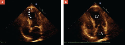
In a patient with reduced ejection fraction, the systolic image (A) reveals septal apical akinesis (arrows) and hypokinesis in the remainder of the left ventricle. The diastolic image (B) is unremarkable.
Normalization of EF is not a cure
In treating patients with HF, it is crucial to distinguish between what is reversible and what is not. Diuretics, oxygen, and other supportive therapy may reverse symptoms of congestion. Reversible causes of HF may include alcohol toxicity, thyroid disease, tachycardia, anemia, valvular heart disease, and CAD, among others. However, reversing the symptoms or the cause does not necessarily reverse HF itself. Further, normalization of EF does not necessarily imply that HF has been cured.
In a prospective study of 42 HF patients whose EF had normalized, the aggregate initial EF was 26%. It increased to ≥40%, with an absolute increase in EF ≥10%. During 41 months of follow-up, 19% of the patients had a recurrence of reduced EF. The likelihood of recurrence was greater among those who had discontinued their HF medications, the researchers found.17
In another prospective study, researchers followed 110 patients with reduced EF who were managed medically according to guideline-prescribed therapy.18 During a 17-month follow-up period, 18% had a normal EF at some point—but in more than half the cases (55%), the improvement was transient. Factors that were predictive of normalization included the presence of arterial hypertension (odds ratio [OR]=8.5; P=.01), nonischemic etiology (OR=4.9; P=.02), the absence of diabetes (OR=9.5; P=.01), beta-blocker therapy with carvedilol (OR=3.9, P=.02), and a higher beta-blocker dosage (OR=1.1; P=.04). Normalization occurred, on average, at 13 months (±6 months). The only difference between those who had a sustained improvement and those for whom the normalization was transient was the rate of chronic obstructive pulmonary disease (COPD). None of the patients who had a sustained improvement had COPD; 36% of those with transient improvement did (P=.04).
Finally, researchers used an Italian registry to follow prospectively 581 patients with dilated cardiomyopathy who were enrolled over a 25-year period.19 The team found that “healing” (reverse remodeling) occurred in 16% of the patients in response to treatment with ACE inhibitors and beta-blockers. Thus, in the vast majority of patients, the underlying disorder that caused the cardiomyopathy was maintained.
Keep patients on the same meds
There are no prospective RCTs investigating the continuation of ACE inhibitors, beta-blockers, or other therapy among HF patients whose EF normalized in response to treatment. Given the dramatic benefit of these medications for patients with a reduced EF, no such trial is likely to be performed. The trials noted above are instructive, however, and show that maintenance of HF medications17 (and the absence of COPD18) are predictors of sustained improvement.
Other factors to consider: Diastolic dysfunction is an ill-defined condition, and normalization of EF does not necessarily restore a patient to the same status as that of someone who never had a reduced EF. In addition, many patients with a reduced EF have concomitant CAD. In such cases, beta-blockers and ACE inhibitors are indicated as part of secondary prevention—another reason for continuation of the medication regimen, despite EF normalization.
CASE That was true for Joe, who suffered from a host of comorbidities, including CAD, as well as HF, and had been hospitalized for chest pain. His negative stress echocardiogram and improved EF suggested—although neither was definitive evidence—that his chest pain may have had a noncardiac cause. At his postdischarge follow-up visit, he was not experiencing any additional pain.
Despite Joe’s improved EF, however, his medical regimen remains unchanged. He comes in every 1 to 2 months for surveillance.
CORRESPONDENCE
William E. Chavey, MD, MS, University of Michigan, 1500 East Medical Center Drive, L2003 Women’s Hospital, Ann Arbor, Mich 48109-5239; [email protected]
1. Schocken DD, Benjamin EJ, Fonarow GC, et al. American Heart Association Council on Epidemiology and Prevention; American Heart Association Council on Clinical Cardiology; American Heart Association Council on Cardiovascular Nursing; American Heart Association Council on High Blood Pressure Research; Quality of Care and Outcomes Research Interdisciplinary Working Group; Functional Genomics and Translational Biology Interdisciplinary Working Group Prevention of heart failure: a scientific statement from the American Heart Association Councils on Epidemiology and Prevention, Clinical Cardiology, Cardiovascular Nursing, and High Blood Pressure Research; Quality of Care and Outcomes Research Interdisciplinary Working Group; and Functional Genomics and Translational Biology Interdisciplinary Working Group. Circulation. 2008;117:2544-2565.
2. Quinones MA. Assessment of diastolic dysfunction. Prog Cardiovasc Dis. 2005;47:340-355
3. Smiseth OA, Thompson CR. Atrioventricular filling dynamics, diastolic dysfunction and dysfunction. Heart Fail Rev. 2000;5:291-299.
4. Pinamonti B, Zecchin M, DiLenarda A, et al. Persistence of restrictive left ventricular filling pattern in dilated cardiomyopathy: an ominous prognostic sign. J Am Coll Cardiol. 1997;29:604-612.
5. Yamamoto K, Sakata Y, Ohtani T, et al. Heart failure with preserved ejection fraction—what is known and unknown. Circ J. 2009;73:404-410.
6. Task Force for Diagnosis and Treatment of Acute and Chronic Heart Failure 2008 of European Society of Cardiology, Dickstein K, Cohen-Solal A, Filippatos G, et al. ESC Guidelines for the diagnosis and treatment of acute and chronic heart failure 2008. Eur Heart J. 2008;29:2388-2442.
7. Jessup M, Abraham WT, Casey DE, et al. Focused update: ACCF/AHA guidelines for the diagnosis and management of heart failure in adults: a report of the American College of Cardiology Foundation/American Heart Association Task Force on Practice Guidelines: developed in collaboration with the International Society for Heart and Lung Transplantation. Circulation. 2009;199:1977-2016
8. Chavey WE, Bleske BE, Van Harrison R, et al. Pharmacologic management of heart failure caused by systolic dysfunction. Am Fam Physician. 2008;77:957-968.
9. Joint Commission Heart failure core measure set. Oak-brook Park, IL. Available at: http://www.jointcommission.org/PerformanceMeasurement/PerformanceMeasurement/Heart+Failure+
Core+Measure+Set.htm. Accessed February 5, 2010.
10. Yusuf S, Pfeffer MA, Swedberg K, et al. Effects of candesartan in patients with chronic heart failure and preserved ejection fraction: the CHARM-Preserved Trial. Lancet. 2003;362:777-781.
11. Moreo A de Chiara B, Cataldo G, et al. Prognostic value of serial measurements of left ventricular function and exercise performance in chronic heart failure [in Spanish]. Rev Esp Cardiol. 2006;59:905-910.
12. Metra M, Nodari S, Parrinello G, et al. Marked improvement in left ventricular ejection fraction during long-term beta blockade in patients with chronic heart failure: clinical correlates and prognostic significance. Am Heart J. 2003;145:292-299.
13. Swedberg K, Cleland J, Dargie H, et al. For the Task Force for the Diagnosis and Treatment of Chronic Heart Failurwe of the European Society of Cardiology. Guidelines for the diagnosis and treatment of chronic heart failure: executive summary (update 2005). Eur Heart J. 2005;26:1115-1140.
14. Malm S, Frigstad S, Sagberg E, et al. Accurate and reproducible measurement of left ventricular volume and ejection fraction by contrast echocardiography. J Am Coll Cardiol. 2004;44:1030-1035.
15. McGowan JH, Cleland J. Reliability of reporting left ventricular systolic function by echocardiography: a systematic review of three methods. Am Heart J. 2003;146:388-397.
16. Lang RM, Bierig M, Devereux RB, et al. Recommendations for chamber quantification: a report from the American Society of Echocardiography’s Guidelines and Standards Committee and the Chamber Quantification Writing Group, developed in conjunction with the European Association of Echocardiography. J Am Soc Echocardiogr. 2005;18:1440-1463.
17. Moon J, Ko YG, Chung N, et al. Recovery and recurrence of left ventricular systolic dysfunction in patients with idiopathic dilated cardiomyopathy. Can J Cardiol. 2009;25:e147-e150.
18. Cioffi G, Stefenelli C, Tarantini L, et al. Chronic left ventricular failure in the community: prevalence, prognosis, and predictors of the complete clinical recovery with return of cardiac size and function to normal in patients undergoing optimal therapy. J Card Fail. 2004;10:250-257.
19. Di Lenarda A, Sabbadini G, Perkan A, et al. Apparent healing in dilated cardiomyopathy: incidence, long-term persistence and predictive factors. The heart muscle disease registry of Trieste [abstract]. Ital Heart J. 2001;2(suppl 2):S97.-
• Assess the ejection fraction (EF) of all heart failure patients, and treat those with reduced EF according to established guidelines. A
• Reassess EF only when the clinical situation demands it; there is no need for routine EF surveillance. B
• Continue to treat patients with heart failure medications even after their EF has normalized. C
Strength of recommendation (SOR)
A Good-quality patient-oriented evidence
B Inconsistent or limited-quality patient-oriented evidence
C Consensus, usual practice, opinion, disease-oriented evidence, case series
CASE Joe H is a 64-year-old African American man with a history of heart failure, hypertension, coronary artery disease (CAD), type 2 diabetes, chronic kidney disease, atrial fibrillation, and gout. His ejection fraction (EF), measured several years ago by echocardiography, was 20%, and he has New York Heart Association class II–III symptoms. Joe is taking an angiotensin-converting enzyme (ACE) inhibitor, a beta-blocker, an aldosterone antagonist, a loop diuretic, a nitrate, and digoxin, and he has an implantable cardioverter defibrillator (ICD). He recently spent 2 days in the hospital after being admitted for chest pain.
As an inpatient, Joe underwent stress echocardiography, which showed no inducible ischemia and an EF of 50%. Should the normalization of Joe’s EF prompt a change in his therapy?
Heart failure (HF), which affects an estimated 5 million Americans, is the leading cause of hospitalization in people older than 65 years.1 The condition—characterized by signs and symptoms of congestion and objective evidence of structural or functional heart disease—has historically been divided into 2 categories: Patients with HF and a reduced ejection fraction (EF) were said to have systolic dysfunction, while the term “diastolic dysfunction” was applied to those with HF and a preserved EF.
The distinction between systolic and diastolic dysfunction is not so simple, though, and the definition of diastolic dysfunction, in particular, is not so clearcut.
Diastolic dysfunction is sometimes described on the basis of echocardiographic criteria, such as the ratio of early-to-late diastolic filling, short deceleration times, and isovolumic relaxation times.2,3 But demographic and physiologic variables make interpretation of these parameters difficult, and the parameters themselves are not uniformly applied. What’s more, echocardiographic evidence of diastolic dysfunction is not specific to HF with a preserved EF.4 Some patients may be shown to have diastolic dysfunction and reduced EF.
As understanding of these variations grows, momentum about the need to change the clinical terminology has begun to develop. The suggested revision is simply to distinguish between HF with reduced EF and HF with preserved EF.5
To provide the best possible care for HF patients—including those who, like Joe, have gone from a reduced to a normal EF after receiving aggressive treatment—you need to be familiar with these changing parameters, recent research findings, and implications for treatment.
Managing both types of HF: What the evidence shows
Evidence-based management of HF with reduced EF is distinctly different from that of HF with preserved EF (TABLE).6-8 In fact, the vast majority of the evidence involves patients with reduced EF, as most randomized clinical trials (RCTs)—and the only trials demonstrating a reduction in mortality—have excluded patients with preserved EF. Thus, in diagnosing and treating HF patients, it is crucial to assess for, and to distinguish between, the 2 EF states. Documentation of this assessment is a core quality measure for HF management, according to the Joint Commission.9
Treating HF with reduced EF. Barring any contraindications, ACE inhibitors and beta-blockers are core treatments for patients with reduced EF.8 Aldosterone antagonists are also indicated for patients with reduced EF who have, or recently had, rest dyspnea. They are also indicated for patients with reduced EF who are 3 to 14 days post-MI and have diabetes or symptomatic HF.8 Nitrates are indicated for African American patients who have persistent symptoms despite treatment with ACE inhibitors, beta-blockers, and diuretics, as needed.8 Consider an ICD as well, as these devices have been found to significantly reduce the risk of death for patients who have an EF <35% with either ischemic cardiomyopathy or symptomatic HF.
Treating HF with preserved EF. Because of the dearth of trials involving patients with HF and a preserved EF, there is limited evidence-based treatment. Nonetheless, it is reasonable to control signs and symptoms of congestion with diuretics.6 In addition, the CHARM-Preserved trial demonstrated the efficacy of candesartan—an angiotensin receptor blocker—in decreasing rates of hospital admissions among symptomatic patients with a preserved EF.10
TABLE
Heart failure: Ejection fraction status dictates treatment6-8
| Intervention | Indications in patients with reduced EF (evidence supporting its use) | Indications in patients with preserved EF (evidence supporting its use) |
|---|---|---|
| ACEIs | •All patients (reduced mortality) | No evidence |
| Beta-blockers | • All symptomatic patients (reduced mortality) | No evidence |
| Aldosterone antagonists | • Rest dyspnea • Post-MI with diabetes or symptomatic HF (reduced mortality) | No evidence |
| Nitrates | • African American patients with persistent symptoms despite treatment with ACEIs, beta-blockers, and diuretics • Intolerance to ACEI/ARBs due to renal impairment (reduced mortality) | No evidence |
| Diuretics | As needed for fluid overload | As needed for fluid overload |
| ARBs | • Intolerance to ACEIs due to cough • Consider for patients with ACEI intolerance due to angioedema* (reduced mortality) | Symptomatic patients (reduced hospitalization rates) |
| Digoxin | • Persistent symptoms despite background therapy • HF and atrial fibrillation (reduced hospitalization rates) | No evidence |
| ICDs | • EF <35% and ischemic cardiomyopathy or symptomatic HF (reduced mortality) | No evidence |
| *There is a possibility of cross-reactivity. | ||
| ACEIs, angiotensin-converting enzyme inhibitors; ARBs, angiotensin receptor blockers; EF, ejection fraction; HF, heart failure; ICDs, implantable cardioverter defibrillators; MI, myocardial infarction. | ||
How to treat the patient with normalized EF
These 2 options, however, do not clearly address the question of what to do with patients like Joe, whose case is described in our opener. Should patients whose EF has normalized after months, or years, of aggressive treatment remain on the medication regimen they followed when they had reduced EF? Should they be treated as HF patients with preserved EF? Is there another option? How often should patients who initially had a reduced EF be reassessed? To answer these questions, let’s take a closer look at the evidence.
Measurement of EF. No large clinical trials have investigated the impact of serial measurement of EF. Two small studies showed prognostic significance with serial measurements that demonstrated improvements in EF,11,12 but their reproducibility and clinical significance are unclear. The American College of Cardiology/American Heart Association and the European Society of Cardiology recommend repeat measurement of EF only when it is clinically indicated.6,13
Accuracy of the results. Echocardiogram is among the most widely used cardiac imaging modalities because it is fast, portable, and noninvasive. However, physiologic limitations and the challenges of calculating a 3-dimensional parameter with 2-dimensional images (FIGURE) limit the usefulness of echocardiography for the measurement of EF. Among other things, echocardiography lacks the ability to reliably identify small changes or improvements. Newer techniques—such as contrast or radionuclide ventriculography—have shown improved reliability in early studies.14 Compared with echocardiography, however, ventriculography is more costly and more invasive.
McGowan et al15 conducted a systematic review of studies comparing echocardiography with reference standards of contrast or radionuclide ventriculography. They concluded that no method in general use to calculate EF from echocardiographic images could provide a 95% confidence interval (CI) of <±10% in the measurement of EF. The American Society of Echocardiography has published standards to improve measurement technique and minimize variability in EF measurement.16
FIGURE
Echocardiographic evidence of heart failure? Systolic image tells the story
In a patient with reduced ejection fraction, the systolic image (A) reveals septal apical akinesis (arrows) and hypokinesis in the remainder of the left ventricle. The diastolic image (B) is unremarkable.
Normalization of EF is not a cure
In treating patients with HF, it is crucial to distinguish between what is reversible and what is not. Diuretics, oxygen, and other supportive therapy may reverse symptoms of congestion. Reversible causes of HF may include alcohol toxicity, thyroid disease, tachycardia, anemia, valvular heart disease, and CAD, among others. However, reversing the symptoms or the cause does not necessarily reverse HF itself. Further, normalization of EF does not necessarily imply that HF has been cured.
In a prospective study of 42 HF patients whose EF had normalized, the aggregate initial EF was 26%. It increased to ≥40%, with an absolute increase in EF ≥10%. During 41 months of follow-up, 19% of the patients had a recurrence of reduced EF. The likelihood of recurrence was greater among those who had discontinued their HF medications, the researchers found.17
In another prospective study, researchers followed 110 patients with reduced EF who were managed medically according to guideline-prescribed therapy.18 During a 17-month follow-up period, 18% had a normal EF at some point—but in more than half the cases (55%), the improvement was transient. Factors that were predictive of normalization included the presence of arterial hypertension (odds ratio [OR]=8.5; P=.01), nonischemic etiology (OR=4.9; P=.02), the absence of diabetes (OR=9.5; P=.01), beta-blocker therapy with carvedilol (OR=3.9, P=.02), and a higher beta-blocker dosage (OR=1.1; P=.04). Normalization occurred, on average, at 13 months (±6 months). The only difference between those who had a sustained improvement and those for whom the normalization was transient was the rate of chronic obstructive pulmonary disease (COPD). None of the patients who had a sustained improvement had COPD; 36% of those with transient improvement did (P=.04).
Finally, researchers used an Italian registry to follow prospectively 581 patients with dilated cardiomyopathy who were enrolled over a 25-year period.19 The team found that “healing” (reverse remodeling) occurred in 16% of the patients in response to treatment with ACE inhibitors and beta-blockers. Thus, in the vast majority of patients, the underlying disorder that caused the cardiomyopathy was maintained.
Keep patients on the same meds
There are no prospective RCTs investigating the continuation of ACE inhibitors, beta-blockers, or other therapy among HF patients whose EF normalized in response to treatment. Given the dramatic benefit of these medications for patients with a reduced EF, no such trial is likely to be performed. The trials noted above are instructive, however, and show that maintenance of HF medications17 (and the absence of COPD18) are predictors of sustained improvement.
Other factors to consider: Diastolic dysfunction is an ill-defined condition, and normalization of EF does not necessarily restore a patient to the same status as that of someone who never had a reduced EF. In addition, many patients with a reduced EF have concomitant CAD. In such cases, beta-blockers and ACE inhibitors are indicated as part of secondary prevention—another reason for continuation of the medication regimen, despite EF normalization.
CASE That was true for Joe, who suffered from a host of comorbidities, including CAD, as well as HF, and had been hospitalized for chest pain. His negative stress echocardiogram and improved EF suggested—although neither was definitive evidence—that his chest pain may have had a noncardiac cause. At his postdischarge follow-up visit, he was not experiencing any additional pain.
Despite Joe’s improved EF, however, his medical regimen remains unchanged. He comes in every 1 to 2 months for surveillance.
CORRESPONDENCE
William E. Chavey, MD, MS, University of Michigan, 1500 East Medical Center Drive, L2003 Women’s Hospital, Ann Arbor, Mich 48109-5239; [email protected]
1. Schocken DD, Benjamin EJ, Fonarow GC, et al. American Heart Association Council on Epidemiology and Prevention; American Heart Association Council on Clinical Cardiology; American Heart Association Council on Cardiovascular Nursing; American Heart Association Council on High Blood Pressure Research; Quality of Care and Outcomes Research Interdisciplinary Working Group; Functional Genomics and Translational Biology Interdisciplinary Working Group Prevention of heart failure: a scientific statement from the American Heart Association Councils on Epidemiology and Prevention, Clinical Cardiology, Cardiovascular Nursing, and High Blood Pressure Research; Quality of Care and Outcomes Research Interdisciplinary Working Group; and Functional Genomics and Translational Biology Interdisciplinary Working Group. Circulation. 2008;117:2544-2565.
2. Quinones MA. Assessment of diastolic dysfunction. Prog Cardiovasc Dis. 2005;47:340-355
3. Smiseth OA, Thompson CR. Atrioventricular filling dynamics, diastolic dysfunction and dysfunction. Heart Fail Rev. 2000;5:291-299.
4. Pinamonti B, Zecchin M, DiLenarda A, et al. Persistence of restrictive left ventricular filling pattern in dilated cardiomyopathy: an ominous prognostic sign. J Am Coll Cardiol. 1997;29:604-612.
5. Yamamoto K, Sakata Y, Ohtani T, et al. Heart failure with preserved ejection fraction—what is known and unknown. Circ J. 2009;73:404-410.
6. Task Force for Diagnosis and Treatment of Acute and Chronic Heart Failure 2008 of European Society of Cardiology, Dickstein K, Cohen-Solal A, Filippatos G, et al. ESC Guidelines for the diagnosis and treatment of acute and chronic heart failure 2008. Eur Heart J. 2008;29:2388-2442.
7. Jessup M, Abraham WT, Casey DE, et al. Focused update: ACCF/AHA guidelines for the diagnosis and management of heart failure in adults: a report of the American College of Cardiology Foundation/American Heart Association Task Force on Practice Guidelines: developed in collaboration with the International Society for Heart and Lung Transplantation. Circulation. 2009;199:1977-2016
8. Chavey WE, Bleske BE, Van Harrison R, et al. Pharmacologic management of heart failure caused by systolic dysfunction. Am Fam Physician. 2008;77:957-968.
9. Joint Commission Heart failure core measure set. Oak-brook Park, IL. Available at: http://www.jointcommission.org/PerformanceMeasurement/PerformanceMeasurement/Heart+Failure+
Core+Measure+Set.htm. Accessed February 5, 2010.
10. Yusuf S, Pfeffer MA, Swedberg K, et al. Effects of candesartan in patients with chronic heart failure and preserved ejection fraction: the CHARM-Preserved Trial. Lancet. 2003;362:777-781.
11. Moreo A de Chiara B, Cataldo G, et al. Prognostic value of serial measurements of left ventricular function and exercise performance in chronic heart failure [in Spanish]. Rev Esp Cardiol. 2006;59:905-910.
12. Metra M, Nodari S, Parrinello G, et al. Marked improvement in left ventricular ejection fraction during long-term beta blockade in patients with chronic heart failure: clinical correlates and prognostic significance. Am Heart J. 2003;145:292-299.
13. Swedberg K, Cleland J, Dargie H, et al. For the Task Force for the Diagnosis and Treatment of Chronic Heart Failurwe of the European Society of Cardiology. Guidelines for the diagnosis and treatment of chronic heart failure: executive summary (update 2005). Eur Heart J. 2005;26:1115-1140.
14. Malm S, Frigstad S, Sagberg E, et al. Accurate and reproducible measurement of left ventricular volume and ejection fraction by contrast echocardiography. J Am Coll Cardiol. 2004;44:1030-1035.
15. McGowan JH, Cleland J. Reliability of reporting left ventricular systolic function by echocardiography: a systematic review of three methods. Am Heart J. 2003;146:388-397.
16. Lang RM, Bierig M, Devereux RB, et al. Recommendations for chamber quantification: a report from the American Society of Echocardiography’s Guidelines and Standards Committee and the Chamber Quantification Writing Group, developed in conjunction with the European Association of Echocardiography. J Am Soc Echocardiogr. 2005;18:1440-1463.
17. Moon J, Ko YG, Chung N, et al. Recovery and recurrence of left ventricular systolic dysfunction in patients with idiopathic dilated cardiomyopathy. Can J Cardiol. 2009;25:e147-e150.
18. Cioffi G, Stefenelli C, Tarantini L, et al. Chronic left ventricular failure in the community: prevalence, prognosis, and predictors of the complete clinical recovery with return of cardiac size and function to normal in patients undergoing optimal therapy. J Card Fail. 2004;10:250-257.
19. Di Lenarda A, Sabbadini G, Perkan A, et al. Apparent healing in dilated cardiomyopathy: incidence, long-term persistence and predictive factors. The heart muscle disease registry of Trieste [abstract]. Ital Heart J. 2001;2(suppl 2):S97.-
Chlamydia screening: How we can better serve patients
Detection of Chlamydia trachomatis infection—the most commonly reported bacterial infection in the United States—falls primarily to patients’ personal physicians, not to sexually transmitted disease (STD) clinics or local health departments, as we’ll describe in a bit. And yet, fewer than half of personal physicians routinely screen for it.1
Left untreated or allowed to recur, chlamydial infections are significant causes of pelvic inflammatory disease, chronic pelvic pain, ectopic pregnancy, and infertility. Chlamydia cases reported to the Centers for Disease Control and Prevention (CDC) exceeded 1 million for the first time in 2006, and the CDC estimates that more than 2 million Americans between the ages of 14 and 39 years are infected.2 Most, if not all, of this increase is likely due to increased test sensitivity, expanded screening services and opportunities, and improved reporting, as well as the continuing high disease burden. Recent work suggests there may also be an increase in prevalence in the Pacific Northwest.3
The CDC established screening guidelines for chlamydial infection more than 10 years ago. However, despite long-standing and widely published screening guidelines, most young women who should receive this service do not. This fact has led the US Preventive Services Task Force (USPSTF) to identify chlamydia screening as one of the most important underused clinical preventive services.4,5
USPSTF screening guidelines for chlamydia infection6
- Screen for chlamydial infection in all sexually active nonpregnant women ages 24 and younger, and in older nonpregnant women who are at increased risk. (Grade A recommendation)
- Screen for chlamydial infection in all pregnant women ages 24 and younger, and in older pregnant women who are at increased risk. (Grade B recommendation)
- Routinely screening for chlamydial infection in women ages 25 and older is not recommended, whether or not they are pregnant, if they are not at increased risk. (Grade C recommendation)
- Current evidence is insufficient to assess the balance of benefits and harms of screening for chlamydial infection in men. (Grade I statement)
Most patients are screened by their personal physicians
To help improve screening rates, we must understand the status of screening site availability as well as usage patterns. Using state case and Infertility Prevention Program data, we examined screening sites in Illinois counties (defined as urban or rural) for women who tested positive for chlamydia in the period 2002 through 2006. For both urban and rural counties, more cases were identified by personal physicians than any other site or provider type (39% of all urban cases; 58% of all rural cases). Personal physicians and hospitals, combined, accounted for more than 53% of reported cases in each group. This is important because most chlamydia cases were identified at health care sites other than those that usually receive federal and state funding for chlamydia screening—local health departments and STD clinics. Reliance on these institutions to significantly reduce the chlamydia epidemic is unrealistic.
To address the issue of screening by personal physicians, the CDC’s Division of STD Prevention collaborated with the National Committee for Quality Assurance to develop the “Chlamydia Screening in Women” measure for the Health Care Effectiveness Data and Information Set (HEDIS). Chlamydia screening is now a covered benefit in many managed care plans, but actual screening rates in physician offices are still only approximately 37%.1 What are the barriers that contribute to such a low rate?
Barriers to chlamydia screening
There are many reasons for inconsistent screening, including flaws in training and practice at the individual level and flaws in regional and national health system design and implementation. Following are several barriers that we must overcome to optimize screening:
- Lack of awareness by physicians. The extent of chlamydial infections, locally and nationally, may not be sufficiently understood. Likewise, physicians may lack awareness of screening guidelines and not appreciate their role in detecting infection. Every medical school and training program (even postgraduate) should use the guidelines to reinforce the critical role personal physicians have in addressing this national epidemic. Without consistent and widespread compliance with screening guidelines by physicians, reductions in chlamydia rates are unlikely.
- Uneasy patient-physician relationships. Many patients and their physicians lack relationships that foster open discussions of sexual issues. Adolescents and young adults are notoriously reticent about discussing sexual behavior, and approximately 70% of chlamydial infections in females are asymptomatic.7 Therefore, physicians cannot rely solely on suggestive health histories and clinical presentations to prompt a discussion of chlamydia.
- Wide prevalence of chlamydial infection. While there are significant correlations between minority and socioeconomic status and infection rates, chlamydial infection is too widespread to base screening decisions on these criteria alone. A particularly important factor contributing to the chain of infection is local social-sexual networks. Perpetuation of these networks contributes to sustaining endemic disease levels through infection and reinfection. Interrupting these networks can directly affect the health of a wide circle of individuals.
- Insufficient time and reimbursement. Primary care physicians report significant dissatisfaction with the short time allocated for individual visits and a relative decline in reimbursement. Such time pressure often leads to a focus on chief complaints; health maintenance and screening discussions are often omitted. Future payment mechanisms for care coordination and performance may remedy this problem.
- Inadequate health information technology. The promises of health information technology are often unfulfilled, and office-based electronic health records (EHRs) frequently do not provide point-of-service information that would improve screening compliance. Lack of efficient interfaces for electronic records and databases makes regional health information exchange difficult. Improving electronic communication among health care professionals will likely improve primary and secondary prevention measures.
- Generally poor integration of public health and medicine. The United States spends a smaller portion of its health care budget on public health than most other industrialized nations. Academically, a vast divide exists between most institutions of public health and medicine. And at the community level, there is a lack of integration of public health services and physician practices. The example of chlamydia screening underscores the need for reform of the US health care system to include greater emphasis on, and integration with, public health, preventive medicine, and primary care.
- Deficient access to health care. One-sixth of the US population is without health insurance,8 and many who have insurance lack benefits that fully cover preventive medicine and screening. Reform of the US health care system to provide easy access to care is more likely to improve a broad range of health outcomes than the development of smaller, fragmented programs focused on specific conditions. This, again, has direct implications for chlamydia control and spread via social-sexual networks.
Our recommendations
- Reacquaint yourself with the USPSTF screening guidelines and commit to following them rigorously in practice. This will necessitate examining your relationship with eligible patients, developing mechanisms to regularly discuss sexual health and STD issues, and consistently providing screening.
- Make screening a routine part of care at recommended opportunities. If you use an EHR, consider working with the vendor to construct appropriate automatic prompts. Those with traditional systems may want to include a systematic chart addition and audit.
- Consider becoming a policy advocate. There are serious health system flaws that hinder efforts to stem the chlamydia epidemic. Many of these system flaws are best addressed by state or national policy change and through new incentives for financial rewards for physicians.
- Actively partner with local public health departments to expand screening services to those at risk. A study by Ward demonstrated that increasing screening in communities with endemic disease might have the greatest effect on the local population and its sexual networks.9
CORRESPONDENCE
Wiley D. Jenkins, Department of Family and Community Medicine, Southern Illinois University School of Medicine, 913 N. Rutledge Street, P.O. Box 19671, Springfield, IL 62794-9671; [email protected]
1. National Center for Quality Assurance. Improving Chlamydia Screening: Strategies From Top Performing Health Plans. Atlanta, GA: U.S. Department of Health and Human Services, Centers for Disease Control and Prevention; December 2007.
2. Centers for Disease Control and Prevention. Sexually Transmitted Disease Surveillance 2006 Supplement, Chlamydia Prevalence Monitoring Project Annual Report 2006. Atlanta, GA: U.S. Department of Health and Human Services, Centers for Disease Control and Prevention; December 2007.
3. Fine D, Dicker L, Mosure D, et al. Increasing chlamydia positivity in women screened in family planning clinics: do we know why. Sex Transm Dis. 2008;35:47-52.
4. Coffield AB, Maciosek MV, McGinnis JM, et al. Priorities among recommended clinical preventive services. Am J Prev Med. 2001;21:1-9.
5. Maciosek MV, Coffield AB, Edwards NM, et al. Priorities among effective clinical preventive services: results of a systematic review and analysis. Am J Prev Med. 2006;31:52-61.
6. US Preventive Services Task Force. Screening for chlamydial infection: US Preventive Services Task Force Recommendation Statement. Ann Intern Med. 2007;147:128-133.
7. Centers for Disease Control and Prevention Chlamydia–CDC fact sheet. Available at: http://www.cdc.gov/std/chlamydia/STDFact-Chlamydia.htm. Accessed January 1, 2010.
8. US Census Bureau. Income, poverty, and health insurance coverage in the United States: 2008. Available at: http://www.census.gov/prod/2009pubs/p60-236.pdf. Accessed: December 15, 2009.
9. Ward H. Prevention strategies for sexually transmitted infections: importance of sexual network structure and epidemic phase. Sex Transm Infect. 2007;83(suppl 1):i43-i49.
Detection of Chlamydia trachomatis infection—the most commonly reported bacterial infection in the United States—falls primarily to patients’ personal physicians, not to sexually transmitted disease (STD) clinics or local health departments, as we’ll describe in a bit. And yet, fewer than half of personal physicians routinely screen for it.1
Left untreated or allowed to recur, chlamydial infections are significant causes of pelvic inflammatory disease, chronic pelvic pain, ectopic pregnancy, and infertility. Chlamydia cases reported to the Centers for Disease Control and Prevention (CDC) exceeded 1 million for the first time in 2006, and the CDC estimates that more than 2 million Americans between the ages of 14 and 39 years are infected.2 Most, if not all, of this increase is likely due to increased test sensitivity, expanded screening services and opportunities, and improved reporting, as well as the continuing high disease burden. Recent work suggests there may also be an increase in prevalence in the Pacific Northwest.3
The CDC established screening guidelines for chlamydial infection more than 10 years ago. However, despite long-standing and widely published screening guidelines, most young women who should receive this service do not. This fact has led the US Preventive Services Task Force (USPSTF) to identify chlamydia screening as one of the most important underused clinical preventive services.4,5
USPSTF screening guidelines for chlamydia infection6
- Screen for chlamydial infection in all sexually active nonpregnant women ages 24 and younger, and in older nonpregnant women who are at increased risk. (Grade A recommendation)
- Screen for chlamydial infection in all pregnant women ages 24 and younger, and in older pregnant women who are at increased risk. (Grade B recommendation)
- Routinely screening for chlamydial infection in women ages 25 and older is not recommended, whether or not they are pregnant, if they are not at increased risk. (Grade C recommendation)
- Current evidence is insufficient to assess the balance of benefits and harms of screening for chlamydial infection in men. (Grade I statement)
Most patients are screened by their personal physicians
To help improve screening rates, we must understand the status of screening site availability as well as usage patterns. Using state case and Infertility Prevention Program data, we examined screening sites in Illinois counties (defined as urban or rural) for women who tested positive for chlamydia in the period 2002 through 2006. For both urban and rural counties, more cases were identified by personal physicians than any other site or provider type (39% of all urban cases; 58% of all rural cases). Personal physicians and hospitals, combined, accounted for more than 53% of reported cases in each group. This is important because most chlamydia cases were identified at health care sites other than those that usually receive federal and state funding for chlamydia screening—local health departments and STD clinics. Reliance on these institutions to significantly reduce the chlamydia epidemic is unrealistic.
To address the issue of screening by personal physicians, the CDC’s Division of STD Prevention collaborated with the National Committee for Quality Assurance to develop the “Chlamydia Screening in Women” measure for the Health Care Effectiveness Data and Information Set (HEDIS). Chlamydia screening is now a covered benefit in many managed care plans, but actual screening rates in physician offices are still only approximately 37%.1 What are the barriers that contribute to such a low rate?
Barriers to chlamydia screening
There are many reasons for inconsistent screening, including flaws in training and practice at the individual level and flaws in regional and national health system design and implementation. Following are several barriers that we must overcome to optimize screening:
- Lack of awareness by physicians. The extent of chlamydial infections, locally and nationally, may not be sufficiently understood. Likewise, physicians may lack awareness of screening guidelines and not appreciate their role in detecting infection. Every medical school and training program (even postgraduate) should use the guidelines to reinforce the critical role personal physicians have in addressing this national epidemic. Without consistent and widespread compliance with screening guidelines by physicians, reductions in chlamydia rates are unlikely.
- Uneasy patient-physician relationships. Many patients and their physicians lack relationships that foster open discussions of sexual issues. Adolescents and young adults are notoriously reticent about discussing sexual behavior, and approximately 70% of chlamydial infections in females are asymptomatic.7 Therefore, physicians cannot rely solely on suggestive health histories and clinical presentations to prompt a discussion of chlamydia.
- Wide prevalence of chlamydial infection. While there are significant correlations between minority and socioeconomic status and infection rates, chlamydial infection is too widespread to base screening decisions on these criteria alone. A particularly important factor contributing to the chain of infection is local social-sexual networks. Perpetuation of these networks contributes to sustaining endemic disease levels through infection and reinfection. Interrupting these networks can directly affect the health of a wide circle of individuals.
- Insufficient time and reimbursement. Primary care physicians report significant dissatisfaction with the short time allocated for individual visits and a relative decline in reimbursement. Such time pressure often leads to a focus on chief complaints; health maintenance and screening discussions are often omitted. Future payment mechanisms for care coordination and performance may remedy this problem.
- Inadequate health information technology. The promises of health information technology are often unfulfilled, and office-based electronic health records (EHRs) frequently do not provide point-of-service information that would improve screening compliance. Lack of efficient interfaces for electronic records and databases makes regional health information exchange difficult. Improving electronic communication among health care professionals will likely improve primary and secondary prevention measures.
- Generally poor integration of public health and medicine. The United States spends a smaller portion of its health care budget on public health than most other industrialized nations. Academically, a vast divide exists between most institutions of public health and medicine. And at the community level, there is a lack of integration of public health services and physician practices. The example of chlamydia screening underscores the need for reform of the US health care system to include greater emphasis on, and integration with, public health, preventive medicine, and primary care.
- Deficient access to health care. One-sixth of the US population is without health insurance,8 and many who have insurance lack benefits that fully cover preventive medicine and screening. Reform of the US health care system to provide easy access to care is more likely to improve a broad range of health outcomes than the development of smaller, fragmented programs focused on specific conditions. This, again, has direct implications for chlamydia control and spread via social-sexual networks.
Our recommendations
- Reacquaint yourself with the USPSTF screening guidelines and commit to following them rigorously in practice. This will necessitate examining your relationship with eligible patients, developing mechanisms to regularly discuss sexual health and STD issues, and consistently providing screening.
- Make screening a routine part of care at recommended opportunities. If you use an EHR, consider working with the vendor to construct appropriate automatic prompts. Those with traditional systems may want to include a systematic chart addition and audit.
- Consider becoming a policy advocate. There are serious health system flaws that hinder efforts to stem the chlamydia epidemic. Many of these system flaws are best addressed by state or national policy change and through new incentives for financial rewards for physicians.
- Actively partner with local public health departments to expand screening services to those at risk. A study by Ward demonstrated that increasing screening in communities with endemic disease might have the greatest effect on the local population and its sexual networks.9
CORRESPONDENCE
Wiley D. Jenkins, Department of Family and Community Medicine, Southern Illinois University School of Medicine, 913 N. Rutledge Street, P.O. Box 19671, Springfield, IL 62794-9671; [email protected]
1. National Center for Quality Assurance. Improving Chlamydia Screening: Strategies From Top Performing Health Plans. Atlanta, GA: U.S. Department of Health and Human Services, Centers for Disease Control and Prevention; December 2007.
2. Centers for Disease Control and Prevention. Sexually Transmitted Disease Surveillance 2006 Supplement, Chlamydia Prevalence Monitoring Project Annual Report 2006. Atlanta, GA: U.S. Department of Health and Human Services, Centers for Disease Control and Prevention; December 2007.
3. Fine D, Dicker L, Mosure D, et al. Increasing chlamydia positivity in women screened in family planning clinics: do we know why. Sex Transm Dis. 2008;35:47-52.
4. Coffield AB, Maciosek MV, McGinnis JM, et al. Priorities among recommended clinical preventive services. Am J Prev Med. 2001;21:1-9.
5. Maciosek MV, Coffield AB, Edwards NM, et al. Priorities among effective clinical preventive services: results of a systematic review and analysis. Am J Prev Med. 2006;31:52-61.
6. US Preventive Services Task Force. Screening for chlamydial infection: US Preventive Services Task Force Recommendation Statement. Ann Intern Med. 2007;147:128-133.
7. Centers for Disease Control and Prevention Chlamydia–CDC fact sheet. Available at: http://www.cdc.gov/std/chlamydia/STDFact-Chlamydia.htm. Accessed January 1, 2010.
8. US Census Bureau. Income, poverty, and health insurance coverage in the United States: 2008. Available at: http://www.census.gov/prod/2009pubs/p60-236.pdf. Accessed: December 15, 2009.
9. Ward H. Prevention strategies for sexually transmitted infections: importance of sexual network structure and epidemic phase. Sex Transm Infect. 2007;83(suppl 1):i43-i49.
Treating dyslipidemia in the high-risk patient
• Clinical trials demonstrate that cardiovascular event rates diminish as low-density lipoprotein (LDL)-cholesterol and apolipoprotein B–containing particles are lowered with statin treatment. A
• The relationship between LDL-cholesterol and cardiovascular events seems to be linear, with no lowest threshold. A
• While LDL-cholesterol remains the primary target for coronary heart disease prevention, non–HDL-cholesterol is an important secondary target in patients with mixed dyslipidemia. A
Strength of recommendation (SOR)
A Good-quality patient-oriented evidence
B Inconsistent or limited-quality patient-oriented evidence
C Consensus, usual practice, opinion, disease-oriented evidence, case series
Dyslipidemia is a primary contributor to coronary heart disease (CHD), the No. 1 cause of death in the United States.1,2 Mortality rates from CHD have declined sharply over the last 3 decades as a result of improvements in acute care and in secondary prevention, notably by means of lipid-lowering statin therapy.3,4 Studies confirm that the decline in cardiovascular events is largely due to decreases in low-density lipoprotein (LDL)-cholesterol and other atherogenic particles.2,5 Still, despite these gains, CHD continues to be a major threat, and the search to find ways to lower CHD risk further continues. Additional avenues being explored include reducing triglycerides and raising high-density lipoprotein (HDL)-cholesterol through lifestyle intervention and other means in patients with mixed dyslipidemia whose statin therapy is already optimal.2 This article reviews the LDL-cholesterol lowering “standard of care” and discusses the potential of addressing other lipoproteins to reduce the residual cardiovascular risk that frequently remains.
Lower LDL levels remain the primary target
The National Cholesterol Education Program (NCEP) has focused on reduction of serum levels of LDL-cholesterol for the primary and secondary prevention of CHD. This approach is biologically reasonable, because LDL is the major atherogenic lipoprotein and is directly implicated in the development of atherosclerosis. Further, the benefit of LDL lowering has been validated by the results of many randomized clinical trials using a combination of lifestyle changes and statins.2
Data on 90,000 patients confirm efficacy of lowering LDL
In an important meta-analysis, the Cholesterol Treatment Trialists’ (CTT) Collaborators compiled data from more than 90,000 people in 14 large-scale, randomized statin trials that included high-risk populations.6 The investigators found that each 39-mg/dL (1-mmol/L) reduction in LDL-cholesterol sustained over 5 years reduced the incidence of a first major coronary event by 23% and the incidence of CHD death by 19%. In the high-risk subgroup with preexisting CHD, a 39-mg/dL (1-mmol/L) reduction in LDL-cholesterol prevented 14 deaths per 1000 participants. These benefits were significant even in the first year of treatment (P<.0001) and were greater in subsequent years.6 The nearly linear relationship between lower LDL-cholesterol levels and fewer major vascular events held true regardless of baseline LDL-cholesterol levels, even when the baseline levels were <100 mg/dL.6
The greatest absolute benefit was noted in the high-risk and very-high-risk groups, especially in individuals with diabetes and those older than 75 years. Moreover, long-term follow-up comparing the original statin-treated participants with the original placebo group showed that lowering LDL-cholesterol continued to reduce cardiovascular risk for 10 years after the study ended.7
Optimal LDL levels may be even lower than we thought
Observational studies have suggested that the relationship between cholesterol and CHD mortality has no apparent lower threshold, and that the physiologic norm for LDL-cholesterol may be lower than that typically seen in Western countries. For example, in a study done in the 1970s in an urban Chinese population of more than 9000 middle-aged men and women, the mean baseline total cholesterol level was 162 mg/dL, and only 7% of deaths were attributed to CHD in 13 years of follow-up.8 Nevertheless, there was an independent and strongly positive (P<.001) relationship between total cholesterol and risk of CHD death, starting at a level as low as 147 mg/dL, which may be equivalent to an LDL-cholesterol of 100 mg/dL. Some data indicate the physiologic norm for LDL-cholesterol levels may be in the range of 50 to 70 mg/dL, which is much lower than the US average of approximately 130 mg/dL.9
Lower LDL-cholesterol protects against atherosclerosis
Lifetime exposure to a lower LDL-cholesterol level may be responsible for a lower burden of atherosclerotic disease later in life. Analyses of data from major statin clinical trials indicate that atherosclerosis does not progress when LDL-cholesterol levels are maintained at <67 mg/dL, while other data suggest that CHD event rates could be minimized at LDL-cholesterol levels of 57 mg/dL for primary prevention and 30 mg/dL for secondary prevention.9
Intensive regimens yield better outcomes
Controlled clinical trials have compared more intensive and less intensive statin and lifestyle modification regimens in high-risk subjects, most of whom already had CHD. These trials found that lower LDL-cholesterol values achieved by more intensive regimens produced incremental CHD benefits.10-16 Major findings of these trials are summarized in TABLE 1.
TABLE 1
Intensive LDL-C lowering in high-risk patients: What the research tells us
| Trial Name | Daily statin treatment; patient population | Mean baseline LDL-C level | Mean achieved LDL-C level,* % reduction | Major findings |
|---|---|---|---|---|
| Antihypertensive and Lipid-Lowering Treatment to Prevent Heart Attack Trial—Lipid-Lowering Trial (ALLHAT-LLT)15 | Pravastatin 40 mg vs usual care; ≥55 y, moderately hypertensive and hypercholesterolemic | 146 mg/dL | 104 mg/dL, 29% (pravastatin); 121 mg/dL, 17% (usual care) | CHD event rates not significantly reduced, except in blacks (27%, P=.03) |
| Anglo-Scandinavian Cardiac Outcomes Trial—Lipid-Lowering Arm (ASCOT-LLA)14 | Atorvastatin 10 mg vs placebo; hypertensive, multiple risk factors | 132 mg/dL | 90 mg/dL, 32% (atorvastatin); 126 mg/dL (placebo) | Atorvastatin 10 mg added to an antihypertensive regimen reduced major CV events by 36% (P=.0005) |
| Heart Protection Study (HPS)13 | Simvastatin 40 mg vs placebo; high-risk coronary or other occlusive arterial disease, or diabetes | 132 mg/dL | 89 mg/dL, 33% (simvastatin); 128 mg/dL (placebo) | Significant 18% decrease in coronary deaths, even in individuals with baseline LDL-C <116 mg/dL |
| Incremental Decrease in End Points through Aggressive Lipid Lowering (IDEAL)11 | Atorvastatin 80 mg vs simvastatin 20 mg; history of MI | 122 mg/dL | 80 mg/dL, 34% (atorvastatin); 100 mg/dL, 17% (simvastatin) | Nonsignificant reduction in primary outcome, but significant reductions in selected secondary outcomes: 13% (P<.02) for major CV events, 16% (P<.001) for any CHD event, 16% (P<.001) for any CV event |
| Prospective Study of Pravastatin in the Elderly at Risk (PROSPER)16 | Pravastatin 40 mg vs placebo; 70-82 y with CVD or at high risk | 147 mg/dL | 97 mg/dL, 34% (pravastatin) | 15% (P=.014) reduction in composite incidence of coronary death, nonfatal MI, and stroke vs placebo |
| Pravastatin or Atorvastatin Evaluation and Infection Therapy—Thrombolysis in Myocardial Infarction 22 (PROVE IT–TIMI 22)12 | Atorvastatin 80 mg vs pravastatin 40 mg; hospitalized for ACS | 106 mg/dL | 62 mg/dL, 42% (atorvastatin, 80 mg); 95 mg/dL, 10% (pravastatin, 40 mg) | Intensive therapy reduced risk of death and major CV events early in treatment vs standard therapy |
| Treating to New Targets (TNT)10 | Atorvastatin 80 mg vs atorvastatin 10 mg; stable CHD | 98 mg/dL | 77 mg/dL, 21% (80 mg); 101 mg/dL, (10 mg) | Intensive therapy reduced rate of major CV events by 22% vs moderate therapy |
| ACS, acute coronary syndromes; CHD, coronary heart disease; CV, cardiovascular; LDL-C, low-density lipoprotein cholesterol; MI, myocardial infarction. | ||||
| *PROVE IT values reflect the median. | ||||
Treating to New Targets (TNT)
After an 8-week run-in period with atorvastatin 10 mg/d, the TNT researchers randomized 10,000 patients with stable CHD and mean baseline LDL-cholesterol levels of 98 mg/dL to atorvastatin 80 mg/d or continued with atorvastatin 10 mg/d.10 Patients in the high-dose group achieved a mean LDL-cholesterol level of 77 mg/dL, which was associated with a 22% relative reduction in risk of a major cardiovascular event (P<.001) and significant reductions in stroke (25%) and cerebrovascular events (23%).10,17
PROVE IT
The Pravastatin or Atorvastatin Evaluation and Infection Therapy–Thrombolysis in Myocardial Infarction 22 (PROVE IT-TIMI 22) trial enrolled 4162 patients who had been hospitalized for acute coronary syndrome within the previous 10 days.12 Patients were randomly assigned to intensive (atorvastatin 80 mg/d) or moderate (pravastatin 40 mg/d) therapy for 24 months—in addition to therapeutic lifestyle interventions. Median LDL-cholesterol levels fell from 106 mg/dL at baseline to 62 mg/dL in the intensive-therapy group and to 95 mg/dL in the standard-therapy group. At 2 years, the primary end point—a composite of cardiovascular events—was 16% lower (P=.005) in patients on intensive therapy than in patients on moderate therapy, with the greatest apparent benefit in those with baseline LDL-cholesterol levels of at least 125 mg/dL.12 Favorable outcomes were more closely related to the on-treatment levels of LDL-cholesterol and C-reactive protein than to the specific agent used.18
Taken together, the TNT and PROVE IT trials show that in high-risk patients with CHD, achieving LDL-cholesterol levels of 60 to 80 mg/dL results in better outcomes than regimens that achieve LDL-cholesterol levels of approximately 100 mg/dL.
Moving the goal posts
The 2002 NCEP Adult Treatment Panel III (ATP III) guidelines recommend LDL-cholesterol goals depending on the patient’s level of risk, with <100 mg/dL as the goal for those in the highest risk category.2 Statin-treated patients in the Heart Protection Study (HPS) achieved a mean LDL-cholesterol level of 89 mg/dL, and investigators reported a “highly significant” 18% reduction in coronary deaths (P=.0005), even in individuals who entered the study with baseline LDL-cholesterol level of <116 mg/dL.13 No indication of a threshold effect was found. For that reason, the HPS investigators suggested that reducing LDL-cholesterol still further with dietary and statin therapy might produce even greater reductions in cardiovascular events.13
In 2004, <70 was a “therapeutic option”
The 2004 update of the NCEP guidelines took into account the findings of the HPS and several other statin trials—most of them secondary prevention studies—that provided further evidence for the benefit of lowering LDL-cholesterol to levels well below 100 mg/dL.12-16,19 The mean achieved LDL-cholesterol levels in these trials and the impact on CHD events are summarized in TABLE 1. The more intensive vs less intensive LDL-cholesterol lowering trials discussed earlier provided evidence that reducing LDL-cholesterol levels to <70 mg/dL is a “therapeutic option” for people at very high CHD risk. The “very-high-risk” category includes those with established cardiovascular disease and additional risk factors such as diabetes mellitus, continued cigarette smoking, metabolic syndrome, and acute coronary syndrome.19 TABLE 2 summarizes the 2004 NCEP goals.
TABLE 2
NCEP risk categories and LDL-cholesterol goals2,19
| Risk category | 10-Year CHD risk | LDL-C goal | Initiate drug therapy |
|---|---|---|---|
| High and very high risk Established CHD and/or CHD risk equivalents | >20% | <100 mg/dL; <70 mg/dL is a reasonable option | ≥100 mg/dL (<100 mg/dL: consider drug options) |
| Moderately high risk Multiple (2+) risk factors | 10%-20% | <130 mg/dL (optional: <100 mg/dL) | ≥130 mg/dL (100-129 mg/dL: consider drug options) |
| Moderate risk Multiple (2+) risk factors | <10% | <130 mg/dL | ≥160 mg/dL |
| Lower risk 0-1 risk factor | <10% | <160 mg/dL | ≥190 mg/dL (160-189 mg/dL: consider drug options) |
| CHD, coronary heart disease; LDL-C, low-density lipoprotein cholesterol; NCEP, National Cholesterol Education Program. | |||
In 2006, <70 became a “reasonable goal”
Guidelines for secondary prevention jointly issued by the American Heart Association and the American College of Cardiology in 2006 and endorsed by the National Heart, Lung, and Blood Institute (NHLBI) agree that a goal of <70 mg/dL is “reasonable” for all patients with CHD and other clinical forms of atherosclerotic disease, even those whose baseline LDL-cholesterol level is between 70 and 100 mg/dL.5
Lowering LDL may cause atherosclerosis to regress
Intensive lipid lowering has shown promise in inducing regression of atherosclerotic plaque.20,21 The Reversal of Atherosclerosis with Aggressive Lipid Lowering (REVERSAL) trial compared the effects on atheroma volume, as measured by intracoronary intravascular ultrasound, of intensive (atorvastatin 80 mg/d) vs moderate (pravastatin 40 mg/d) lipid lowering over 18 months in patients who had 1 or more vessels with a luminal narrowing of 20% or more.20 In the intensive treatment group, which attained a mean LDL-cholesterol level of 79 mg/dL, the 0.4% reduction in atheroma volume indicated no disease progression from baseline and a significantly lower progression rate (P=.02). By contrast, the group on moderate treatment that achieved a mean LDL-cholesterol level of 110 mg/dL had a 2.7% increase in atheroma volume, indicating net progression of atheroma volume compared with baseline.20
ASTEROID shows actual regression
A study of the effect of rosuvastatin on disease progression (A Study to Evaluate the Effect of Rosuvastatin on Intravascular Ultrasound-Derived Coronary Atheroma Burden, or ASTEROID) evaluated the effect of rosuvastatin 40 mg/d on coronary disease progression assessed by intravascular ultrasound at baseline and after 24 months, with the patients serving as their own controls.21 The results showed the mean change in percent atheroma volume (PAV), a measure of disease progression and regression, for the entire vessel being measured was –0.98%, compared with baseline (P<.001). A second efficacy measure, change in atheroma volume in the 10-mm subsegment with the greatest disease severity, also showed a reduction, with a mean change of –6.1 mm3 compared with baseline (P<.001).21 The ASTEROID investigators attributed disease regression to intensive statin treatment, leading to an LDL-cholesterol mean of 61 mg/dL together with significantly increasing HDL-cholesterol levels to 49 mg/dL, up 5% from baseline.21
Combination therapy fails to ENHANCE atherosclerosis regression
In a controversial study in patients with familial hypercholesterolemia utilizing B-mode ultrasound measurements of carotid intima-media thickness, lowered LDL-cholesterol levels did not result in regression of atherosclerosis. The Ezetimibe and Simvastatin in Hypercholesterolemia Enhances Atherosclerosis Regression (ENHANCE) trial evaluated simvastatin 80 mg plus ezetimibe 10 mg compared with simvastatin 80 mg alone.22 Despite a significant 16.5% greater lowering of LDL-cholesterol with combination therapy (P<.01), no difference was observed in progression of carotid intima-media thickness between the 2 treatment groups.
No LDL is too low for safety
Given the physiologic importance of cholesterol in the body, the very low cholesterol levels achieved with intensive statin therapy in some trials has raised questions about the safety of such an approach.23 A substudy of the PROVE IT-TIMI 22 trial focused on the 11% of 1825 atorvastatin-treated patients whose LDL-cholesterol levels had dropped to 40 mg/dL or lower after 4 months of treatment. There were fewer cardiovascular events in this group compared with the patients with LDL-cholesterol levels between 80 and 100 mg/dL, and no relationship between this low level and adverse events over 24 months.23 Similarly, the TNT study group analyzed cardiovascular events across quintiles of LDL-cholesterol and found that the lowest quintile (LDL <64 mg/dL, mean 54 mg/dL) had the lowest event rate, without a difference in adverse events over 5 years.24
LDL isn’t the whole story
It is clear from the statin clinical trials that cardiovascular events occur even after LDL-cholesterol is optimally treated. Why is this so? One possibility is that levels of other lipids—too-high triglycerides or too-low HDL-cholesterol—also contribute to CHD risk. These lipid abnormalities often cluster with other risk factors, including obesity, insulin resistance, hyperglycemia, and hypertension. Such patients are considered to have mixed, or atherogenic, dyslipidemia, and frequently include those with metabolic syndrome and type 2 diabetes. In patients whose triglyceride levels remain high (>200 mg/dL) or HDL-cholesterol levels low (<40 mg/dL) even after they have achieved their LDL-cholesterol goals, the NCEP ATP III guidelines recommend non–HDL-cholesterol as a secondary target of therapy.2 Non–HDL-cholesterol (calculated as total cholesterol minus HDL-cholesterol) is a measure of all the atherogenic, apolipoprotein B-containing lipoproteins (LDL, intermediate-density lipoprotein [IDL], and very-low-density lipoprotein [VLDL]).
In mixed, or atherogenic, dyslipidemia, the LDL particles are usually smaller and the calculated LDL-cholesterol content does not reflect the increased particle number. Several observational studies suggest that non–HDL-cholesterol is a better predictor of risk at any given LDL-cholesterol level.
The IDEAL predictor
To highlight the predictive value of non–HDL-cholesterol, Kastelein and colleagues analyzed pooled data from TNT and IDEAL (Incremental Decrease in End Points through Aggressive Lipid Lowering). IDEAL was a large (N=8888), randomized clinical trial in which patients with established CHD were assigned to usual-dose or high-dose statin treatment.25 In the investigators’ statistical models, while LDL-cholesterol levels were positively associated with cardiovascular outcome, that relationship turned out to be less significant than the relationship with non–HDL-cholesterol and apolipoprotein B. The ratio of total cholesterol to HDL (Total/HDL) and the ratio of apolipoprotein B to apolipoprotein A-I were each more closely associated with outcome than any of the individual proatherogenic lipoprotein parameters.25
Another post hoc analysis of TNT data has shown that HDL-cholesterol levels in patients receiving statins predicted major cardiovascular events. Among subjects with LDL-cholesterol levels <70 mg/dL, those in the highest quintile of HDL-cholesterol were at less risk for major cardiovascular events than those in the lowest quintile (P=.03).26 Both of these analyses support the concept that there is residual CHD risk after optimal statin treatment, and that the easily obtained non–HDL-cholesterol and HDL-cholesterol levels are predictive of that risk.
Setting goals for non–HDL-cholesterol
The ATP III–recommended goal for non–HDL-cholesterol is 30 mg/dL above the LDL goal, since maximum acceptable cholesterol carried in the triglyceride-rich lipoproteins (VLDL/IDL) is one-fifth of the acceptable triglyceride level (150/5=30 mg/dL). Thus, a high-risk person whose LDL-cholesterol goal is <100 mg/dL would have a non–HDL-cholesterol goal of <130 mg/dL. ATP III recommends lowering non–HDL-cholesterol by intensifying statin therapy to further reduce LDL as well as considering the addition of niacin or a fibrate to further decrease VLDL and triglycerides.2 Although not specifically recommended by ATP III, omega-3 fatty acids at a sufficient dose (3-4 g/d of ecosapentanoic acid + decosahexanoic acid) can reduce triglycerides as monotherapy, or when added to statins.27
Total atherogenic particles
A 2008 consensus conference report from the American Diabetes Association and the American College of Cardiology states that in patients with high cardiometabolic risk, LDL-cholesterol levels alone do not adequately capture risk and that measurements of total atherogenic particles are better.28 These measurements include non–HDL-cholesterol, apolipoprotein B, and the number of LDL particles identified by nuclear magnetic resonance. In individuals in the highest-risk category (known clinical cardiovascular disease or diabetes plus 1 or more CHD risk factors in addition to dyslipidemia), the report recommends a non–HDL-cholesterol goal of <100 mg/dL and an apolipoprotein B goal of <80 mg/dL.28
Combining therapies: AIM-HIGH and ACCORD
Two ongoing trials are comparing combination therapy (statin with either niacin or a fibrate) with statin therapy alone in patients with atherogenic dyslipidemia to assess the incremental benefit of combination therapy. AIM-HIGH (Atherothrombosis Intervention in Metabolic syndrome with low HDL/high triglycerides and Impact on Global Health outcomes) is a 5-year study in 3300 patients with vascular disease and low HDL-cholesterol. This study is designed to find out whether lowering LDL to <80 mg/dL with simvastatin plus niacin can delay the time to a first major cardiovascular event for longer than simvastatin therapy alone.29
The 6-year ACCORD trial (Action to Control Cardiovascular Risk in Diabetes) randomizes patients with type 2 diabetes into 2 groups, 1 receiving statin-fibrate combination therapy and the other statin monotherapy. ACCORD is designed to find out whether raising HDL-cholesterol and lowering triglycerides along with targeted reductions in LDL-cholesterol will improve CHD outcomes more than LDL lowering alone.29
Raising HDL-cholesterol is promising, but complex
Improving residual CHD risk after statin treatment has emphasized raising HDL-cholesterol as a therapeutic target. The validity of raising HDL-cholesterol is supported by epidemiologic evidence showing an inverse relationship between HDL-cholesterol levels and cardiovascular risk: an increase of 1 mg/dL in HDL-cholesterol is associated with a 2% to 3% decrease in risk of cardiovascular disease.30 Therapeutic lifestyle changes, such as weight loss, exercise, and smoking cessation are effective at increasing HDL-cholesterol and these interventions are always encouraged. Most statins modestly (5%-10%) increase HDL-cholesterol, with rosuvastatin generally producing the largest increases, as shown in the ASTEROID study.
Niacin raises HDL
Currently, the most efficacious HDL-raising drug is niacin. As monotherapy, niacin can increase HDL-cholesterol by 15% to 35%.2 The problem is that niacin often causes flushing, a side effect patients find unpleasant enough that they don’t continue therapy.31 Extended-release preparations cause less flushing than immediate-release forms of niacin, and specific flush-reducing agents (laropiprant) are under investigation to improve tolerance.32
Fibrates, alone and combined, with statins
By activating the nuclear transcription factor peroxisome proliferator-activated receptor-α, fibrates increase HDL-cholesterol by 8% to 35%.33 The Veterans Affairs HDL Intervention Trial (VA-HIT)34 studied the effects of gemfibrozil in men with CHD and HDL-cholesterol <40 mg/dL. After a median follow-up of 5 years, gemfibrozil raised HDL-cholesterol by 6% more than placebo and lowered triglycerides by 31% more (P<.001 for both), but did not affect levels of LDL-cholesterol. Compared to placebo, gemfibrozil treatment reduced the risk of CHD death and nonfatal myocardial infarction by 22% (P=.006). In post hoc analysis, each 5-mg/dL increase in HDL-cholesterol was associated with an 11% decrease in the risk of these CHD events.34
The Helsinki Heart Study35 reported similar results with gemfibrozil in a population without CHD. Fibrates may be combined with statins: small studies using rosuvastatin and fenofibrate36 and atorvastatin and fenofibrate37 have shown positive effects on dyslipidemia. Gemfibrozil may be associated with increased risks of myositis, whereas fenofibrate combined with statins has not shown this effect.38
High hopes, sobering findings, for torcetrapib
The glycoprotein cholesteryl ester transfer protein (CETP) can make HDL particles smaller and more readily removed by the kidneys, with the overall effect of a reduction in HDL-cholesterol.39 Inhibiting this effect, then, should raise HDL levels. Expectations were high for torcetrapib, the first CETP inhibitor, which had been shown to increase HDL-cholesterol by >50% in early clinical trials.40 However, a clinical outcomes trial comparing torcetrapib and atorvastatin with atorvastatin alone was stopped early because the combination therapy was associated with a higher incidence of adverse cardiovascular events, including total mortality.41 Significant increases in average systolic blood pressure with torcetrapib were reported, but it is not clear if this was the cause of the unfavorable outcome. Further, substantial HDL-cholesterol increases of 54% and 61% achieved with torcetrapib in 2 surrogate outcomes trials did not have a beneficial effect on atherosclerosis.42,43
Other CETP inhibitors are currently in development that, investigators hope, will not have the adverse effects associated with torcetrapib.44,45 Additional investigative approaches for increasing HDL-cholesterol levels or increasing reverse cholesterol transport include both intravenous and oral therapies such as apolipoprotein A1-Milano, apolipoprotein A1-mimetic peptides, and phospholipid-directed therapies.46-49
CORRESPONDENCE Peter Jones, MD, 6565 Fannin Street, MSA-601, Suite B157, Houston, TX 77030; [email protected]
1. Rosamond W, Flegal K, Furie K, et al. For the American Heart Association Statistics Committee and Stroke Statistics Subcommittee. Heart disease and stroke statistics 2008 update: a report from the American Heart Association Statistics Committee and Stroke Statistics Subcommittee. Circulation. 2008;117:e25-e146.
2. Expert Panel on Detection, Evaluation, and Treatment of High Blood Cholesterol in Adults. Third Report of the National Cholesterol Education Program (NCEP) Expert Panel on Detection, Evaluation, and Treatment of High Blood Cholesterol in Adults (Adult Treatment Panel III): final report. Circulation. 2002;106:3143-3421.
3. Rosamond WD, Chambless LE, Folsom AR, et al. Trends in the incidence of myocardial infarction and in mortality due to coronary heart disease, 1987 to 1994. N Engl J Med. 1998;339:861-867.
4. Fox CS, Evans JC, Larson MG, et al. Temporal trends in coronary heart disease mortality and sudden cardiac death from 1950 to 1999: the Framingham Heart Study. Circulation. 2004;110:522-527.
5. Smith SC, Jr, Allen J, Blair SN, et al. AHA/ACC guidelines for secondary prevention for patients with coronary and other atherosclerotic vascular disease: 2006 update. J Am Coll Cardiol. 2006;47:2130-2139.
6. Cholesterol Treatment Trialists’ (CTT) Collaborators. Efficacy and safety of cholesterol-lowering treatment: prospective meta-analysis of data from 90 056 participants in 14 randomized trials of statins. Lancet. 2005;366:1267-1278.
7. Ford I, Murray H, Packard CJ, et al. For the West of Scotland Coronary Prevention Study Group. Long-term follow-up of the West of Scotland Coronary Prevention Study. N Engl J Med. 2007;357:1477-1486.
8. Chen Z, Peto R, Collins R, et al. Serum cholesterol concentration and coronary heart disease in population with low cholesterol concentrations. BMJ. 1991;303:276-282.
9. O’Keefe JH, Jr, Cordain L, Harris WH, et al. Optimal low-density lipoprotein is 50 to 70 mg/dL: lower is better and physiologically normal. J Am Coll Cardiol. 2004;43:2142-2146.
10. LaRosa JC, Grundy SM, Waters DD, et al. For the Treating to New Targets (TNT) Investigators. Intensive lipid lowering with atorvastatin in patients with stable coronary disease. N Engl J Med. 2005;352:1425-1435.
11. Pedersen TR, Faergeman O, Kastelein JJP, et al. For the Incremental Decrease in End Points Through Aggressive Lipid Lowering (IDEAL) Study Group. High-dose atorvastatin vs usual-dose simvastatin for secondary prevention after myocardial infarction. The IDEAL Study: a randomized controlled trial. JAMA. 2005;294:2437-2445.
12. Cannon CP, Braunwald E, McCabe C, et al. For the Pravastatin or Atorvastatin Evaluation and Infection Therapy–Thrombolysis in Myocardial Infarction 22 Investigators. Intensive versus moderate lipid lowering with statins after acute coronary syndromes. N Engl J Med. 2004;350:1495-1504.
13. Heart Protection Study Collaborative Group. MRC/BHF Heart Protection Study of cholesterol lowering with simvastatin in 20 536 high-risk individuals: a randomized placebo-controlled trial. Lancet. 2002;360:7-22.
14. Sever PS, Dahlöf B, Poulter NR, et al. For the ASCOT investigators. Prevention of coronary and stroke events with atorvastatin in hypertensive patients who have average or lower-than-average cholesterol concentrations, in the Anglo-Scandinavian Cardiac Outcomes Trial–Lipid Lowering Arm (ASCOT-LLA): a multicentre randomized controlled trial. Lancet. 2003;361:1149-1158.
15. The ALLHAT Officers and Coordinators for the ALLHAT Collaborative Research Group. Major outcomes in moderately hypercholesterolemic, hypertensive patients randomized to pravastatin vs usual care: the Antihypertensive and Lipid-Lowering Treatment to Prevent Heart Attack Trial (ALLHAT-LLT). JAMA. 2002;288:2998-3007.
16. Shepherd J, Blauw GJ, Murphy MB, et al. On behalf of the PROSPER study group. Pravastatin in elderly individuals at risk of vascular disease (PROSPER): a randomized controlled trial. Lancet. 2002;360:1623-1630.
17. Waters DD, LaRosa JC, Barter P, et al. Effects of high-dose atorvastatin on cerebrovascular events in patients with stable coronary disease in the TNT (Treating to New Targets) study. J Am Coll Cardiol. 2006;48:1793-1799.
18. Ridker PM, Cannon CP, Morrow D, et al. For the Pravastatin or Atorvastatin Evaluation and Infection Therapy–Thrombolysis in Myocardial Infarction 22 (PROVE IT–TIMI 22) Investigators. Creactive protein levels and outcomes after statin therapy. N Engl J Med. 2005;352:20-28.
19. Grundy SM, Cleeman JI, Bairey Merz CN, et al. For the Coordinating Committee of the National Cholesterol Education Program. Implications of recent clinical trials for the National Cholesterol Education Program Adult Treatment Panel III guidelines. Circulation. 2004;110:227-239.
20. Nissen SE, Tuzcu EM, Schoenhagen P, et al. For the REVERSAL Investigators. Effect of intensive compared with moderate lipid-lowering therapy on progression of coronary atherosclerosis: a randomized controlled trial. JAMA. 2004;291:1071-1080.
21. Nissen SE, Nicholls SJ, Sipahi I, et al. For the ASTEROID Investigators. Effect of very high-intensity statin therapy on regression of coronary atherosclerosis: the ASTEROID trial. JAMA. 2006;295:1556-1565.
22. Kastelein JJP, Akdim F, Stroes ESG, et al. For the ENHANCE Investigators. Simvastatin with or without ezetimibe in familial hypercholesterolemia. N Engl J Med. 2008;358:1431-1443.
23. Wiviott SD, Cannon CP, Morrow DA, et al. For the PROVE IT–TIMI Investigators. Can low-density lipoprotein be too low? The safety and efficacy of achieving very low low-density lipoprotein with intensive statin therapy: a PROVE IT-TIMI 22 substudy. J Am Coll Cardiol. 2005;46:1411-1416.
24. LaRosa JC, Grundy SM, Kastelein JJ, et al. For the Treating to New Targets (TNT) Steering Committee and Investigators. Safety and efficacy of atorvastatin-induced very low-density lipoprotein cholesterol levels in patients with coronary heart disease (a post hoc analysis of the treating to new targets [TNT] study). Am J Cardiol. 2007;100:747-752.
25. Kastelein JJP, van der Steeg WA, Holme I, et al. For the TNT and IDEAL Study Groups. Lipids, apolipoproteins, and their ratios in relation to cardiovascular events with statin treatment. Circulation. 2008;117:3002-3009.
26. Barter P, Gotto AM, LaRosa JC, et al. For the Treating to New Targets Investigators. HDL cholesterol, very low levels of LDL cholesterol, and cardiovascular events. N Engl J Med. 2007;357:1301-1310.
27. Davidson MH, Stein EA, Bays HE, et al. Efficacy and tolerability of adding prescription omega-3 fatty acids 4 g/d to simvastatin 40 mg in hypertriglyceridemic patients: an 8-week, randomized, double-blind, placebo-controlled study. Clin Ther. 2007;29:1354-1367.
28. Brunzell JD, Davidson M, Furberg CD, et al. Lipoprotein management in patients with cardiometabolic risk: consensus conference report from the American Diabetes Association and the American College of Cardiology Foundation. J Am Coll Cardiol. 2008;51:1512-1524.
29. Miller M. Optimal treatment of dyslipidemia in high-risk patients: intensive statin treatment or combination therapy? Prev Cardiol. 2007;10:31-35.
30. Gordon DJ, Probstfield JL, Garrison RJ, et al. High-density lipoprotein cholesterol and cardiovascular disease: four prospective American studies. Circulation. 1989;79:8-15.
31. McKenney JM. Effect of drugs on high-density lipoprotein. J Clin Lipidol. 2007;1:74-87.
32. Paolini JF, Mitchel YB, Reyes R, et al. Effects of laropiprant on nicotinic acid-induced flushing in patients with dyslipidemia. Am J Cardiol. 2008;101:625-630.
33. Fruchart JC, Staels B, Duriez P. PPARS, metabolic disease, and atherosclerosis. Pharmacol Res. 2001;44:345-352.
34. Robins SJ, Collins D, Wittes JT, et al. Relation of gemfibrozil treatment and lipid levels with major coronary events: VA-HIT: a randomized controlled trial. JAMA. 2001;285:1585-1591.
35. Frick MH, Elo O, Haapa K, et al. Helsinki Heart Study: primary-prevention trial with gemfibrozil in middle-aged men with dyslipidemia. Safety of treatment, changes in risk factors, and incidence of coronary heart disease. N Engl J Med. 1987;317:1237-1245.
36. Durrington PN, Tuomilehto J, Hamann A, et al. Rosuvastatin and fenofibrate alone and in combination in type 2 diabetic patients with combined hyperlipidemia. Diabetes Res Clin Pract. 2004;64:137-151.
37. Athyros VG, Papageorgiou AA, Athyrou VV, et al. Atorvastatin and micronized fenofibrate alone and in combination in type 2 diabetes patients with combined hyperlipidemia. Diabetes Care. 2002;25:1198-1202.
38. Jones PH, Davidson MH. Reporting rate of rhabdomyolysis with fenofibrate + statin versus gemfibrozil + any statin. Am J Cardiol. 2005;95:120-122.
39. Barter PJ, Kastelein JJ. Targeting cholesteryl ester transfer protein for the prevention and management of cardiovascular disease. J Am Coll Cardiol. 2006;47:492-499.
40. Brousseau ME, Schaefer EJ, Wolfe ML, et al. Effects of an inhibitor of cholesteryl ester transfer protein on HDL cholesterol. N Engl J Med. 2004;350:1505-1515.
41. Tall AR, Yvan-Charvet L, Wang N. The failure of torcetrapib: was it the molecule or the mechanism? Arterioscler Thromb Vasc Biol. 2007;27:257-260.
42. Kastelein JJP, van Leuven SI, Burgess L, et al. For the RADIANCE 1 Investigators. Effect of torcetrapib on carotid atherosclerosis in familial hypercholesterolemia. N Engl J Med. 2007;356:1620-1630.
43. Nissen SE, Tardif J-C, Nicholls SJ, et al. For the ILLUSTRATE Investigators. Effect of torcetrapib on the progression of coronary atherosclerosis. N Engl J Med. 2007;356:1304-1316.
44. de Grooth GJ, Kuivenhoven JA, Stalenhoef AF, et al. Efficacy and safety of a novel cholesteryl ester transfer protein inhibitor, JTT-705, in humans: a randomized phase II dose-response study. Circulation. 2002;105:2159-2165.
45. Kuivenhoven JA, de Grooth GJ, Kawamura H, et al. Effectiveness of inhibition of cholesteryl ester transfer protein by JTT-705 in combination with pravastatin in type II dyslipidemia. Am J Cardiol. 2005;95:1085-1088.
46. Nissen SE, Tsunoda T, Tuzcu EM, et al. Effect of recombinant ApoA-1 Milano on coronary atherosclerosis in patients with acute coronary syndromes: a randomized controlled trial. JAMA. 2003;290:2292-2300.
47. Nicholls SJ, Tuzcu EM, Sipahi I, et al. Relationship between atheroma regression and change in lumen size after infusion of apolipoprotein A-1 Milano. J Am Coll Cardiol. 2006;47:992-997.
48. Cesena FH, Faria-Neto JR, Shah PK. Apolipoprotein A-Imimetic peptides: state-of-the-art perspectives. Int J Atheroscler. 2006;1:137-142.
49. Singh IM, Shishehbor MH, Ansell BJ. High-density lipoprotein as a therapeutic target: a systematic review. JAMA. 2007;298:786-798.
• Clinical trials demonstrate that cardiovascular event rates diminish as low-density lipoprotein (LDL)-cholesterol and apolipoprotein B–containing particles are lowered with statin treatment. A
• The relationship between LDL-cholesterol and cardiovascular events seems to be linear, with no lowest threshold. A
• While LDL-cholesterol remains the primary target for coronary heart disease prevention, non–HDL-cholesterol is an important secondary target in patients with mixed dyslipidemia. A
Strength of recommendation (SOR)
A Good-quality patient-oriented evidence
B Inconsistent or limited-quality patient-oriented evidence
C Consensus, usual practice, opinion, disease-oriented evidence, case series
Dyslipidemia is a primary contributor to coronary heart disease (CHD), the No. 1 cause of death in the United States.1,2 Mortality rates from CHD have declined sharply over the last 3 decades as a result of improvements in acute care and in secondary prevention, notably by means of lipid-lowering statin therapy.3,4 Studies confirm that the decline in cardiovascular events is largely due to decreases in low-density lipoprotein (LDL)-cholesterol and other atherogenic particles.2,5 Still, despite these gains, CHD continues to be a major threat, and the search to find ways to lower CHD risk further continues. Additional avenues being explored include reducing triglycerides and raising high-density lipoprotein (HDL)-cholesterol through lifestyle intervention and other means in patients with mixed dyslipidemia whose statin therapy is already optimal.2 This article reviews the LDL-cholesterol lowering “standard of care” and discusses the potential of addressing other lipoproteins to reduce the residual cardiovascular risk that frequently remains.
Lower LDL levels remain the primary target
The National Cholesterol Education Program (NCEP) has focused on reduction of serum levels of LDL-cholesterol for the primary and secondary prevention of CHD. This approach is biologically reasonable, because LDL is the major atherogenic lipoprotein and is directly implicated in the development of atherosclerosis. Further, the benefit of LDL lowering has been validated by the results of many randomized clinical trials using a combination of lifestyle changes and statins.2
Data on 90,000 patients confirm efficacy of lowering LDL
In an important meta-analysis, the Cholesterol Treatment Trialists’ (CTT) Collaborators compiled data from more than 90,000 people in 14 large-scale, randomized statin trials that included high-risk populations.6 The investigators found that each 39-mg/dL (1-mmol/L) reduction in LDL-cholesterol sustained over 5 years reduced the incidence of a first major coronary event by 23% and the incidence of CHD death by 19%. In the high-risk subgroup with preexisting CHD, a 39-mg/dL (1-mmol/L) reduction in LDL-cholesterol prevented 14 deaths per 1000 participants. These benefits were significant even in the first year of treatment (P<.0001) and were greater in subsequent years.6 The nearly linear relationship between lower LDL-cholesterol levels and fewer major vascular events held true regardless of baseline LDL-cholesterol levels, even when the baseline levels were <100 mg/dL.6
The greatest absolute benefit was noted in the high-risk and very-high-risk groups, especially in individuals with diabetes and those older than 75 years. Moreover, long-term follow-up comparing the original statin-treated participants with the original placebo group showed that lowering LDL-cholesterol continued to reduce cardiovascular risk for 10 years after the study ended.7
Optimal LDL levels may be even lower than we thought
Observational studies have suggested that the relationship between cholesterol and CHD mortality has no apparent lower threshold, and that the physiologic norm for LDL-cholesterol may be lower than that typically seen in Western countries. For example, in a study done in the 1970s in an urban Chinese population of more than 9000 middle-aged men and women, the mean baseline total cholesterol level was 162 mg/dL, and only 7% of deaths were attributed to CHD in 13 years of follow-up.8 Nevertheless, there was an independent and strongly positive (P<.001) relationship between total cholesterol and risk of CHD death, starting at a level as low as 147 mg/dL, which may be equivalent to an LDL-cholesterol of 100 mg/dL. Some data indicate the physiologic norm for LDL-cholesterol levels may be in the range of 50 to 70 mg/dL, which is much lower than the US average of approximately 130 mg/dL.9
Lower LDL-cholesterol protects against atherosclerosis
Lifetime exposure to a lower LDL-cholesterol level may be responsible for a lower burden of atherosclerotic disease later in life. Analyses of data from major statin clinical trials indicate that atherosclerosis does not progress when LDL-cholesterol levels are maintained at <67 mg/dL, while other data suggest that CHD event rates could be minimized at LDL-cholesterol levels of 57 mg/dL for primary prevention and 30 mg/dL for secondary prevention.9
Intensive regimens yield better outcomes
Controlled clinical trials have compared more intensive and less intensive statin and lifestyle modification regimens in high-risk subjects, most of whom already had CHD. These trials found that lower LDL-cholesterol values achieved by more intensive regimens produced incremental CHD benefits.10-16 Major findings of these trials are summarized in TABLE 1.
TABLE 1
Intensive LDL-C lowering in high-risk patients: What the research tells us
| Trial Name | Daily statin treatment; patient population | Mean baseline LDL-C level | Mean achieved LDL-C level,* % reduction | Major findings |
|---|---|---|---|---|
| Antihypertensive and Lipid-Lowering Treatment to Prevent Heart Attack Trial—Lipid-Lowering Trial (ALLHAT-LLT)15 | Pravastatin 40 mg vs usual care; ≥55 y, moderately hypertensive and hypercholesterolemic | 146 mg/dL | 104 mg/dL, 29% (pravastatin); 121 mg/dL, 17% (usual care) | CHD event rates not significantly reduced, except in blacks (27%, P=.03) |
| Anglo-Scandinavian Cardiac Outcomes Trial—Lipid-Lowering Arm (ASCOT-LLA)14 | Atorvastatin 10 mg vs placebo; hypertensive, multiple risk factors | 132 mg/dL | 90 mg/dL, 32% (atorvastatin); 126 mg/dL (placebo) | Atorvastatin 10 mg added to an antihypertensive regimen reduced major CV events by 36% (P=.0005) |
| Heart Protection Study (HPS)13 | Simvastatin 40 mg vs placebo; high-risk coronary or other occlusive arterial disease, or diabetes | 132 mg/dL | 89 mg/dL, 33% (simvastatin); 128 mg/dL (placebo) | Significant 18% decrease in coronary deaths, even in individuals with baseline LDL-C <116 mg/dL |
| Incremental Decrease in End Points through Aggressive Lipid Lowering (IDEAL)11 | Atorvastatin 80 mg vs simvastatin 20 mg; history of MI | 122 mg/dL | 80 mg/dL, 34% (atorvastatin); 100 mg/dL, 17% (simvastatin) | Nonsignificant reduction in primary outcome, but significant reductions in selected secondary outcomes: 13% (P<.02) for major CV events, 16% (P<.001) for any CHD event, 16% (P<.001) for any CV event |
| Prospective Study of Pravastatin in the Elderly at Risk (PROSPER)16 | Pravastatin 40 mg vs placebo; 70-82 y with CVD or at high risk | 147 mg/dL | 97 mg/dL, 34% (pravastatin) | 15% (P=.014) reduction in composite incidence of coronary death, nonfatal MI, and stroke vs placebo |
| Pravastatin or Atorvastatin Evaluation and Infection Therapy—Thrombolysis in Myocardial Infarction 22 (PROVE IT–TIMI 22)12 | Atorvastatin 80 mg vs pravastatin 40 mg; hospitalized for ACS | 106 mg/dL | 62 mg/dL, 42% (atorvastatin, 80 mg); 95 mg/dL, 10% (pravastatin, 40 mg) | Intensive therapy reduced risk of death and major CV events early in treatment vs standard therapy |
| Treating to New Targets (TNT)10 | Atorvastatin 80 mg vs atorvastatin 10 mg; stable CHD | 98 mg/dL | 77 mg/dL, 21% (80 mg); 101 mg/dL, (10 mg) | Intensive therapy reduced rate of major CV events by 22% vs moderate therapy |
| ACS, acute coronary syndromes; CHD, coronary heart disease; CV, cardiovascular; LDL-C, low-density lipoprotein cholesterol; MI, myocardial infarction. | ||||
| *PROVE IT values reflect the median. | ||||
Treating to New Targets (TNT)
After an 8-week run-in period with atorvastatin 10 mg/d, the TNT researchers randomized 10,000 patients with stable CHD and mean baseline LDL-cholesterol levels of 98 mg/dL to atorvastatin 80 mg/d or continued with atorvastatin 10 mg/d.10 Patients in the high-dose group achieved a mean LDL-cholesterol level of 77 mg/dL, which was associated with a 22% relative reduction in risk of a major cardiovascular event (P<.001) and significant reductions in stroke (25%) and cerebrovascular events (23%).10,17
PROVE IT
The Pravastatin or Atorvastatin Evaluation and Infection Therapy–Thrombolysis in Myocardial Infarction 22 (PROVE IT-TIMI 22) trial enrolled 4162 patients who had been hospitalized for acute coronary syndrome within the previous 10 days.12 Patients were randomly assigned to intensive (atorvastatin 80 mg/d) or moderate (pravastatin 40 mg/d) therapy for 24 months—in addition to therapeutic lifestyle interventions. Median LDL-cholesterol levels fell from 106 mg/dL at baseline to 62 mg/dL in the intensive-therapy group and to 95 mg/dL in the standard-therapy group. At 2 years, the primary end point—a composite of cardiovascular events—was 16% lower (P=.005) in patients on intensive therapy than in patients on moderate therapy, with the greatest apparent benefit in those with baseline LDL-cholesterol levels of at least 125 mg/dL.12 Favorable outcomes were more closely related to the on-treatment levels of LDL-cholesterol and C-reactive protein than to the specific agent used.18
Taken together, the TNT and PROVE IT trials show that in high-risk patients with CHD, achieving LDL-cholesterol levels of 60 to 80 mg/dL results in better outcomes than regimens that achieve LDL-cholesterol levels of approximately 100 mg/dL.
Moving the goal posts
The 2002 NCEP Adult Treatment Panel III (ATP III) guidelines recommend LDL-cholesterol goals depending on the patient’s level of risk, with <100 mg/dL as the goal for those in the highest risk category.2 Statin-treated patients in the Heart Protection Study (HPS) achieved a mean LDL-cholesterol level of 89 mg/dL, and investigators reported a “highly significant” 18% reduction in coronary deaths (P=.0005), even in individuals who entered the study with baseline LDL-cholesterol level of <116 mg/dL.13 No indication of a threshold effect was found. For that reason, the HPS investigators suggested that reducing LDL-cholesterol still further with dietary and statin therapy might produce even greater reductions in cardiovascular events.13
In 2004, <70 was a “therapeutic option”
The 2004 update of the NCEP guidelines took into account the findings of the HPS and several other statin trials—most of them secondary prevention studies—that provided further evidence for the benefit of lowering LDL-cholesterol to levels well below 100 mg/dL.12-16,19 The mean achieved LDL-cholesterol levels in these trials and the impact on CHD events are summarized in TABLE 1. The more intensive vs less intensive LDL-cholesterol lowering trials discussed earlier provided evidence that reducing LDL-cholesterol levels to <70 mg/dL is a “therapeutic option” for people at very high CHD risk. The “very-high-risk” category includes those with established cardiovascular disease and additional risk factors such as diabetes mellitus, continued cigarette smoking, metabolic syndrome, and acute coronary syndrome.19 TABLE 2 summarizes the 2004 NCEP goals.
TABLE 2
NCEP risk categories and LDL-cholesterol goals2,19
| Risk category | 10-Year CHD risk | LDL-C goal | Initiate drug therapy |
|---|---|---|---|
| High and very high risk Established CHD and/or CHD risk equivalents | >20% | <100 mg/dL; <70 mg/dL is a reasonable option | ≥100 mg/dL (<100 mg/dL: consider drug options) |
| Moderately high risk Multiple (2+) risk factors | 10%-20% | <130 mg/dL (optional: <100 mg/dL) | ≥130 mg/dL (100-129 mg/dL: consider drug options) |
| Moderate risk Multiple (2+) risk factors | <10% | <130 mg/dL | ≥160 mg/dL |
| Lower risk 0-1 risk factor | <10% | <160 mg/dL | ≥190 mg/dL (160-189 mg/dL: consider drug options) |
| CHD, coronary heart disease; LDL-C, low-density lipoprotein cholesterol; NCEP, National Cholesterol Education Program. | |||
In 2006, <70 became a “reasonable goal”
Guidelines for secondary prevention jointly issued by the American Heart Association and the American College of Cardiology in 2006 and endorsed by the National Heart, Lung, and Blood Institute (NHLBI) agree that a goal of <70 mg/dL is “reasonable” for all patients with CHD and other clinical forms of atherosclerotic disease, even those whose baseline LDL-cholesterol level is between 70 and 100 mg/dL.5
Lowering LDL may cause atherosclerosis to regress
Intensive lipid lowering has shown promise in inducing regression of atherosclerotic plaque.20,21 The Reversal of Atherosclerosis with Aggressive Lipid Lowering (REVERSAL) trial compared the effects on atheroma volume, as measured by intracoronary intravascular ultrasound, of intensive (atorvastatin 80 mg/d) vs moderate (pravastatin 40 mg/d) lipid lowering over 18 months in patients who had 1 or more vessels with a luminal narrowing of 20% or more.20 In the intensive treatment group, which attained a mean LDL-cholesterol level of 79 mg/dL, the 0.4% reduction in atheroma volume indicated no disease progression from baseline and a significantly lower progression rate (P=.02). By contrast, the group on moderate treatment that achieved a mean LDL-cholesterol level of 110 mg/dL had a 2.7% increase in atheroma volume, indicating net progression of atheroma volume compared with baseline.20
ASTEROID shows actual regression
A study of the effect of rosuvastatin on disease progression (A Study to Evaluate the Effect of Rosuvastatin on Intravascular Ultrasound-Derived Coronary Atheroma Burden, or ASTEROID) evaluated the effect of rosuvastatin 40 mg/d on coronary disease progression assessed by intravascular ultrasound at baseline and after 24 months, with the patients serving as their own controls.21 The results showed the mean change in percent atheroma volume (PAV), a measure of disease progression and regression, for the entire vessel being measured was –0.98%, compared with baseline (P<.001). A second efficacy measure, change in atheroma volume in the 10-mm subsegment with the greatest disease severity, also showed a reduction, with a mean change of –6.1 mm3 compared with baseline (P<.001).21 The ASTEROID investigators attributed disease regression to intensive statin treatment, leading to an LDL-cholesterol mean of 61 mg/dL together with significantly increasing HDL-cholesterol levels to 49 mg/dL, up 5% from baseline.21
Combination therapy fails to ENHANCE atherosclerosis regression
In a controversial study in patients with familial hypercholesterolemia utilizing B-mode ultrasound measurements of carotid intima-media thickness, lowered LDL-cholesterol levels did not result in regression of atherosclerosis. The Ezetimibe and Simvastatin in Hypercholesterolemia Enhances Atherosclerosis Regression (ENHANCE) trial evaluated simvastatin 80 mg plus ezetimibe 10 mg compared with simvastatin 80 mg alone.22 Despite a significant 16.5% greater lowering of LDL-cholesterol with combination therapy (P<.01), no difference was observed in progression of carotid intima-media thickness between the 2 treatment groups.
No LDL is too low for safety
Given the physiologic importance of cholesterol in the body, the very low cholesterol levels achieved with intensive statin therapy in some trials has raised questions about the safety of such an approach.23 A substudy of the PROVE IT-TIMI 22 trial focused on the 11% of 1825 atorvastatin-treated patients whose LDL-cholesterol levels had dropped to 40 mg/dL or lower after 4 months of treatment. There were fewer cardiovascular events in this group compared with the patients with LDL-cholesterol levels between 80 and 100 mg/dL, and no relationship between this low level and adverse events over 24 months.23 Similarly, the TNT study group analyzed cardiovascular events across quintiles of LDL-cholesterol and found that the lowest quintile (LDL <64 mg/dL, mean 54 mg/dL) had the lowest event rate, without a difference in adverse events over 5 years.24
LDL isn’t the whole story
It is clear from the statin clinical trials that cardiovascular events occur even after LDL-cholesterol is optimally treated. Why is this so? One possibility is that levels of other lipids—too-high triglycerides or too-low HDL-cholesterol—also contribute to CHD risk. These lipid abnormalities often cluster with other risk factors, including obesity, insulin resistance, hyperglycemia, and hypertension. Such patients are considered to have mixed, or atherogenic, dyslipidemia, and frequently include those with metabolic syndrome and type 2 diabetes. In patients whose triglyceride levels remain high (>200 mg/dL) or HDL-cholesterol levels low (<40 mg/dL) even after they have achieved their LDL-cholesterol goals, the NCEP ATP III guidelines recommend non–HDL-cholesterol as a secondary target of therapy.2 Non–HDL-cholesterol (calculated as total cholesterol minus HDL-cholesterol) is a measure of all the atherogenic, apolipoprotein B-containing lipoproteins (LDL, intermediate-density lipoprotein [IDL], and very-low-density lipoprotein [VLDL]).
In mixed, or atherogenic, dyslipidemia, the LDL particles are usually smaller and the calculated LDL-cholesterol content does not reflect the increased particle number. Several observational studies suggest that non–HDL-cholesterol is a better predictor of risk at any given LDL-cholesterol level.
The IDEAL predictor
To highlight the predictive value of non–HDL-cholesterol, Kastelein and colleagues analyzed pooled data from TNT and IDEAL (Incremental Decrease in End Points through Aggressive Lipid Lowering). IDEAL was a large (N=8888), randomized clinical trial in which patients with established CHD were assigned to usual-dose or high-dose statin treatment.25 In the investigators’ statistical models, while LDL-cholesterol levels were positively associated with cardiovascular outcome, that relationship turned out to be less significant than the relationship with non–HDL-cholesterol and apolipoprotein B. The ratio of total cholesterol to HDL (Total/HDL) and the ratio of apolipoprotein B to apolipoprotein A-I were each more closely associated with outcome than any of the individual proatherogenic lipoprotein parameters.25
Another post hoc analysis of TNT data has shown that HDL-cholesterol levels in patients receiving statins predicted major cardiovascular events. Among subjects with LDL-cholesterol levels <70 mg/dL, those in the highest quintile of HDL-cholesterol were at less risk for major cardiovascular events than those in the lowest quintile (P=.03).26 Both of these analyses support the concept that there is residual CHD risk after optimal statin treatment, and that the easily obtained non–HDL-cholesterol and HDL-cholesterol levels are predictive of that risk.
Setting goals for non–HDL-cholesterol
The ATP III–recommended goal for non–HDL-cholesterol is 30 mg/dL above the LDL goal, since maximum acceptable cholesterol carried in the triglyceride-rich lipoproteins (VLDL/IDL) is one-fifth of the acceptable triglyceride level (150/5=30 mg/dL). Thus, a high-risk person whose LDL-cholesterol goal is <100 mg/dL would have a non–HDL-cholesterol goal of <130 mg/dL. ATP III recommends lowering non–HDL-cholesterol by intensifying statin therapy to further reduce LDL as well as considering the addition of niacin or a fibrate to further decrease VLDL and triglycerides.2 Although not specifically recommended by ATP III, omega-3 fatty acids at a sufficient dose (3-4 g/d of ecosapentanoic acid + decosahexanoic acid) can reduce triglycerides as monotherapy, or when added to statins.27
Total atherogenic particles
A 2008 consensus conference report from the American Diabetes Association and the American College of Cardiology states that in patients with high cardiometabolic risk, LDL-cholesterol levels alone do not adequately capture risk and that measurements of total atherogenic particles are better.28 These measurements include non–HDL-cholesterol, apolipoprotein B, and the number of LDL particles identified by nuclear magnetic resonance. In individuals in the highest-risk category (known clinical cardiovascular disease or diabetes plus 1 or more CHD risk factors in addition to dyslipidemia), the report recommends a non–HDL-cholesterol goal of <100 mg/dL and an apolipoprotein B goal of <80 mg/dL.28
Combining therapies: AIM-HIGH and ACCORD
Two ongoing trials are comparing combination therapy (statin with either niacin or a fibrate) with statin therapy alone in patients with atherogenic dyslipidemia to assess the incremental benefit of combination therapy. AIM-HIGH (Atherothrombosis Intervention in Metabolic syndrome with low HDL/high triglycerides and Impact on Global Health outcomes) is a 5-year study in 3300 patients with vascular disease and low HDL-cholesterol. This study is designed to find out whether lowering LDL to <80 mg/dL with simvastatin plus niacin can delay the time to a first major cardiovascular event for longer than simvastatin therapy alone.29
The 6-year ACCORD trial (Action to Control Cardiovascular Risk in Diabetes) randomizes patients with type 2 diabetes into 2 groups, 1 receiving statin-fibrate combination therapy and the other statin monotherapy. ACCORD is designed to find out whether raising HDL-cholesterol and lowering triglycerides along with targeted reductions in LDL-cholesterol will improve CHD outcomes more than LDL lowering alone.29
Raising HDL-cholesterol is promising, but complex
Improving residual CHD risk after statin treatment has emphasized raising HDL-cholesterol as a therapeutic target. The validity of raising HDL-cholesterol is supported by epidemiologic evidence showing an inverse relationship between HDL-cholesterol levels and cardiovascular risk: an increase of 1 mg/dL in HDL-cholesterol is associated with a 2% to 3% decrease in risk of cardiovascular disease.30 Therapeutic lifestyle changes, such as weight loss, exercise, and smoking cessation are effective at increasing HDL-cholesterol and these interventions are always encouraged. Most statins modestly (5%-10%) increase HDL-cholesterol, with rosuvastatin generally producing the largest increases, as shown in the ASTEROID study.
Niacin raises HDL
Currently, the most efficacious HDL-raising drug is niacin. As monotherapy, niacin can increase HDL-cholesterol by 15% to 35%.2 The problem is that niacin often causes flushing, a side effect patients find unpleasant enough that they don’t continue therapy.31 Extended-release preparations cause less flushing than immediate-release forms of niacin, and specific flush-reducing agents (laropiprant) are under investigation to improve tolerance.32
Fibrates, alone and combined, with statins
By activating the nuclear transcription factor peroxisome proliferator-activated receptor-α, fibrates increase HDL-cholesterol by 8% to 35%.33 The Veterans Affairs HDL Intervention Trial (VA-HIT)34 studied the effects of gemfibrozil in men with CHD and HDL-cholesterol <40 mg/dL. After a median follow-up of 5 years, gemfibrozil raised HDL-cholesterol by 6% more than placebo and lowered triglycerides by 31% more (P<.001 for both), but did not affect levels of LDL-cholesterol. Compared to placebo, gemfibrozil treatment reduced the risk of CHD death and nonfatal myocardial infarction by 22% (P=.006). In post hoc analysis, each 5-mg/dL increase in HDL-cholesterol was associated with an 11% decrease in the risk of these CHD events.34
The Helsinki Heart Study35 reported similar results with gemfibrozil in a population without CHD. Fibrates may be combined with statins: small studies using rosuvastatin and fenofibrate36 and atorvastatin and fenofibrate37 have shown positive effects on dyslipidemia. Gemfibrozil may be associated with increased risks of myositis, whereas fenofibrate combined with statins has not shown this effect.38
High hopes, sobering findings, for torcetrapib
The glycoprotein cholesteryl ester transfer protein (CETP) can make HDL particles smaller and more readily removed by the kidneys, with the overall effect of a reduction in HDL-cholesterol.39 Inhibiting this effect, then, should raise HDL levels. Expectations were high for torcetrapib, the first CETP inhibitor, which had been shown to increase HDL-cholesterol by >50% in early clinical trials.40 However, a clinical outcomes trial comparing torcetrapib and atorvastatin with atorvastatin alone was stopped early because the combination therapy was associated with a higher incidence of adverse cardiovascular events, including total mortality.41 Significant increases in average systolic blood pressure with torcetrapib were reported, but it is not clear if this was the cause of the unfavorable outcome. Further, substantial HDL-cholesterol increases of 54% and 61% achieved with torcetrapib in 2 surrogate outcomes trials did not have a beneficial effect on atherosclerosis.42,43
Other CETP inhibitors are currently in development that, investigators hope, will not have the adverse effects associated with torcetrapib.44,45 Additional investigative approaches for increasing HDL-cholesterol levels or increasing reverse cholesterol transport include both intravenous and oral therapies such as apolipoprotein A1-Milano, apolipoprotein A1-mimetic peptides, and phospholipid-directed therapies.46-49
CORRESPONDENCE Peter Jones, MD, 6565 Fannin Street, MSA-601, Suite B157, Houston, TX 77030; [email protected]
1. Rosamond W, Flegal K, Furie K, et al. For the American Heart Association Statistics Committee and Stroke Statistics Subcommittee. Heart disease and stroke statistics 2008 update: a report from the American Heart Association Statistics Committee and Stroke Statistics Subcommittee. Circulation. 2008;117:e25-e146.
2. Expert Panel on Detection, Evaluation, and Treatment of High Blood Cholesterol in Adults. Third Report of the National Cholesterol Education Program (NCEP) Expert Panel on Detection, Evaluation, and Treatment of High Blood Cholesterol in Adults (Adult Treatment Panel III): final report. Circulation. 2002;106:3143-3421.
3. Rosamond WD, Chambless LE, Folsom AR, et al. Trends in the incidence of myocardial infarction and in mortality due to coronary heart disease, 1987 to 1994. N Engl J Med. 1998;339:861-867.
4. Fox CS, Evans JC, Larson MG, et al. Temporal trends in coronary heart disease mortality and sudden cardiac death from 1950 to 1999: the Framingham Heart Study. Circulation. 2004;110:522-527.
5. Smith SC, Jr, Allen J, Blair SN, et al. AHA/ACC guidelines for secondary prevention for patients with coronary and other atherosclerotic vascular disease: 2006 update. J Am Coll Cardiol. 2006;47:2130-2139.
6. Cholesterol Treatment Trialists’ (CTT) Collaborators. Efficacy and safety of cholesterol-lowering treatment: prospective meta-analysis of data from 90 056 participants in 14 randomized trials of statins. Lancet. 2005;366:1267-1278.
7. Ford I, Murray H, Packard CJ, et al. For the West of Scotland Coronary Prevention Study Group. Long-term follow-up of the West of Scotland Coronary Prevention Study. N Engl J Med. 2007;357:1477-1486.
8. Chen Z, Peto R, Collins R, et al. Serum cholesterol concentration and coronary heart disease in population with low cholesterol concentrations. BMJ. 1991;303:276-282.
9. O’Keefe JH, Jr, Cordain L, Harris WH, et al. Optimal low-density lipoprotein is 50 to 70 mg/dL: lower is better and physiologically normal. J Am Coll Cardiol. 2004;43:2142-2146.
10. LaRosa JC, Grundy SM, Waters DD, et al. For the Treating to New Targets (TNT) Investigators. Intensive lipid lowering with atorvastatin in patients with stable coronary disease. N Engl J Med. 2005;352:1425-1435.
11. Pedersen TR, Faergeman O, Kastelein JJP, et al. For the Incremental Decrease in End Points Through Aggressive Lipid Lowering (IDEAL) Study Group. High-dose atorvastatin vs usual-dose simvastatin for secondary prevention after myocardial infarction. The IDEAL Study: a randomized controlled trial. JAMA. 2005;294:2437-2445.
12. Cannon CP, Braunwald E, McCabe C, et al. For the Pravastatin or Atorvastatin Evaluation and Infection Therapy–Thrombolysis in Myocardial Infarction 22 Investigators. Intensive versus moderate lipid lowering with statins after acute coronary syndromes. N Engl J Med. 2004;350:1495-1504.
13. Heart Protection Study Collaborative Group. MRC/BHF Heart Protection Study of cholesterol lowering with simvastatin in 20 536 high-risk individuals: a randomized placebo-controlled trial. Lancet. 2002;360:7-22.
14. Sever PS, Dahlöf B, Poulter NR, et al. For the ASCOT investigators. Prevention of coronary and stroke events with atorvastatin in hypertensive patients who have average or lower-than-average cholesterol concentrations, in the Anglo-Scandinavian Cardiac Outcomes Trial–Lipid Lowering Arm (ASCOT-LLA): a multicentre randomized controlled trial. Lancet. 2003;361:1149-1158.
15. The ALLHAT Officers and Coordinators for the ALLHAT Collaborative Research Group. Major outcomes in moderately hypercholesterolemic, hypertensive patients randomized to pravastatin vs usual care: the Antihypertensive and Lipid-Lowering Treatment to Prevent Heart Attack Trial (ALLHAT-LLT). JAMA. 2002;288:2998-3007.
16. Shepherd J, Blauw GJ, Murphy MB, et al. On behalf of the PROSPER study group. Pravastatin in elderly individuals at risk of vascular disease (PROSPER): a randomized controlled trial. Lancet. 2002;360:1623-1630.
17. Waters DD, LaRosa JC, Barter P, et al. Effects of high-dose atorvastatin on cerebrovascular events in patients with stable coronary disease in the TNT (Treating to New Targets) study. J Am Coll Cardiol. 2006;48:1793-1799.
18. Ridker PM, Cannon CP, Morrow D, et al. For the Pravastatin or Atorvastatin Evaluation and Infection Therapy–Thrombolysis in Myocardial Infarction 22 (PROVE IT–TIMI 22) Investigators. Creactive protein levels and outcomes after statin therapy. N Engl J Med. 2005;352:20-28.
19. Grundy SM, Cleeman JI, Bairey Merz CN, et al. For the Coordinating Committee of the National Cholesterol Education Program. Implications of recent clinical trials for the National Cholesterol Education Program Adult Treatment Panel III guidelines. Circulation. 2004;110:227-239.
20. Nissen SE, Tuzcu EM, Schoenhagen P, et al. For the REVERSAL Investigators. Effect of intensive compared with moderate lipid-lowering therapy on progression of coronary atherosclerosis: a randomized controlled trial. JAMA. 2004;291:1071-1080.
21. Nissen SE, Nicholls SJ, Sipahi I, et al. For the ASTEROID Investigators. Effect of very high-intensity statin therapy on regression of coronary atherosclerosis: the ASTEROID trial. JAMA. 2006;295:1556-1565.
22. Kastelein JJP, Akdim F, Stroes ESG, et al. For the ENHANCE Investigators. Simvastatin with or without ezetimibe in familial hypercholesterolemia. N Engl J Med. 2008;358:1431-1443.
23. Wiviott SD, Cannon CP, Morrow DA, et al. For the PROVE IT–TIMI Investigators. Can low-density lipoprotein be too low? The safety and efficacy of achieving very low low-density lipoprotein with intensive statin therapy: a PROVE IT-TIMI 22 substudy. J Am Coll Cardiol. 2005;46:1411-1416.
24. LaRosa JC, Grundy SM, Kastelein JJ, et al. For the Treating to New Targets (TNT) Steering Committee and Investigators. Safety and efficacy of atorvastatin-induced very low-density lipoprotein cholesterol levels in patients with coronary heart disease (a post hoc analysis of the treating to new targets [TNT] study). Am J Cardiol. 2007;100:747-752.
25. Kastelein JJP, van der Steeg WA, Holme I, et al. For the TNT and IDEAL Study Groups. Lipids, apolipoproteins, and their ratios in relation to cardiovascular events with statin treatment. Circulation. 2008;117:3002-3009.
26. Barter P, Gotto AM, LaRosa JC, et al. For the Treating to New Targets Investigators. HDL cholesterol, very low levels of LDL cholesterol, and cardiovascular events. N Engl J Med. 2007;357:1301-1310.
27. Davidson MH, Stein EA, Bays HE, et al. Efficacy and tolerability of adding prescription omega-3 fatty acids 4 g/d to simvastatin 40 mg in hypertriglyceridemic patients: an 8-week, randomized, double-blind, placebo-controlled study. Clin Ther. 2007;29:1354-1367.
28. Brunzell JD, Davidson M, Furberg CD, et al. Lipoprotein management in patients with cardiometabolic risk: consensus conference report from the American Diabetes Association and the American College of Cardiology Foundation. J Am Coll Cardiol. 2008;51:1512-1524.
29. Miller M. Optimal treatment of dyslipidemia in high-risk patients: intensive statin treatment or combination therapy? Prev Cardiol. 2007;10:31-35.
30. Gordon DJ, Probstfield JL, Garrison RJ, et al. High-density lipoprotein cholesterol and cardiovascular disease: four prospective American studies. Circulation. 1989;79:8-15.
31. McKenney JM. Effect of drugs on high-density lipoprotein. J Clin Lipidol. 2007;1:74-87.
32. Paolini JF, Mitchel YB, Reyes R, et al. Effects of laropiprant on nicotinic acid-induced flushing in patients with dyslipidemia. Am J Cardiol. 2008;101:625-630.
33. Fruchart JC, Staels B, Duriez P. PPARS, metabolic disease, and atherosclerosis. Pharmacol Res. 2001;44:345-352.
34. Robins SJ, Collins D, Wittes JT, et al. Relation of gemfibrozil treatment and lipid levels with major coronary events: VA-HIT: a randomized controlled trial. JAMA. 2001;285:1585-1591.
35. Frick MH, Elo O, Haapa K, et al. Helsinki Heart Study: primary-prevention trial with gemfibrozil in middle-aged men with dyslipidemia. Safety of treatment, changes in risk factors, and incidence of coronary heart disease. N Engl J Med. 1987;317:1237-1245.
36. Durrington PN, Tuomilehto J, Hamann A, et al. Rosuvastatin and fenofibrate alone and in combination in type 2 diabetic patients with combined hyperlipidemia. Diabetes Res Clin Pract. 2004;64:137-151.
37. Athyros VG, Papageorgiou AA, Athyrou VV, et al. Atorvastatin and micronized fenofibrate alone and in combination in type 2 diabetes patients with combined hyperlipidemia. Diabetes Care. 2002;25:1198-1202.
38. Jones PH, Davidson MH. Reporting rate of rhabdomyolysis with fenofibrate + statin versus gemfibrozil + any statin. Am J Cardiol. 2005;95:120-122.
39. Barter PJ, Kastelein JJ. Targeting cholesteryl ester transfer protein for the prevention and management of cardiovascular disease. J Am Coll Cardiol. 2006;47:492-499.
40. Brousseau ME, Schaefer EJ, Wolfe ML, et al. Effects of an inhibitor of cholesteryl ester transfer protein on HDL cholesterol. N Engl J Med. 2004;350:1505-1515.
41. Tall AR, Yvan-Charvet L, Wang N. The failure of torcetrapib: was it the molecule or the mechanism? Arterioscler Thromb Vasc Biol. 2007;27:257-260.
42. Kastelein JJP, van Leuven SI, Burgess L, et al. For the RADIANCE 1 Investigators. Effect of torcetrapib on carotid atherosclerosis in familial hypercholesterolemia. N Engl J Med. 2007;356:1620-1630.
43. Nissen SE, Tardif J-C, Nicholls SJ, et al. For the ILLUSTRATE Investigators. Effect of torcetrapib on the progression of coronary atherosclerosis. N Engl J Med. 2007;356:1304-1316.
44. de Grooth GJ, Kuivenhoven JA, Stalenhoef AF, et al. Efficacy and safety of a novel cholesteryl ester transfer protein inhibitor, JTT-705, in humans: a randomized phase II dose-response study. Circulation. 2002;105:2159-2165.
45. Kuivenhoven JA, de Grooth GJ, Kawamura H, et al. Effectiveness of inhibition of cholesteryl ester transfer protein by JTT-705 in combination with pravastatin in type II dyslipidemia. Am J Cardiol. 2005;95:1085-1088.
46. Nissen SE, Tsunoda T, Tuzcu EM, et al. Effect of recombinant ApoA-1 Milano on coronary atherosclerosis in patients with acute coronary syndromes: a randomized controlled trial. JAMA. 2003;290:2292-2300.
47. Nicholls SJ, Tuzcu EM, Sipahi I, et al. Relationship between atheroma regression and change in lumen size after infusion of apolipoprotein A-1 Milano. J Am Coll Cardiol. 2006;47:992-997.
48. Cesena FH, Faria-Neto JR, Shah PK. Apolipoprotein A-Imimetic peptides: state-of-the-art perspectives. Int J Atheroscler. 2006;1:137-142.
49. Singh IM, Shishehbor MH, Ansell BJ. High-density lipoprotein as a therapeutic target: a systematic review. JAMA. 2007;298:786-798.
Detecting the other reflux disease
• Suspect laryngopharyngeal reflux (LPR) in a patient with chronic laryngitis; 50% to 60% of such cases are related to LPR. B
• Refer patients with risk factors for head and neck cancer or whose symptoms persist despite lifestyle modification and medical management to an otolaryngologist. A
• While symptoms of LPR should show improvement after 6 to 8 weeks of proton pump inhibitor therapy, advise patients to continue treatment for 4 to 6 months to ensure that laryngeal lesions and edema resolve. B
Strength of recommendation (SOR)
A Good-quality patient-oriented evidence
B Inconsistent or limited-quality patient-oriented evidence
C Consensus, usual practice, opinion, disease-oriented evidence, case series
Laryngopharyngeal reflux (LPR), the retrograde movement of gastric content into the upper aerodigestive tract, is a common—and commonly underdiagnosed—condition. Characterized by inflammation of the laryngopharynx, LPR can coexist with gastroesophageal reflux disease (GERD), but it is a distinct disorder.1 In GERD, the lower esophageal sphincter malfunctions, whereas LPR involves a dysfunctional upper esophageal sphincter.
Because both conditions involve acid reflux, LPR is sometimes mistaken for GERD. Often, too, patients and physicians alike attribute LPR’s signs and symptoms, which are largely nonspecific, to other causes. The hoarseness and laryngitis that are characteristic of LPR may be blamed on vocal cord abuse or smoking, for instance; the chronic cough and throat clearing associated with LPR thought to be caused by allergies; and the sore throat and postnasal drip that often accompany LPR attributed to infection. Another reason LPR is underdiagnosed: Primary care physicians, who are often the first clinicians from whom symptomatic patients seek treatment, are often unfamiliar with this lesser-known reflux disease.2
The failure to recognize and provide timely treatment for LPR may increase patients’ risk for a number of conditions, including laryngeal ulcers, granulomas, subglottic stenosis, chronic sinusitis, laryngospasm, nasal congestion, and asthma.1 Evidence suggests that LPR increases the risk for esophageal and laryngeal carcinomas,3,4 and for laryngeal injury from intubation, as well.1 To minimize these risks, it is important for primary care physicians to promptly identify this disorder, treat it appropriately, and recognize red flags that warrant referral to a specialist.
How LPR develops, what to look for
There is no gold standard for the diagnosis of LPR. Nonetheless, a review of the pathophysiology and clinical presentation of this reflux disorder and the ways in which it differs from GERD will help you identify cases of LPR. The prevalence of LPR in the general population is uncertain. But reports suggest that as many as 10% of otolaryngology referrals are for patients with a classic presentation of LPR, and that 50% to 60% of cases of chronic laryngitis are related to LPR.1,5,6
The laryngopharynx becomes irritated and inflamed
When the physiological barriers protecting the laryngopharynx from the retrograde flow of gastric content break down, gastric contents can directly irritate the ciliated columnar epithelial cells of the upper respiratory tract, leading to ciliary dysfunction. A lack of mucous clearance leads to mucous stasis and, subsequently, to excessive throat clearing and the sensation of postnasal drip.7 In addition, the laryngopharyngeal epithelium becomes inflamed, and this affects the sensitivity of laryngeal sensory endings and leads to laryngospasm and coughing.8 The inflammatory reaction in turn leads to vocal fold edema, contact ulcers, and granulomas. These changes make patients with LPR particularly prone to developing hoarseness, globus pharyngeus—a sensation of a foreign body in the larynx—and sore throat.5,7 The gastric content can also act indirectly by initiating laryngeal reflexes through irritation of the esophagus, leading to vagally mediated changes such as chronic cough and bronchoconstriction.
Enzyme production declines. Under normal circumstances, carbonic anhydrase isoenzyme III (CAIII) is produced in the posterior aspect of the larynx, catalyzing the production of bicarbonate and neutralizing stomach acid.9-11 In LPR, however, the production of CAIII decreases significantly, thereby exposing the larynx to stomach acid without the enzyme’s protective effect.9,10 At the same time, a marked increase in pepsin levels intensifies laryngeal injury.10,12,13
The larynx is highly vulnerable. The laryngopharynx is much more susceptible to pathology from gastric reflux than the esophagus, for a number of reasons. Damage can occur with much less exposure to acid,1 not only because of the decrease in CAIII, but also because of the absence of peristalsis in the larynx.
What’s more, the esophagus has the ability to clear gastric reflux and minimize damage to the epithelial layer.9,10,14,15 In most patients who develop signs and symptoms of LPR, there has been enough gastric reflux to damage the laryngopharynx but not enough to overcome the protective mechanisms of the esophagus. That’s why most LPR patients have little or none of the heartburn and esophagitis that are classic symptoms of GERD.
Common signs and symptoms that signal LPR
LPR is primarily a clinical diagnosis based on signs and symptoms—which are also used to rule out GERD. Notably, less than 50% of patients with LPR suffer from heartburn and regurgitation.16 Those who do have heartburn and regurgitation typically suffer with reflux during the day, when they’re in an upright position, whereas reflux associated with GERD develops primarily at night.16 The results of a recent survey of members of the American Bronchoesophagological Association highlight the most common signs and symptoms of LPR, listed below from the most to the least frequent:17
- throat clearing
- persistent cough
- globus sensation
- hoarseness
- choking episodes.
Additional signs and symptoms include excessive and chronic throat clearing, sore throat, postnasal drip, and dysphagia.
Use a validated symptom index. To further assess the probability and severity of LPR, use the Reflux Symptom Index18 (TABLE 1). A recent cohort study validated the index, with an average score of 21.2 for those with LPR, vs an average of 11.6 for controls (P<.001). A score >13 is suggestive of LPR (odds ratio=9.19), the researchers found.18
If the diagnosis remains uncertain and the patient continues to be troubled by signs and symptoms suggestive of LPR, refer him or her to an otolaryngologist for further investigation. A referral is needed, too, to rule out malignancy in any patient with 3 or more of the following red flags: older than 50 years, otalgia, weight loss, progressive hoarseness, neck mass, a significant history of alcohol use, and a history of smoking.7
TABLE 1
The Reflux Symptom Index for laryngopharyngeal reflux
| Within the last month, how did the following problems affect you? | 0 = NO PROBLEM 5 = SEVERE PROBLEM (CIRCLE THE APPROPRIATE RESPONSE) | |||||
|---|---|---|---|---|---|---|
| 1. Hoarseness or a problem with your voice | 0 | 1 | 2 | 3 | 4 | 5 |
| 2. Clearing your throat | 0 | 1 | 2 | 3 | 4 | 5 |
| 3. Excess throat mucus or postnasal drip | 0 | 1 | 2 | 3 | 4 | 5 |
| 4. Difficulty swallowing food, liquids, or pills | 0 | 1 | 2 | 3 | 4 | 5 |
| 5. Coughing after you ate or after lying down | 0 | 1 | 2 | 3 | 4 | 5 |
| 6. Breathing difficulties or choking episodes | 0 | 1 | 2 | 3 | 4 | 5 |
| 7. Troublesome or annoying cough | 0 | 1 | 2 | 3 | 4 | 5 |
| 8. Sensations of something sticking in your throat or a lump in your throat | 0 | 1 | 2 | 3 | 4 | 5 |
| 9. Heartburn, chest pain, indigestion, or stomach acid coming up | 0 | 1 | 2 | 3 | 4 | 5 |
| TOTAL SCORE= | ||||||
| *A score >13 is considered suggestive of laryngopharyngeal reflux. | ||||||
| Source: Belafsky PC et al. J Voice. 2002.18 Reprinted with permission. | ||||||
Diagnostic tools the specialists will use
Fiberoptic laryngoscopy is the most common test used by otolaryngologists to confirm LPR and rule out other pathology. The test reveals inflammatory findings (FIGURE), such as erythema, edema, granulomas, and contact ulcers, in several anatomical locations of the larynx—especially the posterior aspect and the true vocal folds. It is important to note, however, that as many as 70% of the general population will have some laryngeal inflammation, so these findings alone are not definitive evidence of LPR.7
FIGURE
Fiberoptic laryngoscopy: Before and after treatment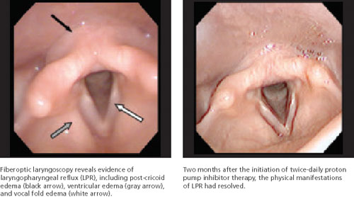
Ambulatory 24-hour dual sensor pH probe monitoring is sometimes used as an adjunctive test to confirm LPR. In 2005, 2 meta-analyses found that the pH probe is reliable and sensitive and specific enough to identify significantly more acid reflux in patients with LPR than in controls.19,20 However, not everyone agrees: Some clinicians question its use as a diagnostic tool for LPR, citing problems with observer reliability, among other things. Because it increases costs to the patient and is impractical, pH monitoring is not widely used by specialists, but primarily as a research tool.21,22
Barium swallow and esophagogastroduodenoscopy (EGD) are relatively common diagnostic tools used to identify anatomical abnormalities in the gastrointestinal tract, such as a Schatzki’s ring or hiatal hernia, that can lead to symptoms of GERD and/or LPR. Some studies suggest that all patients with symptoms of LPR should undergo EGD to screen for esophageal adenocarcinoma.23,24 Because LPR symptoms are relatively common, however, many clinicians believe that EGD should be considered only when heartburn is a primary complaint in a patient with signs and symptoms of LPR—or when a patient believed to have LPR fails to respond to medical management.24
Treating LPR: Lifestyle changes, drug therapy
For all patients with LPR, dietary and lifestyle modifications have been shown to be both clinically effective and cost effective.25 In addition to dietary restrictions (TABLE 2), advise patients to avoid eating too rapidly or drinking large quantities of fluid. Late night meals—indeed, eating within 3 hours of bedtime—should also be avoided, as should heavy lunches and dinners. Tell patients to eat small, frequent meals instead.7,25,26
Recommend other behavioral changes, as well. Tell patients to avoid tight clothing, lying down immediately after a meal, and applying pressure to the abdomen, whether through exercise, heavy lifting, singing, or bending over. Smoking and overuse (or misuse) of the voice—screaming at a concert or singing for hours, for instance—are contraindicated, as well.7,25,26
Tell patients that weight loss, as needed, is likely to bring some symptom relief. Using wooden blocks to elevate the head of the bed about 4 to 6 inches may also be helpful, particularly for patients who suffer from both LPR and GERD.7,25,26
TABLE 2
Dietary management of LPR: What to tell patients7,14,24
| Avoid* | Enjoy† |
|---|---|
| Caffeine | Meat |
| Alcohol | Poultry |
| Spicy foods | Seafood |
| Tomatoes | Milk |
| Chocolate | Fresh vegetables‡ |
| Fats | |
| Citrus fruits | |
| Carbonated beverages | |
| Jams/jellies | |
| Barbecue sauces | |
| Salad dressings | |
| Hot mustard | |
| Curry | |
| Hot peppers | |
| *Other acidic foods. | |
| †Other foods and beverages that are neither spicy nor acidic. | |
| ‡ Except tomatoes. | |
Drug therapy: Straightforward, but not without controversy
Acid suppression with proton pump inhibitors (PPIs) is the primary treatment for LPR, as it is for GERD. But because the larynx is extremely susceptible to injury from acid reflux, LPR typically requires more aggressive and prolonged treatment, compared with GERD.1,5
Clinical trials have shown that PPIs do not inhibit acid production to an intragastric pH of >4 for more than 16.8 hours.1,27 Thus, most patients need twice-daily dosing (although once-a-day dosing or conservative management may be sufficient for those with mild and intermittent symptoms).27,28 Regardless of dosing, PPIs should be taken on an empty stomach, 30 minutes before a meal to increase bioavailability. For maximum benefits, patients should continue the twice-daily regimen for 4 to 6 months, although the optimal duration is unknown.26
One study found 4 months of therapy to be effective;28 others suggest that while symptom relief should begin after 6 to 8 weeks of treatment, 6 months of PPI therapy is needed for laryngeal lesions and edema to resolve.1,8 Despite the time frame, patients should be weaned gradually to prevent the delayed rebound effect associated with abrupt cessation of PPIs.
The PPI controversy. Not only the length of treatment is controversial, however, but the efficacy of PPIs for LPR. Many studies, including several prospective cohort studies and 9 RCTs, have reported significant improvement in laryngeal symptoms, but evidence that PPIs are significantly better than placebo is weak.25,29,30 In fact, a systematic review and 2 meta-analyses concluded that not only is there a lack of sufficient evidence to draw reliable conclusions about the efficacy of PPIs vs placebo for the treatment of LPR, but there seems to be a significant response to placebo among patients with this condition, as well.25,29,30
The role of adjunctive therapy. Histamine type 2 (H2) blockers have been shown to be helpful in the treatment of GERD. But data showing their efficacy for LPR, either as a single agent or in combination with a PPI, are limited. Indeed, 3 clinical trials have found that H2 blockers do not provide any added benefit to PPI therapy for LPR. All 3 were cohort studies that compared the treatment outcomes of PPI alone vs PPI and H2 blockers, and found no statistically significant difference (P>.05).28,31,32 Despite these findings, recent studies suggest that 300 mg ranitidine twice a day provides added benefit (P<.01).33,34 Given these mixed findings, H2 blockers may be considered as adjuvant therapy to the PPI regimen to further reduce acid production in patients with more severe symptoms. Ant-acids and prokinetic agents are sometimes used for this purpose, as well.
When medical management fails
Surgery has a limited, but useful, role in the treatment of LPR.
Nissen fundoplication—a procedure in which the fundus of the stomach is passed posteriorly behind the esophagus to encircle it and provide mechanical obstruction to the retrograde movement of acid—may be considered for patients with a confirmed diagnosis, severe symptoms, and little response to treatment. However, there is little evidence that this procedure will result in long-term improvement in LPR symptoms. Laryngeal surgery can be used to treat vocal fold sequelae of LPR, such as granulomas—with a higher likelihood of success.35
CORRESPONDENCE Kevin Fung, MD, FRCSC, FACS, University of Western Ontario, London Health Sciences Center-Victoria Hospital, 800 Commissioners Road East, Room B3-427, London, Ontario, Canada, N6A 4G5; [email protected]
1. Koufman JA, Aviv JE, Casiano RR, et al. Laryngopharyngeal reflux: position statement of the Committee on Speech, Voice, and Swallowing Disorders of the American Academy of Otolaryngology–Head and Neck Surgery. Otolaryngol Head Neck Surg. 2002;127:32-35.
2. Karkos PD, Thomas L, Temple RH, et al. Awareness of general practitioners towards treatment of laryngopharyngeal reflux: a British survey. Otolaryngol Head Neck Surg. 2005;133:505-508.
3. Morrison MD. Is chronic gastroesophageal reflux a causative factor in glottic carcinoma? Otolaryngol Head Neck Surg. 1988;99:370-373.
4. Ward PH, Hanson DG. Reflux as an etiologic factor of carcinoma of the laryngopharynx. Laryngoscope. 1988;98:1195-1199.
5. Koufman JA. The otolaryngologic manifestations of gastroesophageal reflux disease (GERD): a clinical investigation of 225 patients using ambulatory 24-hour pH monitoring and an experimental investigation of the role of acid and pepsin in the development of laryngeal injury. Laryngoscope. 1991;101(4 pt 2 suppl 53):S1-S78.
6. Koufman JA, Amin MR, Panetti M. Prevalence of reflux in 113 consecutive patients with laryngeal and voice disorders. Otolaryngol Head Neck Surg. 2000;123:385-388.
7. Ford CN. Evaluation and management of laryngopharyngeal reflux. JAMA. 2005;294:1534-1540.
8. Aviv JE, Liu H, Parides M, et al. Laryngopharyngeal sensory deficits in patients with laryngopharyngeal reflux and dysphagia. Ann Otol Rhinol Laryngol. 2000;109:1000-1006.
9. Johnston N, Bulmer D, Gill GA, et al. Cell biology of laryngeal epithelial defenses in health and disease: further studies. Ann Otol Rhinol Laryngol. 2003;112:481.-
10. Gill GA, Johnston N, Buda A, et al. Laryngeal epithelial defenses against laryngopharyngeal reflux: investigations of E-cadherin, carbonic anhydrase isoenzyme III, and pepsin. Ann Otol Rhinol Laryngol. 2005;114:913-921.
11. Okamura H, Sugai N, Kanno T, et al. Histochemical localization of carbonic anhydrase in the trachea of the guinea pig. Histochem Cell Biol. 1996;106:257-260.
12. Johnston N, Knight J, Dettmar PW, et al. Pepsin and carbonic anhydrase isoenzyme III as diagnostic markers for laryngopharyngeal reflux disease. Laryngoscope. 2004;114:2129-2134.
13. Johnston N, Dettmar PW, Bishwokarma B, et al. Activity/stability of human pepsin: implications for reflux attributed laryngeal disease. Laryngoscope. 2007;117:1036-1039.
14. Axford SE, Sharp N, Ross PE, et al. Cell biology of laryngeal epithelial defenses in health and disease: preliminary studies. Ann Otol Rhinol Laryngol. 2001;110:1099-1108.
15. Toros SZ, Toros AB, Yüksel OD, et al. Association of laryngopharyngeal manifestations and gastroesophageal reflux. Eur Arch Otorhinolaryngol. 2009;266:403-409.
16. Koufman JA. Laryngopharyngeal reflux is different from classic gastroesophageal reflux disease Ear Nose Throat J. 2002;81(9 suppl 2):S7-S9.
17. Book DT, Rhee JS, Toohill RJ, et al. Perspectives in laryngopharyngeal reflux: an international survey. Laryngoscope. 2002;112(8 Pt 1):1399-1406.
18. Belafsky PC, Postma GN, Koufman JA. Validity and reliability of the Reflux Symptom Index (RSI). J Voice. 2002;16:274-277.
19. Ulualp SO, Roland PS, Toohill RJ, et al. Prevalence of gastroesophagopharyngeal acid reflux events: an evidence-based systematic review. Am J Otolaryngol. 2005;26:239-244.
20. Merati AL, Lim HJ, Ulualp SO, et al. Meta-analysis of upper probe measurements in normal subjects and patients with laryngopharyngeal reflux. Ann Otol Rhinol Laryngol. 2005;114(3):177-82.
21. Vaezi MF, Schroeder PL, Richter JE. Reproducibility of proximal probe pH parameters in 24-hour ambulatory esophageal pH monitoring. Am J Gastroenterol. 1997;92:825-829.
22. Vincent DA, Jr., Garrett JD, Radionoff SL, et al. The proximal probe in esophageal pH monitoring: development of a normative database. J Voice. 2000;142:247-254.
23. Reavis KM, Morris CD, Gopal DV, et al. Laryngopharyngeal reflux symptoms better predict the presence of esophageal adenocarcinoma than typical gastroesophageal reflux symptoms. Ann Surg. 2004;239:849-858.
24. Gupta R, Sataloff RT. Laryngopharyngeal reflux: current concepts and questions. Curr Opin Otolaryngol Head Neck Surg. 2009;17:143-148.
25. Tsunoda K, Ishimoto S, Suzuki M, et al. An effective management regimen for laryngeal granuloma caused by gastro-esophageal reflux: combination therapy with suggestions for lifestyle modifications. Acta Otolaryngol. 2007;127:88-92.
26. Hopkins C, Yousaf U, Pedersen M. Acid reflux treatment for hoarseness [protocol]. Cochrane Database Syst Rev. 2006;(1):CD005054.-
27. Kahrilas PJ, Falk GW, Johnson DA, et al. The Esomeprazole Study Investigators Esomeprazole improves healing and symptom resolution as compared with omeprazole in reflux oesophagitis patients: a randomized controlled trial. Aliment Pharmacol Ther. 2000;14:1249-1458.
28. Park W, Hicks DM, Khandwala F, et al. Laryngopharyngeal reflux: prospective cohort study evaluating optimal dose of proton-pump inhibitor therapy and pretherapy predictors of response. Laryngoscope. 2005;115:1230-1238.
29. Qadeer MA, Phillips CO, Lopez AR, et al. Proton pump inhibitor therapy for suspected GERD-related chronic laryngitis: a meta-analysis of randomized controlled trials. Am J Gastroenterol. 2006;101:2646-2654.
30. Karkos PD, Wilson JA. Empiric treatment of laryngopharyngeal reflux with proton pump inhibitors: a systematic review. Laryngoscope. 2006;116:144-148.
31. Fackler WK, Ours TM, Vaezi MF, et al. Long-term effect of H2RA therapy on nocturnal gastric acid breakthrough. Gastroenterology. 2002;122:625-632.
32. Ours TM, Fackler WK, Richter JE, et al. Nocturnal acid breakthrough: clinical significance and correlation with esophageal acid exposure. Am J Gastroenterol. 2003;98:545-550.
33. Sato K. Laryngopharyngeal reflux disease with nocturnal gastric acid breakthrough while on proton pump inhibitor therapy. Eur Arch Otorhinolaryngol. 2006;263:1121-1126.
34. Mainie I, Tutuian R, Castell DO. Addition of a H2 receptor antagonist to PPI improves acid control and decreases nocturnal acid breakthrough. J Clin Gastroenterol. 2008;42:676-679.
35. Koufman JA, Rees CJ, Frazier WD, et al. Office-based laryngeal laser surgery: a review of 443 cases using three wavelengths. Otolaryngol Head Neck Surg. 2007;137:146-151.
• Suspect laryngopharyngeal reflux (LPR) in a patient with chronic laryngitis; 50% to 60% of such cases are related to LPR. B
• Refer patients with risk factors for head and neck cancer or whose symptoms persist despite lifestyle modification and medical management to an otolaryngologist. A
• While symptoms of LPR should show improvement after 6 to 8 weeks of proton pump inhibitor therapy, advise patients to continue treatment for 4 to 6 months to ensure that laryngeal lesions and edema resolve. B
Strength of recommendation (SOR)
A Good-quality patient-oriented evidence
B Inconsistent or limited-quality patient-oriented evidence
C Consensus, usual practice, opinion, disease-oriented evidence, case series
Laryngopharyngeal reflux (LPR), the retrograde movement of gastric content into the upper aerodigestive tract, is a common—and commonly underdiagnosed—condition. Characterized by inflammation of the laryngopharynx, LPR can coexist with gastroesophageal reflux disease (GERD), but it is a distinct disorder.1 In GERD, the lower esophageal sphincter malfunctions, whereas LPR involves a dysfunctional upper esophageal sphincter.
Because both conditions involve acid reflux, LPR is sometimes mistaken for GERD. Often, too, patients and physicians alike attribute LPR’s signs and symptoms, which are largely nonspecific, to other causes. The hoarseness and laryngitis that are characteristic of LPR may be blamed on vocal cord abuse or smoking, for instance; the chronic cough and throat clearing associated with LPR thought to be caused by allergies; and the sore throat and postnasal drip that often accompany LPR attributed to infection. Another reason LPR is underdiagnosed: Primary care physicians, who are often the first clinicians from whom symptomatic patients seek treatment, are often unfamiliar with this lesser-known reflux disease.2
The failure to recognize and provide timely treatment for LPR may increase patients’ risk for a number of conditions, including laryngeal ulcers, granulomas, subglottic stenosis, chronic sinusitis, laryngospasm, nasal congestion, and asthma.1 Evidence suggests that LPR increases the risk for esophageal and laryngeal carcinomas,3,4 and for laryngeal injury from intubation, as well.1 To minimize these risks, it is important for primary care physicians to promptly identify this disorder, treat it appropriately, and recognize red flags that warrant referral to a specialist.
How LPR develops, what to look for
There is no gold standard for the diagnosis of LPR. Nonetheless, a review of the pathophysiology and clinical presentation of this reflux disorder and the ways in which it differs from GERD will help you identify cases of LPR. The prevalence of LPR in the general population is uncertain. But reports suggest that as many as 10% of otolaryngology referrals are for patients with a classic presentation of LPR, and that 50% to 60% of cases of chronic laryngitis are related to LPR.1,5,6
The laryngopharynx becomes irritated and inflamed
When the physiological barriers protecting the laryngopharynx from the retrograde flow of gastric content break down, gastric contents can directly irritate the ciliated columnar epithelial cells of the upper respiratory tract, leading to ciliary dysfunction. A lack of mucous clearance leads to mucous stasis and, subsequently, to excessive throat clearing and the sensation of postnasal drip.7 In addition, the laryngopharyngeal epithelium becomes inflamed, and this affects the sensitivity of laryngeal sensory endings and leads to laryngospasm and coughing.8 The inflammatory reaction in turn leads to vocal fold edema, contact ulcers, and granulomas. These changes make patients with LPR particularly prone to developing hoarseness, globus pharyngeus—a sensation of a foreign body in the larynx—and sore throat.5,7 The gastric content can also act indirectly by initiating laryngeal reflexes through irritation of the esophagus, leading to vagally mediated changes such as chronic cough and bronchoconstriction.
Enzyme production declines. Under normal circumstances, carbonic anhydrase isoenzyme III (CAIII) is produced in the posterior aspect of the larynx, catalyzing the production of bicarbonate and neutralizing stomach acid.9-11 In LPR, however, the production of CAIII decreases significantly, thereby exposing the larynx to stomach acid without the enzyme’s protective effect.9,10 At the same time, a marked increase in pepsin levels intensifies laryngeal injury.10,12,13
The larynx is highly vulnerable. The laryngopharynx is much more susceptible to pathology from gastric reflux than the esophagus, for a number of reasons. Damage can occur with much less exposure to acid,1 not only because of the decrease in CAIII, but also because of the absence of peristalsis in the larynx.
What’s more, the esophagus has the ability to clear gastric reflux and minimize damage to the epithelial layer.9,10,14,15 In most patients who develop signs and symptoms of LPR, there has been enough gastric reflux to damage the laryngopharynx but not enough to overcome the protective mechanisms of the esophagus. That’s why most LPR patients have little or none of the heartburn and esophagitis that are classic symptoms of GERD.
Common signs and symptoms that signal LPR
LPR is primarily a clinical diagnosis based on signs and symptoms—which are also used to rule out GERD. Notably, less than 50% of patients with LPR suffer from heartburn and regurgitation.16 Those who do have heartburn and regurgitation typically suffer with reflux during the day, when they’re in an upright position, whereas reflux associated with GERD develops primarily at night.16 The results of a recent survey of members of the American Bronchoesophagological Association highlight the most common signs and symptoms of LPR, listed below from the most to the least frequent:17
- throat clearing
- persistent cough
- globus sensation
- hoarseness
- choking episodes.
Additional signs and symptoms include excessive and chronic throat clearing, sore throat, postnasal drip, and dysphagia.
Use a validated symptom index. To further assess the probability and severity of LPR, use the Reflux Symptom Index18 (TABLE 1). A recent cohort study validated the index, with an average score of 21.2 for those with LPR, vs an average of 11.6 for controls (P<.001). A score >13 is suggestive of LPR (odds ratio=9.19), the researchers found.18
If the diagnosis remains uncertain and the patient continues to be troubled by signs and symptoms suggestive of LPR, refer him or her to an otolaryngologist for further investigation. A referral is needed, too, to rule out malignancy in any patient with 3 or more of the following red flags: older than 50 years, otalgia, weight loss, progressive hoarseness, neck mass, a significant history of alcohol use, and a history of smoking.7
TABLE 1
The Reflux Symptom Index for laryngopharyngeal reflux
| Within the last month, how did the following problems affect you? | 0 = NO PROBLEM 5 = SEVERE PROBLEM (CIRCLE THE APPROPRIATE RESPONSE) | |||||
|---|---|---|---|---|---|---|
| 1. Hoarseness or a problem with your voice | 0 | 1 | 2 | 3 | 4 | 5 |
| 2. Clearing your throat | 0 | 1 | 2 | 3 | 4 | 5 |
| 3. Excess throat mucus or postnasal drip | 0 | 1 | 2 | 3 | 4 | 5 |
| 4. Difficulty swallowing food, liquids, or pills | 0 | 1 | 2 | 3 | 4 | 5 |
| 5. Coughing after you ate or after lying down | 0 | 1 | 2 | 3 | 4 | 5 |
| 6. Breathing difficulties or choking episodes | 0 | 1 | 2 | 3 | 4 | 5 |
| 7. Troublesome or annoying cough | 0 | 1 | 2 | 3 | 4 | 5 |
| 8. Sensations of something sticking in your throat or a lump in your throat | 0 | 1 | 2 | 3 | 4 | 5 |
| 9. Heartburn, chest pain, indigestion, or stomach acid coming up | 0 | 1 | 2 | 3 | 4 | 5 |
| TOTAL SCORE= | ||||||
| *A score >13 is considered suggestive of laryngopharyngeal reflux. | ||||||
| Source: Belafsky PC et al. J Voice. 2002.18 Reprinted with permission. | ||||||
Diagnostic tools the specialists will use
Fiberoptic laryngoscopy is the most common test used by otolaryngologists to confirm LPR and rule out other pathology. The test reveals inflammatory findings (FIGURE), such as erythema, edema, granulomas, and contact ulcers, in several anatomical locations of the larynx—especially the posterior aspect and the true vocal folds. It is important to note, however, that as many as 70% of the general population will have some laryngeal inflammation, so these findings alone are not definitive evidence of LPR.7
FIGURE
Fiberoptic laryngoscopy: Before and after treatment
Ambulatory 24-hour dual sensor pH probe monitoring is sometimes used as an adjunctive test to confirm LPR. In 2005, 2 meta-analyses found that the pH probe is reliable and sensitive and specific enough to identify significantly more acid reflux in patients with LPR than in controls.19,20 However, not everyone agrees: Some clinicians question its use as a diagnostic tool for LPR, citing problems with observer reliability, among other things. Because it increases costs to the patient and is impractical, pH monitoring is not widely used by specialists, but primarily as a research tool.21,22
Barium swallow and esophagogastroduodenoscopy (EGD) are relatively common diagnostic tools used to identify anatomical abnormalities in the gastrointestinal tract, such as a Schatzki’s ring or hiatal hernia, that can lead to symptoms of GERD and/or LPR. Some studies suggest that all patients with symptoms of LPR should undergo EGD to screen for esophageal adenocarcinoma.23,24 Because LPR symptoms are relatively common, however, many clinicians believe that EGD should be considered only when heartburn is a primary complaint in a patient with signs and symptoms of LPR—or when a patient believed to have LPR fails to respond to medical management.24
Treating LPR: Lifestyle changes, drug therapy
For all patients with LPR, dietary and lifestyle modifications have been shown to be both clinically effective and cost effective.25 In addition to dietary restrictions (TABLE 2), advise patients to avoid eating too rapidly or drinking large quantities of fluid. Late night meals—indeed, eating within 3 hours of bedtime—should also be avoided, as should heavy lunches and dinners. Tell patients to eat small, frequent meals instead.7,25,26
Recommend other behavioral changes, as well. Tell patients to avoid tight clothing, lying down immediately after a meal, and applying pressure to the abdomen, whether through exercise, heavy lifting, singing, or bending over. Smoking and overuse (or misuse) of the voice—screaming at a concert or singing for hours, for instance—are contraindicated, as well.7,25,26
Tell patients that weight loss, as needed, is likely to bring some symptom relief. Using wooden blocks to elevate the head of the bed about 4 to 6 inches may also be helpful, particularly for patients who suffer from both LPR and GERD.7,25,26
TABLE 2
Dietary management of LPR: What to tell patients7,14,24
| Avoid* | Enjoy† |
|---|---|
| Caffeine | Meat |
| Alcohol | Poultry |
| Spicy foods | Seafood |
| Tomatoes | Milk |
| Chocolate | Fresh vegetables‡ |
| Fats | |
| Citrus fruits | |
| Carbonated beverages | |
| Jams/jellies | |
| Barbecue sauces | |
| Salad dressings | |
| Hot mustard | |
| Curry | |
| Hot peppers | |
| *Other acidic foods. | |
| †Other foods and beverages that are neither spicy nor acidic. | |
| ‡ Except tomatoes. | |
Drug therapy: Straightforward, but not without controversy
Acid suppression with proton pump inhibitors (PPIs) is the primary treatment for LPR, as it is for GERD. But because the larynx is extremely susceptible to injury from acid reflux, LPR typically requires more aggressive and prolonged treatment, compared with GERD.1,5
Clinical trials have shown that PPIs do not inhibit acid production to an intragastric pH of >4 for more than 16.8 hours.1,27 Thus, most patients need twice-daily dosing (although once-a-day dosing or conservative management may be sufficient for those with mild and intermittent symptoms).27,28 Regardless of dosing, PPIs should be taken on an empty stomach, 30 minutes before a meal to increase bioavailability. For maximum benefits, patients should continue the twice-daily regimen for 4 to 6 months, although the optimal duration is unknown.26
One study found 4 months of therapy to be effective;28 others suggest that while symptom relief should begin after 6 to 8 weeks of treatment, 6 months of PPI therapy is needed for laryngeal lesions and edema to resolve.1,8 Despite the time frame, patients should be weaned gradually to prevent the delayed rebound effect associated with abrupt cessation of PPIs.
The PPI controversy. Not only the length of treatment is controversial, however, but the efficacy of PPIs for LPR. Many studies, including several prospective cohort studies and 9 RCTs, have reported significant improvement in laryngeal symptoms, but evidence that PPIs are significantly better than placebo is weak.25,29,30 In fact, a systematic review and 2 meta-analyses concluded that not only is there a lack of sufficient evidence to draw reliable conclusions about the efficacy of PPIs vs placebo for the treatment of LPR, but there seems to be a significant response to placebo among patients with this condition, as well.25,29,30
The role of adjunctive therapy. Histamine type 2 (H2) blockers have been shown to be helpful in the treatment of GERD. But data showing their efficacy for LPR, either as a single agent or in combination with a PPI, are limited. Indeed, 3 clinical trials have found that H2 blockers do not provide any added benefit to PPI therapy for LPR. All 3 were cohort studies that compared the treatment outcomes of PPI alone vs PPI and H2 blockers, and found no statistically significant difference (P>.05).28,31,32 Despite these findings, recent studies suggest that 300 mg ranitidine twice a day provides added benefit (P<.01).33,34 Given these mixed findings, H2 blockers may be considered as adjuvant therapy to the PPI regimen to further reduce acid production in patients with more severe symptoms. Ant-acids and prokinetic agents are sometimes used for this purpose, as well.
When medical management fails
Surgery has a limited, but useful, role in the treatment of LPR.
Nissen fundoplication—a procedure in which the fundus of the stomach is passed posteriorly behind the esophagus to encircle it and provide mechanical obstruction to the retrograde movement of acid—may be considered for patients with a confirmed diagnosis, severe symptoms, and little response to treatment. However, there is little evidence that this procedure will result in long-term improvement in LPR symptoms. Laryngeal surgery can be used to treat vocal fold sequelae of LPR, such as granulomas—with a higher likelihood of success.35
CORRESPONDENCE Kevin Fung, MD, FRCSC, FACS, University of Western Ontario, London Health Sciences Center-Victoria Hospital, 800 Commissioners Road East, Room B3-427, London, Ontario, Canada, N6A 4G5; [email protected]
1. Koufman JA, Aviv JE, Casiano RR, et al. Laryngopharyngeal reflux: position statement of the Committee on Speech, Voice, and Swallowing Disorders of the American Academy of Otolaryngology–Head and Neck Surgery. Otolaryngol Head Neck Surg. 2002;127:32-35.
2. Karkos PD, Thomas L, Temple RH, et al. Awareness of general practitioners towards treatment of laryngopharyngeal reflux: a British survey. Otolaryngol Head Neck Surg. 2005;133:505-508.
3. Morrison MD. Is chronic gastroesophageal reflux a causative factor in glottic carcinoma? Otolaryngol Head Neck Surg. 1988;99:370-373.
4. Ward PH, Hanson DG. Reflux as an etiologic factor of carcinoma of the laryngopharynx. Laryngoscope. 1988;98:1195-1199.
5. Koufman JA. The otolaryngologic manifestations of gastroesophageal reflux disease (GERD): a clinical investigation of 225 patients using ambulatory 24-hour pH monitoring and an experimental investigation of the role of acid and pepsin in the development of laryngeal injury. Laryngoscope. 1991;101(4 pt 2 suppl 53):S1-S78.
6. Koufman JA, Amin MR, Panetti M. Prevalence of reflux in 113 consecutive patients with laryngeal and voice disorders. Otolaryngol Head Neck Surg. 2000;123:385-388.
7. Ford CN. Evaluation and management of laryngopharyngeal reflux. JAMA. 2005;294:1534-1540.
8. Aviv JE, Liu H, Parides M, et al. Laryngopharyngeal sensory deficits in patients with laryngopharyngeal reflux and dysphagia. Ann Otol Rhinol Laryngol. 2000;109:1000-1006.
9. Johnston N, Bulmer D, Gill GA, et al. Cell biology of laryngeal epithelial defenses in health and disease: further studies. Ann Otol Rhinol Laryngol. 2003;112:481.-
10. Gill GA, Johnston N, Buda A, et al. Laryngeal epithelial defenses against laryngopharyngeal reflux: investigations of E-cadherin, carbonic anhydrase isoenzyme III, and pepsin. Ann Otol Rhinol Laryngol. 2005;114:913-921.
11. Okamura H, Sugai N, Kanno T, et al. Histochemical localization of carbonic anhydrase in the trachea of the guinea pig. Histochem Cell Biol. 1996;106:257-260.
12. Johnston N, Knight J, Dettmar PW, et al. Pepsin and carbonic anhydrase isoenzyme III as diagnostic markers for laryngopharyngeal reflux disease. Laryngoscope. 2004;114:2129-2134.
13. Johnston N, Dettmar PW, Bishwokarma B, et al. Activity/stability of human pepsin: implications for reflux attributed laryngeal disease. Laryngoscope. 2007;117:1036-1039.
14. Axford SE, Sharp N, Ross PE, et al. Cell biology of laryngeal epithelial defenses in health and disease: preliminary studies. Ann Otol Rhinol Laryngol. 2001;110:1099-1108.
15. Toros SZ, Toros AB, Yüksel OD, et al. Association of laryngopharyngeal manifestations and gastroesophageal reflux. Eur Arch Otorhinolaryngol. 2009;266:403-409.
16. Koufman JA. Laryngopharyngeal reflux is different from classic gastroesophageal reflux disease Ear Nose Throat J. 2002;81(9 suppl 2):S7-S9.
17. Book DT, Rhee JS, Toohill RJ, et al. Perspectives in laryngopharyngeal reflux: an international survey. Laryngoscope. 2002;112(8 Pt 1):1399-1406.
18. Belafsky PC, Postma GN, Koufman JA. Validity and reliability of the Reflux Symptom Index (RSI). J Voice. 2002;16:274-277.
19. Ulualp SO, Roland PS, Toohill RJ, et al. Prevalence of gastroesophagopharyngeal acid reflux events: an evidence-based systematic review. Am J Otolaryngol. 2005;26:239-244.
20. Merati AL, Lim HJ, Ulualp SO, et al. Meta-analysis of upper probe measurements in normal subjects and patients with laryngopharyngeal reflux. Ann Otol Rhinol Laryngol. 2005;114(3):177-82.
21. Vaezi MF, Schroeder PL, Richter JE. Reproducibility of proximal probe pH parameters in 24-hour ambulatory esophageal pH monitoring. Am J Gastroenterol. 1997;92:825-829.
22. Vincent DA, Jr., Garrett JD, Radionoff SL, et al. The proximal probe in esophageal pH monitoring: development of a normative database. J Voice. 2000;142:247-254.
23. Reavis KM, Morris CD, Gopal DV, et al. Laryngopharyngeal reflux symptoms better predict the presence of esophageal adenocarcinoma than typical gastroesophageal reflux symptoms. Ann Surg. 2004;239:849-858.
24. Gupta R, Sataloff RT. Laryngopharyngeal reflux: current concepts and questions. Curr Opin Otolaryngol Head Neck Surg. 2009;17:143-148.
25. Tsunoda K, Ishimoto S, Suzuki M, et al. An effective management regimen for laryngeal granuloma caused by gastro-esophageal reflux: combination therapy with suggestions for lifestyle modifications. Acta Otolaryngol. 2007;127:88-92.
26. Hopkins C, Yousaf U, Pedersen M. Acid reflux treatment for hoarseness [protocol]. Cochrane Database Syst Rev. 2006;(1):CD005054.-
27. Kahrilas PJ, Falk GW, Johnson DA, et al. The Esomeprazole Study Investigators Esomeprazole improves healing and symptom resolution as compared with omeprazole in reflux oesophagitis patients: a randomized controlled trial. Aliment Pharmacol Ther. 2000;14:1249-1458.
28. Park W, Hicks DM, Khandwala F, et al. Laryngopharyngeal reflux: prospective cohort study evaluating optimal dose of proton-pump inhibitor therapy and pretherapy predictors of response. Laryngoscope. 2005;115:1230-1238.
29. Qadeer MA, Phillips CO, Lopez AR, et al. Proton pump inhibitor therapy for suspected GERD-related chronic laryngitis: a meta-analysis of randomized controlled trials. Am J Gastroenterol. 2006;101:2646-2654.
30. Karkos PD, Wilson JA. Empiric treatment of laryngopharyngeal reflux with proton pump inhibitors: a systematic review. Laryngoscope. 2006;116:144-148.
31. Fackler WK, Ours TM, Vaezi MF, et al. Long-term effect of H2RA therapy on nocturnal gastric acid breakthrough. Gastroenterology. 2002;122:625-632.
32. Ours TM, Fackler WK, Richter JE, et al. Nocturnal acid breakthrough: clinical significance and correlation with esophageal acid exposure. Am J Gastroenterol. 2003;98:545-550.
33. Sato K. Laryngopharyngeal reflux disease with nocturnal gastric acid breakthrough while on proton pump inhibitor therapy. Eur Arch Otorhinolaryngol. 2006;263:1121-1126.
34. Mainie I, Tutuian R, Castell DO. Addition of a H2 receptor antagonist to PPI improves acid control and decreases nocturnal acid breakthrough. J Clin Gastroenterol. 2008;42:676-679.
35. Koufman JA, Rees CJ, Frazier WD, et al. Office-based laryngeal laser surgery: a review of 443 cases using three wavelengths. Otolaryngol Head Neck Surg. 2007;137:146-151.
The skin disorders of pregnancy: A family physician’s guide
• Pemphigoid gestationis is best managed with oral prednisone at doses from 20 to 60 mg per day to control symptoms. B
• The pruritus associated with pruritic urticarial papules and plaques of pregnancy can be safely and effectively managed with topical corticosteroids and oral antihistamines. A
• Treat intrahepatic cholestasis of pregnancy with ursodeoxycholic acid, which likely reduces serum bile acids as well as associated fetal morbidity and mortality. B
Strength of recommendation (SOR)
A Good-quality patient-oriented evidence
B Inconsistent or limited-quality patient-oriented evidence
C Consensus, usual practice, opinion, disease-oriented evidence, case series
The dermatoses of pregnancy are a poorly understood group of dermatologic conditions. The only thing they have in common is a tendency to appear during pregnancy.
Only 3 are considered unique to pregnancy, however; the others are probably exacerbations of preexisting conditions triggered by pregnancy. There isn’t even complete agreement on what to call them. And to make management even more complex, 2 patients—the mother and the fetus—need to be considered.
Who will manage these patients is another matter. These conditions fall into an overlapping area of health care, where family physicians, obstetricians, and dermatologists all have some share in the responsibility for diagnosis and treatment. As a family physician who probably cares for any number of pregnant patients on a weekly basis, you need to be sufficiently familiar with these conditions so that you can differentiate those that can be treated symptomatically and those that require a referral to a specialist. This review and handy TABLE will help you toward that end.
TABLE
Skin disorders of pregnancy: What you’ll see, how to treat
| Disorder | Lesions | Diagnosis and sequelae | Treatment | Recurrence |
|---|---|---|---|---|
| PG3,5 | Erythematous papules, progressing to vesicles, bullae; periumbilical distribution, sparing face, palms, and soles | Mean onset at 21 weeks; postpartum in 20% of cases. Direct immunofluorescence microscopy shows linear C3 deposition. Newborn may be small for gestational age, but no associated morbidity or mortality | Oral corticosteroids 20-60 mg/d, IVIG, or cyclosporine in refractory cases | Frequent. Skips a pregnancy 8% of the time |
| PUPPP8-10 | Urticarial papules and plaques on abdomen, legs, arms, buttocks, chest, and back | Usually present after 34th week, but can present at any stage. Diagnosis is clinical. No increase in fetal morbidity or mortality | Topical steroids and antihistamines | Uncommon |
| ICP14,17,19-22 | No primary lesions; secondary excoriations in any area patient can reach | Onset after 30th week in 80% of patients. Strongly indicated by serum bile acid >11 mcmol/L. Increased fetal mortality | Ursodeoxycholic acid 450-1200 mg/d | Frequent |
| EP/PP4,10,24 | Grouped, crusted erythematous papules, patches, and plaques, most commonly on extensor surfaces of arms and legs or on abdomen | Onset at any point in pregnancy. Clinical diagnosis. No increase in fetal morbidity or mortality | Symptomatic treatment with topical steroids or antihistamines | Frequent |
| APPP27-29 | Erythematous plaques and pustules starting on inner thighs and groin and spreading to trunk and extremities | Onset at any point in pregnancy. Clinical diagnosis by appearance of lesions and association with systemic illness. Increased incidence of miscarriage and stillbirth, and maternal mortality | Prednisone 15-60 mg/d, cyclosporine 100 mg twice daily in refractory cases, management of associated hypocalcemia | Unknown |
| PFP24,29 | Papules and pustules concentrated around hair follicles, often beginning on abdomen and spreading to extremities | Onset most often in third trimester. Clinical diagnosis. No associated fetal morbidity or mortality | Topical steroids | Unknown |
| APPP, acute pustular psoriasis of pregnancy; EP/PP, eczema of pregnancy/pruritus of pregnancy; ICP, intrahepatic cholestasis of pregnancy; IVIG, intravenous immunoglobulin; PFP, pruritic folliculitis of pregnancy; PG, pemphigoid gestationis; PUPPP, pruritic urticarial papules and plaques of pregnancy. | ||||
Dermatoses unique to pregnancy
Pemphigoid gestationis
Years ago, this disorder was referred to as herpes gestationis, because the lesions are herpetiform. Pemphigoid gestationis (PG) has an incidence of approximately 1 in 10,000 pregnancies.1,2 The time of onset is usually about the 21st week of gestation, although in about 20% of cases, the eruption appears immediately postpartum.3
Presentation. The disease usually begins with urticarial papules and plaques around the umbilicus and extremities. Bullous lesions tend to develop as the disease progresses, and are often not present on first presentation (FIGURE 1). PG lesions tend to spare the face, palms, and soles. Mucosal surfaces are involved in less than 20% of cases. In about 75% of cases, PG flares around the time of delivery, regressing spontaneously after the baby is born.4
Pathophysiology. The pathophysiology is nearly identical to that of bullous pemphigoid, a blistering skin disorder more often seen in elderly patients.5 Pemphigoid disorders are immune processes, involving an immunoglobulin G (IgG) immune response directed at a 180-kDa hemidesmosome transmembrane glycoprotein. This protein is the common target of several subepidermal blistering diseases.
Differential. Disorders that may have some of the same features as PG include pruritic urticarial papules and plaques of pregnancy (PUPPP), erythema multiforme, intrahepatic cholestasis of pregnancy (ICP), contact dermatitis, and drug reactions.
Diagnosis. A biopsy is necessary for the definitive diagnosis. Direct immunofluorescence (DIF) microscopy of a sample of perilesional skin can show tissue-bound immunoreactants. Linear deposition of the complement component protein C3 along the basement membrane zone is diagnostic for PG. IgG is also deposited about 40% of the time.3 Serum enzyme-linked immunosorbent assay (ELISA) studies are also helpful in diagnosis. They have excellent sensitivity and specificity, as well as the capacity to monitor levels of antibody, which correlate with the severity of disease.1
Treatment. Oral corticosteroids are the first-line treatment for PG, typically 20 to 60 mg per day of prednisone. Oral corticosteroids are typically most effective in ameliorating the patient’s symptoms. Prednisone at doses of 40 to 80 mg per day for a short time has not been associated with congenital abnormalities.6 PG patients can also be treated successfully with intravenous immunoglobulin (IVIG) and cyclosporine in refractory cases.7
Pruritus associated with this condition can interfere with day-to-day activity and with the patient’s ability to sleep. Patients may also complain that the rash is painful, particularly if bullae rupture, leading to superficial ulcerations. Fortunately, the patient’s quality of life can be dramatically improved with systemic corticosteroids—with no significant risk to the fetus.
Sequelae. PG uniformly resolves within a few weeks, but the mother’s autoantibodies can passively be transferred to the fetus, causing vesicles and bullae in the newborn.8 An increased incidence of small-for-gestational age (SGA) infants has also been noted in PG, although no lasting morbidity or mortality in the offspring has been noted.5 This disease tends to recur with future pregnancies, but will skip a pregnancy 8% of the time.5
FIGURE 1
Pemphigoid gestationis
Pruritic urticarial papules and plaques of pregnancy
This condition is known by many names besides PUPPP: polymorphic eruption of pregnancy, toxemic erythema of pregnancy, and late prurigo of pregnancy.1 It is a pruritic, inflammatory skin disorder that has been variously estimated to occur in anywhere from 1 in 120 to 1 in 240 pregnancies.8 PUPPP is second only to eczema as the most common dermatosis of pregnancy.
Presentation. As the name implies, the lesions of PUPPP are itchy, red papules that often coalesce into plaques (FIGURE 2). The lesions usually occur in primigravidas after the 34th week of gestation, although they may be seen at any time from the first trimester through the postpartum period.9
Lesions are classically found on the abdomen, sparing the umbilical area, and are found primarily in the striae. This distribution helps to differentiate PUPPP from PG, where the lesions typically cluster around the umbilicus. Most PUPPP lesions (80% in 1 study) are dispersed on the abdomen, legs, arms, buttocks, chest, and back. Another 17% appear only on the abdomen and proximal thighs, and the remaining 3% on the limbs.10 Nearly 50% of the time, lesions also include discrete vesicles.11 There are no reported cases of mucosal involvement.
Patients with this condition are often very uncomfortable. The associated pruritus is severe enough to interfere with sleep. Despite the itching, however, lesions are seldom excoriated.
Pathophysiology. The disorder has been strongly associated with maternal weight gain and multiple gestations. One working hypothesis is that rapid abdominal distention observed in the third trimester leads to damage of the connective tissue, which then releases antigenic molecules, causing an inflammatory reaction.12 Another hypothesis is that increased levels of fetal DNA that have been detected in the skin of PUPPP patients may contribute to the pathology. One study detected male DNA in 6 of 10 PUPPP sufferers, but found none in any of 26 controls— pregnant women without PUPPP pathology.5 There is some evidence that patients with atopy may be predisposed to PUPPP, as well as patients who are hypertensive or obese.10,13
Differential. Initially, PUPPP lesions can be difficult to differentiate from urticarial PG lesions. The distribution of the lesions is the best clue: PG lesions cluster around the umbilicus, whereas PUPPP lesions uniformly spare the umbilical area. Additional disorders in the PUPPP differential are atopic dermatitis, superficial urticarial allergic eruption, viral exanthema, and contact or irritant dermatitis.
Diagnosis. PUPPP can only be diagnosed through clinical observation. None of the available laboratory tests—immunofluorescence, histology, serology—yield findings specific for PUPPP, although histology and immunofluorescence can readily differentiate between this condition and PG.
Treatment. Because the disease holds no real danger for mother or fetus, treatment can be aimed solely at symptomatic relief. Mild to potent topical steroids (consider triamcinolone or fluocinonide) should relieve pruritus within 48 to 72 hours.8 Antihistamines and occasionally low-dose systemic steroids may also be used. Consider hydroxyzine, although diphenhydramine has the more proven safety profile in pregnancy.
Nonpharmacologic treatments such as oil baths and emollients should also be considered. If the condition appears classic for PUPPP, it can be managed symptomatically. However, if there is any question about the diagnosis, referral to a dermatologist is prudent.
Sequelae. No increase in maternal or fetal morbidity or mortality is associated with PUPPP. Recurrence is fairly uncommon, as the disease primarily affects women during their first pregnancy.
FIGURE 2
Pruritic urticarial papules and plaques of pregnancy
Intrahepatic cholestasis of pregnancy
This condition is also called recurrent or idiopathic jaundice of pregnancy, obstetric cholestasis, and pruritus gravidarum. ICP is caused by disruption of hepatic bile flow during pregnancy. It has been recorded at a rate of approximately 10 to 150 per 10,000 pregnancies in Europe and 70 per 10,000 in the United States.12 In 80% of patients, the time of onset is after the 30th week.14 Although this disorder is not primarily a dermatosis of pregnancy, it is a pruritic condition that often presents with excoriations in pregnant women and is associated with fetal morbidity and mortality. It’s important to be able to identify this disease early to minimize sequelae.
Presentation. There are no primary lesions with ICP. The primary presenting symptom is a generalized pruritus affecting the palms and soles, and sometimes extending to the legs and abdomen (FIGURE 3). This itching is often so severe that it leads to chronic insomnia. You may see secondary skin lesions, such as erythema and excoriations. Observable jaundice occurs in 10% to 20% of patients.3 These patients do not develop the encephalopathy that is associated with cholestasis in the nonpregnant state, however.14
Pathophysiology. The genesis of this condition is thought to be a combination of genetic and environmental factors. A family history of the disorder is present in half the cases, and cases with a familial component tend to be more severe.15 ICP may be an exaggerated response to increased estrogen levels in pregnancy, but the mechanism of this response is unknown.16
Differential. Other conditions that must be considered in making the diagnosis are viral hepatitis, gallbladder disease, PG, PUPPP, drug hepatotoxicity, primary biliary cirrhosis, and uremia.
Diagnosis. Laboratory values are the definitive diagnostic tool in this condition. Increased serum bile acids are the single most sensitive test. Average levels of serum bile acids in pregnancy are 6.6 mcmol/L, with an upper limit of 11. The average value in women with ICP is 47 mcmol/L.17
While serum acids remain the gold standard, a recent study showed elevated urine bile acids to have 100% sensitivity and 83% specificity for ICP.18 In 55% to 60% of cases, the liver enzymes aspartate aminotransferase (AST) and alanine aminotransferase (ALT) are mildly increased. Steatorrhea is often noted by the patient, and is followed by vitamin K deficiency.17
Treatment. The current standard of care for ICP is treatment with ursodeoxycholic acid (UDCA). In 4 controlled trials, UDCA showed a sustained decrease in serum bile acids.19-22 Doses used in these trials have varied between 450 and 1200 mg per day.
Before UDCA treatment was available, the disorder was treated with cholestyramine, which could bring about a 70% rate of response. The drawback to cholestyramine treatment is that it precipitates vitamin K, which is already compromised by the disease process. Further, its onset of action is slow.3
Elective delivery is indicated for ICP, particularly in patients with significant clinical presentations.12 Delivery for ICP should be performed around week 37 to 38, as stillbirths tend to cluster around weeks 37 to 39.14 Given the significant fetal mortality associated with this condition (see below), ICP should be managed by a clinician experienced with the disease, likely a gastroenterologist.
Sequelae. The impact of this maternal disorder on the fetus can be disastrous: a 10% to 15% rate of perinatal death, and a 30% to 40% rate of premature labor have been seen with ICP.14 Fortunately, rates of preterm labor are strongly correlated with levels of bile acids, so that as bile acid levels are reduced with UDCA treatment, rates of preterm labor also go down. Currently, management of the condition has reduced rates of perinatal death to 3.5%. There is no evidence of fetal growth retardation.14
FIGURE 3
Intrahepatic cholestasis of pregnancy
Dermatoses triggered by pregnancy
Eczema of pregnancy/prurigo of pregnancy
Eczema of pregnancy/prurigo of pregnancy (EP/PP) may not actually be correlated with the pregnant state. Both conditions manifest as eczematous lesions in an atopic distribution. Although they have been described in the literature as separate entities, the lack of clinical distinction between them led Ambros-Rudolph and colleagues to combine them under the umbrella term, atopic eruption of pregnancy.23 In some patients, at least, they may be preexisting conditions that pregnancy exacerbates. One study of 255 patients with the condition found that 20% had had the lesions before they became pregnant.23 However, the tendency of the condition to be markedly worsened by pregnancy leads us to include it here.
PP has an estimated incidence of 1 in 450 pregnancies.11 But while many authorities consider EP to be the most common dermatosis of pregnancy, no clear estimation of its prevalence has been established.23,24 Taken together, these 2 conditions have the highest prevalence of all pregnancy-induced dermatoses. PP is also known as popular dermatitis of Spangler, Nurse’s early prurigo of pregnancy, and linear IgM disease of pregnancy.3,23,25
Presentation. The typical presentation consists of grouped, crusted, erythematous papules, patches, and plaques—frequently with excoriations. The lesions typically present on the extensor surfaces of the arms and legs or on the abdomen (FIGURE 4).4 Recurrence in later pregnancies is common.
Pathophysiology. The pathophysiology of EP/PP is not understood. Many patients who present with EP/PP have a history of atopy.10
Differential. Conditions that need to be considered in making the diagnosis include tinea infection, scabies, contact dermatitis, ICP, pruritic folliculitis of pregnancy (PFP), and PG.
Diagnosis. History and physical examination determine the diagnosis. Serology, histopathology, and immunofluorescence are not specific, and correlation with increased IgE is marginal, at best.24,26
Treatment. These conditions are treated symptomatically with topical steroids or systemic antihistamines.
Sequelae. No maternal or fetal increase in morbidity or mortality is associated with these conditions.
FIGURE 4
Eczema of pregnancy
Acute pustular psoriasis of pregnancy
Whether or not APPP is actually a pregnancyinduced dermatosis is subject to debate.
There is evidence that APPP is not unique to pregnancy, but is simply a manifestation of ordinary psoriasis. Clinically and histologically, APPP is indistinguishable from pustular psoriasis. Unlike most cases of acute psoriasis, however, APPP often appears in pregnancy without any personal or family history of psoriasis, and usually ceases when the pregnancy is concluded. This fact, combined with reports of increased fetal and maternal morbidity and mortality associated with APPP, lead us to include it here.27
Presentation. APPP is a rare condition that may have an onset at any point in pregnancy. The characteristic lesions begin as erythematous plaques with pustules on the inner thighs, flexural areas, and groin and spread to the trunk and extremities. As the plaques enlarge, the center becomes eroded and crusted. Nails may become onycholytic. The hands, feet, and face are usually spared. Oral and esophageal erosions can occur. Pruritus is typically mild, although the lesions are often painful and flu-like symptoms are often present.28
Pathophysiology. The pathophysiology of this disease is unknown.
Differential. Conditions with similar presentations include an adverse drug reaction, pityriasis rosea, lichen simplex chronicus, eczema, lupus, and pityriasis rubra pilaris.
Diagnosis. Clinical history and association with systemic illness are the basis for a diagnosis of APPP. Cultures of the pustules are negative for any infective pathology, though as the disease progresses, pustules may become superinfected. Lab tests may show an increased erythrocyte sedimentation rate (ESR), hypocalcemia, and low levels of vitamin D.
Treatment. Prednisone 15 to 60 mg per day is often sufficient to control the disease.28 Cyclosporine 100 mg twice daily has also been shown to be useful.29 Cyclosporine in pregnancy is a category C drug. Data on fetal malformation associated with cyclosporine therapy are limited, but the risk appears to be minimal.6 Maternal hypocalcemia should be monitored and treated appropriately. If disease progression is judged serious enough, early induction of labor is indicated, since delivery will almost always lead to swift resolution.
Sequelae. There have been a number of case reports that link APPP to serious sequelae, including fetal growth retardation, hypocalcemia, and stillbirth.28,30,31 The condition is too rare, however, for good data on specific sequelae. While the disease does give significant cause for concern, it would appear that some of the traditional apprehension comes from older publications reporting a rate of maternal mortality of 70% to 90%.32 This statistic has not been borne out in clinical practice. It does appear that the mother will frequently suffer from systemic symptoms, including fever and malaise.
Pruritic folliculitis of pregnancy
Accounts of PFP’s prevalence vary widely: Some sources report fewer than 30 cases in all of the literature, while others indicate that the prevalence is equivalent to that of PG, 1 in 10,000.3,11 PFP most commonly presents in the third trimester. It often resolves before delivery, but uniformly clears within 2 weeks of delivery.
Presentation. PFP presents as papules and pustules concentrated around hair follicles (FIGURE 5). Often lesions begin on the abdomen and spread to the extremities.24,29 The condition is often, but not always, pruritic. Patients are more likely to be concerned about what the condition means for their health, rather than being distressed by the symptoms.
Pathophysiology. Like so many of these conditions, the pathophysiology of PFP is unknown. There is little evidence that the condition is immunologically or hormonally mediated, and there is no evidence of an infectious component.24,29
Differential. PFP must be distinguished from infectious folliculitis, acneiform disorders, HIV-associated eosinophilic folliculitis, and a drug reaction.
Diagnosis. The clinical diagnosis is based on presenting symptoms and third trimester onset. No specific lab or histological analysis can be used to make a definitive diagnosis.
Treatment. As the condition is, by definition, a nonmicrobial folliculitis, the most effective therapy tends to be with mid- to lowpotency topical steroids such as triamcinolone or desonide. Additionally, benzoyl peroxide wash can be effective.
Sequelae. One study reports an increased incidence of low birth weight, but currently no associated morbidity or mortality has been reported.24
FIGURE 5
Pruritic folliculitis of pregnancy
CORRESPONDENCE
Matthew Bremmer, MD, 419 W Redwood Street, Department of Dermatology, Baltimore, MD 21230; [email protected]
1. Cassian S, Powell J, Messer G, et al. Immunoblotting and enzymelinked immunosorbent assay for the diagnosis of pemphigoid gestationis. Obstet Gynecol. 2004;103:757-763.
2. Engineer L, Bohl K, Ahmed AR. Pemphigoid gestationis: a review. Am J Obstet Gynecol. 2000;183:483-491.
3. Kroumpouzos G, Cohen LM. Specific dermatoses of pregnancy: an evidence based systematic review. Am J Obstet Gynecol, 2003;188:1083-1092.
4. Shornick JD. Dermatoses of pregnancy. Semin Cutan Med Surg. 1998;17:172-181.
5. Shornick JK, Bangert JL, Freeman RG, et al. Herpes gestationis: clinical and histological features in 28 cases. J Am Acad Dermatol. 1983;8:214-224.
6. Leachman S, Reed B. The use of dermatologic drugs in pregnancy and lactation. Dermatol Clin. 2004;24:167-197.
7. Hern S, Harman K, Bhogal BS, et al. A severe persistent case of pemphigoid gestationis treated with intravenous immunoglobulins and cyclosporine. Clin Exp Dermatol. 1998;23:185-188.
8. Fitzpatrick TP. Diseases in pregnancy. Color Atlas and Synopsis of Clinical Dermatology. New York, NY: McGraw Hill; 1997:414–419.
9. Aractingi D, Berkane N, Bertheau P, et al. Fetal DNA in skin of polymorphic eruptions of pregnancy. Lancet. 1998;352:1898-1901.
10. Rudolph CM, Al-Fares S, Vaughan-Jones SA, et al. Polymorphic eruption of pregnancy: clinicopathology and potential trigger factors in 181 patients. Clin Lab Invest. 2006;154:54-60.
11. Roger D, Vaillant L, Fignon A, et al. Specific pruritic diseases of pregnancy. A prospective study of 3192 pregnant women. Arch Dermatol. 1994;130:734-739.
12. McDonald J. Cholestasis of pregnancy. J Gastroenterol Hepatol. 1999;14:515-518.
13. Ohel I, Levy A, Silberstein T, et al. Pregnancy outcome of patients with pruritic urticarial papules and plaques of pregnancy. J Maternal-Fetal Neonat Med. 2006;19:305-308.
14. Geenes V, Williamson C. Intrahepatic cholestasis of pregnancy. World J Gastroenterol. 2009;15:2049-2066.
15. Shaw D, Frohlich J, Wittmann BA, et al. A prospective study of 18 patients with cholestasis of pregnancy. Am J Obstet Gynecol. 1982;142:621-625.
16. Reyes H. The spectrum of liver and gastrointestinal disease seen in cholestasis of pregnancy. Gastroenterol Clin North Am. 1992;21:905-921.
17. Lammert F, Marschall H, Glantz A, et al. Intrahepatic cholestasis of pregnancy; molecular pathogenesis, diagnosis and management. J Hepatol. 2000;33:1012-1021.
18. Huang WM, Seubert DE, Donnelly JG, et al. Intrahepatic cholestasis of pregnancy: detection with urinary bile acid assays. J Perinat Med. 2007;35:486-491.
19. Palma J, Reyes H, Ribalta J, et al. Ursodeoxycholic acid in the treatment of intrahepatic cholestasis of pregnancy: a randomized, double blind study controlled with placebo. J Hepatol. 1997;27:1022-1028.
20. Diaferia A, Nicastri PL, Tartagni M, et al. Ursodeoxycholic acid therapy in pregnant women with cholestasis. Int J Gynaecol Obstet. 1996;52:133-140.
21. Nicastri PL, Diaferia A, Tartagni M, et al. A randomised placebocontrolled trial of ursodeoxycholic acid and S-adenosylmethionine in the treatment of intrahepatic cholestasis of pregnancy. Br J Obstet Gynaecol. 1998;105:1205-1207.
22. Glantz A, Marschall HU, Lammert F, et al. Intrahepatic cholestasis in pregnancy: a randomized controlled trial comparing dexamethasone and ursodeoxycholic acid. Hepatology. 2005;42:1399-1405.
23. Ambros-Rudolph CM, Mullegger MM, Vaughan-Jones SA, et al. The specific dermatoses of pregnancy revisited and reclassified: results of a retrospective two-center study on 505 pregnant patients. J Am Acad Dermatol. 2006;54:395-404.
24. Vaughan-Jones SA, Hern S, Nelson-Piercy C, et al. A prospective study of 200 women with dermatoses of pregnancy correlating clinical findings with hormonal and immunopathological profiles. Br J Dermatol. 1999;141:71-81.
25. Shornick JK. Dermatoses of pregnancy. Semin Cutan Med Surg. 1998;17:172-181.
26. Holmes RC, Black MM. The specific dermatoses of pregnancy. J Am Acad Dermatol. 1983;8:405-412.
27. Bukhari IA. Impetigo herpetiformis in a primagravida: successful treatment with etretinate. J Drugs Dermatol. 2004;3:449-451.
28. Oumeish OY, Parish JL. Impetigo herpetiformis. Clin in Dermatol. 2006;24:101-104.
29. Kroumpouzos G, Cohen LM. Pruritic folliculitis of pregnancy. J Am Acad Dermatol. 2000;43:132-134.
30. Sahin HG, Hasin HA, Metin A, et al. Recurrent impetigo herpetiformis in a pregnant adolescent: a case report. Eur J Obstet Gynecol Reprod Endocrinol. 2002;101:201-203.
31. Aka N, Kuscu NK, Yazicioglu E. Impetigo herpetiformis at the 36th week of gestation. Int J Gynecol Obstet. 2000;69:153-154.
32. Wade TR, Wade SL, Jones HE. Skin changes and diseases associated with pregnancy. Obstet Gynecol. 1978;52:233-242.
• Pemphigoid gestationis is best managed with oral prednisone at doses from 20 to 60 mg per day to control symptoms. B
• The pruritus associated with pruritic urticarial papules and plaques of pregnancy can be safely and effectively managed with topical corticosteroids and oral antihistamines. A
• Treat intrahepatic cholestasis of pregnancy with ursodeoxycholic acid, which likely reduces serum bile acids as well as associated fetal morbidity and mortality. B
Strength of recommendation (SOR)
A Good-quality patient-oriented evidence
B Inconsistent or limited-quality patient-oriented evidence
C Consensus, usual practice, opinion, disease-oriented evidence, case series
The dermatoses of pregnancy are a poorly understood group of dermatologic conditions. The only thing they have in common is a tendency to appear during pregnancy.
Only 3 are considered unique to pregnancy, however; the others are probably exacerbations of preexisting conditions triggered by pregnancy. There isn’t even complete agreement on what to call them. And to make management even more complex, 2 patients—the mother and the fetus—need to be considered.
Who will manage these patients is another matter. These conditions fall into an overlapping area of health care, where family physicians, obstetricians, and dermatologists all have some share in the responsibility for diagnosis and treatment. As a family physician who probably cares for any number of pregnant patients on a weekly basis, you need to be sufficiently familiar with these conditions so that you can differentiate those that can be treated symptomatically and those that require a referral to a specialist. This review and handy TABLE will help you toward that end.
TABLE
Skin disorders of pregnancy: What you’ll see, how to treat
| Disorder | Lesions | Diagnosis and sequelae | Treatment | Recurrence |
|---|---|---|---|---|
| PG3,5 | Erythematous papules, progressing to vesicles, bullae; periumbilical distribution, sparing face, palms, and soles | Mean onset at 21 weeks; postpartum in 20% of cases. Direct immunofluorescence microscopy shows linear C3 deposition. Newborn may be small for gestational age, but no associated morbidity or mortality | Oral corticosteroids 20-60 mg/d, IVIG, or cyclosporine in refractory cases | Frequent. Skips a pregnancy 8% of the time |
| PUPPP8-10 | Urticarial papules and plaques on abdomen, legs, arms, buttocks, chest, and back | Usually present after 34th week, but can present at any stage. Diagnosis is clinical. No increase in fetal morbidity or mortality | Topical steroids and antihistamines | Uncommon |
| ICP14,17,19-22 | No primary lesions; secondary excoriations in any area patient can reach | Onset after 30th week in 80% of patients. Strongly indicated by serum bile acid >11 mcmol/L. Increased fetal mortality | Ursodeoxycholic acid 450-1200 mg/d | Frequent |
| EP/PP4,10,24 | Grouped, crusted erythematous papules, patches, and plaques, most commonly on extensor surfaces of arms and legs or on abdomen | Onset at any point in pregnancy. Clinical diagnosis. No increase in fetal morbidity or mortality | Symptomatic treatment with topical steroids or antihistamines | Frequent |
| APPP27-29 | Erythematous plaques and pustules starting on inner thighs and groin and spreading to trunk and extremities | Onset at any point in pregnancy. Clinical diagnosis by appearance of lesions and association with systemic illness. Increased incidence of miscarriage and stillbirth, and maternal mortality | Prednisone 15-60 mg/d, cyclosporine 100 mg twice daily in refractory cases, management of associated hypocalcemia | Unknown |
| PFP24,29 | Papules and pustules concentrated around hair follicles, often beginning on abdomen and spreading to extremities | Onset most often in third trimester. Clinical diagnosis. No associated fetal morbidity or mortality | Topical steroids | Unknown |
| APPP, acute pustular psoriasis of pregnancy; EP/PP, eczema of pregnancy/pruritus of pregnancy; ICP, intrahepatic cholestasis of pregnancy; IVIG, intravenous immunoglobulin; PFP, pruritic folliculitis of pregnancy; PG, pemphigoid gestationis; PUPPP, pruritic urticarial papules and plaques of pregnancy. | ||||
Dermatoses unique to pregnancy
Pemphigoid gestationis
Years ago, this disorder was referred to as herpes gestationis, because the lesions are herpetiform. Pemphigoid gestationis (PG) has an incidence of approximately 1 in 10,000 pregnancies.1,2 The time of onset is usually about the 21st week of gestation, although in about 20% of cases, the eruption appears immediately postpartum.3
Presentation. The disease usually begins with urticarial papules and plaques around the umbilicus and extremities. Bullous lesions tend to develop as the disease progresses, and are often not present on first presentation (FIGURE 1). PG lesions tend to spare the face, palms, and soles. Mucosal surfaces are involved in less than 20% of cases. In about 75% of cases, PG flares around the time of delivery, regressing spontaneously after the baby is born.4
Pathophysiology. The pathophysiology is nearly identical to that of bullous pemphigoid, a blistering skin disorder more often seen in elderly patients.5 Pemphigoid disorders are immune processes, involving an immunoglobulin G (IgG) immune response directed at a 180-kDa hemidesmosome transmembrane glycoprotein. This protein is the common target of several subepidermal blistering diseases.
Differential. Disorders that may have some of the same features as PG include pruritic urticarial papules and plaques of pregnancy (PUPPP), erythema multiforme, intrahepatic cholestasis of pregnancy (ICP), contact dermatitis, and drug reactions.
Diagnosis. A biopsy is necessary for the definitive diagnosis. Direct immunofluorescence (DIF) microscopy of a sample of perilesional skin can show tissue-bound immunoreactants. Linear deposition of the complement component protein C3 along the basement membrane zone is diagnostic for PG. IgG is also deposited about 40% of the time.3 Serum enzyme-linked immunosorbent assay (ELISA) studies are also helpful in diagnosis. They have excellent sensitivity and specificity, as well as the capacity to monitor levels of antibody, which correlate with the severity of disease.1
Treatment. Oral corticosteroids are the first-line treatment for PG, typically 20 to 60 mg per day of prednisone. Oral corticosteroids are typically most effective in ameliorating the patient’s symptoms. Prednisone at doses of 40 to 80 mg per day for a short time has not been associated with congenital abnormalities.6 PG patients can also be treated successfully with intravenous immunoglobulin (IVIG) and cyclosporine in refractory cases.7
Pruritus associated with this condition can interfere with day-to-day activity and with the patient’s ability to sleep. Patients may also complain that the rash is painful, particularly if bullae rupture, leading to superficial ulcerations. Fortunately, the patient’s quality of life can be dramatically improved with systemic corticosteroids—with no significant risk to the fetus.
Sequelae. PG uniformly resolves within a few weeks, but the mother’s autoantibodies can passively be transferred to the fetus, causing vesicles and bullae in the newborn.8 An increased incidence of small-for-gestational age (SGA) infants has also been noted in PG, although no lasting morbidity or mortality in the offspring has been noted.5 This disease tends to recur with future pregnancies, but will skip a pregnancy 8% of the time.5
FIGURE 1
Pemphigoid gestationis
Pruritic urticarial papules and plaques of pregnancy
This condition is known by many names besides PUPPP: polymorphic eruption of pregnancy, toxemic erythema of pregnancy, and late prurigo of pregnancy.1 It is a pruritic, inflammatory skin disorder that has been variously estimated to occur in anywhere from 1 in 120 to 1 in 240 pregnancies.8 PUPPP is second only to eczema as the most common dermatosis of pregnancy.
Presentation. As the name implies, the lesions of PUPPP are itchy, red papules that often coalesce into plaques (FIGURE 2). The lesions usually occur in primigravidas after the 34th week of gestation, although they may be seen at any time from the first trimester through the postpartum period.9
Lesions are classically found on the abdomen, sparing the umbilical area, and are found primarily in the striae. This distribution helps to differentiate PUPPP from PG, where the lesions typically cluster around the umbilicus. Most PUPPP lesions (80% in 1 study) are dispersed on the abdomen, legs, arms, buttocks, chest, and back. Another 17% appear only on the abdomen and proximal thighs, and the remaining 3% on the limbs.10 Nearly 50% of the time, lesions also include discrete vesicles.11 There are no reported cases of mucosal involvement.
Patients with this condition are often very uncomfortable. The associated pruritus is severe enough to interfere with sleep. Despite the itching, however, lesions are seldom excoriated.
Pathophysiology. The disorder has been strongly associated with maternal weight gain and multiple gestations. One working hypothesis is that rapid abdominal distention observed in the third trimester leads to damage of the connective tissue, which then releases antigenic molecules, causing an inflammatory reaction.12 Another hypothesis is that increased levels of fetal DNA that have been detected in the skin of PUPPP patients may contribute to the pathology. One study detected male DNA in 6 of 10 PUPPP sufferers, but found none in any of 26 controls— pregnant women without PUPPP pathology.5 There is some evidence that patients with atopy may be predisposed to PUPPP, as well as patients who are hypertensive or obese.10,13
Differential. Initially, PUPPP lesions can be difficult to differentiate from urticarial PG lesions. The distribution of the lesions is the best clue: PG lesions cluster around the umbilicus, whereas PUPPP lesions uniformly spare the umbilical area. Additional disorders in the PUPPP differential are atopic dermatitis, superficial urticarial allergic eruption, viral exanthema, and contact or irritant dermatitis.
Diagnosis. PUPPP can only be diagnosed through clinical observation. None of the available laboratory tests—immunofluorescence, histology, serology—yield findings specific for PUPPP, although histology and immunofluorescence can readily differentiate between this condition and PG.
Treatment. Because the disease holds no real danger for mother or fetus, treatment can be aimed solely at symptomatic relief. Mild to potent topical steroids (consider triamcinolone or fluocinonide) should relieve pruritus within 48 to 72 hours.8 Antihistamines and occasionally low-dose systemic steroids may also be used. Consider hydroxyzine, although diphenhydramine has the more proven safety profile in pregnancy.
Nonpharmacologic treatments such as oil baths and emollients should also be considered. If the condition appears classic for PUPPP, it can be managed symptomatically. However, if there is any question about the diagnosis, referral to a dermatologist is prudent.
Sequelae. No increase in maternal or fetal morbidity or mortality is associated with PUPPP. Recurrence is fairly uncommon, as the disease primarily affects women during their first pregnancy.
FIGURE 2
Pruritic urticarial papules and plaques of pregnancy
Intrahepatic cholestasis of pregnancy
This condition is also called recurrent or idiopathic jaundice of pregnancy, obstetric cholestasis, and pruritus gravidarum. ICP is caused by disruption of hepatic bile flow during pregnancy. It has been recorded at a rate of approximately 10 to 150 per 10,000 pregnancies in Europe and 70 per 10,000 in the United States.12 In 80% of patients, the time of onset is after the 30th week.14 Although this disorder is not primarily a dermatosis of pregnancy, it is a pruritic condition that often presents with excoriations in pregnant women and is associated with fetal morbidity and mortality. It’s important to be able to identify this disease early to minimize sequelae.
Presentation. There are no primary lesions with ICP. The primary presenting symptom is a generalized pruritus affecting the palms and soles, and sometimes extending to the legs and abdomen (FIGURE 3). This itching is often so severe that it leads to chronic insomnia. You may see secondary skin lesions, such as erythema and excoriations. Observable jaundice occurs in 10% to 20% of patients.3 These patients do not develop the encephalopathy that is associated with cholestasis in the nonpregnant state, however.14
Pathophysiology. The genesis of this condition is thought to be a combination of genetic and environmental factors. A family history of the disorder is present in half the cases, and cases with a familial component tend to be more severe.15 ICP may be an exaggerated response to increased estrogen levels in pregnancy, but the mechanism of this response is unknown.16
Differential. Other conditions that must be considered in making the diagnosis are viral hepatitis, gallbladder disease, PG, PUPPP, drug hepatotoxicity, primary biliary cirrhosis, and uremia.
Diagnosis. Laboratory values are the definitive diagnostic tool in this condition. Increased serum bile acids are the single most sensitive test. Average levels of serum bile acids in pregnancy are 6.6 mcmol/L, with an upper limit of 11. The average value in women with ICP is 47 mcmol/L.17
While serum acids remain the gold standard, a recent study showed elevated urine bile acids to have 100% sensitivity and 83% specificity for ICP.18 In 55% to 60% of cases, the liver enzymes aspartate aminotransferase (AST) and alanine aminotransferase (ALT) are mildly increased. Steatorrhea is often noted by the patient, and is followed by vitamin K deficiency.17
Treatment. The current standard of care for ICP is treatment with ursodeoxycholic acid (UDCA). In 4 controlled trials, UDCA showed a sustained decrease in serum bile acids.19-22 Doses used in these trials have varied between 450 and 1200 mg per day.
Before UDCA treatment was available, the disorder was treated with cholestyramine, which could bring about a 70% rate of response. The drawback to cholestyramine treatment is that it precipitates vitamin K, which is already compromised by the disease process. Further, its onset of action is slow.3
Elective delivery is indicated for ICP, particularly in patients with significant clinical presentations.12 Delivery for ICP should be performed around week 37 to 38, as stillbirths tend to cluster around weeks 37 to 39.14 Given the significant fetal mortality associated with this condition (see below), ICP should be managed by a clinician experienced with the disease, likely a gastroenterologist.
Sequelae. The impact of this maternal disorder on the fetus can be disastrous: a 10% to 15% rate of perinatal death, and a 30% to 40% rate of premature labor have been seen with ICP.14 Fortunately, rates of preterm labor are strongly correlated with levels of bile acids, so that as bile acid levels are reduced with UDCA treatment, rates of preterm labor also go down. Currently, management of the condition has reduced rates of perinatal death to 3.5%. There is no evidence of fetal growth retardation.14
FIGURE 3
Intrahepatic cholestasis of pregnancy
Dermatoses triggered by pregnancy
Eczema of pregnancy/prurigo of pregnancy
Eczema of pregnancy/prurigo of pregnancy (EP/PP) may not actually be correlated with the pregnant state. Both conditions manifest as eczematous lesions in an atopic distribution. Although they have been described in the literature as separate entities, the lack of clinical distinction between them led Ambros-Rudolph and colleagues to combine them under the umbrella term, atopic eruption of pregnancy.23 In some patients, at least, they may be preexisting conditions that pregnancy exacerbates. One study of 255 patients with the condition found that 20% had had the lesions before they became pregnant.23 However, the tendency of the condition to be markedly worsened by pregnancy leads us to include it here.
PP has an estimated incidence of 1 in 450 pregnancies.11 But while many authorities consider EP to be the most common dermatosis of pregnancy, no clear estimation of its prevalence has been established.23,24 Taken together, these 2 conditions have the highest prevalence of all pregnancy-induced dermatoses. PP is also known as popular dermatitis of Spangler, Nurse’s early prurigo of pregnancy, and linear IgM disease of pregnancy.3,23,25
Presentation. The typical presentation consists of grouped, crusted, erythematous papules, patches, and plaques—frequently with excoriations. The lesions typically present on the extensor surfaces of the arms and legs or on the abdomen (FIGURE 4).4 Recurrence in later pregnancies is common.
Pathophysiology. The pathophysiology of EP/PP is not understood. Many patients who present with EP/PP have a history of atopy.10
Differential. Conditions that need to be considered in making the diagnosis include tinea infection, scabies, contact dermatitis, ICP, pruritic folliculitis of pregnancy (PFP), and PG.
Diagnosis. History and physical examination determine the diagnosis. Serology, histopathology, and immunofluorescence are not specific, and correlation with increased IgE is marginal, at best.24,26
Treatment. These conditions are treated symptomatically with topical steroids or systemic antihistamines.
Sequelae. No maternal or fetal increase in morbidity or mortality is associated with these conditions.
FIGURE 4
Eczema of pregnancy
Acute pustular psoriasis of pregnancy
Whether or not APPP is actually a pregnancyinduced dermatosis is subject to debate.
There is evidence that APPP is not unique to pregnancy, but is simply a manifestation of ordinary psoriasis. Clinically and histologically, APPP is indistinguishable from pustular psoriasis. Unlike most cases of acute psoriasis, however, APPP often appears in pregnancy without any personal or family history of psoriasis, and usually ceases when the pregnancy is concluded. This fact, combined with reports of increased fetal and maternal morbidity and mortality associated with APPP, lead us to include it here.27
Presentation. APPP is a rare condition that may have an onset at any point in pregnancy. The characteristic lesions begin as erythematous plaques with pustules on the inner thighs, flexural areas, and groin and spread to the trunk and extremities. As the plaques enlarge, the center becomes eroded and crusted. Nails may become onycholytic. The hands, feet, and face are usually spared. Oral and esophageal erosions can occur. Pruritus is typically mild, although the lesions are often painful and flu-like symptoms are often present.28
Pathophysiology. The pathophysiology of this disease is unknown.
Differential. Conditions with similar presentations include an adverse drug reaction, pityriasis rosea, lichen simplex chronicus, eczema, lupus, and pityriasis rubra pilaris.
Diagnosis. Clinical history and association with systemic illness are the basis for a diagnosis of APPP. Cultures of the pustules are negative for any infective pathology, though as the disease progresses, pustules may become superinfected. Lab tests may show an increased erythrocyte sedimentation rate (ESR), hypocalcemia, and low levels of vitamin D.
Treatment. Prednisone 15 to 60 mg per day is often sufficient to control the disease.28 Cyclosporine 100 mg twice daily has also been shown to be useful.29 Cyclosporine in pregnancy is a category C drug. Data on fetal malformation associated with cyclosporine therapy are limited, but the risk appears to be minimal.6 Maternal hypocalcemia should be monitored and treated appropriately. If disease progression is judged serious enough, early induction of labor is indicated, since delivery will almost always lead to swift resolution.
Sequelae. There have been a number of case reports that link APPP to serious sequelae, including fetal growth retardation, hypocalcemia, and stillbirth.28,30,31 The condition is too rare, however, for good data on specific sequelae. While the disease does give significant cause for concern, it would appear that some of the traditional apprehension comes from older publications reporting a rate of maternal mortality of 70% to 90%.32 This statistic has not been borne out in clinical practice. It does appear that the mother will frequently suffer from systemic symptoms, including fever and malaise.
Pruritic folliculitis of pregnancy
Accounts of PFP’s prevalence vary widely: Some sources report fewer than 30 cases in all of the literature, while others indicate that the prevalence is equivalent to that of PG, 1 in 10,000.3,11 PFP most commonly presents in the third trimester. It often resolves before delivery, but uniformly clears within 2 weeks of delivery.
Presentation. PFP presents as papules and pustules concentrated around hair follicles (FIGURE 5). Often lesions begin on the abdomen and spread to the extremities.24,29 The condition is often, but not always, pruritic. Patients are more likely to be concerned about what the condition means for their health, rather than being distressed by the symptoms.
Pathophysiology. Like so many of these conditions, the pathophysiology of PFP is unknown. There is little evidence that the condition is immunologically or hormonally mediated, and there is no evidence of an infectious component.24,29
Differential. PFP must be distinguished from infectious folliculitis, acneiform disorders, HIV-associated eosinophilic folliculitis, and a drug reaction.
Diagnosis. The clinical diagnosis is based on presenting symptoms and third trimester onset. No specific lab or histological analysis can be used to make a definitive diagnosis.
Treatment. As the condition is, by definition, a nonmicrobial folliculitis, the most effective therapy tends to be with mid- to lowpotency topical steroids such as triamcinolone or desonide. Additionally, benzoyl peroxide wash can be effective.
Sequelae. One study reports an increased incidence of low birth weight, but currently no associated morbidity or mortality has been reported.24
FIGURE 5
Pruritic folliculitis of pregnancy
CORRESPONDENCE
Matthew Bremmer, MD, 419 W Redwood Street, Department of Dermatology, Baltimore, MD 21230; [email protected]
1. Cassian S, Powell J, Messer G, et al. Immunoblotting and enzymelinked immunosorbent assay for the diagnosis of pemphigoid gestationis. Obstet Gynecol. 2004;103:757-763.
2. Engineer L, Bohl K, Ahmed AR. Pemphigoid gestationis: a review. Am J Obstet Gynecol. 2000;183:483-491.
3. Kroumpouzos G, Cohen LM. Specific dermatoses of pregnancy: an evidence based systematic review. Am J Obstet Gynecol, 2003;188:1083-1092.
4. Shornick JD. Dermatoses of pregnancy. Semin Cutan Med Surg. 1998;17:172-181.
5. Shornick JK, Bangert JL, Freeman RG, et al. Herpes gestationis: clinical and histological features in 28 cases. J Am Acad Dermatol. 1983;8:214-224.
6. Leachman S, Reed B. The use of dermatologic drugs in pregnancy and lactation. Dermatol Clin. 2004;24:167-197.
7. Hern S, Harman K, Bhogal BS, et al. A severe persistent case of pemphigoid gestationis treated with intravenous immunoglobulins and cyclosporine. Clin Exp Dermatol. 1998;23:185-188.
8. Fitzpatrick TP. Diseases in pregnancy. Color Atlas and Synopsis of Clinical Dermatology. New York, NY: McGraw Hill; 1997:414–419.
9. Aractingi D, Berkane N, Bertheau P, et al. Fetal DNA in skin of polymorphic eruptions of pregnancy. Lancet. 1998;352:1898-1901.
10. Rudolph CM, Al-Fares S, Vaughan-Jones SA, et al. Polymorphic eruption of pregnancy: clinicopathology and potential trigger factors in 181 patients. Clin Lab Invest. 2006;154:54-60.
11. Roger D, Vaillant L, Fignon A, et al. Specific pruritic diseases of pregnancy. A prospective study of 3192 pregnant women. Arch Dermatol. 1994;130:734-739.
12. McDonald J. Cholestasis of pregnancy. J Gastroenterol Hepatol. 1999;14:515-518.
13. Ohel I, Levy A, Silberstein T, et al. Pregnancy outcome of patients with pruritic urticarial papules and plaques of pregnancy. J Maternal-Fetal Neonat Med. 2006;19:305-308.
14. Geenes V, Williamson C. Intrahepatic cholestasis of pregnancy. World J Gastroenterol. 2009;15:2049-2066.
15. Shaw D, Frohlich J, Wittmann BA, et al. A prospective study of 18 patients with cholestasis of pregnancy. Am J Obstet Gynecol. 1982;142:621-625.
16. Reyes H. The spectrum of liver and gastrointestinal disease seen in cholestasis of pregnancy. Gastroenterol Clin North Am. 1992;21:905-921.
17. Lammert F, Marschall H, Glantz A, et al. Intrahepatic cholestasis of pregnancy; molecular pathogenesis, diagnosis and management. J Hepatol. 2000;33:1012-1021.
18. Huang WM, Seubert DE, Donnelly JG, et al. Intrahepatic cholestasis of pregnancy: detection with urinary bile acid assays. J Perinat Med. 2007;35:486-491.
19. Palma J, Reyes H, Ribalta J, et al. Ursodeoxycholic acid in the treatment of intrahepatic cholestasis of pregnancy: a randomized, double blind study controlled with placebo. J Hepatol. 1997;27:1022-1028.
20. Diaferia A, Nicastri PL, Tartagni M, et al. Ursodeoxycholic acid therapy in pregnant women with cholestasis. Int J Gynaecol Obstet. 1996;52:133-140.
21. Nicastri PL, Diaferia A, Tartagni M, et al. A randomised placebocontrolled trial of ursodeoxycholic acid and S-adenosylmethionine in the treatment of intrahepatic cholestasis of pregnancy. Br J Obstet Gynaecol. 1998;105:1205-1207.
22. Glantz A, Marschall HU, Lammert F, et al. Intrahepatic cholestasis in pregnancy: a randomized controlled trial comparing dexamethasone and ursodeoxycholic acid. Hepatology. 2005;42:1399-1405.
23. Ambros-Rudolph CM, Mullegger MM, Vaughan-Jones SA, et al. The specific dermatoses of pregnancy revisited and reclassified: results of a retrospective two-center study on 505 pregnant patients. J Am Acad Dermatol. 2006;54:395-404.
24. Vaughan-Jones SA, Hern S, Nelson-Piercy C, et al. A prospective study of 200 women with dermatoses of pregnancy correlating clinical findings with hormonal and immunopathological profiles. Br J Dermatol. 1999;141:71-81.
25. Shornick JK. Dermatoses of pregnancy. Semin Cutan Med Surg. 1998;17:172-181.
26. Holmes RC, Black MM. The specific dermatoses of pregnancy. J Am Acad Dermatol. 1983;8:405-412.
27. Bukhari IA. Impetigo herpetiformis in a primagravida: successful treatment with etretinate. J Drugs Dermatol. 2004;3:449-451.
28. Oumeish OY, Parish JL. Impetigo herpetiformis. Clin in Dermatol. 2006;24:101-104.
29. Kroumpouzos G, Cohen LM. Pruritic folliculitis of pregnancy. J Am Acad Dermatol. 2000;43:132-134.
30. Sahin HG, Hasin HA, Metin A, et al. Recurrent impetigo herpetiformis in a pregnant adolescent: a case report. Eur J Obstet Gynecol Reprod Endocrinol. 2002;101:201-203.
31. Aka N, Kuscu NK, Yazicioglu E. Impetigo herpetiformis at the 36th week of gestation. Int J Gynecol Obstet. 2000;69:153-154.
32. Wade TR, Wade SL, Jones HE. Skin changes and diseases associated with pregnancy. Obstet Gynecol. 1978;52:233-242.
Spotting—and treating—PTSD in primary care
• Adopt a staged screening approach to PTSD, starting with a validated 4-question screen for patients with risk factors, and following up, as needed, with a longer (17-item) symptom checklist. C
• Prescribe SSRIs as first-line medication for PTSD, augmented by other agents, if necessary, for symptom control. A
• Enhance your ability to recognize and respond to patients with PTSD through continuing education, psychotherapy, participation in a Balint group, and/or expert consultation. C
Strength of recommendation (SOR)
A Good-quality patient-oriented evidence
B Inconsistent or limited-quality patient-oriented evidence
C Consensus, usual practice, opinion, disease-oriented evidence, case series
CASE. Maureen S,* a patient in her late 50s with chronic pain due to severe degenerative disc disease and early-stage chronic obstructive pulmonary disease (COPD) from years of smoking, was referred to me (JRF) 5 years ago after her physician relocated. Because of her chronic health problems, I began scheduling monthly visits. But she rarely followed through on my recommendations, whether for smoking cessation, physical therapy, pain management, or mammography screening. As I got to know Maureen, it was clear that she was chronically depressed and anxious. I began asking her why she didn’t take better care of herself.
Gradually, she provided the answers. Throughout her childhood, Maureen confided, her mother had subjected her to severe corporal punishment. From the time she was 7 until she reached her teens, Maureen’s “uncle” had sexually abused her. As an adult, she’d had a series of abusive relationships. The patient’s poor health and failure to care for herself, I suspected, were related to chronic post-traumatic stress disorder (PTSD).
CASE. Dominic T,* a 46-year-old construction worker, had always been in good health and remained active, both on the job and off. He came to my office (JRF) for the first time because he was “a little down” and suffering from insomnia.
As I examined Dominic and took a medical history, it was easy to understand why. Three months earlier, he had been involved in an industrial explosion. Dominic had sustained burns on his arms, neck, and upper torso; his buddy, who had been working beside him, died. Upon questioning the patient further, I discovered that he was also having nightmares and panic attacks, often triggered by loud noises. I suspected that he, like Maureen, suffered from PTSD.
* Patients’ names and certain details of their cases have been changed to protect their privacy.
The lifetime prevalence of PTSD in the general population is estimated at 7% to 8%, with about 10% of women and 5% of men developing the disorder at some point in their lives.1 But in primary care settings, where patients often seek medical care related to the situations or experiences that are associated with PTSD, it is generally believed that the rates are 2 to 3 times higher.2,3
In women, rape is the leading cause of PTSD.3 Nearly 13% of US women will be sexually assaulted at some point in their lives,4 and 25% to 50% of them will develop PTSD as a result.3 In men, violence—often combat-related—is the No. 1 cause.3
Overall, PTSD costs an estimated $3 billion a year in lost productivity in the United States—similar to that of major depression.3 A more recent estimate, based solely on PTSD among US troops and on the assumption that 15% of those who return from deployment will develop PTSD, projects a 2-year cost of $3.98 billion for this population alone.5 Clearly, PTSD is not a condition we can afford to overlook.
Untreated PTSD: The impact is severe
The effect of PTSD on patients, families, and society is profound. Mental health comorbidities, including depression, other anxiety disorders, alcohol abuse, and suicidal ideation commonly complicate treatment.2,3 PTSD is associated with functional impairment—underachievement in school and work, and relationship difficulties—and behaviors that represent health risks, such as smoking, overeating, inactivity, and nonadherence to treatment. In addition, PTSD often goes hand-in-hand with chronic conditions such as diabetes and COPD.2,6
While patients with PTSD are often hesitant to talk about psychological symptoms, they often present with vague, and persistent, physical complaints.2,6 Experienced primary care physicians sometimes discover that their most troubling patients—those who are chronically depressed, anxious, or preoccupied with somatization, and nearly impossible to console—may actually be suffering from chronic PTSD.3,7
Diagnostic criteria and PTSD risks
There are 6 diagnostic criteria for PTSD (TABLE 1).8 In addition to 1 or multiple traumatic precipitating events, the patient must suffer from intrusive thoughts or reactions (re-experiencing), detachment or other withdrawal (avoidance/numbing), and sleep disturbance, hypervigilance, or other disturbing reactions (arousal) as a result. In addition, the symptoms must persist for 1 month or more and impair the patient’s academic, occupational, or social functioning.8
The time that has elapsed since the traumatic event is also a factor in diagnosis (TABLE 2).8 While PTSD can be acute (lasting months prior to full resolution), symptoms more often follow a chronic, recurrent course,2,3 as in Maureen’s case.
However, the 4 categories—acute, chronic, delayed, and subclinical PTSD—are not mutually exclusive; an acute case may become chronic if it is unrecognized and untreated; subclinical PTSD may be reactivated by recent reminders of a past traumatic event.2 Several years after Dominic returned to baseline function, for example, a fire in a neighbor’s home triggered another acute episode.
TABLE 1
Diagnostic criteria for PTSD
| Diagnostic criteria | Distinguishing features |
|---|---|
| Traumatic event | Experienced, witnessed, or was confronted with an event involving actual or threatened death or serious injury, or threat to physical integrity; responded with intense fear, helplessness, or horror |
| Reexperiencing trauma (≥1 feature) | Intrusive thoughts, nightmares, flashbacks, intense psychological distress to internal/external cues; physiologic reactivity to cues |
| Avoidance/numbing (≥3 features) | Avoidance of internal/external cues; trauma-related amnesia; diminished interest or participation, social detachment/estrangement; restricted affect; sense of foreshortened future |
| Arousal (≥2 features) | Sleep disturbance, irritability/anger, concentration difficulty, hypervigilance, exaggerated startle response |
| Duration | Symptoms persist ≥1 month |
| Functional impairment | Academic, occupational, social |
| PTSD, posttraumatic stress disorder. | |
| Adapted from: American Psychiatric Association. DSM IV-TR. 2000.8 | |
TABLE 2
PTSD diagnosis: How time affects it
| Diagnosis | Distinguishing features |
|---|---|
| Acute PTSD | Symptoms 1-3 months posttrauma; better prognosis than chronic PTSD |
| Chronic PTSD | ≥3 months of symptoms; worse prognosis than acute PTSD |
| Delayed PTSD | < 5% of cases; symptom onset ≥6 months after trauma exposure |
| Subclinical PTSD | Symptoms may wax and wane; physical or psychological stress may reactivate symptoms |
| PTSD, posttraumatic stress disorder. | |
| Adapted from: American Psychiatric Association. DSM IV-TR. 2000.8 | |
When to suspect PTSD: A review of risk factors
A history of trauma (particularly rape or other forms of sexual assault, physical assault involving weapons, severe injury or perceived life threat, or combat exposure); personal or family mental health problems; substance abuse; vague, persistent medically unexplained physical symptoms; physical injuries; and pregnancy (4%-8% of US women are physically abused during pregnancy9) are key risk factors for PTSD.2,3,10-12
Straightforward as that may seem, physicians frequently fail to consider PTSD in the differential diagnosis—and patients often fail to discuss symptoms. Some patients avoid talking about their problems because of the stigma of mental illness. Others, like Maureen, know little about PTSD and are unaware that events that occurred many years ago can have a profound effect on them now.13
Barriers to detection
There are no specific recommendations for screening for PTSD in primary care, at least in civilian settings. The US Preventive Services Task Force (USPSTF) recommends that physicians address health behaviors that are potential outcomes of violence—tobacco and alcohol use, depression, illicit drug use, and suicidal ideation among them—but does not address PTSD itself.10
A number of other factors work against routine screening for trauma and PTSD, some on the provider side and others on the part of patients.
Provider barriers7,10,14-16 include:
- Education deficit. (The medical aspects of, and sequelae to, violence are not sufficiently addressed; violence is not seen as a medical issue.)
- Time constraints
- Physician discomfort addressing violence. (Repeatedly victimized patients typically display a sense of vulnerability that can induce negative feelings in physicians, potentially causing them to act counterproductively.)
- Misunderstanding of patient needs. (The physician may not realize the importance of providing a psychologically safe environment in which the patient is neither shamed for his or her behavior nor excused from responsibility for self-care.)
- Lack of awareness/limited knowledge of PTSD resources and treatment.
Physicians who have little experience with serious mental health issues may need to take steps to develop the knowledge and skills to work with patients with PTSD—continuing medical education and professional reading, participation in Balint groups (small groups of physicians who meet, typically for 1-2 years, for the purpose of learning to better manage doctor-patient relationships), psychotherapy, and/or professional consultation. You can learn more about Balint groups from the American Balint Society (http://www.americanbalintsociety.org). Information about PTSD and lists of clinicians who specialize in treating it are available at the National Center for PTSD (http://www.ptsd.va.gov), National Crime Victims Research and Treatment Center (http://colleges.musc.edu/ncvc), and Eye Movement Desensitization and Reprocessing Institute, Inc. (www.emdr.com).
Patient barriers13,17 include:
- Fear of retribution. (Victims of violence are often threatened with further harm if they tell anyone about the abuse.)
- Embarrassment, guilt, and shame. (Ironically, refraining from talking about traumatic events can reinforce patients’ sense of shame.)
- Low self-esteem
- Learned helplessness
- Limited insight.
Many of these barriers were affecting Maureen: She had been threatened by the man who sexually abused her. And, because of repeated trauma, she suffered from learned helplessness; she did not consider herself competent enough to take steps to improve her health or otherwise help herself. Nor did she realize that her past continued to profoundly affect her—until she underwent screening for PTSD.
Suspect PTSD? Start with a 4-question screen
Several brief screening instruments have been developed to minimize the time required to identify patients who have (or have a high likelihood of having) PTSD.18-20 One of the most useful is the Primary Care PTSD Screen (PC-PTSD) (TABLE 3). This 4-item test was developed for a study of 88 men and women attending general medicine and women’s health clinics at a Veterans Administration (VA) medical center.18 The questions address the reexperiencing, avoidance/numbing, and hyperarousal that are unique to PTSD.
Using a cutoff score of 3, the PC-PTSD showed 78% sensitivity and 87% specificity compared with the gold standard—the structured diagnostic interview.18 Other studies have confirmed similar results for the PC-PTSD among primary care patients in both VA and civilian primary care settings (JRF, North American Primary Care Research Group [NAPCRG] annual meeting, November 2009).21
Follow up with a more detailed screen or structured interview. A study in a civilian primary care setting found the PC-PTSD to have a positive predictive value (PPV) of 36.7%. Adding a second PTSD screen—the 17-item PTSD symptom checklist, civilian [PCL-C],22-24 a self-administered test in which the patient rates the severity of a range of symptoms over a specified time period—increased the PPV to 47.3% and the negative predictive value to >99% (JRF, NAPCRG, November 2009).
VA and military settings use such a staged approach. All primary care patients in these settings undergo annual screening with the PC-PTSD, and anyone with a score of 3 or higher undergoes additional evaluation.25 Such an approach might also be valuable in civilian primary care settings.
Some physicians resist the idea of a staged approach to identifying PTSD because of time constraints. This is a legitimate concern, considering that the USPSTF alone recommends nearly 100 areas for doctors to consider for screening or basic intervention.14 However, we would counter that argument by noting that PTSD often has such a profound impact on the patient’s well-being and overall health that you can’t afford not to conduct screening.
We recommend a systems-based approach, similar to scheduled HbA1C tests, for PTSD: Patients with any of the risk factors described earlier should be screened with the 4-item PC-PTSD; those with positive results on the brief screen should take the 17-item PCL-C. Nurses or other support staff can be trained to administer PTSD screening tests, with physicians following up on positive results.
TABLE 3
The 4-question Primary Care PTSD Screen
| In your life, have you ever had any experience that was so frightening, horrible or upsetting that, in the past month, you:* | |
| 1. Have had nightmares about it or thought about it when you did not want to? | Yes or No |
| 2. Tried hard not to think about it or went out of your way to avoid situations that reminded you of it? | Yes or No |
| 3. Were constantly on guard, watchful, or easily startled? | Yes or No |
| 4. Felt numb or detached from others, activities, or your surroundings? | Yes or No |
| *A score of 3 or higher should prompt additional evaluation. | |
| Source: Prins A et al. Prim Care Psychiatry. 2003.18 | |
Positive identification of PTSD: Then what?
When screening leads to a diagnosis of PTSD—as it did for both Maureen and Dominic—the first thing you’ll need to do is provide patient education. Talk about the effects of trauma and the fact that PTSD is treatable. Answer questions directly and truthfully, but be calm and reassuring. You may also need to be somewhat directive—for example, stress the importance of adhering to a treatment plan and coming to you, rather than discontinuing treatment, if doubts or difficulties arise.
An important lesson in caring for patients with PTSD is that it is more important to listen empathically than to try to “fix” their problems. It will often be necessary to provide a referral to psychotherapy—primarily cognitive behavioral therapy (CBT)—and prescribe medication, as well.
Evidence-based psychotherapeutic approaches for PTSD include stress inoculation training, trauma-focused CBT, cognitive processing therapy, eye movement desensitization and reprocessing therapy, and exposure therapy. Using typical treatment protocols, the number needed to treat (NNT) for these proven psychotherapy approaches is approximately 12.26
We recommend that primary care physicians work closely with 1 or more local mental health providers skilled in an evidence-based approach to PTSD; keep in mind, however, that no single form of CBT has been found to be superior to the other approaches.26
Selective serotonin reuptake inhibitors (SSRIs) are first-line agents for both the acute and long-term management of PTSD. (For fluoxetine, paroxetine, or sertraline, the NNT=4-5).2,27 Research indicates that 12 weeks is the adequate time for a medication trial for a patient with PTSD (vs 6-8 weeks for major depression), and 12 months is the minimal length of medication treatment.28-30
Research also supports the use of second-line medications to target specific PTSD symptoms. Other classes of antidepressants, benzodiazepines and nonbenzodiazepine hypnotics, atypical antipsychotics, and mood-stabilizing agents are typically used in addition to SSRIs, and in combination with psychotherapy.2
Collaborative care is an ideal approach. In addition to being the setting in which chronic mental and physical health problems are often managed,1,2 primary care is well suited for a collaborative approach to PTSD.2,31 In a collaborative care model, the efforts of the primary care physician might be extended with the help of midlevel providers and periodic consultation with a mental health specialist. Collaborative care management of depression in primary care has been found to be superior to the usual care, and this model is promising for PTSD.32-35
CASE. Maureen: It has now been 5 years since Maureen’s diagnosis: a moderately severe case of chronic PTSD. Since then, I have continued to see her regularly, and have been able to cut back on her pain medication, adjust her dosage of psychotropic medication, and convince her to enter psychotherapy. While Maureen still has multiple health problems, her functioning has improved and, for the first time, she has been able to undergo vital health screening, including Pap smears, mammography, and colonoscopy.
CASE. Dominic: After his PTSD diagnosis, it took Dominic nearly 18 months to return to baseline, with the help of frequent primary care visits, an SSRI, and CBT. He began feeling so much better that we stopped the medication after 2 years of treatment, and he didn’t return to the office for 2 or 3 years, other than for flu shots and other routine health needs. When the fire in the house down the street reactivated his PTSD symptoms, we restarted the SSRI and I met with him monthly for about 6 months to monitor his PTSD symptoms and provide support. By the end of 6 months, Dominic’s PTSD symptoms had largely resolved. Recognizing that PTSD can remain subclinical for a long time but that symptoms may wax and wane, we decided to keep him on antidepressant therapy indefinitely. Dominic is feeling well, and comes in for yearly follow-up.
CORRESPONDENCE
John R. Freedy, University Family Medicine, 9228 Medical Plaza Drive, Charleston, SC 29406; [email protected]
1. US Department of Veterans Affairs. National Center for PTSD. How common is PTSD? Available at: www.ptsd.va.gov/public/pages/how-common-is-ptsd.asp. Accessed January 18, 2010.
2. Freedy JR. Post-traumatic stress disorder. In: Ebell M, ed. Evidence Based Medicine. New York: John Wiley and Sons; in press.
3. Kessler RC. Posttraumatic stress disorder: the burden to the individual and to society. J Clin Psychiatry. 2001;61:4-12.
4. Rape, Abuse & Incest National Network. Statistics. Available at: www.rainn.org/statistics. Accessed January 13, 2010.
5. RAND. Invisible wounds of war. 2008. Available at: www.rand.org/pubs/monograph/MG720. Accessed January 13, 2010.
6. Rheingold AA, Acierno R, Resnick HS. Trauma, PTSD, and health risk behaviors. In: Schnurr PP, Green BL, eds. Trauma and Health: Physical Health Consequences of Exposure to Extreme Stress. Washington, DC: American Psychological Association; 2004.
7. Brock CD, Johnson AH, Koopman RJ, et al. Difficult doctor-patient relationships that cause diagnostic and management dilemmas. J Balint Soc. 2006;33:6-10.
8. American Psychiatric Association. Diagnostic and Statistical Manual of Mental Disorders (DSM IV-TR). Washington, DC: American Psychiatric Association; 2000.
9. SJ Morewitz, ed. Domestic Violence and Maternal and Child Health. New York: Kluwer Academic/Plenum Publishers; 2007:224.
10. Freedy JR, Magruder KM, Zoller JS, et al. Traumatic events and mental health in civilian primary care: Implications for training and practice. Fam Med. In press.
11. Breslau N, Chilcoat HD, Kessler RC, et al. Previous exposure to trauma and PTSD effects of subsequent trauma: results from the Detroit Area Survey of Trauma. Am J Psychiatry. 1999;156:902-907.
12. McQuaid JR, Pedrelli P, McCahill ME, et al. Reported trauma, post-traumatic stress disorder and major depression among primary care patients. Psychol Med. 2001;31:1249-1257.
13. Freedy JR, Hobfoll SE. Traumatic Stress: From Theory to Practice. New York: Plenum Publishing; 1995.
14. Freedy JR, Monnier J, Shaw DL. Putting a comprehensive violence curriculum on the fast track. Acad Med. 2001;76:348-350.
15. US Department of Veterans Affairs. National Center for PTSD. Trauma, PTSD, and the primary care provider. Available at: http://www.ptsd.va.gov/professional/pagestrauma-ptsd-provider.asp. Accessed January 15, 2010.
16. Freedy JR, Brock CD, Blue AB, et al. The art of professionalism: being in role as the fundamental skill. J Balint Soc. 2009;37:51-54.
17. Leskin GA, Ruzek JI, Friedman MJ, et al. Effective clinical management of PTSD in primary care settings: screening and treatment options. Prim Care Psychiatry. 1999;5:3-12.
18. Prins A, Ouimette P, Kimerling R, et al. The primary care PTSD screen (PC-PTSD): development and operating characteristics. Prim Care Psychiatry. 2003;9:9-14.
19. Breslau N, Peterson EL, Kessler RC, et al. Short screening scale of DSM-IV posttraumatic stress disorder. Am J Psychiatry. 1999;156:908-911.
20. Meltzer-Brody S, Churchill E, Davidson JRT. Derivation of the SPAN, a brief diagnostic screening test for post-traumatic stress disorder. Psychiatry Res. 1999;88:63-70.
21. Kimerling R, Ouimette P, Prins A, et al. Brief report: utility of a short screening scale for DSM-IV PTSD in primary care. J Gen Intern Med. 2006;21:65-67.
22. PTSD CheckList–Civilian Version (PCL-C) Available at: www.mirecc.va.gov/docs/visn6/3_PTSD_CheckList_and_Scoring.pdf. Accessed January 18, 2010.
23. Ruggiero KJ, Del B, Scotti JR, et al. Psychometric properties of the PTSD Checklist-Civilian Version. J Trauma Stress. 2003;16:495-502.
24. Walker EA, Newman E, Dobie DJ, et al. Validation of the PTSD checklist in an HMO sample of women. Gen Hosp Psychiatry. 2002;24:375-380.
25. National Quality Measures Clearinghouse Brief Summary. Mental health: percent of eligible patients screened at required intervals for PTSD. Available at: http://www.qualitymeasures.ahrq.gov/summary/summary.aspx?doc_id=10208. Accessed January 18, 2010.
26. Benish SG, Imel ZE, Wampold BE. The relative efficacy of bona fine psychotherapies for treating post-traumatic stress disorder: a meta-analysis of direct comparisons. Clin Psychol Rev. 2008;28:746-758.
27. Stein DJ, Ipser JC, Seedat S. Pharmacotherapy for post traumatic stress disorder (PTSD). Cochrane Database Syst Rev. 2006;(1):CD002795.-
28. Veterans Health Administration, Department of Defense. VA/DoD clinical practice guideline for the management of post-traumatic stress. Version 1.0. Washington, DC: Veterans Health Administration, Department of Defense; January 2004. Available at: http://guidelines.gov/summary/summary.aspx?doc_id=5187&nbr=003569&string=PTSD. Accessed January 19, 2010.
29. American Psychiatric Association. Practice guideline for the treatment of patients with acute stress disorder and posttraumatic stress disorder. Arlington, VA: American Psychiatric Association; November 2004. Available at: http://guidelines.gov/summary/summary.aspx?doc_id=5954&nbr=003920&string=PTSD. Accessed January 19, 2010.
30. National Collaborating Centre for Mental Health. Post-traumatic stress disorder: the management of PTSD in adults and children in primary and secondary care. London, UK: National Institute for Clinical Excellence (NICE); 2005. Available at: http://guidelines.gov/summary/summary.aspx?doc_id=6850&nbr=004204&string=PTSD. Accessed January 19, 2010.
31. Dietrich AJ, Oxman TE, Burns MR, et al. Application of a depression management office system in community practice: a demonstration. J Am Board Fam Pract. 2003;16:107-114.
32. Dietrich AJ, Oxman TE, Williams JW, et al. Going to scale: re-engineering systems for primary care treatment of depression. Ann Fam Med. 2004;2:1-8.
33. Oxman TE, Dietrich AJ, Schulberg HC. Evidence-based models of integrated management of depression in primary care. Psychiatr Clin North Am. 2005;28:1061-1077.
34. Katon W, Von Korff M, Lin E, et al. Collaborative management to achieve treatment guidelines. Impact on depression in primary care. JAMA. 1995;273:1026-1031.
35. Engel CC, Oxman T, Yamamoto C, et al. RESPECT-Mil: feasibility of a systems-level collaborative care approach to depression and post-traumatic stress disorder in military primary care. Mil Med. 2008;173:935-940.
• Adopt a staged screening approach to PTSD, starting with a validated 4-question screen for patients with risk factors, and following up, as needed, with a longer (17-item) symptom checklist. C
• Prescribe SSRIs as first-line medication for PTSD, augmented by other agents, if necessary, for symptom control. A
• Enhance your ability to recognize and respond to patients with PTSD through continuing education, psychotherapy, participation in a Balint group, and/or expert consultation. C
Strength of recommendation (SOR)
A Good-quality patient-oriented evidence
B Inconsistent or limited-quality patient-oriented evidence
C Consensus, usual practice, opinion, disease-oriented evidence, case series
CASE. Maureen S,* a patient in her late 50s with chronic pain due to severe degenerative disc disease and early-stage chronic obstructive pulmonary disease (COPD) from years of smoking, was referred to me (JRF) 5 years ago after her physician relocated. Because of her chronic health problems, I began scheduling monthly visits. But she rarely followed through on my recommendations, whether for smoking cessation, physical therapy, pain management, or mammography screening. As I got to know Maureen, it was clear that she was chronically depressed and anxious. I began asking her why she didn’t take better care of herself.
Gradually, she provided the answers. Throughout her childhood, Maureen confided, her mother had subjected her to severe corporal punishment. From the time she was 7 until she reached her teens, Maureen’s “uncle” had sexually abused her. As an adult, she’d had a series of abusive relationships. The patient’s poor health and failure to care for herself, I suspected, were related to chronic post-traumatic stress disorder (PTSD).
CASE. Dominic T,* a 46-year-old construction worker, had always been in good health and remained active, both on the job and off. He came to my office (JRF) for the first time because he was “a little down” and suffering from insomnia.
As I examined Dominic and took a medical history, it was easy to understand why. Three months earlier, he had been involved in an industrial explosion. Dominic had sustained burns on his arms, neck, and upper torso; his buddy, who had been working beside him, died. Upon questioning the patient further, I discovered that he was also having nightmares and panic attacks, often triggered by loud noises. I suspected that he, like Maureen, suffered from PTSD.
* Patients’ names and certain details of their cases have been changed to protect their privacy.
The lifetime prevalence of PTSD in the general population is estimated at 7% to 8%, with about 10% of women and 5% of men developing the disorder at some point in their lives.1 But in primary care settings, where patients often seek medical care related to the situations or experiences that are associated with PTSD, it is generally believed that the rates are 2 to 3 times higher.2,3
In women, rape is the leading cause of PTSD.3 Nearly 13% of US women will be sexually assaulted at some point in their lives,4 and 25% to 50% of them will develop PTSD as a result.3 In men, violence—often combat-related—is the No. 1 cause.3
Overall, PTSD costs an estimated $3 billion a year in lost productivity in the United States—similar to that of major depression.3 A more recent estimate, based solely on PTSD among US troops and on the assumption that 15% of those who return from deployment will develop PTSD, projects a 2-year cost of $3.98 billion for this population alone.5 Clearly, PTSD is not a condition we can afford to overlook.
Untreated PTSD: The impact is severe
The effect of PTSD on patients, families, and society is profound. Mental health comorbidities, including depression, other anxiety disorders, alcohol abuse, and suicidal ideation commonly complicate treatment.2,3 PTSD is associated with functional impairment—underachievement in school and work, and relationship difficulties—and behaviors that represent health risks, such as smoking, overeating, inactivity, and nonadherence to treatment. In addition, PTSD often goes hand-in-hand with chronic conditions such as diabetes and COPD.2,6
While patients with PTSD are often hesitant to talk about psychological symptoms, they often present with vague, and persistent, physical complaints.2,6 Experienced primary care physicians sometimes discover that their most troubling patients—those who are chronically depressed, anxious, or preoccupied with somatization, and nearly impossible to console—may actually be suffering from chronic PTSD.3,7
Diagnostic criteria and PTSD risks
There are 6 diagnostic criteria for PTSD (TABLE 1).8 In addition to 1 or multiple traumatic precipitating events, the patient must suffer from intrusive thoughts or reactions (re-experiencing), detachment or other withdrawal (avoidance/numbing), and sleep disturbance, hypervigilance, or other disturbing reactions (arousal) as a result. In addition, the symptoms must persist for 1 month or more and impair the patient’s academic, occupational, or social functioning.8
The time that has elapsed since the traumatic event is also a factor in diagnosis (TABLE 2).8 While PTSD can be acute (lasting months prior to full resolution), symptoms more often follow a chronic, recurrent course,2,3 as in Maureen’s case.
However, the 4 categories—acute, chronic, delayed, and subclinical PTSD—are not mutually exclusive; an acute case may become chronic if it is unrecognized and untreated; subclinical PTSD may be reactivated by recent reminders of a past traumatic event.2 Several years after Dominic returned to baseline function, for example, a fire in a neighbor’s home triggered another acute episode.
TABLE 1
Diagnostic criteria for PTSD
| Diagnostic criteria | Distinguishing features |
|---|---|
| Traumatic event | Experienced, witnessed, or was confronted with an event involving actual or threatened death or serious injury, or threat to physical integrity; responded with intense fear, helplessness, or horror |
| Reexperiencing trauma (≥1 feature) | Intrusive thoughts, nightmares, flashbacks, intense psychological distress to internal/external cues; physiologic reactivity to cues |
| Avoidance/numbing (≥3 features) | Avoidance of internal/external cues; trauma-related amnesia; diminished interest or participation, social detachment/estrangement; restricted affect; sense of foreshortened future |
| Arousal (≥2 features) | Sleep disturbance, irritability/anger, concentration difficulty, hypervigilance, exaggerated startle response |
| Duration | Symptoms persist ≥1 month |
| Functional impairment | Academic, occupational, social |
| PTSD, posttraumatic stress disorder. | |
| Adapted from: American Psychiatric Association. DSM IV-TR. 2000.8 | |
TABLE 2
PTSD diagnosis: How time affects it
| Diagnosis | Distinguishing features |
|---|---|
| Acute PTSD | Symptoms 1-3 months posttrauma; better prognosis than chronic PTSD |
| Chronic PTSD | ≥3 months of symptoms; worse prognosis than acute PTSD |
| Delayed PTSD | < 5% of cases; symptom onset ≥6 months after trauma exposure |
| Subclinical PTSD | Symptoms may wax and wane; physical or psychological stress may reactivate symptoms |
| PTSD, posttraumatic stress disorder. | |
| Adapted from: American Psychiatric Association. DSM IV-TR. 2000.8 | |
When to suspect PTSD: A review of risk factors
A history of trauma (particularly rape or other forms of sexual assault, physical assault involving weapons, severe injury or perceived life threat, or combat exposure); personal or family mental health problems; substance abuse; vague, persistent medically unexplained physical symptoms; physical injuries; and pregnancy (4%-8% of US women are physically abused during pregnancy9) are key risk factors for PTSD.2,3,10-12
Straightforward as that may seem, physicians frequently fail to consider PTSD in the differential diagnosis—and patients often fail to discuss symptoms. Some patients avoid talking about their problems because of the stigma of mental illness. Others, like Maureen, know little about PTSD and are unaware that events that occurred many years ago can have a profound effect on them now.13
Barriers to detection
There are no specific recommendations for screening for PTSD in primary care, at least in civilian settings. The US Preventive Services Task Force (USPSTF) recommends that physicians address health behaviors that are potential outcomes of violence—tobacco and alcohol use, depression, illicit drug use, and suicidal ideation among them—but does not address PTSD itself.10
A number of other factors work against routine screening for trauma and PTSD, some on the provider side and others on the part of patients.
Provider barriers7,10,14-16 include:
- Education deficit. (The medical aspects of, and sequelae to, violence are not sufficiently addressed; violence is not seen as a medical issue.)
- Time constraints
- Physician discomfort addressing violence. (Repeatedly victimized patients typically display a sense of vulnerability that can induce negative feelings in physicians, potentially causing them to act counterproductively.)
- Misunderstanding of patient needs. (The physician may not realize the importance of providing a psychologically safe environment in which the patient is neither shamed for his or her behavior nor excused from responsibility for self-care.)
- Lack of awareness/limited knowledge of PTSD resources and treatment.
Physicians who have little experience with serious mental health issues may need to take steps to develop the knowledge and skills to work with patients with PTSD—continuing medical education and professional reading, participation in Balint groups (small groups of physicians who meet, typically for 1-2 years, for the purpose of learning to better manage doctor-patient relationships), psychotherapy, and/or professional consultation. You can learn more about Balint groups from the American Balint Society (http://www.americanbalintsociety.org). Information about PTSD and lists of clinicians who specialize in treating it are available at the National Center for PTSD (http://www.ptsd.va.gov), National Crime Victims Research and Treatment Center (http://colleges.musc.edu/ncvc), and Eye Movement Desensitization and Reprocessing Institute, Inc. (www.emdr.com).
Patient barriers13,17 include:
- Fear of retribution. (Victims of violence are often threatened with further harm if they tell anyone about the abuse.)
- Embarrassment, guilt, and shame. (Ironically, refraining from talking about traumatic events can reinforce patients’ sense of shame.)
- Low self-esteem
- Learned helplessness
- Limited insight.
Many of these barriers were affecting Maureen: She had been threatened by the man who sexually abused her. And, because of repeated trauma, she suffered from learned helplessness; she did not consider herself competent enough to take steps to improve her health or otherwise help herself. Nor did she realize that her past continued to profoundly affect her—until she underwent screening for PTSD.
Suspect PTSD? Start with a 4-question screen
Several brief screening instruments have been developed to minimize the time required to identify patients who have (or have a high likelihood of having) PTSD.18-20 One of the most useful is the Primary Care PTSD Screen (PC-PTSD) (TABLE 3). This 4-item test was developed for a study of 88 men and women attending general medicine and women’s health clinics at a Veterans Administration (VA) medical center.18 The questions address the reexperiencing, avoidance/numbing, and hyperarousal that are unique to PTSD.
Using a cutoff score of 3, the PC-PTSD showed 78% sensitivity and 87% specificity compared with the gold standard—the structured diagnostic interview.18 Other studies have confirmed similar results for the PC-PTSD among primary care patients in both VA and civilian primary care settings (JRF, North American Primary Care Research Group [NAPCRG] annual meeting, November 2009).21
Follow up with a more detailed screen or structured interview. A study in a civilian primary care setting found the PC-PTSD to have a positive predictive value (PPV) of 36.7%. Adding a second PTSD screen—the 17-item PTSD symptom checklist, civilian [PCL-C],22-24 a self-administered test in which the patient rates the severity of a range of symptoms over a specified time period—increased the PPV to 47.3% and the negative predictive value to >99% (JRF, NAPCRG, November 2009).
VA and military settings use such a staged approach. All primary care patients in these settings undergo annual screening with the PC-PTSD, and anyone with a score of 3 or higher undergoes additional evaluation.25 Such an approach might also be valuable in civilian primary care settings.
Some physicians resist the idea of a staged approach to identifying PTSD because of time constraints. This is a legitimate concern, considering that the USPSTF alone recommends nearly 100 areas for doctors to consider for screening or basic intervention.14 However, we would counter that argument by noting that PTSD often has such a profound impact on the patient’s well-being and overall health that you can’t afford not to conduct screening.
We recommend a systems-based approach, similar to scheduled HbA1C tests, for PTSD: Patients with any of the risk factors described earlier should be screened with the 4-item PC-PTSD; those with positive results on the brief screen should take the 17-item PCL-C. Nurses or other support staff can be trained to administer PTSD screening tests, with physicians following up on positive results.
TABLE 3
The 4-question Primary Care PTSD Screen
| In your life, have you ever had any experience that was so frightening, horrible or upsetting that, in the past month, you:* | |
| 1. Have had nightmares about it or thought about it when you did not want to? | Yes or No |
| 2. Tried hard not to think about it or went out of your way to avoid situations that reminded you of it? | Yes or No |
| 3. Were constantly on guard, watchful, or easily startled? | Yes or No |
| 4. Felt numb or detached from others, activities, or your surroundings? | Yes or No |
| *A score of 3 or higher should prompt additional evaluation. | |
| Source: Prins A et al. Prim Care Psychiatry. 2003.18 | |
Positive identification of PTSD: Then what?
When screening leads to a diagnosis of PTSD—as it did for both Maureen and Dominic—the first thing you’ll need to do is provide patient education. Talk about the effects of trauma and the fact that PTSD is treatable. Answer questions directly and truthfully, but be calm and reassuring. You may also need to be somewhat directive—for example, stress the importance of adhering to a treatment plan and coming to you, rather than discontinuing treatment, if doubts or difficulties arise.
An important lesson in caring for patients with PTSD is that it is more important to listen empathically than to try to “fix” their problems. It will often be necessary to provide a referral to psychotherapy—primarily cognitive behavioral therapy (CBT)—and prescribe medication, as well.
Evidence-based psychotherapeutic approaches for PTSD include stress inoculation training, trauma-focused CBT, cognitive processing therapy, eye movement desensitization and reprocessing therapy, and exposure therapy. Using typical treatment protocols, the number needed to treat (NNT) for these proven psychotherapy approaches is approximately 12.26
We recommend that primary care physicians work closely with 1 or more local mental health providers skilled in an evidence-based approach to PTSD; keep in mind, however, that no single form of CBT has been found to be superior to the other approaches.26
Selective serotonin reuptake inhibitors (SSRIs) are first-line agents for both the acute and long-term management of PTSD. (For fluoxetine, paroxetine, or sertraline, the NNT=4-5).2,27 Research indicates that 12 weeks is the adequate time for a medication trial for a patient with PTSD (vs 6-8 weeks for major depression), and 12 months is the minimal length of medication treatment.28-30
Research also supports the use of second-line medications to target specific PTSD symptoms. Other classes of antidepressants, benzodiazepines and nonbenzodiazepine hypnotics, atypical antipsychotics, and mood-stabilizing agents are typically used in addition to SSRIs, and in combination with psychotherapy.2
Collaborative care is an ideal approach. In addition to being the setting in which chronic mental and physical health problems are often managed,1,2 primary care is well suited for a collaborative approach to PTSD.2,31 In a collaborative care model, the efforts of the primary care physician might be extended with the help of midlevel providers and periodic consultation with a mental health specialist. Collaborative care management of depression in primary care has been found to be superior to the usual care, and this model is promising for PTSD.32-35
CASE. Maureen: It has now been 5 years since Maureen’s diagnosis: a moderately severe case of chronic PTSD. Since then, I have continued to see her regularly, and have been able to cut back on her pain medication, adjust her dosage of psychotropic medication, and convince her to enter psychotherapy. While Maureen still has multiple health problems, her functioning has improved and, for the first time, she has been able to undergo vital health screening, including Pap smears, mammography, and colonoscopy.
CASE. Dominic: After his PTSD diagnosis, it took Dominic nearly 18 months to return to baseline, with the help of frequent primary care visits, an SSRI, and CBT. He began feeling so much better that we stopped the medication after 2 years of treatment, and he didn’t return to the office for 2 or 3 years, other than for flu shots and other routine health needs. When the fire in the house down the street reactivated his PTSD symptoms, we restarted the SSRI and I met with him monthly for about 6 months to monitor his PTSD symptoms and provide support. By the end of 6 months, Dominic’s PTSD symptoms had largely resolved. Recognizing that PTSD can remain subclinical for a long time but that symptoms may wax and wane, we decided to keep him on antidepressant therapy indefinitely. Dominic is feeling well, and comes in for yearly follow-up.
CORRESPONDENCE
John R. Freedy, University Family Medicine, 9228 Medical Plaza Drive, Charleston, SC 29406; [email protected]
1. US Department of Veterans Affairs. National Center for PTSD. How common is PTSD? Available at: www.ptsd.va.gov/public/pages/how-common-is-ptsd.asp. Accessed January 18, 2010.
2. Freedy JR. Post-traumatic stress disorder. In: Ebell M, ed. Evidence Based Medicine. New York: John Wiley and Sons; in press.
3. Kessler RC. Posttraumatic stress disorder: the burden to the individual and to society. J Clin Psychiatry. 2001;61:4-12.
4. Rape, Abuse & Incest National Network. Statistics. Available at: www.rainn.org/statistics. Accessed January 13, 2010.
5. RAND. Invisible wounds of war. 2008. Available at: www.rand.org/pubs/monograph/MG720. Accessed January 13, 2010.
6. Rheingold AA, Acierno R, Resnick HS. Trauma, PTSD, and health risk behaviors. In: Schnurr PP, Green BL, eds. Trauma and Health: Physical Health Consequences of Exposure to Extreme Stress. Washington, DC: American Psychological Association; 2004.
7. Brock CD, Johnson AH, Koopman RJ, et al. Difficult doctor-patient relationships that cause diagnostic and management dilemmas. J Balint Soc. 2006;33:6-10.
8. American Psychiatric Association. Diagnostic and Statistical Manual of Mental Disorders (DSM IV-TR). Washington, DC: American Psychiatric Association; 2000.
9. SJ Morewitz, ed. Domestic Violence and Maternal and Child Health. New York: Kluwer Academic/Plenum Publishers; 2007:224.
10. Freedy JR, Magruder KM, Zoller JS, et al. Traumatic events and mental health in civilian primary care: Implications for training and practice. Fam Med. In press.
11. Breslau N, Chilcoat HD, Kessler RC, et al. Previous exposure to trauma and PTSD effects of subsequent trauma: results from the Detroit Area Survey of Trauma. Am J Psychiatry. 1999;156:902-907.
12. McQuaid JR, Pedrelli P, McCahill ME, et al. Reported trauma, post-traumatic stress disorder and major depression among primary care patients. Psychol Med. 2001;31:1249-1257.
13. Freedy JR, Hobfoll SE. Traumatic Stress: From Theory to Practice. New York: Plenum Publishing; 1995.
14. Freedy JR, Monnier J, Shaw DL. Putting a comprehensive violence curriculum on the fast track. Acad Med. 2001;76:348-350.
15. US Department of Veterans Affairs. National Center for PTSD. Trauma, PTSD, and the primary care provider. Available at: http://www.ptsd.va.gov/professional/pagestrauma-ptsd-provider.asp. Accessed January 15, 2010.
16. Freedy JR, Brock CD, Blue AB, et al. The art of professionalism: being in role as the fundamental skill. J Balint Soc. 2009;37:51-54.
17. Leskin GA, Ruzek JI, Friedman MJ, et al. Effective clinical management of PTSD in primary care settings: screening and treatment options. Prim Care Psychiatry. 1999;5:3-12.
18. Prins A, Ouimette P, Kimerling R, et al. The primary care PTSD screen (PC-PTSD): development and operating characteristics. Prim Care Psychiatry. 2003;9:9-14.
19. Breslau N, Peterson EL, Kessler RC, et al. Short screening scale of DSM-IV posttraumatic stress disorder. Am J Psychiatry. 1999;156:908-911.
20. Meltzer-Brody S, Churchill E, Davidson JRT. Derivation of the SPAN, a brief diagnostic screening test for post-traumatic stress disorder. Psychiatry Res. 1999;88:63-70.
21. Kimerling R, Ouimette P, Prins A, et al. Brief report: utility of a short screening scale for DSM-IV PTSD in primary care. J Gen Intern Med. 2006;21:65-67.
22. PTSD CheckList–Civilian Version (PCL-C) Available at: www.mirecc.va.gov/docs/visn6/3_PTSD_CheckList_and_Scoring.pdf. Accessed January 18, 2010.
23. Ruggiero KJ, Del B, Scotti JR, et al. Psychometric properties of the PTSD Checklist-Civilian Version. J Trauma Stress. 2003;16:495-502.
24. Walker EA, Newman E, Dobie DJ, et al. Validation of the PTSD checklist in an HMO sample of women. Gen Hosp Psychiatry. 2002;24:375-380.
25. National Quality Measures Clearinghouse Brief Summary. Mental health: percent of eligible patients screened at required intervals for PTSD. Available at: http://www.qualitymeasures.ahrq.gov/summary/summary.aspx?doc_id=10208. Accessed January 18, 2010.
26. Benish SG, Imel ZE, Wampold BE. The relative efficacy of bona fine psychotherapies for treating post-traumatic stress disorder: a meta-analysis of direct comparisons. Clin Psychol Rev. 2008;28:746-758.
27. Stein DJ, Ipser JC, Seedat S. Pharmacotherapy for post traumatic stress disorder (PTSD). Cochrane Database Syst Rev. 2006;(1):CD002795.-
28. Veterans Health Administration, Department of Defense. VA/DoD clinical practice guideline for the management of post-traumatic stress. Version 1.0. Washington, DC: Veterans Health Administration, Department of Defense; January 2004. Available at: http://guidelines.gov/summary/summary.aspx?doc_id=5187&nbr=003569&string=PTSD. Accessed January 19, 2010.
29. American Psychiatric Association. Practice guideline for the treatment of patients with acute stress disorder and posttraumatic stress disorder. Arlington, VA: American Psychiatric Association; November 2004. Available at: http://guidelines.gov/summary/summary.aspx?doc_id=5954&nbr=003920&string=PTSD. Accessed January 19, 2010.
30. National Collaborating Centre for Mental Health. Post-traumatic stress disorder: the management of PTSD in adults and children in primary and secondary care. London, UK: National Institute for Clinical Excellence (NICE); 2005. Available at: http://guidelines.gov/summary/summary.aspx?doc_id=6850&nbr=004204&string=PTSD. Accessed January 19, 2010.
31. Dietrich AJ, Oxman TE, Burns MR, et al. Application of a depression management office system in community practice: a demonstration. J Am Board Fam Pract. 2003;16:107-114.
32. Dietrich AJ, Oxman TE, Williams JW, et al. Going to scale: re-engineering systems for primary care treatment of depression. Ann Fam Med. 2004;2:1-8.
33. Oxman TE, Dietrich AJ, Schulberg HC. Evidence-based models of integrated management of depression in primary care. Psychiatr Clin North Am. 2005;28:1061-1077.
34. Katon W, Von Korff M, Lin E, et al. Collaborative management to achieve treatment guidelines. Impact on depression in primary care. JAMA. 1995;273:1026-1031.
35. Engel CC, Oxman T, Yamamoto C, et al. RESPECT-Mil: feasibility of a systems-level collaborative care approach to depression and post-traumatic stress disorder in military primary care. Mil Med. 2008;173:935-940.
A new glucose monitoring option
• Practitioners and patients can use continuous glucose monitoring (CGM) data to modify medications and institute lifestyle alterations. A
• CGM systems must be calibrated with conventional blood glucose monitors to ensure accuracy. A
• CGM systems can set off an alarm to alert patients to glucose thresholds above and below established norms. A
Strength of recommendation (SOR)
A Good-quality patient-oriented evidence
B Inconsistent or limited-quality patient-oriented evidence
C Consensus, usual practice, opinion, disease-oriented evidence, case series
We all know that the key to optimal diabetes management is tight glucose control, which can be achieved with multiple daily fingersticks, good record keeping of the results, and appropriate modification of the medication regimen, diet, and exercise schedule.
But patients find the routine burdensome, and many skip fingersticks or abandon the process entirely. And even those who follow the program faithfully may find that it fails to protect them from unpleasant and potentially dangerous episodes of hyper- and hypoglycemia.
The newer technology of continuous glucose monitoring systems (CGMS) offers the promise of overcoming these limitations. But how do these new systems work and what does the evidence tell us about their potential benefits and remaining uncertainties? Read on.
The old way: Take a snapshot
The variables that affect blood glucose levels—meals and snacks, exercise or the lack of it, dosages and timing of medication, and stress, among others—keep changing throughout the day and night. The impact of these variables cannot be adequately captured in snapshot blood glucose levels taken at isolated moments in the patient’s day. Achieving glycemic control with blood glucose monitors can be difficult for some patients, especially since the data generated are dependent on the patient’s willingness and ability to self-monitor his or her glucose levels.
The new way: Monitor continuously
CGMS measure the amount of glucose in the interstitial fluid—not in the blood. These measurements are taken every 5 minutes or so, depending on the system. Each system consists of a sensor, transmitter, and receiver. The sensor is a fine wire—about the diameter of 2 human hairs—that sticks into the skin of the abdomen or upper arm and is kept in place by an adhesive pad. The transmitter fits on the sensor pad and sends information to the receiver via radio waves. Sensors are disposable; they last for 3 to 5 days and then must be replaced. The system is wireless, so your patient isn’t tethered to the equipment.
Calibration with a glucose meter is still necessary. To be sure that interstitial glucose measurements reflect actual blood glucose levels, currently available systems require daily calibration with conventional blood glucose monitors. Patients will still have to do fingersticks, but far less frequently. The FDA has approved CGMS for use only in conjunction with conventional glucose testing. Traditional glucose self-monitoring may also be necessary when CGM results do not correspond to symptoms patients are experiencing.
Receiver displays data, can set off an alarm. Glucose measurements from the CGMS are displayed and stored in the receiver, and the data can be downloaded to a computer using the manufacturer’s data software. Continuous readings over a 24-hour period for up to 7 days allow the user to detect variation and identify trends. High and low glucose value thresholds can be customized for the individual patient and fed into the system. When these thresholds are exceeded, an alarm will sound. The receiver displays directional arrows showing the rate of change in glucose levels, allowing the patient to predict—and possibly prevent—hypoglycemic episodes.
Impact of events can be noted. The systems also allow for input of additional information about events that may affect blood glucose levels, such as medication, exercise, and food intake. Patients can use information about how these events affect their glucose levels to adjust the prandial or basal insulin dose, modify the insulin correction algorithm, or adjust their diet. Patients can bring computer-generated charts and graphs to office visits as a basis for joint decision-making about their care. Short-term, periodic use of a CGMS in patients with type 2 diabetes may identify times when patients need more frequent self-testing or guide further therapy selection.
These systems are available now
The systems available in the United States include:
- the iPro Continuous Glucose Monitor, Guardian Real-Time System, and Mini Med Paradigm Real-Time System—all from Medtronic, Inc.
- the SEVEN PLUS, from DexCom
- the FreeStyle Navigator, from Abbott.1-3
The SEVEN PLUS and the FreeStyle Navigator are FDA approved for adults only. Pediatric versions of Medtronic’s MiniMed Paradigm and Guardian systems are approved for use in patients ages 7 to 17. All these systems require a prescription. For detailed comparisons of the features of these systems, see the TABLE.
TABLE
Continuous glucose monitoring systems: The options
 |  |  |  | ||
|---|---|---|---|---|---|
| SEVEN PLUS | FreeStyle Navigator | Guardian Real-Time System | MiniMed Paradigm Real-Time System | iPro Continuous Glucose Monitor* | |
| Manufacturer | DexCom | Abbott | Medtronic, Inc. | Medtronic, Inc. | Medtronic, Inc. |
| URL | www.dexcom.com | www.freestylenavigator.com | www.minimed.com | www.minimed.com | www.minimed.com |
| Price | $799 for system; $399 for 4 sensors; $79 for software | $1250 for system; $450 for 6 sensors | $1350 for system, including 4 sensors; $350 for 10 sensors | $999, plus cost of insulin pump; $35 per sensor | $1090 for start-up; $350 for 10 sensors |
| Receiver range | 5 feet | 10 feet | 6 feet | 6 feet | |
| Sensor life | Up to 7 days | Up to 5 days | Up to 3 days | Up to 3 days | Up to 3 days |
| Calibration | 2 hours after insertion, then every 12 hours | At least 4 times over a 5-day period at 10, 12, 24, and 72 hours after insertion | 2 hours after insertion, again within 6 hours, then every 12 hours | 2 hours after insertion, then within next 6 hours, then every 12 hours | |
| User-set alarm for highs/lows | Yes, plus factory alarm at 55 mg/dL that can’t be disabled | Yes | Yes | Yes | |
| Glucose reading display frequency | Every 5 minutes | Once every minute | Measures every minute, displays an average of every 5 minutes | Measures every minute, displays an average of every 5 minutes | |
| Displays directional trends | Yes | Yes | Yes | Yes | |
| Sources: Diabetes Network. Diabetes technology. Available at: www.diabetesnet.com/diabetes_technology/continuous_monitoring.php. Accessed January 6, 2010. | |||||
| DexCom. Available at: www.dexcom.com. Accessed January 6, 2010. | |||||
| FreeStyle Navigator. Available at: www.freestylenavigator.com. Accessed January 6, 2010. | |||||
| Medtronic. Available at: www.minimed.com. Accessed January 6, 2010. | |||||
| Conversations with Robert Sala, sales representative, DexCom, on May 1 and May 8, 2009. | |||||
| * iPro consists of sensor and transmitter only; no receiver. Sensor is inserted by provider; data are uploaded in provider’s office to help guide therapeutic decision-making. | |||||
Patients with severe diabetes benefit most
Patients with type 1 diabetes who use an insulin pump or are being switched from multiple injections to pumps, and patients who have problems with hypoglycemia are good candidates for CGMS. The latter group includes those who are not aware of their hypoglycemic state, those who have nighttime hypoglycemia, and those who experience severe episodes of hypoglycemia. The category also includes patients who keep their blood glucose levels higher than appropriate goals would indicate, because of their fear of hypoglycemia.
An additional group of patients who might benefit, although they do not fit currently approved indications for these devices, are pregnant women who should maintain tight glucose control. Other patients who might find CGMS useful are those with glycemic variability or those who have not achieved their A1C goal and want to be proactive.
Your letter of medical necessity can qualify patients like these for Medicare or private insurance reimbursement for the CGMS and for ongoing sensor supplies. You may also choose to purchase a system yourself for patients to use, and bill the patient’s insurance company for the service.
Accuracy continues to be a concern
Currently available systems are more accurate than the first generation of these devices. When glucose is rapidly changing, users need to be aware that there may be a time lag before the interstitial glucose reaches the same level as the blood glucose. So, while medication changes can be made based on CGMS, values should be confirmed with a fingerstick.
SEVEN and Navigator are comparable
A number of studies have confirmed the accuracy of CGMS.4-7 A study by Garg and colleagues compared the accuracy of the DexCom SEVEN and the FreeStyle Navigator.6 Fourteen patients wore sensors from both systems for 3 consecutive, 5-day periods. Laboratory reference measurements of venous blood glucose were taken every 15 minutes through an 8-hour period on days 5, 10, and 15 in clinic using the YSI STAT Plus Glucose Analyzer. Sensors were replaced at the end of the clinic day on days 5 and 10, and the sensors were removed at the end of day 15. The mean absolute relative difference for CGM compared with laboratory glucose measures was 16.8% for the SEVEN and 16.1% for the Navigator (P=.38), an insignificant difference between the 2 systems.
The 2 systems were also compared using continuous glucose error grid analysis, which evaluates how accurately CGM data lead to an appropriate clinical response by the patient. The error grid is divided into 5 zones and superimposed on the correlation plot. Plots in Zone A are a perfect fit and plots in Zone B are “benign error” that does not result in an inaccurate clinical response. The percentage of data points in Zones A or B was 94.8% for the SEVEN and 93.2% for the Navigator. The SEVEN provided better agreement with laboratory glucose measures for the range 40 to 80 mg/dL (P<.001).
Guardian evaluation has similar results
A similar study done by Medtronic in 2004 evaluated the Guardian RT, an earlier version of the Guardian, in 16 patients.7 Values from the Guardian RT were compared with reference YSI STAT Plus glucose analyzer glucose values taken every 30 minutes in clinic. The mean absolute percent difference was 19.7%±18.4%. Of the 3941 total paired glucose measurements, 96% fell in the clinically acceptable error grid Zones A or B. For low glucose values between 40 and 80 mg/dL, 76.1% of readings fell in Zones A or B; for high values, over 240 mg/dL, 86.8% of readings fell in Zones A or B. Accuracy in the hypoglycemic ranges declined as the time increased from insertion of the sensor.
Safety risks are few, minor
Insertion of the sensor can pose minor safety risks, including infection, inflammation, and bleeding. Adverse events reported in 1 study consisted mainly of mild sensor site reactions such as blisters, bullae, edema, and erythema, none of which required treatment.6 The CGMS must be removed prior to magnetic imaging studies and the devices are not approved for use on airplanes. When the FreeStyle Navigator sensor is removed, a portion of the membrane polymer is left in the skin. The company reports no health effects in clinical studies, aside from sensor site reactions mentioned above, but long-term effects of sensor membrane fragments remaining in the skin are unknown.8
CGMS have the potential to reduce diabetic complications
Glycemic fluctuations that occur throughout the day may be an independent risk factor in the development of diabetic complications.9-11 Continuous monitoring that can detect such fluctuations could, potentially, reduce complications, but further studies are needed to determine whether CGMS users actually experience fewer complications. Several studies have shown a relationship between postpran-dial glucose fluctuations and macrovascular disease.12-14 An analysis of data from the Diabetes Control and Complications Trial (DCCT) showed that A1C, mean blood glucose, and glycemic variability were independent risk factors for severe hypoglycemia.15 Reducing glycemic fluctuations may, therefore, reduce the risk of severe hypoglycemia.
CGMS data can change behavior, reduce hypoglycemia. The data a CGMS generates could be used to adjust medications or diet on the basis of real-time glucose levels, identify glucose trends, and aid in pattern management by providing retrospective, nearly continuous glucose values. One study evaluated the benefit of using a CGM in 90 type 1 and type 2 patients receiving insulin.4 All patients wore the monitor at home and at work during daily activities. Patients were randomized to a control group that was blinded to their glucose data and an experimental group that saw the display readings, could review trends, and received alerts and alarms from the system.
The results showed that the group that saw the display spent 21% less time in a hypoglycemic state and 26% more time in the target glycemic range than the control group. Nocturnal hypoglycemia was also significantly reduced in the group that had access to the display. These improvements were seen even though no prescribed plan to adjust therapy on the basis of glucose readings was in place, and must therefore have been the result of diet or insulin changes patients made on their own initiative in response to their CGM readings. Thus, in this study, providing more frequent glucose readings to patients improved safety of insulin and glycemic control.
Studies have also been done comparing the efficacy of CGM and traditional monitoring systems on hemoglobin A1C.16 These studies revealed a trend toward lower A1C with the use of CGMS, but the results were not statistically significant (0.22%; 95% confidence interval, -0.439% to 0.004%; P=.055).
Crossing the barriers to adoption
Before CGMS can become widespread in the primary care setting, barriers to their adoption must be addressed. Some clinicians continue to be dubious about the accuracy of the readings because CGMS measure interstitial glucose levels, rather than blood glucose. As we have seen, studies have been published that indicate a high level of accuracy for CGM readings, but more research needs to be done.
In the real world of the caregiver’s office, physicians and patients will have much to learn before CGMS come into widespread acceptance. Patients and providers both need to learn to use the new equipment and how to apply the data it provides. Physicians and patients will need to take account of the time lag before a CGMS reading catches up with a standard reading, and check with a standard blood glucose meter before making medication adjustments. Patients will need to understand the time to onset and peak of their insulins so that they can make appropriate adjustments.
Providers will have to find ways to incorporate the technology into their already busy clinical practice. Integrating CGMS data into electronic medical records or downloading data before scheduled office visits may streamline the process.
So where does this leave you, the busy family physician?
CGMS can provide useful information to select patients, making it possible for them to alter their diet and lifestyle choices and make better insulin treatment decisions. Although CGMS may not be able to eliminate the need for traditional self-monitoring of blood glucose entirely, using the 2 methods together does offer additional advantages. These new devices may help prevent hypo- and hyperglycemic episodes, improve patients’ quality of life, and potentially reduce the likelihood that complications will develop. Long-term studies will be necessary to confirm these potential benefits.
CORRESPONDENCE Rachel B. Hrabchak, PharmD, Clinical Assistant Professor, AHEC Pharmacy Coordinator, Department of Pharmacotherapy and Translational Research, University of Florida College of Pharmacy, 101 South Newell Drive, HPNP Building 212, Room 3309, Gainesville, FL 32611; [email protected]
1. Dexcom. The new SEVEN PLUS. Available at: www.dexcom.com. Accessed January 6, 2010.
2. FreeStyle Navigator Continuous Glucose Monitoring System Available at: www.freestylenavigator.com. Accessed January 6, 2010.
3. Medtronic Diabetes Available at: www.minimed.com. Accessed January 6, 2010.
4. Garg S, Zisser H, Schwartz S, et al. Improvement in glycemic excursions with a transcutaneous, real time continuous glucose sensor: a randomized controlled trial. Diabetes Care. 2006;29:44-50.
5. Weinstein R, Schwartz S, Bragz R, et al. Accuracy of the 5-day FreeStyle Navigator Continuous Glucose Monitoring System. Diabetes Care. 2007;30:1125-1130.
6. Garg S, Smith J, Beatson C, et al. Comparison of accuracy and safety of the SEVEN and the Navigator continuous glucose monitoring systems. Diabetes Technol Ther. 2009;11:65-72.
7. Medtronic User Guide, Guardian Real-Time Continuous Glucose Monitoring System. Appendix A: Sensor Accuracy, pp. 131-146. Available at: www.medtronicdiabetes.com/pdf/guardian_real_time_user_guide.pdf. Accessed January 11, 2010.
8. FDA Approves Abbott’s FreeStyle Navigator Continuous Glucose Monitoring System Available at: www.abbottdiabetescare.com/adc_dotcom/url/content/en_US/10.10:10/
general_content/General_Content_0000163.htm. Accessed January 6, 2010.
9. Brownlee M, Hirsch IB. Glycemic variability: a hemoglobin A1C-independent risk factor for diabetic complications. JAMA. 2006;295:1707-1708.
10. Hirsch IB, Brownlee M. Should minimal blood glucose variability become the gold standard of glycemic control? J Diabetes Complications. 2005;19:178-181.
11. Monnier L, Mas E, Ginet C, et al. Activation of oxidative stress by acute glucose fluctuations compared with sustained chronic hyperglycemia in patients with type 2 diabetes. JAMA. 2006;295:1681-1687.
12. DECODE Study Group. Glucose tolerance and mortality: comparison of WHO and American Diabetes Association diagnostic criteria. Lancet. 1999;354:617-621.
13. Donahue RP, Abbott RD, Reed DM, et al. Postchallenge glucose concentration and coronary heart disease in men of Japanese ancestry. Honolulu Heart Program. Diabetes. 1987;36:689-692.
14. Temelkova-Kurktschiev TS, Koehler C, Henkl E, et al. Postchallenge plasma glucose and glycemic spikes are more strongly associated with atherosclerosis than fasting glucose or HbA1C level. Diabetes Care. 2000;23:1830-1834.
15. Kilpatrick ES, Rigby AS, Goode K, et al. Relating mean blood glucose and glucose variability to the risk of multiple episodes of hypoglycemia in type 1 diabetes. Diabetologia. 2007;50:2553-2561.
16. Chetty VT, Almulla A, Odueyungbo A, et al. The effect of subcutaneous glucose monitoring (CGMS) versus intermittent whole blood finger-stick glucose monitoring (SBGM) on hemoglobin A1c (HbA1c) levels in type 1 diabetic patients: a systematic review. Diabetes Res Clin Pract. 2008;81:79-87.
• Practitioners and patients can use continuous glucose monitoring (CGM) data to modify medications and institute lifestyle alterations. A
• CGM systems must be calibrated with conventional blood glucose monitors to ensure accuracy. A
• CGM systems can set off an alarm to alert patients to glucose thresholds above and below established norms. A
Strength of recommendation (SOR)
A Good-quality patient-oriented evidence
B Inconsistent or limited-quality patient-oriented evidence
C Consensus, usual practice, opinion, disease-oriented evidence, case series
We all know that the key to optimal diabetes management is tight glucose control, which can be achieved with multiple daily fingersticks, good record keeping of the results, and appropriate modification of the medication regimen, diet, and exercise schedule.
But patients find the routine burdensome, and many skip fingersticks or abandon the process entirely. And even those who follow the program faithfully may find that it fails to protect them from unpleasant and potentially dangerous episodes of hyper- and hypoglycemia.
The newer technology of continuous glucose monitoring systems (CGMS) offers the promise of overcoming these limitations. But how do these new systems work and what does the evidence tell us about their potential benefits and remaining uncertainties? Read on.
The old way: Take a snapshot
The variables that affect blood glucose levels—meals and snacks, exercise or the lack of it, dosages and timing of medication, and stress, among others—keep changing throughout the day and night. The impact of these variables cannot be adequately captured in snapshot blood glucose levels taken at isolated moments in the patient’s day. Achieving glycemic control with blood glucose monitors can be difficult for some patients, especially since the data generated are dependent on the patient’s willingness and ability to self-monitor his or her glucose levels.
The new way: Monitor continuously
CGMS measure the amount of glucose in the interstitial fluid—not in the blood. These measurements are taken every 5 minutes or so, depending on the system. Each system consists of a sensor, transmitter, and receiver. The sensor is a fine wire—about the diameter of 2 human hairs—that sticks into the skin of the abdomen or upper arm and is kept in place by an adhesive pad. The transmitter fits on the sensor pad and sends information to the receiver via radio waves. Sensors are disposable; they last for 3 to 5 days and then must be replaced. The system is wireless, so your patient isn’t tethered to the equipment.
Calibration with a glucose meter is still necessary. To be sure that interstitial glucose measurements reflect actual blood glucose levels, currently available systems require daily calibration with conventional blood glucose monitors. Patients will still have to do fingersticks, but far less frequently. The FDA has approved CGMS for use only in conjunction with conventional glucose testing. Traditional glucose self-monitoring may also be necessary when CGM results do not correspond to symptoms patients are experiencing.
Receiver displays data, can set off an alarm. Glucose measurements from the CGMS are displayed and stored in the receiver, and the data can be downloaded to a computer using the manufacturer’s data software. Continuous readings over a 24-hour period for up to 7 days allow the user to detect variation and identify trends. High and low glucose value thresholds can be customized for the individual patient and fed into the system. When these thresholds are exceeded, an alarm will sound. The receiver displays directional arrows showing the rate of change in glucose levels, allowing the patient to predict—and possibly prevent—hypoglycemic episodes.
Impact of events can be noted. The systems also allow for input of additional information about events that may affect blood glucose levels, such as medication, exercise, and food intake. Patients can use information about how these events affect their glucose levels to adjust the prandial or basal insulin dose, modify the insulin correction algorithm, or adjust their diet. Patients can bring computer-generated charts and graphs to office visits as a basis for joint decision-making about their care. Short-term, periodic use of a CGMS in patients with type 2 diabetes may identify times when patients need more frequent self-testing or guide further therapy selection.
These systems are available now
The systems available in the United States include:
- the iPro Continuous Glucose Monitor, Guardian Real-Time System, and Mini Med Paradigm Real-Time System—all from Medtronic, Inc.
- the SEVEN PLUS, from DexCom
- the FreeStyle Navigator, from Abbott.1-3
The SEVEN PLUS and the FreeStyle Navigator are FDA approved for adults only. Pediatric versions of Medtronic’s MiniMed Paradigm and Guardian systems are approved for use in patients ages 7 to 17. All these systems require a prescription. For detailed comparisons of the features of these systems, see the TABLE.
TABLE
Continuous glucose monitoring systems: The options
| | | | ||
|---|---|---|---|---|---|
| SEVEN PLUS | FreeStyle Navigator | Guardian Real-Time System | MiniMed Paradigm Real-Time System | iPro Continuous Glucose Monitor* | |
| Manufacturer | DexCom | Abbott | Medtronic, Inc. | Medtronic, Inc. | Medtronic, Inc. |
| URL | www.dexcom.com | www.freestylenavigator.com | www.minimed.com | www.minimed.com | www.minimed.com |
| Price | $799 for system; $399 for 4 sensors; $79 for software | $1250 for system; $450 for 6 sensors | $1350 for system, including 4 sensors; $350 for 10 sensors | $999, plus cost of insulin pump; $35 per sensor | $1090 for start-up; $350 for 10 sensors |
| Receiver range | 5 feet | 10 feet | 6 feet | 6 feet | |
| Sensor life | Up to 7 days | Up to 5 days | Up to 3 days | Up to 3 days | Up to 3 days |
| Calibration | 2 hours after insertion, then every 12 hours | At least 4 times over a 5-day period at 10, 12, 24, and 72 hours after insertion | 2 hours after insertion, again within 6 hours, then every 12 hours | 2 hours after insertion, then within next 6 hours, then every 12 hours | |
| User-set alarm for highs/lows | Yes, plus factory alarm at 55 mg/dL that can’t be disabled | Yes | Yes | Yes | |
| Glucose reading display frequency | Every 5 minutes | Once every minute | Measures every minute, displays an average of every 5 minutes | Measures every minute, displays an average of every 5 minutes | |
| Displays directional trends | Yes | Yes | Yes | Yes | |
| Sources: Diabetes Network. Diabetes technology. Available at: www.diabetesnet.com/diabetes_technology/continuous_monitoring.php. Accessed January 6, 2010. | |||||
| DexCom. Available at: www.dexcom.com. Accessed January 6, 2010. | |||||
| FreeStyle Navigator. Available at: www.freestylenavigator.com. Accessed January 6, 2010. | |||||
| Medtronic. Available at: www.minimed.com. Accessed January 6, 2010. | |||||
| Conversations with Robert Sala, sales representative, DexCom, on May 1 and May 8, 2009. | |||||
| * iPro consists of sensor and transmitter only; no receiver. Sensor is inserted by provider; data are uploaded in provider’s office to help guide therapeutic decision-making. | |||||
Patients with severe diabetes benefit most
Patients with type 1 diabetes who use an insulin pump or are being switched from multiple injections to pumps, and patients who have problems with hypoglycemia are good candidates for CGMS. The latter group includes those who are not aware of their hypoglycemic state, those who have nighttime hypoglycemia, and those who experience severe episodes of hypoglycemia. The category also includes patients who keep their blood glucose levels higher than appropriate goals would indicate, because of their fear of hypoglycemia.
An additional group of patients who might benefit, although they do not fit currently approved indications for these devices, are pregnant women who should maintain tight glucose control. Other patients who might find CGMS useful are those with glycemic variability or those who have not achieved their A1C goal and want to be proactive.
Your letter of medical necessity can qualify patients like these for Medicare or private insurance reimbursement for the CGMS and for ongoing sensor supplies. You may also choose to purchase a system yourself for patients to use, and bill the patient’s insurance company for the service.
Accuracy continues to be a concern
Currently available systems are more accurate than the first generation of these devices. When glucose is rapidly changing, users need to be aware that there may be a time lag before the interstitial glucose reaches the same level as the blood glucose. So, while medication changes can be made based on CGMS, values should be confirmed with a fingerstick.
SEVEN and Navigator are comparable
A number of studies have confirmed the accuracy of CGMS.4-7 A study by Garg and colleagues compared the accuracy of the DexCom SEVEN and the FreeStyle Navigator.6 Fourteen patients wore sensors from both systems for 3 consecutive, 5-day periods. Laboratory reference measurements of venous blood glucose were taken every 15 minutes through an 8-hour period on days 5, 10, and 15 in clinic using the YSI STAT Plus Glucose Analyzer. Sensors were replaced at the end of the clinic day on days 5 and 10, and the sensors were removed at the end of day 15. The mean absolute relative difference for CGM compared with laboratory glucose measures was 16.8% for the SEVEN and 16.1% for the Navigator (P=.38), an insignificant difference between the 2 systems.
The 2 systems were also compared using continuous glucose error grid analysis, which evaluates how accurately CGM data lead to an appropriate clinical response by the patient. The error grid is divided into 5 zones and superimposed on the correlation plot. Plots in Zone A are a perfect fit and plots in Zone B are “benign error” that does not result in an inaccurate clinical response. The percentage of data points in Zones A or B was 94.8% for the SEVEN and 93.2% for the Navigator. The SEVEN provided better agreement with laboratory glucose measures for the range 40 to 80 mg/dL (P<.001).
Guardian evaluation has similar results
A similar study done by Medtronic in 2004 evaluated the Guardian RT, an earlier version of the Guardian, in 16 patients.7 Values from the Guardian RT were compared with reference YSI STAT Plus glucose analyzer glucose values taken every 30 minutes in clinic. The mean absolute percent difference was 19.7%±18.4%. Of the 3941 total paired glucose measurements, 96% fell in the clinically acceptable error grid Zones A or B. For low glucose values between 40 and 80 mg/dL, 76.1% of readings fell in Zones A or B; for high values, over 240 mg/dL, 86.8% of readings fell in Zones A or B. Accuracy in the hypoglycemic ranges declined as the time increased from insertion of the sensor.
Safety risks are few, minor
Insertion of the sensor can pose minor safety risks, including infection, inflammation, and bleeding. Adverse events reported in 1 study consisted mainly of mild sensor site reactions such as blisters, bullae, edema, and erythema, none of which required treatment.6 The CGMS must be removed prior to magnetic imaging studies and the devices are not approved for use on airplanes. When the FreeStyle Navigator sensor is removed, a portion of the membrane polymer is left in the skin. The company reports no health effects in clinical studies, aside from sensor site reactions mentioned above, but long-term effects of sensor membrane fragments remaining in the skin are unknown.8
CGMS have the potential to reduce diabetic complications
Glycemic fluctuations that occur throughout the day may be an independent risk factor in the development of diabetic complications.9-11 Continuous monitoring that can detect such fluctuations could, potentially, reduce complications, but further studies are needed to determine whether CGMS users actually experience fewer complications. Several studies have shown a relationship between postpran-dial glucose fluctuations and macrovascular disease.12-14 An analysis of data from the Diabetes Control and Complications Trial (DCCT) showed that A1C, mean blood glucose, and glycemic variability were independent risk factors for severe hypoglycemia.15 Reducing glycemic fluctuations may, therefore, reduce the risk of severe hypoglycemia.
CGMS data can change behavior, reduce hypoglycemia. The data a CGMS generates could be used to adjust medications or diet on the basis of real-time glucose levels, identify glucose trends, and aid in pattern management by providing retrospective, nearly continuous glucose values. One study evaluated the benefit of using a CGM in 90 type 1 and type 2 patients receiving insulin.4 All patients wore the monitor at home and at work during daily activities. Patients were randomized to a control group that was blinded to their glucose data and an experimental group that saw the display readings, could review trends, and received alerts and alarms from the system.
The results showed that the group that saw the display spent 21% less time in a hypoglycemic state and 26% more time in the target glycemic range than the control group. Nocturnal hypoglycemia was also significantly reduced in the group that had access to the display. These improvements were seen even though no prescribed plan to adjust therapy on the basis of glucose readings was in place, and must therefore have been the result of diet or insulin changes patients made on their own initiative in response to their CGM readings. Thus, in this study, providing more frequent glucose readings to patients improved safety of insulin and glycemic control.
Studies have also been done comparing the efficacy of CGM and traditional monitoring systems on hemoglobin A1C.16 These studies revealed a trend toward lower A1C with the use of CGMS, but the results were not statistically significant (0.22%; 95% confidence interval, -0.439% to 0.004%; P=.055).
Crossing the barriers to adoption
Before CGMS can become widespread in the primary care setting, barriers to their adoption must be addressed. Some clinicians continue to be dubious about the accuracy of the readings because CGMS measure interstitial glucose levels, rather than blood glucose. As we have seen, studies have been published that indicate a high level of accuracy for CGM readings, but more research needs to be done.
In the real world of the caregiver’s office, physicians and patients will have much to learn before CGMS come into widespread acceptance. Patients and providers both need to learn to use the new equipment and how to apply the data it provides. Physicians and patients will need to take account of the time lag before a CGMS reading catches up with a standard reading, and check with a standard blood glucose meter before making medication adjustments. Patients will need to understand the time to onset and peak of their insulins so that they can make appropriate adjustments.
Providers will have to find ways to incorporate the technology into their already busy clinical practice. Integrating CGMS data into electronic medical records or downloading data before scheduled office visits may streamline the process.
So where does this leave you, the busy family physician?
CGMS can provide useful information to select patients, making it possible for them to alter their diet and lifestyle choices and make better insulin treatment decisions. Although CGMS may not be able to eliminate the need for traditional self-monitoring of blood glucose entirely, using the 2 methods together does offer additional advantages. These new devices may help prevent hypo- and hyperglycemic episodes, improve patients’ quality of life, and potentially reduce the likelihood that complications will develop. Long-term studies will be necessary to confirm these potential benefits.
CORRESPONDENCE Rachel B. Hrabchak, PharmD, Clinical Assistant Professor, AHEC Pharmacy Coordinator, Department of Pharmacotherapy and Translational Research, University of Florida College of Pharmacy, 101 South Newell Drive, HPNP Building 212, Room 3309, Gainesville, FL 32611; [email protected]
1. Dexcom. The new SEVEN PLUS. Available at: www.dexcom.com. Accessed January 6, 2010.
2. FreeStyle Navigator Continuous Glucose Monitoring System Available at: www.freestylenavigator.com. Accessed January 6, 2010.
3. Medtronic Diabetes Available at: www.minimed.com. Accessed January 6, 2010.
4. Garg S, Zisser H, Schwartz S, et al. Improvement in glycemic excursions with a transcutaneous, real time continuous glucose sensor: a randomized controlled trial. Diabetes Care. 2006;29:44-50.
5. Weinstein R, Schwartz S, Bragz R, et al. Accuracy of the 5-day FreeStyle Navigator Continuous Glucose Monitoring System. Diabetes Care. 2007;30:1125-1130.
6. Garg S, Smith J, Beatson C, et al. Comparison of accuracy and safety of the SEVEN and the Navigator continuous glucose monitoring systems. Diabetes Technol Ther. 2009;11:65-72.
7. Medtronic User Guide, Guardian Real-Time Continuous Glucose Monitoring System. Appendix A: Sensor Accuracy, pp. 131-146. Available at: www.medtronicdiabetes.com/pdf/guardian_real_time_user_guide.pdf. Accessed January 11, 2010.
8. FDA Approves Abbott’s FreeStyle Navigator Continuous Glucose Monitoring System Available at: www.abbottdiabetescare.com/adc_dotcom/url/content/en_US/10.10:10/
general_content/General_Content_0000163.htm. Accessed January 6, 2010.
9. Brownlee M, Hirsch IB. Glycemic variability: a hemoglobin A1C-independent risk factor for diabetic complications. JAMA. 2006;295:1707-1708.
10. Hirsch IB, Brownlee M. Should minimal blood glucose variability become the gold standard of glycemic control? J Diabetes Complications. 2005;19:178-181.
11. Monnier L, Mas E, Ginet C, et al. Activation of oxidative stress by acute glucose fluctuations compared with sustained chronic hyperglycemia in patients with type 2 diabetes. JAMA. 2006;295:1681-1687.
12. DECODE Study Group. Glucose tolerance and mortality: comparison of WHO and American Diabetes Association diagnostic criteria. Lancet. 1999;354:617-621.
13. Donahue RP, Abbott RD, Reed DM, et al. Postchallenge glucose concentration and coronary heart disease in men of Japanese ancestry. Honolulu Heart Program. Diabetes. 1987;36:689-692.
14. Temelkova-Kurktschiev TS, Koehler C, Henkl E, et al. Postchallenge plasma glucose and glycemic spikes are more strongly associated with atherosclerosis than fasting glucose or HbA1C level. Diabetes Care. 2000;23:1830-1834.
15. Kilpatrick ES, Rigby AS, Goode K, et al. Relating mean blood glucose and glucose variability to the risk of multiple episodes of hypoglycemia in type 1 diabetes. Diabetologia. 2007;50:2553-2561.
16. Chetty VT, Almulla A, Odueyungbo A, et al. The effect of subcutaneous glucose monitoring (CGMS) versus intermittent whole blood finger-stick glucose monitoring (SBGM) on hemoglobin A1c (HbA1c) levels in type 1 diabetic patients: a systematic review. Diabetes Res Clin Pract. 2008;81:79-87.