User login
PTSD ‘updates’ in DSM-5 concerning
As a seasoned psychiatrist, I try to take most events in stride. My main reaction to unsettling events is to flatten down and take my own pulse.
However, when I saw the article in the Lancet Psychiatry (2014 Aug. 14 [doi:10.1016/S2215-0366(14)70235-4]) by my longtime colleague, Col. (Ret.) Charles W. Hoge, M.D., and his coauthors, my pulse went way up, and "Oh, my God" was my very unscientific reaction.

As readers may recall, the new definition of posttraumatic stress disorder raises the number of symptoms from 17 to 20, and 8 of those original symptoms were substantially reworded. In addition, PTSD was moved in the new manual from an anxiety disorder to disorders related to trauma and stressors.
In their study, Dr. Hoge and his coauthors administered surveys to soldiers looking at DSM-IV-TR and DSM-5 criteria. In brief, about a third of soldiers who met DSM-IV-TR criteria for PTSD did not meet DSM-5 criteria. Almost a third were in the opposite camp, meeting DSM-5 but not the older criteria, wrote Dr. Hoge of the Center for Psychiatry and Neuroscience at the Walter Reed Army Institute of Research in Silver Spring, Md. The main issue is about criterion C and the splitting up of avoidance symptoms from depressive symptoms.
Why was my reaction so strong? I had thought that the new criteria would widen those eligible for the diagnosis. Instead, it eliminates almost a third of them, mainly because they did not meet the avoidant criteria. (Please read the article for the full complex details.)
In the disability system in the military and Veterans Affairs system, the diagnosis of PTSD carries major weight. So what will happen if the criteria exclude them?
The good news is that both Veterans Affairs and the Department of Defense have made it clear that service members and veterans who already have the diagnosis according to the DSM-IV will not have it changed as a result of DSM-5, so the new definition mostly pertains to those newly seeking care or benefits now. It remains unclear what diagnosis should be used for those veterans who clearly would have met the previous definition (which has been used for more than 25 years), but not the new one. The DSM-5 recommends the use of adjustment disorder in this case, but some experts are concerned that the use of this diagnosis for this purpose will have negative effects. A major issue is that service members can be separated without benefits for an adjustment disorder. Questions also remain about whether adjustment disorder should have even been paired with PTSD in the same chapter in the new DSM-5.
In the accompanying commentary, Dr. Alexander C. McFarlane, of the Centre for Traumatic Studies at the University of Adelaide, Australia, warns about the negative consequences of the change in definition (Lancet Psychiatry 2014 Aug. 14 [doi:10.1016/S2215-0366(14)70321-9]. He also urges caution when the new diagnosis is used in forensic or disability evaluations.
I recommend that readers review this important article and commentary, and that the military and the VA also take a cautious approach.
Dr. Ritchie is former chief of psychiatry for the U.S. Army and current chief clinical officer in the behavioral health department for the District of Columbia.
As a seasoned psychiatrist, I try to take most events in stride. My main reaction to unsettling events is to flatten down and take my own pulse.
However, when I saw the article in the Lancet Psychiatry (2014 Aug. 14 [doi:10.1016/S2215-0366(14)70235-4]) by my longtime colleague, Col. (Ret.) Charles W. Hoge, M.D., and his coauthors, my pulse went way up, and "Oh, my God" was my very unscientific reaction.
As readers may recall, the new definition of posttraumatic stress disorder raises the number of symptoms from 17 to 20, and 8 of those original symptoms were substantially reworded. In addition, PTSD was moved in the new manual from an anxiety disorder to disorders related to trauma and stressors.
In their study, Dr. Hoge and his coauthors administered surveys to soldiers looking at DSM-IV-TR and DSM-5 criteria. In brief, about a third of soldiers who met DSM-IV-TR criteria for PTSD did not meet DSM-5 criteria. Almost a third were in the opposite camp, meeting DSM-5 but not the older criteria, wrote Dr. Hoge of the Center for Psychiatry and Neuroscience at the Walter Reed Army Institute of Research in Silver Spring, Md. The main issue is about criterion C and the splitting up of avoidance symptoms from depressive symptoms.
Why was my reaction so strong? I had thought that the new criteria would widen those eligible for the diagnosis. Instead, it eliminates almost a third of them, mainly because they did not meet the avoidant criteria. (Please read the article for the full complex details.)
In the disability system in the military and Veterans Affairs system, the diagnosis of PTSD carries major weight. So what will happen if the criteria exclude them?
The good news is that both Veterans Affairs and the Department of Defense have made it clear that service members and veterans who already have the diagnosis according to the DSM-IV will not have it changed as a result of DSM-5, so the new definition mostly pertains to those newly seeking care or benefits now. It remains unclear what diagnosis should be used for those veterans who clearly would have met the previous definition (which has been used for more than 25 years), but not the new one. The DSM-5 recommends the use of adjustment disorder in this case, but some experts are concerned that the use of this diagnosis for this purpose will have negative effects. A major issue is that service members can be separated without benefits for an adjustment disorder. Questions also remain about whether adjustment disorder should have even been paired with PTSD in the same chapter in the new DSM-5.
In the accompanying commentary, Dr. Alexander C. McFarlane, of the Centre for Traumatic Studies at the University of Adelaide, Australia, warns about the negative consequences of the change in definition (Lancet Psychiatry 2014 Aug. 14 [doi:10.1016/S2215-0366(14)70321-9]. He also urges caution when the new diagnosis is used in forensic or disability evaluations.
I recommend that readers review this important article and commentary, and that the military and the VA also take a cautious approach.
Dr. Ritchie is former chief of psychiatry for the U.S. Army and current chief clinical officer in the behavioral health department for the District of Columbia.
The Medical Roundtable: New Marker of Heart Failure Outcomes: Galectin-3
With me is Dr. Rudolf de Boer, from University Medical Center, Groningen, The Netherlands, who has done some of the basic science work; Dr. Salvatore Di Somma, who is an emergency physician at San Andrea Hospital at the University Sapienza in Rome; and Dr. Alan Maisel, a world-renowned biomarker researcher from the University of California San Diego at the Veterans Administration. I am Frank Peacock, a Professor in Emergency Medicine and the Director of Research at the Baylor College of Medicine.
Welcome, gentlemen. I’d like to open this up with Dr. Boer, starting with some discussion on what he knows about this marker.
Dr. de Boer: Yes, thank you, Dr. Peacock, for your introduction. I will briefly introduce galectin-3 to you, although I’m sure most of you have become familiar with it to some extent. It was actually not so long ago that galectin-3 was scientifically discovered in the 1980s. When it was discovered, its function was not fully recognized. It was only 8 years ago that galectin-3 was associated with heart failure development and cardiac remodeling.1
Galectin-3 is a member of the galectin family, which is a large family comprising about 20 galectins. Galectin-3 is unique because it’s a so-called chimera-type galectin, meaning that upon lectin binding, it forms multidimers, shaping a mesh, which is thought to add to the stiffness of the interstitial matrix, and in the case of heart failure, it adds stiffness to the extracellular myocardial matrix.2 Galectin-3 can be activated by all kinds of lectins, and in the normal heart, these ligands are believed to be major cellular proteins such as laminin and collagens. In damaged organs in general, and in heart failure in particular, galectin-3 is predominantly released by macrophages and the activating lectins are clearly more abundant, and this is generally thought to be the mechanism of galectin-3 activation in heart failure.
As I mentioned, a first seminal paper1 on the potential role of galectin-3 in heart failure was published in 2004. In a rat model with transition from compensated to decompensated heart failure, galectin-3 was the most strongly differentially expressed gene in this transitional phase. With further experimentation, the investigators showed that adding galectin-3 into the pericardial sac rendered superphysiological levels of galectin-3, and with this perturbation, they were able to provoke adverse cardiac remodeling with collagen deposition and left ventricular dysfunction. So, not only does galectin-3 seem to be associated with heart failure development, but may also be a part of the pathophysiology of cardiac remodeling and heart failure. This, of course, is very interesting.
Furthermore, galectin-3 seems to be a factor that is very dominant in the matrix: it does not predominantly act on cardiomyocytes, but on fibroblasts and the matrix compartment of the heart. It is strictly colocalized with sites of fibrosis where injury occurs, and it was shown that the primary source of galectin-3 in the heart includes all kinds of inflammatory cells, mainly macrophages, at the site of injury, for example, post-myocardial infarction or in hypertensive models. However, some other cells also produce galectin-3 including cardiac fibroblasts and mast cells. Mast cells are clearly less abundant, and fibroblasts are very abundant but produce a rather low amount of galectin-3. So overall, the primary source of galectin-3 is believed to be macrophages. As it is currently understood, when galectin-3 is produced, it turns cardiac fibroblasts into matrix-producing myofibroblasts, which then adds to the remodeling, specifically extracellular matrix production and fibrogenesis, not so much left ventricular hypertrophy, and this may contribute to the ultimate culmination into overt heart failure.
This is a general scheme of how we think of galectin-3 right now. As said before, interestingly, galectin-3 is associated with the disease process, but when activated, it has also been shown to add to the pathophysiology. Besides the naturally occurring binding lectins, investigators have developed neutralizing lectins that may bind to galectin-3 and render it inactive. This approach may actually downregulate galectin-3 activity with the aim to attenuate the galectin-3—driven progress of cardiac remodeling and to halt heart failure development. Recently, these neutralizing lectins have been tested in several animal models of hypertension, and it was shown that galectin-3 can be inhibited; this is associated with the attenuation of end-organ damage.3,4
Besides cardiac fibrosis, it has been observed that galectin-3 is also activated in liver fibrosis,5 kidney fibrosis,6 and lung fibrosis.7 Experimental studies using pharmacological compounds specifically targeting galectin-3 in non-cardiac disease have also been quite successful in attenuating tissue fibrosis.7 I think this adds an extra level of interest to this protein—that is, it’s not only a marker of disease severity, but also some kind of culprit biomarker, suggesting that it is part of the pathophysiology of heart failure, and can be a target of specific therapy.
Dr. Peacock: Perfect, Dr. Boer, that’s a really nice review of what we know up to this point about galectin-3. I think the next question is—is it ready for the clinical world? And if so, how would we be using it? Dr. Di Somma, what do you think about this and the use of galectin-3 in the emergency department?
Dr. Di Somma: That’s a very important question, because for patients presenting to an emergency department for shortness of breath due to acute heart failure, the physician has 3 main important jobs to do. First, a quick diagnosis is needed to start immediately with treatment because from papers in the literature,8 it is very clear that how fast you are in making the diagnosis and starting the treatment is going to have an important impact on both the patient’s outcome and length of stay. So, the first job is to give an appropriate diagnosis, but of no less importance is to immediately perform a risk specification, which is very important in terms of determining how aggressive your treatment must be, as well as the disposition based on this certification analysis of your patients with acute heart failure.
It seems possible now to discriminate between patients arriving in the Emergency Department with signs and symptoms of acute heart failure with galectin-3 levels of <17.8 pg/mL compared to patients with a galectin-3 level greater than this cutoff. Patients with a galectin-3 level of <17.8 pg/mL have a better prognosis after 30 days on follow-up examination, which, from the emergency physician’s point of view, is of great importance in immediately understanding the severity of the patient’s condition, because the disposition for these patients should be different.
Galectin-3 levels could be influenced by age and the kidney function. So if the patient is very old (> 75 years) and his estimated glomerular filtration rate is below 30 mL/min, the galectin-3 levels could be higher than expected. This could be related to the ongoing fibrotic process in the kidney that galectin-3 is mirroring. Moreover, in patients presenting with concomitant oncologic disease, galectin levels could be also higher.
For acute heart failure treatment, we now have many options, but in majority of the cases, the therapy is based on the use of intravenous diuretics. The current guidelines recommend high doses of intravenous diuretics, but also consider the possibility that, if immediately after a patient presents to the emergency department, you are able to make a correct diagnosis and determine the treatment and the following disposition based on biomarkers, the dosage of diuretic should also be based on appropriate patient risk stratification. A galectin-3 level of >17.8 pg/mL for instance indicates that a patient is at high risk for hospitalization, so you must be more cautious with this patient, possibly with a more aggressive intravenous diuretic treatment. Furthermore, the emergency department physician could also decide on the basis of a galectin-3 level of >17.8 pg/mL whether or not to discharge these patients only after infusion or to admit this patient to the hospital. On the contrary, since the job of the emergency department physician is mostly to rule out less severe cases, a galectin-3 level of <17.8 pg/mL (indicating less heart fibrosis) would probably indicate that it is safe to discharge a patient after just a small increase in the diuretic treatment dosage.
The use of galectin-3 will also have an important impact on the problem of overcrowding in the emergency department. In the recertification of patients coming to the emergency department with acute heart failure, there is an important possibility that adding the opportunity to measure galectin-3 can positively impact treatment from the emergency department physician.
Dr. Peacock: I think you brought up some extremely important points. You have to make a diagnosis first. Galectin-3 does not diagnose heart failure, but as you said, once you’ve established the probability that the patient has heart failure, galectin-3 is an outstanding prognostic agent. As Dr. de Boer said, it reflects generalized fibrosis, and there is a lack of specificity in that situation. However, once you’ve decided that the diagnosis is heart failure, then it’s an excellent prognostic, and if you define the cutoff point as 17.8 pg/mL, it identifies patients who would be at lower or higher risk for adverse outcomes subsequent to discharge.
This is really important for emergency medicine because we really don’t have anything that tells us what the future will look like in heart failure, except for B-type natriuretic peptide (BNP), which is really poor at predicting the next 2 weeks, and troponin, which is excellent, but is only elevated in a very small minority of cases using the contemporary assays available in the United States at this time.
Dr. Maisel: You can also make a case that once a diagnosis of heart failure is made—it’s sort of a moderate or mild-to-moderate heart failure, the BNP level is just a little above the grey zone, maybe 500 or 600 pg/mL, and the patient responds well to the diuretic—this isn’t like a troponin-positive acute coronary syndrome patient where, even if it’s just slightly positive, you’re still going to admit them. But, if you could show a propensity to have fibrosis in the short term and long term in this case of heart failure, and especially, if the patient is new to the system, then you may want to admit the patient to make sure this person gets on excellent therapy—and I’ll be talking about the role of mineralocorticoid receptor antagonists in a few minutes.
I think it’s significant to say, “Maybe this guy should not go home.” His level is above 17.8 pg/mL, and he has mild heart failure. He’s prone to just go downhill with each recurrent heart failure admission, and we need to take a closer look. If, in fact, you don’t admit him, then this is a person I would bring back within probably the first week to keep a close eye on.
Dr. Peacock: Dr. Maisel, what are you going to do when Dr. Di Somma sees a patient in the emergency department, their galectin-3 level is 27 pg/mL, and he sends the patient upstairs? Now, you’re at the receiving end.
It’s not a complete answer, but I think that as we go forward with research, we know this is a high-risk patient. Not only for short- and long-term prognosis, but at a high risk even to be readmitted in the next 30 days. Data from the Coordinating Study Evaluating Outcomes of Advising and Counseling in Heart Failure (COACH) clearly show this.9 We need to cut into the vicious cycle of heart failure, which we used to think of as just neural hormones. You give renin and blockers—angiotensin blockers and beta blockers—and you reduce that vicious cycle of vasoconstriction and increased afterload.
Now, we know there are other events that occur in acute heart failure. Some of these are inflammatory, some are ischemic, and some may easily link to activation of fibroblasts via galectin. We want to cut into this vicious cycle, if we can.
Dr. Peacock: What about aldosterone antagonists?
Dr. Maisel: Why don’t I take a few minutes and just give an overview on heart failure and how I see as a cardiologist that galectin-3 should perhaps have a role. Amongst ourselves, we know how common heart failure is: 6 million people in the United States, and almost 15 million people in Europe. The burden is more than all cancers combined, and what we’re finding out is that you don’t just say nowadays that a person has heart failure. They don’t all funnel into this one specific pathway, so they all look alike. In fact, heart failure is not necessarily a disease as much as a heterogeneous group of conditions that cannot meet the body’s demand for work.
So, you could probably look at the general heart failure population and pull out a number of things. You could pull out some genetic disorders. You could pull out endocrine disorders. You could also pull out people who had drug problems, and that’s often what we try to do. But, I think galectin-3 also gives us a window into another category that I will call galectin-3–mediated heart failure, which is an active scarring or fibrosis process. It’s progressive, and these patients are at higher risk for adverse outcomes.
Why is this important? Because these patients may respond to a drug differently than do other groups. In fact, early on, they may respond to drugs that we normally don’t give early, like aldosterone blockade, and later on, when they have very severe heart failure and high galectin-3 levels, they may be burned out enough to no longer respond to the treatment as we expect.
As a biomarker researcher, I also believe that we need multiple biomarkers to separate complex patients, and heart failure patients certainly are complex in the way they present with concomitant kidney disease, lung disease, diabetes, etc. So, the one biomarker that we all use is the natriuretic peptides. I like the fact that galectin-3 and the natriuretic peptides seem to be synergistic. You have a marker for fibrosis: the natriuretic peptides, markers of cardiac stress, and the fibrosis marker galectin-3 are relatively stable. BNP, to an extent, then tells you how much volume you need to remove to get back to baseline, dry BNP levels. When you look at prognosis, galectin-3 clearly adds to what you get from BNP. Galectin-3 can identify between 10% and 20% more patients at additional risk for adverse events.
One really interesting facet of galectin-3 is in preventing heart failure. To try to prevent heart failure, we need to get patients at the stage-A and stage-B level. That is why we went from New York Heart Association class to the American College of Cardiology/American Heart Association stages of heart failure, stage A being that of high risk but without structural disease and stage B, with structural disease but no signs or symptoms of heart failure yet.
As part of the Framingham Offspring Study, the researchers evaluated about 3500 subjects based on galectin-3 quartiles.10 First, they found that galectin-3 seemed to correlate with other risk factors including age, blood pressure, and other factors like diabetes, renal dysfunction, etc.
But, when they followed these patients for 8 years, for those who were otherwise normal, you could show very good gradation of risk for quartiles 1 to 4, not just for the development of heart failure, but also for the development of death. The hazard ratios were about 1:4, which is very significant.10
So what does this suggest? There’s a group of people who perhaps just have diabetes and slight hypertension, and what I’m going to do, as soon as I get the assay, is start measuring galectin-3 levels. If it’s high, I’m going to sit these patients down and say, “What this tells me is that you are going to have big problems if we don’t control things.” That’s very important.
As Dr. Di Somma mentioned, we now have a number: 17.8 pg/mL has been substantiated in a number of studies as a fairly robust cutoff point, below which you do well, and above which, you don’t do well. As a heart-failure doctor, a biomarker researcher, and an associate editor for the Journal of the American College of Cardiology, I see a lot of biomarker papers. We don’t like prognostic papers anymore unless you can do something about it. What do you do with this biomarker? We talk about pre-heart failure, but when you have heart failure, what do we do with it?
Well, a lot of the markers that we study (BNPs, troponins, etc) are death markers. We don’t know if we can do something about the marker levels increasing, but at least in my opinion, galectin-3 gives us an opportunity early in the disease to prevent progressive fibrosis.
So, let’s say you have 2 patients, one has a galectin-3 level of 8, and the other has a galectin-3 level of 35. They both have diabetes and both have slight heart failure. The one with the high galectin-3 is going to progress at a much steeper descent, because with each episode, you’re activating myofibroblasts to release galectin and deposit collagen. Well, galectin-3 gives us opportunities to use a biomarker to potentially guide treatment, so that we can personalize medicine. The guidelines for heart failure medications say that everybody should be given the same dose of everything, and we know that’s not usually what really works in practice.
One of the things that we’ve been talking about at some of our galectin-3 meetings is whether we could use this to guide therapy, and where the unmet need is. Well, one of the unmet needs is in acute decompensated heart failure. In the United States, with the “Obamacare” situation, we’re not paying hospitals—and maybe physicians, because of this, won’t get paid—if a patient is sent home and then readmitted within 30 days. And, that readmission rate around the country is 20% or higher.
We know molecules like BNP, troponin, and galectin-3 are related. They’re biomarkers that are related to readmission, which gives us an opportunity to treat a patient. Well, what other treatment do we use? Normally, we use diuretics or vasodilators. People often withhold angiotensin-converting enzyme (ACE) inhibitors if there’s any kidney dysfunction present. Aldosterone blockade is never given acutely in the hospital. I think this is mostly because physicians are afraid of hypercalcemia, an interaction with ACE inhibitors, etc. But, we know in other forms of heart failure, way back to the Randomized Aldactone Evaluation Study (RALES)11 and then the Eplerenone Post-Acute Myocardial Infarction Heart Failure Efficacy and Survival Study (EPHESUS),12 which is actually a trial of in-house acute heart failure after a myocardial infarction, and then more recently, the Eplerenone in Mild Patients Hospitalization and Survival Study in Heart Failure (EMPHASIS-HF)13 has basically begun to switch guidelines toward giving aldosterone blockers earlier in the course of chronic heart failure.4
What about acute heart failure? We don’t have good strategies, as I mentioned, to prevent readmissions. Nothing we have done has really worked. The idea behind aldosterone 3 was really formulated in a large part by Bert Pitt in an article a few years ago,12 which talked about the possibility of using mineralocorticoid receptor antagonists in acute heart failure, and makes a lot of sense. Aldosterone levels are so high in acute heart failure that it is left relatively unchecked by ACE and angiotensin receptor blockers and does everything to promote a vicious cycle of vasoconstriction, salt and water retention, ischemia, arrhythmias, etc, and yet, nothing is recommended. Even in the guidelines, which the European Society of Cardiology is just coming out with, regarding earlier treatment of patients with aldosterone blockers, there’s nothing about in-hospitalization treatment.
Using an aldosterone blocker in the acute treatment of heart failure may be an unmet need. We are going to be starting a study to try this. This will be the second biomarker-guided heart failure trial, the first one was called ACAT Intravascular Atherosclerosis Treatment Evaluation (ACTIVATE),14 which will use copeptin to put the patients on a copeptin antagonist, called tolvaptan. This study will be called the Reduction in Events with Galectin-3 and Aldosterone Blockade in Acute Heart Failure (REGAL) trial. It will be a multicenter study in Europe and the United States; Dr. Boer will be very involved in this, I know. In the case of patients with acute heart failure, if they don’t have a contraindication and are not on an aldosterone blocker, their galectin-3 levels will be measured. If the levels are high, they will be placed on spironolactone or placebo, and the follow-up is looking for 90-day events.
Now, the interesting thing about this study, which is what a lot of people were worried about, is how you can keep a patient on a placebo for 90 days, since aldosterone blockers are indicated in those with ejection fractions under 35%? The answer is that we will include patients with ejection fractions over 35%. In other words, we will take mild systolic dysfunction and heart failure with preserved ejection fraction and enroll those patients.
Some others can comment, but there’s some decent data. I think Dr. Boer has some data on this, and some other scientists have data suggesting that diastolic dysfunction is, in part, associated with fibrosis and that galectin-3 may be a very important signal.15 We will get a chance to look at this. It will be a relatively easy study to do. For the primary endpoint, we’re going to look at a decrease in events compared to the placebo. So, I’m very excited about that as a heart-failure researcher.
The other question I am excited about answering is whether we can use galectin-3 as a predictor for a response to cardiac resynchronization therapy (CRT) and implantable cardioverter-defibrillator, especially these resynchronization devices that are undergoing a lot of change. They’re very expensive. We have a moving target for which patients we’re being reimbursed. The latest is that, if a patient has heart failure and a wide QRS, but not a left bundle branch block, he/she may no longer qualify for CRT or implantable cardioverter-defibrillator. Earlier, it was used for any patient with a wide QRS. Now, it’s just basically moving to the left bundle.
So, there are probably people in between who will do better, and there’s some early unpublished data, which we could talk about a little bit, from Boston Scientific, arising from the Multicenter Automatic Defibrillator Implantation Trial with Cardiac Resynchronization Therapy (MADIT-CRT) study,16 which showed that galectin-3 predicted adverse outcomes in patients despite optimal therapy. In a small number of patients thus far, it has been seen that if a high galectin-3 level was observed at the time of implantation, there was more of a reduction of events than that observed if a patient had low galectin-3 levels. This observation needs to be substantiated, but it certainly is very exciting.
Again, as a heart-failure and biomarker researcher, I’m very excited to try to integrate new biomarkers into panels that will complement each other, but in ways that allow us to separate patients out, guide institute treatment, and then bio-monitor those patients with the biomarker. So far, we haven’t seen a robust change in galectin-3 levels with treatment, but we’ll need to see as we go on. In any event, a high level puts you in a very important category that I think that we can address right now.
FoxP2 Media LLC is the publisher of The Medical Roundtable.
FoxP2 Media LLC

A deadly chain reaction, on one condition
Story
DM was a 60-year-old woman with a history of granulomatosis polyangiitis controlled with cyclophosphamide. She was sent to the hospital as a direct admission by her nephrologist because of worsening renal function and the possibility that DM was suffering from uncontrolled vasculitis. DM was admitted to the hospital by Dr. Hospitalist 1. The history and physical documented that DM was generally weak, but otherwise without specific complaints.
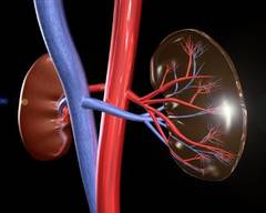
The serum creatinine was 3.89 mg/dL, and coagulation studies were normal. DM’s comorbidities were numerous, including hypertension, coronary artery disease, peripheral vascular disease, and paroxysmal atrial fibrillation. The referring nephrologist had already written orders for a pulsed steroid protocol (Solu-Medrol intravenously for 3 days), in addition to arranging for a renal biopsy by interventional radiology (IR) the following day.
Dr. Hospitalist 1 wrote standard admission orders and made DM nothing per os after midnight. For venous thromboembolism (VTE) prophylaxis, Dr. Hospitalist 1 also wrote for DM to receive mechanical compression to her legs and to begin Lovenox 30 mg subcutaneously daily after the biopsy, only when OK with IR.
The next day, DM was seen in the morning by her nephrologist and Dr. Hospitalist 2 on rounds. Later that afternoon, DM underwent a right renal biopsy under CT guidance by IR. A small hematoma was noted immediately following the procedure. At 8 p.m. that night, DM received 30 mg of Lovenox subcutaneously.
Overnight, DM became tachycardic and complained of right flank pain. Her morning labs drawn at 5 a.m. found a significant drop in her blood count (Hgb, 6.1 mg/dL) and decline in her renal function (serum creatinine, 4.46 mg/dL). A repeat CT of the abdomen and pelvis found an enlarging perinephric hematoma. Dr. Hospitalist 2 immediately moved DM to the ICU and began supportive transfusions.

DM’s blood counts and renal function stabilized over the next 24-36 hrs. However, on the evening of hospital day 4 (ICU day 2), DM experienced shortness of breath followed quickly by a ventricular fibrillation arrest. Telemetry strips before the code demonstrated ST-elevation myocardial infarction. It took almost 45minutes of resuscitation, but DM regained her pulse. At this point, she was now intubated and critically ill. Cardiology was consulted, but given DM’s recent perinephric hematoma, it was felt that she would not tolerate antiplatelet or antithrombotic therapies. Subsequent echocardiogram demonstrated a severely damaged left ventricle from her MI. Even worse, DM went into acute renal failure and shock liver with coagulopathy.
After multiple discussions with the family in the ensuing week, care was ultimately withdrawn and DM passed away.
Complaint
The family understood the bleeding risk of a renal biopsy, but they were upset to learn that DM received blood thinners around the time of her procedure. DM had a previous bleed on Coumadin years earlier, and she even had a "Coumadin allergy" documented in the chart. A lawsuit was filed and both the hospital (employer of the nurses) and Dr. Hospitalist 1 were named in the suit. The complaint alleged that it was substandard for DM to have received Lovenox in this case and that the Lovenox caused the hemorrhage, which in turn led to a myocardial infarction and death.
Scientific principles
Granulomatosis polyangiitis increases the risk for venous and arterial thrombosis. Patients at risk for thrombosis benefit from prophylactic anticoagulation. However, bleeding is the primary complication of renal biopsy and anticoagulation increases that risk. Postbiopsy bleeding can occur into one of three sites: 1) the collecting system (leading to microscopic or gross hematuria); 2) underneath the renal capsule (leading to pressure tamponade and pain); or 3) into the perinephric space (leading to hematoma formation and a possibly large drop in hemoglobin). Most clinically significant bleeding is recognized within 12-24 hours of the biopsy, but bleeding may occur up to several days after the procedure. Anticoagulation should not be used for at least 12-24 hours postbiopsy, and if possible, withholding anticoagulants for a more prolonged period of up to 1 week may be preferable.
Complaint rebuttal and discussion
Discovery in this case confirmed that the original order by Dr. Hospitalist 1 was not transcribed into the medical administration record with any conditions, and IR was not contacted to approve the Lovenox prior to administration. The nurse who gave the Lovenox was simply following the incomplete order that existed on the administration record.
As a result, the hospital settled with the plaintiff early in the process; yet the plaintiff continued to assert that the order itself (whether it was followed properly or not) was substandard and Dr. Hospitalist 1 was also negligent. Dr. Hospitalist 1 defended her Lovenox order as being appropriate and reasonable because DM was at risk for VTE, the dose prescribed was reduced for renal impairment, and it was a conditional order that required approval by interventional radiology prior to medication administration. Dr. Hospitalist 1 was resolute that if her order had just been followed, DM would never have received the medication.
Conclusion
Hospitalists write conditional orders every day. In fact, all "PRN" orders are conditional and presume the use of nursing clinical judgment. However, some conditional orders are directed at other physicians or services (for example, "Discharge the patient if OK with surgery" or "Resume Coumadin if OK with orthopedics").
Often these orders are expedient and typically replace direct communication. But hospitalists should remember that such conditional orders trigger a potential act of commission with nursing as the communication "go-between." There was nothing unreasonable about the conditional order for Lovenox in this case per se. Yet, had this order not been written at all – the chain reaction that subsequently occurred may have been avoided altogether. The jury in this case returned a defense verdict in favor of the hospitalist.
Dr. Michota is director of academic affairs in the hospital medicine department at the Cleveland Clinic and medical editor of Hospitalist News. He has been involved in peer review both within and outside the legal system. Read past columns at ehospitalistnews.com/Lessons.
Story
DM was a 60-year-old woman with a history of granulomatosis polyangiitis controlled with cyclophosphamide. She was sent to the hospital as a direct admission by her nephrologist because of worsening renal function and the possibility that DM was suffering from uncontrolled vasculitis. DM was admitted to the hospital by Dr. Hospitalist 1. The history and physical documented that DM was generally weak, but otherwise without specific complaints.
The serum creatinine was 3.89 mg/dL, and coagulation studies were normal. DM’s comorbidities were numerous, including hypertension, coronary artery disease, peripheral vascular disease, and paroxysmal atrial fibrillation. The referring nephrologist had already written orders for a pulsed steroid protocol (Solu-Medrol intravenously for 3 days), in addition to arranging for a renal biopsy by interventional radiology (IR) the following day.
Dr. Hospitalist 1 wrote standard admission orders and made DM nothing per os after midnight. For venous thromboembolism (VTE) prophylaxis, Dr. Hospitalist 1 also wrote for DM to receive mechanical compression to her legs and to begin Lovenox 30 mg subcutaneously daily after the biopsy, only when OK with IR.
The next day, DM was seen in the morning by her nephrologist and Dr. Hospitalist 2 on rounds. Later that afternoon, DM underwent a right renal biopsy under CT guidance by IR. A small hematoma was noted immediately following the procedure. At 8 p.m. that night, DM received 30 mg of Lovenox subcutaneously.
Overnight, DM became tachycardic and complained of right flank pain. Her morning labs drawn at 5 a.m. found a significant drop in her blood count (Hgb, 6.1 mg/dL) and decline in her renal function (serum creatinine, 4.46 mg/dL). A repeat CT of the abdomen and pelvis found an enlarging perinephric hematoma. Dr. Hospitalist 2 immediately moved DM to the ICU and began supportive transfusions.
DM’s blood counts and renal function stabilized over the next 24-36 hrs. However, on the evening of hospital day 4 (ICU day 2), DM experienced shortness of breath followed quickly by a ventricular fibrillation arrest. Telemetry strips before the code demonstrated ST-elevation myocardial infarction. It took almost 45minutes of resuscitation, but DM regained her pulse. At this point, she was now intubated and critically ill. Cardiology was consulted, but given DM’s recent perinephric hematoma, it was felt that she would not tolerate antiplatelet or antithrombotic therapies. Subsequent echocardiogram demonstrated a severely damaged left ventricle from her MI. Even worse, DM went into acute renal failure and shock liver with coagulopathy.
After multiple discussions with the family in the ensuing week, care was ultimately withdrawn and DM passed away.
Complaint
The family understood the bleeding risk of a renal biopsy, but they were upset to learn that DM received blood thinners around the time of her procedure. DM had a previous bleed on Coumadin years earlier, and she even had a "Coumadin allergy" documented in the chart. A lawsuit was filed and both the hospital (employer of the nurses) and Dr. Hospitalist 1 were named in the suit. The complaint alleged that it was substandard for DM to have received Lovenox in this case and that the Lovenox caused the hemorrhage, which in turn led to a myocardial infarction and death.
Scientific principles
Granulomatosis polyangiitis increases the risk for venous and arterial thrombosis. Patients at risk for thrombosis benefit from prophylactic anticoagulation. However, bleeding is the primary complication of renal biopsy and anticoagulation increases that risk. Postbiopsy bleeding can occur into one of three sites: 1) the collecting system (leading to microscopic or gross hematuria); 2) underneath the renal capsule (leading to pressure tamponade and pain); or 3) into the perinephric space (leading to hematoma formation and a possibly large drop in hemoglobin). Most clinically significant bleeding is recognized within 12-24 hours of the biopsy, but bleeding may occur up to several days after the procedure. Anticoagulation should not be used for at least 12-24 hours postbiopsy, and if possible, withholding anticoagulants for a more prolonged period of up to 1 week may be preferable.
Complaint rebuttal and discussion
Discovery in this case confirmed that the original order by Dr. Hospitalist 1 was not transcribed into the medical administration record with any conditions, and IR was not contacted to approve the Lovenox prior to administration. The nurse who gave the Lovenox was simply following the incomplete order that existed on the administration record.
As a result, the hospital settled with the plaintiff early in the process; yet the plaintiff continued to assert that the order itself (whether it was followed properly or not) was substandard and Dr. Hospitalist 1 was also negligent. Dr. Hospitalist 1 defended her Lovenox order as being appropriate and reasonable because DM was at risk for VTE, the dose prescribed was reduced for renal impairment, and it was a conditional order that required approval by interventional radiology prior to medication administration. Dr. Hospitalist 1 was resolute that if her order had just been followed, DM would never have received the medication.
Conclusion
Hospitalists write conditional orders every day. In fact, all "PRN" orders are conditional and presume the use of nursing clinical judgment. However, some conditional orders are directed at other physicians or services (for example, "Discharge the patient if OK with surgery" or "Resume Coumadin if OK with orthopedics").
Often these orders are expedient and typically replace direct communication. But hospitalists should remember that such conditional orders trigger a potential act of commission with nursing as the communication "go-between." There was nothing unreasonable about the conditional order for Lovenox in this case per se. Yet, had this order not been written at all – the chain reaction that subsequently occurred may have been avoided altogether. The jury in this case returned a defense verdict in favor of the hospitalist.
Dr. Michota is director of academic affairs in the hospital medicine department at the Cleveland Clinic and medical editor of Hospitalist News. He has been involved in peer review both within and outside the legal system. Read past columns at ehospitalistnews.com/Lessons.

App use increased physical activity
People who used a smartphone pedometer app walked 22% more steps (or about half a mile) per day after 8 weeks than did people in a control group in a small, randomized, open-label study.
The 90 participants in rural western Ireland were older than 16 years and owned an Android smartphone, which they were asked to keep charged and to carry during waking hours. Investigators downloaded the app to all phones and calibrated it to each individual to record step counts accurately. All participants were given physical activity goals and information on the benefits of exercise.

In the first week, the app display was not visible while it recorded baseline activity data. Mean step counts in the first week were 5,138 steps/day in the control group and 4,365/day in the intervention group. That difference was not statistically significant, but the investigators still adjusted for it in final analyses, Dr. Liam G. Glynn and his associates reported in the British Journal of General Practice.
The intervention group was then showed the app’s features and settings and encouraged to interact with the app to help meet activity goals. The control group’s app continued to track steps without any interaction.
Among 40 people in the control group and 37 in the intervention group with follow-up data 7 weeks later, the daily counts decreased by 386 steps in the control group and increased by 1,631 steps in the intervention group (Br. J. Gen. Pract. 2014;64:e384-91).
Participants in both groups increased their walking initially, but only the intervention group maintained the increased activity, accounting for the step count difference between groups at 8 weeks, said Dr. Glynn, a general practice physician at the National University of Ireland, Galway.
The study used the Accupedo-Pro pedometer app because it included preestablished, desirable features such as automatic feedback and tracking of step counts and calories burned, visually appealing graphics of step count history, the ability to input goals, and feedback on goal achievement. The Accupedo-Pro app is available for iPhones and Android devices for $3.99. Hundreds of other activity-tracking apps of varying quality are available for free or for sale.
After researchers adjusted for factors unrelated to the study that may have increased step counts during the study period, they found that the mean daily count at baseline for the intervention group was 4,771 steps/day. The difference between the intervention and control groups by the end of 8 weeks was a mean increase of 1,029 steps/day favoring the app use, a 22% gain, compared with the intervention group’s baseline, Dr. Glynn reported.
That’s enough of a change to be clinically meaningful if it’s maintained, based on a large body of literature on exercise and health, he said. Dr. Glynn and his associates now are following app users in several European countries to monitor long-term health outcomes.
The current study was too small to show a difference in health measures such as blood pressure, weight, or body mass index. And it was too short to show whether or not apps embedded in phones can overcome one of the weak points of separate devices used solely as pedometers or activity trackers – namely, the tendency for many people to stop using them over time.

One in three people who owned a fitness tracker stopped using the device in the past year, mainly because they lost interest in tracking their activity level, according to a survey by International Data Corp.
Nonetheless, pedometers and other small hardware-based fitness trackers remain popular. Florida insurer AvMed recently announced that it will cover the cost of the Fitlinxx Pebble activity meter for Medicare Advantage members enrolled in the Walkadoo pedometer-based walking program run by MeYou Health.
Separate data suggest that, among mobile phone customers, 60% in the United States and 57% in Ireland have a smartphone, Dr. Glynn said, and that 90% of U.S. mobile phone users have their phone with them 24 hours a day. Theoretically, a smartphone pedometer app might get more use than a conventional pedometer because it doesn’t require carrying a separate piece of technology.
"This intervention is now firmly on my list (as a clinician) of options for patients to whom I am recommending increased physical activity," Dr. Glynn said in an interview.
He reported having no financial disclosures.
On Twitter @sherryboschert
People who used a smartphone pedometer app walked 22% more steps (or about half a mile) per day after 8 weeks than did people in a control group in a small, randomized, open-label study.
The 90 participants in rural western Ireland were older than 16 years and owned an Android smartphone, which they were asked to keep charged and to carry during waking hours. Investigators downloaded the app to all phones and calibrated it to each individual to record step counts accurately. All participants were given physical activity goals and information on the benefits of exercise.
In the first week, the app display was not visible while it recorded baseline activity data. Mean step counts in the first week were 5,138 steps/day in the control group and 4,365/day in the intervention group. That difference was not statistically significant, but the investigators still adjusted for it in final analyses, Dr. Liam G. Glynn and his associates reported in the British Journal of General Practice.
The intervention group was then showed the app’s features and settings and encouraged to interact with the app to help meet activity goals. The control group’s app continued to track steps without any interaction.
Among 40 people in the control group and 37 in the intervention group with follow-up data 7 weeks later, the daily counts decreased by 386 steps in the control group and increased by 1,631 steps in the intervention group (Br. J. Gen. Pract. 2014;64:e384-91).
Participants in both groups increased their walking initially, but only the intervention group maintained the increased activity, accounting for the step count difference between groups at 8 weeks, said Dr. Glynn, a general practice physician at the National University of Ireland, Galway.
The study used the Accupedo-Pro pedometer app because it included preestablished, desirable features such as automatic feedback and tracking of step counts and calories burned, visually appealing graphics of step count history, the ability to input goals, and feedback on goal achievement. The Accupedo-Pro app is available for iPhones and Android devices for $3.99. Hundreds of other activity-tracking apps of varying quality are available for free or for sale.
After researchers adjusted for factors unrelated to the study that may have increased step counts during the study period, they found that the mean daily count at baseline for the intervention group was 4,771 steps/day. The difference between the intervention and control groups by the end of 8 weeks was a mean increase of 1,029 steps/day favoring the app use, a 22% gain, compared with the intervention group’s baseline, Dr. Glynn reported.
That’s enough of a change to be clinically meaningful if it’s maintained, based on a large body of literature on exercise and health, he said. Dr. Glynn and his associates now are following app users in several European countries to monitor long-term health outcomes.
The current study was too small to show a difference in health measures such as blood pressure, weight, or body mass index. And it was too short to show whether or not apps embedded in phones can overcome one of the weak points of separate devices used solely as pedometers or activity trackers – namely, the tendency for many people to stop using them over time.
One in three people who owned a fitness tracker stopped using the device in the past year, mainly because they lost interest in tracking their activity level, according to a survey by International Data Corp.
Nonetheless, pedometers and other small hardware-based fitness trackers remain popular. Florida insurer AvMed recently announced that it will cover the cost of the Fitlinxx Pebble activity meter for Medicare Advantage members enrolled in the Walkadoo pedometer-based walking program run by MeYou Health.
Separate data suggest that, among mobile phone customers, 60% in the United States and 57% in Ireland have a smartphone, Dr. Glynn said, and that 90% of U.S. mobile phone users have their phone with them 24 hours a day. Theoretically, a smartphone pedometer app might get more use than a conventional pedometer because it doesn’t require carrying a separate piece of technology.
"This intervention is now firmly on my list (as a clinician) of options for patients to whom I am recommending increased physical activity," Dr. Glynn said in an interview.
He reported having no financial disclosures.
On Twitter @sherryboschert

ALS ice bucket challenge: It’s hard to argue with success
The ALS ice bucket challenge has taken the social media world by storm and surprised many by how fast and far it has spread. People simply make a video of themselves dumping a bucket of ice water on their heads and then post it on a social media site and challenge others to do the same within 24 hours or make a donation to ALS research (or both).
The stunt, which began early in the summer as a challenge unrelated to amyotrophic lateral sclerosis, has resonated with many people in the dog days of summer and has been helped by many celebrities taking up the challenge. It became linked to ALS when Peter Frates, a 29-year-old man with the disease, took the challenge – albeit by nodding his head to the song "Ice Ice Baby" instead of having ice water dumped on him – and asked others to do the same.

According to the ALS Association, as of Aug. 19, existing donors and more than 450,000 new donors have contributed $22.9 million since July 29, compared with $1.9 million during the same period last year. The ALS has a four-out-of-four stars rating on Charity Navigator, and an overall score of 90.73 out of 100. Overall, 72% of its expenses are spent on the programs and services it delivers, 11% on administration, and 17% on fundraising.
Some critics have suggested that the stunt promotes click and post activism, keeping people from doing real activism, or is "narcissism masked as altruism," but most people have embraced it as fun for a good cause.
I asked a few Clinical Neurology News editorial advisory board members to weigh in:
• Dr. Richard J. Caselli, professor of neurology at the Mayo Clinic, Scottsdale, Ariz.: "If it is raising money for ALS research, what’s not to like? Not everyone was destined to be a molecular biologist or clinical trialist, and this gives people a way to contribute that seems to be culturally in synch with the ‘social media’ community. ... The ice water is an interesting twist in that it implies that if you turn a blind eye to this cause you should punish yourself, and I suspect many people harbor feelings of at least slight guilt when they feel they are not contributing to worthwhile needs."

• Matthew Huentelman, Ph.D., associate professor of neurogenomics at the Translational Genomics Research Institute, Phoenix: "If a campaign works (and doesn’t harm) then it is hard to argue against. I think that any awareness campaign that actually gets a response from the White House has probably been a useful one. President Obama confirmed he would be donating but not doing the ice bucket thing. ... I suspect that a lot of research foundations are going to be having ‘tough’ conversations at this month’s board meetings as they compare their awareness and fundraising attempts to the simple ALS ice bucket challenge. It just demonstrates to all of us again that social media is relevant for both spreading the word and getting results. There are a few keys from this too: (1) visual "stuff" matters – short video clips is now how the world communicates; (2) challenging your friends/colleagues by name is important, too – it sort of forces a response from them; and (3) celebs are still key to pushing something viral in a truly short period of time."
(While you contemplate taking the ice bucket challenge, you might as well visit Dr. Huentelman’s social media project, MindCrowd, a site leveraging social media to recruit participants into a brain research study.)
The ALS ice bucket challenge has taken the social media world by storm and surprised many by how fast and far it has spread. People simply make a video of themselves dumping a bucket of ice water on their heads and then post it on a social media site and challenge others to do the same within 24 hours or make a donation to ALS research (or both).
The stunt, which began early in the summer as a challenge unrelated to amyotrophic lateral sclerosis, has resonated with many people in the dog days of summer and has been helped by many celebrities taking up the challenge. It became linked to ALS when Peter Frates, a 29-year-old man with the disease, took the challenge – albeit by nodding his head to the song "Ice Ice Baby" instead of having ice water dumped on him – and asked others to do the same.
According to the ALS Association, as of Aug. 19, existing donors and more than 450,000 new donors have contributed $22.9 million since July 29, compared with $1.9 million during the same period last year. The ALS has a four-out-of-four stars rating on Charity Navigator, and an overall score of 90.73 out of 100. Overall, 72% of its expenses are spent on the programs and services it delivers, 11% on administration, and 17% on fundraising.
Some critics have suggested that the stunt promotes click and post activism, keeping people from doing real activism, or is "narcissism masked as altruism," but most people have embraced it as fun for a good cause.
I asked a few Clinical Neurology News editorial advisory board members to weigh in:
• Dr. Richard J. Caselli, professor of neurology at the Mayo Clinic, Scottsdale, Ariz.: "If it is raising money for ALS research, what’s not to like? Not everyone was destined to be a molecular biologist or clinical trialist, and this gives people a way to contribute that seems to be culturally in synch with the ‘social media’ community. ... The ice water is an interesting twist in that it implies that if you turn a blind eye to this cause you should punish yourself, and I suspect many people harbor feelings of at least slight guilt when they feel they are not contributing to worthwhile needs."
• Matthew Huentelman, Ph.D., associate professor of neurogenomics at the Translational Genomics Research Institute, Phoenix: "If a campaign works (and doesn’t harm) then it is hard to argue against. I think that any awareness campaign that actually gets a response from the White House has probably been a useful one. President Obama confirmed he would be donating but not doing the ice bucket thing. ... I suspect that a lot of research foundations are going to be having ‘tough’ conversations at this month’s board meetings as they compare their awareness and fundraising attempts to the simple ALS ice bucket challenge. It just demonstrates to all of us again that social media is relevant for both spreading the word and getting results. There are a few keys from this too: (1) visual "stuff" matters – short video clips is now how the world communicates; (2) challenging your friends/colleagues by name is important, too – it sort of forces a response from them; and (3) celebs are still key to pushing something viral in a truly short period of time."
(While you contemplate taking the ice bucket challenge, you might as well visit Dr. Huentelman’s social media project, MindCrowd, a site leveraging social media to recruit participants into a brain research study.)

The Medical Roundtable: Hypertension in Pregnancy
That’s one of the reasons we’re having this conversation. One of the more controversial issues in medicine has been that of hypertension in pregnant women. The guidelines have not changed much in the last 50 years, although some effort to update these guidelines has been made recently. Today, we discuss the following: (1) The definition of elevated BP in pregnancy: is it too complicated? (2) Indications for therapy; should you wait until the BP increases to levels of 160/105 to 110 mm Hg? Is there any danger in allowing the systolic BP to remain elevated for a few months in a pregnant woman? (3) The indications for interruption of pregnancy; should we begin treatment earlier and continue to use the drugs that have been suggested over the years? During pregnancy, hypertension is typically classified as chronic hypertension (pre-existing or onset prior to 20 weeks gestation), gestational hypertension, preeclampsia, or preeclampsia superimposed on chronic hypertension.
I’m Dr. Marvin Moser, Clinical Professor of Medicine at Yale. We have with us Dr. Phyllis August, Professor of Research and Medicine and OB/GYN at Cornell Weill Medical College; Vesna Garovic, a Professor of Medicine at the Mayo Clinic who has done a great deal of work on hypertension in pregnancy; and Dr. Carl Rose, Associate Professor of Maternal-Fetal Medicine at the Mayo Clinic, to discuss this controversial subject.
Dr. August, let’s start with you. What about the definition of hypertension? We have a very complicated list of definitions of hypertension in pregnancy; do you want to review some of those? Should we simplify the criteria for clinicians?
Dr. August: I think we need to distinguish between the diagnostic category and the definition of hypertension. The definition of hypertension for the general, non-pregnant population is not very different from that for pregnant women. Obstetricians speak about mild hypertension in pregnancy, which they define as a systolic pressure of 140 to 150 mm Hg and diastolic pressure of 90 to 109 mm Hg, and then severe hypertension. There are 2 categories for hypertension: severe hypertension is a pressure of 160/110 mm Hg and above, and mild to moderate hypertension is a pressure below that level. The diagnosis of hypertension is made at a pressure of 140/90 mm Hg and above, so really, it’s not very different from Stages 1 and 2 of essential hypertension outlined in the last Joint National Committee report.1
Preeclampsia, preeclampsia superimposed on chronic hypertension, chronic hypertension alone, and gestational hypertension are the diagnostic categories within which you can have either mild to moderate or severe hypertension.
Dr. Moser: Do we need all these designations? I know that the pathophysiology of preeclampsia is different from that of essential hypertension, but are the definitions helpful for the clinician? What’s the difference, for example, if you have someone with chronic hypertension who becomes pregnant? What if you have someone with pre-eclampsia superimposed on chronic hypertension, and you have these 4 categories. Are they helpful? Does this definition affect therapy?
Dr. August: They are helpful because what we really need to identify is the condition of the woman who, because of the pregnancy, is either developing new hypertension, or that of a woman who had hypertension before, but because of the pregnancy, her hypertension is worsening. That situation can change quickly, leading to serious maternal vmorbidity and fetal morbidity. This also helps to identify women who really need to be watched closely and addressed in a different way from someone who, at the beginning of pregnancy, had a pressure of 150/100 mm Hg and has the same pressure even at mid-pregnancy. I think the diagnostic categories are useful, and the fact that they’ve been used for many decades supports their effectiveness. They’re not perfect, but they are useful.
Dr. Moser: Dr. Rose, do you want to add to that?
Dr. Rose: As an obstetrician, I would suggest that these categories allow us to communicate with one another using common terminology that we collectively understand. It also influences the obstetric management of these patients.
Dr. Garovic: It is likely that young women who don’t see a physician and present with a BP of 150/100 mm Hg when they are pregnant, have had hypertension for a longer period, which has been asymptomatic and subclinical and that it has been brought to the attention of the clinician only due to the pregnancy.
Having said that, I’m not sure that it helps that much with respect to treatment because target levels at which BP treatment is started are decreasing, especially in the tertiary centers. My approach is to treat a pregnant woman with a documented systolic BP of 150 mm Hg on at least 2 occasions, whether symptomatic or asymptomatic and at risk for preeclampsia or not.
Dr. Moser: In the last American Society of Hypertension recommendations,2 it was clearly stated that the level of the BP should guide treatment and the decision of when to interrupt or deliver the pregnancy. So, if it is the level of BP that the clinician is following, why do we need sub-designations? In other words, if the woman has chronic hypertension to start with, you’re obviously going to try and keep the systolic BP below 140 mm Hg. If she develops hypertension in the first trimester or second trimester, which is a rare occurrence, you’re going to reduce the BP. If she develops hypertension in the third trimester with or without proteinuria, for example, you’re going to treat it. So, why don’t we simplify this and treat the BP without 4 or 5 definitions? Of course, if the BP rises and there is proteinuria in the third trimester, other options will also need to be considered. Also, proteinuria in the second trimester is an indication of preeclampsia.
Dr. August: In my experience, if you have a patient who has a BP reading of 120/80 mm Hg at the start of pregnancy, and then at 20 weeks, the BP is 150/100 mm Hg, even with no proteinuria, you have to be very worried about the patient because she’s developing either superimposed preeclampsia or gestational hypertension, until proven otherwise. That’s different from a BP of 150/100 mm Hg that was stable and started out that way at 8 weeks of pregnancy, and then at 20 weeks, it still is 150/100 mm Hg. There has been no change, and no evidence for an evolving process that is potentially dangerous to the mother and fetus, so I would disagree that it’s just a number.
Dr. Moser: How would you treat the patient differently, Dr. August?
Dr. August: If a woman had a clear, well-documented pressure of 110 to 120/70 to 80 mm Hg early in pregnancy and then in the second trimester and developed mild to moderate or Stage 1 hypertension, I would make sure to check all her laboratory data and rule out preeclampsia, but I would be very worried that she was developing a pregnancy-related disorder like pre-eclampsia or gestational hypertension, and I would examine her very frequently.
Dr. Moser: But would you treat her BP?
Dr. August: I would probably treat her BP if it was 150/95 to 99 mm Hg. Even though the guidelines say 160 mm Hg, 150 mm Hg is close to 160 mm Hg and since the patient’s pressure is currently at 150 mm Hg, it may increase to 160 mm Hg in an hour from now when the patient is a little more stressed and uncomfortable; therefore, 150 mm Hg is my cutoff.
Dr. Moser: Consider this: the normal systolic pressure of a pregnant woman is 110 mm Hg, 120 mm Hg, or even lower sometimes, and even in the second and third trimester, it shouldn’t be more than 120 mm Hg, and 130 mm Hg at the most. Now, if a woman’s pressure exceeds the 140/90 mm Hg cutoff of hypertension, why do obstetricians wait until the level increases to 150 mm Hg, or as you said, 160 mm Hg?
Dr. August: They wait because they think lowering the BP too much might compromise the placental perfusion.
Dr. Moser: Dr. Garovic or Dr. Rose, can you comment on the concept that an elevated systolic pressure for a couple of months in pregnancy isn’t going to do any harm?
Dr. Rose: I agree with Dr. August that the pathophysiology of the processes is different. In other words, I do not believe that we should simply treat hypertension and categorize all the accompanying processes as a similar prenatal complication. If a mother develops new-onset hypertension late in pregnancy, certainly this represents a different clinical scenario from one who develops hypertension at 18 weeks of gestation, although implications for the pregnancy may ultimately be similar.
Dr. August: But I would argue that a woman who develops hypertension at 18 weeks and didn’t have it at 12 weeks is at a more serious risk for preterm birth than the woman who develops hypertension in the third trimester.
Dr. Moser: Let me argue on the other side again. Such patients are at risk, but what are you going to do about the risk? You’re going to check some enzymes and chemistries, etc, but what is the only thing that you might do to reduce the risk of progression of hypertension or a small placenta, which may be a result of infarcts from elevated BP? I’m trying to determine whether just lowering the BP, even though the pathophysiology is different, might prevent progression of hypertension. I know it may not prevent the progression of preeclampsia, but early treatment might prevent or delay interruption of pregnancy, based upon the criteria of an elevated BP.
Dr. Garovic: I just want to clarify the point that I was discussing earlier. A BP of 150/100 mm Hg at 21 weeks’ gestation may represent a significantly elevated BP, as the patient’s BP, at that time of pregnancy, should be even lower than her pre-pregnancy levels. I would not treat somebody with a BP of 150/100 mm Hg based on a single office reading for the same reason that we may not treat hypertension based on a single office reading in the general population. However, if I have enough evidence that a patient has sustained hypertension, especially in the second trimester when the BP should be lower than non-pregnant levels, I will treat that patient. Treatment is given after a 6-hour BP reading or nurse-monitored BP readings over a 30-minute to 1-hour period to generate enough evidence to show that BP is certainly steadily elevated.
I would like to discuss a recent patient: she was a 36-year-old woman with a BP of 150/100 mm Hg. It was her first pregnancy, and she was approximately 12 weeks pregnant. She was referred to us from one of the regional clinics close to Rochester. She underwent a retroperitoneal ultrasound, which showed bilateral renal artery stenosis/fibromuscular secondary to fibromuscular dysplasia. So, she had a BP of 150/100 mm Hg, but by the time she saw us, her systolic pressure was 160 to 170 mm Hg and diastolic pressure was in the 100s range. Dr. Rose and I aggressively treated her with 300 mg labetalol twice a day and nifedipine once a day; her BP effectively was reduced to 120 to 130 mm Hg systolic pressure and 70 to 80 mm Hg diastolic pressure, and she delivered a full-term baby boy at 38 gestational weeks.
Dr. August: Let me ask you a question: did she really have renovascular hypertension? Was she successfully revascularized and did her blood pressure normalize after revascularization?
Dr. Garovic: At 6 months post-partum, she underwent a renal angiogram with bilateral angioplasties with a subsequent normalization of her BP. She remains normotensive, when off BP medications.
Dr. Moser: Do you believe that if we treat elevated BP early, we might delay or prevent iatrogenic preterm deliveries?
Dr. August: Yes, I think your point is that if somebody in the third trimester has preeclampsia or gestational hypertension and severe hypertension, and you lower their BP and, if all other parameters are normal, the baby is fine, and the mother’s platelet count is normal, you can prevent her from having a preterm birth. You can extend the pregnancy by preventing the BP from reaching levels that are too dangerous and thus require delivery.
Dr. August: I don’t think there’s any such strong evidence, but there are several issues to consider. One is that you need to weigh the risks and benefits. We don’t have convincing data on the effects, except the hemodynamic effects, of the drugs on the baby’s neurologic development and organ development. We have certain drugs that we think are safe, but have they been rigorously evaluated? Can we say with a 100% certainty that these drugs have absolutely no adverse effects on fetal growth and development? No, we can’t.
During pregnancy, we avoid exposure to any drug. The question is “what are the risks of treating mild hypertension and what are the benefits?” The treatment benefits to the mother maybe fairly small at 140/90 mm Hg. The risks to the baby are not fully known yet. Hemodynamically, we don’t know that the placental blood flow is being compromised. The data that suggest that smaller babies are a result of lowering the BP are very poor. However, you have to think that it’s probably better to use as few medications as possible during pregnancy for those reasons. Dr. Rose, I’d like to hear your opinion on this.
Dr. Rose: I would concur with Dr. August. Although Peter von Dadelszen’s 2002 metaanalysis3 suggests an association between BP control and fetal growth restriction, the more recent systematic review from Abalos4 suggests that treatment of chronic hypertension during pregnancy for lowering the BPs to thresholds lower than historically considered for therapy, should have a very small effect on neonatal birth weight.4
Dr. Moser: Maybe, some newborns born of hypertensive women are smaller because of the placental infarcts.
Dr. August: It is entirely plausible that hypertension contributes to placental pathology; The question of which is worse—mild hypertension or antihypertensive therapy—has simply not been adequately studied either. It’s just the general principle of whether you can avoid using a medication during pregnancy; if you can, you avoid it, whatever be the medication.
Dr. Garovic: I don’t know whether Dr. Rose and Dr. August would agree, but that meta-analysis3 has one important limitation that is hardly ever addressed. It was published in 2002; so, basically, it included studies published before 2000 when doctors were even more reluctant to treat hypertension. Who was getting treated then? It was likely severely hypertensive women and those with diabetes and other risk factors that are associated with an increased risk for intrauterine growth restriction from the beginning of their pregnancies. So, there may have been an initial selection bias about whom to treat. Therefore, intrauterine growth restriction in these women is likely, at least in part, related to their underlying comorbidities.
Dr. August: I completely agree with Dr. Garovic about that, and yes, I think you make a very good point.
Dr. Moser: To summarize this particular part of the discussion, all of you would agree that treating a patient with a BP > 140/90 mm Hg, even though that’s the definition of hypertension, may not be a good idea because the use of any medication in pregnancy is to be avoided, if possible. I believe that 140/90 mm Hg is an abnormal BP in a pregnant woman, whether it’s at the beginning of pregnancy or later on at 20 weeks or 30 weeks of gestation. I would favor treatment and lowering of BP slowly, because I don’t think the evidence showing fetal harm is very strong. The general consensus, however, of the experts thus far is that they are reluctant to treat a patient until the pressure reaches 150/100 or 155/100 mm Hg.
Dr. August: Well, it’s toward 150 mm Hg. One thing I can say with certainty is that we don’t have good data. We don’t have data to indicate that that treating a pressure of 140/90 mm Hg is harmful, nor do we have good data to show that treating mild hypertension is beneficial.
Dr. Garovic: I don’t know whether current theories would agree with that, but if a young patient (say about 28 years old) without any risk factors has a steady elevation of BP above 140/90 mm Hg, and I see her when she is 12 weeks pregnant, ie, early in pregnancy, I will treat her and follow-up her BP closely.
Dr. August: I agree with that.
Dr. Garovic: Again, I don’t think that it is a wise decision to treat hypertension based on a single BP reading. For me, a steady BP above 140/90 mm Hg in a young patient is an indication for treatment. We are trying to identify these patients very early and keep their BPs at 130/80 mm Hg throughout their pregnancies.
Dr. August: I would agree with that.
Dr. Moser: Would all of you agree that if a woman came in pregnant with chronic hypertension on medication, the medication should be continued, except for agents that effect the renin-angiotensin system, with the caveat that in the first trimester, you might have to lower the dosage because the pressures may be lower?
Dr. August: Most of the time, we do that. We don’t stop all the medications. We follow-up the patients closely. We see if the pregnancy has caused a decrease in BP. If the patient’s BP is 110/70, we would consider reducing the BP medications; we keep many women on medications during pregnancy, as long as they’re safe under them.
Dr. Moser: But you’d keep them on a diuretic or calcium channel blocker?
Dr. August: I would choose nifedipine because there’s more data about nifedipine.
Dr. Garovic: I agree with that.
Dr. August: I think amlodipine is safe, but unfortunately, there aren’t a lot of data on it. I have used verapamil; there are some data on it, although not comparable to the data on nifedipine.
Dr. Garovic: Verapamil also acts faster than amlodipine: it starts working earlier.
Dr. Moser: What about the continual use of diuretics, for example?
Dr. August: I continue administering them, particularly in patients with salt-sensitive hypertension. It is often possible to lower the dose during pregnancy if blood pressure decreases. I stop spironolactone but not hydrochlorothiazide. I don’t think there are a lot of data on chlorthalidone in pregnancy.
Dr. Moser: Dr. Rose, would you agree that if a person with known hypertension came in on medication, you tend to continue it, with the caveat, as Dr. August said, that if the pressure reduced, which it does in the first trimester, you might have to adjust the dosage downward, and, of course, never continue an angiotensin-converting-enzyme inhibitor, angiotensin receptor blocker, or renin inhibitor because of damage to the fetus?
Dr. Rose: Absolutely. During the second trimester, when the physiologic fall in BP occurs, one may need to discontinue medications in some patients, anticipating that therapy will be resumed later in gestation during the third trimester when BPs begin to rise.
Dr. Garovic: We are now trying to conduct a population-based study and see the effects of that “short” duration of hypertension in pregnancy with respect to cardiovascular risk later in life. Chances are that hypertension of any duration may induce vascular damage. For example, magnetic resonance imaging studies of the brain in women with eclampsia showed vasogenic edema involving the posterior circulation, which, with neurological signs and symptoms, was consistent with the diagnosis of posterior reversible encephalopathy syndrome. Follow-up studies of these patients have suggested that permanent cerebrovascular damage of the brain may occur in some of them.5,6 These changes may have some long-term effects on their neurological health and cognitive functioning later in life.
Dr. Moser: So, you’re convinced that a BP of 150 or 160 mm Hg, even for just 4 or 5 months in a young woman, might produce some long-term vascular changes?
Dr. Garovic: I think that there is a high possibility for that. Again, we don’t have data to support it, but the absence of evidence is not the evidence of absence. I would be delighted to see a study following up these women after their affected pregnancies. A paper published in the Journal of American College of Obstetrics and Gynecology7 indicated that women with a history of eclampsia have self-reported poor cognitive functioning years after their pregnancies. So, preeclampsia and brain neurological deficit are concerning conditions, and we don’t know whether they are related to only the BP or some other mechanisms. For example, endothelial dysfunction, which is a systemic condition in pre-eclamptic pregnancy, may actually facilitate any kind of vascular damage induced by high BPs.
Dr. Moser: There is evidence of endothelial dysfunction in hypertension too. Drs. August and Rose, would you agree or disagree that systolic BPs of 150 or 160 mm Hg for 4 or 5 months can produce permanent vascular changes?
Dr. August: I wouldn’t rule it out. It could happen in some people, and maybe there’s some heterogeneity; some might be able to tolerate it with impunity, but it may be more dangerous in people with other vascular risk factors or older individuals. High BP for 4 to 6 months is not very good.
Dr. Rose: I would agree. I think that at this point, we don’t have substantive data that speak one way or the other. But, it certainly seems very reasonable.
Dr. Moser: Yes, in some of the hypertension studies done in pregnant women, it would appear that if you treat early, the benefit is greater. But as you say, the data are certainly not compelling.
Since we don’t completely agree on when you should start treatment, although I think there’s a tendency toward earlier intervention, perhaps we should discuss what medicines should be used now. Obstetricians have been using alpha methyldopa and hydralazine for many years, because they’re comfortable with them and because studies have shown that they’re well tolerated. All of us in the hypertension field, however, know that these may not be the most effective drugs, and if you use them and the pressure keeps increasing, there’s a tendency for pregnancy interruption. What about changing the treatment algorithms, even though, as Dr. August has pointed out many times, there is little evidence of long-term outcomes with some of the newer drugs. Except for the angiotensin-converting–enzyme inhibitors and the angiotensin receptor blockers, there is very little evidence of fetal problems with beta blockers, although there is a report of smaller babies, but that was questionable as obstetricians are reluctant to use beta blockers, calcium channel blockers, or diuretics, partly because of lack of experience and possible litigation.
Dr. Rose: We are a relatively conservative group.
Dr. August: We use labetalol a lot. I think everybody’s happy with labetalol, which is an alpha-beta blocker. It’s reasonably effective, so I think that’s become the drug that everybody reaches for first, to treat hypertension in pregnancy, and I would say the next is nifedipine. I just got an E-mail today from a patient who I started administering nifedipine to, and she’s already complaining of swollen ankles and headache. You tend to get more side effects from nifedipine than labetalol, even though it may be more effective. Methyldopa is not the best drug, but it does work in pregnancy. There’s some evidence that sympathetic nervous system activity is part of the pathogenesis of preeclampsia, and I’ve seen methyldopa work. It makes people tired, which means that they don’t do much and spend a lot of time resting which is not necessarily bad.
Dr. Moser: Oh, yes, and depressed.
Dr. August: But, I’ve written a few prescriptions for methyldopa in the last 12 months. It’s not my first choice; I’d say it’s my third. I don’t initiate diuretics for preeclampsia or gestational hypertension. However, I might start somebody with chronic hypertension on a diuretic.
Dr. Moser: What about the effect or concern for litigation? You deliver babies, and those who deliver babies are very concerned about this because any drug, as Dr. August mentioned, has a potential for doing something, and some lawyer somewhere is going to find a reason to say it’s a terrible drug to use. Do you think that plays a role in obstetricians’ activities?
Dr. August: I think the legal profession has ruined the field of OB/GYN. They’ve decimated it. They’ve made it impossible; I don’t know what Dr. Rose has to say about it, but I think it’s one of the great tragedies of the last 30 years in medicine.
Dr. Rose is a hero for persisting in this environment and actually taking the risks to be involved in the process of delivering babies when it is such a hostile environment. I feel strongly about it because I’ve seen it, and I’m sure Dr. Rose does too.
Dr. Rose: Additionally, dosing frequency perhaps should be a consideration in the treatment of hypertension, with any type of medication requiring more frequent dosing intervals, the subsequent compliance rates are much lower. Thus, when I consider any type of medication that I'm prescribing more than twice a day, I'm not sure how realistic it is to expect good compliance.
Dr. Moser: I’m going to ask each of you to imagine that you were the chairperson of a new committee and summarize the definitions of hypertension and decide when to treat and how to treat. Would you make any major suggestions that are different from the present guidelines that have been used for so many years?
Dr. Rose: Currently, the American Congress of Obstetricians and Gynecologists recommends instituting medical therapy for patients with chronic hypertension at threshold values of 150–160/100–110 mm Hg and at diastolic values of 105–110 mm Hg for patients with preeclampsia.8
Dr. Moser: What about the drugs that we use? Would you expand the recommendations, as Dr. August mentioned, to include calcium channel blockers, drugs like labetalol, and diuretics?
Dr. Rose: Yes.
Dr. Garovic: With respect to medications, I completely agree with what other speakers have said. In general, young women, especially those with either severe hypertension in the absence of a family history or with features of secondary hypertension (such as an abdominal bruit) should be ruled out for secondary hypertension, optimally before pregnancy.
Dr. August: I would argue for a clinical trial similar to, but not exactly the same as, the Systolic Blood Pressure Intervention Trial (SPRINT), in non-pregnant people, where you could at least establish the safety of lowering BP to a normal BP (eg, 120/80 mm Hg) in women with chronic hypertension. I think one of the problems with studies like Action to Control Cardiovascular Risk in Diabetes (ACCORD), and maybe SPRINT, is that there are certain patients in whom you should not force the BP to 120/80 mm Hg. If you have to combine 3 or 4 drugs, it’s probably not worth it. So, I think the design of such a study would have to be carefully considered. But, I would like to know whether it’s safe; should we keep women at approximately normal BPs during pregnancy and look at the outcomes that are more pregnancy-related as well as maternal?
Dr. Moser: Is it advisable not to wait until their pressure gets as high as presently supported by guideline committees, just because there is no evidence and no definitive studies to prove otherwise?
Dr. August: Yes. But a large, cooperative, collaborative, multi-center study would need to be conducted to prove this.
Dr. Moser: I thank you all very much.
FoxP2 Media LLC is the publisher of The Medical Roundtable.
FoxP2 Media LLC

Sunshine Act – a reminder
Last year, when the Physician Payment Sunshine Act became law, I recommended that all physicians involved in any sort of financial relationship with the pharmaceutical industry review the data reported about them prior to public posting of the information. Since that posting is due to occur at the end of this month, a reminder is in order.
Under a new bureaucracy created by the Affordable Care Act – formally called the Open Payments program – all manufacturers of drugs, devices, and medical supplies covered by federal health care programs must report any financial interactions with physicians and teaching hospitals to the Centers for Medicare & Medicaid Services (CMS).
Reportable interactions include consulting; food; ownership or investment interest; direct compensation for speakers at education programs; and research. Compensation for conducting clinical trials must be reported, but will not be posted on the website until the product receives approval from the Food and Drug Administration or until 4 years after the payment, whichever is earlier. Payments for trials involving a new indication for an approved drug will be posted immediately.
There are a number of specific exclusions, such as certified and accredited continuing medical education activities funded by manufacturers, and product samples for patient use. Medical students and residents are excluded entirely.
Under the law, you are allowed to review your data and seek corrections before it is published. Publication is scheduled to occur on Sept. 30, so if you have not yet done this, there is no time to waste. Although you will have an additional 2 years to pursue corrections after the online content goes live, any erroneous information will remain on the site until corrections can be made, so the best time to find and fix errors is now.
If you don’t see drug reps, accept sponsored lunches, or give sponsored talks, don’t assume that you won’t be on the website. Check anyway; you might be indirectly involved in a compensation situation that you were not aware of, or you may have been reported in error.
To review your data, register at the CMS Enterprise Portal, and request access to the Open Payments system.
Once you are satisfied that your interactions have been reported accurately, the question of what effect the law will have on research, continuing education, and private practice remains. The short answer is that no one knows. Much will depend on how the public interprets the data – if they take notice at all.
Sunshine laws have been in effect for several years in six states: California, Colorado, Massachusetts, Minnesota, Vermont, and West Virginia; plus the District of Columbia. (Maine repealed its law in 2011.) Observers disagree on their impact. Studies in Maine and West Virginia showed no significant public reaction or changes in prescribing patterns, according to a 2012 article in Archives of Internal Medicine (Arch. Intern. Med. 2012;172:819-21).
Potential effects on physician-patient interactions are equally unclear. Do patients think less of doctors who accept the occasional industry-sponsored lunch for their employees? Do they think more of doctors who speak at meetings or conduct industry-sponsored clinical research? There are no objective data, as far as I know.
My guess is that attorneys, activists, and the occasional reporter will data-mine the website on a regular basis, and perhaps use their findings as ammunition in any agenda that they might be pushing, but few patients will ever bother to visit. Nevertheless, you should review each year’s reportage to ensure the accuracy of anything posted about you.
The data must be reported to CMS by March 31 each year, so you will need to set aside time each April or May to review it. If you have many or complex industry relationships, you should probably contact each company in January or February and ask to see the data before it is submitted. Then, review it again once CMS gets it, to be sure nothing was changed. A free app is available to help physicians track payments and other reportable industry interactions; search for "Open Payments" at your favorite app store.
Maintaining accurate financial records has always been important, but it will be even more so now, to effectively dispute any inconsistencies. While the extra work may turn out to have been unnecessary, it is still a prudent precaution, given the possible consequences of any increased government or public scrutiny that may (or may not) result.
Dr. Eastern practices dermatology and dermatologic surgery in Belleville, N.J. He is the author of numerous articles and textbook chapters, and is a long-time monthly columnist for Skin & Allergy News.

The Medical Roundtable: Familial Hypercholesterolemia
In 2011, the National Lipid Association (NLA) released clinical guidance and expert panel recommendations regarding the screening and treatment of patients with FH.1 Two US advocacy groups have emerged that promote awareness and screening for this condition, and several new LDL-lowering drugs are in different phases of development. These agents have and will be evaluated in the FH population.
Recent observational data remind us that medication is the current cornerstone therapy for FH. Lipid-lowering therapies reduce the risk of CVD in these patients as compared with those who are not treated. There remain, however, many patients who cannot tolerate therapy or who, despite maximal pharmacologic treatment, do not achieve the recommended LDL targets. For these patients, LDL apheresis is an option available at approximately 35 centers throughout the United States. Finally, with new national cholesterol guidelines underway, there is renewed interest and awareness regarding the identification and treatment of patients with lipid disorders.
I’m Dr. James Underberg, Clinical Assistant Professor of Medicine at the NYU School of Medicine, NYU Center for Cardiovascular Disease. I am also Director of the lipid clinic at Bellevue Hospital in New York. I’m joined today by 3 internationally recognized experts in the field of hypercholesterolemia: Dr. Eliot Brinton, President of the Utah Lipid Center and Director of Atherometabolic Research at the Utah Foundation for Biomedical Research; Dr. Patrick Moriarty of the University of Kansas Medical Center; and Dr. Mary McGowan, Chief Medical Officer of the FH Foundation.
Dr. Brinton, can you briefly review the epidemiology, associated cardiovascular risk, and different diagnostic criteria for FH?
DR. BRINTON: This is among the most commonly occurring, clinically significant, monogenic metabolic disorders. It is codominant, so the clinical picture differs among homozygotes, heterozygotes, and the unaffected.
Heterozygous FH occurs in approximately 1 of 300 to 1 of 500 persons in the general population, although certain populations have a much higher prevalence. In groups such as the French Canadians and Dutch Afrikaners, 1 in 100 individuals are heterozygotes due to a founder effect. FH homozygotes are less common—about 1 in a million—and it is estimated that up to 10 000 homozygous FH patients are present worldwide.
One of the biggest clinical problems that we face with FH is detecting it or finding affected patients in the general population. The disease is treatable, but it is important to start treatment as early as possible. It has been very hard to identify such patients because they tend to be scattered throughout the population. Many of them may have never had even a single lipid panel, much less been referred to a lipidologist for proper care of their cholesterol disorder.
Another enormous challenge that we face in dealing with FH is that the patients have very, very high LDL-C levels—usually in the range of 250 to 500 mg/dL—in heterozygotes and usually above 500 mg/dL in homozygotes. These are untreated levels, and of course, once treatment is started, the levels fall. The underlying cause of FH in most cases is a defect in the LDL receptor.
The action of the LDL receptor in the liver is the major mechanism responsible for clearing LDL particles from the bloodstream, and if one has a defective receptor such that it is either never synthesized or has little or no capacity to bind to the LDL particle, then the LDL levels greatly increase in the blood. Of course, if someone is heterozygous, he/she will have one functioning receptor and one missing or malfunctioning receptor. By definition, homozygotes have defects in both copies of the LDL receptor gene; therefore, they have very little, if any, LDL receptor activity. This is classic FH, but there are other causes of severe hypercholesterolemia too. Although they are genetically and causally distinct, the increase in LDL-C levels may be similar to that in classic FH. When 2 distinct mutations cause the same phenotype, they are said to be phenocopies of each other.
For example, approximately 10% of patients with FH have a defect in apolipoprotein B (apoB), which is the major ligand for the LDL receptor, rather than a defect in the LDL receptor itself. A much smaller number of patients appear to have a defect in proprotein convertase subtilisin/kexin type 9 (PCSK9), which is a factor that helps process the LDL receptor. Because this factor functions to reduce the activity of LDL receptors, it is a rare gain-of-function mutation that reduces LDL receptor activity, thereby causing an increase in LDL-C levels.
Interestingly, even though a defect in the LDL receptor is by far the most common cause of FH, these defects are very heterogeneous in nature; thousands of individual mutations of the LDL receptor have been discovered in genotyping studies of FH patients. However, genetically isolated populations often have a founder effect with more uniform mutations as well as a higher prevalence of FH. Generally, the most striking aspect of FH is that even though the phenotype tends to be quite similar from genotype to genotype, there is incredible genetic heterogeneity.
FH was one of the very first monogenic disorders with important clinical sequelae ever discovered. These patients tend to have premature CVD, which is CVD occurring in men younger than 45 years and women younger than 55 years of age. CVD is quite uncommon among people in these age groups in the general population. The risk ratio of CVD for people with to those without heterozygous FH is very high at these younger ages, and we generally see a 5- to 20-fold increase, even though, in absolute terms, there are few events. The risk ratios and absolute rates of CVD are much higher in FH homozygotes, especially in those younger than 25 years of age.
Past middle age, CVD becomes quite common in the general population, although obviously, FH patients are also at progressively increased risk for developing CVD at older ages. In homozygous FH patients, CVD can occur before the age of 10 years. It’s really very troubling to see a young child have a heart attack, for example. Thankfully, with improved diagnosis and treatment, we’re now seeing homozygous FH patients getting well into their teens and even sometimes into adulthood before they have their first cardiovascular event.
DR. UNDERBERG: So, how would you diagnose the condition?
DR. BRINTON: That’s a great question. The diagnosis usually should be made clinically, in my opinion. In this modern era, it is tempting to jump right ahead to genetic testing, and indeed, genetic testing can help make or confirm a diagnosis of FH; however, in a significant percentage of patients who truly have FH, the genetic abnormality cannot be determined. More importantly, treatment must be guided by the lipid levels, not the genotype; therefore, lipid testing is much more important in patient management.
Screening can start with determining just the total cholesterol level in a non-fasting specimen. If it’s above 200 mg/dL, then FH may be present, and a fasting lipid panel with an LDL-C level is needed. In children or adolescents, if the LDL-C level is above 160 mg/dL or the non-high density lipoprotein (HDL), which is total cholesterol minus HDL cholesterol (HDL-C), is above 190 mg/dL, then FH is strongly suspected.
In adults over the age of 20 years, the cutoffs are higher because the LDL-C level increases with age, and FH is suspected if the LDL-C level is above 190 mg/dL or if the non-HDL-C level is above 220 mg/dL. FH is very likely at higher levels: for individuals aged below 20 years, this level is an LDL-C of above 190 mg/dL; for those aged 20 to 30 years, it’s an LDL-C level of above 220 mg/dL; and for those aged 30 years or older, the LDL-C threshold is 250 mg/dL.
DR. UNDERBERG: Are those numbers are from the Make Early Diagnosis to Prevent Early Deaths (MEDPED) criteria?2
DR. BRINTON: Yes, and they are also from the NLA Expert Panel statement,3 which, as you mentioned, came out in the middle of 2011.
DR. UNDERBERG: That’s US-based. Are there any other criteria that are used globally?
DR. BRINTON: These are non-US criteria, but they’re fairly similar, so I think we should focus on the US-based guidelines. Screening and diagnostic cutoffs are, of course, always a tradeoff between sensitivity and specificity. Lower cutoffs will have greater sensitivity but less specificity. Thus, you’re going to have a larger number of non-FH patients that you think might have FH. If you set the criteria or the cutoffs higher, then you’re much more likely to have a true FH case, but you will miss some patients with FH due to variability in LDL-C levels, even among patients with an identical FH mutation. These LDL-C variations can be due to variability in other genetic factors and environmental influences.
DR. UNDERBERG: Dr. McGowan, we’ve heard a little bit about diagnostic criteria. How are we doing right now with regard to diagnosis both here and globally? What options do we have to improve the diagnosis-making process with respect to screening, awareness, and other factors?
DR. MCGOWAN: Unfortunately, we’re doing fairly poorly, both in the United States and abroad. Roughly 20% of people with FH have received a diagnosis of FH. That doesn’t mean that people who have FH and have not yet been diagnosed are not being treated with lipid lowering agents: they may be. However, they are often not receiving an adequate dose of medication, and the failure to diagnose means a missed opportunity to educate patients and screen relatives; remember, this is an autosomal codominant disorder. Roughly half of the first-degree relatives of a person living with FH will also have FH.
General practitioners and even cardiologists often don’t think of FH when they have a young person with a coronary event admitted to their critical care unit. This is very unfortunate. It may occur in part because coronary disease is such a common disorder in the United States. When physicians miss the diagnosis of FH, they miss the opportunity to educate patients about cascade screening. This is the process I just referred to. Cascade screening involves screening all first-degree relatives of an index case and repeating the exercise in the first-degree relatives of the newly identified patients. As Dr. Brinton pointed out, FH is treatable, and the sooner we make the diagnosis, the more likely we are to prevent a cardiac event.
There are certainly some physical findings that we look for in patients with FH, but these are often quite subtle and are frequently missed. We can look for xanthomas in the Achilles tendon or the extensor tendons of the hands. Corneal arcus is not pathognomonic for FH, but when seen in the correct setting, it may help you make a diagnosis.
These physical findings are actually used in some of the FH-screening tools. Dr. Brinton mentioned the MEDPED, which was developed in Utah. Other screening tools include the Simon Broome criteria4 and the Dutch Lipid Clinic Network criteria.5 Both the Simon Broome and the Dutch Lipid Clinic Network criteria evaluate lipid levels in the context of physical findings and determine the probability that a person has FH.
DR. UNDERBERG: Does making the diagnosis of FH alter the way you would treat someone, especially with respect to how aggressively you would treat someone?
DR. MCGOWAN: This is a very important question. When we think about patients with FH, one of the things we know is that they’ve had elevated lipids since birth. In fact, if a person inherits the FH from his/her mother, he/she has been exposed to the mother’s very elevated lipids in utero and may have a greater cardiac risk than somebody who inherited the FH from his/her father. Having elevated lipids from birth is very different from gaining weight at the age of 40 years and developing hyperlipidemia in mid-life. The NLA has specifically pointed out that we should not be using the Framingham Risk Score in patients with FH because, in fact, they should all be considered very high risk. Anyone with FH should be treated very aggressively.
When we compare children with FH to their unaffected siblings, we see a significant difference in terms of carotid intima-media thickness by approximately 12 years of age.6 The vascular trajectory is very different in people with FH as compared to people who develop hyperlipidemia later in life. We should not be satisfied with achieving an LDL-C level of 190, 160, or even 130 mg/dL. We should try to reduce the LDL level as far as possible. Sometimes, this is very difficult because patients with FH don’t respond to lipid-lowering agents as well as patients with polygenic hyperlipidemia.
For example, homozygotes achieve about a 25% reduction in the LDL level at maximum doses of statins in combination with ezetimibe.7 Clearly, homozygous FH patients are at very high risk, and unfortunately, they don’t get the 50% to 60% reduction that we see when we treat other people with high doses of statins.8 Heterozygotes have a better response, but it’s still not the same response that you see in people with polygenic hyperlipidemia. Therefore, the answer to your question is yes, we should treat FH patients differently.
DR. BRINTON: I generally agree with what Dr. McGowan has said, but I am slightly more agnostic. In my view, there isn’t much clinical value in making a genetic diagnosis of FH, and here’s why: For everyone with suspected FH, we need to obtain the best-possible family history. If the person has a family history of CVD, especially before middle age and especially in more than 1 close relative, 2 things should occur: first, aggressive LDL lowering in the patient, and second, aggressive screening in family members. In cases where little or no family history can be obtained—for example, if the patient was adopted—then we can either do genetic testing or simply assume the presence of FH and proceed to aggressive treatment and screening of any blood relatives of the patient. Therefore, a positive family history and a very high LDL-C level will, in fact, lead us to the aggressive treatment that Dr. McGowan mentioned and to family screening as well.
A lipidologist may want to make the diagnosis of FH in a formal genetic manner, and I can think of some scenarios in which this might be useful. I believe that some of the difficulty that we’ve had in the broader physician community is that FH can seem like an arcane entity with many complicated criteria. I fear that, often, primary care doctors may give up in despair and leave patients undiagnosed and untreated, or undertreated, and do not refer them. I think we need to keep our messages about FH very simple—not only for patients but also for general physicians.
Even though I’ve spent much of my career working in genetically oriented academic institutions, I feel that we and our patients are best served by focusing on the phenotype, or lipid levels, rather than on the genotype in cases of severe hypercholesterolemia. By definition, the levels in these cases are far above the LDL-C cutoffs, and we need to be prepared to give such patients high doses of more than one LDL-lowering medication, as Dr. McGowan has said. Given limited resources, I don’t think we should be worrying so much about the finer details of how to diagnose FH. Instead, we should put that effort toward aggressive screening and treatment.
DR. UNDERBERG: Dr. McGowan, there are some criteria elucidated by the NLA that might create subsets of FH patients that we should target for LDL-C lowering. Do you want to point some of these out?
Notably, once diagnosed with FH, many patients feel somewhat at a loss. They feel like they’re all by themselves with this genetic disorder. Certainly, they are likely to have family members with the same diagnosis, but they may still feel alone and different. I would encourage health care providers to refer their patients to the website of the FH Foundation. This foundation is very unique because it was started by a group of patients with FH, and their specific stated mission is to raise awareness of FH through education, advocacy, and research. The goal of the foundation is to save lives by increasing the rate of early diagnosis and encouraging proactive treatment.
Patients who would like to learn more about FH would really be well served by going on the FH website: www.thefhfoundation.com. The NLA also has a wonderful patient page at www.lipidfoundation.org, and this too will give patients some very valuable information about FH.
DR. UNDERBERG: So, Dr. Moriarty, we’ve been talking about the epidemiology and diagnosis rates, but let’s talk about the current treatment paradigms. Where are we now, and how would you proceed in the management of a patient with FH?
DR. MORIARTY: To start with, because this is a genetic disorder, lifestyle changes do not have a major effect on the treatment or prevention of the disease. Pharmacotherapy has been the major treatment for FH patients, but it has had little success, particularly in the homozygote population.
The primary defect of most FH patients involves their LDL receptors, which are either dysfunctional or lacking in number, and the statins are somewhat ineffective due to their inability to alter the receptors in this patient population. Resins, which inhibit bile acid reabsorption in the gut, are also unable to significantly change LDL-C levels in the FH population, and the same can be said for niacin. Alternate nonpharmacological therapies include liver transplant and ileal bypass surgery, both of which are rarely used. Presently, lipid apheresis is the most common therapy for FH patients who are resistant to lipid-lowering therapy.
DR. UNDERBERG: Dr. Moriarty, you have quite a lot of experience with apheresis. From your perspective, how widely accessible is it for the patients, and what has your experience been with it?
DR. MORIARTY: Despite the large number of individuals diagnosed with FH, there are only 500 patients receiving regular apheresis treatments in North America. I believe the cause of this low number is multifactorial: There are only about 50 sites that offer this therapy, and team effort is required including that from the patient, the medical staff, and the healthcare provider. Patients must commit to twice monthly or once weekly 3-hour sessions, and they must understand that a shunt/fistula may be needed for venous access. A week of training is mandatory for the nursing staff, and at least 10 to 20 treatments must be performed before they will be fully confident in the procedure. Clinic space will be needed for machines, beds, and supplies. The cost of kits for one treatment ($1 000 to $1 500) can be daunting, and healthcare providers must understand the investment for the therapy.
Finally, another reason for the low number of patients receiving apheresis treatments is that many patients in this country do not know that they have FH. Dr. McGowan talked about the FH Foundation expanding the knowledge and understanding of FH to both the lay population and to medical personnel.
DR. UNDERBERG: Dr. McGowan had mentioned patients with elevated apolipoprotein(a) (Lp(a)). What has been your experience with them?
DR. MORIARTY: Lp(a) is a very interesting lipoprotein that can be elevated in both the FH and general population. Basically, Lp(a) is an LDL-like particle that is linked to apolipoprotein(a) (apo[a]). It promotes atherosclerosis, inflammation, and thrombosis. Plasma levels of Lp(a) are primarily genetically determined and generally resistant to diet and pharmacotherapy with the exception of some inconsistent and small reductions with niacin.
Lipid apheresis can lower Lp(a) by 80%. The European Atherosclerosis Society recently approved lipid apheresis for patients with progressive coronary disease and markedly elevated plasma Lp(a).9
DR. UNDERBERG: Are there any data regarding either symptoms or outcomes?
DR. MORIARTY: Yes. In a longitudinal cohort study performed in Germany,10 involving 120 patients with elevated Lp(a) levels and coronary artery disease (CAD), the risk of major adverse coronary events was significantly reduced with lipid apheresis irrespective of their baseline LDL-C levels. The important point was that elevated plasma levels of Lp(a) can place a patient at risk, and lowering these levels will reduce adverse events.
Now, interestingly, the German government wanted these lipidologists and apheresis centers to perform a double-blinded placebo-controlled study on these patients despite the data that was already published. These scientists and clinicians refused and said that it was unethical for them to even attempt to perform blinded, sham therapy on patients who had a known CAD risk and elevated Lp(a).
Presently, we still do not have a placebo-controlled trial to demonstrate the benefits of pharmacotherapy for treating Lp(a). Hopefully, the 2 classes of drugs (mipomersen and PCSK9 inhibitors) that lower Lp(a) in addition to LDL-C will demonstrate a reduction of CAD based on lower Lp(a) levels.
DR. UNDERBERG: As another director of a lipid center, Dr. Brinton, I think you’re probably deeply aware of the large gap in treatment efficacy that we see in these difficult-to-manage patients and getting them to their targeted LDL levels—either with pharmacologic treatment or just complacency with apheresis.
So, where are we with regards to new drugs in development? Would you start us off with the antisense apoB therapies that are in development?
DR. BRINTON: Yes. Let me first comment on what Dr. Moriarty said. In addition to the prothrombotic, procoagulant effect of Lp(a), there’s quite a bit of research showing a prooxidative effect.
Apo(a), the protein that turns an LDL particle into Lp(a), seems to be a free radical scavenger but also tends to spread those free radicals around, and so, it actually tends to promote oxidation. Another interesting aspect of Lp(a) in the context of FH is its decreased ability to bind to the LDL receptor. Familial hypercholesterolemia patients have impaired receptor activity, while patients with high Lp(a) levels have impaired ligand activity. Somehow, apo(a) makes the apoB less accessible to the LDL receptor. This effect adds to the atherogenicity of Lp(a) and is likely worsened by any underlying abnormality of the LDL receptor such as FH.
Thus, the finding of elevated Lp(a) level in FH is actually expected to some degree, and certainly, as Dr. Moriarty mentioned, it exacerbates the situation clinically.
I’d like to add that at least 1 of the cholesterylester transfer protein (CETP) inhibitors, anacetrapib, appears to lower the Lp(a) level significantly. We’re not focusing on this class of drugs in this conversation because none have been approved for clinical use, and their effects on LDL-C levels are fairly modest. Two CETP inhibitors, anacetrapib and evacetrapib, are currently undergoing phase III testing to see if they will reduce cardiovascular events.
Development of 1 CETP inhibitor, dalcetrapib, was recently stopped due to lack of a reduction in cardiovascular events. But, dalcetrapib did not lower Lp(a) or LDL-C levels, and that maybe another reason for anacetrapib and evacetrapib to succeed where dalcetrapib could not.
DR. UNDERBERG: Interestingly, there are some data published on FH patients with abnormal HDL function, and specifically in that group, the CETP inhibitors could potentially be a therapeutic option, especially the 2 remaining ones because they seem to be the 2 drugs that had better LDL-lowering effects than the other 2 drugs that have already failed. So, if you combine that with the Lp(a) effect, it does represent a potential fourth drug class that we could consider. That’s actually fascinating.
DR. BRINTON: Yes, that is very interesting, and I’m glad you pointed that out. So, back to drugs more clearly focused on LDL lowering. There is a new antisense oligonucleotide for apoB that has been developed by Isis and Genzyme called mipomersen. It consists of a single-stranded nucleotide that matches the messenger RNA (mRNA) for apoB.
So, the apoB gene produces mRNA, and the mRNA goes to the endoplasmic reticulum where the apoB protein is produced. The antisense apoB oligonucleotide binds to the apoB mRNA, prevents it from producing protein, and sets it up for catabolism.
The reason for this is that double-stranded mRNA, such as that which is created by the binding of single-stranded mipomersen to single-stranded apoB mRNA, is very susceptible to degradation. This greatly reduces the production of apoB. Some of the details of its mechanisms and effects are yet to be determined, in human subjects in particular, but we do know that it blocks the production of apoB. This, in turn, blocks the production and secretion of very low-density lipoprotein (VLDL), which is the precursor of LDL. In this way, LDL levels are reduced.
Mipomersen has been studied now in several different populations starting with homozygous FH patients, where it caused a decent LDL cholesterol reduction of about 25% to 35%. Interestingly, it lowers the apoB levels by approximately the same percentage. This is in contrast to the statins, which tend to lower LDL cholesterol to a greater degree than apoB. So, the use of mipomersen is fairly beneficial for homozygous FH patients, and this is in addition to aggressive therapy that we’ve been discussing with the currently available agents.11
DR. UNDERBERG: Do we see an effect on Lp(a)?
DR. BRINTON: Mipomersen has resulted in an Lp(a) reduction of somewhere between 20% and 30%. This is very exciting because we have so few drugs that can lower Lp(a), and so, anything that can help us in that regard is potentially useful.
In addition to the potential clinical benefits, this provides a clue regarding the metabolism of Lp(a). It is interesting to know that reducing the production of VLDL—which then is later catabolized to LDL—can also reduce Lp(a) levels. It suggests that we can address high Lp(a) levels on the production side.
Mipomersen appears to be very useful for homozygous FH, but it’s too early to know if it actually reduces cardiovascular events. It should do so because it lowers LDL-C and Lp(a) levels. All this, however, remains to be proven.
In addition to homozygous FH patients, heterozygotes have been studied. They tend to achieve an approximate 20% to 40% decrease in LDL-C and apoB levels. Some very interesting recent studies have looked at statin-intolerant patients who don’t have FH. Their LDL-C levels are not high enough to meet the criteria for FH, and yet, they are in trouble clinically because they can’t tolerate our best class of drugs for LDL-lowering. This means that their LDL-C level is generally far above the recommended goals, and it appears that mipomersen works well in these patients too, but it does not have a Food and Drug Administration (FDA)-approved indication for heterozygous FH patients.
DR. UNDERBERG: Do you see this drug being used in addition to statins, in place of statins, or before statins?
DR. BRINTON: Even assuming that mipomersen is proven to reduce CVD events, I think it’s unlikely that it will be used before statins. Mipomersen is cumbersome to use because it is a subcutaneous injection, and although it only needs to be given once a week, this is generally harder to do than taking a pill each day.
There are also some side effects including site reactions, which are quite common and in some cases, can be really bothersome. These can recur later at a previously injected site, and they have been bad enough to cause some patients to drop out of clinical trials.
Another concern is the elevation of transaminases and increases in liver fat content. This may be an on-target effect because the liver gets rid of triglycerides by making apoB and a VLDL particle. So, if we inhibit apoB synthesis sufficiently, the liver may be unable to get rid of triglycerides efficiently and may fill with fat, which may drive the transaminase levels up and cause other undesirable effects such as long-term hepatic inflammation.
Given these 2 concerns in terms of side effects and the far greater cost, I don’t think that mipomersen will replace statins as a first-line treatment any time soon. However, it could be considered as an adjunct to statins and other established treatments for homozygous and potentially heterozygous FH patients who have not achieved their goals with older treatments. Additionally, it might be used in place of statins to treat someone who does not tolerate statins. In this regard, however, we do have drug classes that are already approved and have been studied more extensively than mipomersen.
DR. UNDERBERG: Dr. McGowan, moving from an injectable to oral medication, there have been some issues and concerns with regard to fatty liver as well as steatorrhea with the use of microsomal triglyceride transfer protein (MTP) inhibitors.
DR. MCGOWAN: Yes. The MTP inhibitors are now under evaluation in homozygous FH patients.
DR. UNDERBERG: Can you tell us more about this new medication ?
There have been previous early phase studies with lomitapide. The current evaluation is in a population of 29 homozygous FH patients. Microsomal triglyceride transfer protein is necessary for both the secretion and assembly of VLDL and chylomicrons. Lomitapide inhibits MTP, and by doing so, it can decrease LDL quite substantially—by about 40%.
Lomitapide inhibits MTP in not only the liver, but also the gut; thus, one of the hurdles that needed to be overcome was steatorrhea. The way the investigator overcame this hurdle was by using very low doses of lomitapide and gradually increasing the dose while keeping people on a very low-fat diet. That combination of a gradual increase in the dose and a very low-fat diet allowed some patients to tolerate a dose of 60 mg, which resulted in a substantial reduction in LDL levels. The other major issue with lomitapide is the propensity of this agent to increase transaminases and hepatic fat. Lomitapide was presented at an FDA advisory board in October.
DR. UNDERBERG: Dr. Moriarty, you made a reference to newer drugs when we were discussing Lp(a). The last class of drugs I wanted to discuss today is the PCSK9 inhibitors, which are also injectable agents. What can you tell us about them?
DR. MORIARTY: As Dr. Brinton discussed earlier, the PCSK9 pathway of lipid metabolism was discovered only 10 years ago, and progressing from that discovery to the development of a new PCSK9 inhibitor has been a commendable feat. There are more than 6 pharmaceutical companies investigating PCSK9 inhibitors, and some of them have found a significant benefit in reducing LDL-C levels.
Again, as Dr. Brinton mentioned, the subcutaneous injectable drug mipomersen appears to significantly lower LDL-C levels when added to statin therapy.
Unlike mipomersen, which can cause flu-like symptoms, injection-site reactions, and fatty liver, the side effects of PCSK9 inhibitors appear to be almost non-existent at present. There are some side effects related to injection sites, as Dr. Brinton mentioned, but nothing close to the severity that we find with mipomersen. There’s no sign of liver toxicity such as fatty liver deposits, most likely since the drugs act on LDL receptors rather than within the liver tissue. So, this class of drugs has great potential for future use in the FH population.
Phase III outcome studies have been initiated with PCSK9 inhibitors, which will analyze hard cardiovascular endpoints to validate the effectiveness of the drug. Interestingly, statins actually upregulate PCSK9 production, so this drug could be used synergistically with statins. This would be another added benefit for the FH population.
DR. BRINTON: Dr. Underberg, let me just interject something here to add to what Dr. Moriarty has said. An interesting nuance here is that most of the PCSK9 inhibitors advancing now are monoclonal antibodies, which is a relatively new technology. There are other such drugs that have been approved and are under use for a limited number of conditions, so there is a precedent for injecting monoclonal antibodies to a protein that naturally occurs in the body, but it’s a relatively new approach. I think that’s one problem we should maintain caution about.
One thing that makes this treatment approach more exciting—and this is in agreement with what Dr. Moriarty was saying—is that there are some people in the population who naturally have low PCSK9 activity. These are people with natural mutations in PCSK9 who have had a lifelong reduction in their LDL-C levels simply because PCSK9 is not functional in these patients. The only obvious clinical finding in these patients is fewer cardiovascular events. There doesn’t seem to be any adverse consequence of having low levels of PCSK9 over one’s entire lifespan, so I think that makes the PCSK9 inhibitors more likely to be a safe treatment option.
DR. MORIARTY: You mentioned that Lp(a) does not have a high affinity for the LDL receptor, but it appears that the PCSK9 inhibitors significantly reduce Lp(a) by a mechanism that is not fully understood at present.
DR. UNDERBERG: People often ask about the role of genetic screening. Currently, in the US, it’s not something that we use routinely in the diagnosis of FH, but with targeted therapies such as those focusing on apoB or PCSK9, do you think that genetic testing can play a role in identifying patients who might respond better to one or another of these therapies?
DR. BRINTON: There is clearly potential for this. For example, if we knew that somebody had a gain-of-function mutation in PCSK9, then maybe a PCSK9 inhibitor would work much better. We might target therapy in that way.
In cases of FH that are due to a mutation in the apoB gene that causes apoB to bind poorly to the LDL receptor, the antisense apoB oligonucleotide might confer a greater benefit. Of course, this assumes that the antisense apoB oligo somehow still bound well to the mutated apoB mRNA.
But yes, I think we’re entering an era where the clinical benefit from genetic screening might increase, at least in certain cases. My own personal view—and I’d be interested to hear what the other panelists would say—is that there’s very limited clinical benefit from genetic screening at the moment for any of these disorders. However, if it would help the patient or maybe help the family to understand the disease and cooperate better with screening and treatment, then genetic testing might be warranted.
For now, my primary viewpoint is that routine screening to establish a diagnosis of FH is often difficult because of the many potential mutations, and the genetic cause doesn’t matter as much as the degree of LDL-C elevation at baseline and how well the patient responds to a given treatment.
In other words, we need to treat a patient empirically because it is so hard to predict the lipid response genetically, and CVD risk is largely driven by time-averaged LDL-C levels.
DR. UNDERBERG: Dr. McGowan, have you found that genetic testing is helpful for convincing parents to have their children screened? For example, if you identify an abnormality in a parent, you can also look for it in a child?
DR. MCGOWAN: You know, I think that we are moving to a point where it is going to be more important to do genetic screening, but I would agree with Dr. Brinton that we’re not quite there yet.
I was involved in a situation where I was treating a homozygous child who had 1 parent who very clearly had FH, while the other parent had an elevated LDL level but didn’t really meet the criteria for FH. That family would probably have benefited from knowing what their genetic situation was. I’m not saying that would necessarily change the outcome. However, there is clearly some variation in the LDL levels of heterozygotes. You can imagine 2 heterozygotes having children and not realizing their situation, and this is certainly a situation where genetic testing would potentially be valuable.
I’ve found that genetic screening often helps motivate patients—it helps them feel like they understand their disease better. It’s certainly expensive and not covered by all insurances, but if it’s going to help inform a patient and help patients be more motivated to stick with their treatment plan, I think it’s worthwhile. I also think that ultimately, we will find that certain genetic mutations respond better to certain drugs.
In the United States, things are quite complex because we have such a diverse population, whereas in Canada, for example, there may be very few mutations that lead to FH. The same is true for Spain. It may therefore be more difficult to perform genetic testing in the US. There are many more mutations here than we see elsewhere, although I believe genetic screening will evolve over the next 5 to 10 years.
DR. BRINTON: Dr. Underberg, may I interject something again? Two things that Dr. McGowan mentioned earlier are quite important. One is this concept of cascade screening: If you find somebody who has a very high LDL-C level and/or very premature atherosclerosis, it is critical to screen as many close family members as possible.
We start, of course, with first-degree relatives, but hopefully we go on to second-degree relatives—a group that includes so many people who are otherwise so hard to identify. Of course, there are good arguments in favor of population-wide screening, but cascade screening is much more cost-effective.
Second, I am impressed by Dr. McGowan and all the others who are working on the FH Foundation and the websites and social media magnets where people with very high cholesterol levels can experience a sense of community. I also appreciate their work to help both the general population and physicians recognize FH more easily. The promotion of wider screening and the organization of FH patients are the keys to success when fighting this very serious but treatable disease.
DR. MORIARTY: Speaking of genes, under the direction of Dan Rader and the University of Pennsylvania, we hope to initiate a phase I trial examining the use of gene therapy for homozygous FH patients next year.
Dan and his colleagues at the University of Pennsylvania have developed an LDL receptor genome with adenovirus 8 as the vector. A similar study was successfully executed in patients with hemophilia B and was recently published in the New England Journal of Medicine.12
Our site will be involved with the trial, and if the outcome is anything close to the hemophilia B study, it will be very exciting and hopefully will add another mode of treatment for the FH population.
DR. UNDERBERG: A new gene therapy was recently approved for the management of patients with familial hyperchylomicronemia and recurrent pancreatitis in Europe. So clearly, the path has been laid, and this actually makes it very easy for me to summarize because I feel like we’re at the crest of a wave when it comes to a variety of different forces at play in the field of treating patients with FH.
As Dr. Brinton pointed out, we’re becoming more aware of the cardiovascular risks and better at understanding the epidemiology as well as the pathophysiology of this condition since the past several years. This coincides with more awareness about the disease state, thanks to the foundations that Dr. McGowan brought to our attention: the FH Foundation of the NLA. There’s actually even a Facebook page for patients with FH that doctors can direct their patients to, and very interesting dialogues go on there.13
There are some gaps in our currently available options, but at the same time, we now have several potential new tools that we may be able to use—some of them very soon hopefully—to help in the treatment and management of these patients.
For those of us who treat patients often, it’s an exciting time, but I think it’s also exciting for those who come in contact with these patients but don’t think about them as much, for example, a cardiologist or someone doing any type of cardiovascular risk management or lipid management. We now understand how important it is to identify these patients because of everything we’ve talked about today, and I think it becomes even more evident with every passing day.
I would like to thank all of you for your participation, discussion, and insights, and I look forward to getting together and talking again in the future. Thank you.
FoxP2 Media LLC is the publisher of The Medical Roundtable.
FoxP2 Media LLC

Beware these pitfalls when seeking a social media consultant
In my last column, I addressed the question of whether or not to outsource your practice’s social media efforts. While some medical practices do their own social media, others simply don’t have the time, resources, or desire to do it themselves. That’s where social media consultants come in. Hiring the right consultant offers numerous benefits such as devising a strategic plan that aligns with your practice’s unique goals, as well helping you develop and market your personal brand.
Last time, I offered guidelines to help you choose the right consultant for your practice. This time, I’ll offer some pitfalls to avoid when choosing a social media consultant or agency. If a consultant promises any of the following, be skeptical:
• Thousands of followers in just a few weeks! Authentic social media takes time because it’s about building relationships. There are digital programs that let you buy followers, but I don’t recommend them. It’s much better to have a smaller, genuine, truly engaged target audience than a huge, inauthentic one. Building a genuine target audience takes longer to cultivate, but it is far more effective in the long run.
• Your videos will go viral! We’ve all seen videos that have gone viral (many involve grumpy cats, which I don’t recommend using for your brand). Chances are if you’re making a 2-minute video on how to treat rosacea or ringworm, it won’t go viral. (Unless it involves a grumpy cat.) Instead, focus on realistic expectations. Your consultant should offer creative ways to market your brand, whether it’s video or text. But getting hung up on going viral is like waiting to win the lottery. It’s better to regularly create and share high-quality content that over time will garner an audience.
• You’ll double your patients in weeks! The number one way physicians acquire new patients is through word of mouth. Social media is word of mouth enhanced by technology, so it has the potential to grow your patient base. But it takes time. If acquiring new patients is one of your goals, then be certain that your consultant creates a realistic plan to achieve that goal.
• We’re your one-stop shop! Although I advocate using social media to market your practice, I believe it should be only one spoke in your marketing wheel. Traditional forms of marketing, such as newspaper and magazine advertisements, newsletters, and local radio and television appearances, still offer real benefits for many practices. If someone is trying to convince you to abandon all your other marketing efforts, be cautious.
• We’ve had 100% success with all of our clients! Would you believe your fellow doctor if he said he hadn’t had a patient complaint in 3 years? I didn’t think so. Don’t believe a consultant who tells you he’s never made mistakes or had an unhappy client. Ask what a consultant has learned from negative and positive experiences. What insight have they gleaned from that unsuccessful experience? How can these insights help when devising your strategy?
With these guidelines in place, you’re putting your practice and your reputation in safer hands.
Dr. Benabio is a partner physician in the department of dermatology of the Southern California Permanente Group in San Diego and a volunteer clinical assistant professor at the University of California, San Diego. Dr. Benabio is @dermdoc on Twitter.

An overseas solution to a worsening problem
Like most doctors, I have a few patients without insurance.
Raj is a hard-working guy who, like me, has his own business and a family to support. He pays his bills, but really can’t afford insurance.
Recently, he tripped over his dog and suffered a back injury. I tried to manage it conservatively, but things kept getting worse and it became obvious that an MRI was needed. I found a decent place that gave him a cash discount and got the study.

Unfortunately, he had a pretty bad disk herniation with severe canal stenosis and radicular impingement. It was obvious he needed surgery.
He made some calls around the state, looking to get a decent surgical package put together. The best he was able to get, including surgery, anesthesia, and a few hospital days, was $60,000. This didn’t include any costs that might arise from complications.
Raj, like most of us, didn’t have that kind of money lying around. Nor was he going to go to an emergency department to make the rest of us pay for it.
But he also was having increasing problems walking. He and I had a few phone calls trying to find a solution, without any clear ideas.
His answer was to contact his grandparents, who live in India. They were able to get him names of established spine surgeons in the country. He flew there with his MRI disk, saw the surgeon, had a successful operation, and was back home after 10 days. He’s now back at work, without any complications, and doing fine.
Total cost (not including plane fare): $4,000.
I have nothing against the American health care system. I’m a part of it. But I’m left wondering why a successful back surgery would have such a dramatic cost difference between two countries. I’m sure malpractice issues are part of it, but not the whole issue. Are equipment and drug costs lower in India? Labor?
There are probably a lot of factors, which I won’t pretend to understand. But it raises a question. In an era when American medicine is trying to do more with less, what can we learn from other countries? A lot of major breakthroughs are made here that travel elsewhere, but that doesn’t mean we have all the answers.
I may be naive, but if we can learn ways to improve our system by looking elsewhere, we have to. Our patients deserve it.
Dr. Block has a solo neurology practice in Scottsdale, Ariz.
Like most doctors, I have a few patients without insurance.
Raj is a hard-working guy who, like me, has his own business and a family to support. He pays his bills, but really can’t afford insurance.
Recently, he tripped over his dog and suffered a back injury. I tried to manage it conservatively, but things kept getting worse and it became obvious that an MRI was needed. I found a decent place that gave him a cash discount and got the study.
Unfortunately, he had a pretty bad disk herniation with severe canal stenosis and radicular impingement. It was obvious he needed surgery.
He made some calls around the state, looking to get a decent surgical package put together. The best he was able to get, including surgery, anesthesia, and a few hospital days, was $60,000. This didn’t include any costs that might arise from complications.
Raj, like most of us, didn’t have that kind of money lying around. Nor was he going to go to an emergency department to make the rest of us pay for it.
But he also was having increasing problems walking. He and I had a few phone calls trying to find a solution, without any clear ideas.
His answer was to contact his grandparents, who live in India. They were able to get him names of established spine surgeons in the country. He flew there with his MRI disk, saw the surgeon, had a successful operation, and was back home after 10 days. He’s now back at work, without any complications, and doing fine.
Total cost (not including plane fare): $4,000.
I have nothing against the American health care system. I’m a part of it. But I’m left wondering why a successful back surgery would have such a dramatic cost difference between two countries. I’m sure malpractice issues are part of it, but not the whole issue. Are equipment and drug costs lower in India? Labor?
There are probably a lot of factors, which I won’t pretend to understand. But it raises a question. In an era when American medicine is trying to do more with less, what can we learn from other countries? A lot of major breakthroughs are made here that travel elsewhere, but that doesn’t mean we have all the answers.
I may be naive, but if we can learn ways to improve our system by looking elsewhere, we have to. Our patients deserve it.
Dr. Block has a solo neurology practice in Scottsdale, Ariz.