User login
Like texting and driving: The human cost of AI
A recent medical meeting I attended included multiple sessions on the use of artificial intelligence (AI), a mere preview, I suspect, of what is to come for both patients and physicians.
I vow not to be a contrarian, but I have concerns. If we’d known how cell phones would permeate nearly every waking moment of our lives, would we have built in more protections from the onset?
Although anyone can see the enormous potential of AI in medicine, harnessing the wonders of it without guarding against the dangers could be paramount to texting and driving.

A palpable disruption in the common work-a-day human interaction is a given. CEOs who mind the bottom line will seek every opportunity to cut personnel whenever machine learning can deliver. As our dependence on algorithms increases, our need to understand electrocardiogram interpretation and echocardiographic calculations will wane. Subtle case information will go undetected. Nuanced subconscious alerts regarding the patient condition will go unnoticed.
These realities are never reflected in the pronouncements of companies who promote and develop AI.
The 2-minute echo
In September 2020, Carolyn Lam, MBBS, PhD, and James Hare, MBA, founders of the AI tech company US2.AI, told Healthcare Transformers that AI advances in echocardiology will turn “a manual process of 30 minutes, 250 clicks, with up to 21% variability among fully trained sonographers analyzing the same exam, into an AI-automated process taking 2 minutes, 1 click, with 0% variability.”
Let’s contrast this 2-minute human-machine interaction with the standard 20- to 30-minute human-to-human echocardiography procedure.
Take Mrs. Smith, for instance. She is referred for echocardiography for shortness of breath. She’s shown to a room and instructed to lie down on a table, where she undergoes a brief AI-directed acquisition of images and then a cheery dismissal from the imaging lab. Medical corporate chief financial officers will salivate at the efficiency, the decrease in cost for personnel, and the sharp increase in put-through for the echo lab schedule.
But what if Mrs. Smith gets a standard 30-minute sonographer-directed exam and the astute echocardiographer notes a left ventricular ejection fraction of 38%. A conversation with the patient reveals that she lost her son a few weeks ago. Upon completion of the study, the patient stands up and then adds, “I hope I can sleep in my bed tonight.” Thinking there may be more to the patient’s insomnia than grief-driven anxiety, the sonographer asks her to explain. “I had to sleep in a chair last night because I couldn’t breathe,” Mrs. Smith replies.
The sonographer reasons correctly that Mrs. Smith is likely a few weeks past an acute coronary syndrome for which she didn’t seek attention and is now in heart failure. The consulting cardiologist is alerted. Mrs. Smith is worked into the office schedule a week earlier than planned, and a costly in-patient stay for acute heart failure or worse is avoided.
Here’s a true-life example (some details have been changed to protect the patient’s identity): Mr. Rodriquez was referred for echocardiography because of dizziness. The sonographer notes significant mitral regurgitation and a decline in left ventricular ejection fraction from moderately impaired to severely reduced. When the sonographer inquires about a fresh bruise over Mr. Rodriguez’s left eye, he replies that he “must have fallen, but can’t remember.” The sonographer also notes runs of nonsustained ventricular tachycardia on the echo telemetry, and after a phone call from the echo lab to the ordering physician, Mr. Rodriquez is admitted. Instead of chancing a sudden death at home while awaiting follow-up, he undergoes catheterization and gets an implantable cardioverter defibrillator.
These scenarios illustrate that a 2-minute visit for AI-directed acquisition of echocardiogram images will never garner the protections of a conversation with a human. Any attempts at downplaying the importance of these human interactions are misguided.
Sometimes we embrace the latest advances in medicine while failing to tend to the most rudimentary necessities of data analysis and reporting. Catherine M. Otto, MD, director of the heart valve clinic and a professor of cardiology at the University of Washington Medical Center, Seattle, is a fan of the basics.
At the recent annual congress of the European Society of Cardiology, she commented on the AI-ENHANCED trial, which used an AI decision support algorithm to identify patients with moderate to severe aortic stenosis, which is associated with poor survival if left untreated. She correctly highlighted that while we are discussing the merits of AI-driven assessment of aortic stenosis, we are doing so in an era when many echo interpreters exclude critical information. The vital findings of aortic valve area, Vmax, and ejection fraction are often nowhere to be seen on reports. We should attend to our basic flaws in interpretation and reporting before we shift our focus to AI.
Flawed algorithms
Incorrect AI algorithms that are broadly adopted could negatively affect the health of millions.
Perhaps the most unsettling claim is made by causaLens: “Causal AI is the only technology that can reason and make choices like humans do,” the website states. A tantalizing tag line that is categorically untrue.
Our mysterious and complex neurophysiological function of reasoning still eludes understanding, but one thing is certain: medical reasoning originates with listening, seeing, and touching.
As AI infiltrates mainstream medicine, opportunities for hearing, observing, and palpating will be greatly reduced.
Folkert Asselbergs from University Medical Center Utrecht, the Netherlands, who has cautioned against overhyping AI, was the discussant for an ESC study on the use of causal AI to improve cardiovascular risk estimation.
He flashed a slide of a 2019 Science article on racial bias in an algorithm that U.S. health care systems use. Remedying that bias “would increase the percentage of Black people receiving additional help from 17.7% to 46.5%,” according to the authors.
Successful integration of AI-driven technology will come only if we build human interaction into every patient encounter.
I hope I don’t live to see the rise of the physician cyborg.
Artificial intelligence could be the greatest boon since the invention of the stethoscope, but it will be our downfall if we stop administering a healthy dose of humanity to every patient encounter.
Melissa Walton-Shirley, MD, is a clinical cardiologist in Nashville, Tenn., who has retired from full-time invasive cardiology. She disclosed no relevant conflicts of interest.
A version of this article first appeared on Medscape.com.
A recent medical meeting I attended included multiple sessions on the use of artificial intelligence (AI), a mere preview, I suspect, of what is to come for both patients and physicians.
I vow not to be a contrarian, but I have concerns. If we’d known how cell phones would permeate nearly every waking moment of our lives, would we have built in more protections from the onset?
Although anyone can see the enormous potential of AI in medicine, harnessing the wonders of it without guarding against the dangers could be paramount to texting and driving.
A palpable disruption in the common work-a-day human interaction is a given. CEOs who mind the bottom line will seek every opportunity to cut personnel whenever machine learning can deliver. As our dependence on algorithms increases, our need to understand electrocardiogram interpretation and echocardiographic calculations will wane. Subtle case information will go undetected. Nuanced subconscious alerts regarding the patient condition will go unnoticed.
These realities are never reflected in the pronouncements of companies who promote and develop AI.
The 2-minute echo
In September 2020, Carolyn Lam, MBBS, PhD, and James Hare, MBA, founders of the AI tech company US2.AI, told Healthcare Transformers that AI advances in echocardiology will turn “a manual process of 30 minutes, 250 clicks, with up to 21% variability among fully trained sonographers analyzing the same exam, into an AI-automated process taking 2 minutes, 1 click, with 0% variability.”
Let’s contrast this 2-minute human-machine interaction with the standard 20- to 30-minute human-to-human echocardiography procedure.
Take Mrs. Smith, for instance. She is referred for echocardiography for shortness of breath. She’s shown to a room and instructed to lie down on a table, where she undergoes a brief AI-directed acquisition of images and then a cheery dismissal from the imaging lab. Medical corporate chief financial officers will salivate at the efficiency, the decrease in cost for personnel, and the sharp increase in put-through for the echo lab schedule.
But what if Mrs. Smith gets a standard 30-minute sonographer-directed exam and the astute echocardiographer notes a left ventricular ejection fraction of 38%. A conversation with the patient reveals that she lost her son a few weeks ago. Upon completion of the study, the patient stands up and then adds, “I hope I can sleep in my bed tonight.” Thinking there may be more to the patient’s insomnia than grief-driven anxiety, the sonographer asks her to explain. “I had to sleep in a chair last night because I couldn’t breathe,” Mrs. Smith replies.
The sonographer reasons correctly that Mrs. Smith is likely a few weeks past an acute coronary syndrome for which she didn’t seek attention and is now in heart failure. The consulting cardiologist is alerted. Mrs. Smith is worked into the office schedule a week earlier than planned, and a costly in-patient stay for acute heart failure or worse is avoided.
Here’s a true-life example (some details have been changed to protect the patient’s identity): Mr. Rodriquez was referred for echocardiography because of dizziness. The sonographer notes significant mitral regurgitation and a decline in left ventricular ejection fraction from moderately impaired to severely reduced. When the sonographer inquires about a fresh bruise over Mr. Rodriguez’s left eye, he replies that he “must have fallen, but can’t remember.” The sonographer also notes runs of nonsustained ventricular tachycardia on the echo telemetry, and after a phone call from the echo lab to the ordering physician, Mr. Rodriquez is admitted. Instead of chancing a sudden death at home while awaiting follow-up, he undergoes catheterization and gets an implantable cardioverter defibrillator.
These scenarios illustrate that a 2-minute visit for AI-directed acquisition of echocardiogram images will never garner the protections of a conversation with a human. Any attempts at downplaying the importance of these human interactions are misguided.
Sometimes we embrace the latest advances in medicine while failing to tend to the most rudimentary necessities of data analysis and reporting. Catherine M. Otto, MD, director of the heart valve clinic and a professor of cardiology at the University of Washington Medical Center, Seattle, is a fan of the basics.
At the recent annual congress of the European Society of Cardiology, she commented on the AI-ENHANCED trial, which used an AI decision support algorithm to identify patients with moderate to severe aortic stenosis, which is associated with poor survival if left untreated. She correctly highlighted that while we are discussing the merits of AI-driven assessment of aortic stenosis, we are doing so in an era when many echo interpreters exclude critical information. The vital findings of aortic valve area, Vmax, and ejection fraction are often nowhere to be seen on reports. We should attend to our basic flaws in interpretation and reporting before we shift our focus to AI.
Flawed algorithms
Incorrect AI algorithms that are broadly adopted could negatively affect the health of millions.
Perhaps the most unsettling claim is made by causaLens: “Causal AI is the only technology that can reason and make choices like humans do,” the website states. A tantalizing tag line that is categorically untrue.
Our mysterious and complex neurophysiological function of reasoning still eludes understanding, but one thing is certain: medical reasoning originates with listening, seeing, and touching.
As AI infiltrates mainstream medicine, opportunities for hearing, observing, and palpating will be greatly reduced.
Folkert Asselbergs from University Medical Center Utrecht, the Netherlands, who has cautioned against overhyping AI, was the discussant for an ESC study on the use of causal AI to improve cardiovascular risk estimation.
He flashed a slide of a 2019 Science article on racial bias in an algorithm that U.S. health care systems use. Remedying that bias “would increase the percentage of Black people receiving additional help from 17.7% to 46.5%,” according to the authors.
Successful integration of AI-driven technology will come only if we build human interaction into every patient encounter.
I hope I don’t live to see the rise of the physician cyborg.
Artificial intelligence could be the greatest boon since the invention of the stethoscope, but it will be our downfall if we stop administering a healthy dose of humanity to every patient encounter.
Melissa Walton-Shirley, MD, is a clinical cardiologist in Nashville, Tenn., who has retired from full-time invasive cardiology. She disclosed no relevant conflicts of interest.
A version of this article first appeared on Medscape.com.
Food insecurity a growing problem for many with CVD
A growing number of Americans with cardiovascular disease (CVD) have limited or uncertain access to food, results of a new study suggest.
An analysis of data from the National Health and Nutrition Examination Survey (NHANES) representing more than 300 million American adults found that, overall, 38.1% of people with cardiovascular disease were food insecure in 2017-2019.

Twenty years earlier, that rate was 16.3%.
“What really stood out from our study is how frequent food insecurity is among people with cardiovascular disease, compared to those without cardiovascular disease,” lead author, Eric J. Brandt, MD, MHS, a cardiologist at the University of Michigan Health Frankel Cardiovascular Center, Ann Arbor, said in an interview.
“We believe that the relationship between food insecurity and cardiovascular disease is bidirectional. Food insecurity puts people at risk for cardiovascular disease, which then makes them vulnerable to events like myocardial infarction or stroke, which in turn may make them less able to work, thereby worsening their financial situation and increasing their vulnerability to food insecurity,” Dr. Brandt said.
For the analysis, Dr. Brandt and his team used an analytic sample of 57,517 adults to represent 312 million non-institutionalized adults in the United States.
Overall, 6,770 individuals (11.8%) in the analytic sample reported food insecurity.
Food insecurity was more prevalent among Hispanic people (n = 1,938, 24.0%) and non-Hispanic Black people (n = 1,202, 18.2%), compared with non-Hispanic Asian people (n = 100, 8.0%), and non-Hispanic White people (n = 3,221, 8.5%).
The prevalence of cardiovascular disease in the sample was 7.9% (n = 4,527).
Hypertension was the most prevalent CVD risk factor, reported in 49.6% of the sample. This was followed by obesity in 33.2%, dyslipidemia in 30.8%, and diabetes in 11.2%.
The findings were published online in JAMA Cardiology.
“All cardiovascular disease and cardiometabolic diseases except coronary artery disease were more prevalent among those with food insecurity,” Dr. Brandt noted.
“The results of our study are especially timely, as the White House just hosted its first conference on Hunger, Nutrition, and Health in over 50 years. Food insecurity is a focus of that conference. In the last few years, especially in relation to the pandemic, there has been expansion of some of the federal programs to prevent food insecurity. I would like to see a continued effort to solve this,” he said.
Dr. Brandt added that he hopes clinicians will be more cognizant of the problem of food insecurity and other social determinants of health when they see their patients.
“If someone is not going to be able to afford the food on their table, they’re probably not going to pay for their medications. Recognizing these social determinants in the clinical setting and helping our patients access local resources may address the underlying factors contributing to heart disease,” he said.
Uphill battle
Johanna Contreras, MD, advanced heart failure and transplant cardiologist at the Mount Sinai Hospital, New York, treats food insecure cardiovascular patients in her practice and tries to educate them about good nutrition. But it is an uphill battle.
“A lot of my patients live in the South Bronx. They have hypertension, hypercholesterolemia, and there are no grocery stores where they can buy fresh vegetables. I talk to them about eating healthy. They tell me it’s impossible. The stores only have pre-packaged foods. So even in the South Bronx, even though it is in New York, it is very hard to get fresh food. And when it is available, it is very expensive,” Dr. Contreras told this news organization.
“Fresh pineapples can cost $8. A fast-food burger costs $3. So that is what they buy: It’s what they can afford. Even the store managers don’t want to stock fresh produce because it can spoil. They open stores, like Whole Foods, but in the more affluent neighborhoods. They should open one in poor neighborhoods,” she said.
Dr. Contreras says she spends much of her time educating her patients about good nutrition. She asks them to keep a food diary and analyzes the results at each visit.
“I look at what they eat, and I try to see how I can use this information in a good way. I advise them to use frozen foods, and avoid canned, because it is a lot healthier. I am pragmatic, because I know that if I tell my patients to eat salmon, for example, they aren’t going to be able to afford it, if they can even access it.”
She also informs them about relatively healthy fast-food choices.
“I tell them to order 100% fruit juice, water, or milk when they go to McDonalds or other fast-food places. So I think this study is very important. Food insecurity is a very important component of cardiovascular disease, and unfortunately, minority communities are where this occurs.”
Dr. Brandt and Dr. Contreras report no relevant financial relationships.
A version of this article first appeared on Medscape.com.
A growing number of Americans with cardiovascular disease (CVD) have limited or uncertain access to food, results of a new study suggest.
An analysis of data from the National Health and Nutrition Examination Survey (NHANES) representing more than 300 million American adults found that, overall, 38.1% of people with cardiovascular disease were food insecure in 2017-2019.
Twenty years earlier, that rate was 16.3%.
“What really stood out from our study is how frequent food insecurity is among people with cardiovascular disease, compared to those without cardiovascular disease,” lead author, Eric J. Brandt, MD, MHS, a cardiologist at the University of Michigan Health Frankel Cardiovascular Center, Ann Arbor, said in an interview.
“We believe that the relationship between food insecurity and cardiovascular disease is bidirectional. Food insecurity puts people at risk for cardiovascular disease, which then makes them vulnerable to events like myocardial infarction or stroke, which in turn may make them less able to work, thereby worsening their financial situation and increasing their vulnerability to food insecurity,” Dr. Brandt said.
For the analysis, Dr. Brandt and his team used an analytic sample of 57,517 adults to represent 312 million non-institutionalized adults in the United States.
Overall, 6,770 individuals (11.8%) in the analytic sample reported food insecurity.
Food insecurity was more prevalent among Hispanic people (n = 1,938, 24.0%) and non-Hispanic Black people (n = 1,202, 18.2%), compared with non-Hispanic Asian people (n = 100, 8.0%), and non-Hispanic White people (n = 3,221, 8.5%).
The prevalence of cardiovascular disease in the sample was 7.9% (n = 4,527).
Hypertension was the most prevalent CVD risk factor, reported in 49.6% of the sample. This was followed by obesity in 33.2%, dyslipidemia in 30.8%, and diabetes in 11.2%.
The findings were published online in JAMA Cardiology.
“All cardiovascular disease and cardiometabolic diseases except coronary artery disease were more prevalent among those with food insecurity,” Dr. Brandt noted.
“The results of our study are especially timely, as the White House just hosted its first conference on Hunger, Nutrition, and Health in over 50 years. Food insecurity is a focus of that conference. In the last few years, especially in relation to the pandemic, there has been expansion of some of the federal programs to prevent food insecurity. I would like to see a continued effort to solve this,” he said.
Dr. Brandt added that he hopes clinicians will be more cognizant of the problem of food insecurity and other social determinants of health when they see their patients.
“If someone is not going to be able to afford the food on their table, they’re probably not going to pay for their medications. Recognizing these social determinants in the clinical setting and helping our patients access local resources may address the underlying factors contributing to heart disease,” he said.
Uphill battle
Johanna Contreras, MD, advanced heart failure and transplant cardiologist at the Mount Sinai Hospital, New York, treats food insecure cardiovascular patients in her practice and tries to educate them about good nutrition. But it is an uphill battle.
“A lot of my patients live in the South Bronx. They have hypertension, hypercholesterolemia, and there are no grocery stores where they can buy fresh vegetables. I talk to them about eating healthy. They tell me it’s impossible. The stores only have pre-packaged foods. So even in the South Bronx, even though it is in New York, it is very hard to get fresh food. And when it is available, it is very expensive,” Dr. Contreras told this news organization.
“Fresh pineapples can cost $8. A fast-food burger costs $3. So that is what they buy: It’s what they can afford. Even the store managers don’t want to stock fresh produce because it can spoil. They open stores, like Whole Foods, but in the more affluent neighborhoods. They should open one in poor neighborhoods,” she said.
Dr. Contreras says she spends much of her time educating her patients about good nutrition. She asks them to keep a food diary and analyzes the results at each visit.
“I look at what they eat, and I try to see how I can use this information in a good way. I advise them to use frozen foods, and avoid canned, because it is a lot healthier. I am pragmatic, because I know that if I tell my patients to eat salmon, for example, they aren’t going to be able to afford it, if they can even access it.”
She also informs them about relatively healthy fast-food choices.
“I tell them to order 100% fruit juice, water, or milk when they go to McDonalds or other fast-food places. So I think this study is very important. Food insecurity is a very important component of cardiovascular disease, and unfortunately, minority communities are where this occurs.”
Dr. Brandt and Dr. Contreras report no relevant financial relationships.
A version of this article first appeared on Medscape.com.
Coffee linked to reduced cardiovascular disease and mortality risk
Drinking two to three daily cups of – ground, instant, or decaffeinated – is associated with significant reductions in new cardiovascular disease (CVD) and mortality risk, compared with avoiding coffee, a new analysis of the prospective UK Biobank suggests.
Ground and instant coffee, but not decaffeinated coffee, also was associated with reduced risk of new-onset arrhythmia, including atrial fibrillation.
“Our study is the first to look at differences in coffee subtypes to tease out important differences which may explain some of the mechanisms through which coffee works,” Peter M. Kistler, MD, of the Alfred Hospital and Baker Heart and Diabetes Institute, Melbourne, Australia, told this news organization.
“Daily coffee intake should not be discouraged by physicians but rather considered part of a healthy diet,” Dr. Kistler said.
“This study supports that coffee is safe and even potentially beneficial, which is consistent with most of the prior evidence,” Carl “Chip” Lavie, MD, who wasn’t involved in the study, told this news organization.
“We do not prescribe coffee to patients, but for the majority who like coffee, they can be encouraged it is fine to take a few cups daily,” said Dr. Lavie, with the Ochsner Heart and Vascular Institute in New Orleans.
The study was published online in the European Journal of Preventive Cardiology.
Clear cardiovascular benefits
A total of 449,563 UK Biobank participants (median age 58 years; 55% women), who were free of arrhythmias or other CVD at baseline, reported in questionnaires their level of daily coffee intake and preferred type of coffee.
During more than 12.5 years of follow-up, 27,809 participants (6.2%) died.
Drinking one to five cups per day of ground or instant coffee (but not decaffeinated coffee) was associated with a significant reduction in incident arrhythmia. The lowest risk was with four to five cups per day for ground coffee (hazard ratio [HR] 0.83; 95% confidence interval [CI], 0.76-0.91; P < .0001) and two to three cups per day for instant coffee (HR, 0.88; 95% CI, 0.85-0.92; P < .0001).
Habitual coffee drinking of up to five cups perday was also associated with significant reductions in the risk of incident CVD, when compared with nondrinkers.
Significant reductions in the risk of incident coronary heart disease (CHD) were associated with habitual coffee intake of up to five cups per day, with the lowest risk for CHD observed in those who consumed two to three cups per day (HR 0.89; 95% CI, 0.86-0.91; P < .0001).
Coffee consumption at all levels was linked to significant reduction in the risk of congestive cardiac failure (CCF) and ischemic stroke. The lowest risks were observed in those who consumed two to three cups per day, with HR, 0.83 (95% CI, 0.79-0.87; P < .0001) for CCF and HR, 0.84 (95% CI, 0.78-0.90; P < .0001) for ischemic stroke.
Death from any cause was significantly reduced for all coffee subtypes, with the greatest risk reduction seen with two to three cups per day for decaffeinated (HR, 0.86; 95% CI, 0.81-0.91; P < .0001); ground (HR, 0.73; 95% CI, 0.69-0.78; P < .0001); and instant coffee (HR, 0.89; 95% CI, 0.86-0.93; P < .0001).
“Coffee consumption is associated with cardiovascular benefits and should not empirically be discontinued in those with underlying heart rhythm disorders or cardiovascular disease,” Dr. Kistler told this news organization.

Plausible mechanisms
There are a number of proposed mechanisms to explain the benefits of coffee on CVD.
“Caffeine has antiarrhythmic properties through adenosine A1 and A2A receptor inhibition, hence the difference in effects of decaf vs. full-strength coffee on heart rhythm disorders,” Dr. Kistler explained.
Coffee has vasodilatory effects and coffee also contains antioxidant polyphenols, which reduce oxidative stress and modulate metabolism.
“The explanation for improved survival with habitual coffee consumption remains unclear,” Dr. Kistler said.
“Putative mechanisms include improved endothelial function, circulating antioxidants, improved insulin sensitivity, and reduced inflammation. Another potential mechanism includes the beneficial effects of coffee on metabolic syndrome,” he said.
“Caffeine has a role in weight loss through inhibition of gut fatty acid absorption and increase in basal metabolic rate. Furthermore, coffee has been associated with a significantly lower incidence of type 2 diabetes mellitus,” Dr. Kistler added.
Direction of relationship unclear
Charlotte Mills, PhD, University of Reading, England, said this study “adds to the body of evidence from observational trials associating moderate coffee consumption with cardioprotection, which looks promising.”
However, with the observational design, it’s unclear “which direction the relationship goes – for example, does coffee make you healthy or do inherently healthier people consume coffee? Randomized controlled trials are needed to fully understand the relationship between coffee and health before recommendations can be made,” Dr. Mills told the UK nonprofit Science Media Centre.
Annette Creedon, PhD, nutrition scientist with the British Nutrition Foundation, said it’s possible that respondents over- or underestimated the amount of coffee that they were consuming at the start of the study when they self-reported their intake.
“It is therefore difficult to determine whether the outcomes can be directly associated with the behaviors in coffee consumption reported at the start of the study,” she told the Science Media Centre.
The study had no funding. Dr. Kistler has received funding from Abbott Medical for consultancy and speaking engagements and fellowship support from Biosense Webster. Dr. Lavie has no relevant disclosures. Dr. Mills has worked in collaboration with Nestle on research relating to coffee and health funded by UKRI. Dr. Creedon has reported no relevant financial relationships.
A version of this article first appeared on Medscape.com.
Drinking two to three daily cups of – ground, instant, or decaffeinated – is associated with significant reductions in new cardiovascular disease (CVD) and mortality risk, compared with avoiding coffee, a new analysis of the prospective UK Biobank suggests.
Ground and instant coffee, but not decaffeinated coffee, also was associated with reduced risk of new-onset arrhythmia, including atrial fibrillation.
“Our study is the first to look at differences in coffee subtypes to tease out important differences which may explain some of the mechanisms through which coffee works,” Peter M. Kistler, MD, of the Alfred Hospital and Baker Heart and Diabetes Institute, Melbourne, Australia, told this news organization.
“Daily coffee intake should not be discouraged by physicians but rather considered part of a healthy diet,” Dr. Kistler said.
“This study supports that coffee is safe and even potentially beneficial, which is consistent with most of the prior evidence,” Carl “Chip” Lavie, MD, who wasn’t involved in the study, told this news organization.
“We do not prescribe coffee to patients, but for the majority who like coffee, they can be encouraged it is fine to take a few cups daily,” said Dr. Lavie, with the Ochsner Heart and Vascular Institute in New Orleans.
The study was published online in the European Journal of Preventive Cardiology.
Clear cardiovascular benefits
A total of 449,563 UK Biobank participants (median age 58 years; 55% women), who were free of arrhythmias or other CVD at baseline, reported in questionnaires their level of daily coffee intake and preferred type of coffee.
During more than 12.5 years of follow-up, 27,809 participants (6.2%) died.
Drinking one to five cups per day of ground or instant coffee (but not decaffeinated coffee) was associated with a significant reduction in incident arrhythmia. The lowest risk was with four to five cups per day for ground coffee (hazard ratio [HR] 0.83; 95% confidence interval [CI], 0.76-0.91; P < .0001) and two to three cups per day for instant coffee (HR, 0.88; 95% CI, 0.85-0.92; P < .0001).
Habitual coffee drinking of up to five cups perday was also associated with significant reductions in the risk of incident CVD, when compared with nondrinkers.
Significant reductions in the risk of incident coronary heart disease (CHD) were associated with habitual coffee intake of up to five cups per day, with the lowest risk for CHD observed in those who consumed two to three cups per day (HR 0.89; 95% CI, 0.86-0.91; P < .0001).
Coffee consumption at all levels was linked to significant reduction in the risk of congestive cardiac failure (CCF) and ischemic stroke. The lowest risks were observed in those who consumed two to three cups per day, with HR, 0.83 (95% CI, 0.79-0.87; P < .0001) for CCF and HR, 0.84 (95% CI, 0.78-0.90; P < .0001) for ischemic stroke.
Death from any cause was significantly reduced for all coffee subtypes, with the greatest risk reduction seen with two to three cups per day for decaffeinated (HR, 0.86; 95% CI, 0.81-0.91; P < .0001); ground (HR, 0.73; 95% CI, 0.69-0.78; P < .0001); and instant coffee (HR, 0.89; 95% CI, 0.86-0.93; P < .0001).
“Coffee consumption is associated with cardiovascular benefits and should not empirically be discontinued in those with underlying heart rhythm disorders or cardiovascular disease,” Dr. Kistler told this news organization.
Plausible mechanisms
There are a number of proposed mechanisms to explain the benefits of coffee on CVD.
“Caffeine has antiarrhythmic properties through adenosine A1 and A2A receptor inhibition, hence the difference in effects of decaf vs. full-strength coffee on heart rhythm disorders,” Dr. Kistler explained.
Coffee has vasodilatory effects and coffee also contains antioxidant polyphenols, which reduce oxidative stress and modulate metabolism.
“The explanation for improved survival with habitual coffee consumption remains unclear,” Dr. Kistler said.
“Putative mechanisms include improved endothelial function, circulating antioxidants, improved insulin sensitivity, and reduced inflammation. Another potential mechanism includes the beneficial effects of coffee on metabolic syndrome,” he said.
“Caffeine has a role in weight loss through inhibition of gut fatty acid absorption and increase in basal metabolic rate. Furthermore, coffee has been associated with a significantly lower incidence of type 2 diabetes mellitus,” Dr. Kistler added.
Direction of relationship unclear
Charlotte Mills, PhD, University of Reading, England, said this study “adds to the body of evidence from observational trials associating moderate coffee consumption with cardioprotection, which looks promising.”
However, with the observational design, it’s unclear “which direction the relationship goes – for example, does coffee make you healthy or do inherently healthier people consume coffee? Randomized controlled trials are needed to fully understand the relationship between coffee and health before recommendations can be made,” Dr. Mills told the UK nonprofit Science Media Centre.
Annette Creedon, PhD, nutrition scientist with the British Nutrition Foundation, said it’s possible that respondents over- or underestimated the amount of coffee that they were consuming at the start of the study when they self-reported their intake.
“It is therefore difficult to determine whether the outcomes can be directly associated with the behaviors in coffee consumption reported at the start of the study,” she told the Science Media Centre.
The study had no funding. Dr. Kistler has received funding from Abbott Medical for consultancy and speaking engagements and fellowship support from Biosense Webster. Dr. Lavie has no relevant disclosures. Dr. Mills has worked in collaboration with Nestle on research relating to coffee and health funded by UKRI. Dr. Creedon has reported no relevant financial relationships.
A version of this article first appeared on Medscape.com.
FROM EUROPEAN JOURNAL OF PREVENTIVE CARDIOLOGY
Amulet, Watchman 2.5 LAAO outcomes neck and neck at 3 years
The Amplatzer Amulet (Abbott) and first-generation Watchman 2.5 (Boston Scientific) devices provide relatively comparable results out to 3 years after left atrial appendage occlusion (LAAO), longer follow-up from the Amplatzer Amulet Left Atrial Appendage Occluder Versus Watchman Device for Stroke Prophylaxis (Amulet IDE) trial shows.

“The dual-seal Amplatzer Amulet left atrial appendage occluder continued to demonstrate safety and effectiveness through 3 years,” principal investigator Dhanunjaya Lakkireddy, MD, said in a late-breaking session at the recent Transcatheter Cardiovascular Therapeutics annual meeting.
Preliminary results, reported last year, showed that procedural complications were higher with the Amplatzer but that it provided superior closure of the left atrial appendage (LAA) at 45 days and was noninferior with respect to safety at 12 months and efficacy at 18 months.
Amulet IDE is the largest head-to-head comparison of the two devices, enrolling 1,878 high-risk patients with nonvalvular atrial fibrillation undergoing LAA closure to reduce the risk of stroke.
Three-year follow-up was higher with the Amulet device than with the Watchman, at 721 vs. 659 patients, driven by increased deaths (85 vs. 63) and withdrawals (50 vs. 23) in the Watchman group within 18 months, noted Dr. Lakkireddy, Kansas City Heart Rhythm Institute and Research Foundation, Overland Park, Kan.
Use of oral anticoagulation was higher in the Watchman group at 6 months (2.8% vs. 4.7%; P = .04), 18 months (3.1% vs. 5.6%; P = .01), and 3 years (3.7% vs. 7.3%; P < .01).
This was primarily driven by more late device-related thrombus (DRT) after 6 months with the Watchman device than with the Amulet occluder (23 vs. 10). “Perhaps the dual-closure mechanism of the Amulet explains this fundamental difference, where you have a nice smooth disc that covers the ostium,” he posited.
At 3 years, rates of cardiovascular death trended lower with Amulet than with Watchman (6.6% vs. 8.5%; P = .14), as did all-cause deaths (14.6% vs. 17.9%; P = .07).
Most cardiovascular deaths in the Amulet group were not preceded by a device factor, whereas DRT (1 vs. 4) and peridevice leak 3 mm or more (5 vs. 15) frequently preceded these deaths in the Watchman group, Dr. Lakkireddy observed. No pericardial effusion-related deaths occurred in either group.
Major bleeding, however, trended higher for the Amulet, at 16.1%, compared with 14.7% for the Watchman (P = .46). Ischemic stroke and systemic embolic rates also trended higher for Amulet, at 5%, and 4.6% for Watchman.
The protocol recommended aspirin only for both groups after 6 months. None of the 29 Amulet and 3 of the 29 Watchman patients with an ischemic stroke were on oral anticoagulation at the time of the stroke.
Device factors, however, frequently preceded ischemic strokes in the Watchman group, Dr. Lakkireddy said. DRT occurred in 1 patient with Amulet and 2 patients with Watchman and peridevice leak in 3 with Amulet and 15 with Watchman. “Again, the peridevice leak issue really stands out as an important factor,” he said at the meeting, which was sponsored by the Cardiovascular Research Foundation.
Based on “data from the large trials, it’s clearly evident that the presence of peridevice leak significantly raises the risk of stroke in follow-up,” he said. “So, attention has to be paid to the choice of the device and how we can mitigate the risk of peridevice leaks in these patients.”
The composite of stroke, systemic embolism, and cardiovascular death occurred in 11.1% of patients with Amulet and 12.7% with Watchman (P = .31).
Asked following the formal presentation whether the results justify use of one device over the other for LAA occlusion, Dr. Lakkireddy said he likes the dual closure mechanism of the Amulet and is more likely to use it in patients with proximal lobes, very large appendages, or a relatively shallow appendage. “In the rest of the cases, I think it’s a toss-up.”
As for how generalizable the results are, he noted that the study tested the Amulet against the legacy Watchman 2.5 but that the second-generation Watchman FLX is available in a larger size and has shown improved performance.
The Amplatzer Amulet does not require oral anticoagulants at discharge. However, the indication for the Watchman FLX was recently expanded to include 45-day dual antiplatelet therapy as a postprocedure alternative to oral anticoagulation plus aspirin.
Going forward, the “next evolution” is to test the Watchman FLX and Amulet on either single antiplatelet or a dual antiplatelet regimen without oral anticoagulation, he suggested.
Results from SWISS APERO, the first randomized trial to compare the Amulet and Watchman FLX (and a handful of 2.5 devices) in 221 patients, showed that the devices are not interchangeable for rates of complications or leaks.
During a press conference prior to the presentation, discussant Federico Asch, MD, MedStar Health Research Institute, Washington, said, “the most exciting thing here is that we have good options. We now can start to tease out which patients will benefit best from one or the other because we actually have two options.”
The Amulet IDE trial was funded by Abbott. Dr. Lakkireddy reports that he or his spouse/partner have received grant/research support from Abbott, AtriCure, Alta Thera, Medtronic, Biosense Webster, Biotronik, and Boston Scientific; and speaker honoraria from Abbott, Medtronic, Biotronik, and Boston Scientific.
A version of this article first appeared on Medscape.com.
The Amplatzer Amulet (Abbott) and first-generation Watchman 2.5 (Boston Scientific) devices provide relatively comparable results out to 3 years after left atrial appendage occlusion (LAAO), longer follow-up from the Amplatzer Amulet Left Atrial Appendage Occluder Versus Watchman Device for Stroke Prophylaxis (Amulet IDE) trial shows.
“The dual-seal Amplatzer Amulet left atrial appendage occluder continued to demonstrate safety and effectiveness through 3 years,” principal investigator Dhanunjaya Lakkireddy, MD, said in a late-breaking session at the recent Transcatheter Cardiovascular Therapeutics annual meeting.
Preliminary results, reported last year, showed that procedural complications were higher with the Amplatzer but that it provided superior closure of the left atrial appendage (LAA) at 45 days and was noninferior with respect to safety at 12 months and efficacy at 18 months.
Amulet IDE is the largest head-to-head comparison of the two devices, enrolling 1,878 high-risk patients with nonvalvular atrial fibrillation undergoing LAA closure to reduce the risk of stroke.
Three-year follow-up was higher with the Amulet device than with the Watchman, at 721 vs. 659 patients, driven by increased deaths (85 vs. 63) and withdrawals (50 vs. 23) in the Watchman group within 18 months, noted Dr. Lakkireddy, Kansas City Heart Rhythm Institute and Research Foundation, Overland Park, Kan.
Use of oral anticoagulation was higher in the Watchman group at 6 months (2.8% vs. 4.7%; P = .04), 18 months (3.1% vs. 5.6%; P = .01), and 3 years (3.7% vs. 7.3%; P < .01).
This was primarily driven by more late device-related thrombus (DRT) after 6 months with the Watchman device than with the Amulet occluder (23 vs. 10). “Perhaps the dual-closure mechanism of the Amulet explains this fundamental difference, where you have a nice smooth disc that covers the ostium,” he posited.
At 3 years, rates of cardiovascular death trended lower with Amulet than with Watchman (6.6% vs. 8.5%; P = .14), as did all-cause deaths (14.6% vs. 17.9%; P = .07).
Most cardiovascular deaths in the Amulet group were not preceded by a device factor, whereas DRT (1 vs. 4) and peridevice leak 3 mm or more (5 vs. 15) frequently preceded these deaths in the Watchman group, Dr. Lakkireddy observed. No pericardial effusion-related deaths occurred in either group.
Major bleeding, however, trended higher for the Amulet, at 16.1%, compared with 14.7% for the Watchman (P = .46). Ischemic stroke and systemic embolic rates also trended higher for Amulet, at 5%, and 4.6% for Watchman.
The protocol recommended aspirin only for both groups after 6 months. None of the 29 Amulet and 3 of the 29 Watchman patients with an ischemic stroke were on oral anticoagulation at the time of the stroke.
Device factors, however, frequently preceded ischemic strokes in the Watchman group, Dr. Lakkireddy said. DRT occurred in 1 patient with Amulet and 2 patients with Watchman and peridevice leak in 3 with Amulet and 15 with Watchman. “Again, the peridevice leak issue really stands out as an important factor,” he said at the meeting, which was sponsored by the Cardiovascular Research Foundation.
Based on “data from the large trials, it’s clearly evident that the presence of peridevice leak significantly raises the risk of stroke in follow-up,” he said. “So, attention has to be paid to the choice of the device and how we can mitigate the risk of peridevice leaks in these patients.”
The composite of stroke, systemic embolism, and cardiovascular death occurred in 11.1% of patients with Amulet and 12.7% with Watchman (P = .31).
Asked following the formal presentation whether the results justify use of one device over the other for LAA occlusion, Dr. Lakkireddy said he likes the dual closure mechanism of the Amulet and is more likely to use it in patients with proximal lobes, very large appendages, or a relatively shallow appendage. “In the rest of the cases, I think it’s a toss-up.”
As for how generalizable the results are, he noted that the study tested the Amulet against the legacy Watchman 2.5 but that the second-generation Watchman FLX is available in a larger size and has shown improved performance.
The Amplatzer Amulet does not require oral anticoagulants at discharge. However, the indication for the Watchman FLX was recently expanded to include 45-day dual antiplatelet therapy as a postprocedure alternative to oral anticoagulation plus aspirin.
Going forward, the “next evolution” is to test the Watchman FLX and Amulet on either single antiplatelet or a dual antiplatelet regimen without oral anticoagulation, he suggested.
Results from SWISS APERO, the first randomized trial to compare the Amulet and Watchman FLX (and a handful of 2.5 devices) in 221 patients, showed that the devices are not interchangeable for rates of complications or leaks.
During a press conference prior to the presentation, discussant Federico Asch, MD, MedStar Health Research Institute, Washington, said, “the most exciting thing here is that we have good options. We now can start to tease out which patients will benefit best from one or the other because we actually have two options.”
The Amulet IDE trial was funded by Abbott. Dr. Lakkireddy reports that he or his spouse/partner have received grant/research support from Abbott, AtriCure, Alta Thera, Medtronic, Biosense Webster, Biotronik, and Boston Scientific; and speaker honoraria from Abbott, Medtronic, Biotronik, and Boston Scientific.
A version of this article first appeared on Medscape.com.
FROM TCT 2022
Early or delayed menopause and irregular periods tied to new-onset atrial fibrillation
Takeaway
- Early or delayed menopause and a history of irregular menstrual cycles were significantly associated with a greater risk of new-onset atrial fibrillation (AF) in women.
- Women with nulliparity and multiparity had a greater risk of new-onset AF compared with those with one to two live births.
Why this matters
- Findings highlight the significance of considering the reproductive history of women while developing tailored screening and prevention strategies for AF.
Study design
- A population-based cohort study of 235,191 women (age, 40-69 years) without AF and a history of hysterectomy and/or bilateral oophorectomy, identified from the UK Biobank (2006-2010).
- Funding: Gender and Prevention Grant from ZonMw and other.
Key results
- During a median follow-up of 11.6 years, 4,629 (2.0%) women were diagnosed with new-onset AF.
- A history of irregular menstrual cycle was associated with higher risk of new-onset AF (adjusted HR, 1.34; 95% confidence interval, 1.01-1.79; P = .04).
- Compared with women who experienced menarche at the age of 12 years, the risk of new-onset AF was significantly higher in those who experienced menarche:
- –Earlier between the ages of 7 and 11 years (aHR, 1.10; 95% CI, 1.00-1.21; P = .04) and
- –Later between the ages of 13 and 18 years (aHR, 1.08; 95% CI, 1.00-1.17; P = .05).
- The risk of new-onset AF was significantly higher in women who experienced menopause:
- –At the age of < 35 years (aHR, 2.25; 95% CI, 1.48-3.43; P < .001);
- –Between the ages of 35 and 44 years (aHR, 1.24; 95% CI, 1.10-1.39; P < .001); and
- –At the age of ≥ 60 years (aHR, 1.34; 95% CI, 1.10-1.78; P = .04).
- Women with no live births (aHR, 1.13; 95% CI, 1.04-1.24; P < .01), four to six live births (aHR, 1.12; 95% CI, 1.01-1.24; P = .04), and ≥ seven live births (aHR, 1.67; 95% CI, 1.03-2.70; P = .03) vs. those with one to two live births had a significantly higher risk of new-onset AF.
Limitations
- Observational design.
A version of this article first appeared on Medscape UK.
Reference
Lu Z, Aribas E, Geurts S, Roeters van Lennep JE, Ikram MA, Bos MM, de Groot NMS, Kavousi M. Association Between Sex-Specific Risk Factors and Risk of New-Onset Atrial Fibrillation Among Women. JAMA Netw Open. 2022;5(9):e2229716. doi: 10.1001/jamanetworkopen.2022.29716. PMID: 36048441.
FROM JAMA NETWORK OPEN
New ESC guidelines for cutting CV risk in noncardiac surgery
The European Society of Cardiology guidelines on cardiovascular assessment and management of patients undergoing noncardiac surgery have seen extensive revision since the 2014 version.
They still have the same aim – to prevent surgery-related bleeding complications, perioperative myocardial infarction/injury (PMI), stent thrombosis, acute heart failure, arrhythmias, pulmonary embolism, ischemic stroke, and cardiovascular (CV) death.
Cochairpersons Sigrun Halvorsen, MD, PhD, and Julinda Mehilli, MD, presented highlights from the guidelines at the annual congress of the European Society of Cardiology and the document was simultaneously published online in the European Heart Journal.
The document classifies noncardiac surgery into three levels of 30-day risk of CV death, MI, or stroke. Low (< 1%) risk includes eye or thyroid surgery; intermediate (1%-5%) risk includes knee or hip replacement or renal transplant; and high (> 5%) risk includes aortic aneurysm, lung transplant, or pancreatic or bladder cancer surgery (see more examples below).
It classifies patients as low risk if they are younger than 65 without CV disease or CV risk factors (smoking, hypertension, diabetes, dyslipidemia, family history); intermediate risk if they are 65 or older or have CV risk factors; and high risk if they have CVD.
In an interview, Dr. Halvorsen, professor in cardiology, University of Oslo, zeroed in on three important revisions:
First, recommendations for preoperative ECG and biomarkers are more specific, he noted.
The guidelines advise that before intermediate- or high-risk noncardiac surgery, in patients who have known CVD, CV risk factors (including age 65 or older), or symptoms suggestive of CVD:
- It is recommended to obtain a preoperative 12-lead ECG (class I).
- It is recommended to measure high-sensitivity cardiac troponin T (hs-cTn T) or high-sensitivity cardiac troponin I (hs-cTn I). It is also recommended to measure these biomarkers at 24 hours and 48 hours post surgery (class I).
- It should be considered to measure B-type natriuretic peptide or N-terminal of the prohormone BNP (NT-proBNP).
However, for low-risk patients undergoing low- and intermediate-risk noncardiac surgery, it is not recommended to routinely obtain preoperative ECG, hs-cTn T/I, or BNP/NT-proBNP concentrations (class III).
Troponins have a stronger class I recommendation, compared with the IIA recommendation for BNP, because they are useful for preoperative risk stratification and for diagnosis of PMI, Dr. Halvorsen explained. “Patients receive painkillers after surgery and may have no pain,” she noted, but they may have PMI, which has a bad prognosis.
Second, the guidelines recommend that “all patients should stop smoking 4 weeks before noncardiac surgery [class I],” she noted. Clinicians should also “measure hemoglobin, and if the patient is anemic, treat the anemia.”
Third, the sections on antithrombotic treatment have been significantly revised. “Bridging – stopping an oral antithrombotic drug and switching to a subcutaneous or IV drug – has been common,” Dr. Halvorsen said, “but recently we have new evidence that in most cases that increases the risk of bleeding.”
“We are [now] much more restrictive with respect to bridging” with unfractionated heparin or low-molecular-weight heparin, she said. “We recommend against bridging in patients with low to moderate thrombotic risk,” and bridging should only be considered in patients with mechanical prosthetic heart valves or with very high thrombotic risk.
More preoperative recommendations
In the guideline overview session at the congress, Dr. Halverson highlighted some of the new recommendations for preoperative risk assessment.
If time allows, it is recommended to optimize guideline-recommended treatment of CVD and control of CV risk factors including blood pressure, dyslipidemia, and diabetes, before noncardiac surgery (class I).
Patients commonly have “murmurs, chest pain, dyspnea, and edema that may suggest severe CVD, but may also be caused by noncardiac disease,” she noted. The guidelines state that “for patients with a newly detected murmur and symptoms or signs of CVD, transthoracic echocardiography is recommended before noncardiac surgery (class I).
“Many studies have been performed to try to find out if initiation of specific drugs before surgery could reduce the risk of complications,” Dr. Halvorsen noted. However, few have shown any benefit and “the question of presurgery initiation of beta-blockers has been greatly debated,” she said. “We have again reviewed the literature and concluded ‘Routine initiation of beta-blockers perioperatively is not recommended (class IIIA).’ “
“We adhere to the guidelines on acute and chronic coronary syndrome recommending 6-12 months of dual antiplatelet treatment as a standard before elective surgery,” she said. “However, in case of time-sensitive surgery, the duration of that treatment can be shortened down to a minimum of 1 month after elective PCI and a minimum of 3 months after PCI and ACS.”
Patients with specific types of CVD
Dr. Mehilli, a professor at Landshut-Achdorf (Germany) Hospital, highlighted some new guideline recommendations for patients who have specific types of cardiovascular disease.
Coronary artery disease (CAD). “For chronic coronary syndrome, a cardiac workup is recommended only for patients undergoing intermediate risk or high-risk noncardiac surgery.”
“Stress imaging should be considered before any high risk, noncardiac surgery in asymptomatic patients with poor functional capacity and prior PCI or coronary artery bypass graft (new recommendation, class IIa).”
Mitral valve regurgitation. For patients undergoing scheduled noncardiac surgery, who remain symptomatic despite guideline-directed medical treatment for mitral valve regurgitation (including resynchronization and myocardial revascularization), consider a valve intervention – either transcatheter or surgical – before noncardiac surgery in eligible patients with acceptable procedural risk (new recommendation).
Cardiac implantable electronic devices (CIED). For high-risk patients with CIEDs undergoing noncardiac surgery with high probability of electromagnetic interference, a CIED checkup and necessary reprogramming immediately before the procedure should be considered (new recommendation).
Arrhythmias. “I want only to stress,” Dr. Mehilli said, “in patients with atrial fibrillation with acute or worsening hemodynamic instability undergoing noncardiac surgery, an emergency electrical cardioversion is recommended (class I).”
Peripheral artery disease (PAD) and abdominal aortic aneurysm. For these patients “we do not recommend a routine referral for a cardiac workup. But we recommend it for patients with poor functional capacity or with significant risk factors or symptoms (new recommendations).”
Chronic arterial hypertension. “We have modified the recommendation, recommending avoidance of large perioperative fluctuations in blood pressure, and we do not recommend deferring noncardiac surgery in patients with stage 1 or 2 hypertension,” she said.
Postoperative cardiovascular complications
The most frequent postoperative cardiovascular complication is PMI, Dr. Mehilli noted.
“In the BASEL-PMI registry, the incidence of this complication around intermediate or high-risk noncardiac surgery was up to 15% among patients older than 65 years or with a history of CAD or PAD, which makes this kind of complication really important to prevent, to assess, and to know how to treat.”
“It is recommended to have a high awareness for perioperative cardiovascular complications, combined with surveillance for PMI in patients undergoing intermediate- or high-risk noncardiac surgery” based on serial measurements of high-sensitivity cardiac troponin.
The guidelines define PMI as “an increase in the delta of high-sensitivity troponin more than the upper level of normal,” Dr. Mehilli said. “It’s different from the one used in a rule-in algorithm for non-STEMI acute coronary syndrome.”
Postoperative atrial fibrillation (AFib) is observed in 2%-30% of noncardiac surgery patients in different registries, particularly in patients undergoing intermediate or high-risk noncardiac surgery, she noted.
“We propose an algorithm on how to prevent and treat this complication. I want to highlight that in patients with hemodynamic unstable postoperative AF[ib], an emergency cardioversion is indicated. For the others, a rate control with the target heart rate of less than 110 beats per minute is indicated.”
In patients with postoperative AFib, long-term oral anticoagulation therapy should be considered in all patients at risk for stroke, considering the anticipated net clinical benefit of oral anticoagulation therapy as well as informed patient preference (new recommendations).
Routine use of beta-blockers to prevent postoperative AFib in patients undergoing noncardiac surgery is not recommended.
The document also covers the management of patients with kidney disease, diabetes, cancer, obesity, and COVID-19. In general, elective noncardiac surgery should be postponed after a patient has COVID-19, until he or she recovers completely, and coexisting conditions are optimized.
The guidelines are available from the ESC website in several formats: pocket guidelines, pocket guidelines smartphone app, guidelines slide set, essential messages, and the European Heart Journal article.
Noncardiac surgery risk categories
The guideline includes a table that classifies noncardiac surgeries into three groups, based on the associated 30-day risk of death, MI, or stroke:
- Low (< 1%): breast, dental, eye, thyroid, and minor gynecologic, orthopedic, and urologic surgery.
- Intermediate (1%-5%): carotid surgery, endovascular aortic aneurysm repair, gallbladder surgery, head or neck surgery, hernia repair, peripheral arterial angioplasty, renal transplant, major gynecologic, orthopedic, or neurologic (hip or spine) surgery, or urologic surgery
- High (> 5%): aortic and major vascular surgery (including aortic aneurysm), bladder removal (usually as a result of cancer), limb amputation, lung or liver transplant, pancreatic surgery, or perforated bowel repair.
The guidelines were endorsed by the European Society of Anaesthesiology and Intensive Care. The guideline authors reported numerous disclosures.
A version of this article first appeared on Medscape.com.
FROM ESC CONGRESS 2022
Extravascular ICD surpasses goals in pivotal trial
BARCELONA – A novel “extravascular” implantable cardioverter defibrillator (ICD) that uses substernally placed electrodes surpassed its prespecified efficacy and safety targets in the device’s pivotal trial with 299 patients who received an implant.
The results showed that the extravascular ICD “provides antitachycardia pacing and low energy defibrillation while avoiding the vascular space” for lead placement, Ian Crozier, MD, said at the annual congress of the European Society of Cardiology.

“The results are fantastic; they exceeded our expectations,” said Dr. Crozier in an interview, adding that he expects the new device to receive marketing approval from regulatory agencies based on the findings. “This will be the next generation of ICD going forward,” predicted Dr. Crozier, an electrophysiologist cardiologist at Christchurch (New Zealand) Hospital.
Moving beyond transvenous and subcutaneous ICDs
Traditional ICDs use transvenous leads, which can cause vascular injury, are prone to lead fracture over time, and can produce serious infections as well as other potential complications. The U.S. Food and Drug Administration first approved an alternative-design, subcutaneous ICD in 2012 that avoids the need for transvenous leads and the risks they pose. But subcutaneous ICDs have their own limitations: an inability to provide antitachycardia pacing or chronic pacing; a limited ability to provide bradycardia pacing; and an increased device size with shorter battery life, because of the high shock power needed for effective performance. These drawbacks have collectively hindered uptake, Dr. Crozier said.
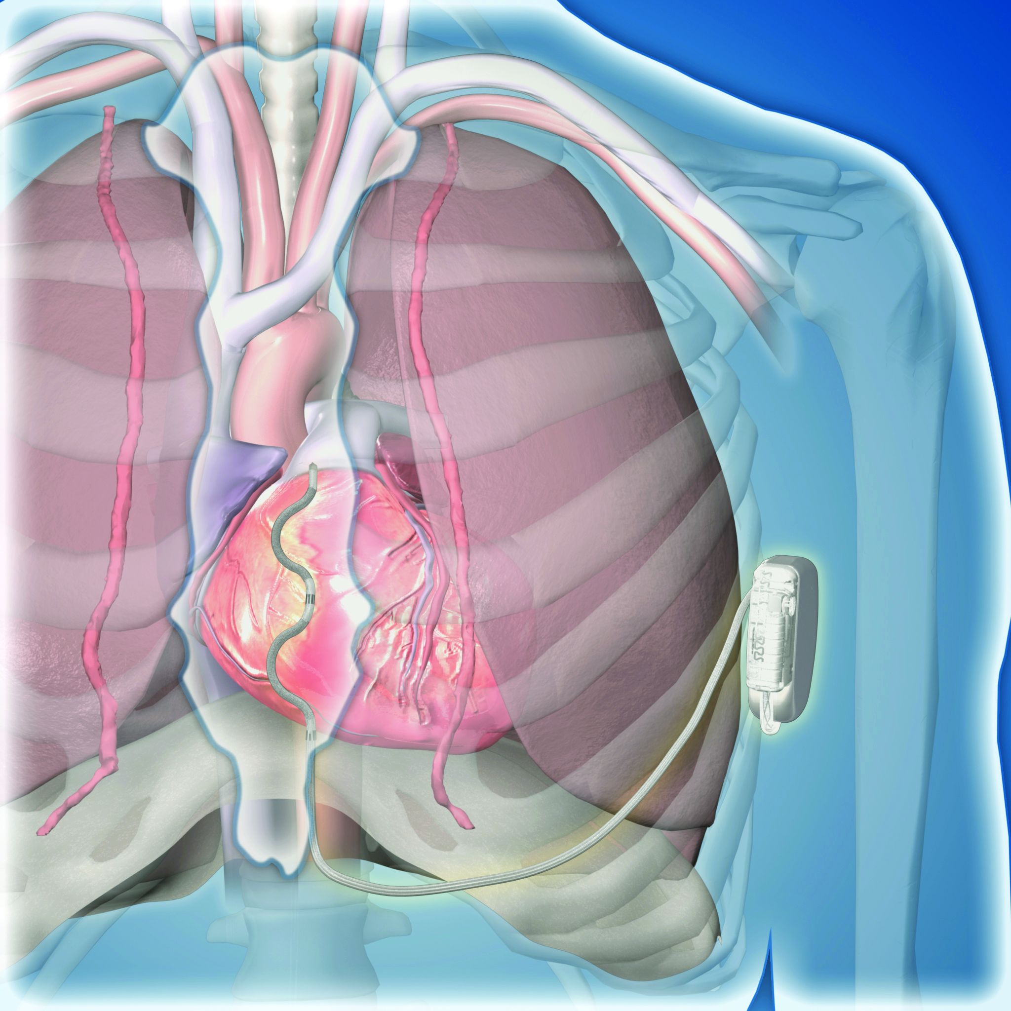
This led to development of the extravascular ICD – 10 years in the making – which uses substernally placed leads that allow antitachycardia pacing and backup pacing in a device with the size of and the anticipated battery longevity of a transvenous ICD device, noted Dr. Crozier.
A 98.7% rate of arrhythmia termination at implant
The pivotal trial’s primary efficacy endpoint was successful defibrillation based on terminating an induced, sustained, shockable ventricular arrhythmia at the time of implantation. The rate was 98.7%, compared with a prespecified target of 88%. All patients had a class I or IIa indication for an ICD.
The primary safety endpoint was freedom from major system- or procedure-related complications at 6 months, which occurred at a rate of 92.6%, compared with the study’s prespecified target rate of 79%. Both targets were derived from the historical rates of ICDs with transvenous leads.
Simultaneously with Dr. Crozier’s report at the congress, the results also appeared online in the New England Journal of Medicine.
Although the pivotal study met both prespecified endpoints, the evidence has limitations that make it likely that regulatory bodies will seek additional data, commented Fred Kusumoto, MD, director of heart rhythm services for the Mayo Clinic in Jacksonville, Fla.
Short follow-up; questions remain
“Follow-up was relatively short, less than a year,” and “questions remain” about the extravascular ICD’s performance, Dr. Kusumoto said in an interview. “Inappropriate shocks occurred in nearly 10% of patients after 11 month follow-up,” he noted, and also cited the 29 patients who needed revisions including two cases with lead fractures.
“The extravascular lead strategy has an advantage over transvenous systems because of the lower risk for extraction or explant,” and it also provides the antitachycardia pacing that’s not available with subcutaneous ICDs, he granted. But in the new study, antitachycardia termination was delivered to only 10 patients and had “reasonable” effectiveness by resolving 70% of these episodes. “Wide adoption by clinicians will depend on results from larger studies with longer follow-up,” Dr. Kusumoto maintained. He also wanted to see confirmation of the ease of lead removal after longer periods of implantation.
Implantation ‘is not difficult’
The trial ran at 46 sites in 17 countries during September 2019 to October 2021. It enrolled patients with a class I or IIa indication for an ICD, excluding patients with a prior sternotomy or need for chronic pacing, and those unable to undergo defibrillation testing.
Clinicians attempted an implantation in 316 patients and had successful placement in 299 (314 had successful placement of their substernal leads), with 292 having a functional device after 6 months, and 284 completing their planned 6-month follow-up. The median procedure time was 66 minutes, including the time for defibrillation testing.
All of the cardiologists who did the implants had received a full day of training prior to performing the procedure. “This is not a difficult procedure, but it is not a region [the substernal space] that cardiologists are familiar working in,” noted Dr. Crozier, explaining the rationale behind a policy of required implantation training.
Twenty-five adverse events occurred in 23 patients. Eighteen of these events required a system revision, including nine lead dislodgments and five infections. The seven adverse events that did not require a revision included three wound-related episodes and three hospitalizations for inappropriate shock. No patients died, nor were there any cardiac injuries as result of the implant.
During average follow-up of 10.6 months, the implanted devices delivered antitachycardia pacing to 10 patients, successfully terminating 32 of 46 episodes (70%), a rate that Dr. Crozier called “very good, and very comparable to transvenous devices.” The devices also delivered 18 appropriate shocks that successfully converted all 18 episodes.
A 10% rate of inappropriate shocks
However, 29 patients (10% of the study cohort) received inappropriate shocks in 81 episodes, with a total of 118 inappropriate shocks delivered, including 34 episodes (42%) triggered by oversensing of a P wave.
“We fully acknowledge that the inappropriate shock rate is higher than what’s seen with transvenous ICDs, but the rate is comparable to what was seen in the early trials with subcutaneous ICDs,” said Dr. Crozier. “We have a number of strategies to reduce the inappropriate shock rate to what we’d expect with conventional devices,” such as making sure that P waves are not detected by the device at the time of implantation, using new algorithms to mitigate P wave sensing, and other programming changes, he added.
Two patients had lead fractures that Dr. Crozier attributed to atypical lead locations and that are likely avoidable in the future. He expressed optimism that the extravascular ICD will avoid the high lead fracture rate over time that remains a problem for ICDs with transvenous leads.
The study also followed a subgroup of 36 patients who underwent a prespecified protocol of chronic defibrillation testing that was successful in all 36.
Dr. Crozier conceded that the extravascular ICD cannot currently deliver chronic pacing, but he expressed optimism that this capability will be available in the future.
“This innovative [extravascular] ICD system would be particularly beneficial for patients with ventricular arrhythmias that can be reliably pace terminated and avoid a transvenous endocardial lead, but more information is required,” concluded Dr. Kusumoto.
The study was sponsored by Medtronic, the company that is developing the extravascular ICD. Dr. Crozier is a consultant to and has received research funding from Medtronic. Dr. Kusumoto had no disclosures.
BARCELONA – A novel “extravascular” implantable cardioverter defibrillator (ICD) that uses substernally placed electrodes surpassed its prespecified efficacy and safety targets in the device’s pivotal trial with 299 patients who received an implant.
The results showed that the extravascular ICD “provides antitachycardia pacing and low energy defibrillation while avoiding the vascular space” for lead placement, Ian Crozier, MD, said at the annual congress of the European Society of Cardiology.
“The results are fantastic; they exceeded our expectations,” said Dr. Crozier in an interview, adding that he expects the new device to receive marketing approval from regulatory agencies based on the findings. “This will be the next generation of ICD going forward,” predicted Dr. Crozier, an electrophysiologist cardiologist at Christchurch (New Zealand) Hospital.
Moving beyond transvenous and subcutaneous ICDs
Traditional ICDs use transvenous leads, which can cause vascular injury, are prone to lead fracture over time, and can produce serious infections as well as other potential complications. The U.S. Food and Drug Administration first approved an alternative-design, subcutaneous ICD in 2012 that avoids the need for transvenous leads and the risks they pose. But subcutaneous ICDs have their own limitations: an inability to provide antitachycardia pacing or chronic pacing; a limited ability to provide bradycardia pacing; and an increased device size with shorter battery life, because of the high shock power needed for effective performance. These drawbacks have collectively hindered uptake, Dr. Crozier said.
This led to development of the extravascular ICD – 10 years in the making – which uses substernally placed leads that allow antitachycardia pacing and backup pacing in a device with the size of and the anticipated battery longevity of a transvenous ICD device, noted Dr. Crozier.
A 98.7% rate of arrhythmia termination at implant
The pivotal trial’s primary efficacy endpoint was successful defibrillation based on terminating an induced, sustained, shockable ventricular arrhythmia at the time of implantation. The rate was 98.7%, compared with a prespecified target of 88%. All patients had a class I or IIa indication for an ICD.
The primary safety endpoint was freedom from major system- or procedure-related complications at 6 months, which occurred at a rate of 92.6%, compared with the study’s prespecified target rate of 79%. Both targets were derived from the historical rates of ICDs with transvenous leads.
Simultaneously with Dr. Crozier’s report at the congress, the results also appeared online in the New England Journal of Medicine.
Although the pivotal study met both prespecified endpoints, the evidence has limitations that make it likely that regulatory bodies will seek additional data, commented Fred Kusumoto, MD, director of heart rhythm services for the Mayo Clinic in Jacksonville, Fla.
Short follow-up; questions remain
“Follow-up was relatively short, less than a year,” and “questions remain” about the extravascular ICD’s performance, Dr. Kusumoto said in an interview. “Inappropriate shocks occurred in nearly 10% of patients after 11 month follow-up,” he noted, and also cited the 29 patients who needed revisions including two cases with lead fractures.
“The extravascular lead strategy has an advantage over transvenous systems because of the lower risk for extraction or explant,” and it also provides the antitachycardia pacing that’s not available with subcutaneous ICDs, he granted. But in the new study, antitachycardia termination was delivered to only 10 patients and had “reasonable” effectiveness by resolving 70% of these episodes. “Wide adoption by clinicians will depend on results from larger studies with longer follow-up,” Dr. Kusumoto maintained. He also wanted to see confirmation of the ease of lead removal after longer periods of implantation.
Implantation ‘is not difficult’
The trial ran at 46 sites in 17 countries during September 2019 to October 2021. It enrolled patients with a class I or IIa indication for an ICD, excluding patients with a prior sternotomy or need for chronic pacing, and those unable to undergo defibrillation testing.
Clinicians attempted an implantation in 316 patients and had successful placement in 299 (314 had successful placement of their substernal leads), with 292 having a functional device after 6 months, and 284 completing their planned 6-month follow-up. The median procedure time was 66 minutes, including the time for defibrillation testing.
All of the cardiologists who did the implants had received a full day of training prior to performing the procedure. “This is not a difficult procedure, but it is not a region [the substernal space] that cardiologists are familiar working in,” noted Dr. Crozier, explaining the rationale behind a policy of required implantation training.
Twenty-five adverse events occurred in 23 patients. Eighteen of these events required a system revision, including nine lead dislodgments and five infections. The seven adverse events that did not require a revision included three wound-related episodes and three hospitalizations for inappropriate shock. No patients died, nor were there any cardiac injuries as result of the implant.
During average follow-up of 10.6 months, the implanted devices delivered antitachycardia pacing to 10 patients, successfully terminating 32 of 46 episodes (70%), a rate that Dr. Crozier called “very good, and very comparable to transvenous devices.” The devices also delivered 18 appropriate shocks that successfully converted all 18 episodes.
A 10% rate of inappropriate shocks
However, 29 patients (10% of the study cohort) received inappropriate shocks in 81 episodes, with a total of 118 inappropriate shocks delivered, including 34 episodes (42%) triggered by oversensing of a P wave.
“We fully acknowledge that the inappropriate shock rate is higher than what’s seen with transvenous ICDs, but the rate is comparable to what was seen in the early trials with subcutaneous ICDs,” said Dr. Crozier. “We have a number of strategies to reduce the inappropriate shock rate to what we’d expect with conventional devices,” such as making sure that P waves are not detected by the device at the time of implantation, using new algorithms to mitigate P wave sensing, and other programming changes, he added.
Two patients had lead fractures that Dr. Crozier attributed to atypical lead locations and that are likely avoidable in the future. He expressed optimism that the extravascular ICD will avoid the high lead fracture rate over time that remains a problem for ICDs with transvenous leads.
The study also followed a subgroup of 36 patients who underwent a prespecified protocol of chronic defibrillation testing that was successful in all 36.
Dr. Crozier conceded that the extravascular ICD cannot currently deliver chronic pacing, but he expressed optimism that this capability will be available in the future.
“This innovative [extravascular] ICD system would be particularly beneficial for patients with ventricular arrhythmias that can be reliably pace terminated and avoid a transvenous endocardial lead, but more information is required,” concluded Dr. Kusumoto.
The study was sponsored by Medtronic, the company that is developing the extravascular ICD. Dr. Crozier is a consultant to and has received research funding from Medtronic. Dr. Kusumoto had no disclosures.
AT ESC CONGRESS 2022
Warfarin associated with higher upper GI bleeding rates, compared with DOACs
Warfarin is associated with higher rates of upper gastrointestinal bleeding but not overall or lower GI bleeding rates, compared with direct oral anticoagulants (DOACs), according to a new nationwide report from Iceland.
In addition, warfarin is associated with higher rates of major GI bleeding, compared with apixaban.
“Although there has been a myriad of studies comparing GI bleeding rates between warfarin and DOACs, very few studies have compared upper and lower GI bleeding rates specifically,” Arnar Ingason, MD, PhD, a gastroenterology resident at the University of Iceland and Landspitali University Hospital, Reykjavik, said in an interview.
“Knowing whether the risk of upper and lower GI bleeding differs between warfarin and DOACs is important, as it can help guide oral anticoagulant selection,” he said.
“Given that warfarin was associated with higher rates of upper GI bleeding compared to DOACs in our study, warfarin may not be optimal for patients with high risk of upper GI bleeding, such as patients with previous history of upper GI bleeding,” Dr. Ingason added.
The study was published online in Clinical Gastroenterology and Hepatology.
Analyzing bleed rates
Dr. Ingason and colleagues analyzed data from electronic medical records for more than 7,000 patients in Iceland who began a prescription for oral anticoagulants between 2014 and 2019. They used inverse probability weighting to yield balanced study groups and calculate the rates of overall, major, upper, and lower GI bleeding. All events of gastrointestinal bleeding were manually confirmed by chart review.
Clinically relevant GI bleeding was defined as bleeding that led to medical intervention, unscheduled physician contact, or temporary cessation of treatment. Upper GI bleeding was defined as hematemesis or a confirmed upper GI bleed site on endoscopy, whereas lower gastrointestinal bleeding was defined as hematochezia or a confirmed lower GI bleed site on endoscopy. Patients with melena and uncertain bleeding site on endoscopy were classified as having a gastrointestinal bleed of unknown location.
Major bleeding was defined as a drop in hemoglobin of at least 20 g/L, transfusion of two or more packs of red blood cells, or bleeding into a closed compartment such as the retroperitoneum.
In total, 295 gastrointestinal bleed events were identified, with 150 events (51%) classified as lower, 105 events (36%) classified as upper, and 40 events (14%) of an unknown location. About 71% required hospitalization, and 63% met the criteria for major bleeding. Five patients died, including three taking warfarin and the other two taking apixaban and rivaroxaban.
Overall, warfarin was associated with double the rate of upper GI bleeding, with 1.7 events per 100 person-years, compared with 0.8 events per 100 person-years for DOACs. The rates of lower GI bleeding were similar for the drugs.
Specifically, warfarin was associated with nearly 5.5 times higher rates of upper gastrointestinal bleeding, compared with dabigatran (Pradaxa, Boehringer Ingelheim), 2.6 times higher than apixaban (Eliquis, Bristol-Myers Squibb), and 1.7 times higher than rivaroxaban (Xarelto, Janssen). The risk for upper GI bleeding also was higher in men taking warfarin.
Warfarin was associated with higher rates of major bleeding, compared with apixaban, with 2.3 events per 100 person-years versus 1.5 events per 100 person-years. Otherwise, overall and major bleed rates were similar for users of warfarin and DOACs.
“GI bleeding among cardiac patients on anticoagulants and antiplatelets is the fastest growing group of GI bleeders,” Neena Abraham, MD, professor of medicine and a gastroenterologist at the Mayo Clinic in Scottsdale, Ariz., said in an interview.
Dr. Abraham, who wasn’t involved with this study, runs a dedicated cardiogastroenterology practice and has studied these patients’ bleeding risk for 20 years.
“This is a group that is ever increasing with aging baby boomers,” she said. “It is anticipated by 2040 that more than 40% of the U.S. adult population will have one or more cardiovascular conditions requiring the chronic prescription of anticoagulant or antiplatelet drugs.”
Considering future research
In this study, peptic ulcer disease was a proportionally less common cause of upper GI bleeding for warfarin at 18%, compared with DOACs at 39%. At the same time, the absolute propensity-weighted incidence rates of peptic ulcer–induced bleeding were similar, with 0.3 events per 100 person-years for both groups.
“As warfarin is not thought to induce peptic ulcer disease but rather promote bleeding from pre-existing lesions, one explanation may be that peptic ulcer disease almost always leads to overt bleeding in anticoagulated patients, while other lesions, such as mucosal erosions and angiodysplasias, may be more likely to lead to overt bleeding in warfarin patients due to a potentially more intense anticoagulation,” Dr. Ingason said.
Dr. Ingason and colleagues now plan to compare GI bleeding severity between warfarin and DOACs. Previous studies have suggested that GI bleeding may be more severe in patients receiving warfarin than in those receiving DOACs, he said.
In addition, large studies with manual verification of GI bleed events could better estimate the potential differences in the sources of upper and lower bleeding between warfarin and DOACs, Dr. Ingason noted.
“Some DOACs, specifically dabigatran, are known to have a mucosal effect on the luminal GI tract, as well as a systemic effect,” Dr. Abraham said. “This pharmacologic effect may contribute to an increase in lower gastrointestinal bleeding in the setting of colonic diverticulosis or mucosal injuries from inflammatory processes.”
Ongoing research should also look at different ways to reduce anticoagulant-related GI bleeding among cardiac patients, she noted.
“Our research group continues to study the risk of cardiac and bleeding adverse events in patients prescribed to DOACs compared to those patients who receive a left atrial appendage occlusion device,” Dr. Abraham said. “This device often permits patients at high risk of GI bleeding to transition off anticoagulant and antiplatelet drugs.”
The study was funded by the Icelandic Centre for Research and the Landspitali University Hospital Research Fund. The funders had no role in the design, conduct, or reporting of the study. The authors declared no competing interests. Dr. Abraham reported no relevant financial relationships.
A version of this article first appeared on Medscape.com.
FROM CLINICAL GASTROENTEROLOGY AND HEPATOLOGY
Early rhythm control improves cardiovascular outcomes in AFib patients regardless of stroke risk
These findings broaden support for early rhythm control, suggesting that physicians should be presenting the option to all patients diagnosed with AFib in routine clinical practice, lead author Daehoon Kim, MD, of Yonsei University, Seoul, South Korea, and colleagues reported.
In 2020, the EAST-AFNET 4 trial showed that early rhythm control was better than rate control for reducing adverse cardiovascular outcomes, but the trial only included patients at risk of stroke with a CHA2DS2-VASc score of at least 2, leaving it unclear whether healthier patients might benefit from the same approach.
“Although the primary indication for rhythm control is to alleviate AF[ib]-related symptoms and improve quality of life, the current guidelines suggest younger age and no or few comorbid conditions as factors favoring rhythm control,” the investigators wrote in Annals of Internal Medicine. “Thus, the effect of rhythm control on cardiovascular outcomes in this population requires elucidation.”
Methods and results
The present study aimed to address this knowledge gap by reviewing data from 54,216 patients with AFib who had rhythm control (ablation or medication) or rate control within one year of diagnosis. Among these patients, 69.3% would have qualified for the EAST-AFNET 4 trial based on higher stroke risk, while the remaining 30.7% of patients would not have been eligible because of lower stroke risk. Median age, consequently, was higher in the former group, at 70 years, versus 54 years in the latter group.
Evaluating the same primary composite outcome as the EAST-AFNET 4 trial (cardiovascular death, ischemic stroke, hospitalization for heart failure, or MI) showed that patients benefited from rhythm control over rate control regardless of risk group.
Those in the higher risk group had a 14% reduced risk of negative cardiovascular outcomes (weighted hazard ratio, 0.86; 95% confidence interval, 0.81-0.92), while those in the lower risk group had a 19% reduced risk of adverse cardiovascular outcomes (weighted HR, 0.81; 95% CI, 0.66-0.98). Safety profiles were similar across groups and management strategies.
Rhythm control well supported from statistical perspective
“We think that physicians should pursue early rhythm control in all patients diagnosed with AF[ib],” principal author Boyoung Joung, MD, PhD, of Yonsei University said in an interview. “Like catheter ablation, we support the idea that early rhythm control can be more effective and safely performed in younger and less frail populations.”
Xiaoxi Yao, PhD, MPH, associate professor of health services research at Mayo Clinic, Rochester, Minn., agreed that rhythm control is now well supported from a statistical perspective, but patients and physicians need to look beyond relative risk improvements, and remain pragmatic.

“There is a benefit, but the benefit is consistent in terms of hazard ratio, or relative risk,” Dr. Yao said in an interview. “You still find a smaller absolute risk difference.”
Patients in the United States – versus Korea where the investigators are based – also need to consider the out-of-pocket costs involved in rhythm control, Dr. Yao said, noting that unclear cost effectiveness may also prevent changes to American guidelines. Medication side effects and procedural risks should also be considered, she added, as well as time off from work needed for ablation.
Dr. Yao, who published a similar paper in June and previously evaluated the role of catheter ablation in routine practice, suggested that the youngest patients may have the most to gain from rhythm control. This is because even a small absolute benefit is magnified with time, she said.
“Since [younger patients] have another several decades to live ... then yes, there might be very significant long-term effects in terms of both symptom control and cardiovascular death and stroke,” Dr. Yao said.
For optimal patient selection, however, more advanced tools are needed, which is why Dr. Yao and her colleagues are exploring new technologies to improve risk-benefit analysis.
“We are not only interested in [a patient’s] baseline high or low risk, but also the extent of risk reduction [that rhythm control provides],” Dr. Yao said. “We are trying to see if there is an [artificial intelligence] or machine-learning approach that can help us provide each patient with a more accurate, individualized estimate to help them make their decision.”
Until then, Dr. Yao encouraged physicians to engage in shared decision-making with patients, making sure to discuss both statistical and practical considerations.
The study was funded by the Ministry of Health and Welfare and the Ministry of Food and Drug Safety of the Republic of Korea. The investigators and Dr. Yao reported no conflicts.
These findings broaden support for early rhythm control, suggesting that physicians should be presenting the option to all patients diagnosed with AFib in routine clinical practice, lead author Daehoon Kim, MD, of Yonsei University, Seoul, South Korea, and colleagues reported.
In 2020, the EAST-AFNET 4 trial showed that early rhythm control was better than rate control for reducing adverse cardiovascular outcomes, but the trial only included patients at risk of stroke with a CHA2DS2-VASc score of at least 2, leaving it unclear whether healthier patients might benefit from the same approach.
“Although the primary indication for rhythm control is to alleviate AF[ib]-related symptoms and improve quality of life, the current guidelines suggest younger age and no or few comorbid conditions as factors favoring rhythm control,” the investigators wrote in Annals of Internal Medicine. “Thus, the effect of rhythm control on cardiovascular outcomes in this population requires elucidation.”
Methods and results
The present study aimed to address this knowledge gap by reviewing data from 54,216 patients with AFib who had rhythm control (ablation or medication) or rate control within one year of diagnosis. Among these patients, 69.3% would have qualified for the EAST-AFNET 4 trial based on higher stroke risk, while the remaining 30.7% of patients would not have been eligible because of lower stroke risk. Median age, consequently, was higher in the former group, at 70 years, versus 54 years in the latter group.
Evaluating the same primary composite outcome as the EAST-AFNET 4 trial (cardiovascular death, ischemic stroke, hospitalization for heart failure, or MI) showed that patients benefited from rhythm control over rate control regardless of risk group.
Those in the higher risk group had a 14% reduced risk of negative cardiovascular outcomes (weighted hazard ratio, 0.86; 95% confidence interval, 0.81-0.92), while those in the lower risk group had a 19% reduced risk of adverse cardiovascular outcomes (weighted HR, 0.81; 95% CI, 0.66-0.98). Safety profiles were similar across groups and management strategies.
Rhythm control well supported from statistical perspective
“We think that physicians should pursue early rhythm control in all patients diagnosed with AF[ib],” principal author Boyoung Joung, MD, PhD, of Yonsei University said in an interview. “Like catheter ablation, we support the idea that early rhythm control can be more effective and safely performed in younger and less frail populations.”
Xiaoxi Yao, PhD, MPH, associate professor of health services research at Mayo Clinic, Rochester, Minn., agreed that rhythm control is now well supported from a statistical perspective, but patients and physicians need to look beyond relative risk improvements, and remain pragmatic.
“There is a benefit, but the benefit is consistent in terms of hazard ratio, or relative risk,” Dr. Yao said in an interview. “You still find a smaller absolute risk difference.”
Patients in the United States – versus Korea where the investigators are based – also need to consider the out-of-pocket costs involved in rhythm control, Dr. Yao said, noting that unclear cost effectiveness may also prevent changes to American guidelines. Medication side effects and procedural risks should also be considered, she added, as well as time off from work needed for ablation.
Dr. Yao, who published a similar paper in June and previously evaluated the role of catheter ablation in routine practice, suggested that the youngest patients may have the most to gain from rhythm control. This is because even a small absolute benefit is magnified with time, she said.
“Since [younger patients] have another several decades to live ... then yes, there might be very significant long-term effects in terms of both symptom control and cardiovascular death and stroke,” Dr. Yao said.
For optimal patient selection, however, more advanced tools are needed, which is why Dr. Yao and her colleagues are exploring new technologies to improve risk-benefit analysis.
“We are not only interested in [a patient’s] baseline high or low risk, but also the extent of risk reduction [that rhythm control provides],” Dr. Yao said. “We are trying to see if there is an [artificial intelligence] or machine-learning approach that can help us provide each patient with a more accurate, individualized estimate to help them make their decision.”
Until then, Dr. Yao encouraged physicians to engage in shared decision-making with patients, making sure to discuss both statistical and practical considerations.
The study was funded by the Ministry of Health and Welfare and the Ministry of Food and Drug Safety of the Republic of Korea. The investigators and Dr. Yao reported no conflicts.
FROM ANNALS OF INTERNAL MEDICINE
Rivaroxaban outmatched by VKAs for AFib in rheumatic heart disease
Contrary to expectations, vitamin K antagonists (VKAs) reduced the risk for ischemic stroke and death, compared with the factor Xa inhibitor rivaroxaban, (Xarelto, Janssen) in patients with rheumatic heart disease and atrial fibrillation (AFib), in the INVICTUS trial.
Patients receiving a VKA, typically warfarin, had a 25% lower risk for the primary outcome – a composite of stroke, systemic embolism, myocardial infarction, or death from vascular or unknown causes outcome – than receiving rivaroxaban (hazard ratio, 1.25; 95% confidence interval, 1.10-1.41).
This difference was driven primarily by a significant reduction in the risk for death in the VKA group, and without a significant increase in major bleeding, reported Ganesan Karthikeyan, MD, from the All India Institute of Medical Sciences in New Delhi.
“VKA should remain the standard of care for patients with rheumatic heart disease and atrial fibrillation,” he concluded in a hotline session at the annual congress of the European Society of Cardiology.
The study, simultaneously published in the New England Journal of Medicine, is the first randomized controlled trial to assess anticoagulant therapy in patients with rheumatic heart disease and AFib.

“Who could have possibly guessed these results? Certainly not me,” said invited discussant Renato D. Lopes, MD, MHS, PhD, Duke Clinical Research Institute, Durham, N.C. “To me, this is one more classical example of why we need to do randomized trials, since they are the only reliable way to determine treatment effects and drive clinical practice.”
Evidence gap
Rheumatic heart disease affects over 40 million people, mainly living in low- and low- to middle-income countries. About 20% of symptomatic patients have AF and an elevated stroke risk, but previous AFib trials excluded these patients, Dr. Karthikeyan noted.
INVICTUS was led by the Population Health Research Institute in Hamilton, Ont., and enrolled 4,565 patients from 24 countries in Africa, Asia, and Latin America who had rheumatic heart disease, AFib or atrial flutter, and an increased stroke risk caused by any of the following: CHA2DS2VASc score of 2 or more, moderate to severe mitral stenosis (valve area ≤ 2.0 cm2), left atrial spontaneous echo contrast, or left atrial thrombus.
Participants were randomly assigned to receive rivaroxaban, 20 mg once daily (15 mg/d if creatinine clearance was 15-49 mL/min), or a VKA titrated to an international normalized ratio (INR) of 2.0-3.0.
Warfarin was used in 79%-85% of patients assigned to VKA, with the percentage varying between visits. The INR was in therapeutic range in 33.2% of patients at baseline, 65.1% at 3 years, and 64.1% at 4 years.
During an average follow-up of 3.1 years, the primary outcome occurred in 446 patients in the VKA group (6.49% per year) and 560 patients in the rivaroxaban group (8.21% per year). The restricted mean survival time for the primary outcome was 1,675 vs. 1,599 days, respectively (difference, –76 days; 95% CI, –121 to –31 days; P for superiority < .001).
The rate of stroke or systemic embolism was similar between the VKA and rivaroxaban groups (75 vs. 94 events), although ischemic strokes were significantly lower with VKA (48 vs. 74 events).
No easy explanation
Deaths were significantly lower with VKA than rivaroxaban, at 442 versus 552 (restricted mean survival time for death, 1,608 vs. 1,587 days; difference, −72 days; 95% CI, –117 to –28 days).
“This reduction is not easily explained,” Dr. Karthikeyan acknowledged. “We cannot explain this reduction by the reduction in stroke that we saw because the number of deaths that are prevented by VKA are far larger than the number of strokes that are prevented. Moreover, the number of deaths were mainly heart failure or sudden deaths.”
Numbers of patients with major bleeding were also similar in the VKA and rivaroxaban groups (56 vs. 40 patients; P = .18), although numbers with fatal bleeding were lower with rivaroxaban (15 vs. 4, respectively).
By design, there were more physician interactions for monthly monitoring of INR in the VKA group, “but we do not believe such a large reduction can be explained entirely by increased health care contact,” he said. Moreover, there was no significant between-group difference in heart failure medications or hospitalizations or the need for valve replacement.
Almost a quarter (23%) of patients in the rivaroxaban group permanently discontinued the study drug versus just 6% in the VKA group.
Importantly, the mortality benefit emerged much later than in other trials and coincided with the time when the INR became therapeutic at about 3 years, Dr. Karthikeyan said. But it is unknown whether this is because of the INR or an unrelated effect.
More physician contact
Following the presentation, session cochair C. Michael Gibson, MD, Baim Institute for Clinical Research, Harvard Medical School, Boston, questioned the 23% discontinuation rate for rivaroxaban. “Is this really a superiority of warfarin or is this superiority of having someone come in and see their physician for a lot of checks on their INR?”
In response, Dr. Karthikeyan said that permanent discontinuation rates were about 20%-25% in shorter-duration direct oral anticoagulant trials, such as RELY, ROCKET-AF, and ARISTOLE, and exceeded 30% in ENGAGE-AF with 2.8 years’ follow-up.
“So, this is not new,” he said, adding that 31.4% of rivaroxaban patients did so for valve replacement surgery and subsequently received nonstudy VKA.
Dr. Lopes said it is important to keep in mind that INVICTUS enrolled a “very different population” that was younger (mean age, 50.5 years), was much more often female (72.3%), and had fewer comorbidities than patients with AFib who did not have rheumatic heart disease in the pivotal trials.
“It will be interesting to see the treatment effect according to mitral stenosis severity, since we had about 30% with mild mitral stenosis and additionally 18% of patients without mitral stenosis,” he added.
Co–principal investigator Stuart J. Connolly, MD, from the Population Health Research Institute, said physician contacts may be a factor but that the mortality difference was clear, highly significant, and sufficiently powered.
“What’s amazing is that what we’re seeing here is something that hasn’t been previously described with VKA or warfarin, which is that it reduces mortality,” he said in an interview.
Rivaroxaban has never been shown to reduce mortality in any particular condition, and a meta-analysis of other novel oral anticoagulants shows only a small reduction in mortality, caused almost completely by less intracranial hemorrhage than warfarin, he added. “So, we don’t think this is a problem with rivaroxaban. In some ways, rivaroxaban is an innocent bystander to a trial of warfarin in patients with rheumatic heart disease and atrial fibrillation.”
Dr. Connolly said more work is needed to explain the findings and analyses are planned to see which patients are at highest risk for death as well as looking at the relationship between INR control and outcomes.
“We need to do more research on what it is about VKA that could explain this,” he said. “Is it affecting the myocardium in some way, is it preventing fibrosis, is there some off target effect, not on the anticoagulation system, that could explain this?”
Athena Poppas, MD, chief of cardiology at Brown University, Providence, R.I., and past president of the American College of Cardiology, said “INVICTUS is an incredibly important study that needed to be done.”
“The results – though disappointing and surprising in some ways – I don’t think we can explain them away and change what we are doing right now,” she said in an interview.
Although warfarin is a cheap drug, Dr. Poppas said, it would be tremendously helpful to have an alternative treatment for these patients. Mechanistic studies are needed to understand the observed mortality advantage and low bleeding rates but that trials of other novel anticoagulants are also needed.
“But I’m not sure that will happen,” she added. “It’s unlikely to be industry sponsored, so it would be a very expensive lift with a low likelihood of success.”
In an editorial accompanying the paper, Gregory Y.H. Lip, MD, University of Liverpool (England), pointed out that observational data show similar or even higher risks for major bleeding with rivaroxaban than with warfarin. “To improve outcomes in these patients, we therefore need to look beyond anticoagulation alone or beyond a type of anticoagulation drug per se. Indeed, a one-size-fits-all approach may not be appropriate.”
The study was funded by an unrestricted grant from Bayer. Dr. Karthikeyan and Dr. Poppas reported no relevant conflicts of interest.
A version of this article first appeared on Medscape.com.
Contrary to expectations, vitamin K antagonists (VKAs) reduced the risk for ischemic stroke and death, compared with the factor Xa inhibitor rivaroxaban, (Xarelto, Janssen) in patients with rheumatic heart disease and atrial fibrillation (AFib), in the INVICTUS trial.
Patients receiving a VKA, typically warfarin, had a 25% lower risk for the primary outcome – a composite of stroke, systemic embolism, myocardial infarction, or death from vascular or unknown causes outcome – than receiving rivaroxaban (hazard ratio, 1.25; 95% confidence interval, 1.10-1.41).
This difference was driven primarily by a significant reduction in the risk for death in the VKA group, and without a significant increase in major bleeding, reported Ganesan Karthikeyan, MD, from the All India Institute of Medical Sciences in New Delhi.
“VKA should remain the standard of care for patients with rheumatic heart disease and atrial fibrillation,” he concluded in a hotline session at the annual congress of the European Society of Cardiology.
The study, simultaneously published in the New England Journal of Medicine, is the first randomized controlled trial to assess anticoagulant therapy in patients with rheumatic heart disease and AFib.
“Who could have possibly guessed these results? Certainly not me,” said invited discussant Renato D. Lopes, MD, MHS, PhD, Duke Clinical Research Institute, Durham, N.C. “To me, this is one more classical example of why we need to do randomized trials, since they are the only reliable way to determine treatment effects and drive clinical practice.”
Evidence gap
Rheumatic heart disease affects over 40 million people, mainly living in low- and low- to middle-income countries. About 20% of symptomatic patients have AF and an elevated stroke risk, but previous AFib trials excluded these patients, Dr. Karthikeyan noted.
INVICTUS was led by the Population Health Research Institute in Hamilton, Ont., and enrolled 4,565 patients from 24 countries in Africa, Asia, and Latin America who had rheumatic heart disease, AFib or atrial flutter, and an increased stroke risk caused by any of the following: CHA2DS2VASc score of 2 or more, moderate to severe mitral stenosis (valve area ≤ 2.0 cm2), left atrial spontaneous echo contrast, or left atrial thrombus.
Participants were randomly assigned to receive rivaroxaban, 20 mg once daily (15 mg/d if creatinine clearance was 15-49 mL/min), or a VKA titrated to an international normalized ratio (INR) of 2.0-3.0.
Warfarin was used in 79%-85% of patients assigned to VKA, with the percentage varying between visits. The INR was in therapeutic range in 33.2% of patients at baseline, 65.1% at 3 years, and 64.1% at 4 years.
During an average follow-up of 3.1 years, the primary outcome occurred in 446 patients in the VKA group (6.49% per year) and 560 patients in the rivaroxaban group (8.21% per year). The restricted mean survival time for the primary outcome was 1,675 vs. 1,599 days, respectively (difference, –76 days; 95% CI, –121 to –31 days; P for superiority < .001).
The rate of stroke or systemic embolism was similar between the VKA and rivaroxaban groups (75 vs. 94 events), although ischemic strokes were significantly lower with VKA (48 vs. 74 events).
No easy explanation
Deaths were significantly lower with VKA than rivaroxaban, at 442 versus 552 (restricted mean survival time for death, 1,608 vs. 1,587 days; difference, −72 days; 95% CI, –117 to –28 days).
“This reduction is not easily explained,” Dr. Karthikeyan acknowledged. “We cannot explain this reduction by the reduction in stroke that we saw because the number of deaths that are prevented by VKA are far larger than the number of strokes that are prevented. Moreover, the number of deaths were mainly heart failure or sudden deaths.”
Numbers of patients with major bleeding were also similar in the VKA and rivaroxaban groups (56 vs. 40 patients; P = .18), although numbers with fatal bleeding were lower with rivaroxaban (15 vs. 4, respectively).
By design, there were more physician interactions for monthly monitoring of INR in the VKA group, “but we do not believe such a large reduction can be explained entirely by increased health care contact,” he said. Moreover, there was no significant between-group difference in heart failure medications or hospitalizations or the need for valve replacement.
Almost a quarter (23%) of patients in the rivaroxaban group permanently discontinued the study drug versus just 6% in the VKA group.
Importantly, the mortality benefit emerged much later than in other trials and coincided with the time when the INR became therapeutic at about 3 years, Dr. Karthikeyan said. But it is unknown whether this is because of the INR or an unrelated effect.
More physician contact
Following the presentation, session cochair C. Michael Gibson, MD, Baim Institute for Clinical Research, Harvard Medical School, Boston, questioned the 23% discontinuation rate for rivaroxaban. “Is this really a superiority of warfarin or is this superiority of having someone come in and see their physician for a lot of checks on their INR?”
In response, Dr. Karthikeyan said that permanent discontinuation rates were about 20%-25% in shorter-duration direct oral anticoagulant trials, such as RELY, ROCKET-AF, and ARISTOLE, and exceeded 30% in ENGAGE-AF with 2.8 years’ follow-up.
“So, this is not new,” he said, adding that 31.4% of rivaroxaban patients did so for valve replacement surgery and subsequently received nonstudy VKA.
Dr. Lopes said it is important to keep in mind that INVICTUS enrolled a “very different population” that was younger (mean age, 50.5 years), was much more often female (72.3%), and had fewer comorbidities than patients with AFib who did not have rheumatic heart disease in the pivotal trials.
“It will be interesting to see the treatment effect according to mitral stenosis severity, since we had about 30% with mild mitral stenosis and additionally 18% of patients without mitral stenosis,” he added.
Co–principal investigator Stuart J. Connolly, MD, from the Population Health Research Institute, said physician contacts may be a factor but that the mortality difference was clear, highly significant, and sufficiently powered.
“What’s amazing is that what we’re seeing here is something that hasn’t been previously described with VKA or warfarin, which is that it reduces mortality,” he said in an interview.
Rivaroxaban has never been shown to reduce mortality in any particular condition, and a meta-analysis of other novel oral anticoagulants shows only a small reduction in mortality, caused almost completely by less intracranial hemorrhage than warfarin, he added. “So, we don’t think this is a problem with rivaroxaban. In some ways, rivaroxaban is an innocent bystander to a trial of warfarin in patients with rheumatic heart disease and atrial fibrillation.”
Dr. Connolly said more work is needed to explain the findings and analyses are planned to see which patients are at highest risk for death as well as looking at the relationship between INR control and outcomes.
“We need to do more research on what it is about VKA that could explain this,” he said. “Is it affecting the myocardium in some way, is it preventing fibrosis, is there some off target effect, not on the anticoagulation system, that could explain this?”
Athena Poppas, MD, chief of cardiology at Brown University, Providence, R.I., and past president of the American College of Cardiology, said “INVICTUS is an incredibly important study that needed to be done.”
“The results – though disappointing and surprising in some ways – I don’t think we can explain them away and change what we are doing right now,” she said in an interview.
Although warfarin is a cheap drug, Dr. Poppas said, it would be tremendously helpful to have an alternative treatment for these patients. Mechanistic studies are needed to understand the observed mortality advantage and low bleeding rates but that trials of other novel anticoagulants are also needed.
“But I’m not sure that will happen,” she added. “It’s unlikely to be industry sponsored, so it would be a very expensive lift with a low likelihood of success.”
In an editorial accompanying the paper, Gregory Y.H. Lip, MD, University of Liverpool (England), pointed out that observational data show similar or even higher risks for major bleeding with rivaroxaban than with warfarin. “To improve outcomes in these patients, we therefore need to look beyond anticoagulation alone or beyond a type of anticoagulation drug per se. Indeed, a one-size-fits-all approach may not be appropriate.”
The study was funded by an unrestricted grant from Bayer. Dr. Karthikeyan and Dr. Poppas reported no relevant conflicts of interest.
A version of this article first appeared on Medscape.com.
FROM ESC CONGRESS 2022