User login
Treating IBD in medical home reduces costs
DALLAS – In the midst of the ever-increasing costs of patient care for chronic disease, one model for care of a specific, complex condition is the medical home, according to a presentation at the American Gastroenterological Association’s Partners in Value meeting.

The medical home concept came out of pediatrics and primary care, where patients’ health care needs could vary greatly over several years but benefited from coordinated care, Miguel Regueiro, MD, AGAF, professor of medicine and chair of the department of gastroenterology, hepatology, and nutrition at the Cleveland Clinic, told attendees at the meeting.
The medical home is ideal for a disease such as inflammatory bowel disease because it brings together the different care providers essential for such a complex condition and allows for the kind of coordinated, holistic care that’s uncommon in America’s typically fragmented health care system.
The two key components of a specialist medical home are a population of patients whose principal care requires a specialist and a health plan partnership around a chronic disease. The major attributes of a medical home, he explained, are accessibility; comprehensive, coordinated care; compassionate, culturally sensitive, patient-and family-centered care; and team-based delivery.
After initially building an IBD medical home in Pittsburgh, Dr. Regueiro brought the concept to Cleveland Clinic and shared with attendees how he did it and the challenges and benefits it involved.
He advises starting with a small team and expanding as demands or needs dictate. He began with a GI specialist, a psychiatrist, a dietitian, a social worker, a nurse, and three in-house schedulers. The patient ratio was 500 patients per nurse and 1,000 patients per gastroenterologist, psychiatrist and dietitian.
Dr. Regueiro explained the patient flow through the medical home, starting with a preclinic referral and patient questionnaire. The actual visit moves from intake and triage to the actual exam to a comprehensive care plan involving all relevant providers, plus any necessary referrals to any outside services, such as surgery or pain management. The work continues, however, after the patient leaves the clinic, with follow-up calls and telemedicine follow-up, including psychosocial telemedicine.
The decision to include in-house schedulers is among the most important, though it may admittedly be one of the more difficult for those trying to build a medical home from the ground up.
“I think that central scheduling is the worst thing that’s ever happened to medicine,” Dr. Regueiro told attendees. It’s too depersonalized to serve patients well, he said. His center’s embedded schedulers begin the clinical experience from a patient’s first phone call. They ask patients their top three problems and the top three things they want from their visit.
“If we don’t ask our patients what they want, the focus becomes physician-centered instead of patient-centered,” Dr. Regueiro said, sharing anecdotes of patients who came in with problems, expectations, and requests that differed, sometimes dramatically, from what he anticipated. Many of these needs were psychosocial, and the medical home model is ideally suited to address them in tandem with physical medical care.
“I firmly believe that the secret sauce of all our medical homes is the psychosocial care of patients by understanding the interactions between biological and environmental factors in the mind-body illness interface,” he said.
The center also uses provider team huddles before meeting a patient at intake and then afterward for follow-up. Part of team communication involves identifying patients as “red,” “yellow,” or “green” based on the magnitude of their needs and care utilization.
“There are a lot of green-zone patients: They see you once a year and really don’t need the intensive care” his clinic can provide, he said. “We did as much as we could to keep the patient at home, in their community, at school, more than anything else,” Dr. Regueiro said. “It’s not just about their quality of life and disease but about the impact on their work-life balance.”
One way the clinic addressed those needs was by involving patient stakeholders to find out early on – as they were setting up the center – what the patient experience was and what needed to improve. As they learned about logistical issues that frustrated the patient experience, such as lost medical records, central scheduling, or inadequate parking, they could work to identify solutions – thereby also addressing patients’ psychosocial needs.
But Dr. Regueiro was upfront about the substantial investment and challenges involved in setting up an IBD medical home. He would not have been able to meet his relative-value unit targets in this model, so those were cut in half. When an audience member asked how the clinic successfully worked with a variety of commercial insurers given the billing challenges, Dr. Regueiro said he didn’t have a good answer, though several large insurers have approached him with interest in the model.
“I don’t know what’s going to happen, but the appetite seems to be there,” he said. “I do think the insurers are interested because of the cost [savings] part of this.”
Those cost savings showed up in the long-term outcomes. At the Pittsburgh center, total emergency department visits dropped by nearly half (47%) from the year before the medical home total care model was implemented to the year after, from 508 total ED visits among the patient population to 264 visits. Hospitalizations similarly declined by a third (36%), from 208 to 134.
Part of the reason for that decline, as Dr. Regueiro showed in a case study example, was halting the repetitive testing and interventions in the ED that did not actually address – or even find out – the patient’s needs, particularly when those needs were psychosocial. And many psychosocial needs could even be met outside the clinic: 35% of all behavioral visits were telemedicine.
Still, payment models remain a challenge for creating medical homes. Other challenges include preventing team burnout, which can also deter interest in this model in the first place, and the longitudinal coordination of care with the medical neighborhood.
Despite his caveats, Dr. Regueiro’s presentation made a strong impression on attendees.
Mark Tsuchiyose, MD, a gastroenterologist with inSite Digestive Health Care in Daly City, Calif., found the presentation “fantastic” and said using medical homes for chronic GI care is “unquestionably the right thing to do.” But the problem, again, is reimbursement and a payer model that works with a medical home, he said. Dr. Regueiro needed to reduce his relative-value unit targets and was able to get funding for the care team, including in-house schedulers, Dr. Tsuchiyose noted, and that’s simply not feasible for most providers in most areas right now.
Sanjay Sandhir, MD, of Dayton (Ohio) Gastroenterology, said he appreciated the discussion of patient engagement apps in the medical home and helping patients with anxiety, depression, stress, and other psychosocial needs. While acknowledging the payer hurdles to such a model, he expressed optimism.
“If we go to the payers, and the payers are willing to understand and can get their head around and accept [this model], and we can give good data, it’s possible,” Dr. Sandhir said. “It’s worked in other cities, but it has to be a paradigm shift in the way people think.”
John Garrett, MD, a gastroenterologist with Mission Health and Asheville (N.C.) Gastroenterology, said he found the talk – and the clinic itself – “truly amazing.”
“It truly requires a multidisciplinary approach to identify the problems your IBD patients have and manage them most effectively,” he said. But the model is also “incredibly labor-intensive,” he added.
“I think few of us could mobilize a team as large, effective, and well-funded as his, but I think we can all take pieces of that and do it on a much more economical level, and still get good results,” he said, pointing specifically to incorporating depression screenings and other psychosocial elements into care. “I think most important would be to identify whether significant psychosocial issues are present and be ready to treat those.”
Dr. Regueiro has consulted for Abbvie, Allergan, Amgen, Celgene, Janssen, Pfizer, Takeda, and UCB, and has received research grants from Abbvie, Janssen, and Takeda. Dr. Tsuchiyose, Dr. Sandhir, and Dr. Garrett had no disclosures.
Gastroenterology has released a special collection of IBD articles, which gathers the best IBD research published over the past 2 years. View it at https://www.gastrojournal.org/content/inflammatory_bowel_disease.
DALLAS – In the midst of the ever-increasing costs of patient care for chronic disease, one model for care of a specific, complex condition is the medical home, according to a presentation at the American Gastroenterological Association’s Partners in Value meeting.
The medical home concept came out of pediatrics and primary care, where patients’ health care needs could vary greatly over several years but benefited from coordinated care, Miguel Regueiro, MD, AGAF, professor of medicine and chair of the department of gastroenterology, hepatology, and nutrition at the Cleveland Clinic, told attendees at the meeting.
The medical home is ideal for a disease such as inflammatory bowel disease because it brings together the different care providers essential for such a complex condition and allows for the kind of coordinated, holistic care that’s uncommon in America’s typically fragmented health care system.
The two key components of a specialist medical home are a population of patients whose principal care requires a specialist and a health plan partnership around a chronic disease. The major attributes of a medical home, he explained, are accessibility; comprehensive, coordinated care; compassionate, culturally sensitive, patient-and family-centered care; and team-based delivery.
After initially building an IBD medical home in Pittsburgh, Dr. Regueiro brought the concept to Cleveland Clinic and shared with attendees how he did it and the challenges and benefits it involved.
He advises starting with a small team and expanding as demands or needs dictate. He began with a GI specialist, a psychiatrist, a dietitian, a social worker, a nurse, and three in-house schedulers. The patient ratio was 500 patients per nurse and 1,000 patients per gastroenterologist, psychiatrist and dietitian.
Dr. Regueiro explained the patient flow through the medical home, starting with a preclinic referral and patient questionnaire. The actual visit moves from intake and triage to the actual exam to a comprehensive care plan involving all relevant providers, plus any necessary referrals to any outside services, such as surgery or pain management. The work continues, however, after the patient leaves the clinic, with follow-up calls and telemedicine follow-up, including psychosocial telemedicine.
The decision to include in-house schedulers is among the most important, though it may admittedly be one of the more difficult for those trying to build a medical home from the ground up.
“I think that central scheduling is the worst thing that’s ever happened to medicine,” Dr. Regueiro told attendees. It’s too depersonalized to serve patients well, he said. His center’s embedded schedulers begin the clinical experience from a patient’s first phone call. They ask patients their top three problems and the top three things they want from their visit.
“If we don’t ask our patients what they want, the focus becomes physician-centered instead of patient-centered,” Dr. Regueiro said, sharing anecdotes of patients who came in with problems, expectations, and requests that differed, sometimes dramatically, from what he anticipated. Many of these needs were psychosocial, and the medical home model is ideally suited to address them in tandem with physical medical care.
“I firmly believe that the secret sauce of all our medical homes is the psychosocial care of patients by understanding the interactions between biological and environmental factors in the mind-body illness interface,” he said.
The center also uses provider team huddles before meeting a patient at intake and then afterward for follow-up. Part of team communication involves identifying patients as “red,” “yellow,” or “green” based on the magnitude of their needs and care utilization.
“There are a lot of green-zone patients: They see you once a year and really don’t need the intensive care” his clinic can provide, he said. “We did as much as we could to keep the patient at home, in their community, at school, more than anything else,” Dr. Regueiro said. “It’s not just about their quality of life and disease but about the impact on their work-life balance.”
One way the clinic addressed those needs was by involving patient stakeholders to find out early on – as they were setting up the center – what the patient experience was and what needed to improve. As they learned about logistical issues that frustrated the patient experience, such as lost medical records, central scheduling, or inadequate parking, they could work to identify solutions – thereby also addressing patients’ psychosocial needs.
But Dr. Regueiro was upfront about the substantial investment and challenges involved in setting up an IBD medical home. He would not have been able to meet his relative-value unit targets in this model, so those were cut in half. When an audience member asked how the clinic successfully worked with a variety of commercial insurers given the billing challenges, Dr. Regueiro said he didn’t have a good answer, though several large insurers have approached him with interest in the model.
“I don’t know what’s going to happen, but the appetite seems to be there,” he said. “I do think the insurers are interested because of the cost [savings] part of this.”
Those cost savings showed up in the long-term outcomes. At the Pittsburgh center, total emergency department visits dropped by nearly half (47%) from the year before the medical home total care model was implemented to the year after, from 508 total ED visits among the patient population to 264 visits. Hospitalizations similarly declined by a third (36%), from 208 to 134.
Part of the reason for that decline, as Dr. Regueiro showed in a case study example, was halting the repetitive testing and interventions in the ED that did not actually address – or even find out – the patient’s needs, particularly when those needs were psychosocial. And many psychosocial needs could even be met outside the clinic: 35% of all behavioral visits were telemedicine.
Still, payment models remain a challenge for creating medical homes. Other challenges include preventing team burnout, which can also deter interest in this model in the first place, and the longitudinal coordination of care with the medical neighborhood.
Despite his caveats, Dr. Regueiro’s presentation made a strong impression on attendees.
Mark Tsuchiyose, MD, a gastroenterologist with inSite Digestive Health Care in Daly City, Calif., found the presentation “fantastic” and said using medical homes for chronic GI care is “unquestionably the right thing to do.” But the problem, again, is reimbursement and a payer model that works with a medical home, he said. Dr. Regueiro needed to reduce his relative-value unit targets and was able to get funding for the care team, including in-house schedulers, Dr. Tsuchiyose noted, and that’s simply not feasible for most providers in most areas right now.
Sanjay Sandhir, MD, of Dayton (Ohio) Gastroenterology, said he appreciated the discussion of patient engagement apps in the medical home and helping patients with anxiety, depression, stress, and other psychosocial needs. While acknowledging the payer hurdles to such a model, he expressed optimism.
“If we go to the payers, and the payers are willing to understand and can get their head around and accept [this model], and we can give good data, it’s possible,” Dr. Sandhir said. “It’s worked in other cities, but it has to be a paradigm shift in the way people think.”
John Garrett, MD, a gastroenterologist with Mission Health and Asheville (N.C.) Gastroenterology, said he found the talk – and the clinic itself – “truly amazing.”
“It truly requires a multidisciplinary approach to identify the problems your IBD patients have and manage them most effectively,” he said. But the model is also “incredibly labor-intensive,” he added.
“I think few of us could mobilize a team as large, effective, and well-funded as his, but I think we can all take pieces of that and do it on a much more economical level, and still get good results,” he said, pointing specifically to incorporating depression screenings and other psychosocial elements into care. “I think most important would be to identify whether significant psychosocial issues are present and be ready to treat those.”
Dr. Regueiro has consulted for Abbvie, Allergan, Amgen, Celgene, Janssen, Pfizer, Takeda, and UCB, and has received research grants from Abbvie, Janssen, and Takeda. Dr. Tsuchiyose, Dr. Sandhir, and Dr. Garrett had no disclosures.
Gastroenterology has released a special collection of IBD articles, which gathers the best IBD research published over the past 2 years. View it at https://www.gastrojournal.org/content/inflammatory_bowel_disease.
REPORTING FROM 2018 AGA PARTNERS IN VALUE
Antibiotics trigger proteolytic activity that leads to chronic colitis
Antibiotics are associated with increased large intestinal proteolytic activity and gut barrier disruption, thereby raising the risk of chronic colitis in susceptible individuals, a recent study found.
Although the association between antibiotics and chronic colitis has been previously described, this is the first study to demonstrate the causative role of high proteolytic activity, reported lead author Hongsup Yoon, PhD, chair of nutrition and immunology at Technische Universität München in Freising-Weihenstephan, Germany, and colleagues. The team’s experiments support development of antiproteolytic strategies in susceptible humans.
“In the context of IBD, several clinical studies have already revealed that early and frequent antibiotic therapies, especially metronidazole or fluoroquinolone treatments, are associated with increased risk for Crohn’s disease,” the authors wrote in Cellular and Molecular Gastroenterology and Hepatology. “However, the causal role of antibiotic therapies in the disease development and the mechanisms underlying this [potentially] serious long-term adverse effect of antibiotics on the intestinal immune homeostasis remain unknown.”
Previous studies have shown that antibiotic therapy often causes high luminal proteolytic activity in the large intestine, likely because of the elimination of antiproteolytic bacteria that normally control pancreatic protease levels. Other studies have shown that exposing murine colonic mucosa to fecal supernatants with high proteolytic activity increases gut barrier permeability, which triggers chronic inflammation via translocation of luminal antigens.
“In view of these data,” the authors wrote, “we hypothesized that the antibiotic-increased proteolytic activity in the large intestine is a relevant risk factor for the development of colitis in susceptible organisms.”
The first component of the study used transwell experiments to evaluate the impact of high proteolytic activity on gut barrier integrity. High proteolytic activity was induced by several antibiotics, including fluoroquinolones with or without an imidazole (ciprofloxacin and levofloxacin plus or minus metronidazole), a beta-lactam (amoxicillin + clavulanate), cephalosporins with or without a macrolide (azithromycin and ceftriaxone plus or minus azithromycin), and a rifamycin (rifaximin).
“All tested antibiotic classes mediated a major proteolytic activity increase in some patients but not in others,” the authors wrote, “demonstrating individual-specific vulnerability of the intestinal microbiota toward antibiotic therapies, which is likely caused by the high interindividual variability of human microbial ecosystems.”
One-quarter of patients had a 400% or greater increase in large intestinal proteolytic activity following antibiotic therapy, and several had an increase greater than 900%. Analysis indicated that proteolytic activity was caused by pancreatic proteases such as chymotrypsin and trypsin.
Subsequent cell line testing showed that stool supernatants with high proteolytic activity damaged the epithelial barrier, but samples with low proteolytic activity did not. Of note, the negative impact of high proteolytic activity on epithelial cells could be mitigated by incubating stool supernatants with a serine protease inhibitor.
In analogous experiments, mice were given a combination of vancomycin and metronidazole (V/M). In contrast with the various proteolytic activity levels observed in humans, all mice had high proteolytic activity levels following treatment, suggesting that V/M eliminated almost all antiproteolytic bacteria.
The loss of antiproteolytic bacteria was clarified by cecal microbiota transplantation tests. Transplants from untreated mice were capable of normalizing proteolytic activity levels in germ-free mice (which have high proteolytic activity levels), but transplants from V/M-treated mice were ineffective, suggesting a near-total loss of antiproteolytic bacteria. The identity of these antiproteolytic bacteria remains a mystery.
“Although our data are in line with published literature suggesting specific strains of the order Bacteroidales to play a role in the physiological inactivation of pancreatic proteases,” the authors wrote, “the identity of relevant antiproteolytic species/strains remains to be elucidated.”
The next part of the study involved wild-type and interleukin (IL)-10–/– mice, the latter of which serves as a model of human colitis. Both types of mice were given V/M with or without an oral serine protease inhibitor, a potential therapy intended to limit proteolytic activity and associated intestinal barrier damage.
Although both wild-type and IL-10–/– mice had increased intestinal permeability after V/M treatment, only IL-10–/– mice showed lasting inflammation. Of note, coadministration of an oral serine protease inhibitor with V/M protected against colitis in IL-10–/– mice.
The protective benefit of an oral serine protease inhibitor in IL-10–/– mice prompts the development of antiproteolytic strategies in humans. These would target “large intestinal proteolytic activity [e.g., oral administration of encapsulated serine protease inhibitors, commensal antiproteolytic bacteria, or genetically modified bacteria expressing protease inhibitors] to protect the large intestinal mucosa from adverse effects of antibiotic-induced or diarrhea-induced high proteolytic activity,” the authors wrote.
The study was funded by the Deutscher Akademischer Austauschdienst. No conflicts of interest were reported.
SOURCE: Yoon H-S et al. Cell Mol Gastroenterol Hepatol. 2018 May 29. doi: 10.1016/j.jcmgh.2018.05.008.
FROM CELLULAR AND MOLECULAR GASTROENTEROLOGY AND HEPATOLOGY
Key clinical point: In patients susceptible to inflammatory bowel disease, antibiotics cause increased proteolytic activity in the large intestine that disrupts the gut barrier, thereby increasing risk of chronic colitis.
Major finding: One-quarter of patients had a 400% or greater increase in large intestinal proteolytic activity following antibiotic therapy.
Study details: A prospective study involving mice and humans treated with antibiotics.
Disclosures: The study was funded by the Deutscher Akademischer Austauschdienst. No conflicts of interest were reported.
Source: Yoon H et al. Cell Mol Gastroenterol Hepatol. 2018 May 29. doi: 10.1016/j.jcmgh.2018.05.008.
Adalimumab safety profile similar in children and adults
The safety profile for adalimumab in children is similar to that of adults, according to findings published in the Journal of Pediatrics.

In an analysis of data from seven clinical trials from 2002-2015, the most common adverse events across indications were upper respiratory tract infection (24 events per 100 patient-years), nasopharyngitis (17 events per 100 PY), and headache (20 events per 100 PY). Serious infections were the most frequent adverse events across indications (8% of all patients; 4 events per 100 PY), reported Gerd Horneff, MD, of the department of general pediatrics at Asklepios Klinik Sankt Augustin (Germany), and his coauthors.
All of the clinical trials were funded by AbbVie, and included 577 pediatric patients with juvenile idiopathic arthritis (JIA), psoriasis, or Crohn’s disease. Patients received subcutaneous injection of adalimumab at a dosage of either 40 mg/0.8 mL or 20 mg/0.4 mL.
Adverse events that occurred after the first adalimumab dose and up to 70 days after the last dose were included. Serious adverse events were defined as “events that were fatal or immediately life-threatening; required inpatient or prolonged hospitalization; resulted in persistent or significant disability/incapacity, congenital anomaly, or spontaneous or elective abortion; or required medical or surgical intervention to prevent a serious outcome,” the authors said.
Infections occurred in 82% of JIA patients (151 events per 100 PY), 74% of patients with psoriasis (169 events per 100 PY), and 76% of patients with CD (132 events per 100 PY). The most common events for JIA, psoriasis, and Crohn’s were injection-site pain (22% of patients; 75 events per 100 PY), headache (30% of patients; 47 events per 100 PY), and worsening of Crohn’s disease (55% of patients; 37 events per 100 PY), respectively.
Serious adverse events occurred in 29% of patients. Rates for JIA, psoriasis, and Crohn’s were 14, 7, and 32 events per 100 PY, respectively. Serious infections were the most common serious adverse event, with rates of 3, 1, and 7 events per 100 PY for JIA, psoriasis, and Crohn’s disease, respectively. Pneumonia was the most commonly reported serious infection (1% of patients; 1 event per 100 PY). One death, due to an accidental fall, occurred in an adolescent patient with psoriasis.
The study findings add to “a more complete understanding of the established safety profile of adalimumab,” and suggest that in pediatric patients, “the overall safety profile was comparable and consistent with that in adults,” Dr. Horneff and his associates added.
AbbVie funded the study. Dr. Horneff has received grants from AbbVie, Chugai, Novartis, Pfizer, and Roche. Seven of the investigators are or were employees of AbbVie and may own AbbVie stock and stock options. Two of the investigators disclosed ties with a number of pharmaceutical companies.
SOURCE: Horneff G et al. J Pediatr. 2018 Oct. doi: 10.1016/j.jpeds.2018.05.042.
The findings of this study underscore the importance of being “aware of the safety profile of this widely used biologic medication,” Philip J. Hashkes, MD, MSc, wrote in an accompanying editorial.
“The major finding was that the safety profile is similar to that seen in adults,” he added. “Although almost all patients developed adverse effects, especially infections, most were usual pediatric infections (including the serious infections) with very few opportunistic infections.” Patients with Crohn’s disease had more serious adverse effects and infections.
Future research should go a step further and focus on “post-marketing surveillance in ‘real life’ settings,” he concluded.
Dr. Hashkes is a pediatric rheumatologist at the Cleveland Clinic. His editorial in response to the article by Horneff et al. appeared in the Journal of Pediatrics (J Pediatr. 2018 Oct;201:2-3).
The safety profile for adalimumab in children is similar to that of adults, according to findings published in the Journal of Pediatrics.
In an analysis of data from seven clinical trials from 2002-2015, the most common adverse events across indications were upper respiratory tract infection (24 events per 100 patient-years), nasopharyngitis (17 events per 100 PY), and headache (20 events per 100 PY). Serious infections were the most frequent adverse events across indications (8% of all patients; 4 events per 100 PY), reported Gerd Horneff, MD, of the department of general pediatrics at Asklepios Klinik Sankt Augustin (Germany), and his coauthors.
All of the clinical trials were funded by AbbVie, and included 577 pediatric patients with juvenile idiopathic arthritis (JIA), psoriasis, or Crohn’s disease. Patients received subcutaneous injection of adalimumab at a dosage of either 40 mg/0.8 mL or 20 mg/0.4 mL.
Adverse events that occurred after the first adalimumab dose and up to 70 days after the last dose were included. Serious adverse events were defined as “events that were fatal or immediately life-threatening; required inpatient or prolonged hospitalization; resulted in persistent or significant disability/incapacity, congenital anomaly, or spontaneous or elective abortion; or required medical or surgical intervention to prevent a serious outcome,” the authors said.
Infections occurred in 82% of JIA patients (151 events per 100 PY), 74% of patients with psoriasis (169 events per 100 PY), and 76% of patients with CD (132 events per 100 PY). The most common events for JIA, psoriasis, and Crohn’s were injection-site pain (22% of patients; 75 events per 100 PY), headache (30% of patients; 47 events per 100 PY), and worsening of Crohn’s disease (55% of patients; 37 events per 100 PY), respectively.
Serious adverse events occurred in 29% of patients. Rates for JIA, psoriasis, and Crohn’s were 14, 7, and 32 events per 100 PY, respectively. Serious infections were the most common serious adverse event, with rates of 3, 1, and 7 events per 100 PY for JIA, psoriasis, and Crohn’s disease, respectively. Pneumonia was the most commonly reported serious infection (1% of patients; 1 event per 100 PY). One death, due to an accidental fall, occurred in an adolescent patient with psoriasis.
The study findings add to “a more complete understanding of the established safety profile of adalimumab,” and suggest that in pediatric patients, “the overall safety profile was comparable and consistent with that in adults,” Dr. Horneff and his associates added.
AbbVie funded the study. Dr. Horneff has received grants from AbbVie, Chugai, Novartis, Pfizer, and Roche. Seven of the investigators are or were employees of AbbVie and may own AbbVie stock and stock options. Two of the investigators disclosed ties with a number of pharmaceutical companies.
SOURCE: Horneff G et al. J Pediatr. 2018 Oct. doi: 10.1016/j.jpeds.2018.05.042.
FROM THE JOURNAL OF PEDIATRICS
Key clinical point:
Major finding: The most common adverse events across indications were upper respiratory tract infection (24 events per 100 patient-years), nasopharyngitis (17 events per 100 PY), and headache (20 events per 100 PY).
Study details: An analysis of data for 577 pediatric patients from seven clinical trials between September 2002 and December 2015.
Disclosures: AbbVie funded the study. Dr. Horneff has received grants from AbbVie, Chugai, Novartis, Pfizer, and Roche. Seven of the investigators are or were employees of AbbVie and may own AbbVie stock and stock options. Two of the investigators disclosed ties with a number of pharmaceutical companies.
Source: Horneff G et al. J Pediatr. 2018 Oct. doi: 10.1016/j.jpeds.2018.05.042.

Autologous fecal transplant restores microbiota after allo-HSCT
For patients who undergo allogeneic hematopoietic stem cell transplant (allo-HSCT) with intensive antibiotics, a subsequent autologous fecal transplant (auto-FMT) can restore intestinal microbiota, a recent study found.

Loss of normal gut bacteria after allo-HSCT and antibiotics is a common occurrence and known risk factor for graft-versus-host disease (GVHD) and intestinal infection.
“Overall, patients who lose gut microbiota diversity at the time of hematopoietic stem cell engraftment have higher rates of transplant-related death,” reported Ying Taur, MD, of the Memorial Sloan Kettering Cancer Center in New York, and his colleagues. “We explored whether the microbiota could be restored in allo-HSCT patients through the use of auto-FMT.”
Allo-HSCT patients are immune suppressed for months after engraftment, so safety concerns led the investigators to use auto-FMT rather than a fecal transplant from another individual. The complex population of viruses, fungi, archaea, bacteria, and protozoa that inhabit the human gut remains poorly understood, as does the infectious potential of a heterologous fecal donor.
“Despite remarkable advances in recent years, current technologies are incapable of comprehensively determining fecal composition,” the authors wrote in Science Translational Medicine.
The study involved 25 patients undergoing allo-HSCT with intensive antibiotic therapy. Prior to engraftment, fecal samples were collected from all patients and analyzed for composition and diversity, measured by inverse Simpson index.
Samples were then frozen and stored. Fecal analysis also was performed after engraftment, and again after the auto-FMT time point. Auto-FMT was performed in 14 patients; 11 patients served as controls and did not receive treatment. Patients were followed for 1 year.
The investigators found that all of the patients who underwent auto-FMT recovered their pre–allo-HSCT microbiota composition and diversity, compared with none of the controls (P less than .0001). Further analysis showed that auto-FMT increased diversity (inverse Simpson index) by 64%, compared with 38% in controls.
“We have demonstrated the potential of auto-FMT as a clinical intervention to restore intestinal microbiota diversity to levels deemed safe in patients, thereby reversing the disruptive effects of broad-spectrum antibiotic treatment for patients undergoing allo-HSCT transplant,” the investigators concluded.
Study funding was provided by the Leonard Tow Foundation and the Memorial Sloan Kettering’s Center for Microbes, Inflammation, and Cancer. The authors reported financial relationships with Merck, AbbVie, Nektar Therapeutics, Novartis, and others.
If you perform fecal microbiota transplantation (FMT), learn more about how you can get involved with the AGA FMT National Registry at http://ow.ly/ke1L30m35fj.
SOURCE: Taur Y et al. Sci Transl Med. 2018 Sep 26. doi: 10.1126/scitranslmed.aap9489.
For patients who undergo allogeneic hematopoietic stem cell transplant (allo-HSCT) with intensive antibiotics, a subsequent autologous fecal transplant (auto-FMT) can restore intestinal microbiota, a recent study found.
Loss of normal gut bacteria after allo-HSCT and antibiotics is a common occurrence and known risk factor for graft-versus-host disease (GVHD) and intestinal infection.
“Overall, patients who lose gut microbiota diversity at the time of hematopoietic stem cell engraftment have higher rates of transplant-related death,” reported Ying Taur, MD, of the Memorial Sloan Kettering Cancer Center in New York, and his colleagues. “We explored whether the microbiota could be restored in allo-HSCT patients through the use of auto-FMT.”
Allo-HSCT patients are immune suppressed for months after engraftment, so safety concerns led the investigators to use auto-FMT rather than a fecal transplant from another individual. The complex population of viruses, fungi, archaea, bacteria, and protozoa that inhabit the human gut remains poorly understood, as does the infectious potential of a heterologous fecal donor.
“Despite remarkable advances in recent years, current technologies are incapable of comprehensively determining fecal composition,” the authors wrote in Science Translational Medicine.
The study involved 25 patients undergoing allo-HSCT with intensive antibiotic therapy. Prior to engraftment, fecal samples were collected from all patients and analyzed for composition and diversity, measured by inverse Simpson index.
Samples were then frozen and stored. Fecal analysis also was performed after engraftment, and again after the auto-FMT time point. Auto-FMT was performed in 14 patients; 11 patients served as controls and did not receive treatment. Patients were followed for 1 year.
The investigators found that all of the patients who underwent auto-FMT recovered their pre–allo-HSCT microbiota composition and diversity, compared with none of the controls (P less than .0001). Further analysis showed that auto-FMT increased diversity (inverse Simpson index) by 64%, compared with 38% in controls.
“We have demonstrated the potential of auto-FMT as a clinical intervention to restore intestinal microbiota diversity to levels deemed safe in patients, thereby reversing the disruptive effects of broad-spectrum antibiotic treatment for patients undergoing allo-HSCT transplant,” the investigators concluded.
Study funding was provided by the Leonard Tow Foundation and the Memorial Sloan Kettering’s Center for Microbes, Inflammation, and Cancer. The authors reported financial relationships with Merck, AbbVie, Nektar Therapeutics, Novartis, and others.
If you perform fecal microbiota transplantation (FMT), learn more about how you can get involved with the AGA FMT National Registry at http://ow.ly/ke1L30m35fj.
SOURCE: Taur Y et al. Sci Transl Med. 2018 Sep 26. doi: 10.1126/scitranslmed.aap9489.
FROM SCIENCE TRANSLATIONAL MEDICINE
Key clinical point:
Major finding: All patients who received auto-FMT regained pre–allo-HSCT microbiota composition and diversity (P less than .0001).
Study details: An open-label study involving 25 allo-HSCT patients that compared auto-FMT with no treatment.
Disclosures: Study funding was provided by the Leonard Tow Foundation and the Memorial Sloan Kettering’s Center for Microbes, Inflammation, and Cancer. The authors reported disclosures related Merck, AbbVie, Nektar Therapeutics, Novartis, and others.
Source: Taur Y et al. Sci Transl Med. 2018 Sep 26. doi: 10.1126/scitranslmed.aap9489.
Infliximab biosimilar only moderately less expensive
The infliximab-dyyb biosimilar was only moderately less expensive than the originator infliximab product Remicade in the United States in 2017 under Medicare Part D, an analysis shows.

Infliximab-dyyb (Inflectra) cost 18% less than infliximab, with an annual cost exceeding $14,000 in an analysis published online in JAMA by Jinoos Yazdany, MD, of the division of rheumatology at the University of California, San Francisco, and her coauthors.
However, “without biosimilar gap discounts in 2017, beneficiaries would have paid more than $5,100 for infliximab-dyyb, or nearly $1,700 more in projected out-of-pocket costs than infliximab,” Dr. Yazdany and her coauthors wrote.
Biologics represent only 2% of U.S. prescriptions but made up 38% of drug spending in 2015 and accounted for 70% of growth in drug spending from 2010 to 2015, according to Dr. Yazdany and her colleagues.
Biologics for rheumatoid arthritis (RA) and gastroenterology cost more than $14,000 per year, and in 2015, 3 were among the top 15 drugs in terms of Medicare expenditures, they added.
While biosimilars are supposed to increase competition and lower prices, it’s an open question whether they actually reduce out-of-pocket expenditures for the 43 million individuals with drug benefits under Medicare Part D.
That uncertainty is due in part to the complex cost-sharing design of Part D, which includes an initial deductible, a coverage phase, a coverage gap, and catastrophic coverage.
In 2017, the plan included an initial $400 deductible, followed by the coverage phase, in which the patient paid 25% of drug costs. In the coverage gap, which started at $3,700 in total drug costs, the patient’s share of drug costs increased to 40% for biologics, and 51% for biosimilars. In the catastrophic coverage phase, triggered when out-of-pocket costs exceeded $4,950, the patient was responsible for 5% of drug costs.
“Currently, beneficiaries receive a 50% manufacturer discount during the gap for brand-name drugs and biologics, but not for biosimilars,” Dr. Yazdany and her coauthors noted.
To evaluate cost-sharing for infliximab-dyyb, the authors analyzed data for all Part D plans in the June 2017 Medicare Prescription Drug Plan Formulary, Pharmacy Network, and Pricing Information Files.
Out of 2,547 plans, only 10% covered the biosimilar, while 96% covered infliximab, the authors found.
The mean total cost of infliximab-dyyb was “modestly lower,” they reported. Eight-week prescription costs were $2,185 for infliximab-dyyb versus $2,667 for infliximab, while annual costs were $14,202 for the biosimilar and $17,335 for infliximab.
However, all plans required coinsurance cost-sharing for the biosimilar, they said. The mean coinsurance rate was 26.6% of the total drug cost for the biosimilar and 28.4% for infliximab.
For beneficiaries, projected annual out-of-pocket costs without the gap discount were $5,118 for infliximab-dyyb and $3,432 for infliximab, the researchers said.
Biosimilar gap discounts are set to start in 2019, according to the authors. However, they said those discounts may not substantially reduce out-of-pocket costs for Part D beneficiaries because of the high price of infliximab-dyyb and a coinsurance cost-sharing rate similar to that of infliximab. Because the RA starting dose is typically 3 mg/kg, compared with the 5-mg/kg starting dose for patients with inflammatory bowel disease, cost issues may be worse for GI patients.
“Further policies are needed to address affordability and access to specialty drugs,” Dr. Yazdany and her coauthors concluded.
The study was funded in part by grants from the Agency for Healthcare Research and Quality, the Robert L. Kroc Endowed Chair in Rheumatic and Connective Tissue Diseases, and other sources. Dr. Yazdany reported receiving an independent investigator award from Pfizer. Her coauthors reported no conflicts of interest.
The infliximab-dyyb biosimilar was only moderately less expensive than the originator infliximab product Remicade in the United States in 2017 under Medicare Part D, an analysis shows.
Infliximab-dyyb (Inflectra) cost 18% less than infliximab, with an annual cost exceeding $14,000 in an analysis published online in JAMA by Jinoos Yazdany, MD, of the division of rheumatology at the University of California, San Francisco, and her coauthors.
However, “without biosimilar gap discounts in 2017, beneficiaries would have paid more than $5,100 for infliximab-dyyb, or nearly $1,700 more in projected out-of-pocket costs than infliximab,” Dr. Yazdany and her coauthors wrote.
Biologics represent only 2% of U.S. prescriptions but made up 38% of drug spending in 2015 and accounted for 70% of growth in drug spending from 2010 to 2015, according to Dr. Yazdany and her colleagues.
Biologics for rheumatoid arthritis (RA) and gastroenterology cost more than $14,000 per year, and in 2015, 3 were among the top 15 drugs in terms of Medicare expenditures, they added.
While biosimilars are supposed to increase competition and lower prices, it’s an open question whether they actually reduce out-of-pocket expenditures for the 43 million individuals with drug benefits under Medicare Part D.
That uncertainty is due in part to the complex cost-sharing design of Part D, which includes an initial deductible, a coverage phase, a coverage gap, and catastrophic coverage.
In 2017, the plan included an initial $400 deductible, followed by the coverage phase, in which the patient paid 25% of drug costs. In the coverage gap, which started at $3,700 in total drug costs, the patient’s share of drug costs increased to 40% for biologics, and 51% for biosimilars. In the catastrophic coverage phase, triggered when out-of-pocket costs exceeded $4,950, the patient was responsible for 5% of drug costs.
“Currently, beneficiaries receive a 50% manufacturer discount during the gap for brand-name drugs and biologics, but not for biosimilars,” Dr. Yazdany and her coauthors noted.
To evaluate cost-sharing for infliximab-dyyb, the authors analyzed data for all Part D plans in the June 2017 Medicare Prescription Drug Plan Formulary, Pharmacy Network, and Pricing Information Files.
Out of 2,547 plans, only 10% covered the biosimilar, while 96% covered infliximab, the authors found.
The mean total cost of infliximab-dyyb was “modestly lower,” they reported. Eight-week prescription costs were $2,185 for infliximab-dyyb versus $2,667 for infliximab, while annual costs were $14,202 for the biosimilar and $17,335 for infliximab.
However, all plans required coinsurance cost-sharing for the biosimilar, they said. The mean coinsurance rate was 26.6% of the total drug cost for the biosimilar and 28.4% for infliximab.
For beneficiaries, projected annual out-of-pocket costs without the gap discount were $5,118 for infliximab-dyyb and $3,432 for infliximab, the researchers said.
Biosimilar gap discounts are set to start in 2019, according to the authors. However, they said those discounts may not substantially reduce out-of-pocket costs for Part D beneficiaries because of the high price of infliximab-dyyb and a coinsurance cost-sharing rate similar to that of infliximab. Because the RA starting dose is typically 3 mg/kg, compared with the 5-mg/kg starting dose for patients with inflammatory bowel disease, cost issues may be worse for GI patients.
“Further policies are needed to address affordability and access to specialty drugs,” Dr. Yazdany and her coauthors concluded.
The study was funded in part by grants from the Agency for Healthcare Research and Quality, the Robert L. Kroc Endowed Chair in Rheumatic and Connective Tissue Diseases, and other sources. Dr. Yazdany reported receiving an independent investigator award from Pfizer. Her coauthors reported no conflicts of interest.
JAMA
Five-year follow-up confirms safety of antibiotics for uncomplicated appendicitis
Longer-term outcomes of treating uncomplicated acute appendicitis with antibiotics suggest it is a feasible alternative to appendectomy.
Researchers have presented the 5-year follow-up data from the Appendicitis Acuta (APPAC) multicenter randomized clinical trial comparing appendectomy with antibiotic therapy in 530 patients.
They found that 39.1% (100) of the 257 patients randomized to antibiotic therapy – 3 days of intravenous ertapenem followed by 7 days of oral levofloxacin and metronidazole – experienced a recurrence of appendicitis within 5 years and subsequently had surgery.
However the authors noted that seven of these patients were later found not to have appendicitis, so the true success rate for antibiotic treatment was actually 63.7%.
Seventy of the patients who experienced a recurrence underwent surgery in the first year after randomization, 17 in the second year, 3 in the third year, 5 in the fourth year, and the remaining 5 patients in the fifth year, the authors wrote in an article published in JAMA.
However the overall complication rate was similar in patients who were randomized to undergo appendectomy and in those who were initially randomized to the antibiotic group but later experienced a recurrence and underwent surgery.
“No patient initially treated with antibiotics, who ultimately developed recurrent appendicitis, had any complications related to the delay in surgery,” wrote Paulina Salminen, MD, from Turku (Finland) University Hospital and coauthors. “Nearly two-thirds of all patients who initially presented with uncomplicated appendicitis were successfully treated with antibiotics alone, and those who ultimately developed recurrent disease did not experience any adverse outcomes related to the delay in appendectomy.”
Of the 100 patients randomized to antibiotics who underwent appendectomy after a recurrence, 15 were operated on when they were first hospitalized at study admission.
The authors commented that the study design allowed for surgeons to exercise their clinical judgment in choosing when to perform an appendectomy on patients in the antibiotic group, because antibiotics alone was not considered acceptable treatment for appendicitis.
“This led to some patients undergoing appendectomy who did not have appendicitis or who might have been successfully treated with antibiotics or an another course of antibiotics,” they wrote. “Future studies should investigate protocols for further imaging or antibiotic treatment for patients who develop recurrent appendicitis after they were initially treated with antibiotics.”
In the recurrence group, the majority were found to have uncomplicated appendicitis, but complicated appendicitis was seen in two patients between 2 and 5 years after the index admission.
There was a significant 17.9% higher complication rate in the appendectomy group, compared with the antibiotic group – 24.4% versus 6.5% – at 5 years and two patients in the appendectomy group had severe complications requiring reoperation.
They suggested that the higher complication rate with surgery, which was mostly attributable to infections, could be reduced by the use of laparoscopic appendectomy, which is also associated with faster recovery.
The median length of hospital stay was 3 days for both the appendectomy group and the antibiotics-only group, but patients randomized to appendectomy took a median of 22 days of sick leave, compared with 11 days for those randomized to antibiotics (P less than .001).
In the absence of standard protocol on treating appendicitis with antibiotics, the authors noted that they took a conservative approach, using broad-spectrum antibiotics and keeping patients in hospital for 3 days for observation.
“The success of antibiotic treatment for appendicitis calls into question prior beliefs that appendicitis inevitably results in serious intra-abdominal infection if appendectomy is not performed.”
The study was supported by the Mary and Georg C. Ehrnrooth Foundation, the EVO Foundation, and Turku University. One author declared lecture fees from three pharmaceutical companies but no other conflicts of interest were declared.
SOURCE: Salminen P et al. JAMA 2018;320:1259-65. doi: 10.1001/jama.2018.13201.
“When the earlier results of the APPAC trial were published, showing that 73% of patients with uncomplicated acute appendicitis did not require surgery at 1 year of follow-up, critics raised concerns that many more of these patients would eventually require surgery,” wrote Edward H. Livingston, MD, deputy editor of JAMA in an accompanying editorial (JAMA 2018;320:1245-46).Surgeons have since been waiting until longer-term outcomes were known. These 5-year results are supportive of the antibiotics approach. “They show no increase in major complications in patients who experienced a recurrence and underwent appendectomy after initially being randomized to antibiotic therapy. They challenge the notion that uncomplicated acute appendicitis is a surgical emergency and show that nonsurgical treatment is a reasonable option,” Dr Livingston wrote.
He declared no conflicts of interest.
Longer-term outcomes of treating uncomplicated acute appendicitis with antibiotics suggest it is a feasible alternative to appendectomy.
Researchers have presented the 5-year follow-up data from the Appendicitis Acuta (APPAC) multicenter randomized clinical trial comparing appendectomy with antibiotic therapy in 530 patients.
They found that 39.1% (100) of the 257 patients randomized to antibiotic therapy – 3 days of intravenous ertapenem followed by 7 days of oral levofloxacin and metronidazole – experienced a recurrence of appendicitis within 5 years and subsequently had surgery.
However the authors noted that seven of these patients were later found not to have appendicitis, so the true success rate for antibiotic treatment was actually 63.7%.
Seventy of the patients who experienced a recurrence underwent surgery in the first year after randomization, 17 in the second year, 3 in the third year, 5 in the fourth year, and the remaining 5 patients in the fifth year, the authors wrote in an article published in JAMA.
However the overall complication rate was similar in patients who were randomized to undergo appendectomy and in those who were initially randomized to the antibiotic group but later experienced a recurrence and underwent surgery.
“No patient initially treated with antibiotics, who ultimately developed recurrent appendicitis, had any complications related to the delay in surgery,” wrote Paulina Salminen, MD, from Turku (Finland) University Hospital and coauthors. “Nearly two-thirds of all patients who initially presented with uncomplicated appendicitis were successfully treated with antibiotics alone, and those who ultimately developed recurrent disease did not experience any adverse outcomes related to the delay in appendectomy.”
Of the 100 patients randomized to antibiotics who underwent appendectomy after a recurrence, 15 were operated on when they were first hospitalized at study admission.
The authors commented that the study design allowed for surgeons to exercise their clinical judgment in choosing when to perform an appendectomy on patients in the antibiotic group, because antibiotics alone was not considered acceptable treatment for appendicitis.
“This led to some patients undergoing appendectomy who did not have appendicitis or who might have been successfully treated with antibiotics or an another course of antibiotics,” they wrote. “Future studies should investigate protocols for further imaging or antibiotic treatment for patients who develop recurrent appendicitis after they were initially treated with antibiotics.”
In the recurrence group, the majority were found to have uncomplicated appendicitis, but complicated appendicitis was seen in two patients between 2 and 5 years after the index admission.
There was a significant 17.9% higher complication rate in the appendectomy group, compared with the antibiotic group – 24.4% versus 6.5% – at 5 years and two patients in the appendectomy group had severe complications requiring reoperation.
They suggested that the higher complication rate with surgery, which was mostly attributable to infections, could be reduced by the use of laparoscopic appendectomy, which is also associated with faster recovery.
The median length of hospital stay was 3 days for both the appendectomy group and the antibiotics-only group, but patients randomized to appendectomy took a median of 22 days of sick leave, compared with 11 days for those randomized to antibiotics (P less than .001).
In the absence of standard protocol on treating appendicitis with antibiotics, the authors noted that they took a conservative approach, using broad-spectrum antibiotics and keeping patients in hospital for 3 days for observation.
“The success of antibiotic treatment for appendicitis calls into question prior beliefs that appendicitis inevitably results in serious intra-abdominal infection if appendectomy is not performed.”
The study was supported by the Mary and Georg C. Ehrnrooth Foundation, the EVO Foundation, and Turku University. One author declared lecture fees from three pharmaceutical companies but no other conflicts of interest were declared.
SOURCE: Salminen P et al. JAMA 2018;320:1259-65. doi: 10.1001/jama.2018.13201.
FROM JAMA
Key clinical point: Antibiotics could be used as an alternative to surgery for uncomplicated acute appendicitis.
Major finding: Around two-thirds of patients with acute appendicitis treated with antibiotics only did not experience a recurrence in 5 years.
Study details: Randomized controlled study in 530 patients with uncomplicated acute appendicitis.
Disclosures: The study was supported by the Mary and Georg C. Ehrnrooth Foundation, the EVO Foundation, and Turku University. One author declared lecture fees from three pharmaceutical companies but no other conflicts of interest were declared.
Source: Salminen P et al. JAMA 2018;320:1259-65. doi: 10.1001/jama.2018.13201.
Peripheral opioid blocker eases opioid-induced constipation without inducing withdrawal
LAS VEGAS – .
In three placebo-controlled randomized studies, the drug increased the number of spontaneous bowel movements and improved stool consistency without inducing opioid withdrawal symptoms.
Naldemedine (Symproic) received FDA approval last year based on COMPOSE 1 and COMPOSE 2, which were published in Lancet Gastroenterology and Hepatology last year (2017 Aug 2[8]:555-64). James E. Wild, MD, of Upstate Clinical Research Associates, in Williamsville, N.Y., reported these in a poster presented at the annual PAINWeek.
The year-long COMPOSE 3 trial was not presented at the meeting, but appeared in the journal Pain (2018 May;5:987-94).
Opioids not only affect the central nervous system but also bind to mu-opioid receptors in the gut, decreasing intestinal motility. Naldemedine blocks these receptors from opioid binding but cannot cross the blood-brain barrier. Its peripheral action blocks gut opioid binding, side-stepping the motility problem without inducing any opioid withdrawal symptoms.
COMPOSE 1 (C1) and COMPOSE 2 (C2) comprised a total of 1,095 subjects with chronic, noncancer pain. They were randomized to either naldemedine 0.2 mg daily or placebo for 12 weeks.
Patients were a median of 53 years with a mean opioid use of 61 months. About 60% had a mean daily morphine equivalent of 30-100 mg, and 40% a mean dose of more than 100 mg. At baseline, they had a mean of one spontaneous bowel movement (BM) per week, with a mean of 0.4 deemed “complete.” All the BMs were accompanied by straining.
Response was defined as a patient who had at least three spontaneous BMs per week and an increase of at least one for that week, for at least 9 of the 12 treatment weeks and at least 3 of the last 4 weeks of the trial.
In both studies, the responder rate was significantly higher in the naldemedine group than in the placebo group (C1 48% vs. 34%; C2 53% vs. 33%). Those taking naldemedine had a mean of two more spontaneous BMs per week than did those taking placebo, and significantly more of those were accomplished without straining.
The most common treatment-emergent adverse effects were diarrhea (about 8% vs. 3%) and abdominal pain (about 6% vs. 1%).
Three patients in C1 experienced at least one event of opioid withdrawal (two taking the study drug and one taking placebo). There were no confirmed withdrawal events in C2, but seven patients (five taking the study drug and two taking placebo) experienced possible gastrointestinal withdrawal symptoms.
COMPOSE 3 demonstrated naldemedine’s lasting benefit in this population. It randomized 1,246 patients to 52 weeks of placebo or the 0.2 mg/day dose. Patient demographics were similar to the earlier COMPOSE studies.
The primary endpoint was treatment-emergent adverse events. Additional endpoints included opioid withdrawal, pain intensity, frequency of bowel movements, and constipation-related symptoms.
There was a significant and sustained increase from baseline in the frequency of bowel movements with naldemedine, increasing from about two to four each week. Constipation symptoms and quality of life scores both improved significantly, relative to placebo.
Again, the most common adverse event was diarrhea (11% naldemedine vs. 5% placebo). The drug was not associated with any opioid withdrawal symptoms, nor did it interfere with a patient’s pain control.
AT PAINWEEK 2018
Key clinical point: Naldemedine improves symptoms of opioid-induced constipation over 12 and 52 weeks.
Major finding: The drug doubled the frequency of bowel movements in both 12- and 52-week studies.
Study details: Altogether, COMPOSE 1, 2, and 3 randomized 2,300 patients to placebo or 0.2 mg/day naldemedine.
Disclosures: The studies were sponsored by Shionogi. Dr. Wild is a clinical trialist with Upstate Clinical Research Associates, Williamsville, N.Y.
Source: Wild JE et al. PAINWeek 2018, Abstract 34
Poor gut health tied to increased systemic disease risk
LAKE TAHOE, CALIF. – according to Mark A. Underwood, MD.
“Antibiotic exposure changes the composition of the intestinal microbiota,” he said at the annual meeting of the Society for Pediatric Dermatology. “That clearly causes both antibiotic-associated diarrhea and Clostridium difficile colitis. The bigger question is, is it possible that intestinal dysbiosis is related to a whole bunch of other systemic diseases? In other words, does an insult during a critical window of development cause changes in the intestinal microbiota that can lead to systemic diseases in the brain, the lung, the liver, or the immune system?”
According Dr. Underwood, a pediatrician who is chief of the division of neonatology at the University of California, Davis, the prevalence of dysbiosis is increasing worldwide, particularly in developed countries, where Bifidobacteria are decreasing and Enterobacteriaceae are increasing. “Those changes are associated with gut permeability and alterations in both local and systemic inflammation, and the risk for a number of diseases, including atopic dermatitis,” he said. Key reasons for the increasing prevalence of dysbiosis, he continued, include the use of antibiotics, cesarean section delivery, formula feeding, changes in hygiene that alter the intestinal biota, the high-fat, high-sugar Western diet, and a loss of vertical and horizontal transmission over generations.
In an effort to evaluate the association between early childhood antibiotic use with allergic diseases in later childhood, Japanese researchers followed 1,200 infants to the age of 5 years (Ann Allergy Asthma Immunol. 2017;119:54-8). They found that antibiotic exposure within the first 2 years of life was associated with an increased risk of asthma (adjusted odds ratio 1.72), allergic rhinitis (adjusted OR 1.65), and atopic dermatitis (adjusted OR 1.40). In a more recent, smaller prospective study, 436 Dutch infants were followed to 1 year of age (Pediatr Allergy Immunol. 2018;29[2]:151-8). The researchers found that antibiotic exposure within the first week of life was associated with allergic sensitization (adjusted OR 3.26), colic (adjusted OR 1.66), and wheezing (adjusted OR 1.56).
In a study of 44 term infants with a family history of allergy, changes in the fecal microbiota, especially colonization with Ruminococcus gnavus, preceded the onset of allergic symptoms (Gastroenterol. 2018;154[1]:154-67). The same researchers observed similar findings in an animal model.
One potential mechanism by which intestinal dysbiosis causes systemic disease is stimulation of toll-like receptor 4 (TLR4), “which is a receptor on a variety of mucosal cells that senses the presence of a microbial pathogen-associated patterns, particularly those of Gram-negative Enterobacteriaceae,” Dr. Underwood explained. Other potential mechanisms include increased intestinal permeability, an increase in the pH and decreases in short-chain fatty acids within the gut lumen, and a loss of intraluminal hypoxia. “Think of the colon as an anaerobic chamber,” he said. “The colon lumen should be very low in oxygen. It should be dominated by obligate anaerobes.”
Efforts to prevent or treat dysbiosis-related diseases include the use of probiotics and fecal transplantation. A recent Cochrane review of 8,672 patients found “moderate certainty evidence” that probiotics are effective for preventing C. difficile-associated diarrhea. The analysis included 31 randomized, controlled trials of adults who were treated either with a probiotic or with a placebo. When pooled, the risk ratio was 0.40, which represented a significant protection. In addition, a summary of 7 randomized controlled trials and 30 case series suggests that fecal microbial transplantation is superior to vancomycin for adults with recurrent C. difficile colitis (relative risk 0.23) (Aliment Pharmacol Ther. 2017;46[5]:470-93).
To date, the effect of giving probiotics to pregnant women who have a family history of allergy is less clear. One pooled analysis of such studies put the overall risk ratio at 0.74 (Mil Med. 2014;179[6]:580-92). “While I think the jury’s still out on how to best prevent atopic dermatitis in these families, it looks like there is some potential benefit in treating these moms during pregnancy with probiotics and treating the infant during the first few months of life,” Dr. Underwood said. The most effective were mixtures including one or more Lactobacillus species or L. rhamnosus, mixtures including one or more Bifidobacterium species, or B. lactis by itself. The use of probiotics also has been found to prevent necrotizing enterocolitis, sepsis, and death in premature infants (Semin Pediatr Surg. 2018;27[1]:39-46).
Dr. Underwood disclosed that he has received honoraria from Abbott and that he was a member of the scientific review board for Avexegen. He also chaired the data safety and monitoring board for Infant Bacterial Therapeutics and has received support from Evolve BioSystems to perform a clinical trial.
[email protected]
EXPERT ANALYSIS FROM THE SPD ANNUAL MEETING
Treatment of inflammatory bowel disease
The inflammatory bowel disease (IBD) session covered optimal ways to initiate treatment for ulcerative colitis and Crohn’s disease, when and how to discontinue some of those treatments, and the use of biosimilars as substitutes for biologic therapies. Finally, suggestions were provided regarding the often-challenging identification of irritable bowel complaints in patients with IBD.

Uma Mahadevan, MD, opened by reviewing important patient factors to consider, such as disease type and severity, comorbidities, and previous therapies, before the initiation of anti-inflammatory, immunomodulatory, and/or biologic therapies in patients with IBD. She underlined the importance of risk stratification in selecting optimal therapies as well as the role of steroid-sparing agents, and she discussed the preferability of a “treat to target” strategy that emphasizes clinical, laboratory, and endoscopic response. Finally, Dr. Mahadevan, of the University of California, San Francisco, provided an overview of the role of tofacitinib (Xeljanz), which was recently approved for the treatment of moderate to severe ulcerative colitis.
Millie D. Long, MD, MPH, discussed the clinical challenges associated with stopping biologic and/or immunomodulator therapy in selected patients with IBD. Long-term use of immunomodulators and biologics can be associated with increased risk of malignancy. Dr. Long, of the University of North Carolina, Chapel Hill, reviewed data suggesting that approximately 50% of patients will relapse within 2 years of withdrawing from either class of agents. For patients on dual therapy, similar rates are seen on withdrawal of biologics alone, although most patients can be brought back under control with drug reinitiation. In general, withdrawal is predicted to be most effective in patients who have had a durable response for at least 6-12 months with clinical, laboratory, and endoscopic evidence of remission.
Around the world, use of biosimilar anti–tumor necrosis factor agents is growing. Peter Lakatos, MD, PhD, AGAF, director of IBD Centre at McGill University, Montreal, reviewed the current literature, which demonstrates very similar clinical performance between the first generation of biosimilars and more traditional agents from this class. Food and Drug Administration approval in the United States has been slower than elsewhere. However, it is expected that multiple agents already used or under development outside this country will be available in the United States in the next several years. Biosimilars have already captured 50% of the European market and, because of cost considerations, their use is mandatory in some non-U.S. settings.
In some patients, differentiating an IBD flare from superimposed irritable bowel syndrome symptoms can be difficult. Charles Bernstein, MD, of the University of Manitoba (Canada), emphasized the importance of exploring perceived stress in the life of patients with IBD. Stress can be the dominant factor driving clinical complaints in the absence of identifiable increases in inflammatory activity, he explained.
Dr. Weinberg is chairman of the department of medicine, Fox Chase Cancer Center, Philadelphia. This is a summary provided by the moderator of one of the spring postgraduate course sessions held at DDW 2018.
The inflammatory bowel disease (IBD) session covered optimal ways to initiate treatment for ulcerative colitis and Crohn’s disease, when and how to discontinue some of those treatments, and the use of biosimilars as substitutes for biologic therapies. Finally, suggestions were provided regarding the often-challenging identification of irritable bowel complaints in patients with IBD.
Uma Mahadevan, MD, opened by reviewing important patient factors to consider, such as disease type and severity, comorbidities, and previous therapies, before the initiation of anti-inflammatory, immunomodulatory, and/or biologic therapies in patients with IBD. She underlined the importance of risk stratification in selecting optimal therapies as well as the role of steroid-sparing agents, and she discussed the preferability of a “treat to target” strategy that emphasizes clinical, laboratory, and endoscopic response. Finally, Dr. Mahadevan, of the University of California, San Francisco, provided an overview of the role of tofacitinib (Xeljanz), which was recently approved for the treatment of moderate to severe ulcerative colitis.
Millie D. Long, MD, MPH, discussed the clinical challenges associated with stopping biologic and/or immunomodulator therapy in selected patients with IBD. Long-term use of immunomodulators and biologics can be associated with increased risk of malignancy. Dr. Long, of the University of North Carolina, Chapel Hill, reviewed data suggesting that approximately 50% of patients will relapse within 2 years of withdrawing from either class of agents. For patients on dual therapy, similar rates are seen on withdrawal of biologics alone, although most patients can be brought back under control with drug reinitiation. In general, withdrawal is predicted to be most effective in patients who have had a durable response for at least 6-12 months with clinical, laboratory, and endoscopic evidence of remission.
Around the world, use of biosimilar anti–tumor necrosis factor agents is growing. Peter Lakatos, MD, PhD, AGAF, director of IBD Centre at McGill University, Montreal, reviewed the current literature, which demonstrates very similar clinical performance between the first generation of biosimilars and more traditional agents from this class. Food and Drug Administration approval in the United States has been slower than elsewhere. However, it is expected that multiple agents already used or under development outside this country will be available in the United States in the next several years. Biosimilars have already captured 50% of the European market and, because of cost considerations, their use is mandatory in some non-U.S. settings.
In some patients, differentiating an IBD flare from superimposed irritable bowel syndrome symptoms can be difficult. Charles Bernstein, MD, of the University of Manitoba (Canada), emphasized the importance of exploring perceived stress in the life of patients with IBD. Stress can be the dominant factor driving clinical complaints in the absence of identifiable increases in inflammatory activity, he explained.
Dr. Weinberg is chairman of the department of medicine, Fox Chase Cancer Center, Philadelphia. This is a summary provided by the moderator of one of the spring postgraduate course sessions held at DDW 2018.
AGA Guideline: Treatment of opioid-induced constipation
For patients with suspected opioid-induced constipation, start by taking a careful history of defecation and dietary patterns, stool consistency, incomplete evacuation, and “alarm symptoms,” such as bloody stools or weight loss, state new guidelines from the American Gastroenterological Association in Gastroenterology.
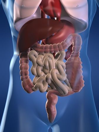
Clinicians also should rule out other causes of constipation, such as pelvic outlet dysfunction, mechanical obstruction, metabolic abnormalities, and comorbidities or concurrent medications, wrote Seth D. Crockett, MD, MPH, of the University of North Carolina at Chapel Hill, together with his associates. The guideline was published online Sept. 1.
Opioid therapy can lead to a range of gastrointestinal symptoms, such as constipation, gastroesophageal reflux, nausea and vomiting, bloating, and abdominal pain. Among these, constipation is by far the most common and debilitating, the guideline notes. In past studies, 40%-80% of patients who received opioids developed opioid-induced constipation (OIC), a more severe presentation that involves a combination of reduced stool frequency in addition to other symptoms, such as harder stools, new or worsening straining during defecation, and a sense of incomplete rectal evacuation.
Treating OIC should start with lifestyle interventions, such as drinking more fluids, toileting as soon as possible when feeling the urge to defecate, and adding regular moderate exercise whenever tolerable, the guideline advises. For patients on oral or parenteral therapy, consider switching to an equianalgesic dose of a less-constipating opioid, such as transdermal fentanyl or oxycodone-naloxone combination therapy.
Many patients with OIC require interventions beyond lifestyle changes or opioid switching. For these patients, the guideline advises starting with conventional laxative therapies based on their safety, low cost, and “established efficacy” in the OIC setting. Options include stool softeners (docusate sodium), osmotic laxatives (polyethylene glycol, magnesium hydroxide, magnesium citrate, and lactulose), lubricants (mineral oil), and stimulant laxatives (bisacodyl, sodium picosulfate, and senna). “Of note, there is little evidence that routine use of stimulant laxatives is harmful to the colon, despite widespread concern to the contrary,” the guideline states. Although randomized, controlled trials have not evaluated particular laxative combinations or titrations for OIC, the best evidence supports stimulant and osmotic laxative therapy, the authors note.
Before deeming any case of OIC laxative refractory, ensure that a patient receives an adequate trial of at least two classes of laxatives administered on a regular schedule, not just “as needed,” the guideline specifies. For example, a patient might receive a 2-week trial of a daily osmotic laxative plus a stimulant laxative two to three times weekly. The guideline authors suggest restricting the use of enemas to rescue therapy. They also note that consuming more fiber tends not to help patients with OIC because fiber does not affect colonic motility.
For truly laxative-refractory OIC, the guidelines recommend escalating treatment to peripherally acting mu-opioid receptor antagonists (PAMORAs). These drugs restore the function of the enteric nervous system by blocking mu-opioid receptors in the gut. Among the PAMORAs, the guideline strongly recommends the use of naldemedine or naloxegol over no treatment, based on robust data from randomized, double-blind, placebo-controlled trials. In the phase 3 COMPOSE 1, 2, and 3 trials, about 52% of patients who received naldemedine achieved at least three spontaneous bowel movements per week, compared with 35% of patients who received placebo. Additionally, in a 52-week safety and efficacy study (COMPOSE 3), naldemedine was associated with one more spontaneous bowel movement per week versus placebo and with a low absolute increase in adverse events.
The guideline bases its strong recommendation for naloxegol on moderate-quality data from three studies, including two phase 3, double-blind, randomized, placebo-controlled trials. Although at least five randomized, controlled trials have evaluated methylnaltrexone, the evidence was low quality and therefore the guideline only conditionally recommends prescribing this PAMORA over no treatment.
The guideline also makes no recommendation on the use of the intestinal secretagogue lubiprostone or the 5HT agonist prucalopride. Studies of lubiprostone were limited by possible reporting bias and showed no clear treatment benefit, the authors state. They describe a similar evidence gap for prucalopride, noting that at least one trial ended early without publication of the findings. They recommend further studying lubiprostone as well as prucalopride and other highly selective 5-HT4 agonists for treating OIC. Head-to-head trials would help guide treatment choice for patients with laxative-refractory OIC, they add. “Cost-effectiveness studies are also lacking in this field, which could inform prescribing strategy, particularly for newer, more expensive agents.”
For patients with suspected opioid-induced constipation, start by taking a careful history of defecation and dietary patterns, stool consistency, incomplete evacuation, and “alarm symptoms,” such as bloody stools or weight loss, state new guidelines from the American Gastroenterological Association in Gastroenterology.
Clinicians also should rule out other causes of constipation, such as pelvic outlet dysfunction, mechanical obstruction, metabolic abnormalities, and comorbidities or concurrent medications, wrote Seth D. Crockett, MD, MPH, of the University of North Carolina at Chapel Hill, together with his associates. The guideline was published online Sept. 1.
Opioid therapy can lead to a range of gastrointestinal symptoms, such as constipation, gastroesophageal reflux, nausea and vomiting, bloating, and abdominal pain. Among these, constipation is by far the most common and debilitating, the guideline notes. In past studies, 40%-80% of patients who received opioids developed opioid-induced constipation (OIC), a more severe presentation that involves a combination of reduced stool frequency in addition to other symptoms, such as harder stools, new or worsening straining during defecation, and a sense of incomplete rectal evacuation.
Treating OIC should start with lifestyle interventions, such as drinking more fluids, toileting as soon as possible when feeling the urge to defecate, and adding regular moderate exercise whenever tolerable, the guideline advises. For patients on oral or parenteral therapy, consider switching to an equianalgesic dose of a less-constipating opioid, such as transdermal fentanyl or oxycodone-naloxone combination therapy.
Many patients with OIC require interventions beyond lifestyle changes or opioid switching. For these patients, the guideline advises starting with conventional laxative therapies based on their safety, low cost, and “established efficacy” in the OIC setting. Options include stool softeners (docusate sodium), osmotic laxatives (polyethylene glycol, magnesium hydroxide, magnesium citrate, and lactulose), lubricants (mineral oil), and stimulant laxatives (bisacodyl, sodium picosulfate, and senna). “Of note, there is little evidence that routine use of stimulant laxatives is harmful to the colon, despite widespread concern to the contrary,” the guideline states. Although randomized, controlled trials have not evaluated particular laxative combinations or titrations for OIC, the best evidence supports stimulant and osmotic laxative therapy, the authors note.
Before deeming any case of OIC laxative refractory, ensure that a patient receives an adequate trial of at least two classes of laxatives administered on a regular schedule, not just “as needed,” the guideline specifies. For example, a patient might receive a 2-week trial of a daily osmotic laxative plus a stimulant laxative two to three times weekly. The guideline authors suggest restricting the use of enemas to rescue therapy. They also note that consuming more fiber tends not to help patients with OIC because fiber does not affect colonic motility.
For truly laxative-refractory OIC, the guidelines recommend escalating treatment to peripherally acting mu-opioid receptor antagonists (PAMORAs). These drugs restore the function of the enteric nervous system by blocking mu-opioid receptors in the gut. Among the PAMORAs, the guideline strongly recommends the use of naldemedine or naloxegol over no treatment, based on robust data from randomized, double-blind, placebo-controlled trials. In the phase 3 COMPOSE 1, 2, and 3 trials, about 52% of patients who received naldemedine achieved at least three spontaneous bowel movements per week, compared with 35% of patients who received placebo. Additionally, in a 52-week safety and efficacy study (COMPOSE 3), naldemedine was associated with one more spontaneous bowel movement per week versus placebo and with a low absolute increase in adverse events.
The guideline bases its strong recommendation for naloxegol on moderate-quality data from three studies, including two phase 3, double-blind, randomized, placebo-controlled trials. Although at least five randomized, controlled trials have evaluated methylnaltrexone, the evidence was low quality and therefore the guideline only conditionally recommends prescribing this PAMORA over no treatment.
The guideline also makes no recommendation on the use of the intestinal secretagogue lubiprostone or the 5HT agonist prucalopride. Studies of lubiprostone were limited by possible reporting bias and showed no clear treatment benefit, the authors state. They describe a similar evidence gap for prucalopride, noting that at least one trial ended early without publication of the findings. They recommend further studying lubiprostone as well as prucalopride and other highly selective 5-HT4 agonists for treating OIC. Head-to-head trials would help guide treatment choice for patients with laxative-refractory OIC, they add. “Cost-effectiveness studies are also lacking in this field, which could inform prescribing strategy, particularly for newer, more expensive agents.”
FROM GASTROENTEROLOGY