User login
Oral SERD camizestrant prolongs PFS vs. fulvestrant in breast cancer
SAN ANTONIO – compared with the first-generation SERD fulvestrant Faslodex, in the SERENA-2 trial, shows a study recently presented at the San Antonio Breast Cancer Symposium.
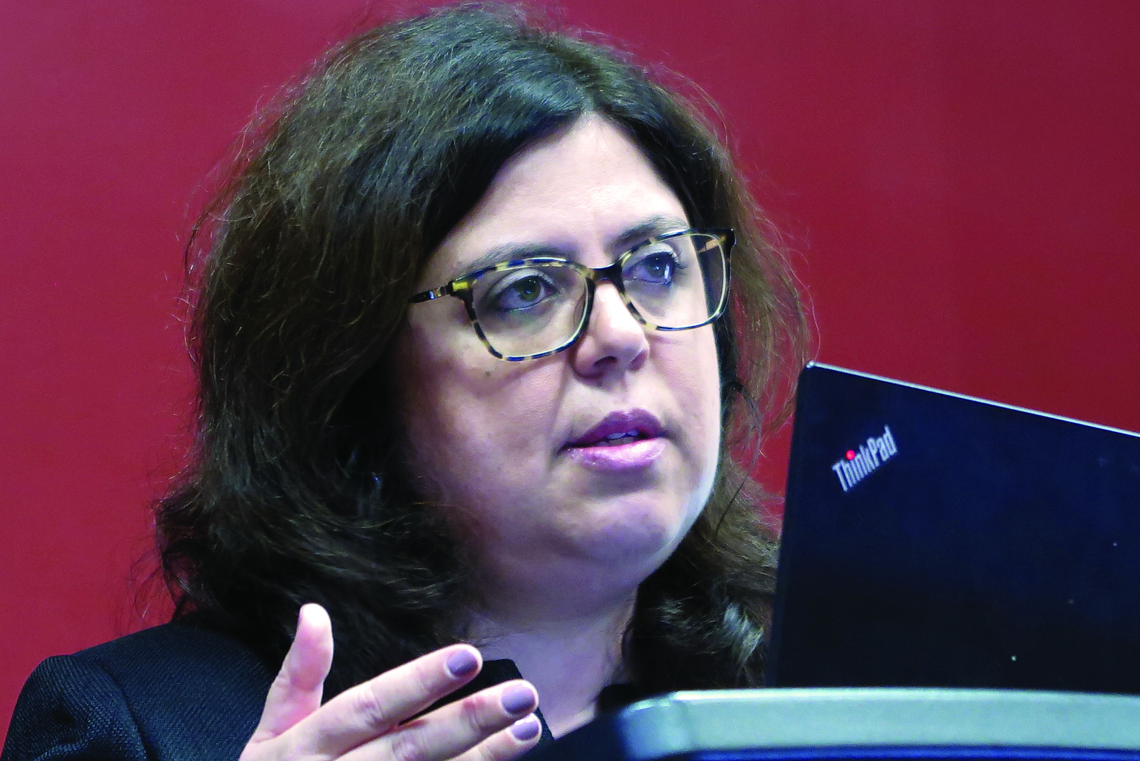
Among 180 postmenopausal women with ER+/HER2– breast cancers that had recurred or progressed following at least one line of endocrine therapy, the median progression-free survival (PFS) after a median follow-up of 16.6-17.4 months was 7.2 months for patients treated at a 75-mg dose of camizestrant and 7.7 months for those treated at a 150-mg dose, compared with 3.7 months for patients who received fulvestrant, reported Mafalda Oliveira, MD, PhD, from Vall d’Hebron University Hospital in Barcelona.
Oral agent
Camizestrant is a next-generation oral SERD and pure estrogen receptor antagonist that was shown in the SERENA-1 trial to be safe and to have clinical activity against ER+ breast cancers.
SERENA-2 pitted camizestrant at doses of 75 mg, 150 mg, or 300 mg against standard-dose fulvestrant, although the 300-mg dose was dropped in a protocol amendment after 20 patients had been assigned to that arm. (Currently planned studies with camizestrant will be conducted with the 75-mg dose.)
The investigators enrolled women with ER+/HER2– advanced breast cancer who had not previously received fulvestrant or an oral SERD. Eligible patients were limited to no more than one prior line of endocrine and one prior line of chemotherapy for advanced breast cancers. The study included patients with both measurable and unmeasurable disease.
The median patient age was about 60 years. Approximately 59% of patients in each arm had either lung or liver metastases. Patients with recurrence in bone only comprised 14.9%-19.4%.
Mutations in ESR1, a gene associated with hormonal resistance, were detectable in 29.7%-47.9% of patients.
Better PFS
As noted before, the primary endpoint of investigator-assessed median PFS favored camizestrant in both the 75-mg arm (7.2 months) and the 150-mg arm (7.7 months), with respective adjusted hazard ratios for progression versus fulvestrant of 0.58 (P = .0124) and 0.67 (P = .0161).
Camizestrant at the 75-mg dose was also superior to fulvestrant among patients who had previously received a cyclin-dependent kinase 4/6 inhibitor, with median PFS of 5.5 months and 3.8 months for the 75-mg and 150-mg doses, respectively, compared with 2.1 months.
The adjusted HR for progression with camizestrant with the 75-mg dose was 0.49, with a 90% confidence interval indicating significance. The 150-mg dose was not significantly superior to fulvestrant, however.
Both camizestrant doses were also superior for prolonging PFS versus fulvestrant among patients with lung and/or liver metastases, with median PFS of 7.2 months, 5.6 months, and 2.0 months, respectively.
The experimental SERD also outperformed fulvestrant in an analysis looking at PFS by ESR1 mutational status and ER-driven disease. Among patients with ESR1 wild type, however, median PFS rates with camizestrant 75 mg and fulvestrant were the same (7.2 months).
The 24-week objective response rates were 15.7% in the 75-mg camizestrant arm, 20% in the 150-mg arm, and 11.8% in the fulvestrant arm. The respective clinical benefit rates, including all patients with responses or stable disease, were 47.3%, 49.3%, and 38.4%. The camizestrant clinical benefit rates did not differ significantly from those with fulvestrant, however.
Treatment-related adverse events of grade 3 or greater occurred in only five patients, and only two patients, both in the 75-mg camizestrant arm, discontinued therapy because of adverse events. There were no treatment-related deaths.
Adverse events that occurred only with camizestrant included photopsia (flashing lights or floaters in the field of vision) and sinus bradycardia.
Promising, but early
Carlos Artega, MD, codirector of SABCS and director of the Simmons Comprehensive Cancer Center at UT Southwestern Medical Center, Dallas, who was not involved in the study, said the data look promising in comparison with fulvestrant.
“There is a clear suggestion that this might be better,” he said. “[Camizestrant] seems to be better at reducing the titer in plasma of the ESR1 mutation, and there is very strong basic science that supports that.”
He noted that the study numbers were relatively small, however.
Dr. Arteaga was speaking at a media briefing held immediately prior to the presentation of the data in an oral abstract session.
Fabrice Andre, MD, from Gustave Roussy in Villejuif, France, the invited discussant for the oral session, noted that, in patients with ESR1 wild type, where fulvestrant shows some efficacy, camizestrant appears to be equally effective, and that the latter agent may be more synergistic with targeted therapies than fulvestrant.
Given high patient dropout rates with currently available SERDs, there is a need for SERDs used in the adjuvant setting that are effective at minimally bioactive doses for patients who are predicted to poorly adherent, Dr. Andre said.
The study was funded by AstraZeneca. Dr. Oliveira has received personal funding from AstraZeneca, Guardant Health, Roche, Merck Sharp & Dohme, Pfizer, Seagen, iTeos Therapeutics, Eisai, Novartis, Relay Therapeutics, and Gilead. Dr. Arteaga is a scientific adviser to AstraZeneca and others, and has received grant support from Pfizer Lilly and Takeda. Dr. Andre disclosed fees to his hospital on his behalf from AstraZeneca, Daiichi Sankyo, Sanofi, Pfizer, Lilly, and Roche.
SAN ANTONIO – compared with the first-generation SERD fulvestrant Faslodex, in the SERENA-2 trial, shows a study recently presented at the San Antonio Breast Cancer Symposium.
Among 180 postmenopausal women with ER+/HER2– breast cancers that had recurred or progressed following at least one line of endocrine therapy, the median progression-free survival (PFS) after a median follow-up of 16.6-17.4 months was 7.2 months for patients treated at a 75-mg dose of camizestrant and 7.7 months for those treated at a 150-mg dose, compared with 3.7 months for patients who received fulvestrant, reported Mafalda Oliveira, MD, PhD, from Vall d’Hebron University Hospital in Barcelona.
Oral agent
Camizestrant is a next-generation oral SERD and pure estrogen receptor antagonist that was shown in the SERENA-1 trial to be safe and to have clinical activity against ER+ breast cancers.
SERENA-2 pitted camizestrant at doses of 75 mg, 150 mg, or 300 mg against standard-dose fulvestrant, although the 300-mg dose was dropped in a protocol amendment after 20 patients had been assigned to that arm. (Currently planned studies with camizestrant will be conducted with the 75-mg dose.)
The investigators enrolled women with ER+/HER2– advanced breast cancer who had not previously received fulvestrant or an oral SERD. Eligible patients were limited to no more than one prior line of endocrine and one prior line of chemotherapy for advanced breast cancers. The study included patients with both measurable and unmeasurable disease.
The median patient age was about 60 years. Approximately 59% of patients in each arm had either lung or liver metastases. Patients with recurrence in bone only comprised 14.9%-19.4%.
Mutations in ESR1, a gene associated with hormonal resistance, were detectable in 29.7%-47.9% of patients.
Better PFS
As noted before, the primary endpoint of investigator-assessed median PFS favored camizestrant in both the 75-mg arm (7.2 months) and the 150-mg arm (7.7 months), with respective adjusted hazard ratios for progression versus fulvestrant of 0.58 (P = .0124) and 0.67 (P = .0161).
Camizestrant at the 75-mg dose was also superior to fulvestrant among patients who had previously received a cyclin-dependent kinase 4/6 inhibitor, with median PFS of 5.5 months and 3.8 months for the 75-mg and 150-mg doses, respectively, compared with 2.1 months.
The adjusted HR for progression with camizestrant with the 75-mg dose was 0.49, with a 90% confidence interval indicating significance. The 150-mg dose was not significantly superior to fulvestrant, however.
Both camizestrant doses were also superior for prolonging PFS versus fulvestrant among patients with lung and/or liver metastases, with median PFS of 7.2 months, 5.6 months, and 2.0 months, respectively.
The experimental SERD also outperformed fulvestrant in an analysis looking at PFS by ESR1 mutational status and ER-driven disease. Among patients with ESR1 wild type, however, median PFS rates with camizestrant 75 mg and fulvestrant were the same (7.2 months).
The 24-week objective response rates were 15.7% in the 75-mg camizestrant arm, 20% in the 150-mg arm, and 11.8% in the fulvestrant arm. The respective clinical benefit rates, including all patients with responses or stable disease, were 47.3%, 49.3%, and 38.4%. The camizestrant clinical benefit rates did not differ significantly from those with fulvestrant, however.
Treatment-related adverse events of grade 3 or greater occurred in only five patients, and only two patients, both in the 75-mg camizestrant arm, discontinued therapy because of adverse events. There were no treatment-related deaths.
Adverse events that occurred only with camizestrant included photopsia (flashing lights or floaters in the field of vision) and sinus bradycardia.
Promising, but early
Carlos Artega, MD, codirector of SABCS and director of the Simmons Comprehensive Cancer Center at UT Southwestern Medical Center, Dallas, who was not involved in the study, said the data look promising in comparison with fulvestrant.
“There is a clear suggestion that this might be better,” he said. “[Camizestrant] seems to be better at reducing the titer in plasma of the ESR1 mutation, and there is very strong basic science that supports that.”
He noted that the study numbers were relatively small, however.
Dr. Arteaga was speaking at a media briefing held immediately prior to the presentation of the data in an oral abstract session.
Fabrice Andre, MD, from Gustave Roussy in Villejuif, France, the invited discussant for the oral session, noted that, in patients with ESR1 wild type, where fulvestrant shows some efficacy, camizestrant appears to be equally effective, and that the latter agent may be more synergistic with targeted therapies than fulvestrant.
Given high patient dropout rates with currently available SERDs, there is a need for SERDs used in the adjuvant setting that are effective at minimally bioactive doses for patients who are predicted to poorly adherent, Dr. Andre said.
The study was funded by AstraZeneca. Dr. Oliveira has received personal funding from AstraZeneca, Guardant Health, Roche, Merck Sharp & Dohme, Pfizer, Seagen, iTeos Therapeutics, Eisai, Novartis, Relay Therapeutics, and Gilead. Dr. Arteaga is a scientific adviser to AstraZeneca and others, and has received grant support from Pfizer Lilly and Takeda. Dr. Andre disclosed fees to his hospital on his behalf from AstraZeneca, Daiichi Sankyo, Sanofi, Pfizer, Lilly, and Roche.
AT SABCS 2022
Chemotherapy meets its match against aggressive ER+/HER2– breast cancers
SAN ANTONIO – Results of a study being hailed as practice changing showed that, for pre- or perimenopausal women with aggressive hormone receptor-positive, HER2-negative (HR+/HER2–) untreated breast cancers,
That’s according to investigators of the phase 2 RIGHT Choice study who found that first-line ribociclib, combined with either letrozole or anastrozole plus goserelin, was associated with a doubling of progression-free survival (PFS), compared with the investigator’s choice of combination chemotherapy, reported Yen-Shen Lu, MD, from National Taiwan University Hospital in Taipei, at the San Antonio Breast Cancer Symposium.
“These results from RIGHT Choice have now shown that first-line ribociclib plus endocrine therapy should be considered the preferred treatment option for this patient population,” he said.
Chemo loses its luster
“This is not time first time that we’ve looked at a CDK4/6 inhibitor compared to chemotherapy, but this is the first time that we’ve seen it compared to a combination chemotherapy,” commented Virginia Kaklamani, MD, from University of Texas Health, San Antonio, who moderated a media briefing held prior to Dr. Lu’s presentation of the data in an oral abstract session.
“I think with this study we’re finding that chemotherapy, at least in the early stages of [estrogen receptor]–positive breast cancer, is probably not appropriate for our patients,” she said.
Chemotherapy is the current standard of care for patients with advanced breast cancers with aggressive disease features that can include rapidly progressive disease, high symptom burden, and/or life-threatening visceral crises requiring rapid control of disease, Dr. Lu said.
Compared with single-agent chemotherapy, combination chemotherapy, for those who can tolerate it, is associated with higher overall response rates and longer PFS.
Although ribociclib plus endocrine therapy has been shown to offer significant PFS and overall survival (OS) benefits, compared with endocrine therapy alone, there have not been any head-to-head studies pitting these agents against combination chemotherapy.
Study details
To rectify this, Dr. Lu and colleagues enrolled 222 pre- or perimenopausal women with HR+/HER2– advanced breast cancers with aggressive features who had not yet received systemic therapy for advanced breast cancer.
After stratification for the presence or absence of liver metastases and by length of disease-free interval (time from complete resection of a primary tumor to documented recurrence), the patients were randomly assigned to receive either ribociclib 600 mg 3 weeks on, 1 week off) plus letrozole or anastrozole and goserelin, or the investigators choice of either docetaxel plus capecitabine, paclitaxel plus gemcitabine, or capecitabine plus vinorelbine.
After a median follow-up of 24.1 months at the time of data cutoff in April 2022, median PFS, the primary endpoint, was 24 months in the ribociclib plus endocrine therapy arm versus 12.3 months in the chemotherapy arm.
This translated into a hazard ratio for progression on ribociclib plus endocrine therapy of 0.54 (P = .0007).
The benefit for the ribociclib combination, compared with combination chemotherapy, was consistent across most patient subgroups, Dr. Lu said.
The median time to treatment failure was also longer with ribociclib, at 18.6 months versus 8.5 months, respectively, translating into a HR of 0.45 favoring ribociclib, with a statistically significant confidence interval.
Overall response rates were similar between the groups, at 65.2% with ribociclib versus 60% with chemotherapy. The respective clinical benefit rates (including complete and partial responses plus stable disease) were 80.4% versus 72.7%.
The time to response was similar between the treatment arms, an important consideration for patients with rapidly progressive disease, Dr. Lu noted.
Adverse events that occurred more frequently with ribociclib were neutropenia and leukopenia. Events more common with chemotherapy included anemia, liver enzyme elevations, nausea, vomiting, diarrhea, alopecia, fatigue, and palmar-plantar erythrodysesthesia.
Confirmation
“These data are just confirming what we’ve already known, and that is that with ER-positive, HER2-negative breast cancer where you have metastatic disease and more aggressive characteristics, treating with a CDK4/6 inhibitor and endocrine therapy leads to high response rates,” breast cancer specialist Matthew P. Goetz, MD, from the Mayo Clinic in Rochester, Minn., said in an interview. Dr. Goetz was not involved in the study.
“What was surprising to me was the fact that the response rates with chemotherapy were not higher,” he said. “We sometime think that the more chemotherapy, the higher the response rates. It was nice to see a direct comparison with chemotherapy, and really to see that giving a target therapy actually led to very, very good results. That tells us that there should be very few situations where we would be prescribing chemotherapy over CDK4/6 inhibitor–based therapies.”
The study was funded by Novartis Pharma. Dr. Lu disclosed personal funding from Novartis and others. Dr. Goetz disclosed grants and other supports for work with the development of abemaciclib and palbociclib, and consulting for Pfizer and others. Dr. Kaklamani disclosed speakers bureau activity for Novartis and others, research support from Eisai, and consulting for other companies.
AT SABCS 2022
Potential cause of worse outcomes among Black breast cancer patients found
SAN ANTONIO – compared with White women, a discovery that may at least partially explain racial differences in breast cancer outcomes, investigators say.
The finding, which comes from a retrospective study comparing differences in tumor microenvironment of metastasis (TMEM) “doorways” between Black and White women suggest that tumors in Black women may have a stronger prometastatic response to neoadjuvant chemotherapy than tumors in White women, reported Maja H. Oktay, MD, PhD, of Montefiore Einstein Cancer Center, Albert Einstein College of Medicine, New York, at the San Antonio Breast Cancer Symposium.
“Looking forward ... we propose to use TMEM doorway density as a prognostic marker for distant recurrence-free survival as a marker of dissemination, and also as a predictive marker of response to drugs that can block TMEM doorways,” she said at a briefing held prior to the presentation of data in an oral abstract session.
Entry points
As their name implies, TMEM doorways are transient entry points or portals that allow cancer cells to disseminate to distant sites. TMEM doorways are composed of tumor cells, macrophages, and endothelial cells that come into direct contact and together create temporary vascular openings that allow tumor cells to cross cell walls into circulation, where they can then hitch a ride and travel to distant organ sites.
Previous studies have shown that TMEM doorway density is a prognostic marker of metastasis in breast cancer patients treated with adjuvant chemotherapy. And as Dr. Oktay and colleagues showed in the current study, TMEM doorway density, as measured by a TMEM doorway score, is a prognostic marker for distant metastatic recurrence of ER+/HER2– breast cancer following neoadjuvant chemotherapy.
They also showed that neoadjuvant chemotherapy may increase the TMEM doorway score and lead to a pro–metastatic tumor microenvironment in some women.
Doorway scores
The investigators measured TMEM doorway scores from residual breast cancers in women who had undergone standard neoadjuvant chemotherapy. The cohort consisted of 96 Black women, 43 of whom had ER+/HER2– breast cancer and 37 of whom had triple-negative breast cancer (TNBC), and 87 White women, 50 with ER+/HER2– cancer and 22 with TNBC. The remaining patients had other breast cancer subtypes.
They found that TNBCs had higher TMEM doorway density score and higher macrophage density scores, which may explain why patients with TNBC often have early recurrence of disease.
They also found that, compared with White patients, Black patients with ER+/HER2– tumors, but not TNBC tumors, had higher TMEM doorway density scores. Similarly, Black patients with ER+/HER– cancers, but not TNBC, had higher macrophage levels than White women, a finding that may explain racial disparity in ER+/HER2– disease, Dr. Oktay said.
For the entire cohort, patients with high TMEM doorway density scores had significantly worse distant recurrence–free survival than patients with intermediate or low scores (P = .008), and there was a trend toward worse DRFS among all patients with ER+/HER2– who were in the highest third of scores, but this did not quite reach statistical significance.
High versus low TMEM doorway density score was also an independent prognostic factor for worse outcomes among the entire cohort (P = .01).
There was no significant difference in TMEM density scores among patients with TNBC.
Neither high macrophage counts nor microvascular density alone were significantly associated with inferior DRFS. TMEM doorway score was the only factor significantly prognostic for worse outcomes among patients in the entire cohort.
Hypothesis needs further testing
Invited discussant Lori Pierce, MD, a radiation oncologist with Michigan Medicine, University of Michigan, Ann Arbor, said it’s unclear whether TMEM doorway density changed following neoadjuvant chemotherapy as there were no prechemotherapy scores available in this study.
“But I think the key part is that, if we think neoadjuvant chemotherapy promotes metastasis, then there should be an inferior outcome compared to adjuvant chemotherapy, but that’s not what we see. Well-powered randomized trials show equivalent outcomes with neoadjuvant chemotherapy as well as adjuvant,” she said.
She noted that a 2018 meta-analysis of individual patient data from 10 randomized trials comparing neoadjuvant with adjuvant chemotherapy in early breast cancer showed no differences in long-term distant recurrences, breast cancer–specific mortality, or all-cause mortality between the two modalities.
“While I think these data are very provocative, I certainly wouldn’t want Black women or any women who need neoadjuvant therapy to be discouraged because of these data. We need these data to be tested rigorously, so I look forward to the clinical trials that will test this question and can really give us more information about this very interesting hypothesis,” Dr. Pierce said.
The study was funded by the National Institutes of Health, New York State Department of Health Peter T. Rowley Breast Cancer Scientific Research Projects, Helen & Irving Spatz Family Foundation, Evelyn Gruss Lipper Charitable Foundation, and the Gruss-Lipper Biophotonics Center and the integrated imaging program at the Albert Einstein College of Medicine. Dr. Oktay reported no conflicts of interests.
AT SABCS 2022
Diagnosed too late
It had only been 3 weeks since I first met this patient. She presented with an advanced case of colon cancer, but instead of treatment,
Within the course of 2 weeks I saw another new patient, but this time with pancreatic cancer that metastasized to the liver. “When can we start treatment?” he asked. Like my female patient with colon cancer, he was diagnosed too late as he was already in an incurable stage. He was shocked to learn that his condition was in stage 4, that achieving remission would be difficult and a cure, not likely. Certainly, standard of care treatments and clinical trials offered him hope, but they were unlikely to change the outcome.
We take a course in this – that is, in giving bad news, but every doctor has his or her own approach. Some are so uncomfortable with the talk, they choose avoidance and adopt the “look like you gotta go approach.” Or, the doctor may schedule another treatment or another test with the intention of avoiding end-of-life discussions. Other doctors opt for straight talk: “I think you should get your affairs in order. You’ve got 3 months to live.” These are extreme behaviors I wouldn’t recommend.
In my practice, I sit with my patients and explain the diagnosis. After discussing all options and the advanced stage and diagnosis, it ultimately comes down to “Win or lose, I will be here to take care of you.” Sometimes there is therapy that may help, but either way, the patient understands that death is a real possibility.
I find that people just want to know if there is hope. A different treatment regimen or a clinical trial may (or may not) extend their life. And while we cannot predict outcomes, we can give them hope. You can’t shut down hope. True for some people the cup is always half empty, but most people want to live and are optimistic no matter how small the chances are.
These conversations are very difficult. I don’t like them, but then I don’t avoid them either. Fortunately, patients don’t usually come to my office for the first visit presenting with advanced disease. In the cases I described above, one patient had been experiencing unexplained weight loss, but didn’t share it with a physician. And, for the patient with pancreatic cancer, other than some discomfort in the last couple of weeks, the disease was not associated with other symptoms. But the absence of symptoms should not in any way rule out a malignant disease. A diagnosis should be based on a complete evaluation of signs and symptoms followed by testing.
We’ve got to be able to take the time to listen to our patients during these encounters. We may not spend as much time as we should because we’re so busy now and we’re slaves to EMRs. It helps if we take more time to probe symptoms a little longer, especially in the primary care setting.
It is possible for a patient with cancer to be asymptomatic up until the later stages of the disease. A study published in ESMO Open in 2020 found that fewer than half of patients with stage 4 non–small cell lung cancer have only one or two symptoms at diagnosis regardless of whether the patient was a smoker. In this study only 33% of patients reported having a cough and 25% had chest pain.
A study presented in October at the United European Gastroenterology Week found that of 600 pancreatic cancer cases, 46 of these were not detected by CT or MRI conducted 3-18 months prior to diagnosis. Of the 46 cases, 26% were not picked up by the radiologist and the rest were largely as a result of imaging changes over time. Radiology techniques are good, but they cannot pick up lesions that are too small. And some lesions, particularly in pancreatic cancer, can grow and metastasize rather quickly.
When a patient is diagnosed with advanced disease, it is most often simply because of the nature of the disease. But sometimes patients put off scheduling a doctor visit because of fear of the potential for bad news or fear of the doctor belittling their symptoms. Some tell me they were “just hoping the symptoms would disappear.” Waiting too long to see a doctor is never a good idea because timing is crucial. In many cases, there is a small window of opportunity to treat disease if remission is to be achieved.
Dr. Henry is a practicing clinical oncologist with PennMedicine in Philadelphia where he also serves as Vice Chair of the Department of Medicine at Pennsylvania Hospital.
This article was updated 12/7/22.
Managing trastuzumab deruxtecan adverse events in the real world
With recent expansions in its breast cancer indications, there has been an increase in the use of trastuzumab deruxtecan (T-DXd; Enhertu).
“A lot of us are using this more frequently now than we were in the past,” explained Sid Yadav, MD, a breast and gynecologic cancer specialist at the Mayo Clinic in Rochester, Minn. However, he added that managing its adverse events has been a “bit of a learning curve for all of us.”
The antibody-drug conjugate has been on the market since 2019 for metastatic human epidermal growth factor receptor (HER2)–positive breast cancer, but it was then approved in May 2022 for earlier use in this patient population and in August 2022 for patients with HER2-low disease. This latest approval was based on data showing an improvement in overall survival that was described as “practice changing.”
In addition, T-DXd is also approved for use in metastatic HER2-mutated non–small cell lung cancer and metastatic HER2-positive gastric and gastroesophageal junction adenocarcinoma.
, Dr. Yadav told this news organization.
Among the eight or so patients he’s seen or treated over 2 months, Dr. Yadav has already seen one case of high-grade interstitial lung disease/pneumonitis, a complication that “everybody worries about” because the label for T-DXd carries a black box warning of this possibility.
There have been other issues at Mayo Clinic, as well. In one recent week, five patients were admitted for possible T-DXd adverse events, including neutropenic fever and sepsis; pneumonitis; severe nausea/vomiting with electrolyte imbalance; pneumonia, and non–ST elevation myocardial infarction with low ejection fraction.
It’s unknown what proportion of T-DXd recipients the five admissions represented. Dr. Yadav’s service has over 10 breast oncologists, so the cases could represent maybe 1%-10% of patients, he said.
His experience prompted Dr. Yadav to turn to Twitter to ask fellow oncologists what complications they’ve seen with T-DXd.
One said that his “real-world toxicity experience [has been] worse than the trial data,” which isn’t unusual, another oncologist noted, because real-world patients are often sicker than trial participants and more vulnerable to toxicities.
A third oncologist countered that she has “found [T-DXd] generally easy for patients to tolerate and [has] not needed to admit anyone” so far.
Overall, Dr. Yadav said that in his experience there are issues that need to be considered with T-DXd beyond interstitial lung disease.
As with any chemotherapy, neutropenia and infections are a concern, as the labeling notes. The interstitial lung disease case has also made Dr. Yadav have a low threshold to order CT in patients with any hints of shortness of breath and to start steroids if there’s any suspicion.
Probably the most common issue, however, is nausea and vomiting. In clinical trials, over 70% of participants reported nausea and over 40% experienced vomiting.
In response, Dr. Yadav and his colleagues have become more aggressive with prophylaxis. Pretreatment includes steroids, palonosetron, and fosaprepitant. Patients are also usually sent home with prochlorperazine, ondansetron, and lorazepam. If these don’t help, the team considers olanzapine.
They have also learned that “it’s important to spend that extra 15-20 minutes upfront” with patients before starting T-DXd to explain the risk for nausea and vomiting and how it will be managed, Dr. Yadav commented. “We do chemotherapy teaching for every patient, but I think we spend more time [now] talking about nausea and vomiting with this subset,” he said.
Dr. Yadav still starts patients on the standard breast cancer dose of T-DXd – 5.4 mg/kg every 3 weeks – but said he’s quicker now to lower the dose if patients aren’t doing well. He estimates he’s done that a couple of times so far.
Approaches at the Mayo Clinic are in line with those in a recent article on managing T-DXd toxicities by Hope Rugo, MD, from the University of California, San Francisco, and colleagues.
These authors conclude that adverse events related to T-DXd are frequent but are most commonly low grade and manageable. Nausea and vomiting are among the most common, and they note that interstitial lung disease/pneumonitis is an important adverse event, for which proactive monitoring, diagnosis, and management are key.
The review describes management practices of other health care providers and institutions with experience in using T-DXd to help with safe and effective management of the drug’s adverse events, particularly since the duration of treatment may be quite long.
Proper management of T-DXd–related adverse events will allow optimal exposure to and benefit from the drug and will help avoid premature discontinuation or improper dose reductions, Dr. Rugo and colleagues commented.
Dr. Yadav reports no relevant conflicts of interest.
A version of this article first appeared on Medscape.com.
Whole breast radiation for breast cancer shown to be safe and effective
The findings from a phase 3 clinical trial are a boon to patient convenience.
“These findings are indeed practice changing. This was a well-designed trial that looked at shortening treatment from 6 weeks down to 3 weeks. And, they showed equivalent local control and importantly, a good cosmetic outcome over time,” said Kathleen Horst, MD, who served as a discussant at a press conference held at the annual meeting of the American Society for Radiation Oncology where the findings were presented.
“This is substantially more convenient. It is cost effective, both for the health care system and for individual patients. Importantly, our patients come in for treatment every day. They’re taking time off of work, they have to arrange for childcare, and they have to arrange for transportation. So this makes a big difference for these patients,” said Dr. Horst, who is a professor of radiation oncology at Stanford (Calif.) Medicine and director of well-being in the radiation department at Stanford Medicine.
The study was presented by Frank A. Vicini, MD, FASTRO, a radiation oncologist with GenesisCare, Farmington Hills, Mich.
“One of the things I think that was surprising is I think all of us were thinking that this might be a more toxic regimen, but as Dr. Vincini showed, over time it was equally effective and with minimal toxicity, and cosmesis over time was stable, and that’s important. Importantly, that included patient-reported outcomes, not just the physician-reported outcomes. Broadly, I think these findings are applicable for many patients, all patients who are receiving whole breast radiotherapy with an added boost. I think over time this is going to improve the quality of life of our patients. It is an innovative change that everyone is going to be excited to embrace,” Dr. Horst said.
Previous randomized, controlled trials showed that an additional radiation dose to the tumor bed following lumpectomy and whole breast irradiation reduces the relative risk of local recurrence by about 35%. However, this increases treatment time for patients who have already endured an extensive regimen. For whole breast irradiation, hypofractionated radiation is in 15-16 fractions over 3 weeks has comparable recurrence rates as a 5-week regimen, but the relevant trials did not examine the effect hypofractionation may have on a radiation boost to the tumor bed of high-risk patients. Because of this lack of evidence, current practice is for the boost to remain sequential in five to eight fractions after completion of whole breast irradiation, which adds a week to a week and a half to treatment length.
The study included 2,262 patients who were randomized to receive a sequential boost or a concomitant boost. After a median follow-up of 7.4 years, there were 54 ipsilateral breast recurrence (IBR) events. The estimated 7-year risk of IBR was 2.2% in the sequential boost and 2.6% in the concurrent risk group (hazard ratio, 1.32; noninferiority test P = .039). Approximately 60% of patients received adjuvant chemotherapy.
Grade 3 or higher adverse events were similar, with a frequency of 3.3% in the sequential group and 3.5% in the concurrent group (P = .79). The researchers used the Global Cosmetic Score to assess outcomes from the perspective of both physicians and patients; 86% of physicians rated the outcome as excellent/good in the sequential group versus 82% in the concurrent group (P = .33).
“For high-risk early-stage breast cancer patients undergoing breast conservation, a concurrent boost with hypofractionated whole breast irradiation as compared to a sequential boost, results in noninferior local recurrence rates with no significant difference in toxicity, noninferior patient-rated cosmesis, no significant difference in physician rated cosmesis, and delivering the entire treatment even at high risk patients in 3 weeks. Just as critical, the use of target volume–based radiation planning for 3-D [three-dimensional] conformal or [intensity-modulated radiation therapy] whole breast irradiation assessed by dose volume analysis is feasible, and resulted in very low toxicity in the treatment arms, regardless of the fractionation schedule, or the boost delivery,” said Dr. Vincini during the press conference.
The study was grant funded. Neither Dr. Vincini nor Dr. Horst had relevant financial disclosures.
FROM ASTRO 2022
Discontinuing immunotherapy: Is the infusion bag half empty or half full?
It’s a “champagne problem” many of us have encountered over the past few years in the clinic.
A patient with advanced non–small cell lung cancer (NSCLC) is fortunate enough to continue to do well for 2 years on ongoing pembrolizumab or perhaps pemetrexed and pembrolizumab as maintenance therapy. The latest CT shows a residual but far smaller primary tumor than what she started with.
In this instance, you may be considering stopping treatment but are concerned about doing so with evidence of disease still present.
Clinical trials of immunotherapy or chemoimmunotherapy have generally terminated treatment in nonprogressing patients after 2 years. We also know that some patients in early trials of immunotherapy stopped treatment after a fixed period of 1 or 2 years and continued to show no evidence of progression many years later.
The reason some patients experience this kind of success: Unlike the mechanism of action of conventional chemotherapy or targeted therapies, where ongoing treatment would be important to continue to exert an inhibitory effect, the active substrate of immunotherapy is the patient’s immune system, which can potentially have a self-sustaining efficacy beyond the stimulatory effect of the checkpoint inhibitor.
One trial directly addressed this question of stopping vs. continuing treatment in patients on immunotherapy. The CheckMate 153 trial, published in 2020, randomly assigned 252 previously treated patients who hadn’t demonstrated progression after 1 year on nivolumab to either discontinue nivolumab or continue nivolumab on an ongoing basis. The results were strongly in favor of ongoing therapy. Both progression-free survival (PFS) and overall survival (OS) were significantly longer in patients who continued therapy: PFS of 24.7 months vs. 9.4 months and OS not reached vs. 32.5 months.
This finding is important, but there’s an important caveat. The study population included many heavily pretreated patients, but, in practice, immunotherapy has generally moved into the first-line setting, where we see dramatic responses in a significant subset of patients.
Even more recent data are emerging that may help us evaluate who will do well off therapy and who should continue treatment.
We now have a growing collection of long-term data on patients who are more likely to have good outcomes with immunotherapy, specifically those with high tumor programmed death-ligand 1 (PD-L1) expression (≥ 50%), from the KEYNOTE-024 trial. In this study, 39 of 151 (25.8%) patients assigned to pembrolizumab completed the planned maximum of 2 years of treatment, among whom 82.1% achieved an objective response; but, only 10% (4 patients) achieved a complete response. The proportion of patients without progression and remaining off therapy wasn’t reported, but the OS rate 3 years after completing treatment was 81.4%.
In addition, restarting immunotherapy after discontinuing appears to be a moderately effective strategy. In the KEYNOTE-024 trial, 12 patients received a second course of pembrolizumab because of disease progression a median of 15.2 months after discontinuing pembrolizumab. In this small cohort, eight of these patients (66.7%) were alive at the data cutoff, and six (50%) achieved stable disease.
Recently, we received additional insight in the follow-up from two chemoimmunotherapy trials that have most shaped my practice for patients with advanced NSCLC and any level of PD-L1 expression. These are the KEYNOTE-189 trial of platinum-pemetrexed with pembrolizumab vs. placebo in those with nonsquamous NSCLC, and the KEYNOTE-407 trial of carboplatin-taxane with pembrolizumab vs. placebo in patients with advanced squamous NSCLC. The National Comprehensive Cancer Network has designated each as a “preferred regimen” for patients with advanced NSCLC.
Both regimens have demonstrated sustained efficacy benefits with prolonged follow-up, including significantly superior objective response rate, PFS, and OS with the addition of pembrolizumab. These findings merely cemented the role of these regimens in our practice, but the trials also reported on the cohort of patients who completed 35 cycles of treatment over 2 years then discontinued therapy. In both, the majority of patients showed an objective response (86% in KEYNOTE-189 and 90% in KEYNOTE-407), with most patients alive at 3 years after 2 years of treatment (71.9% in KEYNOTE-189 and 69.5% in KEYNOTE-407). In addition, the proportion of patients alive without disease progression or subsequent therapy was notable – 40.4% in KEYNOTE-189 and 43.6% KEYNOTE-407.
How should we interpret these data for the patient who is in the exam room with us?
The short answer is that we don’t know. I see this as a half-empty, half-full conundrum.
I’m disappointed that more patients who responded for 2 years will experience disease progression in the 1-3 years that follow. This signals that their immune systems have not perpetuated their initial response over the long-term. But these patients may have demonstrated disease progression even if they had continued therapy.
We also know that some patients can be rechallenged and will respond again. Some of these patients will show stable disease, whereas others will progress with repeat treatment. I would love to be able to better predict which patients are destined to do well without treatment vs. those who benefit from treatment beyond 2 years.
Might the level of PD-L1 expression tell us? Can PET imaging discriminate those with residual hypermetabolism who may need continued treatment from those with no residual uptake who could be spared it? Would serial measurement of circulating tumor DNA (ctDNA) in responding patients identify when they have achieved a point of diminishing returns, potentially indicating that some can safely discontinue treatment after 2 years, whereas others need to continue to suppress on prolonged maintenance therapy?
These questions have yet to be studied systematically. In the meantime, I take an individualized approach with my patients facing this decision. Some have experienced escalating arthralgias and myalgias, cost concerns, or other issues related to immunotherapy that may dissuade us from continuing treatment. But several others have been grateful to continue with their treatment, hesitant to do anything that could change the path of their disease.
In my patients who tolerate therapy well, I’m more worried about potential undertreatment than overtreatment. I tend to favor having my patients continue therapy in the absence of problematic toxicity or practical challenges. There is certainly room for debate here while we await data to better guide these decisions. How do you approach these patients?
Dr. West is Clinical Associate Professor, Department of Medical Oncology, City of Hope Comprehensive Cancer Care, Duarte, Calif. He reported conflicts of interest with Ariad/Takeda, Bristol-Myers Squibb, Boehringer Ingelheim, Spectrum, AstraZeneca, Celgene, Genentech/Roche, Pfizer, Merck, and Eli Lilly.
A version of this article first appeared on Medscape.com.
How AI is, or will soon be, relevant in radiation oncology
Artificial intelligence (AI) is impacting many aspects of health care, and radiation oncology is no exception. It has the potential to cut costs and streamline work flows ranging from image analysis to treatment plan formulation, but its specific place in clinical practice is still being debated.
In a session at the annual meeting of the American Society for Radiation Oncology, researchers discussed some of the ways that AI is or will soon be relevant to the clinic. The general consensus was that
In his talk, Sanjay Aneja, MD focused on practical applications of AI that are in the clinic or close to being ready. One example is image classification. “There has been recent evidence that suggests in a variety of different kind of scenarios, deep-learning models can be very good at image classification in automated ways,” said Dr. Aneja, who is a professor of radiology at Yale University, New Haven, Conn. He described one study that used AI to classify 14 different pathologies on chest x-ray images.
Dr. Aneja described the open-source nnU-net tool, which automatically configures itself and segments biomedical images for research or clinical purposes, including therapy planning support, intraoperative support, and tumor growth monitoring. The researchers who developed it also created a “recipe” to systematize configuration of nnU-net, making it useful as an out-of-the-box tool for image segmentation.
He predicted that AI will improve radiology oncology by assisting in the determination of disease extent, including microscopic areas of disease. It could also help plan treatment volume and monitor treatment response. “I think that these are the types of things that will be moving toward the clinic in the future; very specific applications and models trained on very specific scenarios that will help us answer a very important clinical question,” Dr. Aneja said.
He expects AI to contribute to auto-segmenting and clinical contouring, “but I will caution everyone that these algorithms have not been proven to be better than physician contours. They very frequently fail in the specific use cases when anatomy is distorted by, I don’t know, say a tumor. And so a lot of times, we don’t actually have the ability to just make it an automated process. I think it’ll be something that physicians will use to help them but not necessarily replace their contouring ability,” Dr. Aneja said.
Another, potentially more useful application, is in adaptive radiation planning. “I think that AI auto-contouring will be very helpful in establishing contours in a situation in which a physician doing them would not be feasible. We need to have nimble and computationally efficient auto segmentation algorithms that will be able to be easily deployed at the linear accelerator,” he said.
AI in pathology and treatment selection
In another talk, Osama Mohamad, MD talked about AI in pathology, and specifically treatment selection. He described research from his group that digitized pathology data from 5,500 patients drawn from five randomized, clinical trials. They used AI on data from four of the clinical trials to identify a prognostic biomarker for distant metastasis, then validated it on data from the remaining clinical trial, which compared radiation versus radiation plus short-term hormone therapy in prostate cancer.
The results suggested that most patients should receive hormone therapy, but the AI suggested a more nuanced answer. “Patients who had AI biomarker negative do not see any benefit from adding 4 months of hormone therapy ... whereas patients who have biomarker positive have significant difference and improvement in distant metastasis at 10 years and 15 years. This means that we can save a significant proportion of patients from getting [androgen deprivation therapy], which is hormonal therapy and has very well-known side effects, because they simply they will not benefit,” said Dr. Mohamad, who is an assistant professor of radiation oncology at University of California, San Francisco.
That study relied on the ArteraAI prostate cancer test, which is available through a Clinical Laboratory Improvement Amendment–certified laboratory in Florida.
Another example of AI used to plan treatment is On-line Real-time Benchmarking Informatics Technology for Radiotherapy (ORBIT-RT), developed at the University of California, San Diego. It focuses on radiotherapy treatment plan quality control, and has two main components: creating clinically validated plan routines and a free radiotherapy plan quality control system.
No matter how impressive the technical advances may be, AI contributions won’t impact clinical practice if radiation oncologists, physicians, and patients don’t accept AI. Dr. Aneja’s group surveyed patients about which health field they would feel more comfortable with AI having an important role. Most said they were extremely uncomfortable when it came to cancer. “Now, does that mean that we can’t use AI in oncology? No, I think it just means that we have to be a little bit more nuanced in our approach and how we develop AI solutions for cancer patients,” Dr. Aneja said.
Physicians also show reluctance, according to Alejandro Berlin, MD, who is an affiliate scientist at Princess Margaret Cancer Centre in Toronto. He discussed some research looking at physician acceptance of machine learning. His group looked at physician acceptance of treatment plans for prostate cancer that were generated by physicians and in parallel by machine learning. In a theoretical phase, physicians generally agreed that the machine learning plans were better, but when it came to a phase of the study in which physicians chose which plan to implement in a real patient, the acceptance of machine learning-generated plans dropped by 20%.
This tendency to trust humans over machines is what Dr. Berlin called “automation bias,” and he called for a more collaborative approach to implement AI. “In some cases, [machine learning] is going to be good and sufficient. And in some cases, you will need the expertise of a human.”
Dr. Aneja, who also moderated the session, expressed a similar sentiment when summing up the day’s talks: “I do feel like it’s a disruptive technology ... but I think there will still be a need for us to have people who are trained in order to evaluate and make sure that these algorithms are working correctly and efficiently.”
Dr. Aneja, Dr. Mohamad, and Dr. Berlin have no relevant financial disclosures.
* This article was updated on Nov. 15, 2022.
FROM ASTRO 2022
Chronic stress, especially race related, may hasten cancer death
The American folk hero John Henry pitted his hammer against a mechanical steam drill, only to die of exhaustion after winning the battle. In the legend, John Henry was African American, and it’s a fitting metaphor, according to Justin Xavier Moore, PhD.
It’s a metaphor for accumulated stress over a lifetime, also known as allostatic load. Though it affects everyone, Black, Indigenous, and people of color experience it in excess. “It serves as a symbolism for the plight of African Americans within the United States, that regardless of all the triumph and trying to overcompensate and work just as hard as your counterpart, it oftentimes leads to this overtaxing or exhaustion because your competitor has an unfair advantage. You have Jim Crow laws in the South. We have the history of slavery. We have individuals of racial subgroups that are exposed daily to microaggressions, racial discrimination, stereotypes, redlining, all of these different issues that basically reduce to systemic racism,” said Dr. Moore, who is an assistant professor of medicine at the Medical College of Georgia, Augusta.
Dr. Moore is also a coauthor of a new study published online in SSM–Population Health, which examined the association between increased allostatic load and cancer outcomes among participants in the National Health and Nutrition Examination Survey (NHANES) and the National Death Index. They found that both non-Hispanic Black and non-Hispanic White adults with high allostatic load had about a doubled risk of cancer death.
To determine allostatic load, the researchers looked at nine factors collected in NHANES: abnormal values of BMI, diastolic blood pressure, glycohemoglobin, systolic blood pressure, total cholesterol, serum triglycerides, serum albumin, serum creatinine, and C-reactive protein. “The fact that we’re looking at cardiovascular, metabolic and immune function, all in one gives us a better risk assessment for morbidity and mortality. Allostatic load has actually been associated with cardiovascular disease. I think we are one of the first studies to actually look at whether allostatic load is associated with cancer mortality,” said Dr. Moore.
Previous research coauthored by Dr. Moore showed 20-year old African Americans have an allostatic load comparable with that seen in 30-year-old non-Hispanic Whites. That can lead to a proinflammatory state that might be causing increased cancer risk. But stress isn’t a simple concept to pin down, Dr. Moore said. “One of the founding fathers of public health research and epidemiology, Paracelsus, [said] ‘the dose makes the poison.’ ”
In this case, it means that not all stress is bad. Exercise is good stress. “Your heart rate goes up, you compete, and then it comes back down. That’s healthy. But then there’s those stressful situations like dealing with a horrible job, and a boss that may just be overdemanding. Deadlines, and not having a work-life balance. Too much stress, in this case, can cause cancer death,” Dr. Moore said.
In the study, both non-Hispanic Black adults and non-Hispanic White adults heightened risk of cancer death when dealing with high allostatic load, even though the cause of stress may be different. “It’s almost like the cause of the stress does not matter as much. There are millions of Americans that live in environments that are not conducive to their health. The fact of the matter is that because of racial discrimination, because all these different biases, African Americans may have higher allostatic load, which they did on an average, but high allostatic load for even White people is associated with dying from cancer,” Dr. Moore said.
After adjustment, the (adjusted subdistributed hazard ratio, 1.14; 95% CI, 1.04-1.26). After stratification by age, high allostatic load was associated with an 80% increased risk of cancer death among adults (SHR, 1.80; 95% CI, 1.35-2.41). Non-Hispanic White adults had a 95% increased risk (SHR, 1.95; 95% CI, 1.22-3.12), non-Hispanic Black adults had a twofold increased risk (SHR, 1.06; 95% CI, 1.27-3.34), and Hispanic adults had a 36% increased risk.
Dr. Moore has no relevant financial disclosures.
FROM SSM–POPULATION HEALTH
Third COVID booster benefits cancer patients
though this population still suffers higher risks than those of the general population, according to a new large-scale observational study out of the United Kingdom.
People living with lymphoma and those who underwent recent systemic anti-cancer treatment or radiotherapy are at the highest risk, according to study author Lennard Y.W. Lee, PhD. “Our study is the largest evaluation of a coronavirus third dose vaccine booster effectiveness in people living with cancer in the world. For the first time we have quantified the benefits of boosters for COVID-19 in cancer patients,” said Dr. Lee, UK COVID Cancer program lead and a medical oncologist at the University of Oxford, England.
The research was published in the November issue of the European Journal of Cancer.
Despite the encouraging numbers, those with cancer continue to have a more than threefold increased risk of both hospitalization and death from coronavirus compared to the general population. “More needs to be done to reduce this excess risk, like prophylactic antibody therapies,” Dr. Lee said.
Third dose efficacy was lower among cancer patients who had been diagnosed within the past 12 months, as well as those with lymphoma, and those who had undergone systemic anti-cancer therapy or radiotherapy within the past 12 months.
The increased vulnerability among individuals with cancer is likely due to compromised immune systems. “Patients with cancer often have impaired B and T cell function and this study provides the largest global clinical study showing the definitive meaningful clinical impact of this,” Dr. Lee said. The greater risk among those with lymphoma likely traces to aberrant white cells or immunosuppressant regimens, he said.
“Vaccination probably should be used in combination with new forms of prevention and in Europe the strategy of using prophylactic antibodies is going to provide additional levels of protection,” Dr. Lee said.
Overall, the study reveals the challenges that cancer patients face in a pandemic that remains a critical health concern, one that can seriously affect quality of life. “Many are still shielding, unable to see family or hug loved ones. Furthermore, looking beyond the direct health risks, there is also the mental health impact. Shielding for nearly 3 years is very difficult. It is important to realize that behind this large-scale study, which is the biggest in the world, there are real people. The pandemic still goes on for them as they remain at higher risk from COVID-19 and we must be aware of the impact on them,” Dr. Lee said.
The study included data from the United Kingdom’s third dose booster vaccine program, representing 361,098 individuals who participated from December 2020 through December 2021. It also include results from all coronavirus tests conducted in the United Kingdom during that period. Among the participants, 97.8% got the Pfizer-BioNTech vaccine as a booster, while 1.5% received the Moderna vaccine. Overall, 8,371,139 individuals received a third dose booster, including 230,666 living with cancer. The researchers used a test-negative case-controlled analysis to estimate vaccine efficacy.
The booster shot had a 59.1% efficacy against breakthrough infections, 62.8% efficacy against symptomatic infections, 80.5% efficacy versus coronavirus hospitalization, and 94.5% efficacy against coronavirus death. Patients with solid tumors benefited from higher efficacy versus breakthrough infections 66.0% versus 53.2%) and symptomatic infections (69.6% versus 56.0%).
Patients with lymphoma experienced just a 10.5% efficacy of the primary dose vaccine versus breakthrough infections and 13.6% versus symptomatic infections, and this did not improve with a third dose. The benefit was greater for hospitalization (23.2%) and death (80.1%).
Despite the additional protection of a third dose, patients with cancer had a higher risk than the population control for coronavirus hospitalization (odds ratio, 3.38; P < .000001) and death (odds ratio, 3.01; P < .000001).
Dr. Lee has no relevant financial disclosures.
FROM THE EUROPEAN JOURNAL OF CANCER