User login
Official Newspaper of the American College of Surgeons
MRI assessment of pulmonary vein stenosis predicts outcomes
A retrospective analysis of children who underwent pulmonary vein stenosis repair with preoperative computed tomography and magnetic resonance imaging from 1990 to 2012 showed that smaller upstream or downstream total cross-sectional area indexed (TCSAi) for body surface area led to poorer survival.
The study of 31 patients at a single institution also indicated that early survival seemed especially poor for patients with a greater number of stenotic veins and upstream pulmonary vein (PV) involvement. The study was published in the March issue of the Journal of Thoracic and Cardiovascular Surgery.
Dr. Mauro Lo Rito and his colleagues at The Hospital for Sick Children, Toronto, retrospectively assessed the 31 patients out of 145 who underwent surgical repair who had had preoperative CT and MRI imaging. Complete sutureless repair was done in 18 (58%), single-side sutureless repair in 12 (39%), and pericardial patch reconstruction in 1 (3%). The mean follow-up was 4.3 years; the median patient age at time of operation was 226 days. Stenosis was bilateral in 45% of patients and unilateral in 55 (J Thorac Cardiovasc Surg. 2016;151:657-66).
In-hospital mortality was 9.7%, with an overall survival of 75%, 69%, and 64% at 1, 3, and 5 years, respectively. Univariate analysis showed that a younger age at operation, lower body surface area, smaller upstream TCSAi, and greater number of PV with stenosis/occlusion were associated with an increased risk of death.
Multivariate analysis showed that smaller upstream TCSAi for body surface area (P = .030) and greater number of stenotic PVs (P = .007) were associated with poor early (less than 1 year) survival. There was a nonsignificant tendency for smaller downstream TCSAi to be associated with poor late survival (greater than 1 year). None of the different PV morphologies were found to influence survival, according to Dr. Lo Rito and his colleagues.
Among the 28 hospital survivors, restenosis occurred in 10 patients, 7 of whom did not undergo further surgery (3 of these were alive at last follow-up and 4 died secondary to disease progression). Of the 3 patients who underwent subsequent intervention, 2 were alive at last follow-up.
“Risk stratification for patients with PV stenosis is currently challenging because of the variability in the anatomic configuration and the unknown relationship between these anatomic variants and survival. Our study demonstrates that by using cross-sectional areas, pulmonary vein cross-sectional area indexed to body surface area (PVCSAi) and TCSAi and tabulating the number of stenotic PVs, we can identify high-risk subsets of patients with high predicted mortality.” Dr. Lo Rito and his colleagues stated.
“The upstream total cross-sectional area and the number of stenotic PVs influence early survival and can be used to guide counseling. Smaller downstream cross-sectional area influences late survival, and those patients should be monitored with close follow-up. This methodology could aid in risk stratification for future clinical trials of pharmacologic agents designed to target upstream pulmonary vasculopathy,” the investigators concluded.
The authors reported that they had no conflicts of interest.
A webcast of the original presentation of these results at the 95th American Association for Thoracic Surgery Annual Meeting is available online (http://webcast.aats.org/2015/Video/Tuesday/04-28-15_6A_1615_Lo_Rito.mp4).
“The Toronto group has contributed significantly to our knowledge and management of pulmonary vein stenosis during the past decade. This article by Dr. Lo Rito and coworkers continues that contribution by reinforcing the values of MRI in imaging PVs before intervention and providing a valuable “hint” that preoperative PV size measurements are related to outcome,” Dr. William M. DeCampli wrote in his invited commentary (J Thorac Cardiovasc Surg. 2016;1510:667-8).
“The task of definitively demonstrating this relationship is daunting for any single institution, however, because 1) PVS is relatively rare, 2) MRI and computed tomography are relatively recently used diagnostic modalities, and 3) MRI is not easily used in an important subset of the cohort, small infants.” This limited the study to a small number of covariates,” noted Dr. DeCampli, and prevented the researchers from taking into account a myriad of additional covariates commonly associated with survival in complex congenital heart disease.

|
Dr. William M. DeCampli |
Such covariates included in a sufficiently large model could significantly alter the observed odds ratios otherwise calculated for the included variables in this study, he added, citing a study of PVS by Boston Children’s Hospital (J Thorac Cardiovasc Surg. 2015;150:911-7), which found a different set of covariates associated with death; in that case, age younger than 6 months at operation, weight less than 3 kg at operation, and lesser preoperative right ventricular systolic pressure.
“The challenges in studying PVS encountered by these two high-volume, research-oriented programs leads us to suggest that PVS should be studied in a different way. Perhaps it is time to consider a multi-institutional, mixed or inception cohort registry for PVS. The spring 2015 Society of Thoracic Surgeons Congenital Heart Database report lists 506 cases of PVS repair as the primary procedure between January 2011 and December 2014. If a study were to enroll just one-third of these subjects it would accrue more than 40 subjects per year. Five years hence with an anticipated 50-80 events (deaths), it would be possible to carry out more robust risk-hazard analyses,” Dr. DeCampli suggested.
Dr. DeCampli is a congenital heart surgeon at the department of clinical sciences, University of Central Florida, and the Heart Center at Arnold Palmer Hospital for Children, both in Orlando. He reported having no conflicts.
“The Toronto group has contributed significantly to our knowledge and management of pulmonary vein stenosis during the past decade. This article by Dr. Lo Rito and coworkers continues that contribution by reinforcing the values of MRI in imaging PVs before intervention and providing a valuable “hint” that preoperative PV size measurements are related to outcome,” Dr. William M. DeCampli wrote in his invited commentary (J Thorac Cardiovasc Surg. 2016;1510:667-8).
“The task of definitively demonstrating this relationship is daunting for any single institution, however, because 1) PVS is relatively rare, 2) MRI and computed tomography are relatively recently used diagnostic modalities, and 3) MRI is not easily used in an important subset of the cohort, small infants.” This limited the study to a small number of covariates,” noted Dr. DeCampli, and prevented the researchers from taking into account a myriad of additional covariates commonly associated with survival in complex congenital heart disease.
|
|
Dr. William M. DeCampli |
Such covariates included in a sufficiently large model could significantly alter the observed odds ratios otherwise calculated for the included variables in this study, he added, citing a study of PVS by Boston Children’s Hospital (J Thorac Cardiovasc Surg. 2015;150:911-7), which found a different set of covariates associated with death; in that case, age younger than 6 months at operation, weight less than 3 kg at operation, and lesser preoperative right ventricular systolic pressure.
“The challenges in studying PVS encountered by these two high-volume, research-oriented programs leads us to suggest that PVS should be studied in a different way. Perhaps it is time to consider a multi-institutional, mixed or inception cohort registry for PVS. The spring 2015 Society of Thoracic Surgeons Congenital Heart Database report lists 506 cases of PVS repair as the primary procedure between January 2011 and December 2014. If a study were to enroll just one-third of these subjects it would accrue more than 40 subjects per year. Five years hence with an anticipated 50-80 events (deaths), it would be possible to carry out more robust risk-hazard analyses,” Dr. DeCampli suggested.
Dr. DeCampli is a congenital heart surgeon at the department of clinical sciences, University of Central Florida, and the Heart Center at Arnold Palmer Hospital for Children, both in Orlando. He reported having no conflicts.
A retrospective analysis of children who underwent pulmonary vein stenosis repair with preoperative computed tomography and magnetic resonance imaging from 1990 to 2012 showed that smaller upstream or downstream total cross-sectional area indexed (TCSAi) for body surface area led to poorer survival.
The study of 31 patients at a single institution also indicated that early survival seemed especially poor for patients with a greater number of stenotic veins and upstream pulmonary vein (PV) involvement. The study was published in the March issue of the Journal of Thoracic and Cardiovascular Surgery.
Dr. Mauro Lo Rito and his colleagues at The Hospital for Sick Children, Toronto, retrospectively assessed the 31 patients out of 145 who underwent surgical repair who had had preoperative CT and MRI imaging. Complete sutureless repair was done in 18 (58%), single-side sutureless repair in 12 (39%), and pericardial patch reconstruction in 1 (3%). The mean follow-up was 4.3 years; the median patient age at time of operation was 226 days. Stenosis was bilateral in 45% of patients and unilateral in 55 (J Thorac Cardiovasc Surg. 2016;151:657-66).
In-hospital mortality was 9.7%, with an overall survival of 75%, 69%, and 64% at 1, 3, and 5 years, respectively. Univariate analysis showed that a younger age at operation, lower body surface area, smaller upstream TCSAi, and greater number of PV with stenosis/occlusion were associated with an increased risk of death.
Multivariate analysis showed that smaller upstream TCSAi for body surface area (P = .030) and greater number of stenotic PVs (P = .007) were associated with poor early (less than 1 year) survival. There was a nonsignificant tendency for smaller downstream TCSAi to be associated with poor late survival (greater than 1 year). None of the different PV morphologies were found to influence survival, according to Dr. Lo Rito and his colleagues.
Among the 28 hospital survivors, restenosis occurred in 10 patients, 7 of whom did not undergo further surgery (3 of these were alive at last follow-up and 4 died secondary to disease progression). Of the 3 patients who underwent subsequent intervention, 2 were alive at last follow-up.
“Risk stratification for patients with PV stenosis is currently challenging because of the variability in the anatomic configuration and the unknown relationship between these anatomic variants and survival. Our study demonstrates that by using cross-sectional areas, pulmonary vein cross-sectional area indexed to body surface area (PVCSAi) and TCSAi and tabulating the number of stenotic PVs, we can identify high-risk subsets of patients with high predicted mortality.” Dr. Lo Rito and his colleagues stated.
“The upstream total cross-sectional area and the number of stenotic PVs influence early survival and can be used to guide counseling. Smaller downstream cross-sectional area influences late survival, and those patients should be monitored with close follow-up. This methodology could aid in risk stratification for future clinical trials of pharmacologic agents designed to target upstream pulmonary vasculopathy,” the investigators concluded.
The authors reported that they had no conflicts of interest.
A webcast of the original presentation of these results at the 95th American Association for Thoracic Surgery Annual Meeting is available online (http://webcast.aats.org/2015/Video/Tuesday/04-28-15_6A_1615_Lo_Rito.mp4).
FROM JOURNAL OF THORACIC AND CARDIOVASCULAR SURGERY
Key clinical point: Survival after pulmonary vein stenosis repair was adversely affected by smaller upstream cross-sectional area indexed to body surface area.
Major finding: Smaller upstream total cross-sectional area indexed for body surface area (P = .30) and greater number of stenotic pulmonary veins (P = .007) were associated with increased early risk of death.
Data source: Researchers reviewed the outcomes of 31/145 patients who underwent surgical repair of pulmonary stenosis who had preoperative computed tomography and magnetic resonance imaging between 1990 and 2012.
Disclosures: The authors reported that they had no conflicts of interest.

ISC: Carotid surgery, stenting offer patients balanced alternatives
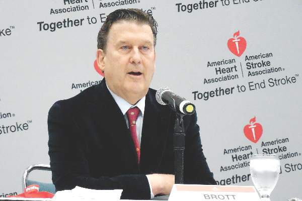
LOS ANGELES – The equipoise between carotid stenting and endarterectomy received a further boost in 10-year results from the landmark Carotid Revascularization Endarterectomy versus Stenting Trial (CREST) that compared the two options head-to-head.
Reported the day after results from another big trial that pitted carotid stenting against surgery, the Asymptomatic Carotid Trial (ACT I), the new long-term results from the CREST study mean that deciding among the options relies largely on patient preference although individual clinical characteristics might favor one approach or the other, experts said.

The big remaining unknown and wild card is whether doing no procedural intervention at all and relying entirely on optimal, contemporary medical treatment works just as well as endarterectomy or carotid stenting. The role for stand-alone medical therapy against carotid surgery or stenting (on top of medical therapy) is currently undergoing a formal, direct comparison in the randomized Carotid Revascularization and Medical Management for Asymptomatic Carotid Stenosis Trial (CREST-2).
Taking the 5-year outcome results from ACT I and the 10-year outcome results from CREST both into account, “we now have a lot of evidence that both carotid stenting and surgery are safe and durable. The results support both options” for either patients with symptomatic carotid artery stenosis or asymptomatic patients with carotid stenosis as extensive as in the patients enrolled in these trials, said Dr. Thomas G. Brott at the International Stroke Conference.
“In routine practice, we lay out the options of endarterectomy, carotid stenting, or no intervention with just medical treatment to patients and let them decide,” noted Dr. Brott, professor of neurology and director of research at the Mayo Clinic in Jacksonville, Fla.
CREST randomized 2,502 symptomatic or asymptomatic patients with significant carotid stenosis during 2000-2008 at 117 U.S. and Canadian centers. From this group, 1,607 consented and were available for long-term follow-up, done at a median of 7.4 years and as long as 10 years after follow-up.
The study’s primary, long-term endpoint was stroke, MI, or death during the periprocedural period (30 days after treatment or 36 days after enrollment depending on when the procedural intervention occurred) plus the rate of ipsilateral stroke during up to 10 years of follow-up. This combined endpoint occurred in 10% of the patients who underwent endarterectomy and in 12% of those who had stenting, a difference that was not statistically significant, Dr. Brott reported. Concurrent with his presentation at the meeting, sponsored by the American Heart Association, the results also were published online (N Engl J Med. 2016 Feb 18. doi: 10.1056/NEJMoa1505215).
The results included a secondary endpoint that showed a significant benefit for endarterectomy. The tally of periprocedural strokes or deaths plus ipsilateral strokes during 10-year follow-up was 8% for the surgical group and 11% for those who received a stent, a 37% excess hazard with stenting.
Dr. Brott attributed this secondary difference between the two arms of the study to a statistically significant excess of stroke or death during the periprocedural period in the patients treated by stenting, and more specifically an excess of strokes. The rate of total periprocedural strokes was 4% with stenting and 2% with endarterectomy, a statistically significant difference. Although an embolic protection device was used “when feasible” during stenting, this protection can be fallible, Dr. Brott noted. In contrast, the results from the ACT I trial showed no statistically significant difference in the rate of periprocedural total strokes between the stented and endarterectomy patients.
Dr. Brott had no relevant disclosures. The CREST trial received partial funding from Abbott Vascular.
On Twitter @mitchelzoler
The 10-year CREST results are good news for patients with carotid disease because they show the durability of both interventions we can offer patients. Having these data and the results from ACT I allows physicians to have an informed discussion with patients about their treatment options. I also hope that with these results from both trials, reimbursement will cease to be a deciding factor and that both surgery and stenting will be on a level playing field for insurance coverage.
Although on a population level stenting and surgery appear to produce comparable results, individual patient characteristics can make one option more appropriate. These include the morphology of a patient’s carotid arteries and stenotic lesions that can make stenting a technical challenge, and a patient’s medical condition and comorbidities which could put them at higher risk for general anesthesia and surgery. Also, a big concern for many patients is how long they will require hospitalization.

|
Dr. Mark J. Alberts |
A major unresolved question now about treating carotid disease is whether medical treatment alone is an equally good third alternative for asymptomatic patients. We are in a relatively new era of medical therapy, with more options for smoking cessation, better and more diverse drugs for blood pressure and hyperglycemia control, and wider use of high-dose statins. Some patients are eager to avoid any intervention and already opt for medical management only, but only after CREST-2 is completed will we know whether they will truly fare as well as patients who have a procedure performed.
Another issue that needs to be considered when extrapolating the results from CREST and ACT I to routine practice is that the surgeons and interventionalists who performed the procedures in these trials were highly selected and experienced. One cannot assume that the results in these trials will be replicated by any surgeon or interventionalist in the community. I suggest that patients investigate the track record of their community hospitals and operators by consulting the performance information that is increasingly posted on the Internet.
Dr. Mark J. Alberts is professor of neurology and medical director of the neurology service at the University of Texas Southwestern Medical Center in Dallas. He had no disclosures. He made these comments in an interview.
The 10-year CREST results are good news for patients with carotid disease because they show the durability of both interventions we can offer patients. Having these data and the results from ACT I allows physicians to have an informed discussion with patients about their treatment options. I also hope that with these results from both trials, reimbursement will cease to be a deciding factor and that both surgery and stenting will be on a level playing field for insurance coverage.
Although on a population level stenting and surgery appear to produce comparable results, individual patient characteristics can make one option more appropriate. These include the morphology of a patient’s carotid arteries and stenotic lesions that can make stenting a technical challenge, and a patient’s medical condition and comorbidities which could put them at higher risk for general anesthesia and surgery. Also, a big concern for many patients is how long they will require hospitalization.
|
|
Dr. Mark J. Alberts |
A major unresolved question now about treating carotid disease is whether medical treatment alone is an equally good third alternative for asymptomatic patients. We are in a relatively new era of medical therapy, with more options for smoking cessation, better and more diverse drugs for blood pressure and hyperglycemia control, and wider use of high-dose statins. Some patients are eager to avoid any intervention and already opt for medical management only, but only after CREST-2 is completed will we know whether they will truly fare as well as patients who have a procedure performed.
Another issue that needs to be considered when extrapolating the results from CREST and ACT I to routine practice is that the surgeons and interventionalists who performed the procedures in these trials were highly selected and experienced. One cannot assume that the results in these trials will be replicated by any surgeon or interventionalist in the community. I suggest that patients investigate the track record of their community hospitals and operators by consulting the performance information that is increasingly posted on the Internet.
Dr. Mark J. Alberts is professor of neurology and medical director of the neurology service at the University of Texas Southwestern Medical Center in Dallas. He had no disclosures. He made these comments in an interview.
LOS ANGELES – The equipoise between carotid stenting and endarterectomy received a further boost in 10-year results from the landmark Carotid Revascularization Endarterectomy versus Stenting Trial (CREST) that compared the two options head-to-head.
Reported the day after results from another big trial that pitted carotid stenting against surgery, the Asymptomatic Carotid Trial (ACT I), the new long-term results from the CREST study mean that deciding among the options relies largely on patient preference although individual clinical characteristics might favor one approach or the other, experts said.
The big remaining unknown and wild card is whether doing no procedural intervention at all and relying entirely on optimal, contemporary medical treatment works just as well as endarterectomy or carotid stenting. The role for stand-alone medical therapy against carotid surgery or stenting (on top of medical therapy) is currently undergoing a formal, direct comparison in the randomized Carotid Revascularization and Medical Management for Asymptomatic Carotid Stenosis Trial (CREST-2).
Taking the 5-year outcome results from ACT I and the 10-year outcome results from CREST both into account, “we now have a lot of evidence that both carotid stenting and surgery are safe and durable. The results support both options” for either patients with symptomatic carotid artery stenosis or asymptomatic patients with carotid stenosis as extensive as in the patients enrolled in these trials, said Dr. Thomas G. Brott at the International Stroke Conference.
“In routine practice, we lay out the options of endarterectomy, carotid stenting, or no intervention with just medical treatment to patients and let them decide,” noted Dr. Brott, professor of neurology and director of research at the Mayo Clinic in Jacksonville, Fla.
CREST randomized 2,502 symptomatic or asymptomatic patients with significant carotid stenosis during 2000-2008 at 117 U.S. and Canadian centers. From this group, 1,607 consented and were available for long-term follow-up, done at a median of 7.4 years and as long as 10 years after follow-up.
The study’s primary, long-term endpoint was stroke, MI, or death during the periprocedural period (30 days after treatment or 36 days after enrollment depending on when the procedural intervention occurred) plus the rate of ipsilateral stroke during up to 10 years of follow-up. This combined endpoint occurred in 10% of the patients who underwent endarterectomy and in 12% of those who had stenting, a difference that was not statistically significant, Dr. Brott reported. Concurrent with his presentation at the meeting, sponsored by the American Heart Association, the results also were published online (N Engl J Med. 2016 Feb 18. doi: 10.1056/NEJMoa1505215).
The results included a secondary endpoint that showed a significant benefit for endarterectomy. The tally of periprocedural strokes or deaths plus ipsilateral strokes during 10-year follow-up was 8% for the surgical group and 11% for those who received a stent, a 37% excess hazard with stenting.
Dr. Brott attributed this secondary difference between the two arms of the study to a statistically significant excess of stroke or death during the periprocedural period in the patients treated by stenting, and more specifically an excess of strokes. The rate of total periprocedural strokes was 4% with stenting and 2% with endarterectomy, a statistically significant difference. Although an embolic protection device was used “when feasible” during stenting, this protection can be fallible, Dr. Brott noted. In contrast, the results from the ACT I trial showed no statistically significant difference in the rate of periprocedural total strokes between the stented and endarterectomy patients.
Dr. Brott had no relevant disclosures. The CREST trial received partial funding from Abbott Vascular.
On Twitter @mitchelzoler
AT THE INTERNATIONAL STROKE CONFERENCE
Key clinical point: Long-term follow-up of the CREST trial out to 10 years showed no statistically significant difference between endarterectomy or carotid stenting for patients with carotid artery stenosis.
Major finding: The primary, long-term endpoint occurred in 10% of endarterectomy patients and 12% of stented patients, a nonsignificant difference.
Data source: The CREST trial, which followed 1,607 patients for up to 10 years after their randomized intervention.
Disclosures: Dr. Brott had no relevant disclosures. The CREST trial received partial funding from Abbott Vascular.
VIDEO: Stenting in asymptomatic patients noninferior to endarterectomy at 5 years
LOS ANGELES – In asymptomatic patients under 80 years old, carotid stenting and endarterectomy perform equally as well for severe carotid stenosis out to 5 years, according to a randomized trial published online in the New England Journal of Medicine.
Overall, 1,032 patients were stented, and 343 had endarterectomies in the trial, called Asymptomatic Carotid Trial I (ACT I). If stenting didn’t look safe on postrandomization angiography, patients were given the option of medical management or crossover into the surgical group. The subjects all had bifurcation carotid stenosis blocking at least 70% of the lumen. None were at high risk for surgical complications. “Asymptomatic” meant they hadn’t had a stroke, transient ischemic attack, or amaurosis fugax in the 6 months before enrollment. Stenting and endarterectomy were done by physicians and centers well experienced in the techniques (N Engl J Med. 2016 Feb 17. doi: 10.1056/NEJMoa1515706).
At 1 year, stenting was noninferior to endarterectomy for the primary composite endpoint of death, stroke, or myocardial infarction within 30 days after the procedure or ipsilateral stroke within 1 year; the event rate was 3.8% among stent patients and 3.4% among endarterectomy patients (P = .01 for noninferiority, with a noninferiority margin of 3 percentage points).
The cumulative 5-year stroke-free survival rate was 93.1% in the stenting group and 94.7% in the endarterectomy group (P = .44).
For now, the results mean that sometimes choosing between carotid endarterectomy or stenting (or medical management) has as much to do with patient and physician preference as medical science, raising the difficult question of how to choose. In a video interview at the International Stroke Conference, investigator Dr. Lawrence Wechsler, professor of neurology/neurosurgery and chair of the department of neurology at the University of Pittsburgh, shared his thoughts on that and the other implications of the study. The work was funded by Abbott Vascular.
The video associated with this article is no longer available on this site. Please view all of our videos on the MDedge YouTube channel
AT The INTERNATIONAL STROKE CONFERENCE

Downturn in reoperation after breast conservation surgery
Rates of reoperation after breast conservation surgery have declined significantly since 2003, but nearly 25% of women are still undergoing repeat operations, new data suggest.
Researchers analyzed data from a population-based sample of 89,448 women undergoing primary breast conservation surgery for breast cancer in New York State from 2003 to 2013.
According to a study published online Feb.17 in JAMA Surgery, the mean 90-day reoperation rate declined from 39.5% in 2003-2004 to 23.1% in 2011-2013 (P less than .001), with an overall rate of 30.9% for the entire study period.
“We believe that the findings of reduced occurrence of reoperations are encouraging and imply improvements in training and patient selection for BCS,” wrote Abby J. Isaacs of Cornell University, New York, and her coauthors.
Reoperation rates were highest in younger women (37.7%), compared with women aged over 65 years (26.3%).
However, the study also showed that women aged 50-64 years were less likely than were older or younger women to undergo breast conservation surgery, compared with mastectomy as a repeat procedure (JAMA Surg 2016 Feb 17. doi:10.1001/jamasurg.2015.5535).
Breast conservation surgery was more common than was mastectomy as a repeat procedure in women who were white, had commercial insurance, had low comorbidity scores, or had in situ disease.
Researchers observed significant surgeon-level variation in rates of reoperation after breast conservation surgery, which they described as “novel and unprecedented.” The rates of reoperation among surgeons ranged from 0% to nearly 100%.
Overall, the mean rate of reoperation for surgeons was 30.8%, but nearly 20% of surgeons had reoperation rates above 50% and 6.1% had reoperation rates above the 99.8% limits.
Similarly, 6% of surgeons had reoperation rates below the 95% confidence limits and 2.9% were below the 99.8% limits.
Reoperation rates were independently associated with surgical volume; surgeons performing fewer than 14 breast conservation surgery procedures had a mean reoperation rate of 35.2% while those who performed 34 or more procedures had a reoperation rate of 27.5%.
Over the entire study period, the overall rate of breast conservation surgery peaked in 2004 with more than 8,500 cases, then decreased to a mean number of 8,078 for 2011-2013.
The decrease in primary breast conservation surgery was significantly greater in women aged 20-49 years (P less than .001) with 1,416 women in this age group undergoing breast conservation surgery in 2013, compared with 3,644 women aged 65 years or older.
“The reduction in the use of BCS in women younger than 50 years and a corresponding reduction in overall reoperation rates over time implies that surgeons may be selecting more appropriate patients for the procedures,” the authors wrote.
They acknowledged that they did not have access to information on tumor size, grade, and staging, which are variables known to have an important influence on margin rates and reoperation rates. “We believe, however, in the context of physician-level outcomes, that these factors will not bias the results; the recommendation for BCS will be made by surgeons based on their knowledge of the patients’ disease, and we have no reason to believe that unknowable patient characteristics will be unbalanced between surgeons.”
Two authors reported involvement with the Food and Drug Administration–funded MDEpiNet Science and Infrastructure Center; no other conflicts of interest were declared.
The recent Society for Surgical Oncology–American Society for Radiation Oncology (SSO-ASTRO) consensus guidelines encourage the use of “no ink on tumor” as the current standard in an era of multimodal treatment and evolving understanding of tumor biology along with tumor burden.

|
Dr. E. Shelley Hwang |
Establishing a rational, evidence-based approach to reexcision as originally proposed by Fisher et al. and supported by the new SSO-ASTRO guidelines can provide substantial national cost savings by eliminating reexcisions for close but negative margins.
Dr. Uttara Nag and Dr. E. Shelley Hwang are with the department of health policy and management in the department of surgery, Duke University, Durham, N.C. These comments are taken from an editorial (JAMA Surg. 2016 Feb 17. doi:10.1001/jamasurg.2015.5555). No conflicts of interest were declared.
The recent Society for Surgical Oncology–American Society for Radiation Oncology (SSO-ASTRO) consensus guidelines encourage the use of “no ink on tumor” as the current standard in an era of multimodal treatment and evolving understanding of tumor biology along with tumor burden.
|
|
Dr. E. Shelley Hwang |
Establishing a rational, evidence-based approach to reexcision as originally proposed by Fisher et al. and supported by the new SSO-ASTRO guidelines can provide substantial national cost savings by eliminating reexcisions for close but negative margins.
Dr. Uttara Nag and Dr. E. Shelley Hwang are with the department of health policy and management in the department of surgery, Duke University, Durham, N.C. These comments are taken from an editorial (JAMA Surg. 2016 Feb 17. doi:10.1001/jamasurg.2015.5555). No conflicts of interest were declared.
Rates of reoperation after breast conservation surgery have declined significantly since 2003, but nearly 25% of women are still undergoing repeat operations, new data suggest.
Researchers analyzed data from a population-based sample of 89,448 women undergoing primary breast conservation surgery for breast cancer in New York State from 2003 to 2013.
According to a study published online Feb.17 in JAMA Surgery, the mean 90-day reoperation rate declined from 39.5% in 2003-2004 to 23.1% in 2011-2013 (P less than .001), with an overall rate of 30.9% for the entire study period.
“We believe that the findings of reduced occurrence of reoperations are encouraging and imply improvements in training and patient selection for BCS,” wrote Abby J. Isaacs of Cornell University, New York, and her coauthors.
Reoperation rates were highest in younger women (37.7%), compared with women aged over 65 years (26.3%).
However, the study also showed that women aged 50-64 years were less likely than were older or younger women to undergo breast conservation surgery, compared with mastectomy as a repeat procedure (JAMA Surg 2016 Feb 17. doi:10.1001/jamasurg.2015.5535).
Breast conservation surgery was more common than was mastectomy as a repeat procedure in women who were white, had commercial insurance, had low comorbidity scores, or had in situ disease.
Researchers observed significant surgeon-level variation in rates of reoperation after breast conservation surgery, which they described as “novel and unprecedented.” The rates of reoperation among surgeons ranged from 0% to nearly 100%.
Overall, the mean rate of reoperation for surgeons was 30.8%, but nearly 20% of surgeons had reoperation rates above 50% and 6.1% had reoperation rates above the 99.8% limits.
Similarly, 6% of surgeons had reoperation rates below the 95% confidence limits and 2.9% were below the 99.8% limits.
Reoperation rates were independently associated with surgical volume; surgeons performing fewer than 14 breast conservation surgery procedures had a mean reoperation rate of 35.2% while those who performed 34 or more procedures had a reoperation rate of 27.5%.
Over the entire study period, the overall rate of breast conservation surgery peaked in 2004 with more than 8,500 cases, then decreased to a mean number of 8,078 for 2011-2013.
The decrease in primary breast conservation surgery was significantly greater in women aged 20-49 years (P less than .001) with 1,416 women in this age group undergoing breast conservation surgery in 2013, compared with 3,644 women aged 65 years or older.
“The reduction in the use of BCS in women younger than 50 years and a corresponding reduction in overall reoperation rates over time implies that surgeons may be selecting more appropriate patients for the procedures,” the authors wrote.
They acknowledged that they did not have access to information on tumor size, grade, and staging, which are variables known to have an important influence on margin rates and reoperation rates. “We believe, however, in the context of physician-level outcomes, that these factors will not bias the results; the recommendation for BCS will be made by surgeons based on their knowledge of the patients’ disease, and we have no reason to believe that unknowable patient characteristics will be unbalanced between surgeons.”
Two authors reported involvement with the Food and Drug Administration–funded MDEpiNet Science and Infrastructure Center; no other conflicts of interest were declared.
FROM JAMA SURGERY
Key clinical point: Rates of reoperation after breast conservation surgery are declining but vary significantly between surgeons.
Major finding: The mean 90-day reoperation rate after breast conservation surgery declined from 39.5% in 2003-2004 to 23.1% in 2011-2013.
Data source: Population-based sample of 89,448 women undergoing primary breast conservation surgery for breast cancer.
Disclosures: Two authors reported involvement with the Food and Drug Administration–funded MDEpiNet Science and Infrastructure Center; no other conflicts of interest were declared.

The no-operation quality assessment ‘blind spot’
JACKSONVILLE, FLA. – About one-third of patients admitted to the hospital for abdominal problems like diverticulitis and small bowel obstruction get discharged without having surgery, but their outcomes are not typically included in quality assessment, leaving this group of patients in a “blind spot” of surgical quality, according to Dr. Michael Wandling.
However, researchers from Northwestern University in Chicago have analyzed data from the Nationwide (National) Inpatient Sample and determined how many cases of diverticulitis, small bowel obstruction (SBO), cholecystitis, and acute appendicitis are managed without surgery and the clinical factors that may influence that, he reported at the Association for Academic Surgery/Society of University Surgeons Academic Surgical Congress.

“Surgeons frequently will admit patients for nonoperative management of diagnoses such as diverticulitis, small bowel obstruction, cholecystitis, and perforated appendicitis,” said Dr. Wandling, a general surgery resident at Northwestern. “Yet nonoperative management does not really factor into current surgical quality assessment. In fact, nonoperative management is not frequently evaluated, and utilization rates have not even really been quantified.”
The researchers’ goal was to evaluate hospital-level variability in nonoperative management practices and identify hospital characteristics associated with high rates of nonoperative management, Dr. Wandling said.
“What we found was that smaller bed size, fewer annual discharges, being a public government-run hospital, being a nonteaching hospital, and being rural or located in the Midwest were all associated with greater use of nonoperative management,” he said.
They extracted a sample from the Nationwide (National) Inpatient Sample that analyzed admission and discharge data on 1.6 million patients admitted for one of the four studied diagnoses from 1998 to 2011. Overall, the four diagnoses accounted for more than 500,000 annual admissions, “and this rate has been increasing over time,” Dr. Wandling said. To calculate rates of nonoperative management for each diagnosis, the researchers concentrated on data from 2010 and 2011. They found the following rates of nonoperative management: 87.1% for diverticulitis, 38.1% for SBO, 11.3% for cholecystitis, and 3.7% for appendicitis. The overall rate of nonoperative management for all four diagnoses was 32.8%, Dr. Wandling said.
They also evaluated the overall rates of nonoperative management for each year from 1998 to 2011 and found they steadily increased from 25.6% to 32.8%, Dr. Wandling said. “Nonoperative management is not uncommon, with approximately 190,000 patients being admitted for nonoperative management each year, and this number has also been increasing,” he said.
Dr. Wandling acknowledged some limitations with the study because it used an administrative dataset with data collected retrospectively and because the data do not track patients after discharge, making it impossible to know if any patients managed nonoperatively were subsequently readmitted for surgery. “Current surgical quality assessment only focuses on patients who have surgery, which can be seen through public reporting programs like Hospital Compare, pay-for-performance initiatives like [Centers for Medicare & Medicaid Services] valued-based purchasing, and clinical data registries,” he said. “As a result, patients who are managed nonoperatively are really left in a blind spot of surgical quality.”
Dr. Wandling said he and his coauthors are working with the American College of Surgeons National Surgical Quality Improvement Program to develop an Emergency General Surgery (EGS) Pilot to evaluate performance in operative and nonoperative care for SBO, cholecystitis, and appendicitis. Fourteen centers have so far collected more than 6 months of data as part of the EGS Pilot, he said, and additional hospitals are currently being recruited to participate.
“Ultimately the goal is to identify optimal nonoperative management strategies in general surgery so that all patients can receive high-quality surgical care, not just those who we operate on,” Dr. Wandling said.
He and his coauthors had no relevant financial disclosures.
JACKSONVILLE, FLA. – About one-third of patients admitted to the hospital for abdominal problems like diverticulitis and small bowel obstruction get discharged without having surgery, but their outcomes are not typically included in quality assessment, leaving this group of patients in a “blind spot” of surgical quality, according to Dr. Michael Wandling.
However, researchers from Northwestern University in Chicago have analyzed data from the Nationwide (National) Inpatient Sample and determined how many cases of diverticulitis, small bowel obstruction (SBO), cholecystitis, and acute appendicitis are managed without surgery and the clinical factors that may influence that, he reported at the Association for Academic Surgery/Society of University Surgeons Academic Surgical Congress.
“Surgeons frequently will admit patients for nonoperative management of diagnoses such as diverticulitis, small bowel obstruction, cholecystitis, and perforated appendicitis,” said Dr. Wandling, a general surgery resident at Northwestern. “Yet nonoperative management does not really factor into current surgical quality assessment. In fact, nonoperative management is not frequently evaluated, and utilization rates have not even really been quantified.”
The researchers’ goal was to evaluate hospital-level variability in nonoperative management practices and identify hospital characteristics associated with high rates of nonoperative management, Dr. Wandling said.
“What we found was that smaller bed size, fewer annual discharges, being a public government-run hospital, being a nonteaching hospital, and being rural or located in the Midwest were all associated with greater use of nonoperative management,” he said.
They extracted a sample from the Nationwide (National) Inpatient Sample that analyzed admission and discharge data on 1.6 million patients admitted for one of the four studied diagnoses from 1998 to 2011. Overall, the four diagnoses accounted for more than 500,000 annual admissions, “and this rate has been increasing over time,” Dr. Wandling said. To calculate rates of nonoperative management for each diagnosis, the researchers concentrated on data from 2010 and 2011. They found the following rates of nonoperative management: 87.1% for diverticulitis, 38.1% for SBO, 11.3% for cholecystitis, and 3.7% for appendicitis. The overall rate of nonoperative management for all four diagnoses was 32.8%, Dr. Wandling said.
They also evaluated the overall rates of nonoperative management for each year from 1998 to 2011 and found they steadily increased from 25.6% to 32.8%, Dr. Wandling said. “Nonoperative management is not uncommon, with approximately 190,000 patients being admitted for nonoperative management each year, and this number has also been increasing,” he said.
Dr. Wandling acknowledged some limitations with the study because it used an administrative dataset with data collected retrospectively and because the data do not track patients after discharge, making it impossible to know if any patients managed nonoperatively were subsequently readmitted for surgery. “Current surgical quality assessment only focuses on patients who have surgery, which can be seen through public reporting programs like Hospital Compare, pay-for-performance initiatives like [Centers for Medicare & Medicaid Services] valued-based purchasing, and clinical data registries,” he said. “As a result, patients who are managed nonoperatively are really left in a blind spot of surgical quality.”
Dr. Wandling said he and his coauthors are working with the American College of Surgeons National Surgical Quality Improvement Program to develop an Emergency General Surgery (EGS) Pilot to evaluate performance in operative and nonoperative care for SBO, cholecystitis, and appendicitis. Fourteen centers have so far collected more than 6 months of data as part of the EGS Pilot, he said, and additional hospitals are currently being recruited to participate.
“Ultimately the goal is to identify optimal nonoperative management strategies in general surgery so that all patients can receive high-quality surgical care, not just those who we operate on,” Dr. Wandling said.
He and his coauthors had no relevant financial disclosures.
AT THE ACADEMIC SURGICAL CONGRESS
Key clinical point: Quality assessment of surgical outcomes does not account for cases that are managed medically without an operation; this study evaluated nonoperative management for four common diagnoses of abdominal pain.
Major finding: The overall rate of nonoperative management for diverticulitis, small bowel obstruction, cholecystitis, and appendicitis was 32.8% with rates increasing steadily over the 13-year study period.
Data source: An analysis of a sampling of 1.6 million admissions from the Nationwide (National) Inpatient Sample from 1998 to 2011, with concentration on data from 2010 and 2011.
Disclosures: The study authors reported having no relevant financial disclosures.

Study measures post-thyroidectomy voice changes
JACKSONVILLE, FLA. – Voice quality changes after thyroid surgery are detectable by using both subjective and objective measures, according to investigators at Monash University in Melbourne, Australia.
After thyroid surgery, up to 80% of patients with functional recurrent laryngeal nerves (RLNs) have reported voice changes, so the investigators set out to evaluate the extent of those voice changes and how the extent of the operation and RLN edema may affect them.

“It has been confirmed that thyroid procedures do alter the voice without necessarily causing a measurable recurrent laryngeal nerve palsy,” said lead investigator Dr. James Lee, “and the change of voice is correlated to the extent of surgery and the amount of nerve swelling.” The findings were presented at the Association of Academic Surgery/Society of University Surgeons Academic Surgical Congress.
The study evaluated 62 patients who had total and partial thyroidectomy surgery between 2010 and 2011 at the Monash University Endocrine Surgery Unit. To subjectively measure voice quality after surgery, the researchers used the Voice Disorder Index (VDI), which measures voice quality on a 0-40 scale from best to worst. After surgery, the mean VDI score in this group showed a 5.2 plus or minus 1.2–point deterioration from 4.2 to 9.4 (P less than .001). For objective evaluation, the researchers used the Dysphonia Severity Index (DSI), which scores voice quality on a scale of –5 to 5 from worst to best. After surgery, the mean DSI score showed a 1.1 plus or minus 0.2–point deterioration from 3.9 to 2.8 (P less than .001). Two speech pathologists conducted the voice assessments.
“Subjective scoring of both hemithyroidectomy and total thyroidectomy reported worse voice postoperatively,” Dr. Lee said. “However, when you take a close look at the numbers, those undergoing total thyroidectomy reported a higher measure of deterioration in their voice.”
Patients who had either partial and total thyroidectomy reported significant subjective deterioration of their voice with mean VDI change from 5.4 to 7.9 (P = 0.02) and 3.5 to 10.6 (P less than .001), respectively. However, on objective evaluation, only the total thyroidectomy patients showed significant voice deterioration, with a mean DSI change from 4 to 2.5 (P less than .001).
Dr. Lee noted that study outcomes between partial and total thyroidectomy patients diverged in another respect: the impact RLN swelling had on voice deterioration. To evaluate RLN swelling, the researchers measured the diameter of the nerve with Vernier calipers before and after the lobectomy during each operation. RLN diameter increased 0.58 plus or minus 0.05 mm on average (P less than .001). In patients who had partial thyroidectomy, the greater the RLN swelling, the worse the subjective score (P = .03). This was not the case in the total thyroidectomy patients where involvement of two nerves complicates the interaction, he said.
During follow-up, the investigators came upon a revelatory finding. “With median 8-month follow-up, the self-reported, VDI scores had returned to baseline levels,” Dr. Lee said. “Interestingly, not only did the objective DSI scores show a return to baseline levels, but it exceeded the baseline levels, meaning the voice had scored better after surgery than before.” However, he noted only 13 patients completed the follow-up.
“Voice change post-thyroidectomy without recurrent laryngeal nerve injury is a complex phenomenon and is likely multifactorial, and we only looked at two of those factors: the extent of surgery and the gradient of recurrent laryngeal nerve injury with nerve edema as a surrogate,” Dr. Lee said. Future studies should evaluate other factors, including the role of the external branch of the superior laryngeal nerve and patient factors such as diabetes or smoking, he said.
Dr. Lee and his coauthors had no disclosures.
JACKSONVILLE, FLA. – Voice quality changes after thyroid surgery are detectable by using both subjective and objective measures, according to investigators at Monash University in Melbourne, Australia.
After thyroid surgery, up to 80% of patients with functional recurrent laryngeal nerves (RLNs) have reported voice changes, so the investigators set out to evaluate the extent of those voice changes and how the extent of the operation and RLN edema may affect them.
“It has been confirmed that thyroid procedures do alter the voice without necessarily causing a measurable recurrent laryngeal nerve palsy,” said lead investigator Dr. James Lee, “and the change of voice is correlated to the extent of surgery and the amount of nerve swelling.” The findings were presented at the Association of Academic Surgery/Society of University Surgeons Academic Surgical Congress.
The study evaluated 62 patients who had total and partial thyroidectomy surgery between 2010 and 2011 at the Monash University Endocrine Surgery Unit. To subjectively measure voice quality after surgery, the researchers used the Voice Disorder Index (VDI), which measures voice quality on a 0-40 scale from best to worst. After surgery, the mean VDI score in this group showed a 5.2 plus or minus 1.2–point deterioration from 4.2 to 9.4 (P less than .001). For objective evaluation, the researchers used the Dysphonia Severity Index (DSI), which scores voice quality on a scale of –5 to 5 from worst to best. After surgery, the mean DSI score showed a 1.1 plus or minus 0.2–point deterioration from 3.9 to 2.8 (P less than .001). Two speech pathologists conducted the voice assessments.
“Subjective scoring of both hemithyroidectomy and total thyroidectomy reported worse voice postoperatively,” Dr. Lee said. “However, when you take a close look at the numbers, those undergoing total thyroidectomy reported a higher measure of deterioration in their voice.”
Patients who had either partial and total thyroidectomy reported significant subjective deterioration of their voice with mean VDI change from 5.4 to 7.9 (P = 0.02) and 3.5 to 10.6 (P less than .001), respectively. However, on objective evaluation, only the total thyroidectomy patients showed significant voice deterioration, with a mean DSI change from 4 to 2.5 (P less than .001).
Dr. Lee noted that study outcomes between partial and total thyroidectomy patients diverged in another respect: the impact RLN swelling had on voice deterioration. To evaluate RLN swelling, the researchers measured the diameter of the nerve with Vernier calipers before and after the lobectomy during each operation. RLN diameter increased 0.58 plus or minus 0.05 mm on average (P less than .001). In patients who had partial thyroidectomy, the greater the RLN swelling, the worse the subjective score (P = .03). This was not the case in the total thyroidectomy patients where involvement of two nerves complicates the interaction, he said.
During follow-up, the investigators came upon a revelatory finding. “With median 8-month follow-up, the self-reported, VDI scores had returned to baseline levels,” Dr. Lee said. “Interestingly, not only did the objective DSI scores show a return to baseline levels, but it exceeded the baseline levels, meaning the voice had scored better after surgery than before.” However, he noted only 13 patients completed the follow-up.
“Voice change post-thyroidectomy without recurrent laryngeal nerve injury is a complex phenomenon and is likely multifactorial, and we only looked at two of those factors: the extent of surgery and the gradient of recurrent laryngeal nerve injury with nerve edema as a surrogate,” Dr. Lee said. Future studies should evaluate other factors, including the role of the external branch of the superior laryngeal nerve and patient factors such as diabetes or smoking, he said.
Dr. Lee and his coauthors had no disclosures.
AT THE ACADEMIC SURGICAL CONGRESS
Key clinical point: Voice changes after thyroid surgery directly correlate to the amount of swelling of the recurrent laryngeal nerves as well as the duration and intensity of the operation.
Major finding: Mean subjective voice scores declined from 4.2 to 9.4 on the Voice Disorder Index and mean objective voice scores declined from 3.9 to 2.8 on the Dysphonia Severity Index after thyroidectomy.
Data source: Sixty-two patients undergoing total and hemithyroidectomy were prospectively recruited from the Monash University Endocrine Surgery Unit between 2010 and 2011.
Disclosures: The study authors reported having no financial disclosures.

Study: Hospitals with EHRs reduce patient harm
Cardiovascular, surgery, and pneumonia patients at hospitals with a fully electronic health record (EHR) system experienced lower rates of in-hospital adverse events, according to a new study published in the Journal of Patient Safety.
A research team led by Dr. Mark L. Metersky, medical director at the UConn Health Center for Bronchiectasis Care in Farmington, Conn., conducted a retrospective analysis of patient discharges using data from the 2012 and 2013 Medicare Patient Safety Monitoring System.

The study sample included patients aged 18 years and older that were hospitalized for one of three conditions: acute cardiovascular disease, pneumonia, or conditions requiring surgery. The main outcome measures were in-hospital adverse events, including hospital-acquired infections, adverse drug events (based on selected medications), general events, and post-procedural events.
Among the 45,235 patients who were at risk for 347,281 adverse events in the study sample, the occurrence rate of adverse events was 2.3%, and 13% of patients were exposed to a fully electronic EHR. In multivariate modeling adjusted for patient and hospital characteristics, investigators discovered that patient exposure to a fully electronic EHR was associated with 17%-30% lower odds of any adverse event for cardiovascular, pneumonia, and surgery patients.
“Beyond the direct effects on safety, EHRs can also indirectly reduce overall adverse event rates by enabling quality improvement initiatives,” said Dr. Metersky and his coauthors.
Read the full study in the Journal of Patient Safety (2016 Feb 6. doi: 10.1097/PTS.0000000000000257).
On Twitter @richpizzi
Cardiovascular, surgery, and pneumonia patients at hospitals with a fully electronic health record (EHR) system experienced lower rates of in-hospital adverse events, according to a new study published in the Journal of Patient Safety.
A research team led by Dr. Mark L. Metersky, medical director at the UConn Health Center for Bronchiectasis Care in Farmington, Conn., conducted a retrospective analysis of patient discharges using data from the 2012 and 2013 Medicare Patient Safety Monitoring System.
The study sample included patients aged 18 years and older that were hospitalized for one of three conditions: acute cardiovascular disease, pneumonia, or conditions requiring surgery. The main outcome measures were in-hospital adverse events, including hospital-acquired infections, adverse drug events (based on selected medications), general events, and post-procedural events.
Among the 45,235 patients who were at risk for 347,281 adverse events in the study sample, the occurrence rate of adverse events was 2.3%, and 13% of patients were exposed to a fully electronic EHR. In multivariate modeling adjusted for patient and hospital characteristics, investigators discovered that patient exposure to a fully electronic EHR was associated with 17%-30% lower odds of any adverse event for cardiovascular, pneumonia, and surgery patients.
“Beyond the direct effects on safety, EHRs can also indirectly reduce overall adverse event rates by enabling quality improvement initiatives,” said Dr. Metersky and his coauthors.
Read the full study in the Journal of Patient Safety (2016 Feb 6. doi: 10.1097/PTS.0000000000000257).
On Twitter @richpizzi
FROM THE JOURNAL OF PATIENT SAFETY

AGA survey finds more patient education needed about OTC pain meds
With patients indicating that they are regularly ignoring dosing guidelines on over-the-counter pain medications, the American Gastroenterological Association is calling for better patient education, including a more active role for doctors.
In particular, AGA is asking doctors to be more proactive in understanding how their patients are using over-the-counter pain medications after a survey revealed that 43% of respondents who suffer from chronic pain said that they knowingly have taken more than the recommended dose, with 28% saying they have experienced complications due to OTC pain medication overdose.

“Patients are self-medicating and actually ignoring the dosing guidelines,” Dr. Charles Melbern Wilcox, professor at the University of Alabama at Birmingham, said during a Jan. 25 teleconference to talk about the survey results. “Commonly, patients are unaware of overdose symptoms or not connecting their symptoms to over-the-counter pain medicine overdose or overusage. Patients are presenting with nausea, abdominal pain, and stomach ulcers due to over-the-counter pain medicine overdose and overuse. Patients with chronic pain are often taking multiple medications.”
AGA hosted the conference call as part of the “Gut Check: Know Your Medicine” campaign to encourage educating consumers on the proper usage and risks associated with OTC pain medication. The survey of 1,015 adults aged 30 years or older (479 were currently experiencing chronic pain at the time of the survey) and 251 licensed gastroenterologists conducted as part of the campaign occurred in September and October 2015.
Additionally, chronic pain sufferers also are taking multisymptom OTC products for allergy, cold, and flu symptoms that could cause the extra intake of pain medication.
“Our survey findings suggest that providers give more attention to patients with chronic pain since they are likely to take more than the recommended dose and also to take more than one pain medicine,” Dr. Byron Cryer, assistant dean for faculty diversity and development at the University of Texas Southwestern Medical Center, Dallas, said during the call to discuss the survey results and AGA campaign to promote OTC pain medication awareness. “Providers should work with patients to determine the best treatment options that address the patient’s pain while using medicines safely.”
Dr. Cryer noted that health care professionals “must start asking simple direct questions about pain medicine use each time they see a patient and must lead the way to improve the lines of communication.”
He added that having these active conversations about pain medications, both OTC and prescribed, can provide the necessary warnings about the products that “are likely to be remembered by patients” and will allow doctors to provide follow-up timing and instructions if the pain does not subside in a certain amount of time.
Another survey finding that is adding to the call for more patient education is that, according to the survey’s executive summary, most chronic pain sufferers “say they don’t always read the full Drug Facts label on an OTC pain medicine they haven’t taken before (66%) and many say the directions on the labels of OTC pain medicines are really just guidelines – they know what works for them (43%).”
Additionally, the survey found that 27% of chronic pain sufferers “are willing to take more of an OTC pain medicine than directed because they incorrectly believe their symptoms will disappear faster.”
With patients indicating that they are regularly ignoring dosing guidelines on over-the-counter pain medications, the American Gastroenterological Association is calling for better patient education, including a more active role for doctors.
In particular, AGA is asking doctors to be more proactive in understanding how their patients are using over-the-counter pain medications after a survey revealed that 43% of respondents who suffer from chronic pain said that they knowingly have taken more than the recommended dose, with 28% saying they have experienced complications due to OTC pain medication overdose.
“Patients are self-medicating and actually ignoring the dosing guidelines,” Dr. Charles Melbern Wilcox, professor at the University of Alabama at Birmingham, said during a Jan. 25 teleconference to talk about the survey results. “Commonly, patients are unaware of overdose symptoms or not connecting their symptoms to over-the-counter pain medicine overdose or overusage. Patients are presenting with nausea, abdominal pain, and stomach ulcers due to over-the-counter pain medicine overdose and overuse. Patients with chronic pain are often taking multiple medications.”
AGA hosted the conference call as part of the “Gut Check: Know Your Medicine” campaign to encourage educating consumers on the proper usage and risks associated with OTC pain medication. The survey of 1,015 adults aged 30 years or older (479 were currently experiencing chronic pain at the time of the survey) and 251 licensed gastroenterologists conducted as part of the campaign occurred in September and October 2015.
Additionally, chronic pain sufferers also are taking multisymptom OTC products for allergy, cold, and flu symptoms that could cause the extra intake of pain medication.
“Our survey findings suggest that providers give more attention to patients with chronic pain since they are likely to take more than the recommended dose and also to take more than one pain medicine,” Dr. Byron Cryer, assistant dean for faculty diversity and development at the University of Texas Southwestern Medical Center, Dallas, said during the call to discuss the survey results and AGA campaign to promote OTC pain medication awareness. “Providers should work with patients to determine the best treatment options that address the patient’s pain while using medicines safely.”
Dr. Cryer noted that health care professionals “must start asking simple direct questions about pain medicine use each time they see a patient and must lead the way to improve the lines of communication.”
He added that having these active conversations about pain medications, both OTC and prescribed, can provide the necessary warnings about the products that “are likely to be remembered by patients” and will allow doctors to provide follow-up timing and instructions if the pain does not subside in a certain amount of time.
Another survey finding that is adding to the call for more patient education is that, according to the survey’s executive summary, most chronic pain sufferers “say they don’t always read the full Drug Facts label on an OTC pain medicine they haven’t taken before (66%) and many say the directions on the labels of OTC pain medicines are really just guidelines – they know what works for them (43%).”
Additionally, the survey found that 27% of chronic pain sufferers “are willing to take more of an OTC pain medicine than directed because they incorrectly believe their symptoms will disappear faster.”

VIDEO: Octogenarians benefit from ischemic stroke thrombectomy
LOS ANGELES – Clot removal to recanalize the occluded intracerebral arteries of acute ischemic stroke patients was as effective for producing good outcomes in patients aged 80 years or older as it was in younger patients, according to results from a pooled analysis of 1,287 patients in five separate but similar randomized trials.
This unprecedented evidence for the safety and efficacy of thrombectomy (also known as embolectomy) in octogenarians experiencing an acute occlusive, large-vessel, proximal anterior-circulation stroke was one of several new findings from the pooled analysis that should help further push thrombectomy to the forefront of acute care for patients undergoing this type of ischemic stroke, predicted Dr. Wade S. Smith in a video interview at the International Stroke Conference.
“By looking at all the data, we have much more refined information on the robustness of the treatment across age groups, which is quite important, especially patients in the 80-plus age group,” commented Dr. Smith, professor of neurology and chief of the neurovascular division at the University of California, San Francisco.
Until now, during the year following the reports in early 2015 from all five studies, “age had been a limiting factor” in applying the practicing-changing intervention of thrombectomy to patients, he noted.
“This [the new pooled analysis] will change that,” Dr. Smith predicted. “It does not apply to patients who were infirm prior to their stroke – but for patients who were otherwise healthy, with a modified Rankin scale level of 0 or 1 at initial presentation, it appears that they benefit [from thrombectomy] regardless of their age.” In the pooled analysis, 198 of the 1,287 total patients (15%) were at least 80 years old.
“It removes age discrimination. A healthy 80-year-old may do extremely well with this treatment,” Dr. Smith said.
The consistency of benefit across a wide range of stroke severity that showed up in the trials as four distinct strata of NIH Stroke Scale scores prior to treatment was another important finding that could not previously be definitively made by analyzing each of the five trials individually.
In patients with stroke-severity scores that ranged from 10 or less (the least severely affected) to patients with scores of 21 or greater, all had post-thrombectomy improvements that clustered around the overall average number-needed-to-treat of 2.6 patients to reduce the disability of one patient at follow-up by at least one level on the modified Rankin scale.
Other notable findings from the pooled analysis were that thrombectomy also produced a consistent benefit to patients across every other subgroup examined, including sex, specific occlusion site, whether or not patients also received thrombolytic treatment with tissue plasminogen activator, and time to thrombectomy treatment (5 or fewer hours from stroke onset or more than 5 hours), reported Dr. Michael D. Hill and Dr. Tudor G. Jovin in a joint presentation at the meeting, sponsored by the American Heart Association.
Their pooled analysis, known as HERMES, pooled data from the MR CLEAN, ESCAPE, REVASCAT, SWIFT PRIME, and EXTEND IA trials, all run during 2010-2014.
“Endovascular treatment is a highly effective treatment across all subgroups,” concluded Dr. Hill and Dr. Jovin as they completed their talk. “These data may provide additional support for endovascular treatment in subgroups not addressed in the individual trials.”
Concurrent with their report at the meeting, the results appeared in a paper published online (Lancet. 2016 Feb 18;doi: 10.1016/S0140-6736(16)00163-X).
Both Dr. Jovin and Dr. Hill shared the enthusiasm of Dr. Smith and others in the packed meeting room about the age finding.
“Older patients seemed to benefit even more” from thrombectomy, compared with younger patients, noted Dr. Jovin, chief of the stroke division at the University of Pittsburgh and a coinvestigator on SWIFT PRIME. “There is no reason to deny this treatment to appropriately selected patients based on age,” he said.
“There is no upper age limit,” agreed Dr. Hill, professor of neurology and director of the stroke unit at the University of Calgary (Alta.) and a coinvestigator on the ESCAPE trial. “If it’s an otherwise healthy 90-year-old who is living independently, you can surely consider them for this treatment.”
HERMES received fundings through an unrestricted grant from Medtronic. Dr. Hill and Dr. Jovin had no personal disclosures. Dr. Smith served on the data safety and monitoring board for a trial funded by Stryker.
The video associated with this article is no longer available on this site. Please view all of our videos on the MDedge YouTube channel
On Twitter @mitchelzoler
AT THE INTERNATIONAL STROKE CONFERENCE
Key clinical point: A pooled analysis of five recent trials of thrombectomy for acute ischemic stroke should help further propel its widespread U.S. adoption.
Major finding: Intracerebral, transcatheter clot removal in acute ischemic stroke was equally effective in octogenarian and younger patients.
Data source: HERMES, a pooled analysis of data from 1,287 acute ischemic stroke patients randomized in five separate but similar trials.
Disclosures: HERMES received fundings through an unrestricted grant from Medtronic. Dr. Hill and Dr. Jovin had no personal disclosures. Dr. Smith served on the data safety and monitoring board for a trial funded by Stryker.

STS: Lactate dehydrogenase of 1,150 IU/L flags LVAD thrombosis
PHOENIX – A blood lactate dehydrogenase level of at least 1,150 IU/L may be the best cutoff for identifying pump thrombosis in patients who have received a left ventricular assist device based on findings from a combined series of 502 pump recipients.
Using this cutoff correlated with a positive predictive value of 74% and a negative predictive value of 92%, Dr. Vikram Sood said at the annual meeting of the Society of Thoracic Surgeons.
Results from several prior studies had established an elevated blood level of lactate dehydrogenase (LDH) as perhaps the best early flag of significant thrombus inside of a left ventricular assist device (LVAD), a condition called pump thrombosis. But prior studies had not identified a specific cutoff value to use that could confidently diagnose pump thrombosis. A level of 1,150 IU/L is about five times the upper limit of normal, noted Dr. Francis D. Pagani, professor of surgery and surgical director of adult heart transplantation at the University of Michigan, Ann Arbor, and a senior author on this report.
Dr. Sood and his associates reviewed records for 502 patients who received a HeartMate II LVAD at either the University of Michigan or at the Mayo Clinic in Rochester, Minn., during 2004-2014. During that time, 58 of the patients required a total of 70 LVAD explants. Fifty-two of the explants were for suspected pump thrombosis, 12 for infection, and 6 for lead fracture. The average time from LVAD placement to explant was 12 months.
Among the 52 LVADs explanted for suspected pump thrombosis, 47 actually had thrombosis. In addition, six of the LVADs that were explanted for other reasons also had a significant amount of thrombus, resulting in 53 total explanted LVAD with confirmed pump thrombosis.
The researchers analyzed blood levels of LDH during the 6 months prior to explant. Among the patients with the 53 LVADs explanted with confirmed pump thrombosis, the median of all LDH levels during the period was 1,061 IU/L and the median maximum LDH level for each patient was 1,940 IU/L, reported Dr. Sood, a cardiac surgeon at the University of Michigan. In contrast, among the patients who had a total of 17 LVADs explanted without pump thrombosis, the median value for all LDH measures during the 6 months prior to explant was 533 IU/L, and their medium maximum level was 504 IU/L.
Analysis of LDH levels prior to explant and subsequent confirmation of pump thrombosis identified a cut off of 1,150 IU/L as able to identify pump thrombosis with a sensitivity of 78% and a specificity of 90%, Dr. Sood reported.
On Twitter @mitchelzoler
AT THE STS ANNUAL MEETING
Key clinical point: In patients who have received a left ventricular assist device, a blood level of lactate dehydrogenase of at least 1,150 IU/L had a good positive and negative predictive value for pump thrombosis.
Major finding: A 1,150 IU/L cutoff for LDH had a positive predictive value of 74% and a negative predictive value of 92%.
Data source: Retrospective review of 502 patients who received a HeartMate II LVAD at either of two U.S. centers during 2004-2014.
Disclosures: Dr. Sood and Dr. Pagani had no disclosures.
