User login
Official Newspaper of the American College of Surgeons
Pulsed-dye laser erased evidence of breast radiation
BOSTON – The appearance of radiation-induced telangectasias of the breast can be significantly improved by treatment with a pulsed-dye laser, investigators reported at the annual meeting of the American Society for Laser Medicine and Surgery.
There were no adverse treatment-associated effects, and the treatment was safe to use in breast cancer patients and women with reconstructed breasts, said Dr. Anthony Rossi, a fellow in procedural dermatology/Mohs surgery at Memorial Sloan-Kettering Cancer Center in New York.
"After treatment, all patients reported improvement, including an improved sense of confidence and aesthetic appearance, and one patient commented that she was now able to change in front of her partner without embarrassment," said Dr. Rossi.
Chronic radiation dermatitis can occur within 1 or 2 years of treatment for breast cancer. In one study, 59% of women had telangectasias within 5 years of undergoing electron-beam radiotherapy, and 72% had telangectasias at the treatment site within 7 years (Br. J. Radiol. 2002;75:444-7).
The clinical characteristics include skin atrophy, hypo- or hyperpigmentation, and prominent lesions believed to be caused by dilation of reduced or poorly supported skin vasculature. Telangectasias of the breast are typically confined to the site of the highest radiation dose and to areas that received radiation boosts, such as surgical scars.
For women who have undergone breast cancer therapy, telangectasias "can serve as a reminder of their cancer, almost akin to a surgical scar, and can prompt fears of recurrence or even social anxiety," Dr. Rossi said.
He and his colleagues conducted a retrospective study of 11 patients treated with a pulsed-dye laser for radiation-induced telangectasias, looking at radiation type and dose received; onset, color, thickness, and distribution of telangectasias; laser fluence parameters; and complications. They also evaluated patient perceptions and quality of life, and had pre- and postlaser clinical photos assessed by two independent raters to judge percentage clearance of telangectasias.
The women had received an average of 5,000 cGy (50 Gy) in 25 fractions, often with radiation boosts to the surgical scars. The telangectasias developed a mean of 3.7 years after radiation exposure.
Five patients were treated with a 595-nm pulsed-dye laser, and two with a 585-nm laser. The endpoint for all treatments was transient purpura.
The mean clearance was 72.7% (range, 50%-90%), after a mean of 4.3 treatments (2-9). The average laser fluence used was 7.2 J/cm2. The energy was applied with a 10-mm spot size in 3- to 6-ms pulses.
The investigators saw no adverse effects of therapy, including in women with reconstructed breasts, whether with implants or flaps.
Based on their findings, the investigators are embarking on a prospective study designed to evaluate the effect of radiation-induced telangiectasias on patient quality of life and changes in quality of life measures after laser therapy, using the Skindex-16 and BREAST-Q validated scales. They also plan to assess long-term effects on quality of life and recurrence, if any, of treated telangectasias.
The study was internally funded. Dr. Rossi reported having no financial disclosures.
AT LASER 2013
Major finding: The mean clearance of radiation-induced telangiectasias with a pulsed-dye laser was 72.7% (range, 50%-90%), after a mean of 4.3 treatments (2-9).
Data source: Retrospective case series of 11 breast cancer patients.
Disclosures: The study was internally funded. Dr. Rossi reported having no financial disclosures
Various repeat procedures succeed for failed POP repair
CHARLESTON, S.C. – Native tissue, vaginal mesh, and abdominal mesh repairs may all be viable treatment options for repeat surgery after failed pelvic organ prolapse repair when used in appropriately selected patients, findings from a retrospective study of 63 cases has shown.
Of 20 patients with a failed initial vaginal mesh procedure, 1 underwent repeat vaginal mesh placement in a different compartment, 10 underwent sacrocolpopexy, and 9 had a native tissue procedure for their repeat surgery. Of 43 patients with a failed initial native tissue procedure, 2 underwent a vaginal mesh repair; 33, sacrocolpopexy; and 8, a repeat native tissue procedure for their repeat surgery, Dr. Lee A. Richter reported at the annual meeting of the Society of Gynecologic Surgeons.
All patients achieved objective success. Mean Pelvic Organ Prolapse Quantification (POP-Q) scores were significantly improved for all patients following their repeat surgery, regardless of the type of initial repair and the type of repeat surgery, and none of the patients required additional surgery or pessary use during follow-up, said Dr. Richter, a fourth-year urology resident at Medstar Washington (D.C.) Hospital Center.
At 1-year follow-up, mean POP-Q measurements at anterior point B improved by 2.18 and 2.25 points in the patients treated initially with native tissue and vaginal mesh, respectively (despite significantly worse preoperative anterior prolapse in the native tissue group). Measurements at posterior point B improved by 2.63 and 2.35 points in the groups, respectively; and measurements at the cervix or vaginal cuff were improved by 8.10 and 7.65 points in the groups.
No major perioperative or postoperative complications occurred.
Of note, the time to repeat surgery was 123 months for those who initially underwent native tissue repair, compared with 30 months for those who initially underwent vaginal mesh repair, Dr. Richter noted at the meeting, jointly sponsored by the American College of Surgeons.
Patients included in the study were treated between 2008 and 2011 for failed initial vaginal native tissue or vaginal mesh repair. Most (63%) were referred from outside institutions for the repeat surgery.
The findings are important because regardless of whether mesh or native tissue is used, surgery for pelvic organ prolapse is not always successful, Dr. Richter said, noting that 13% of patients require reoperation within 5 years, and 29% require reoperation at some point during their lifetime.
In this study, native tissue repair was associated with more anterior failures than vaginal mesh, but total reoperation rates were highest for vaginal mesh.
Although the majority of patients with failed initial procedures were treated with sacrocolpopexy, native tissue and vaginal mesh repairs were equally successful for reoperation.
Dr. Richter reported having no relevant financial disclosures.
AT THE SGS ANNUAL MEETING
Major finding: At 1-year follow-up, mean POP-Q measurements at anterior point B improved by 2.18 and 2.25 points in the patients treated initially with native tissue and vaginal mesh, respectively
Data source: A retrospective chart study involving 63 patients with failed initial vaginal mesh procedures or failed initial native tissue procedures for pelvic organ prolapse.
Disclosures: Dr. Richter reported having no relevant financial disclosures.
Staff/family meetings lower futile care
Futile. It’s a word not often heard in discussions with cancer patients or family members as test results are reviewed and next steps considered. But it may be key, as patients near the end of life, to debates about reducing costs and reducing suffering, dual objectives that so often go hand-in-hand.
As they weigh second-, third- or fourth-line options, should patients ask, "What is the likelihood this round of chemotherapy (or other aggressive treatment) will be futile? Would I gain function, time, or both? And at what physical and financial cost?"
All too often, the question isn’t even asked among colleagues during rounds, at hallway consults, or at the nursing station.
Then, when futile treatment fails and the inevitable family meeting transpires, often in the intensive care unit, the message to family members from the staff may be unclear.
Inevitably, the family member with the most assertive voice wins, more orders get written, and aggressive care continues.
At the City of Hope National Medical Center in Duarte, Calif., a novel project seeks to bring perspective to that process in the hopes of alleviating suffering, preserving patient dignity, and reducing the stress inherent in providing care that is "medically ineffective" through no fault of those ordering or administering that care.
Diane Morrison, licensed clinical social worker and manager of clinical social work, recently offered a snapshot of the Multidisciplinary ICU Team Family Meeting Project during a poster session at the annual meeting of the American Psychosocial Oncology Society.
Several years ago, a multidisciplinary steering committee was formed, made up of "physician champions" from the ICU and medical services, clinical social workers, nurses, and a chaplain. They designed a "shared accountability" strategy that brought goals and values to center stage in the ICU, hinging on structured family meetings.
Critically, a multidisciplinary premeeting is held, either in person or virtually, Ms. Morrison said in an interview after the meeting.
"The premeeting is used to make sure the medical staff is on the same page," she said.
Any disagreements about the course of care are aired and resolved. Details are organized, and expected "next steps" are considered. A primary spokesperson for the team is designated. Often, this will be the physician, but not always.
Perspectives of the family are shared to construct a culturally attuned scaffolding for the meeting to clarify that the patient’s life is ending.
During the meeting, speaking in a kind, professional, and unified voice, the team explains the patient’s medical condition, offers perspective on the role of supportive care and hospice, and provides support to the family or the patient as decisions are made.
As an added benefit, professionals extensively trained in family dynamics and end-of-life discussions lend help during the meetings to medical oncologists, hematologists, and surgeons.
Parents (24%), spouses (13%), siblings (16%), and offspring (26%) have attended the meetings, Ms. Morrison said. In just 5% of cases does the patient attend, a statistic that speaks to how far down the treatment road the case has progressed.
To administrators and budget-minded politicians, the results might as well be surrounded by flashing strobe lights. Among the roughly 100 patients who die in the ICU each year at the institution, the ICU stay was reduced by 4 days, from 14.2 days in 2010 to 10.3 days in 2012. (Approximately 1,850 patients are admitted each year to the ICU.)
But to Ms. Morrison, it’s not about money. It’s about better communication and more thoughtful attention to human values, dignity, and compassion.
Physician feedback has been strongly positive, with 19 of 22 participants agreeing or strongly agreeing that the family meetings have been useful in managing patients. Fully 18 of 22 felt that, by meeting the emotional and psychological needs of patients and caregivers, their time was protected to focus on duties related to their medical expertise.
Moreover, the program is believed to be reducing "moral distress" among nurses.
"We believe," concluded the poster, "that less time in futile care alleviates suffering for patients and family members. We also propose that less time spent providing medically ineffective care reduces stress on staff."
Dr. Freed is a psychologist in Santa Barbara, Calif., and a medical journalist.
Study questions value of Surgical Care Improvement Project quality measure
INDIANAPOLIS – Intraoperative temperature proved unrelated to the risk of surgical site infection following major colorectal surgery in a large patient series.
This finding undercuts the rationale for normothermia as a process measure that’s part of the Surgical Care Improvement Project (SCIP) sponsored by CMS in partnership with the American College of Surgeons and other organizations.
"Our study suggests that perioperative normothermia is not independently associated in and of itself with reduced surgical site infections after colorectal surgery, and this as a process measure may have limited utility in actually decreasing SSIs. We believe that efforts in other areas may be more efficacious," Dr. Genevieve B. Melton-Meaux said at the annual meeting of the American Surgical Association.
She hastened to add that she and her coinvestigators are by no means saying intraoperative warming is unimportant. Indeed, there is compelling evidence that warming has physiologic benefit. Also, it has been shown that intraoperative hypothermia boosts SSI risk by about three-fold (N. Engl. J. Med. 1996; 334:1209-15). But the investigators take issue with the SCIP quality measure mandating documentation of a temperature of exactly 36° C at the end of a surgical case, given that their study demonstrated that this metric had no correlation with SSI rate.
"Our message and belief is that warming is a good thing and hypothermia is not a good thing. Warming is indeed something that should be done," emphasized Dr. Melton-Meaux, a colorectal surgeon at the University of Minnesota, Minneapolis.
She presented an analysis of continuously measured intraoperative temperature data recorded via anesthesia information system in 1,008 adults who underwent major colorectal procedures at the Cleveland Clinic during a recent 1-year period. Roughly two-thirds of the patients had either a partial colectomy, a proctectomy, or total abdominal colectomy. The mean operating time was 173 minutes, and 22% of patients had a laparoscopic approach. The anesthesia information system, Dr. Melton-Meaux observed, is a hitherto largely untapped rich data source for research, since it records temperature and other physiologic data throughout the operation.
Active rewarming was performed in 92% of cases. A total of 91% of patients received an antibiotic within 1 hour prior to incision, in accord with another SCIP performance measure. The mean and median intraoperative temperature was 36.0° C, with an ending temperature of 36.3° C.
The 30-day SSI rate was 17.4%, including an organ/space infection rate of 8.5%. Neither maximum, minimum, median, nor ending temperature differed significantly among patients who developed an SSI and those who didn’t. In a multivariate analysis, the only factors significantly associated with SSI risk were preoperative diabetes, which carried a 1.9-fold increased risk; laparoscopic approach, which was associated with a 41% reduction in risk; and estimated blood loss.
Discussant Dr. Mary T. Hawn characterized the temperature study as an indictment of SCIP.
"Colorectal surgery, as we all know, has been a major focus of the Surgical Care Improvement Project. Yet despite rapid adoption and standardization of some aspects of perioperative care, there is little if any evidence that any meaningful improvements in outcomes have been realized. And the evidence to support many of the SCIP metrics is limited. For instance, the evidence to support the use of prophylactic antibiotics is based upon extensive Level 1 data, but that data is on whether or not the patient received the antibiotic, not whether it was given within 60 minutes prior to incision," said Dr. Hawn of the University of Alabama at Birmingham.
She added that it’s incumbent upon surgeons themselves to develop the evidence for alternative metrics that more meaningfully measure true surgical quality.
"If you Google ‘SCIP normothermia measure,’ the first three sites that come up are companies selling these devices, so I think we need to study them," the surgeon said.
Other audience members decried the fact that hospitals are spending millions of dollars to be compliant with quality scorecards based in large part upon SCIP process measures of unproven value.
"Are we ready to recommend to CMS that they modify their indirect attempts to alter the practice of medicine by telling us exactly what we ought to do with temperature?" commented Dr. Kenneth L. Mattox, professor and vice chairman of the department of surgery at Baylor College of Medicine, Houston.
Dr. Melton-Meaux commented, "I think the intention behind the process measures is the right one: that we should be implementing system-wide best practices. But I think what has happened inadvertently, especially because SCIP has become part of value-based purchasing, is we are all playing a game. We are playing to the measure rather than really focusing on delivering better care and better outcomes."
Reducing surgical site infections has been a major focus of SCIP because they constitute the most common and costly complication of colorectal surgery. Moreover, SSI is the most powerful risk factor for readmission within 30 days.
Dr. Melton-Meaux reported having no financial conflicts.
AT THE ASA ANNUAL MEETING
Major Finding: Intraoperative temperatures in patients undergoing major colorectal surgery proved unrelated to surgical site infection risk.
Data Source: A retrospective study of continuous intraoperative temperature data measured via an anesthesia information system in 1,008 adults undergoing major colorectal procedures.
Disclosures: The study presenter reported having no financial conflicts.
Fever after c-section may not be endometritis
LAS VEGAS – Until recently, residents and surgeons at the University of Cincinnati Medical Center routinely misdiagnosed normal postoperative fever after cesarean section as endometritis, significantly effecting the deep surgical site infection rates reported on websites such as Hospital Compare, according to an investigation by the medical center’s infectious disease experts.
All it took to fix the problem were a few Power Point presentations to make physicians aware of what was going on. Within months, the hospital’s deep-seated c-section infection rate dropped from 2.32 to 0.84 per 100 patients, according to Dr. Madhuri Sopirala, the center’s medical director of infection control, who led the investigation and subsequent educational efforts.
"I don’t believe this is unique to our institution. I am familiar with a lot of other hospitals and practices. This is not uncommon" or limited to c-sections, she said at the annual meeting of the Surgical Infection Society.
"Postoperative fevers happen in a majority of patients," resolve in a day or two, and usually have nothing to do with infection, she noted. Even so, they are often diagnosed and treated as infections out of an abundance of caution.
That’s a problem at a time when surgical site infection rates are among the hospital quality measures reported to the public and, increasingly, affecting the bottom line. Also, "giving antibiotics to patients who don’t need them is not a good thing," Dr. Sopirala added.
The investigation began after she and her colleagues noticed that postcesarean endometritis accounted for a significant proportion of the medical center’s deep surgical site infections.
They found that 78 patients were diagnosed with endometritis after vaginal deliveries or c-sections between January 2011 and June 2012. Forty-four patients were sent home after just a day or two of antibiotics; only 8 patients were readmitted within 30 days.
Most of the 20 post c-section endometritis cases diagnosed between July 2011 and June 2012 got just a few antibiotic doses, too; none of them returned to the hospital.
The numbers just didn’t add up, Dr. Sopirala said. Endometritis is a serious infection; if patients sent home after a dose or two of antibiotics truly had endometritis, more would have been back within a month, seriously ill.
They didn’t come back "because they didn’t need to. It wasn’t really endometritis. These patients most likely had postoperative fever," she said.
"Fever is the most common indication for antibiotics in this country. Whenever a patient has a fever, people give them antibiotics just in case, then find some reason to [justify it]. Endometritis is the most common thing that comes to mind in a patient that’s had a c-section," she said.
Residents and faculty were glad to be made aware of the problem. "The data speak for themselves," Dr. Sopirala said.
Instead of diagnosing postcesarean fevers as endometritis, they "started monitoring temperatures, and could see them coming down on their own. Before, when the fever came down, they assumed it was a response to the antibiotics," she said.
Following the educational efforts, there were no c-section endometritis cases diagnosed at the center between July 2012 and March 2013.
Dr. Sopirala said that she has no disclosures.
AT THE ANNUAL MEETING OF THE SURGICAL INFECTION SOCIETY
Major finding: Following an education campaign to remind surgeons that fevers after c-sections are usually benign, a university medical center’s postcesarean deep surgical infection rate dropped from 2.32 to 0.84 per 100 patients.
Data Source: Review of c-section endometritis cases.
Disclosures: The lead investigator has no disclosures.
Chance of pregnancy good after robotic-assisted myomectomy
CHARLESTON, S.C. – Women who desire pregnancy after undergoing robotic-assisted myomectomy – including those with known infertility prior to the procedure – may have a good chance of conceiving following the procedure, findings from a retrospective case series have shown.
Of 198 women who underwent robotic myomectomy between 2006 and 2011 and who were followed for between 1 and 6.5 years, 52% attempted pregnancy, and 71% of those had a total of 81 pregnancies. A subset of 39% of the participants had known infertility prior to myomectomy, and of those, 62% achieved a total of 52 pregnancies, Dr. Lauren Rascoff reported at the annual meeting of the Society of Gynecologic Surgeons.
Of the 81 pregnancies, 38 (47%) occurred spontaneously, including 14 in the group with preoperative infertility, and 43 (53%) were achieved using assisted reproductive technology, including 39 in the infertility group, said Dr. Rascoff of Mount Sinai Medical Center, New York.
Of 40 deliveries that had occurred by the time of Dr. Rascoff’s presentation, 31 (78%) were done at term, including 19 in the infertility group, and 9 (22%) were preterm, including 8 in the infertility group. Most (37, or 92.5%) of the women were delivered by cesarean section, including 26 in the infertility group, and 3 (7.5%) involved a normal spontaneous vaginal delivery, including 1 in the infertility group.
One pregnancy was complicated by cesarean hysterectomy secondary to a placenta accreta, but no cases of uterine rupture occurred.
Eighteen pregnancies were ongoing, and 18 resulted in spontaneous abortion, including 14 in the infertility group. The rest, except for one molar pregnancy, ended as a result of elective termination or intrauterine fetal demise.
Participants were interviewed by phone using a 27-item survey about pregnancy attempts and outcomes before and after myomectomy, and about preoperative and postoperative infertility treatments. They had a mean age of 37 years, had a mean body mass index of 24.75 kg/m2, and were ethnically diverse. Parity ranged from 0 to 4.
On average, 3.3 fibroids with a mean weight of 332 g each were removed during the procedures. The mean operative time was 152 minutes.
The findings are important because the prevalence of fibroids in an unselected female population ranges from 5% to 25%, and fibroids account for 27% of uterine abnormalities in women attempting to become pregnant. The presence of fibroids is known to have adverse effects on implantation and pregnancy continuation, Dr. Rascoff said.
Although several studies have demonstrated the benefit of fibroid removal on fertility, few address pregnancy outcomes after robotic-assisted myomectomy, she noted.
The current findings are comparable to those of other series reported in the literature, she said.
Dr. Rascoff reported having no relevant financial disclosures.
AT THE SGS ANNUAL MEETING
Major finding: Just over half of robotic myomectomy patients attempted pregnancy, and 71% of those had a total of 81 pregnancies.
Data source: A retrospective case series of 198 women followed for up to 6.5 years.
Disclosures: Dr. Rascoff reported having no relevant financial disclosures.
Lawmakers take aim at ACA implementation: The Policy & Practice podcast
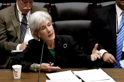
It was a rough week on Capitol Hill for Health and Human Services Secretary Kathleen Sebelius. She appeared before congressional committees in the House and the Senate to defend President Obama’s proposed fiscal year 2014 budget, but spent much of her time fending off attacks on the Affordable Care Act (ACA).
She had a testy exchange with Rep. Michael Burgess (R-Texas), who is critical of how HHS has handled the Pre-Existing Condition Insurance Plan. The PCIP is a program that provides health insurance coverage for uninsured adults with pre-existing conditions and was scheduled to run until Jan. 1, 2014 when the ACA would require health plans to offer insurance regardless of prior medical conditions. But the program is short on funds, so HHS halted new enrollment in February.
Rep. Burgess is a cosponsor for a bill (H.R. 1549) that would infuse the program with $4 billion in new funding taken from the ACA’s Prevention and Public Health Fund. That’s where the controversy comes in. The GOP have frequently called the prevention fund a "slush fund" and tried to eliminate it by using the $15 billion in it for deficit reduction. And HHS officials, who oppose the bill, have been dipping into the fund to cover some ACA implementation costs that Congress has refused to fund, such as education and outreach for the new health insurance exchanges. Just to make it more confusing, some conservative groups, including the Heritage Foundation, said they oppose the plan to add funding to the PCIP, because they oppose federally-funded high-risk insurance pools.
Meanwhile, across town Ms. Sebelius was grilled by Senators about why her department wasn’t doing a better job getting the word out about the exchanges. Sen. Max Baucus (D-Mont.), a supporter of the ACA, said he was concerned the exchange implementation would be a "train wreck."
Hear about all these issues and criticism of Medicare’s electronic health record incentive program in this week’s edition of the Policy and Practice podcast.
Initial surgical management bests expectant management for VVF
CHARLESTON, S.C. – Most simple vesicovaginal fistulae are initially managed by surgery, which is successful in the majority of cases, according to a multicenter review from the Fellows’ Pelvic Research Network.
Conversely, most of those initially managed conservatively by catheter drainage fail to resolve.
Of 226 patients with vesicovaginal fistulae (VVF) included in the review, 166 were managed surgically at the outset, and 77.5% of these resolved with one surgery. Of 37 who did not resolve after initial surgery, 32 underwent a second surgery; 56% of these resolved, Dr. Susan H. Oakley reported at the annual meeting of the Society of Gynecologic Surgeons.
Of the 60 patients who were managed conservatively, only 12% resolved during a median duration of catheterization of 35 days. The 53 who did not resolve went on to have surgery; the majority of these patients resolved. The overall success rate in the study was 83% in the 219 who underwent surgery, said Dr. Oakley of Good Samaritan Hospital, Cincinnati.
The patients (mean age, 49.5 years) were treated for VVF at 1 of 12 participating academic medical centers between July 2006 and June 2011. Most patients were postmenopausal (53%), nonsmokers (59.5%) and white (71%).
Of those initially managed surgically, 41% underwent vaginal repair, 23% underwent abdominal repair, and the remaining patients underwent other types of repair such as electrocautery. Of those initially managed conservatively who underwent subsequent surgery, 41% underwent vaginal repair, 53% underwent abdominal repair, and the remaining patients underwent other types of repair.
"VVF are the most common acquired fistulae of the urinary tract, and in the United States, gynecologic surgery is often the cause. Regardless of the etiology, the physical and emotional effects on patients are profound," Dr. Oakley said, adding that because a standardized algorithm for the management of VVS is lacking, the purpose of this study was to describe practice patterns and treatment outcomes for VVF.
While conservative management is often suggested as first-line therapy, it has been unclear how often this strategy is successful, she said at the meeting, jointly sponsored by the American College of Surgeons.
Although limited by its retrospective nature and by missing data that prevented complete analysis of all of the variables of interest, the study does have several strengths, including the multicenter design and a large number of cases. The findings thus support a recommendation that early surgical management should be considered as the preferred approach to treating simple VVF, Dr. Oakley concluded.
She reported having no relevant financial disclosures.
AT THE SGS ANNUAL MEETING
Major finding: Success following initial surgical management of VVF vs. initial conservative management was 77.5% and 12%, respectively.
Data source: A chart review involving 226 VVF cases.
Disclosures: Dr. Oakley reported having no relevant financial disclosures.
Fractional laser offers new hope for old burn scars
BOSTON – The appearance of mature burn scars significantly improved after treatment with a nonablative fractional laser, based on data from a randomized controlled trial.
In a split-lesion study, laser-treated skin appeared smoother than did adjacent untreated control sites, and within 3 months both patients and clinicians rated laser-treated areas as significantly improved, said Dr. Merete Haedersdal of the University of Copenhagen, Denmark.
"We consider this as a safe treatment, we now have long-term clinical and histological efficacy, and we do have documentation that the efficacy improves over time. We are operating with an intact skin barrier, which may give us a good potential for when we are going to treat patients with large areas of burn scars," she said at the annual meeting of the American Society for Laser Medicine and Surgery.
Dr. Haedersdal and her colleagues in Denmark, Germany, and Belgium examined long-term outcomes from the treatment of burn scars using a 1,540-nm fractional nonablative laser with compression handpieces to deliver energy to deep and superficial tissues.
Twenty patients (median age, 38 years) were enrolled, and 17 completed the study. The patients, all Fitzpatrick skin types II or III, had mature burn scars from fires (75%) or scalding (25%), and 75% had previously received skin grafts. The mature scars, with a median duration of 7 years, involved the trunk and/or extremities.
Side-by-side areas of each lesion were randomly assigned to receive three monthly deep and superficial treatments with Palomar Medical Technologies’ 1,540-nm nonablative fractional laser or to serve as untreated controls.
With the deep (XD) handpiece, the energy was applied with 15-ms pulses at 70 mJ per microbeam in three stacks for 10 passes. The compression tip of the handpiece squeezes moisture away from the applicator end, allowing delivery of energy into deep tissues.
The superficial (XF) handpiece was then used to deliver 50 mJ per microbeam in a 15-ms pulse for one stack with two passes.
"By ultrasound, we saw that we were able to deliver the energy into the deep layers of the skin," Dr. Haedersdal said. She cited the example of one patient whose scar was 1.08 mm thick before treatment, and immediately after treatment it was 3.52 mm thick from edema in the mid and deep dermal layers.
On-site clinical evaluations at 6 months performed by blinded observers rated 15 of 18 treated areas as improved, 3 as showing no response, and none as worsening. In contrast, all 18 control sites were rated as having no response.
The investigators also used the Patient and Observer Scar Assessment Scale (POSAS), which rates vascularity, pigmentation, thickness, relief, pliability, and surface area, as well as overall impression. The scale ranges from 1 for "normal skin" to 10 for "worst scar imaginable." The baseline median score was 7 (range, 3-8) for both groups.
At 1 month after treatment, there was no significant difference in POSAS score between the treated and untreated sides of the scars, but by 3 months the untreated sides were rated as a median of 7, compared with 5 for the treated sides (P = .0185). At 6 months the difference had increased slightly, with a median rating of 7 for the untreated sides and 4 for the treated sides (P = .0008).
The researchers also observed significant improvements in laser-treated (but not control) scars from baseline to 3 months (P = .0185), baseline to 6 months (P = .0008), and from 3 to 6 months (P = .0092), "which actually supports for the first time that when giving these nonablative fractional treatments, there is a continued improvement over time," Dr. Haedersdal said.
The researchers found that meshed (transplanted) skin tended to respond better than nontransplanted skin. Preliminary histology showed treatment-induced remodeling of the stratum corneum, frequently with a thicker epidermal compartment. In addition, post-treatment collagen deposition appeared closer to that found in normal skin, Dr. Haedersdal noted.
The immediate post-treatment responses included edema in 17 of 20 patients, erythema in 18, and purpura in 15, but there was no blistering of skin, and the skin barrier remained intact.
During the study period, 6 of 20 patients had hyperpigmentation, which resolved gradually over time.
"We also saw a grid pattern in three of the patients using these parameters, but we softened up the treatments afterward by giving following treatments with the XF handpiece," Dr. Haedersdal said.
The study was supported by an equipment loan and research grant to Dr. Haedersdal from Palomar Medical Technologies.
AT LASER 2013
Major finding: At 3 months, untreated areas were rated 7 on the POSAS scale, compared with 5 for laser-treated areas (P = .0185).
Data source: A randomized controlled trial in 20 patients, comparing side-by-side areas of untreated and laser-treated mature burn scars.
Disclosures: The study was supported by an equipment loan and research grant to Dr. Haedersdal from Palomar Medical Technologies.
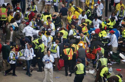
Emergency physicians share lessons from Boston Marathon bombings response
If there’s one piece of advice that Boston emergency physicians have for their peers following the Boston Marathon bombings, it’s this: drill, drill, drill.
"No matter how much drilling you do, you will never really prepare yourself for what this is like," said Dr. Ron M. Walls, chairman of the department of emergency medicine at Brigham and Women’s Hospital.
"But I would encourage everyone to plan those drills, take them seriously, do them well, and do them often. There’s no question that the expertise of emergency physicians and surgeons and nurses is crucial at the time of response, but being part of the systematic response and knowing that you’re part of systematic response is also very important."
On Monday afternoon, April 15, before the two improvised explosive devices detonated near the finish line of the Boston Marathon, killing three and injuring more than 170 people, Dr. Walls was in his office with his department administrator going over the budget. Suddenly, an alert went through the phone system. A news banner popped up on his phone.
"And then I heard an absolute cacophony of sirens, more than I had ever heard anywhere," he said in an interview.
Boston is unique when it comes to emergency and trauma care. It has five level 1 trauma centers. And Massachusetts was one of the first states to ban ambulance diversions in 2009. As a result, hospitals have created protocols to move patients quickly and make room in the ED.
"EMS [emergency medical services] did a beautiful job of distributing the patients at Beth Israel Deaconess Medical Center. There wasn’t any point where we felt truly overwhelmed," said Dr. Richard E. Wolfe, chief of emergency medicine at the medical center.
And the drills and preparations helped. While mass casualty drills have been a part of hospital operations for years, the 9/11 terrorist attacks brought the importance of preparation to the forefront.
"We’ve had a big shift in education," said Col. John McManus, MC, USA (ret.), an emergency physician who was among the last to leave Iraq in 2011. "Even though we learned a lot of these lessons from World War II and Vietnam, we kind of forgot about some of it. They were published, but we didn’t keep them in the cutting edge of medicine. We felt like it was never going to happen here, and that the military had to deal with it. After 9/11, unfortunately, we realized that we were underprepared and undertrained to treat these kinds of casualties."
Brigham and Women’s Hospital has held 73 disaster preparation drills since 2006, including citywide drills with Boston EMS, Dr. Walls said.
Now, the many hospitals have physicians, nurses, and other personnel on staff who have served in combat situations. Federal agencies, medical societies, and private entities are collaborating to update training and medical information. "The majority of scientific meetings include military and blast education," Dr. McManus, vice president of medical affairs at Masimo Corporation, said in an interview.
"What’s happened since Sept. 11 is that people are much more focused on disaster management and mass casualty events," Dr. Wolfe said in an interview. At Beth Israel Deaconess Medical Center, "we’ve had speakers from Israel doing visiting fellowships with us. We had Dr. Chris Caldwell, who helped coordinate the Aurora and Columbine disaster responses, as a visiting professor with us, walking us through the lessons learned in Columbine.
"It’s become part of residency training and didactic programs, and has really developed a culture of knowledge in terms of mass casualty events, in a way that we didn’t have in the 80s and 90s," Dr. Wolfe said.
And after each disaster, be it Hurricane Katrina or the Boston Marathon, hospitals improve their protocols and share what they’ve learned. Both Dr. Walls and Dr. Wolfe said they wanted to improve communication, whether it’s within the hospital and between the departments, or whether it’s with outside staff who may feel left out of the loop.
On Monday afternoon, Dr. Wolfe had just gotten home when he saw television reports of the bombing. He called his ED and headed back to work. Within minutes, ED personnel levels increased fivefold as patients began to arrive.
He quickly established crowd control in the ED, and put some of the volunteers on the reserve and on the sidelines. He also created zones, where people worked in small pockets. There was a disaster coordinator on the scene "so we had all the right people," Dr. Wolfe said.
Out front, social workers dealt with others who were arriving at the hospital. Registration was "very focused on getting real names so that we wouldn’t lose any patients to mistaken identity," he said. Patients were quickly assessed as they arrived, then flowed rapidly to the next stage of care, whether it was operating room or intensive care."
At Brigham and Women’s, the emergency department received a total of 35 patients, 28 of whom arrived in an hour.
"When this started, we had a pretty full ED," said Dr. Walls, professor of medicine at Harvard Medical School. "We decompressed by moving some of the patients out of the ED or into inpatient units. We created a lot of capacity in a short period of time. At the same time, we had alerted the OR, and it happened to be shift change, so we froze the staff, resulting in double staffing. And it was Monday afternoon on a holiday, so the ORs’ slate was lighter. Those two things allowed us to create seven open, staffed, ready ORs in a very short period of time.
"Within the ED, we deployed [emergency physicians] and surgeons and nurses into a number of trauma teams. At one point we had more than 10 teams, and they were all ready to receive patients, which turned out to be very important," Dr. Walls said.
At press time, none of more than 170 patients who were admitted to Boston area hospitals after the marathon had died, and physicians attribute this to great prehospital care, and lots of practice, preparation, and collaboration.
"It’s times like this that bring out the best in everybody," Dr. Wolfe said. "People are willing to help, they’re willing to collaborate. Egos dissipate and people tend to work together the way you just don’t see on a regular basis, which is absolutely lovely. From my standpoint, watching how well the teams worked, sort of was a validation of what emergency medicine is all about, and what emergency medicine training is."
"This was an amazing example for me, not that I really needed one, on how valuable every single person in the emergency department is," Dr. Walls said. "It’s the physicians and nurses, but it is also the aides, and the registration people and the housekeepers and everybody in the ED who have to do their jobs magnificently to make this work. And on that day, they all did."
On Twitter @NaseemSMiller


