User login
Pandemic prompts spike in eating disorder hospitalization for adolescents
Hospital admission for children with eating disorders approximately tripled during the COVID-19 pandemic, based on data from 85 patients.
Eating disorders are common among adolescents and often require hospital admission for nutritional restoration, according to May Shum of Yale University, New Haven, Conn., and colleagues

During the COVID-19 pandemic, the volume of hospital admissions for adolescents with eating disorders has increased, the researchers wrote in a poster presented at the annual meeting of the Pediatric Academic Societies. This increase may be driven both by interruptions in medical care and increased psychological distress, but data on changes in patient characteristics and hospitalization course are lacking, they said.
The researchers reviewed charts from patients with eating disorders admitted to a single center between Jan. 1, 2017, and June 30, 2021. The majority of the patients were female (90.6%), and White (78.8%), had restrictive eating behaviors (97.2%), and had private insurance (80.0%).
Overall, the number of monthly admissions increased from 1.4 before the onset of the pandemic to 3.6 during the pandemic (P < .001).
Length of stay increased significantly from before to during pandemic cases (12.8 days vs. 17.3 days, P = .04) and age younger than 13 years was significantly associated with a longer length of stay (P < .001).
The number of patients for whom psychotropic medications were initiated or changed increased significantly (12.5% vs. 28.3%, P = .04); as did the proportion of patients discharged to partial hospitalization, residential, or inpatient psychiatric treatment rather than discharged home with outpatient therapy (56.2% vs. 75.0%, P = .04).
No significant differences were noted in demographics, comorbidities, admission parameters, EKG abnormalities, electrolyte repletion, or tube feeding.
The study findings were limited by the use of data from a single center. However, the results suggest an increase in severity of hospital admissions that have implications for use of hospital resources, the researchers said.
“In addition to an increase in hospital admissions for eating disorder management during the pandemic, longer inpatient stays of younger children with higher acuity at discharge is an added strain on hospital resources and warrants attention,” they concluded.
Considerations for younger patients
The current study is especially important at this time, Margaret Thew, DNP, FNP-BC, medical director of the department of adolescent medicine at Children’s Wisconsin in Milwaukee, said in an interview. “There have been reports of the rising numbers in eating disorders, but until research has been conducted, we cannot quantify the volumes,” said Ms. Thew, who was not involved in the study. “There have been many reports of the rise in mental health issues during the pandemic, so it seems accurate that the rate of eating disorders would rise,” she said. “Additionally, from a clinical perspective there seemed to be many younger-age patients with eating disorders presenting to the inpatient units who seemed sicker,” she noted.
Ms. Thew said she was not surprised by the study findings. “Working with adolescents with eating disorders we saw the increased numbers of both hospitalizations and outpatient referrals during the pandemic,” said Ms. Thew. “Length of stay was higher across the nation regarding admissions for concerns of eating disorders. These patients are sicker and fewer went home after medical stabilization,” she emphasized.
“Clinicians should be more aware of the rise in patients presenting with eating disorders at younger ages to their clinics and provide early interventions to prevent severe illness and medical instability,” said Ms. Thew. Clinicians also should be more proactive in managing younger children and adolescents who express mood disorders, disordered eating, or weight loss, given the significant rise in eating disorders and mental health concerns, she said.
Additional research is needed to continue following the rate of eating disorders into 2022, said Ms. Thew. More research is needed on early interventions and recognition of eating disorders for preteens and teens to prevent severe illness, as is research on how the younger patient with an eating disorder may present differently to the primary care doctor or emergency department, she said.
“We may need to study treatment of the younger population, as they may not do as well with admissions into behavioral health facilities,” Ms. Thew added.
The study received no outside funding. The researchers had no financial conflicts to disclose. Ms. Thew had no financial conflicts to disclose and serves on the editorial advisory board of Pediatric News.
Hospital admission for children with eating disorders approximately tripled during the COVID-19 pandemic, based on data from 85 patients.
Eating disorders are common among adolescents and often require hospital admission for nutritional restoration, according to May Shum of Yale University, New Haven, Conn., and colleagues
During the COVID-19 pandemic, the volume of hospital admissions for adolescents with eating disorders has increased, the researchers wrote in a poster presented at the annual meeting of the Pediatric Academic Societies. This increase may be driven both by interruptions in medical care and increased psychological distress, but data on changes in patient characteristics and hospitalization course are lacking, they said.
The researchers reviewed charts from patients with eating disorders admitted to a single center between Jan. 1, 2017, and June 30, 2021. The majority of the patients were female (90.6%), and White (78.8%), had restrictive eating behaviors (97.2%), and had private insurance (80.0%).
Overall, the number of monthly admissions increased from 1.4 before the onset of the pandemic to 3.6 during the pandemic (P < .001).
Length of stay increased significantly from before to during pandemic cases (12.8 days vs. 17.3 days, P = .04) and age younger than 13 years was significantly associated with a longer length of stay (P < .001).
The number of patients for whom psychotropic medications were initiated or changed increased significantly (12.5% vs. 28.3%, P = .04); as did the proportion of patients discharged to partial hospitalization, residential, or inpatient psychiatric treatment rather than discharged home with outpatient therapy (56.2% vs. 75.0%, P = .04).
No significant differences were noted in demographics, comorbidities, admission parameters, EKG abnormalities, electrolyte repletion, or tube feeding.
The study findings were limited by the use of data from a single center. However, the results suggest an increase in severity of hospital admissions that have implications for use of hospital resources, the researchers said.
“In addition to an increase in hospital admissions for eating disorder management during the pandemic, longer inpatient stays of younger children with higher acuity at discharge is an added strain on hospital resources and warrants attention,” they concluded.
Considerations for younger patients
The current study is especially important at this time, Margaret Thew, DNP, FNP-BC, medical director of the department of adolescent medicine at Children’s Wisconsin in Milwaukee, said in an interview. “There have been reports of the rising numbers in eating disorders, but until research has been conducted, we cannot quantify the volumes,” said Ms. Thew, who was not involved in the study. “There have been many reports of the rise in mental health issues during the pandemic, so it seems accurate that the rate of eating disorders would rise,” she said. “Additionally, from a clinical perspective there seemed to be many younger-age patients with eating disorders presenting to the inpatient units who seemed sicker,” she noted.
Ms. Thew said she was not surprised by the study findings. “Working with adolescents with eating disorders we saw the increased numbers of both hospitalizations and outpatient referrals during the pandemic,” said Ms. Thew. “Length of stay was higher across the nation regarding admissions for concerns of eating disorders. These patients are sicker and fewer went home after medical stabilization,” she emphasized.
“Clinicians should be more aware of the rise in patients presenting with eating disorders at younger ages to their clinics and provide early interventions to prevent severe illness and medical instability,” said Ms. Thew. Clinicians also should be more proactive in managing younger children and adolescents who express mood disorders, disordered eating, or weight loss, given the significant rise in eating disorders and mental health concerns, she said.
Additional research is needed to continue following the rate of eating disorders into 2022, said Ms. Thew. More research is needed on early interventions and recognition of eating disorders for preteens and teens to prevent severe illness, as is research on how the younger patient with an eating disorder may present differently to the primary care doctor or emergency department, she said.
“We may need to study treatment of the younger population, as they may not do as well with admissions into behavioral health facilities,” Ms. Thew added.
The study received no outside funding. The researchers had no financial conflicts to disclose. Ms. Thew had no financial conflicts to disclose and serves on the editorial advisory board of Pediatric News.
FROM PAS 2022
Majority of pandemic pediatric visits managed with telemedicine
Approximately two-thirds of pediatric acute care concerns managed in telemedicine visits required no additional visits or follow-up, based on data from more than 600 visits.
The increase in use of telemedicine during the first year of the COVID-19 pandemic enabled access to care and connection to doctors for many pediatric patients, said Kristina Kissiova, MD, of Children’s National Health System, Washington, and colleagues. Some advantages of telemedicine include enhanced medical homes, reduced health care costs, and less crowding and wait time for patients in offices and emergency departments; however, the optimal use of telemedicine for acute primary care has not been examined, they said.

In a study presented at the annual meeting of the Pediatric Academic Societies, the researchers conducted a retrospective chart review of 638 acute care telemedicine visits conducted by 21 health care providers at a single practice in Washington in October 2020 and November 2020. Approximately half of the patients were male, 65% were white, and 89% had commercial insurance. The most common age group was 6-12 years (23%), followed by 2-3 years (16%), 3-6 years (15%), and 12-18 years (14%).
The primary outcome was the number and nature of visits completed via telemedicine without the need for referral or a subsequent in-person visit. Telemedicine visits for well-child checks and follow-up visits were excluded.
Overall, 60% of the visits (384 of 638) were completed over telemedicine with no need for additional visits or referrals. The most common acute complaints were upper respiratory infections, dermatologic issues, gastrointestinal issues, COVID-19 related issues, and fever (18.7%, 16.3%, 12.9%, 11.9%, and 10.3%, respectively).
Of these, dermatologic and GI concerns were most often completed via telemedicine (93.3% and 81.7%, respectively), while upper respiratory tract infections and fever issues were the least likely to be completed via telemedicine (22.7% and 13.6%), mainly because of the need to report for in-person COVID-19 testing, the researchers said.
Among other less common chief complaints, 100% of breathing concerns, behavior/mental health concerns, and head trauma or falls were addressed via telemedicine without additional referrals or follow-up visits. In addition, 90.9% of urgent care or emergency department follow-ups, 88.9% of ear concerns, and 87.5% of eye concerns were completely resolved via telemedicine visits.
Overall, 3% of patients who were not referred after a telemedicine visit presented in person for worsening symptoms. Of these who were referred after a telemedicine visit, 90% were seen in person within 48 hours.
The study findings were limited by the inclusion of data from only a single center. However, “These early findings provide insight into the utility of telehealth in the primary care setting for a broad array of urgent concerns,” the researchers concluded.
Pandemic propelled telemedicine to improve patient care
The widespread adoption of telemedicine in primary care has been a beneficial side effect of the COVID-19 pandemic, said Tim Joos, MD, a Seattle-based clinician with a combination internal medicine/pediatrics practice, in an interview.
“Toward the end of World War II and in the push to form the United Nations, Winston Churchill was credited with the saying, ‘Never let a good crisis go to waste,’” said Dr. Joos, who was not connected with the study.
“As awful as this pandemic has been, it has propelled health care delivery at an unprecedented pace into the digital age,” he noted.
The current study is important because it highlights the number of complaints that can be successfully resolved through telemedicine, offering patients and families quicker access and more options for care, Dr. Joos said.
“I feel that giving patients and families an open choice for either telemedicine or in-person visits improves the likelihood that the issue will be resolved efficiently and satisfactorily with fewer visits,” he added.
Approximately two-thirds of pediatric acute care concerns managed in telemedicine visits required no additional visits or follow-up, based on data from more than 600 visits.
The increase in use of telemedicine during the first year of the COVID-19 pandemic enabled access to care and connection to doctors for many pediatric patients, said Kristina Kissiova, MD, of Children’s National Health System, Washington, and colleagues. Some advantages of telemedicine include enhanced medical homes, reduced health care costs, and less crowding and wait time for patients in offices and emergency departments; however, the optimal use of telemedicine for acute primary care has not been examined, they said.
In a study presented at the annual meeting of the Pediatric Academic Societies, the researchers conducted a retrospective chart review of 638 acute care telemedicine visits conducted by 21 health care providers at a single practice in Washington in October 2020 and November 2020. Approximately half of the patients were male, 65% were white, and 89% had commercial insurance. The most common age group was 6-12 years (23%), followed by 2-3 years (16%), 3-6 years (15%), and 12-18 years (14%).
The primary outcome was the number and nature of visits completed via telemedicine without the need for referral or a subsequent in-person visit. Telemedicine visits for well-child checks and follow-up visits were excluded.
Overall, 60% of the visits (384 of 638) were completed over telemedicine with no need for additional visits or referrals. The most common acute complaints were upper respiratory infections, dermatologic issues, gastrointestinal issues, COVID-19 related issues, and fever (18.7%, 16.3%, 12.9%, 11.9%, and 10.3%, respectively).
Of these, dermatologic and GI concerns were most often completed via telemedicine (93.3% and 81.7%, respectively), while upper respiratory tract infections and fever issues were the least likely to be completed via telemedicine (22.7% and 13.6%), mainly because of the need to report for in-person COVID-19 testing, the researchers said.
Among other less common chief complaints, 100% of breathing concerns, behavior/mental health concerns, and head trauma or falls were addressed via telemedicine without additional referrals or follow-up visits. In addition, 90.9% of urgent care or emergency department follow-ups, 88.9% of ear concerns, and 87.5% of eye concerns were completely resolved via telemedicine visits.
Overall, 3% of patients who were not referred after a telemedicine visit presented in person for worsening symptoms. Of these who were referred after a telemedicine visit, 90% were seen in person within 48 hours.
The study findings were limited by the inclusion of data from only a single center. However, “These early findings provide insight into the utility of telehealth in the primary care setting for a broad array of urgent concerns,” the researchers concluded.
Pandemic propelled telemedicine to improve patient care
The widespread adoption of telemedicine in primary care has been a beneficial side effect of the COVID-19 pandemic, said Tim Joos, MD, a Seattle-based clinician with a combination internal medicine/pediatrics practice, in an interview.
“Toward the end of World War II and in the push to form the United Nations, Winston Churchill was credited with the saying, ‘Never let a good crisis go to waste,’” said Dr. Joos, who was not connected with the study.
“As awful as this pandemic has been, it has propelled health care delivery at an unprecedented pace into the digital age,” he noted.
The current study is important because it highlights the number of complaints that can be successfully resolved through telemedicine, offering patients and families quicker access and more options for care, Dr. Joos said.
“I feel that giving patients and families an open choice for either telemedicine or in-person visits improves the likelihood that the issue will be resolved efficiently and satisfactorily with fewer visits,” he added.
FROM PAS 2022
Depression strikes more than half of obese adolescents
More than 50% of obese adolescents met criteria for depression, which also was associated with several components of metabolic syndrome, based on data from 160 individuals.
Previous research shows that the metabolic consequences of obesity are worsened with the coexistence of depression in adults, but a similar relationship in obese adolescents has not been explored, according to Nisha Gupta, a medical student at the University of Texas Health Science Center, Houston, and colleagues.
“This relationship is explained by an overactive stress response and adoption of unhealthy lifestyle habits,” both of which increased during the COVID-19 pandemic, the researchers noted in their abstract.
In a study presented at the Pediatric Academic Societies annual meeting, the researchers reviewed data from 160 obese adolescents seen at a pediatric weight management clinic between July 1, 2018, and Dec. 3, 2021. The data included anthropometric, clinical, and laboratory information. Depression was assessed using the Patient Health Questionnaire–9 (PHQ-9). The goal of the study was to compare the prevalence of metabolic syndrome components in obese youth with and without diagnosed depression.
Overall, 46% of the patients had PHQ-9 scores less than 5, which was defined as no clinically significant depression. A total of 26% had current or prior diagnoses of depression, and 25% met the criteria for moderate to severe depression, with PHQ-9 scores of 10 or higher. Notably, 18% of individuals with no prior history of depression met criteria for moderate to severe depression, the researchers wrote.
Teens who reported daytime fatigue or trouble sleeping, and those who reported eating out seven or more times a week had higher scores than those without these reports.
In laboratory analyses, higher PHQ-9 scores were significantly associated with increasing weight, body mass index, body fat percentage, diastolic blood pressure, and fasting blood insulin (P < .02 for all).
The study findings were limited by the relatively small sample size, the researchers noted. However, the results suggest that depression is common, but often underdiagnosed in obese adolescents, and depression screening should be part of obesity management.
Study highlights need to screen
The current study is important because of the overall increase in obesity in the United States, which extends to children and teens, Tim Joos, MD, a Seattle-based clinician with a combination internal medicine/pediatrics practice, said in an interview.
“With skyrocketing rates of obesity among children and teens over the last decades, we are seeing more ‘adult’ diseases seep into the younger ages, including type 2 diabetes, high blood pressure and now, depression,” he said.
“The results are a wake-up call for the need for better system-wide prevention and management of obesity in adolescents and the importance of screening and managing depression in obese teenagers,” he emphasized.
The study received no outside funding. The researchers had no financial conflicts to disclose. Dr. Joos had no financial conflicts to disclose and serves on the editorial advisory board of Pediatric News.
FROM PAS 2022
Internet intervention improved insomnia in Black women
Data from previous studies suggest that women are up to 40% more likely to experience insomnia disorder compared with men, Eric S. Zhou, PhD, of Harvard Medical School, Boston, and colleagues wrote. The risk is even higher among Black women, but research on tailored treatments for this particular population has been limited.
In their study, published in JAMA Psychiatry, the researchers recruited women with elevated insomnia symptoms who were enrolled in the Black Women’s Health Study, an ongoing national, longitudinal research cohort in the United States. Participants were recruited between October 2019 and June 2020.The participants were randomized to an Internet-delivered behavior intervention (108 women), a stakeholder-informed Internet intervention tailored to Black women (110 women), or non-Internet patient education about sleep (115 women).
The Internet intervention, known as Sleep Healthy Using the Internet (SHUTi), was a 6-session program lasting 45-60 minutes per session and delivered over 6-9 weeks. The program included core elements of cognitive behavioral therapy and took into account information provided by patients about their baseline sleep function, treatment adherence, and sleep progress.
The tailored version of SHUTi for Black women (SHUTi-BWHS) was similar, but included Black actors for video vignettes and the inclusion of content about the cultural and social contexts in which insomnia often occurs for Black women, such while managing neighborhood noise and or living in crowded environments.
A third group received standard patient education material about sleep through a noninteractive website, and served as the control group.
The primary outcome of insomnia severity was measured using the Insomnia Severity Index (ISI), a 0- to 28-point scale. Scores for the ISI are based on responses to seven questions, including some that ask participants to rate the severity of their insomnia symptoms.
Clinically significant improvement in insomnia was defined as a reduction in score of more than 7 points. Patients were assessed at baseline, at 9 weeks, and again at approximately 6 months.
Significantly greater reductions in insomnia severity seen in intervention groups vs. control group
Overall, women randomized to SHUTi or SHUTi-BWHS) reported a significantly greater reduction in insomnia symptoms from baseline to 6 months, compared with the control group (P < .001), with ISI score decreases of 10.0, 9.3, and 3.6, respectively. No statistically significant differences in ISI score changes appeared between the between the SHUTi-BWHS and SHUTi groups.
Also, significantly more women in the SHUTi-BWHS group than in the SHUTi group completed the intervention (78.2% vs. 64.8%).
Treatment response was similar between the SHUTI-BWHS and SHUTi groups; 47.3% and 46.3%, respectively, had a decrease in ISI score of more than 7 points. In addition, 37% of women in the SHUTi-BWHS and 38% of women in the SHUTi groups reached ISI scores of less than 8 points, defined as full resolution of insomnia, by the last follow-up visit.
Both the SHUTi and SHUTi-BWHS interventions had dramatic effects on insomnia, but the increased number of women who completed the intervention in the SHUTi-BWHS group supports the value of tailored intervention, the researchers noted. “Similar to prior SHUTi trials, there was a direct association between the participant’s level of intervention engagement and their improvement in sleep.”
The average age of the participants was 60 years, 62% were single, and 44% had a graduate degree or higher. Approximately 5% were being actively treated for sleep apnea.
The study findings were limited by several factors including the relatively high socioeconomic status of the study participants, lack of data on medical mistrust, and inability to detect smaller differences between SHUTi and SHUTi-BWHS, the researchers noted.
Choose Internet-based CBT first for insomnia
“This was an excellent paper that sought to see the relative efficacy of standard version of Internet-delivered CBT-I [cognitive-behavioral therapy for insomnia] versus a culturally tailored version for Black women,” said Neil Skolnik, MD, professor of family and community medicine at Thomas Jefferson University, Philadelphia, in an interview. “The trial confirmed that, compared with sleep education, which was used as the control, Internet-delivered CBT-I is effective in the treatment of insomnia.”

“These results demonstrate two important things,” said Dr. Skolnik. “The most important is that Internet-delivered CBT-I works, and since it is both safe and effective, should be the first-line therapy for patients who want treatment for insomnia.”
Secondly, “the fact that more people completed culturally tailored versions suggests that, when culturally tailored versions are available, their use is preferable, as it might facilitate a higher proportion of patients being successful in their insomnia treatment,” he added.
The study was supported by the Patient-Centered Outcomes Research Institute. The Black Women’s Health Study is supported by the National Cancer Institute. Dr. Zhou disclosed support from both PCORI and the NCI during the study. Dr. Skolnik, who was not involved in the study, disclosed serving on the advisory board for Idorsia Pharmaceuticals. He is also a member of the editorial advisory board of Family Practice News.
Data from previous studies suggest that women are up to 40% more likely to experience insomnia disorder compared with men, Eric S. Zhou, PhD, of Harvard Medical School, Boston, and colleagues wrote. The risk is even higher among Black women, but research on tailored treatments for this particular population has been limited.
In their study, published in JAMA Psychiatry, the researchers recruited women with elevated insomnia symptoms who were enrolled in the Black Women’s Health Study, an ongoing national, longitudinal research cohort in the United States. Participants were recruited between October 2019 and June 2020.The participants were randomized to an Internet-delivered behavior intervention (108 women), a stakeholder-informed Internet intervention tailored to Black women (110 women), or non-Internet patient education about sleep (115 women).
The Internet intervention, known as Sleep Healthy Using the Internet (SHUTi), was a 6-session program lasting 45-60 minutes per session and delivered over 6-9 weeks. The program included core elements of cognitive behavioral therapy and took into account information provided by patients about their baseline sleep function, treatment adherence, and sleep progress.
The tailored version of SHUTi for Black women (SHUTi-BWHS) was similar, but included Black actors for video vignettes and the inclusion of content about the cultural and social contexts in which insomnia often occurs for Black women, such while managing neighborhood noise and or living in crowded environments.
A third group received standard patient education material about sleep through a noninteractive website, and served as the control group.
The primary outcome of insomnia severity was measured using the Insomnia Severity Index (ISI), a 0- to 28-point scale. Scores for the ISI are based on responses to seven questions, including some that ask participants to rate the severity of their insomnia symptoms.
Clinically significant improvement in insomnia was defined as a reduction in score of more than 7 points. Patients were assessed at baseline, at 9 weeks, and again at approximately 6 months.
Significantly greater reductions in insomnia severity seen in intervention groups vs. control group
Overall, women randomized to SHUTi or SHUTi-BWHS) reported a significantly greater reduction in insomnia symptoms from baseline to 6 months, compared with the control group (P < .001), with ISI score decreases of 10.0, 9.3, and 3.6, respectively. No statistically significant differences in ISI score changes appeared between the between the SHUTi-BWHS and SHUTi groups.
Also, significantly more women in the SHUTi-BWHS group than in the SHUTi group completed the intervention (78.2% vs. 64.8%).
Treatment response was similar between the SHUTI-BWHS and SHUTi groups; 47.3% and 46.3%, respectively, had a decrease in ISI score of more than 7 points. In addition, 37% of women in the SHUTi-BWHS and 38% of women in the SHUTi groups reached ISI scores of less than 8 points, defined as full resolution of insomnia, by the last follow-up visit.
Both the SHUTi and SHUTi-BWHS interventions had dramatic effects on insomnia, but the increased number of women who completed the intervention in the SHUTi-BWHS group supports the value of tailored intervention, the researchers noted. “Similar to prior SHUTi trials, there was a direct association between the participant’s level of intervention engagement and their improvement in sleep.”
The average age of the participants was 60 years, 62% were single, and 44% had a graduate degree or higher. Approximately 5% were being actively treated for sleep apnea.
The study findings were limited by several factors including the relatively high socioeconomic status of the study participants, lack of data on medical mistrust, and inability to detect smaller differences between SHUTi and SHUTi-BWHS, the researchers noted.
Choose Internet-based CBT first for insomnia
“This was an excellent paper that sought to see the relative efficacy of standard version of Internet-delivered CBT-I [cognitive-behavioral therapy for insomnia] versus a culturally tailored version for Black women,” said Neil Skolnik, MD, professor of family and community medicine at Thomas Jefferson University, Philadelphia, in an interview. “The trial confirmed that, compared with sleep education, which was used as the control, Internet-delivered CBT-I is effective in the treatment of insomnia.”
“These results demonstrate two important things,” said Dr. Skolnik. “The most important is that Internet-delivered CBT-I works, and since it is both safe and effective, should be the first-line therapy for patients who want treatment for insomnia.”
Secondly, “the fact that more people completed culturally tailored versions suggests that, when culturally tailored versions are available, their use is preferable, as it might facilitate a higher proportion of patients being successful in their insomnia treatment,” he added.
The study was supported by the Patient-Centered Outcomes Research Institute. The Black Women’s Health Study is supported by the National Cancer Institute. Dr. Zhou disclosed support from both PCORI and the NCI during the study. Dr. Skolnik, who was not involved in the study, disclosed serving on the advisory board for Idorsia Pharmaceuticals. He is also a member of the editorial advisory board of Family Practice News.
FROM JAMA PSYCHIATRY
COVID-19 accelerated psychological problems for critical care clinicians
Approximately one-third of critical care workers reported some degree of depression, anxiety, or somatic symptoms in the early phase of the COVID-19 pandemic, based on survey results from 939 health care professionals.
The emotional response of professionals in a critical care setting in the early phase of the COVID-19 pandemic has not been well studied, Robyn Branca, PhD, and Paul Branca, MD, of Carson Newman University and the University of Tennessee Medical Center, both in Knoxville, wrote in an abstract presented at the virtual Critical Care Congress sponsored by the Society of Critical Care Medicine.
The prevalence of depression, anxiety, and somatization is low in the general population overall, but the researchers predicted that these conditions increased among workers in critical care settings early in the pandemic.
To assess the prevalence of psychological problems during that time, they sent an email survey on April 7, 2020, to members of the Society of Critical Care Medicine. The survey collected data on demographics, perceived caseload, and potential course of the pandemic. The survey also collected responses to assessments for depression (using the Patient Health Questionnaire–9), anxiety (using the Generalized Anxiety Disorder [GAD] Scale–7), and symptom somatization (using the PHQ-15).
Of the 939 survey respondents, 37% were male, 61.4% were female, and 1.4% gave another or no response.
Overall, 32.3% reported encountering 0-50 COVID-19 cases, 31.1% had encountered 51-200 cases, 12.5% had encountered 201-500 cases, 9.4% had encountered 501-1000 cases, and 13.7% had encountered more than 1,000 cases.
Based on the PHQ-9 depression scale, 44.9% of the respondents had minimal symptoms, 31.1% mild symptoms, 14.3% moderate symptoms, and 9.7% met criteria for severe depressive symptoms. Based on the GAD-7 anxiety scale, 35.5% had minimal symptoms, 32.9% mild, 16.8% moderate, and 14.8% had severe symptoms. Based on the PHQ-15 somatization scale, 39.6% of respondents showed minimal symptoms, whereas 38.2% showed mild symptoms, 17.3% moderate symptoms, and 4.9% had a severe degree of somatic symptoms.
The study findings were limited by the reliance on self-reports; however, the results indicate that a high percentage of critical care workers experienced significant, diagnosable levels of depression, anxiety, and somatic symptoms, the researchers said.
The standard guidance is to pursue individual intervention for anyone with scores of moderate or severe on the scales used in the survey, the researchers said.
Therefore, the findings represent “an alarming degree of mental health impact,” they emphasized. “Immediate mitigation efforts are needed to preserve the health of our ICU workforce.”
The study is important at this time because clinician fatigue and occupational stress are at endemic levels, Bernard Chang, MD, of Columbia University Irving Medical Center, New York City, said in an interview. “It is vital that we take stock of how frontline workers in critical care settings are doing overall,” said Dr. Chang.
Dr. Chang, who was not involved with the study but has conducted research on mental health in frontline health care workers during the pandemic, said he was not surprised by the findings. “This work builds on the growing body of literature in the pandemic noting high levels of stress, fatigue, and depression/anxiety symptoms across many frontline workers, from emergency department staff, first responders and others. These are all data points highlighting the urgent need for a broad safety net, not only for patients but the providers serving them.”
The takeaway message: “Clinicians are often so focused on providing care for their patients that they may overlook the need to care for their own well-being and mental health,” said Dr. Chang.
As for additional research, “we need to now take this important data and build on creating and identifying tangible solutions to improve the morale of the acute care/health care workforce to ensure career longevity, professional satisfaction, and overall well-being,” Dr. Chang emphasized. Mental health and morale affect not only health care workers, but also the patients they care for. Well–cared for health care providers can be at their best to provide the optimal care for their patients.
The study received no outside funding. The researchers and Dr. Chang disclosed no relevant financial relationships.
A version of this article first appeared on Medscape.com.
FROM SCCM 2022
Young and older athletes show similar arrhythmia patterns with fQRS
The prevalence of exercise-induced arrhythmias in young athletes with fragmented QRS (fQRS) patterns in lead V1 was 27%, similar to that seen in adult athletes, based on data from nearly 700 individuals.
Recent data suggest that fQRS complex in lead V1 (fQRSV1) in healthy athletes may promote arrhythmias in the context of training-induced right ventricular remodeling, but the prevalence and significance in young athletes has not been well studied, Guilia Quinto, MD, of the University of Padova (Italy) said in a presentation at the annual congress of the European Association of Preventive Cardiology.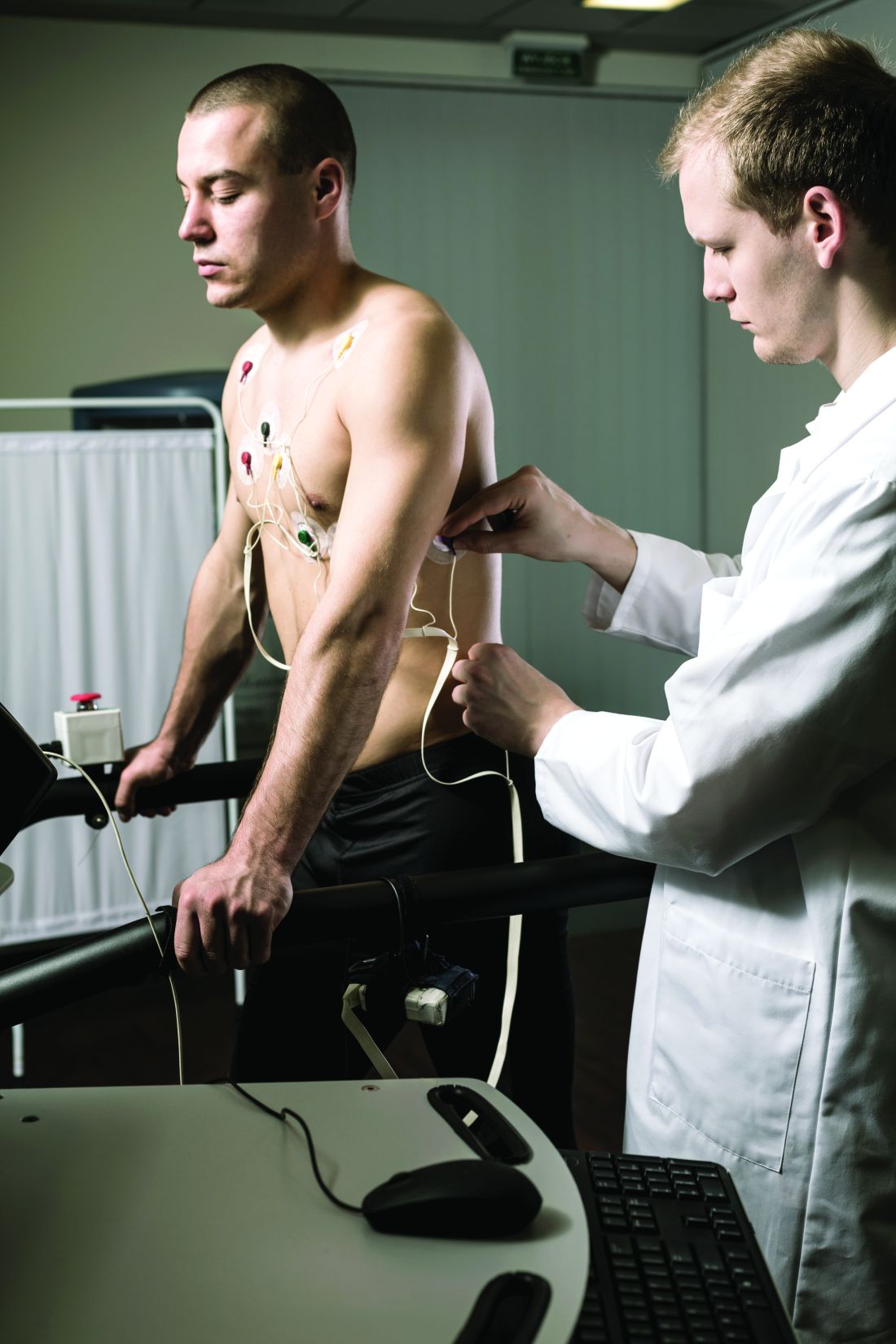
Dr. Quinto and colleagues assessed data from of young athletes on ventricular arrhythmias during exercise tests.
The study population included 684 young athletes with a mean age of 15 years; 64% were male. Baseline data collection included medical history, physical exam, resting ECG, standardized maximum exercise tolerance, and echocardiography evaluation.
The overall prevalence of fQRSV1 was 27%. Individuals with fQRSV1 were significantly less likely than those without fQRSV1 to be female (22% vs. 43%), and to present with a lower resting heart rate (66.98 beats per minute vs. 70.08 beats per minute).
Echocardiographic data showed that individuals with fQRSV1 had significantly different morphological and functional right ventricular characteristics.
Notably, right ventricular end-diastolic diameter was 20.42 mm/m2 among individuals with fQRSV1 and 19.81 mm/m2 in those without, a significant difference (P = .019), Dr. Quinto said. Tricuspid annulus plain systolic excursion also differed significantly; 24.33 mm and 23.75 mm for individuals with and without fQRSV1, respectively (P = .013).
However, the individuals with fQRSV1 showed no increased occurrence of any type of exercise-induced arrhythmias regardless of morphology or complexity, said Dr. Quinto.
The prevalence of common and uncommon arrhythmias among individuals with and without fQRSV1 was 31% versus 34% and 13% versus 11%, respectively; these differences were not significant.
The study findings were limited by the relatively small size, but were strengthened by the review of echocardiographic data by two independent physicians, she said.
The results show that the overall prevalence of fQRSV1 in young athletes is comparable with patterns seen in studies of adult athletes, and no differences in exercise-induced arrhythmias occurred despite differences in right ventricular characteristics, she concluded.
Expanded insight into evaluation
The ECG pattern identified in the current study is often encountered in the evaluation of athletes, but its importance was unknown, Matthew Martinez, MD, a sports cardiologist at the Atlantic Health System in Morristown, N.J., said in an interview.
“Studies of ECG findings in athletes continues to inform us about which findings are important to evaluate. This study furthers our understanding of how to proceed,” and will serve as a guide for additional testing to reduce athlete risk, he said.
Looking ahead, “this study should guide clinicians about additional testing and evaluation when fQRS is present in adolescent athletes compared to adults,” Dr. Martinez noted. However, additional research is needed to determine which is the next best test, and whether the patient requires ongoing surveillance, or whether a single evaluation is sufficient, he said. “Further study should focus on best practices after fQRS is identified and whether outcomes can be linked to this finding.”
The study received no outside funding. Dr. Quinto and Dr. Martinez had no financial conflicts to disclose.
FROM ESC PREVENTIVE CARDIOLOGY 2022
Peripheral muscle fatigue limits post-COVID exercise
Peripheral muscle fatigue was the most common cause of exercise limitation in patients recovered from COVID-19 regardless of disease severity, in a study of nearly 300 individuals.
The source and magnitude of exercise intolerance in post–COVID-19 patients has not been well studied, said Mauricio Milani, MD, of Fitcordis Exercise Medicine Clinic, Brasilia, Brazil, in a presentation at the annual congress of the European Association of Preventive Cardiology.
To assess exercise intolerance, the researchers performed cardiopulmonary exercise testing (CPET) on 144 adults who had recovered from COVID-19 and 144 matched controls who had not had COVID-19. The average age of the participants was 43 years, and 57% were male. COVID-19 was defined as mild, moderate, or severe in 60%, 21%, and 19% of the cases, respectively.
Residual symptoms were present in 41% of cases. CPET was performed at roughly 14 weeks after disease onset.
Among the COVID-19 patients, most of the CPET limitations (92%) were caused by muscle fatigue; cardiovascular limitations were noted in 2%, and pulmonary limitations were noted in 6%.
Data from the post-COVID CPET showed differences in peak oxygen consumption, as well as the first and second ventilatory thresholds (VT1 and VT2) between COVID-19 patients and controls, and with lower values related to higher illness severities, Dr. Milani said. Heart rate also varied according to illness severity, with lower values significantly related to higher illness severities and significant differences between COVID patients and controls.
A total of 42 individuals with COVID-19 had previous CPET data for comparison (27 with mild disease and 15 with moderate or severe disease), Dr. Milani said. In the subgroup with mild disease, the only significant difference in CPET results before and after COVID-19 was peak speed. In the moderate/severe group, the researchers observed higher reductions in peak speed and also reductions in oxygen consumption at peak and thresholds.
However, peak oxygen flows were not different before and after COVID-19 in either the mild or moderate/severe subgroups, Dr. Milani said.
The study findings were limited in part by the relatively small study population; however, the results indicate that peripheral muscle fatigue is the primary etiology in exercise limitation in post–COVID-19 patients.
“Our data suggest that treatment should emphasize comprehensive rehabilitation programs, including aerobic and muscle strengthening components,” Dr. Milani concluded.
COVID challenges remain unclear
“After COVID, patients often display a postviral syndrome with a wide range of symptoms,” Matthew Martinez, MD, a sports cardiologist at the Atlantic Health System in Morristown, N.J., in an interview said. “These conditions frequently lead to a sense of tiredness and weakness, pain, difficulty concentrating, and headaches that linger after the viral infection has cleared,” and these symptoms may continue for weeks.
However, this scenario is not unique to COVID-19: “This study confirms the importance of muscle fatigue in recovery,” said Dr. Martinez. “Recovery from viral illness requires hydration, sleep and slow progression return to exercise.” Consequently, Dr. Martinez said he was not surprised by the current study findings.
The take-home message for clinicians is to be aware that COVID-19 can have postviral syndrome, as is common after other infections, Dr. Martinez noted. The findings provide a starting point for discussing concerns with patients and explaining that a slow return to normal with usual care is expected. “Time to recovery will vary by individual,” he said. “Additional research is needed to identify which specific therapies are most important to help reduce time to recovery, and what new therapies could be developed to help facilitate muscle fatigue recovery and reduce time needed to recover.”
The study was supported by CAPES and CNPq. Dr. Milani had no financial conflicts to disclose. Dr. Martinez had no financial conflicts to disclose.
FROM ESC PREVENTIVE CARDIOLOGY 2022
ILD progression, not diagnosis, triggers palliative care
Most health care providers are comfortable recommending palliative care (PC) for their patients with interstitial lung disease (ILD), but most do so at the time of disease progression, rather than diagnosis, as indicated on survey data from 128 clinicians.
ILD is associated with a high mortality rate and profound symptoms that contribute to poor quality of life, Rebecca A. Gersen, MD, of Johns Hopkins University, Baltimore, and colleagues wrote.
“Nevertheless, there is often a lack of preparedness for death by both patients and providers, contributing to increased distress,” they said. Clinician perspectives on the use of PC for ILD patients have not been well studied, although PC is not limited to end-of-life care and is recommended for ILD patients by professional organizations, including the American Thoracic Society. “PC is successful in improving breathlessness in chronic lung disease and can increase survival.”
In a study published in the journal CHEST®, the researchers surveyed health care providers at 68 Pulmonary Fibrosis Foundation centers across the United States. The survey was sent and collected by email and a restricted social media platform. A total of 128 providers from 34 states completed the survey between October 2020 and January 2021. Of these, 61% were physicians, and 67% identified as White.
Overall, 95% of the respondents agreed or strongly agreed that addressing advance directives is important, but only 66% agreed or strongly agreed that they themselves addressed advance directives in the outpatient ILD clinic setting. A greater number (91%) agreed or strongly agreed that they had a high level of comfort in discussing prognosis, while 88% agreed or strongly agreed that they felt comfortable assessing a patient’s readiness for and acceptance of PC. Approximately two-thirds (67%) agreed or strongly agreed that they use PC services for ILD patients. There were no significant differences in responses from clinicians who had more than 10 years of experience and those who had less.
Of the providers who referred patients to PC, 54% did so at objective disease progression, and 80% did so at objective and/or symptomatic progress; 2% referred patients to PC at initial ILD diagnosis.
Lack of resources
Health care providers who reported that they rarely referred patients to palliative care were significantly more likely to cite a lack of local PC options (P < .01). Those who rarely referred patients for PC also were significantly less likely to feel comfortable discussing prognoses or advance directives in the ILD clinic (P = .03 and P = .02, respectively).
Among the 23% of responders who reported that they rarely referred patients, 66% said they did not have PC at their institution.
“In addition to understanding and addressing barriers to care, educational resources may be key to improving PC delivery to the ILD population,” the researchers wrote.
The study findings were limited by several factors, including voluntary participation, lack of a validated questionnaire, and use of self-reports, which may not reflect physicians’ actual practice, the researchers noted. Other limitations include the use of U.S. data only, which may not generalize to countries with different health care models.
However, the results were strengthened by the use of data from providers at a range of institutions across the United States and by the high overall survey response rate, the researchers said.
“While ILD providers reassuringly demonstrate knowledge and interest in PC involvement, no current system exists to facilitate and monitor response to referral,” they noted. “Future research is desperately needed to address barriers to the provision of PC in order to enhance access to a critical service in the management and care of patients with ILD.”
The study was supported by the National Heart, Lung, and Blood Institute. The researchers disclosed no relevant financial relationships.
A version of this article first appeared on Medscape.com.
FROM THE JOURNAL CHEST®
Aspirin exposure fails to reduce cardiovascular event risk
The benefits of aspirin use for the primary prevention of atherosclerotic cardiovascular disease (ASCVD) have been questioned in light of data showing neutral outcomes in low-risk patients and concerns about increased bleeding risk and mortality in healthy older adults, wrote Rita Del Pinto, MD, of University of L’Aquila (Italy) and colleagues in JAMA Network Open.

In the study, Dr. Del Pinto and colleagues conducted a post hoc analysis of data from more than 2,500 participants in SPRINT (Systolic Blood Pressure Intervention Trial), a multicenter, randomized trial conducted from 2010 to 2013.
The goal of SPRINT was to compare intensive and standard blood pressure–lowering strategies for hypertension patients. The primary outcome of the current study was risk of a first cardiovascular event, which included adjudicated myocardial infarction, non–myocardial infarction acute coronary syndrome, stroke, acute heart failure, and CVD death.“There has been considerable improvement in the management of cardiovascular risk factors since the first reports on aspirin use for cardiovascular prevention,” Dr. Del Pinto said in an interview.
“As for hypertension, not only have more effective antihypertensive medications become available, but also evidence has recently emerged in support of a downwards redefinition of blood pressure targets during treatment,” she said. “In this context, in an era when great attention is paid to the personalization of treatment, no specific studies had addressed the association of aspirin use as a primary prevention strategy in a cohort of relatively old, high-risk individuals with treated systolic blood pressure steadily below the recommended target,” she added.
The researchers assessed whether aspirin use in addition to standard blood pressure management (a target of less than 140 mm Hg) decreased risk and improved survival.
The study population included 2,664 adult patients; 29.3% were women, and 24.5% were aged 75 years and older. Half of the patients (1,332) received aspirin and 1,332 did not.
In a multivariate analysis, 42 cardiovascular events occurred in the aspirin group, compared with 20 events in those not exposed to aspirin (hazard ratio, 2.30). The findings were consistent in subgroup analyses of younger individuals, current and former smokers, and patients on statins.
An additional subgroup analysis of individuals randomized to standard care or intensive care in the SPRINT study showed no significant difference in primary outcome rates between individuals who received aspirin and those who did not. The rates for aspirin use vs. non–aspirin use were 5.85% vs. 3.60% in the standard treatment group and 4.66% vs. 2.56% in the intensive treatment group.
The study findings were limited by several factors, including the post hoc design, short follow-up period, and lack of data on the initiation of aspirin and bleeding events, the researchers wrote. However, the results suggest that modern management of hypertension may have redefined the potential benefits of aspirin in patients with hypertension, they concluded.
Findings confirm value of preventive care
“The study was conducted as a post-hoc analysis on an experimental cohort, which must be considered when interpreting the results,” Dr. Del Pinto said.
Despite the limitations, the study findings affirm that effective treatment of major cardiovascular risk factors, such as hypertension, with proven drugs is “a mainstay of the primary prevention of ASCVD,” she emphasized.
As for additional research, “Testing our findings in a dedicated setting with sufficiently long follow-up, where aspirin dose and indication, as well as any possible bleeding event, are reported could expand the clinical meaning of our observations,” said Dr. Del Pinto. “Also, the clinical impact of aspirin, even in combination with novel cardiovascular drugs such as direct oral anticoagulants, in populations exposed to combinations of risk factors, deserves further investigation.”
Data support shared decision-making
“While recent evidence has not shown a benefit of aspirin in the primary prevention of ASCVD in several populations, the subpopulation of patients with hypertension as an ASCVD risk factor is also of interest to the clinician,” Suman Pal, MD, of the University of New Mexico, Albuquerque, said in an interview. “The lack of benefit of aspirin in this study, despite its limitations, was surprising, and I would be eager to see how the role of aspirin in ASCVD prevention would continue to evolve in conjunction with improvement in other therapies for modification of risk factors.”
“The decision to continue aspirin in this subgroup of patients should warrant a discussion with patients and a reexamination of risks and benefits until further data are available,” Dr. Pal emphasized.
Larger studies with long-term follow-ups would be required to further clarify the role of aspirin in primary prevention of ASCVD in patients with hypertension without diabetes or chronic kidney disease, he added.
Data were supplied courtesy of BioLINCC. The study received no outside funding. The researchers and Dr. Pal had no financial conflicts to disclose.
The benefits of aspirin use for the primary prevention of atherosclerotic cardiovascular disease (ASCVD) have been questioned in light of data showing neutral outcomes in low-risk patients and concerns about increased bleeding risk and mortality in healthy older adults, wrote Rita Del Pinto, MD, of University of L’Aquila (Italy) and colleagues in JAMA Network Open.
In the study, Dr. Del Pinto and colleagues conducted a post hoc analysis of data from more than 2,500 participants in SPRINT (Systolic Blood Pressure Intervention Trial), a multicenter, randomized trial conducted from 2010 to 2013.
The goal of SPRINT was to compare intensive and standard blood pressure–lowering strategies for hypertension patients. The primary outcome of the current study was risk of a first cardiovascular event, which included adjudicated myocardial infarction, non–myocardial infarction acute coronary syndrome, stroke, acute heart failure, and CVD death.“There has been considerable improvement in the management of cardiovascular risk factors since the first reports on aspirin use for cardiovascular prevention,” Dr. Del Pinto said in an interview.
“As for hypertension, not only have more effective antihypertensive medications become available, but also evidence has recently emerged in support of a downwards redefinition of blood pressure targets during treatment,” she said. “In this context, in an era when great attention is paid to the personalization of treatment, no specific studies had addressed the association of aspirin use as a primary prevention strategy in a cohort of relatively old, high-risk individuals with treated systolic blood pressure steadily below the recommended target,” she added.
The researchers assessed whether aspirin use in addition to standard blood pressure management (a target of less than 140 mm Hg) decreased risk and improved survival.
The study population included 2,664 adult patients; 29.3% were women, and 24.5% were aged 75 years and older. Half of the patients (1,332) received aspirin and 1,332 did not.
In a multivariate analysis, 42 cardiovascular events occurred in the aspirin group, compared with 20 events in those not exposed to aspirin (hazard ratio, 2.30). The findings were consistent in subgroup analyses of younger individuals, current and former smokers, and patients on statins.
An additional subgroup analysis of individuals randomized to standard care or intensive care in the SPRINT study showed no significant difference in primary outcome rates between individuals who received aspirin and those who did not. The rates for aspirin use vs. non–aspirin use were 5.85% vs. 3.60% in the standard treatment group and 4.66% vs. 2.56% in the intensive treatment group.
The study findings were limited by several factors, including the post hoc design, short follow-up period, and lack of data on the initiation of aspirin and bleeding events, the researchers wrote. However, the results suggest that modern management of hypertension may have redefined the potential benefits of aspirin in patients with hypertension, they concluded.
Findings confirm value of preventive care
“The study was conducted as a post-hoc analysis on an experimental cohort, which must be considered when interpreting the results,” Dr. Del Pinto said.
Despite the limitations, the study findings affirm that effective treatment of major cardiovascular risk factors, such as hypertension, with proven drugs is “a mainstay of the primary prevention of ASCVD,” she emphasized.
As for additional research, “Testing our findings in a dedicated setting with sufficiently long follow-up, where aspirin dose and indication, as well as any possible bleeding event, are reported could expand the clinical meaning of our observations,” said Dr. Del Pinto. “Also, the clinical impact of aspirin, even in combination with novel cardiovascular drugs such as direct oral anticoagulants, in populations exposed to combinations of risk factors, deserves further investigation.”
Data support shared decision-making
“While recent evidence has not shown a benefit of aspirin in the primary prevention of ASCVD in several populations, the subpopulation of patients with hypertension as an ASCVD risk factor is also of interest to the clinician,” Suman Pal, MD, of the University of New Mexico, Albuquerque, said in an interview. “The lack of benefit of aspirin in this study, despite its limitations, was surprising, and I would be eager to see how the role of aspirin in ASCVD prevention would continue to evolve in conjunction with improvement in other therapies for modification of risk factors.”
“The decision to continue aspirin in this subgroup of patients should warrant a discussion with patients and a reexamination of risks and benefits until further data are available,” Dr. Pal emphasized.
Larger studies with long-term follow-ups would be required to further clarify the role of aspirin in primary prevention of ASCVD in patients with hypertension without diabetes or chronic kidney disease, he added.
Data were supplied courtesy of BioLINCC. The study received no outside funding. The researchers and Dr. Pal had no financial conflicts to disclose.
FROM JAMA NETWORK OPEN

Denosumab boosts bone strength in glucocorticoid users
Bone strength and microarchitecture remained stronger at 24 months after treatment with denosumab compared to risedronate, in a study of 110 adults using glucocorticoids.
Patients using glucocorticoids are at increased risk for vertebral and nonvertebral fractures at both the start of treatment or as treatment continues, wrote Piet Geusens, MD, of Maastricht University, the Netherlands, and colleagues.

Imaging data collected via high-resolution peripheral quantitative computed tomography (HR-pQCT) allow for the assessment of bone microarchitecture and strength, but specific data comparing the impact of bone treatment in patients using glucocorticoids are lacking, they said.
In a study published in the Journal of Bone and Mineral Research, the researchers identified a subset of 56 patients randomized to denosumab and 54 to risedronate patients out of a total of 590 patients who were enrolled in a phase 3 randomized, controlled trial of denosumab vs. risedronate for bone mineral density. The main results of the larger trial – presented at EULAR 2018 – showed greater increases in bone strength with denosumab over risedronate in patients receiving glucocorticoids.
In the current study, the researchers reviewed HR-pQCT scans of the distal radius and tibia at baseline, 12 months, and 24 months. Bone strength and microarchitecture were defined in terms of failure load (FL) as a primary outcome. Patients also were divided into subpopulations of those initiating glucocorticoid treatment (GC-I) and continuing treatment (GC-C).
Baseline characteristics were mainly balanced among the treatment groups within the GC-I and GC-C categories.
Among the GC-I patients, in the denosumab group, FL increased significantly from baseline to 12 months at the radius at tibia (1.8% and 1.7%, respectively) but did not change significantly in the risedronate group, which translated to a significant treatment difference between the drugs of 3.3% for radius and 2.5% for tibia.
At 24 months, the radius measure of FL was unchanged from baseline in denosumab patients but significantly decreased in risedronate patients, with a difference of –4.1%, which translated to a significant between-treatment difference at the radius of 5.6% (P < .001). Changes at the tibia were not significantly different between the groups at 24 months.
Among the GC-C patients, FL was unchanged from baseline to 12 months for both the denosumab and risedronate groups. However, FL significantly increased with denosumab (4.3%) and remained unchanged in the risedronate group.
The researchers also found significant differences between denosumab and risedronate in percentage changes in cortical bone mineral density, and less prominent changes and differences in trabecular bone mineral density.
The study findings were limited by several factors including the use of the HR-pQCT scanner, which limits the measurement of trabecular microarchitecture, and the use of only standard HR-pQCT parameters, which do not allow insight into endosteal changes, and the inability to correct for multiplicity of data, the researchers noted.
However, the results support the superiority of denosumab over risedronate for preventing FL and total bone mineral density loss at the radius and tibia in new glucocorticoid users, and for increasing FL and total bone mineral density at the radius in long-term glucocorticoid users, they said.
Denosumab therefore could be a useful therapeutic option and could inform decision-making in patients initiating GC-therapy or on long-term GC-therapy, they concluded.
The study was supported by Amgen. Dr. Geusens disclosed grants from Amgen, Celgene, Lilly, Merck, Pfizer, Roche, UCB, Fresenius, Mylan, and Sandoz, and grants and other funding from AbbVie, outside the current study.
Bone strength and microarchitecture remained stronger at 24 months after treatment with denosumab compared to risedronate, in a study of 110 adults using glucocorticoids.
Patients using glucocorticoids are at increased risk for vertebral and nonvertebral fractures at both the start of treatment or as treatment continues, wrote Piet Geusens, MD, of Maastricht University, the Netherlands, and colleagues.
Imaging data collected via high-resolution peripheral quantitative computed tomography (HR-pQCT) allow for the assessment of bone microarchitecture and strength, but specific data comparing the impact of bone treatment in patients using glucocorticoids are lacking, they said.
In a study published in the Journal of Bone and Mineral Research, the researchers identified a subset of 56 patients randomized to denosumab and 54 to risedronate patients out of a total of 590 patients who were enrolled in a phase 3 randomized, controlled trial of denosumab vs. risedronate for bone mineral density. The main results of the larger trial – presented at EULAR 2018 – showed greater increases in bone strength with denosumab over risedronate in patients receiving glucocorticoids.
In the current study, the researchers reviewed HR-pQCT scans of the distal radius and tibia at baseline, 12 months, and 24 months. Bone strength and microarchitecture were defined in terms of failure load (FL) as a primary outcome. Patients also were divided into subpopulations of those initiating glucocorticoid treatment (GC-I) and continuing treatment (GC-C).
Baseline characteristics were mainly balanced among the treatment groups within the GC-I and GC-C categories.
Among the GC-I patients, in the denosumab group, FL increased significantly from baseline to 12 months at the radius at tibia (1.8% and 1.7%, respectively) but did not change significantly in the risedronate group, which translated to a significant treatment difference between the drugs of 3.3% for radius and 2.5% for tibia.
At 24 months, the radius measure of FL was unchanged from baseline in denosumab patients but significantly decreased in risedronate patients, with a difference of –4.1%, which translated to a significant between-treatment difference at the radius of 5.6% (P < .001). Changes at the tibia were not significantly different between the groups at 24 months.
Among the GC-C patients, FL was unchanged from baseline to 12 months for both the denosumab and risedronate groups. However, FL significantly increased with denosumab (4.3%) and remained unchanged in the risedronate group.
The researchers also found significant differences between denosumab and risedronate in percentage changes in cortical bone mineral density, and less prominent changes and differences in trabecular bone mineral density.
The study findings were limited by several factors including the use of the HR-pQCT scanner, which limits the measurement of trabecular microarchitecture, and the use of only standard HR-pQCT parameters, which do not allow insight into endosteal changes, and the inability to correct for multiplicity of data, the researchers noted.
However, the results support the superiority of denosumab over risedronate for preventing FL and total bone mineral density loss at the radius and tibia in new glucocorticoid users, and for increasing FL and total bone mineral density at the radius in long-term glucocorticoid users, they said.
Denosumab therefore could be a useful therapeutic option and could inform decision-making in patients initiating GC-therapy or on long-term GC-therapy, they concluded.
The study was supported by Amgen. Dr. Geusens disclosed grants from Amgen, Celgene, Lilly, Merck, Pfizer, Roche, UCB, Fresenius, Mylan, and Sandoz, and grants and other funding from AbbVie, outside the current study.
FROM THE JOURNAL OF BONE AND MINERAL RESEARCH