User login
CCJM delivers practical clinical articles relevant to internists, cardiologists, endocrinologists, and other specialists, all written by known experts.
Copyright © 2019 Cleveland Clinic. All rights reserved. The information provided is for educational purposes only. Use of this website is subject to the disclaimer and privacy policy.
gambling
compulsive behaviors
ammunition
assault rifle
black jack
Boko Haram
bondage
child abuse
cocaine
Daech
drug paraphernalia
explosion
gun
human trafficking
ISIL
ISIS
Islamic caliphate
Islamic state
mixed martial arts
MMA
molestation
national rifle association
NRA
nsfw
pedophile
pedophilia
poker
porn
pornography
psychedelic drug
recreational drug
sex slave rings
slot machine
terrorism
terrorist
Texas hold 'em
UFC
substance abuse
abuseed
abuseer
abusees
abuseing
abusely
abuses
aeolus
aeolused
aeoluser
aeoluses
aeolusing
aeolusly
aeoluss
ahole
aholeed
aholeer
aholees
aholeing
aholely
aholes
alcohol
alcoholed
alcoholer
alcoholes
alcoholing
alcoholly
alcohols
allman
allmaned
allmaner
allmanes
allmaning
allmanly
allmans
alted
altes
alting
altly
alts
analed
analer
anales
analing
anally
analprobe
analprobeed
analprobeer
analprobees
analprobeing
analprobely
analprobes
anals
anilingus
anilingused
anilinguser
anilinguses
anilingusing
anilingusly
anilinguss
anus
anused
anuser
anuses
anusing
anusly
anuss
areola
areolaed
areolaer
areolaes
areolaing
areolaly
areolas
areole
areoleed
areoleer
areolees
areoleing
areolely
areoles
arian
arianed
arianer
arianes
arianing
arianly
arians
aryan
aryaned
aryaner
aryanes
aryaning
aryanly
aryans
asiaed
asiaer
asiaes
asiaing
asialy
asias
ass
ass hole
ass lick
ass licked
ass licker
ass lickes
ass licking
ass lickly
ass licks
assbang
assbanged
assbangeded
assbangeder
assbangedes
assbangeding
assbangedly
assbangeds
assbanger
assbanges
assbanging
assbangly
assbangs
assbangsed
assbangser
assbangses
assbangsing
assbangsly
assbangss
assed
asser
asses
assesed
asseser
asseses
assesing
assesly
assess
assfuck
assfucked
assfucker
assfuckered
assfuckerer
assfuckeres
assfuckering
assfuckerly
assfuckers
assfuckes
assfucking
assfuckly
assfucks
asshat
asshated
asshater
asshates
asshating
asshatly
asshats
assholeed
assholeer
assholees
assholeing
assholely
assholes
assholesed
assholeser
assholeses
assholesing
assholesly
assholess
assing
assly
assmaster
assmastered
assmasterer
assmasteres
assmastering
assmasterly
assmasters
assmunch
assmunched
assmuncher
assmunches
assmunching
assmunchly
assmunchs
asss
asswipe
asswipeed
asswipeer
asswipees
asswipeing
asswipely
asswipes
asswipesed
asswipeser
asswipeses
asswipesing
asswipesly
asswipess
azz
azzed
azzer
azzes
azzing
azzly
azzs
babeed
babeer
babees
babeing
babely
babes
babesed
babeser
babeses
babesing
babesly
babess
ballsac
ballsaced
ballsacer
ballsaces
ballsacing
ballsack
ballsacked
ballsacker
ballsackes
ballsacking
ballsackly
ballsacks
ballsacly
ballsacs
ballsed
ballser
ballses
ballsing
ballsly
ballss
barf
barfed
barfer
barfes
barfing
barfly
barfs
bastard
bastarded
bastarder
bastardes
bastarding
bastardly
bastards
bastardsed
bastardser
bastardses
bastardsing
bastardsly
bastardss
bawdy
bawdyed
bawdyer
bawdyes
bawdying
bawdyly
bawdys
beaner
beanered
beanerer
beaneres
beanering
beanerly
beaners
beardedclam
beardedclamed
beardedclamer
beardedclames
beardedclaming
beardedclamly
beardedclams
beastiality
beastialityed
beastialityer
beastialityes
beastialitying
beastialityly
beastialitys
beatch
beatched
beatcher
beatches
beatching
beatchly
beatchs
beater
beatered
beaterer
beateres
beatering
beaterly
beaters
beered
beerer
beeres
beering
beerly
beeyotch
beeyotched
beeyotcher
beeyotches
beeyotching
beeyotchly
beeyotchs
beotch
beotched
beotcher
beotches
beotching
beotchly
beotchs
biatch
biatched
biatcher
biatches
biatching
biatchly
biatchs
big tits
big titsed
big titser
big titses
big titsing
big titsly
big titss
bigtits
bigtitsed
bigtitser
bigtitses
bigtitsing
bigtitsly
bigtitss
bimbo
bimboed
bimboer
bimboes
bimboing
bimboly
bimbos
bisexualed
bisexualer
bisexuales
bisexualing
bisexually
bisexuals
bitch
bitched
bitcheded
bitcheder
bitchedes
bitcheding
bitchedly
bitcheds
bitcher
bitches
bitchesed
bitcheser
bitcheses
bitchesing
bitchesly
bitchess
bitching
bitchly
bitchs
bitchy
bitchyed
bitchyer
bitchyes
bitchying
bitchyly
bitchys
bleached
bleacher
bleaches
bleaching
bleachly
bleachs
blow job
blow jobed
blow jober
blow jobes
blow jobing
blow jobly
blow jobs
blowed
blower
blowes
blowing
blowjob
blowjobed
blowjober
blowjobes
blowjobing
blowjobly
blowjobs
blowjobsed
blowjobser
blowjobses
blowjobsing
blowjobsly
blowjobss
blowly
blows
boink
boinked
boinker
boinkes
boinking
boinkly
boinks
bollock
bollocked
bollocker
bollockes
bollocking
bollockly
bollocks
bollocksed
bollockser
bollockses
bollocksing
bollocksly
bollockss
bollok
bolloked
bolloker
bollokes
bolloking
bollokly
bolloks
boner
bonered
bonerer
boneres
bonering
bonerly
boners
bonersed
bonerser
bonerses
bonersing
bonersly
bonerss
bong
bonged
bonger
bonges
bonging
bongly
bongs
boob
boobed
boober
boobes
boobies
boobiesed
boobieser
boobieses
boobiesing
boobiesly
boobiess
boobing
boobly
boobs
boobsed
boobser
boobses
boobsing
boobsly
boobss
booby
boobyed
boobyer
boobyes
boobying
boobyly
boobys
booger
boogered
boogerer
boogeres
boogering
boogerly
boogers
bookie
bookieed
bookieer
bookiees
bookieing
bookiely
bookies
bootee
booteeed
booteeer
booteees
booteeing
booteely
bootees
bootie
bootieed
bootieer
bootiees
bootieing
bootiely
booties
booty
bootyed
bootyer
bootyes
bootying
bootyly
bootys
boozeed
boozeer
boozees
boozeing
boozely
boozer
boozered
boozerer
boozeres
boozering
boozerly
boozers
boozes
boozy
boozyed
boozyer
boozyes
boozying
boozyly
boozys
bosomed
bosomer
bosomes
bosoming
bosomly
bosoms
bosomy
bosomyed
bosomyer
bosomyes
bosomying
bosomyly
bosomys
bugger
buggered
buggerer
buggeres
buggering
buggerly
buggers
bukkake
bukkakeed
bukkakeer
bukkakees
bukkakeing
bukkakely
bukkakes
bull shit
bull shited
bull shiter
bull shites
bull shiting
bull shitly
bull shits
bullshit
bullshited
bullshiter
bullshites
bullshiting
bullshitly
bullshits
bullshitsed
bullshitser
bullshitses
bullshitsing
bullshitsly
bullshitss
bullshitted
bullshitteded
bullshitteder
bullshittedes
bullshitteding
bullshittedly
bullshitteds
bullturds
bullturdsed
bullturdser
bullturdses
bullturdsing
bullturdsly
bullturdss
bung
bunged
bunger
bunges
bunging
bungly
bungs
busty
bustyed
bustyer
bustyes
bustying
bustyly
bustys
butt
butt fuck
butt fucked
butt fucker
butt fuckes
butt fucking
butt fuckly
butt fucks
butted
buttes
buttfuck
buttfucked
buttfucker
buttfuckered
buttfuckerer
buttfuckeres
buttfuckering
buttfuckerly
buttfuckers
buttfuckes
buttfucking
buttfuckly
buttfucks
butting
buttly
buttplug
buttpluged
buttpluger
buttpluges
buttpluging
buttplugly
buttplugs
butts
caca
cacaed
cacaer
cacaes
cacaing
cacaly
cacas
cahone
cahoneed
cahoneer
cahonees
cahoneing
cahonely
cahones
cameltoe
cameltoeed
cameltoeer
cameltoees
cameltoeing
cameltoely
cameltoes
carpetmuncher
carpetmunchered
carpetmuncherer
carpetmuncheres
carpetmunchering
carpetmuncherly
carpetmunchers
cawk
cawked
cawker
cawkes
cawking
cawkly
cawks
chinc
chinced
chincer
chinces
chincing
chincly
chincs
chincsed
chincser
chincses
chincsing
chincsly
chincss
chink
chinked
chinker
chinkes
chinking
chinkly
chinks
chode
chodeed
chodeer
chodees
chodeing
chodely
chodes
chodesed
chodeser
chodeses
chodesing
chodesly
chodess
clit
clited
cliter
clites
cliting
clitly
clitoris
clitorised
clitoriser
clitorises
clitorising
clitorisly
clitoriss
clitorus
clitorused
clitoruser
clitoruses
clitorusing
clitorusly
clitoruss
clits
clitsed
clitser
clitses
clitsing
clitsly
clitss
clitty
clittyed
clittyer
clittyes
clittying
clittyly
clittys
cocain
cocaine
cocained
cocaineed
cocaineer
cocainees
cocaineing
cocainely
cocainer
cocaines
cocaining
cocainly
cocains
cock
cock sucker
cock suckered
cock suckerer
cock suckeres
cock suckering
cock suckerly
cock suckers
cockblock
cockblocked
cockblocker
cockblockes
cockblocking
cockblockly
cockblocks
cocked
cocker
cockes
cockholster
cockholstered
cockholsterer
cockholsteres
cockholstering
cockholsterly
cockholsters
cocking
cockknocker
cockknockered
cockknockerer
cockknockeres
cockknockering
cockknockerly
cockknockers
cockly
cocks
cocksed
cockser
cockses
cocksing
cocksly
cocksmoker
cocksmokered
cocksmokerer
cocksmokeres
cocksmokering
cocksmokerly
cocksmokers
cockss
cocksucker
cocksuckered
cocksuckerer
cocksuckeres
cocksuckering
cocksuckerly
cocksuckers
coital
coitaled
coitaler
coitales
coitaling
coitally
coitals
commie
commieed
commieer
commiees
commieing
commiely
commies
condomed
condomer
condomes
condoming
condomly
condoms
coon
cooned
cooner
coones
cooning
coonly
coons
coonsed
coonser
coonses
coonsing
coonsly
coonss
corksucker
corksuckered
corksuckerer
corksuckeres
corksuckering
corksuckerly
corksuckers
cracked
crackwhore
crackwhoreed
crackwhoreer
crackwhorees
crackwhoreing
crackwhorely
crackwhores
crap
craped
craper
crapes
craping
craply
crappy
crappyed
crappyer
crappyes
crappying
crappyly
crappys
cum
cumed
cumer
cumes
cuming
cumly
cummin
cummined
cumminer
cummines
cumming
cumminged
cumminger
cumminges
cumminging
cummingly
cummings
cummining
cumminly
cummins
cums
cumshot
cumshoted
cumshoter
cumshotes
cumshoting
cumshotly
cumshots
cumshotsed
cumshotser
cumshotses
cumshotsing
cumshotsly
cumshotss
cumslut
cumsluted
cumsluter
cumslutes
cumsluting
cumslutly
cumsluts
cumstain
cumstained
cumstainer
cumstaines
cumstaining
cumstainly
cumstains
cunilingus
cunilingused
cunilinguser
cunilinguses
cunilingusing
cunilingusly
cunilinguss
cunnilingus
cunnilingused
cunnilinguser
cunnilinguses
cunnilingusing
cunnilingusly
cunnilinguss
cunny
cunnyed
cunnyer
cunnyes
cunnying
cunnyly
cunnys
cunt
cunted
cunter
cuntes
cuntface
cuntfaceed
cuntfaceer
cuntfacees
cuntfaceing
cuntfacely
cuntfaces
cunthunter
cunthuntered
cunthunterer
cunthunteres
cunthuntering
cunthunterly
cunthunters
cunting
cuntlick
cuntlicked
cuntlicker
cuntlickered
cuntlickerer
cuntlickeres
cuntlickering
cuntlickerly
cuntlickers
cuntlickes
cuntlicking
cuntlickly
cuntlicks
cuntly
cunts
cuntsed
cuntser
cuntses
cuntsing
cuntsly
cuntss
dago
dagoed
dagoer
dagoes
dagoing
dagoly
dagos
dagosed
dagoser
dagoses
dagosing
dagosly
dagoss
dammit
dammited
dammiter
dammites
dammiting
dammitly
dammits
damn
damned
damneded
damneder
damnedes
damneding
damnedly
damneds
damner
damnes
damning
damnit
damnited
damniter
damnites
damniting
damnitly
damnits
damnly
damns
dick
dickbag
dickbaged
dickbager
dickbages
dickbaging
dickbagly
dickbags
dickdipper
dickdippered
dickdipperer
dickdipperes
dickdippering
dickdipperly
dickdippers
dicked
dicker
dickes
dickface
dickfaceed
dickfaceer
dickfacees
dickfaceing
dickfacely
dickfaces
dickflipper
dickflippered
dickflipperer
dickflipperes
dickflippering
dickflipperly
dickflippers
dickhead
dickheaded
dickheader
dickheades
dickheading
dickheadly
dickheads
dickheadsed
dickheadser
dickheadses
dickheadsing
dickheadsly
dickheadss
dicking
dickish
dickished
dickisher
dickishes
dickishing
dickishly
dickishs
dickly
dickripper
dickrippered
dickripperer
dickripperes
dickrippering
dickripperly
dickrippers
dicks
dicksipper
dicksippered
dicksipperer
dicksipperes
dicksippering
dicksipperly
dicksippers
dickweed
dickweeded
dickweeder
dickweedes
dickweeding
dickweedly
dickweeds
dickwhipper
dickwhippered
dickwhipperer
dickwhipperes
dickwhippering
dickwhipperly
dickwhippers
dickzipper
dickzippered
dickzipperer
dickzipperes
dickzippering
dickzipperly
dickzippers
diddle
diddleed
diddleer
diddlees
diddleing
diddlely
diddles
dike
dikeed
dikeer
dikees
dikeing
dikely
dikes
dildo
dildoed
dildoer
dildoes
dildoing
dildoly
dildos
dildosed
dildoser
dildoses
dildosing
dildosly
dildoss
diligaf
diligafed
diligafer
diligafes
diligafing
diligafly
diligafs
dillweed
dillweeded
dillweeder
dillweedes
dillweeding
dillweedly
dillweeds
dimwit
dimwited
dimwiter
dimwites
dimwiting
dimwitly
dimwits
dingle
dingleed
dingleer
dinglees
dingleing
dinglely
dingles
dipship
dipshiped
dipshiper
dipshipes
dipshiping
dipshiply
dipships
dizzyed
dizzyer
dizzyes
dizzying
dizzyly
dizzys
doggiestyleed
doggiestyleer
doggiestylees
doggiestyleing
doggiestylely
doggiestyles
doggystyleed
doggystyleer
doggystylees
doggystyleing
doggystylely
doggystyles
dong
donged
donger
donges
donging
dongly
dongs
doofus
doofused
doofuser
doofuses
doofusing
doofusly
doofuss
doosh
dooshed
doosher
dooshes
dooshing
dooshly
dooshs
dopeyed
dopeyer
dopeyes
dopeying
dopeyly
dopeys
douchebag
douchebaged
douchebager
douchebages
douchebaging
douchebagly
douchebags
douchebagsed
douchebagser
douchebagses
douchebagsing
douchebagsly
douchebagss
doucheed
doucheer
douchees
doucheing
douchely
douches
douchey
doucheyed
doucheyer
doucheyes
doucheying
doucheyly
doucheys
drunk
drunked
drunker
drunkes
drunking
drunkly
drunks
dumass
dumassed
dumasser
dumasses
dumassing
dumassly
dumasss
dumbass
dumbassed
dumbasser
dumbasses
dumbassesed
dumbasseser
dumbasseses
dumbassesing
dumbassesly
dumbassess
dumbassing
dumbassly
dumbasss
dummy
dummyed
dummyer
dummyes
dummying
dummyly
dummys
dyke
dykeed
dykeer
dykees
dykeing
dykely
dykes
dykesed
dykeser
dykeses
dykesing
dykesly
dykess
erotic
eroticed
eroticer
erotices
eroticing
eroticly
erotics
extacy
extacyed
extacyer
extacyes
extacying
extacyly
extacys
extasy
extasyed
extasyer
extasyes
extasying
extasyly
extasys
fack
facked
facker
fackes
facking
fackly
facks
fag
faged
fager
fages
fagg
fagged
faggeded
faggeder
faggedes
faggeding
faggedly
faggeds
fagger
fagges
fagging
faggit
faggited
faggiter
faggites
faggiting
faggitly
faggits
faggly
faggot
faggoted
faggoter
faggotes
faggoting
faggotly
faggots
faggs
faging
fagly
fagot
fagoted
fagoter
fagotes
fagoting
fagotly
fagots
fags
fagsed
fagser
fagses
fagsing
fagsly
fagss
faig
faiged
faiger
faiges
faiging
faigly
faigs
faigt
faigted
faigter
faigtes
faigting
faigtly
faigts
fannybandit
fannybandited
fannybanditer
fannybandites
fannybanditing
fannybanditly
fannybandits
farted
farter
fartes
farting
fartknocker
fartknockered
fartknockerer
fartknockeres
fartknockering
fartknockerly
fartknockers
fartly
farts
felch
felched
felcher
felchered
felcherer
felcheres
felchering
felcherly
felchers
felches
felching
felchinged
felchinger
felchinges
felchinging
felchingly
felchings
felchly
felchs
fellate
fellateed
fellateer
fellatees
fellateing
fellately
fellates
fellatio
fellatioed
fellatioer
fellatioes
fellatioing
fellatioly
fellatios
feltch
feltched
feltcher
feltchered
feltcherer
feltcheres
feltchering
feltcherly
feltchers
feltches
feltching
feltchly
feltchs
feom
feomed
feomer
feomes
feoming
feomly
feoms
fisted
fisteded
fisteder
fistedes
fisteding
fistedly
fisteds
fisting
fistinged
fistinger
fistinges
fistinging
fistingly
fistings
fisty
fistyed
fistyer
fistyes
fistying
fistyly
fistys
floozy
floozyed
floozyer
floozyes
floozying
floozyly
floozys
foad
foaded
foader
foades
foading
foadly
foads
fondleed
fondleer
fondlees
fondleing
fondlely
fondles
foobar
foobared
foobarer
foobares
foobaring
foobarly
foobars
freex
freexed
freexer
freexes
freexing
freexly
freexs
frigg
frigga
friggaed
friggaer
friggaes
friggaing
friggaly
friggas
frigged
frigger
frigges
frigging
friggly
friggs
fubar
fubared
fubarer
fubares
fubaring
fubarly
fubars
fuck
fuckass
fuckassed
fuckasser
fuckasses
fuckassing
fuckassly
fuckasss
fucked
fuckeded
fuckeder
fuckedes
fuckeding
fuckedly
fuckeds
fucker
fuckered
fuckerer
fuckeres
fuckering
fuckerly
fuckers
fuckes
fuckface
fuckfaceed
fuckfaceer
fuckfacees
fuckfaceing
fuckfacely
fuckfaces
fuckin
fuckined
fuckiner
fuckines
fucking
fuckinged
fuckinger
fuckinges
fuckinging
fuckingly
fuckings
fuckining
fuckinly
fuckins
fuckly
fucknugget
fucknuggeted
fucknuggeter
fucknuggetes
fucknuggeting
fucknuggetly
fucknuggets
fucknut
fucknuted
fucknuter
fucknutes
fucknuting
fucknutly
fucknuts
fuckoff
fuckoffed
fuckoffer
fuckoffes
fuckoffing
fuckoffly
fuckoffs
fucks
fucksed
fuckser
fuckses
fucksing
fucksly
fuckss
fucktard
fucktarded
fucktarder
fucktardes
fucktarding
fucktardly
fucktards
fuckup
fuckuped
fuckuper
fuckupes
fuckuping
fuckuply
fuckups
fuckwad
fuckwaded
fuckwader
fuckwades
fuckwading
fuckwadly
fuckwads
fuckwit
fuckwited
fuckwiter
fuckwites
fuckwiting
fuckwitly
fuckwits
fudgepacker
fudgepackered
fudgepackerer
fudgepackeres
fudgepackering
fudgepackerly
fudgepackers
fuk
fuked
fuker
fukes
fuking
fukly
fuks
fvck
fvcked
fvcker
fvckes
fvcking
fvckly
fvcks
fxck
fxcked
fxcker
fxckes
fxcking
fxckly
fxcks
gae
gaeed
gaeer
gaees
gaeing
gaely
gaes
gai
gaied
gaier
gaies
gaiing
gaily
gais
ganja
ganjaed
ganjaer
ganjaes
ganjaing
ganjaly
ganjas
gayed
gayer
gayes
gaying
gayly
gays
gaysed
gayser
gayses
gaysing
gaysly
gayss
gey
geyed
geyer
geyes
geying
geyly
geys
gfc
gfced
gfcer
gfces
gfcing
gfcly
gfcs
gfy
gfyed
gfyer
gfyes
gfying
gfyly
gfys
ghay
ghayed
ghayer
ghayes
ghaying
ghayly
ghays
ghey
gheyed
gheyer
gheyes
gheying
gheyly
gheys
gigolo
gigoloed
gigoloer
gigoloes
gigoloing
gigololy
gigolos
goatse
goatseed
goatseer
goatsees
goatseing
goatsely
goatses
godamn
godamned
godamner
godamnes
godamning
godamnit
godamnited
godamniter
godamnites
godamniting
godamnitly
godamnits
godamnly
godamns
goddam
goddamed
goddamer
goddames
goddaming
goddamly
goddammit
goddammited
goddammiter
goddammites
goddammiting
goddammitly
goddammits
goddamn
goddamned
goddamner
goddamnes
goddamning
goddamnly
goddamns
goddams
goldenshower
goldenshowered
goldenshowerer
goldenshoweres
goldenshowering
goldenshowerly
goldenshowers
gonad
gonaded
gonader
gonades
gonading
gonadly
gonads
gonadsed
gonadser
gonadses
gonadsing
gonadsly
gonadss
gook
gooked
gooker
gookes
gooking
gookly
gooks
gooksed
gookser
gookses
gooksing
gooksly
gookss
gringo
gringoed
gringoer
gringoes
gringoing
gringoly
gringos
gspot
gspoted
gspoter
gspotes
gspoting
gspotly
gspots
gtfo
gtfoed
gtfoer
gtfoes
gtfoing
gtfoly
gtfos
guido
guidoed
guidoer
guidoes
guidoing
guidoly
guidos
handjob
handjobed
handjober
handjobes
handjobing
handjobly
handjobs
hard on
hard oned
hard oner
hard ones
hard oning
hard only
hard ons
hardknight
hardknighted
hardknighter
hardknightes
hardknighting
hardknightly
hardknights
hebe
hebeed
hebeer
hebees
hebeing
hebely
hebes
heeb
heebed
heeber
heebes
heebing
heebly
heebs
hell
helled
heller
helles
helling
hellly
hells
hemp
hemped
hemper
hempes
hemping
hemply
hemps
heroined
heroiner
heroines
heroining
heroinly
heroins
herp
herped
herper
herpes
herpesed
herpeser
herpeses
herpesing
herpesly
herpess
herping
herply
herps
herpy
herpyed
herpyer
herpyes
herpying
herpyly
herpys
hitler
hitlered
hitlerer
hitleres
hitlering
hitlerly
hitlers
hived
hiver
hives
hiving
hivly
hivs
hobag
hobaged
hobager
hobages
hobaging
hobagly
hobags
homey
homeyed
homeyer
homeyes
homeying
homeyly
homeys
homo
homoed
homoer
homoes
homoey
homoeyed
homoeyer
homoeyes
homoeying
homoeyly
homoeys
homoing
homoly
homos
honky
honkyed
honkyer
honkyes
honkying
honkyly
honkys
hooch
hooched
hoocher
hooches
hooching
hoochly
hoochs
hookah
hookahed
hookaher
hookahes
hookahing
hookahly
hookahs
hooker
hookered
hookerer
hookeres
hookering
hookerly
hookers
hoor
hoored
hoorer
hoores
hooring
hoorly
hoors
hootch
hootched
hootcher
hootches
hootching
hootchly
hootchs
hooter
hootered
hooterer
hooteres
hootering
hooterly
hooters
hootersed
hooterser
hooterses
hootersing
hootersly
hooterss
horny
hornyed
hornyer
hornyes
hornying
hornyly
hornys
houstoned
houstoner
houstones
houstoning
houstonly
houstons
hump
humped
humpeded
humpeder
humpedes
humpeding
humpedly
humpeds
humper
humpes
humping
humpinged
humpinger
humpinges
humpinging
humpingly
humpings
humply
humps
husbanded
husbander
husbandes
husbanding
husbandly
husbands
hussy
hussyed
hussyer
hussyes
hussying
hussyly
hussys
hymened
hymener
hymenes
hymening
hymenly
hymens
inbred
inbreded
inbreder
inbredes
inbreding
inbredly
inbreds
incest
incested
incester
incestes
incesting
incestly
incests
injun
injuned
injuner
injunes
injuning
injunly
injuns
jackass
jackassed
jackasser
jackasses
jackassing
jackassly
jackasss
jackhole
jackholeed
jackholeer
jackholees
jackholeing
jackholely
jackholes
jackoff
jackoffed
jackoffer
jackoffes
jackoffing
jackoffly
jackoffs
jap
japed
japer
japes
japing
japly
japs
japsed
japser
japses
japsing
japsly
japss
jerkoff
jerkoffed
jerkoffer
jerkoffes
jerkoffing
jerkoffly
jerkoffs
jerks
jism
jismed
jismer
jismes
jisming
jismly
jisms
jiz
jized
jizer
jizes
jizing
jizly
jizm
jizmed
jizmer
jizmes
jizming
jizmly
jizms
jizs
jizz
jizzed
jizzeded
jizzeder
jizzedes
jizzeding
jizzedly
jizzeds
jizzer
jizzes
jizzing
jizzly
jizzs
junkie
junkieed
junkieer
junkiees
junkieing
junkiely
junkies
junky
junkyed
junkyer
junkyes
junkying
junkyly
junkys
kike
kikeed
kikeer
kikees
kikeing
kikely
kikes
kikesed
kikeser
kikeses
kikesing
kikesly
kikess
killed
killer
killes
killing
killly
kills
kinky
kinkyed
kinkyer
kinkyes
kinkying
kinkyly
kinkys
kkk
kkked
kkker
kkkes
kkking
kkkly
kkks
klan
klaned
klaner
klanes
klaning
klanly
klans
knobend
knobended
knobender
knobendes
knobending
knobendly
knobends
kooch
kooched
koocher
kooches
koochesed
koocheser
koocheses
koochesing
koochesly
koochess
kooching
koochly
koochs
kootch
kootched
kootcher
kootches
kootching
kootchly
kootchs
kraut
krauted
krauter
krautes
krauting
krautly
krauts
kyke
kykeed
kykeer
kykees
kykeing
kykely
kykes
lech
leched
lecher
leches
leching
lechly
lechs
leper
lepered
leperer
leperes
lepering
leperly
lepers
lesbiansed
lesbianser
lesbianses
lesbiansing
lesbiansly
lesbianss
lesbo
lesboed
lesboer
lesboes
lesboing
lesboly
lesbos
lesbosed
lesboser
lesboses
lesbosing
lesbosly
lesboss
lez
lezbianed
lezbianer
lezbianes
lezbianing
lezbianly
lezbians
lezbiansed
lezbianser
lezbianses
lezbiansing
lezbiansly
lezbianss
lezbo
lezboed
lezboer
lezboes
lezboing
lezboly
lezbos
lezbosed
lezboser
lezboses
lezbosing
lezbosly
lezboss
lezed
lezer
lezes
lezing
lezly
lezs
lezzie
lezzieed
lezzieer
lezziees
lezzieing
lezziely
lezzies
lezziesed
lezzieser
lezzieses
lezziesing
lezziesly
lezziess
lezzy
lezzyed
lezzyer
lezzyes
lezzying
lezzyly
lezzys
lmaoed
lmaoer
lmaoes
lmaoing
lmaoly
lmaos
lmfao
lmfaoed
lmfaoer
lmfaoes
lmfaoing
lmfaoly
lmfaos
loined
loiner
loines
loining
loinly
loins
loinsed
loinser
loinses
loinsing
loinsly
loinss
lubeed
lubeer
lubees
lubeing
lubely
lubes
lusty
lustyed
lustyer
lustyes
lustying
lustyly
lustys
massa
massaed
massaer
massaes
massaing
massaly
massas
masterbate
masterbateed
masterbateer
masterbatees
masterbateing
masterbately
masterbates
masterbating
masterbatinged
masterbatinger
masterbatinges
masterbatinging
masterbatingly
masterbatings
masterbation
masterbationed
masterbationer
masterbationes
masterbationing
masterbationly
masterbations
masturbate
masturbateed
masturbateer
masturbatees
masturbateing
masturbately
masturbates
masturbating
masturbatinged
masturbatinger
masturbatinges
masturbatinging
masturbatingly
masturbatings
masturbation
masturbationed
masturbationer
masturbationes
masturbationing
masturbationly
masturbations
methed
mether
methes
mething
methly
meths
militaryed
militaryer
militaryes
militarying
militaryly
militarys
mofo
mofoed
mofoer
mofoes
mofoing
mofoly
mofos
molest
molested
molester
molestes
molesting
molestly
molests
moolie
moolieed
moolieer
mooliees
moolieing
mooliely
moolies
moron
moroned
moroner
morones
moroning
moronly
morons
motherfucka
motherfuckaed
motherfuckaer
motherfuckaes
motherfuckaing
motherfuckaly
motherfuckas
motherfucker
motherfuckered
motherfuckerer
motherfuckeres
motherfuckering
motherfuckerly
motherfuckers
motherfucking
motherfuckinged
motherfuckinger
motherfuckinges
motherfuckinging
motherfuckingly
motherfuckings
mtherfucker
mtherfuckered
mtherfuckerer
mtherfuckeres
mtherfuckering
mtherfuckerly
mtherfuckers
mthrfucker
mthrfuckered
mthrfuckerer
mthrfuckeres
mthrfuckering
mthrfuckerly
mthrfuckers
mthrfucking
mthrfuckinged
mthrfuckinger
mthrfuckinges
mthrfuckinging
mthrfuckingly
mthrfuckings
muff
muffdiver
muffdivered
muffdiverer
muffdiveres
muffdivering
muffdiverly
muffdivers
muffed
muffer
muffes
muffing
muffly
muffs
murdered
murderer
murderes
murdering
murderly
murders
muthafuckaz
muthafuckazed
muthafuckazer
muthafuckazes
muthafuckazing
muthafuckazly
muthafuckazs
muthafucker
muthafuckered
muthafuckerer
muthafuckeres
muthafuckering
muthafuckerly
muthafuckers
mutherfucker
mutherfuckered
mutherfuckerer
mutherfuckeres
mutherfuckering
mutherfuckerly
mutherfuckers
mutherfucking
mutherfuckinged
mutherfuckinger
mutherfuckinges
mutherfuckinging
mutherfuckingly
mutherfuckings
muthrfucking
muthrfuckinged
muthrfuckinger
muthrfuckinges
muthrfuckinging
muthrfuckingly
muthrfuckings
nad
naded
nader
nades
nading
nadly
nads
nadsed
nadser
nadses
nadsing
nadsly
nadss
nakeded
nakeder
nakedes
nakeding
nakedly
nakeds
napalm
napalmed
napalmer
napalmes
napalming
napalmly
napalms
nappy
nappyed
nappyer
nappyes
nappying
nappyly
nappys
nazi
nazied
nazier
nazies
naziing
nazily
nazis
nazism
nazismed
nazismer
nazismes
nazisming
nazismly
nazisms
negro
negroed
negroer
negroes
negroing
negroly
negros
nigga
niggaed
niggaer
niggaes
niggah
niggahed
niggaher
niggahes
niggahing
niggahly
niggahs
niggaing
niggaly
niggas
niggased
niggaser
niggases
niggasing
niggasly
niggass
niggaz
niggazed
niggazer
niggazes
niggazing
niggazly
niggazs
nigger
niggered
niggerer
niggeres
niggering
niggerly
niggers
niggersed
niggerser
niggerses
niggersing
niggersly
niggerss
niggle
niggleed
niggleer
nigglees
niggleing
nigglely
niggles
niglet
nigleted
nigleter
nigletes
nigleting
nigletly
niglets
nimrod
nimroded
nimroder
nimrodes
nimroding
nimrodly
nimrods
ninny
ninnyed
ninnyer
ninnyes
ninnying
ninnyly
ninnys
nooky
nookyed
nookyer
nookyes
nookying
nookyly
nookys
nuccitelli
nuccitellied
nuccitellier
nuccitellies
nuccitelliing
nuccitellily
nuccitellis
nympho
nymphoed
nymphoer
nymphoes
nymphoing
nympholy
nymphos
opium
opiumed
opiumer
opiumes
opiuming
opiumly
opiums
orgies
orgiesed
orgieser
orgieses
orgiesing
orgiesly
orgiess
orgy
orgyed
orgyer
orgyes
orgying
orgyly
orgys
paddy
paddyed
paddyer
paddyes
paddying
paddyly
paddys
paki
pakied
pakier
pakies
pakiing
pakily
pakis
pantie
pantieed
pantieer
pantiees
pantieing
pantiely
panties
pantiesed
pantieser
pantieses
pantiesing
pantiesly
pantiess
panty
pantyed
pantyer
pantyes
pantying
pantyly
pantys
pastie
pastieed
pastieer
pastiees
pastieing
pastiely
pasties
pasty
pastyed
pastyer
pastyes
pastying
pastyly
pastys
pecker
peckered
peckerer
peckeres
peckering
peckerly
peckers
pedo
pedoed
pedoer
pedoes
pedoing
pedoly
pedophile
pedophileed
pedophileer
pedophilees
pedophileing
pedophilely
pedophiles
pedophilia
pedophiliac
pedophiliaced
pedophiliacer
pedophiliaces
pedophiliacing
pedophiliacly
pedophiliacs
pedophiliaed
pedophiliaer
pedophiliaes
pedophiliaing
pedophilialy
pedophilias
pedos
penial
penialed
penialer
peniales
penialing
penially
penials
penile
penileed
penileer
penilees
penileing
penilely
peniles
penis
penised
peniser
penises
penising
penisly
peniss
perversion
perversioned
perversioner
perversiones
perversioning
perversionly
perversions
peyote
peyoteed
peyoteer
peyotees
peyoteing
peyotely
peyotes
phuck
phucked
phucker
phuckes
phucking
phuckly
phucks
pillowbiter
pillowbitered
pillowbiterer
pillowbiteres
pillowbitering
pillowbiterly
pillowbiters
pimp
pimped
pimper
pimpes
pimping
pimply
pimps
pinko
pinkoed
pinkoer
pinkoes
pinkoing
pinkoly
pinkos
pissed
pisseded
pisseder
pissedes
pisseding
pissedly
pisseds
pisser
pisses
pissing
pissly
pissoff
pissoffed
pissoffer
pissoffes
pissoffing
pissoffly
pissoffs
pisss
polack
polacked
polacker
polackes
polacking
polackly
polacks
pollock
pollocked
pollocker
pollockes
pollocking
pollockly
pollocks
poon
pooned
pooner
poones
pooning
poonly
poons
poontang
poontanged
poontanger
poontanges
poontanging
poontangly
poontangs
porn
porned
porner
pornes
porning
pornly
porno
pornoed
pornoer
pornoes
pornography
pornographyed
pornographyer
pornographyes
pornographying
pornographyly
pornographys
pornoing
pornoly
pornos
porns
prick
pricked
pricker
prickes
pricking
prickly
pricks
prig
priged
priger
priges
priging
prigly
prigs
prostitute
prostituteed
prostituteer
prostitutees
prostituteing
prostitutely
prostitutes
prude
prudeed
prudeer
prudees
prudeing
prudely
prudes
punkass
punkassed
punkasser
punkasses
punkassing
punkassly
punkasss
punky
punkyed
punkyer
punkyes
punkying
punkyly
punkys
puss
pussed
pusser
pusses
pussies
pussiesed
pussieser
pussieses
pussiesing
pussiesly
pussiess
pussing
pussly
pusss
pussy
pussyed
pussyer
pussyes
pussying
pussyly
pussypounder
pussypoundered
pussypounderer
pussypounderes
pussypoundering
pussypounderly
pussypounders
pussys
puto
putoed
putoer
putoes
putoing
putoly
putos
queaf
queafed
queafer
queafes
queafing
queafly
queafs
queef
queefed
queefer
queefes
queefing
queefly
queefs
queer
queered
queerer
queeres
queering
queerly
queero
queeroed
queeroer
queeroes
queeroing
queeroly
queeros
queers
queersed
queerser
queerses
queersing
queersly
queerss
quicky
quickyed
quickyer
quickyes
quickying
quickyly
quickys
quim
quimed
quimer
quimes
quiming
quimly
quims
racy
racyed
racyer
racyes
racying
racyly
racys
rape
raped
rapeded
rapeder
rapedes
rapeding
rapedly
rapeds
rapeed
rapeer
rapees
rapeing
rapely
raper
rapered
raperer
raperes
rapering
raperly
rapers
rapes
rapist
rapisted
rapister
rapistes
rapisting
rapistly
rapists
raunch
raunched
rauncher
raunches
raunching
raunchly
raunchs
rectus
rectused
rectuser
rectuses
rectusing
rectusly
rectuss
reefer
reefered
reeferer
reeferes
reefering
reeferly
reefers
reetard
reetarded
reetarder
reetardes
reetarding
reetardly
reetards
reich
reiched
reicher
reiches
reiching
reichly
reichs
retard
retarded
retardeded
retardeder
retardedes
retardeding
retardedly
retardeds
retarder
retardes
retarding
retardly
retards
rimjob
rimjobed
rimjober
rimjobes
rimjobing
rimjobly
rimjobs
ritard
ritarded
ritarder
ritardes
ritarding
ritardly
ritards
rtard
rtarded
rtarder
rtardes
rtarding
rtardly
rtards
rum
rumed
rumer
rumes
ruming
rumly
rump
rumped
rumper
rumpes
rumping
rumply
rumprammer
rumprammered
rumprammerer
rumprammeres
rumprammering
rumprammerly
rumprammers
rumps
rums
ruski
ruskied
ruskier
ruskies
ruskiing
ruskily
ruskis
sadism
sadismed
sadismer
sadismes
sadisming
sadismly
sadisms
sadist
sadisted
sadister
sadistes
sadisting
sadistly
sadists
scag
scaged
scager
scages
scaging
scagly
scags
scantily
scantilyed
scantilyer
scantilyes
scantilying
scantilyly
scantilys
schlong
schlonged
schlonger
schlonges
schlonging
schlongly
schlongs
scrog
scroged
scroger
scroges
scroging
scrogly
scrogs
scrot
scrote
scroted
scroteed
scroteer
scrotees
scroteing
scrotely
scroter
scrotes
scroting
scrotly
scrots
scrotum
scrotumed
scrotumer
scrotumes
scrotuming
scrotumly
scrotums
scrud
scruded
scruder
scrudes
scruding
scrudly
scruds
scum
scumed
scumer
scumes
scuming
scumly
scums
seaman
seamaned
seamaner
seamanes
seamaning
seamanly
seamans
seamen
seamened
seamener
seamenes
seamening
seamenly
seamens
seduceed
seduceer
seducees
seduceing
seducely
seduces
semen
semened
semener
semenes
semening
semenly
semens
shamedame
shamedameed
shamedameer
shamedamees
shamedameing
shamedamely
shamedames
shit
shite
shiteater
shiteatered
shiteaterer
shiteateres
shiteatering
shiteaterly
shiteaters
shited
shiteed
shiteer
shitees
shiteing
shitely
shiter
shites
shitface
shitfaceed
shitfaceer
shitfacees
shitfaceing
shitfacely
shitfaces
shithead
shitheaded
shitheader
shitheades
shitheading
shitheadly
shitheads
shithole
shitholeed
shitholeer
shitholees
shitholeing
shitholely
shitholes
shithouse
shithouseed
shithouseer
shithousees
shithouseing
shithousely
shithouses
shiting
shitly
shits
shitsed
shitser
shitses
shitsing
shitsly
shitss
shitt
shitted
shitteded
shitteder
shittedes
shitteding
shittedly
shitteds
shitter
shittered
shitterer
shitteres
shittering
shitterly
shitters
shittes
shitting
shittly
shitts
shitty
shittyed
shittyer
shittyes
shittying
shittyly
shittys
shiz
shized
shizer
shizes
shizing
shizly
shizs
shooted
shooter
shootes
shooting
shootly
shoots
sissy
sissyed
sissyer
sissyes
sissying
sissyly
sissys
skag
skaged
skager
skages
skaging
skagly
skags
skank
skanked
skanker
skankes
skanking
skankly
skanks
slave
slaveed
slaveer
slavees
slaveing
slavely
slaves
sleaze
sleazeed
sleazeer
sleazees
sleazeing
sleazely
sleazes
sleazy
sleazyed
sleazyer
sleazyes
sleazying
sleazyly
sleazys
slut
slutdumper
slutdumpered
slutdumperer
slutdumperes
slutdumpering
slutdumperly
slutdumpers
sluted
sluter
slutes
sluting
slutkiss
slutkissed
slutkisser
slutkisses
slutkissing
slutkissly
slutkisss
slutly
sluts
slutsed
slutser
slutses
slutsing
slutsly
slutss
smegma
smegmaed
smegmaer
smegmaes
smegmaing
smegmaly
smegmas
smut
smuted
smuter
smutes
smuting
smutly
smuts
smutty
smuttyed
smuttyer
smuttyes
smuttying
smuttyly
smuttys
snatch
snatched
snatcher
snatches
snatching
snatchly
snatchs
sniper
snipered
sniperer
sniperes
snipering
sniperly
snipers
snort
snorted
snorter
snortes
snorting
snortly
snorts
snuff
snuffed
snuffer
snuffes
snuffing
snuffly
snuffs
sodom
sodomed
sodomer
sodomes
sodoming
sodomly
sodoms
spic
spiced
spicer
spices
spicing
spick
spicked
spicker
spickes
spicking
spickly
spicks
spicly
spics
spik
spoof
spoofed
spoofer
spoofes
spoofing
spoofly
spoofs
spooge
spoogeed
spoogeer
spoogees
spoogeing
spoogely
spooges
spunk
spunked
spunker
spunkes
spunking
spunkly
spunks
steamyed
steamyer
steamyes
steamying
steamyly
steamys
stfu
stfued
stfuer
stfues
stfuing
stfuly
stfus
stiffy
stiffyed
stiffyer
stiffyes
stiffying
stiffyly
stiffys
stoneded
stoneder
stonedes
stoneding
stonedly
stoneds
stupided
stupider
stupides
stupiding
stupidly
stupids
suckeded
suckeder
suckedes
suckeding
suckedly
suckeds
sucker
suckes
sucking
suckinged
suckinger
suckinges
suckinging
suckingly
suckings
suckly
sucks
sumofabiatch
sumofabiatched
sumofabiatcher
sumofabiatches
sumofabiatching
sumofabiatchly
sumofabiatchs
tard
tarded
tarder
tardes
tarding
tardly
tards
tawdry
tawdryed
tawdryer
tawdryes
tawdrying
tawdryly
tawdrys
teabagging
teabagginged
teabagginger
teabagginges
teabagginging
teabaggingly
teabaggings
terd
terded
terder
terdes
terding
terdly
terds
teste
testee
testeed
testeeed
testeeer
testeees
testeeing
testeely
testeer
testees
testeing
testely
testes
testesed
testeser
testeses
testesing
testesly
testess
testicle
testicleed
testicleer
testiclees
testicleing
testiclely
testicles
testis
testised
testiser
testises
testising
testisly
testiss
thrusted
thruster
thrustes
thrusting
thrustly
thrusts
thug
thuged
thuger
thuges
thuging
thugly
thugs
tinkle
tinkleed
tinkleer
tinklees
tinkleing
tinklely
tinkles
tit
tited
titer
tites
titfuck
titfucked
titfucker
titfuckes
titfucking
titfuckly
titfucks
titi
titied
titier
tities
titiing
titily
titing
titis
titly
tits
titsed
titser
titses
titsing
titsly
titss
tittiefucker
tittiefuckered
tittiefuckerer
tittiefuckeres
tittiefuckering
tittiefuckerly
tittiefuckers
titties
tittiesed
tittieser
tittieses
tittiesing
tittiesly
tittiess
titty
tittyed
tittyer
tittyes
tittyfuck
tittyfucked
tittyfucker
tittyfuckered
tittyfuckerer
tittyfuckeres
tittyfuckering
tittyfuckerly
tittyfuckers
tittyfuckes
tittyfucking
tittyfuckly
tittyfucks
tittying
tittyly
tittys
toke
tokeed
tokeer
tokees
tokeing
tokely
tokes
toots
tootsed
tootser
tootses
tootsing
tootsly
tootss
tramp
tramped
tramper
trampes
tramping
tramply
tramps
transsexualed
transsexualer
transsexuales
transsexualing
transsexually
transsexuals
trashy
trashyed
trashyer
trashyes
trashying
trashyly
trashys
tubgirl
tubgirled
tubgirler
tubgirles
tubgirling
tubgirlly
tubgirls
turd
turded
turder
turdes
turding
turdly
turds
tush
tushed
tusher
tushes
tushing
tushly
tushs
twat
twated
twater
twates
twating
twatly
twats
twatsed
twatser
twatses
twatsing
twatsly
twatss
undies
undiesed
undieser
undieses
undiesing
undiesly
undiess
unweded
unweder
unwedes
unweding
unwedly
unweds
uzi
uzied
uzier
uzies
uziing
uzily
uzis
vag
vaged
vager
vages
vaging
vagly
vags
valium
valiumed
valiumer
valiumes
valiuming
valiumly
valiums
venous
virgined
virginer
virgines
virgining
virginly
virgins
vixen
vixened
vixener
vixenes
vixening
vixenly
vixens
vodkaed
vodkaer
vodkaes
vodkaing
vodkaly
vodkas
voyeur
voyeured
voyeurer
voyeures
voyeuring
voyeurly
voyeurs
vulgar
vulgared
vulgarer
vulgares
vulgaring
vulgarly
vulgars
wang
wanged
wanger
wanges
wanging
wangly
wangs
wank
wanked
wanker
wankered
wankerer
wankeres
wankering
wankerly
wankers
wankes
wanking
wankly
wanks
wazoo
wazooed
wazooer
wazooes
wazooing
wazooly
wazoos
wedgie
wedgieed
wedgieer
wedgiees
wedgieing
wedgiely
wedgies
weeded
weeder
weedes
weeding
weedly
weeds
weenie
weenieed
weenieer
weeniees
weenieing
weeniely
weenies
weewee
weeweeed
weeweeer
weeweees
weeweeing
weeweely
weewees
weiner
weinered
weinerer
weineres
weinering
weinerly
weiners
weirdo
weirdoed
weirdoer
weirdoes
weirdoing
weirdoly
weirdos
wench
wenched
wencher
wenches
wenching
wenchly
wenchs
wetback
wetbacked
wetbacker
wetbackes
wetbacking
wetbackly
wetbacks
whitey
whiteyed
whiteyer
whiteyes
whiteying
whiteyly
whiteys
whiz
whized
whizer
whizes
whizing
whizly
whizs
whoralicious
whoralicioused
whoraliciouser
whoraliciouses
whoraliciousing
whoraliciously
whoraliciouss
whore
whorealicious
whorealicioused
whorealiciouser
whorealiciouses
whorealiciousing
whorealiciously
whorealiciouss
whored
whoreded
whoreder
whoredes
whoreding
whoredly
whoreds
whoreed
whoreer
whorees
whoreface
whorefaceed
whorefaceer
whorefacees
whorefaceing
whorefacely
whorefaces
whorehopper
whorehoppered
whorehopperer
whorehopperes
whorehoppering
whorehopperly
whorehoppers
whorehouse
whorehouseed
whorehouseer
whorehousees
whorehouseing
whorehousely
whorehouses
whoreing
whorely
whores
whoresed
whoreser
whoreses
whoresing
whoresly
whoress
whoring
whoringed
whoringer
whoringes
whoringing
whoringly
whorings
wigger
wiggered
wiggerer
wiggeres
wiggering
wiggerly
wiggers
woody
woodyed
woodyer
woodyes
woodying
woodyly
woodys
wop
woped
woper
wopes
woping
woply
wops
wtf
wtfed
wtfer
wtfes
wtfing
wtfly
wtfs
xxx
xxxed
xxxer
xxxes
xxxing
xxxly
xxxs
yeasty
yeastyed
yeastyer
yeastyes
yeastying
yeastyly
yeastys
yobbo
yobboed
yobboer
yobboes
yobboing
yobboly
yobbos
zoophile
zoophileed
zoophileer
zoophilees
zoophileing
zoophilely
zoophiles
anal
ass
ass lick
balls
ballsac
bisexual
bleach
causas
cheap
cost of miracles
cunt
display network stats
fart
fda and death
fda AND warn
fda AND warning
fda AND warns
feom
fuck
gfc
humira AND expensive
illegal
madvocate
masturbation
nuccitelli
overdose
porn
shit
snort
texarkana
direct\-acting antivirals
assistance
ombitasvir
support path
harvoni
abbvie
direct-acting antivirals
paritaprevir
advocacy
ledipasvir
vpak
ritonavir with dasabuvir
program
gilead
greedy
financial
needy
fake-ovir
viekira pak
v pak
sofosbuvir
support
oasis
discount
dasabuvir
protest
ritonavir
section[contains(@class, 'nav-hidden')]
footer[@id='footer']
div[contains(@class, 'pane-pub-article-cleveland-clinic')]
div[contains(@class, 'pane-pub-home-cleveland-clinic')]
div[contains(@class, 'pane-pub-topic-cleveland-clinic')]
div[contains(@class, 'panel-panel-inner')]
div[contains(@class, 'pane-node-field-article-topics')]
section[contains(@class, 'footer-nav-section-wrapper')]
Noninvasive tests for liver disease, fibrosis, and cirrhosis: Is liver biopsy obsolete?
Primary care physicians and specialists alike often encounter patients with chronic liver disease. Fortunately, these days we need to resort to liver biopsy less often than in the past.
The purpose of this review is to provide a critical assessment of the growing number of noninvasive tests available for diagnosing liver disease and assessing hepatic fibrosis, and to discuss the implications of these advances related to the indications for needle liver biopsy.
WHEN IS LIVER BIOPSY USEFUL?
In diagnosis
Needle liver biopsy for diagnosis remains important in cases of:
Diagnostic uncertainty (eg, in patients with atypical features)
Coexisting disorders (eg, human immunodeficiency virus [HIV] and hepatitis C virus infection, or alcoholic liver disease and hepatitis C)
An overlapping syndrome (eg, primary biliary cirrhosis with autoimmune hepatitis).
Fatty liver. Needle liver biopsy can distinguish between benign steatosis and progressive steatohepatitis in a patient with a fatty liver found on imaging, subject to the limitations of sampling error.
Because fatty liver disease is common and proven treatments are few, no consensus has emerged about which patients with suspected fatty liver disease should undergo needle biopsy. Many specialists eschew needle biopsy and treat the underlying risk factors of metabolic syndrome, reserving biopsy for patients with findings that raise the concern of cirrhosis.
Hereditary disorders, eg, hemochromatosis, alpha-1 antitrypsin deficiency, and Wilson disease.
In management
Periodic needle biopsy is also valuable in the management of a few diseases.
In autoimmune hepatitis, monitoring the plasma cell score on liver biopsy may help predict relapse when a physician is considering reducing or discontinuing immunosuppressive therapy.1
After liver transplantation, a liver biopsy is highly valuable to assess for rejection and the presence and intensity of disease recurrence.
PROBLEMS WITH LIVER BIOPSY
Liver biopsy is invasive and can cause significant complications. Nearly 30% of patients report having substantial pain after liver biopsy, and some experience serious complications such as pneumothorax, bleeding, or puncture of the biliary tree. In rare cases, patients die of bleeding.2
Furthermore, hepatic pathology, particularly fibrosis, is not always uniformly distributed. Surgical wedge biopsy provides adequate tissue volume to overcome this problem. Needle biopsy, on the other hand, provides a much smaller volume of tissue (1/50,000 of the total mass of the liver).3
As examples of the resulting sampling errors that can occur, consider the two most common chronic liver diseases: hepatitis C and fatty liver disease.
Regev et al4 performed laparoscopically guided biopsy of the right and left hepatic lobes in a series of 124 patients with chronic hepatitis C. Biopsy samples from the right and left lobes differed in the intensity of inflammation in 24.2% of cases, and in the intensity of fibrosis in 33.1%. Differences of more than one grade of inflammation or stage of fibrosis were uncommon. However, in 14.5%, cirrhosis was diagnosed in one lobe but not the other.
In a study in patients with nonalcoholic fatty liver disease, Ratziu et al5 found that none of the features characteristic of nonalcoholic steatohepatitis were highly concordant in paired liver biopsies. Clearly, needle liver biopsy is far from an ideal test.
Increasingly, liver diseases can be diagnosed precisely with laboratory tests, imaging studies, or both. Thus, needle liver biopsy is playing a lesser role in diagnosis.
ADVANCES IN NONINVASIVE DIAGNOSIS OF LIVER DISEASE
Over the past 30 years, substantial strides have been made in our ability to make certain diagnoses through noninvasive means.
Blood tests can be used to diagnose viral hepatitis A, B, and C and many cases of hemochromatosis and primary biliary cirrhosis. For a detailed discussion of how blood tests are used in diagnosing liver diseases, see www.clevelandclinicmeded.com/medicalpubs/diseasemanagement/hepatology/guide-to-common-liver-tests/.
Imaging studies. Primary sclerosing cholangitis can be diagnosed with an imaging study, ie, magnetic resonance cholangiopancreatography (MRCP) or endoscopic retrograde cholangiopancreatography (ERCP). The value of needle biopsy in these patients is limited to assessing the degree of fibrosis to help with management of the disease and, less often, to discovering other liver pathologies.6
Most benign space-occupying liver lesions, both cystic and solid, can be fully characterized by imaging, especially in patients who have no underlying chronic liver disease, and no biopsy is needed. Whether biopsy should be performed to investigate liver lesions depends on the clinical scenario; the topic is beyond the scope of this paper but has been reviewed in detail by Rockey et al.2
CAN NONINVASIVE TESTS DETECT HEPATIC FIBROSIS?
Cirrhosis (stage 4 fibrosis) results in nodular transformation of the liver and impedance of portal blood flow, setting the stage for portal hypertension and its sequelae. Knowing whether cirrhosis is present is important in subsequent management.
In advanced cases, cirrhosis is associated with typical clinical manifestations and laboratory and radiographic findings. In such cases, needle biopsy will add little. However, in most cases, particularly early in the course, clinical, laboratory, and radiologic correlates of cirrhosis are absent. In one study of patients with hepatitis C, 27% had cirrhosis, but in only a small number would cirrhosis have been apparent from clinical signs and laboratory and imaging studies.6
Since a major contemporary role for liver biopsy is in assessing the degree of fibrosis, it is reasonable to ask if newer noninvasive means are available to estimate hepatic fibrosis. The remainder of this review focuses on assessing our increasing ability to stage the degree of fibrosis (including the presence or absence of cirrhosis) by noninvasive means.
Clinical features point to cirrhosis, but not earlier fibrosis
Clinical manifestations help point to the diagnosis of cirrhosis but not to earlier stages of fibrosis.
For example, if a patient is known to have liver disease, the findings of ascites, splenomegaly, or asterixis mean that cirrhosis is highly probable. Similarly, hypersplenism (splenomegaly with a decrease in circulating blood cells but a normal to hyperactive bone marrow) in a patient with liver test abnormalities almost always represents portal hypertension due to cirrhosis, although other, nonhepatic causes are possible, such as congestive heart failure and constrictive pericarditis.
These features generally emerge late in the course of cirrhosis. The absence of such stigmata certainly does not preclude the presence of cirrhosis. Thus, these clinical signs have a high positive predictive value but a low negative predictive value, making them insufficient by themselves to diagnose or stage liver disease.
Laboratory tests are of limited value in assessing the degree of fibrosis
Standard liver tests are of limited value in assessing the degree of fibrosis.
Usual laboratory tests. At one end of the spectrum, anemia, thrombocytopenia, and leukopenia in the presence of liver disease correlate with cirrhosis. At the other end, a serum ferritin concentration of less than 1,000 mg/mL in a patient with hemochromatosis and no confounding features such as hepatitis C, HIV infection, or heavy alcohol use strongly predicts that the patient does not have significant hepatic fibrosis.8
Bilirubin elevation is a late finding in cirrhosis, but in cholestatic diseases bilirubin may be elevated before cirrhosis occurs.
Albumin is made exclusively in the liver, and its concentration falls as liver function worsens with progressive cirrhosis.
The prothrombin time increases as the liver loses its ability to synthesize clotting factors in cirrhosis. Coagulopathy correlates with the degree of liver disease.
Hyponatremia due to impaired ability to excrete free water is seen in patients with cirrhosis and ascites.
In summary, the usual laboratory tests related to liver disease are imprecise and, when abnormal, often indicate not just the presence of cirrhosis, but impending or actual decompensation.
Newer serologic markers, alone or in combination, have been proposed as aids in determining the degree of fibrosis or cirrhosis in the liver. Direct markers of fibrosis measure the turnover or metabolism of extracellular matrix. Indirect markers of fibrosis reflect alterations in hepatic function (see below).
Parkes et al9 reviewed 10 different panels of serum markers of hepatic fibrosis in chronic hepatitis C. Only 35% of patients had fibrosis adequately ruled in or ruled out by these panels, and the stage of fibrosis could not be adequately determined.
These serologic markers have not been validated in other chronic liver diseases or in liver disease due to multiple causes. Thus, although they show promise for use by the general internist, they need to be validated in patients with disease and in normal reference populations before they are ready for “prime time.”
Direct serologic markers of fibrosis
Direct serologic markers of fibrosis include those associated with matrix deposition—eg, procollagen type III amino-terminal peptide (P3NP), type I and IV collagens, laminin, hyaluronic acid, and chondrex.
P3NP is the most widely studied marker of hepatic fibrosis. It is elevated in both acute and chronic liver diseases; serum levels reflect the histologic stage of hepatic fibrosis in various chronic liver diseases, including alcoholic, viral, and primary biliary cirrhosis.10–12 Successful treatment of autoimmune hepatitis has been shown to lead to reductions of P3NP levels.13
Other direct markers of fibrosis are those associated with matrix degradation, ie, matrix metalloproteinases 2 and 3 (MMP-2, MMP-3) and tissue inhibitors of metalloproteinases 1 and 2 (TIMP-1, TIMP-2). Levels of MMP-2 proenzymes and active enzymes are increased in liver disease, but studies are inconsistent in correlating serum levels of MMP-2 to the degree of hepatic fibrosis.14,15 These tests are not commercially available, and the components are not readily available in most clinical laboratories.
Indirect serologic markers of fibrosis
Some indirect markers are readily available:
The AST:ALT ratio. The normal ratio of aspartate aminotransferase (AST) to alanine aminotransferase (ALT) is approximately 0.8. A ratio greater than 1.0 provides evidence of cirrhosis. However, findings have been inconsistent.
The AST:platelet ratio index (APRI), a commonly used index, is calculated by the following formula:
![]()
In studies of hepatitis C and hepatitis C-HIV, the APRI has shown a sensitivity of 37% to 80% and a specificity of 45% to 98%, depending on the cutoff value and whether a diagnosis of severe fibrosis or cirrhosis was being tested.16–19 These sensitivities and specificities are disappointing and do not provide information equal to that provided by needle liver biopsy in most patients with chronic liver disease.
The combination of prothrombin, gamma glutamyl, and apolipoprotein AI levels (PGA index) has been validated in patients with many types of chronic liver disease, and its accuracy for detecting cirrhosis is highest (66%–72%) in patients with alcoholic liver disease.20,21
FibroIndex uses the platelet count, AST level, and gamma globulin level to detect significant fibrosis in chronic hepatitis C, but its accuracy has yet to be validated.22
The FIB-4 index is based on four independent predictors of fibrosis, ie, age, the platelet count, AST level, and ALT level. It has shown good accuracy for detecting advanced fibrosis in two studies in patients with hepatitis C.23,24
Fibrometer (based on the platelet count; the prothrombin index; the levels of AST, alfa-2 macroglobulin, hyaluronate, and blood urea nitrogen; and age) predicted fibrosis well in chronic viral hepatitis.25,26
Fibrotest and Fibrosure are proprietary commercial tests available in many laboratories. They employ a mathematical formula to predict fibrosis (characterized as mild, significant, or indeterminate) using the levels of alpha-2 macroglobulin, alpha-2 globulin, gamma globulin, apolipoprotein A1, gamma glutamyl transferase, and total bilirubin. For detecting significant fibrosis, these tests are reported to have a sensitivity of about 75% and a specificity of 85%.27–29
ActiTest incorporates the ALT level into the Fibrotest to reflect liver fibrosis and necro-inflammatory activity.
A meta-analysis showed that Fibrotest and ActiTest could be reliable alternatives to liver biopsy in patients with chronic hepatitis C.30 The area under the receiver operator characteristic curve for the diagnosis of significant fibrosis ranged from 0.73 to 0.87; for the diagnosis of significant histologic activity it ranged from 0.75 to 0.86. Fibrotest had a negative predictive value for excluding significant fibrosis of 91% with a cutoff of 0.31. ActiTest’s negative predictive value for excluding significant necrosis was 85% with a cutoff of 0.36. None of these serum tests have become part of standard of practice for diagnosing fibrosis or cirrhosis.
The Sequential Algorithm for Fibrosis Evaluation (SAFE) combines the APRI and Fibrotest-Fibrosure tests in a sequential fashion to test for fibrosis and cirrhosis. In a large multicenter study31 validating this algorithm to detect significant fibrosis (stage F2 or greater by the F0–F4 METAVIR scoring system32), its accuracy was 90.1%, the area under the receiver operating characteristic curve was 0.89 (95% CI 0.87–0.90), and it reduced the number of liver biopsies needed by 46.5%. When the algorithm was used to detect cirrhosis, its accuracy was 92.5%, the area under the curve was 0.92 (95% CI 0.89–0.94), and it reduced the number of liver biopsies needed by 81.5%.
Another algorithm was developed to simultaneously detect significant fibrosis and cirrhosis. It had a 97.4% accuracy, but 64% of patients still required a liver biopsy.31
SAFE algorithms have the potential to reduce the number of needle biopsies needed to assess the degree of hepatic fibrosis.
CONVENTIONAL IMAGING STUDIES ARE NOT SENSITIVE FOR FIBROSIS
Standard imaging studies often show findings of cirrhosis but are not particularly sensitive, with a low negative predictive value.
Ultrasonography can show a small, nodular liver in advanced cirrhosis, but surface nodularity or increased echogenicity can be seen in hepatic steatosis as well as in cirrhosis. In one study,33 ultrasonography identified diffuse parenchymal disease but could not reliably distinguish fat from fibrosis or diagnose cirrhosis.
Often, in cirrhosis, the right lobe of the liver is atrophied and the caudate or left lobes are hypertrophied. Efforts to use the ratio of the widths of the lobes to diagnose cirrhosis have shown varying performance characterstics.34,35
One study of the splenic artery pulsatility index has shown this to be an accurate predictor of cirrhosis.36
Computed tomography provides information similar to that of ultrasonography, and it can identify complications of cirrhosis, including portal hypertension and ascites. On the other hand, it costs more and it exposes the patient to radiation and contrast media.
ELASTOGRAPHY, A PROMISING TEST
Hepatic elastography, a method for estimating liver stiffness, is an exciting recent development in the noninvasive measurement of hepatic fibrosis. Currently, elastography can be accomplished by ultrasound or magnetic resonance.
Ultrasound elastography
The FibroScan device (EchoSens, Paris, France) uses a mild-amplitude, low-frequency (50-Hz) vibration transmitted through the liver.37 It induces an elastic shear wave that is detected by pulse-echo ultrasonography as the wave propagates through the organ.
The velocity of the wave correlates with tissue stiffness: the wave travels faster through denser, fibrotic tissue.38,39
Ultrasound elastography (also called transient elastography) can sample a much larger area than liver biopsy can, providing a better understanding of the entire hepatic parenchyma. 40 Moreover, it can be repeated often without risk. This device is in widespread use in many parts of the world, but it is not yet approved in the United States.
A meta-analysis of 50 studies assessed the overall performance of ultrasound elastography for diagnosing liver fibrosis.41 The areas under the receiver operating characteristic curve were as follows:
- For significant fibrosis: 0.84 (95% CI 0.82–0.86)
- For severe fibrosis: 0.89 (95% CI 0.88–0.91)
- For cirrhosis: 0.94 (95% CI 0.93–0.95).
The type of underlying liver disease influenced the diagnosis of significant fibrosis, which was diagnosed most consistently in patients with hepatitis C. The authors concluded that ultrasound elastography had excellent diagnostic accuracy for diagnosing cirrhosis irrespective of the underlying liver disease, while the diagnosis of significant fibrosis had higher variation, which was dependent on the underlying liver disease.
A meta-analysis of nine studies42 showed ultrasound elastography to have a sensitivity of 87% (95% CI 84%–90%) and a specificity of 91% (95% CI 89%–92%) for the diagnosis of cirrhosis. In seven of the nine studies, it diagnosed stage II to IV fibrosis with 70% sensitivity (95% CI 67%–73%) and 84% specificity (95% CI 80%–88%).
Limitations. Ultrasound elastography is less effective in obese patients, as the adipose tissue attenuates the elastic wave, and it has not been reliable in patients with acute viral hepatitis.43 Male sex, body mass index greater than 30, and metabolic syndrome seem to increase liver stiffness, thus limiting the use of this test.44
Until more data are available, the ultimate value of ultrasound elastography in reducing the number of liver biopsies needed remains unknown. However, this test shows potential as a reliable and noninvasive way to assess the degree of fibrosis in patients with liver disease.
Magnetic resonance elastography
Studies have shown a magnetic resonance scoring system that distinguishes Child-Pugh grade A cirrhosis from other grades to be 93% sensitive and 82% specific.45
Cost may limit the use of magnetic resonance elastography, and some patients may be unable to tolerate the procedure because of claustrophobia. It seems clear, though, that this test currently has the most promise in reducing the need for liver biopsy for grading the severity of hepatic fibrosis.
WHERE ARE WE NOW?
The importance of liver biopsy in arriving at a diagnosis of diffuse parenchymal liver disease is being diminished by accurate blood testing strategies for chronic viral hepatitis, autoimmune hepatitis, and primary biliary cirrhosis. Further, imaging tests are superior to liver biopsy in the diagnosis of primary sclerosing cholangitis.
However, many cases remain in which diagnostic confusion exists even after suitable laboratory testing and imaging studies. Diagnosing infiltrative disease (eg, amyloidosis, sarcoidosis), separating benign fatty liver disease from steatohepatitis, and evaluating liver parenchyma after liver transplantation are best accomplished by liver biopsy.
While needle biopsy is still the mainstay in diagnosing hepatic fibrosis, its days of dominance seem limited as technology improves. When physical examination or standard laboratory tests reveal clear-cut signs of portal hypertension, liver biopsy will seldom add useful information. Similarly, when imaging studies provide compelling evidence of cirrhosis and portal hypertension, needle biopsy is not warranted.
The SAFE algorithms warrant further evaluation in all chronic liver diseases, as they may help decrease the number of liver biopsies required. And we believe elastography will play an ever-increasing role in the assessment of hepatic fibrosis and will significantly reduce the need for biopsy in patients with liver disease.
- Verma S, Gunuwan B, Mendler M, Govindrajan S, Redeker A. Factors predicting relapse and poor outcome in type I autoimmune hepatitis: role of cirrhosis development, patterns of transaminases during remission and plasma cell activity in the liver biopsy. Am J Gastroenterol 2004; 99:1510–1516.
- Rockey DC, Caldwell SH, Goodman ZD, Nelson RC, Smith AD; American Association for the Study of Liver Diseases. Liver biopsy. Hepatology 2009; 49:1017–1044.
- Bravo AA, Sheth SG, Chopra S. Liver biopsy. N Engl J Med 2001; 344:495–500.
- Regev A, Berho M, Jeffers LJ, et al. Sampling error and intraobserver variation in liver biopsy in patients with chronic HCV infection. Am J Gastroenterol 2002; 97:2614–2618.
- Ratziu V, Charlotte F, Heurtier A, et al; LIDO Study Group Sampling variability of liver biopsy in nonalcoholic fatty liver disease. Gastroenterology 2005; 128:1898–1906.
- Saadeh S, Cammell G, Carey WD, Younossi Z, Barnes D, Easley K. The role of liver biopsy in chronic hepatitis C. Hepatology 2001; 33:196–200.
- Batts KP, Ludwig J. Chronic hepatitis. An update on terminology and reporting. Am J Surg Pathol 1995; 19:1409–1417.
- Morrison ED, Brandhagen DJ, Phatak PD, et al. Serum ferritin level predicts advanced hepatic fibrosis among U.S. patients with phenotypic hemochromatosis. Ann Intern Med 2003; 138:627–633.
- Parkes J, Guha IN, Roderick P, Rosenberg W. Performance of serum marker panels for liver fibrosis in chronic hepatitis C. J Hepatol 2006; 44:462–474.
- Montalto G, Soresi M, Aragona F, et al. Procollagen III and laminin in chronic viral hepatopathies. Presse Med 1996; 25:59–62.
- Teare JP, Sherman D, Greenfield SM, et al. Comparison of serum procollagen III peptide concentrations and PGA index for assessment of hepatic fibrosis. Lancet 1993; 342:895–898.
- Trinchet JC, Hartmann DJ, Pateron D, et al. Serum type I collagen and N-terminal peptide of type III procollagen in chronic hepatitis. Relationship to liver histology and conventional liver tests. J Hepatol 1991; 12:139–144.
- McCullough AJ, Stassen WN, Wiesner RH, Czaja AJ. Serial determinations of the amino-terminal peptide of type III procollagen in severe chronic active hepatitis. J Lab Clin Med 1987; 109:55–61.
- Takahara T, Furui K, Funaki J, et al. Increased expression of matrix metalloproteinase-II in experimental liver fibrosis in rats. Hepatology 1995; 21:787–795.
- Takahara T, Furui K, Yata Y, et al. Dual expression of matrix metalloproteinase-2 and membrane-type 1-matrix metalloproteinase in fibrotic human livers. Hepatology 1997; 26:1521–1529.
- Wai CT, Greenson JK, Fontana RJ, et al. A simple noninvasive index can predict both significant fibrosis and cirrhosis in patients with chronic hepatitis C. Hepatology 2003; 38:518–526.
- Kelleher TB, Mehta SH, Bhaskar R, et al. Prediction of hepatic fibrosis in HIV/HCV co-infected patients using serum fibrosis markers: the SHASTA index. J Hepatol 2005; 43:78–84.
- Islam S, Antonsson L, Westin J, Lagging M. Cirrhosis in hepatitis C virus-infected patients can be excluded using an index of standard biochemical serum markers. Scand J Gastroenterol 2005; 40:867–872.
- Lackner C, Struber G, Liegl B, et al. Comparison and validation of simple noninvasive tests for prediction of fibrosis in chronic hepatitis C. Hepatology 2005; 41:1376–1382.
- Poynard T, Aubert A, Bedossa P, et al. A simple biological index for detection of alcoholic liver disease in drinkers. Gastroenterology 1991; 100:1397–1402.
- Oberti F, Valsesia E, Pilette C, et al. Noninvasive diagnosis of hepatic fibrosis or cirrhosis. Gastroenterology 1997; 113:1609–1616.
- Koda M, Matunaga Y, Kawakami M, Kishimoto Y, Suou T, Murawaki Y. FibroIndex, a practical index for predicting significant fibrosis in patients with chronic hepatitis C. Hepatology 2007; 45:297–306.
- Vallet-Pichard A, Mallet V, Nalpas B, et al. FIB-4: an inexpensive and accurate marker of fibrosis in HCV infection. Comparison with liver biopsy and fibrotest. Hepatology 2007; 46:32–36.
- Sterling RK, Lissen E, Clumeck N, et al; APRI COT Clinical Investigators. Development of a simple noninvasive index to predict significant fibrosis in patients with HIV/HCV coinfection. Hepatology 2006; 43:1317–1325.
- Calès P, Oberti F, Michalak S, et al. A novel panel of blood markers to assess the degree of liver fibrosis. Hepatology 2005; 42:1373–1381.
- Leroy V, Hilleret MN, Sturm N, et al. Prospective comparison of six non-invasive scores for the diagnosis of liver fibrosis in chronic hepatitis C. J Hepatol 2007; 46:775–782.
- Myers RP, De Torres M, Imbert-Bismut F, Ratziu V, Charlotte F, Poynard T; MULTIVIRC Group. Biochemical markers of fibrosis in patients with chronic hepatitis C: a comparison with prothrombin time, platelet count, and age-platelet index. Dig Dis Sci 2003; 48:146–153.
- Rossi E, Adams L, Prins A, et al. Validation of the FibroTest biochemical markers score in assessing liver fibrosis in hepatitis C patients. Clin Chem 2003; 49:450–454.
- Halfon P, Bourliere M, Deydier R, et al. Independent prospective multicenter validation of biochemical markers (fibrotest-actitest) for the prediction of liver fibrosis and activity in patients with chronic hepatitis C: the fibropaca study. Am J Gastroenterol 2006; 101:547–555.
- Poynard T, Imbert-Bismut F, Munteanu M, et al. Overview of the diagnostic value of biochemical markers of liver fibrosis (FibroTest, HCV FibroSure) and necrosis (ActiTest) in patients with chronic hepatitis C. Comp Hepatol 2004; 3:8.
- Sebastiani G, Halfon P, Castera L, et al. SAFE biopsy: a validated method for large-scale staging of liver fibrosis in chronic hepatitis C. Hepatology 2009; 49:1821–1827.
- The French METAVIR Cooperative Study Group. Intraobserver and interobserver variations in liver biopsy interpretations in patients with chronic hepatitis C. Hepatology 1994; 20:15–20.
- Sanford NL, Walsh P, Matis C, Baddeley H, Powell LW. Is ultrasonography useful in the assessment of diffuse parenchymal liver disease? Gastroenterology 1985; 89:186–191.
- Harbin WP, Robert NJ, Ferrucci JT. Diagnosis of cirrhosis based on regional changes in hepatic morphology: a radiological and pathological analysis. Radiology 1980; 135:273–283.
- Giorgio A, Amoroso P, Lettieri G, et al. Cirrhosis: value of caudate to right lobe ratio in diagnosis with US. Radiology 1986; 161:443–445.
- Liu CH, Hsu SJ, Lin JW, et al. Noninvasive diagnosis of hepatic fibrosis in patients with chronic hepatitis C by splenic Doppler impedance index. Clin Gastroenterol Hepatol 2007; 5:1199–1206.
- Talawalkar JA. Elastography for detecting hepatic fibrosis: options and considerations. Gastroenterology 2008; 135:299–302.
- Sandrin L, Fourquet B, Hasquenoph JM, et al. Transient elastography: a new noninvasive method for assessment of hepatic fibrosis. Ultrasound Med Biol 2003; 29:1705–1713.
- Kettaneh A, Marcellin P, Douvin C, et al. Features associated with success rate and performance of FibroScan measurements for the diagnosis of cirrhosis in HCV patients: a prospective study of 935 patients. J Hepatol 2007; 46:628–634.
- Ziol M, Handra-Luca A, Kettaneh A, et al. Noninvasive assessment of liver fibrosis by measurement of stiffness in patients with chronic hepatitis C. Hepatology 2005; 41:48–54.
- Friedrich-Rust M, Ong MF, Martens S, et al. Performance of transient elastography for the staging of liver fibrosis: a meta-analysis. Gastroenterology 2008; 134:960–974.
- Talwalkar JA, Kurtz DM, Schoenleber SJ, West CP, Montori VM. Ultrasound-based transient elastography for the detection of hepatic fibrosis: systematic review and meta-analysis. Clin Gastroenterol Hepatol 2007; 5:1214–1220.
- Arena U, Vizzutti F, Corti G, et al. Acute viral hepatitis increases liver stiffness values measured by transient elastography. Hepatology 2008; 47:380–384.
- Roulot D, Czernichow S, Le Clésiau H, Costes JL, Vergnaud AC, Beaugrand M. Liver stiffness values in apparently healthy subjects: influence of gender and metabolic syndrome. J Hepatol 2008; 48:606–613.
- Ito K, Mitchell DG, Hann HW, et al. Viral-induced cirrhosis: grading of severity using MR imaging. AJR Am J Roentgenol 1999; 173:591–596.
- Huwart L, Sempoux C, Vicaut E, et al. Magnetic resonance elastography for the noninvasive staging of liver fibrosis. Gastroenterology 2008; 135:32–40.
Primary care physicians and specialists alike often encounter patients with chronic liver disease. Fortunately, these days we need to resort to liver biopsy less often than in the past.
The purpose of this review is to provide a critical assessment of the growing number of noninvasive tests available for diagnosing liver disease and assessing hepatic fibrosis, and to discuss the implications of these advances related to the indications for needle liver biopsy.
WHEN IS LIVER BIOPSY USEFUL?
In diagnosis
Needle liver biopsy for diagnosis remains important in cases of:
Diagnostic uncertainty (eg, in patients with atypical features)
Coexisting disorders (eg, human immunodeficiency virus [HIV] and hepatitis C virus infection, or alcoholic liver disease and hepatitis C)
An overlapping syndrome (eg, primary biliary cirrhosis with autoimmune hepatitis).
Fatty liver. Needle liver biopsy can distinguish between benign steatosis and progressive steatohepatitis in a patient with a fatty liver found on imaging, subject to the limitations of sampling error.
Because fatty liver disease is common and proven treatments are few, no consensus has emerged about which patients with suspected fatty liver disease should undergo needle biopsy. Many specialists eschew needle biopsy and treat the underlying risk factors of metabolic syndrome, reserving biopsy for patients with findings that raise the concern of cirrhosis.
Hereditary disorders, eg, hemochromatosis, alpha-1 antitrypsin deficiency, and Wilson disease.
In management
Periodic needle biopsy is also valuable in the management of a few diseases.
In autoimmune hepatitis, monitoring the plasma cell score on liver biopsy may help predict relapse when a physician is considering reducing or discontinuing immunosuppressive therapy.1
After liver transplantation, a liver biopsy is highly valuable to assess for rejection and the presence and intensity of disease recurrence.
PROBLEMS WITH LIVER BIOPSY
Liver biopsy is invasive and can cause significant complications. Nearly 30% of patients report having substantial pain after liver biopsy, and some experience serious complications such as pneumothorax, bleeding, or puncture of the biliary tree. In rare cases, patients die of bleeding.2
Furthermore, hepatic pathology, particularly fibrosis, is not always uniformly distributed. Surgical wedge biopsy provides adequate tissue volume to overcome this problem. Needle biopsy, on the other hand, provides a much smaller volume of tissue (1/50,000 of the total mass of the liver).3
As examples of the resulting sampling errors that can occur, consider the two most common chronic liver diseases: hepatitis C and fatty liver disease.
Regev et al4 performed laparoscopically guided biopsy of the right and left hepatic lobes in a series of 124 patients with chronic hepatitis C. Biopsy samples from the right and left lobes differed in the intensity of inflammation in 24.2% of cases, and in the intensity of fibrosis in 33.1%. Differences of more than one grade of inflammation or stage of fibrosis were uncommon. However, in 14.5%, cirrhosis was diagnosed in one lobe but not the other.
In a study in patients with nonalcoholic fatty liver disease, Ratziu et al5 found that none of the features characteristic of nonalcoholic steatohepatitis were highly concordant in paired liver biopsies. Clearly, needle liver biopsy is far from an ideal test.
Increasingly, liver diseases can be diagnosed precisely with laboratory tests, imaging studies, or both. Thus, needle liver biopsy is playing a lesser role in diagnosis.
ADVANCES IN NONINVASIVE DIAGNOSIS OF LIVER DISEASE
Over the past 30 years, substantial strides have been made in our ability to make certain diagnoses through noninvasive means.
Blood tests can be used to diagnose viral hepatitis A, B, and C and many cases of hemochromatosis and primary biliary cirrhosis. For a detailed discussion of how blood tests are used in diagnosing liver diseases, see www.clevelandclinicmeded.com/medicalpubs/diseasemanagement/hepatology/guide-to-common-liver-tests/.
Imaging studies. Primary sclerosing cholangitis can be diagnosed with an imaging study, ie, magnetic resonance cholangiopancreatography (MRCP) or endoscopic retrograde cholangiopancreatography (ERCP). The value of needle biopsy in these patients is limited to assessing the degree of fibrosis to help with management of the disease and, less often, to discovering other liver pathologies.6
Most benign space-occupying liver lesions, both cystic and solid, can be fully characterized by imaging, especially in patients who have no underlying chronic liver disease, and no biopsy is needed. Whether biopsy should be performed to investigate liver lesions depends on the clinical scenario; the topic is beyond the scope of this paper but has been reviewed in detail by Rockey et al.2
CAN NONINVASIVE TESTS DETECT HEPATIC FIBROSIS?
Cirrhosis (stage 4 fibrosis) results in nodular transformation of the liver and impedance of portal blood flow, setting the stage for portal hypertension and its sequelae. Knowing whether cirrhosis is present is important in subsequent management.
In advanced cases, cirrhosis is associated with typical clinical manifestations and laboratory and radiographic findings. In such cases, needle biopsy will add little. However, in most cases, particularly early in the course, clinical, laboratory, and radiologic correlates of cirrhosis are absent. In one study of patients with hepatitis C, 27% had cirrhosis, but in only a small number would cirrhosis have been apparent from clinical signs and laboratory and imaging studies.6
Since a major contemporary role for liver biopsy is in assessing the degree of fibrosis, it is reasonable to ask if newer noninvasive means are available to estimate hepatic fibrosis. The remainder of this review focuses on assessing our increasing ability to stage the degree of fibrosis (including the presence or absence of cirrhosis) by noninvasive means.
Clinical features point to cirrhosis, but not earlier fibrosis
Clinical manifestations help point to the diagnosis of cirrhosis but not to earlier stages of fibrosis.
For example, if a patient is known to have liver disease, the findings of ascites, splenomegaly, or asterixis mean that cirrhosis is highly probable. Similarly, hypersplenism (splenomegaly with a decrease in circulating blood cells but a normal to hyperactive bone marrow) in a patient with liver test abnormalities almost always represents portal hypertension due to cirrhosis, although other, nonhepatic causes are possible, such as congestive heart failure and constrictive pericarditis.
These features generally emerge late in the course of cirrhosis. The absence of such stigmata certainly does not preclude the presence of cirrhosis. Thus, these clinical signs have a high positive predictive value but a low negative predictive value, making them insufficient by themselves to diagnose or stage liver disease.
Laboratory tests are of limited value in assessing the degree of fibrosis
Standard liver tests are of limited value in assessing the degree of fibrosis.
Usual laboratory tests. At one end of the spectrum, anemia, thrombocytopenia, and leukopenia in the presence of liver disease correlate with cirrhosis. At the other end, a serum ferritin concentration of less than 1,000 mg/mL in a patient with hemochromatosis and no confounding features such as hepatitis C, HIV infection, or heavy alcohol use strongly predicts that the patient does not have significant hepatic fibrosis.8
Bilirubin elevation is a late finding in cirrhosis, but in cholestatic diseases bilirubin may be elevated before cirrhosis occurs.
Albumin is made exclusively in the liver, and its concentration falls as liver function worsens with progressive cirrhosis.
The prothrombin time increases as the liver loses its ability to synthesize clotting factors in cirrhosis. Coagulopathy correlates with the degree of liver disease.
Hyponatremia due to impaired ability to excrete free water is seen in patients with cirrhosis and ascites.
In summary, the usual laboratory tests related to liver disease are imprecise and, when abnormal, often indicate not just the presence of cirrhosis, but impending or actual decompensation.
Newer serologic markers, alone or in combination, have been proposed as aids in determining the degree of fibrosis or cirrhosis in the liver. Direct markers of fibrosis measure the turnover or metabolism of extracellular matrix. Indirect markers of fibrosis reflect alterations in hepatic function (see below).
Parkes et al9 reviewed 10 different panels of serum markers of hepatic fibrosis in chronic hepatitis C. Only 35% of patients had fibrosis adequately ruled in or ruled out by these panels, and the stage of fibrosis could not be adequately determined.
These serologic markers have not been validated in other chronic liver diseases or in liver disease due to multiple causes. Thus, although they show promise for use by the general internist, they need to be validated in patients with disease and in normal reference populations before they are ready for “prime time.”
Direct serologic markers of fibrosis
Direct serologic markers of fibrosis include those associated with matrix deposition—eg, procollagen type III amino-terminal peptide (P3NP), type I and IV collagens, laminin, hyaluronic acid, and chondrex.
P3NP is the most widely studied marker of hepatic fibrosis. It is elevated in both acute and chronic liver diseases; serum levels reflect the histologic stage of hepatic fibrosis in various chronic liver diseases, including alcoholic, viral, and primary biliary cirrhosis.10–12 Successful treatment of autoimmune hepatitis has been shown to lead to reductions of P3NP levels.13
Other direct markers of fibrosis are those associated with matrix degradation, ie, matrix metalloproteinases 2 and 3 (MMP-2, MMP-3) and tissue inhibitors of metalloproteinases 1 and 2 (TIMP-1, TIMP-2). Levels of MMP-2 proenzymes and active enzymes are increased in liver disease, but studies are inconsistent in correlating serum levels of MMP-2 to the degree of hepatic fibrosis.14,15 These tests are not commercially available, and the components are not readily available in most clinical laboratories.
Indirect serologic markers of fibrosis
Some indirect markers are readily available:
The AST:ALT ratio. The normal ratio of aspartate aminotransferase (AST) to alanine aminotransferase (ALT) is approximately 0.8. A ratio greater than 1.0 provides evidence of cirrhosis. However, findings have been inconsistent.
The AST:platelet ratio index (APRI), a commonly used index, is calculated by the following formula:
![]()
In studies of hepatitis C and hepatitis C-HIV, the APRI has shown a sensitivity of 37% to 80% and a specificity of 45% to 98%, depending on the cutoff value and whether a diagnosis of severe fibrosis or cirrhosis was being tested.16–19 These sensitivities and specificities are disappointing and do not provide information equal to that provided by needle liver biopsy in most patients with chronic liver disease.
The combination of prothrombin, gamma glutamyl, and apolipoprotein AI levels (PGA index) has been validated in patients with many types of chronic liver disease, and its accuracy for detecting cirrhosis is highest (66%–72%) in patients with alcoholic liver disease.20,21
FibroIndex uses the platelet count, AST level, and gamma globulin level to detect significant fibrosis in chronic hepatitis C, but its accuracy has yet to be validated.22
The FIB-4 index is based on four independent predictors of fibrosis, ie, age, the platelet count, AST level, and ALT level. It has shown good accuracy for detecting advanced fibrosis in two studies in patients with hepatitis C.23,24
Fibrometer (based on the platelet count; the prothrombin index; the levels of AST, alfa-2 macroglobulin, hyaluronate, and blood urea nitrogen; and age) predicted fibrosis well in chronic viral hepatitis.25,26
Fibrotest and Fibrosure are proprietary commercial tests available in many laboratories. They employ a mathematical formula to predict fibrosis (characterized as mild, significant, or indeterminate) using the levels of alpha-2 macroglobulin, alpha-2 globulin, gamma globulin, apolipoprotein A1, gamma glutamyl transferase, and total bilirubin. For detecting significant fibrosis, these tests are reported to have a sensitivity of about 75% and a specificity of 85%.27–29
ActiTest incorporates the ALT level into the Fibrotest to reflect liver fibrosis and necro-inflammatory activity.
A meta-analysis showed that Fibrotest and ActiTest could be reliable alternatives to liver biopsy in patients with chronic hepatitis C.30 The area under the receiver operator characteristic curve for the diagnosis of significant fibrosis ranged from 0.73 to 0.87; for the diagnosis of significant histologic activity it ranged from 0.75 to 0.86. Fibrotest had a negative predictive value for excluding significant fibrosis of 91% with a cutoff of 0.31. ActiTest’s negative predictive value for excluding significant necrosis was 85% with a cutoff of 0.36. None of these serum tests have become part of standard of practice for diagnosing fibrosis or cirrhosis.
The Sequential Algorithm for Fibrosis Evaluation (SAFE) combines the APRI and Fibrotest-Fibrosure tests in a sequential fashion to test for fibrosis and cirrhosis. In a large multicenter study31 validating this algorithm to detect significant fibrosis (stage F2 or greater by the F0–F4 METAVIR scoring system32), its accuracy was 90.1%, the area under the receiver operating characteristic curve was 0.89 (95% CI 0.87–0.90), and it reduced the number of liver biopsies needed by 46.5%. When the algorithm was used to detect cirrhosis, its accuracy was 92.5%, the area under the curve was 0.92 (95% CI 0.89–0.94), and it reduced the number of liver biopsies needed by 81.5%.
Another algorithm was developed to simultaneously detect significant fibrosis and cirrhosis. It had a 97.4% accuracy, but 64% of patients still required a liver biopsy.31
SAFE algorithms have the potential to reduce the number of needle biopsies needed to assess the degree of hepatic fibrosis.
CONVENTIONAL IMAGING STUDIES ARE NOT SENSITIVE FOR FIBROSIS
Standard imaging studies often show findings of cirrhosis but are not particularly sensitive, with a low negative predictive value.
Ultrasonography can show a small, nodular liver in advanced cirrhosis, but surface nodularity or increased echogenicity can be seen in hepatic steatosis as well as in cirrhosis. In one study,33 ultrasonography identified diffuse parenchymal disease but could not reliably distinguish fat from fibrosis or diagnose cirrhosis.
Often, in cirrhosis, the right lobe of the liver is atrophied and the caudate or left lobes are hypertrophied. Efforts to use the ratio of the widths of the lobes to diagnose cirrhosis have shown varying performance characterstics.34,35
One study of the splenic artery pulsatility index has shown this to be an accurate predictor of cirrhosis.36
Computed tomography provides information similar to that of ultrasonography, and it can identify complications of cirrhosis, including portal hypertension and ascites. On the other hand, it costs more and it exposes the patient to radiation and contrast media.
ELASTOGRAPHY, A PROMISING TEST
Hepatic elastography, a method for estimating liver stiffness, is an exciting recent development in the noninvasive measurement of hepatic fibrosis. Currently, elastography can be accomplished by ultrasound or magnetic resonance.
Ultrasound elastography
The FibroScan device (EchoSens, Paris, France) uses a mild-amplitude, low-frequency (50-Hz) vibration transmitted through the liver.37 It induces an elastic shear wave that is detected by pulse-echo ultrasonography as the wave propagates through the organ.
The velocity of the wave correlates with tissue stiffness: the wave travels faster through denser, fibrotic tissue.38,39
Ultrasound elastography (also called transient elastography) can sample a much larger area than liver biopsy can, providing a better understanding of the entire hepatic parenchyma. 40 Moreover, it can be repeated often without risk. This device is in widespread use in many parts of the world, but it is not yet approved in the United States.
A meta-analysis of 50 studies assessed the overall performance of ultrasound elastography for diagnosing liver fibrosis.41 The areas under the receiver operating characteristic curve were as follows:
- For significant fibrosis: 0.84 (95% CI 0.82–0.86)
- For severe fibrosis: 0.89 (95% CI 0.88–0.91)
- For cirrhosis: 0.94 (95% CI 0.93–0.95).
The type of underlying liver disease influenced the diagnosis of significant fibrosis, which was diagnosed most consistently in patients with hepatitis C. The authors concluded that ultrasound elastography had excellent diagnostic accuracy for diagnosing cirrhosis irrespective of the underlying liver disease, while the diagnosis of significant fibrosis had higher variation, which was dependent on the underlying liver disease.
A meta-analysis of nine studies42 showed ultrasound elastography to have a sensitivity of 87% (95% CI 84%–90%) and a specificity of 91% (95% CI 89%–92%) for the diagnosis of cirrhosis. In seven of the nine studies, it diagnosed stage II to IV fibrosis with 70% sensitivity (95% CI 67%–73%) and 84% specificity (95% CI 80%–88%).
Limitations. Ultrasound elastography is less effective in obese patients, as the adipose tissue attenuates the elastic wave, and it has not been reliable in patients with acute viral hepatitis.43 Male sex, body mass index greater than 30, and metabolic syndrome seem to increase liver stiffness, thus limiting the use of this test.44
Until more data are available, the ultimate value of ultrasound elastography in reducing the number of liver biopsies needed remains unknown. However, this test shows potential as a reliable and noninvasive way to assess the degree of fibrosis in patients with liver disease.
Magnetic resonance elastography
Studies have shown a magnetic resonance scoring system that distinguishes Child-Pugh grade A cirrhosis from other grades to be 93% sensitive and 82% specific.45
Cost may limit the use of magnetic resonance elastography, and some patients may be unable to tolerate the procedure because of claustrophobia. It seems clear, though, that this test currently has the most promise in reducing the need for liver biopsy for grading the severity of hepatic fibrosis.
WHERE ARE WE NOW?
The importance of liver biopsy in arriving at a diagnosis of diffuse parenchymal liver disease is being diminished by accurate blood testing strategies for chronic viral hepatitis, autoimmune hepatitis, and primary biliary cirrhosis. Further, imaging tests are superior to liver biopsy in the diagnosis of primary sclerosing cholangitis.
However, many cases remain in which diagnostic confusion exists even after suitable laboratory testing and imaging studies. Diagnosing infiltrative disease (eg, amyloidosis, sarcoidosis), separating benign fatty liver disease from steatohepatitis, and evaluating liver parenchyma after liver transplantation are best accomplished by liver biopsy.
While needle biopsy is still the mainstay in diagnosing hepatic fibrosis, its days of dominance seem limited as technology improves. When physical examination or standard laboratory tests reveal clear-cut signs of portal hypertension, liver biopsy will seldom add useful information. Similarly, when imaging studies provide compelling evidence of cirrhosis and portal hypertension, needle biopsy is not warranted.
The SAFE algorithms warrant further evaluation in all chronic liver diseases, as they may help decrease the number of liver biopsies required. And we believe elastography will play an ever-increasing role in the assessment of hepatic fibrosis and will significantly reduce the need for biopsy in patients with liver disease.
- Verma S, Gunuwan B, Mendler M, Govindrajan S, Redeker A. Factors predicting relapse and poor outcome in type I autoimmune hepatitis: role of cirrhosis development, patterns of transaminases during remission and plasma cell activity in the liver biopsy. Am J Gastroenterol 2004; 99:1510–1516.
- Rockey DC, Caldwell SH, Goodman ZD, Nelson RC, Smith AD; American Association for the Study of Liver Diseases. Liver biopsy. Hepatology 2009; 49:1017–1044.
- Bravo AA, Sheth SG, Chopra S. Liver biopsy. N Engl J Med 2001; 344:495–500.
- Regev A, Berho M, Jeffers LJ, et al. Sampling error and intraobserver variation in liver biopsy in patients with chronic HCV infection. Am J Gastroenterol 2002; 97:2614–2618.
- Ratziu V, Charlotte F, Heurtier A, et al; LIDO Study Group Sampling variability of liver biopsy in nonalcoholic fatty liver disease. Gastroenterology 2005; 128:1898–1906.
- Saadeh S, Cammell G, Carey WD, Younossi Z, Barnes D, Easley K. The role of liver biopsy in chronic hepatitis C. Hepatology 2001; 33:196–200.
- Batts KP, Ludwig J. Chronic hepatitis. An update on terminology and reporting. Am J Surg Pathol 1995; 19:1409–1417.
- Morrison ED, Brandhagen DJ, Phatak PD, et al. Serum ferritin level predicts advanced hepatic fibrosis among U.S. patients with phenotypic hemochromatosis. Ann Intern Med 2003; 138:627–633.
- Parkes J, Guha IN, Roderick P, Rosenberg W. Performance of serum marker panels for liver fibrosis in chronic hepatitis C. J Hepatol 2006; 44:462–474.
- Montalto G, Soresi M, Aragona F, et al. Procollagen III and laminin in chronic viral hepatopathies. Presse Med 1996; 25:59–62.
- Teare JP, Sherman D, Greenfield SM, et al. Comparison of serum procollagen III peptide concentrations and PGA index for assessment of hepatic fibrosis. Lancet 1993; 342:895–898.
- Trinchet JC, Hartmann DJ, Pateron D, et al. Serum type I collagen and N-terminal peptide of type III procollagen in chronic hepatitis. Relationship to liver histology and conventional liver tests. J Hepatol 1991; 12:139–144.
- McCullough AJ, Stassen WN, Wiesner RH, Czaja AJ. Serial determinations of the amino-terminal peptide of type III procollagen in severe chronic active hepatitis. J Lab Clin Med 1987; 109:55–61.
- Takahara T, Furui K, Funaki J, et al. Increased expression of matrix metalloproteinase-II in experimental liver fibrosis in rats. Hepatology 1995; 21:787–795.
- Takahara T, Furui K, Yata Y, et al. Dual expression of matrix metalloproteinase-2 and membrane-type 1-matrix metalloproteinase in fibrotic human livers. Hepatology 1997; 26:1521–1529.
- Wai CT, Greenson JK, Fontana RJ, et al. A simple noninvasive index can predict both significant fibrosis and cirrhosis in patients with chronic hepatitis C. Hepatology 2003; 38:518–526.
- Kelleher TB, Mehta SH, Bhaskar R, et al. Prediction of hepatic fibrosis in HIV/HCV co-infected patients using serum fibrosis markers: the SHASTA index. J Hepatol 2005; 43:78–84.
- Islam S, Antonsson L, Westin J, Lagging M. Cirrhosis in hepatitis C virus-infected patients can be excluded using an index of standard biochemical serum markers. Scand J Gastroenterol 2005; 40:867–872.
- Lackner C, Struber G, Liegl B, et al. Comparison and validation of simple noninvasive tests for prediction of fibrosis in chronic hepatitis C. Hepatology 2005; 41:1376–1382.
- Poynard T, Aubert A, Bedossa P, et al. A simple biological index for detection of alcoholic liver disease in drinkers. Gastroenterology 1991; 100:1397–1402.
- Oberti F, Valsesia E, Pilette C, et al. Noninvasive diagnosis of hepatic fibrosis or cirrhosis. Gastroenterology 1997; 113:1609–1616.
- Koda M, Matunaga Y, Kawakami M, Kishimoto Y, Suou T, Murawaki Y. FibroIndex, a practical index for predicting significant fibrosis in patients with chronic hepatitis C. Hepatology 2007; 45:297–306.
- Vallet-Pichard A, Mallet V, Nalpas B, et al. FIB-4: an inexpensive and accurate marker of fibrosis in HCV infection. Comparison with liver biopsy and fibrotest. Hepatology 2007; 46:32–36.
- Sterling RK, Lissen E, Clumeck N, et al; APRI COT Clinical Investigators. Development of a simple noninvasive index to predict significant fibrosis in patients with HIV/HCV coinfection. Hepatology 2006; 43:1317–1325.
- Calès P, Oberti F, Michalak S, et al. A novel panel of blood markers to assess the degree of liver fibrosis. Hepatology 2005; 42:1373–1381.
- Leroy V, Hilleret MN, Sturm N, et al. Prospective comparison of six non-invasive scores for the diagnosis of liver fibrosis in chronic hepatitis C. J Hepatol 2007; 46:775–782.
- Myers RP, De Torres M, Imbert-Bismut F, Ratziu V, Charlotte F, Poynard T; MULTIVIRC Group. Biochemical markers of fibrosis in patients with chronic hepatitis C: a comparison with prothrombin time, platelet count, and age-platelet index. Dig Dis Sci 2003; 48:146–153.
- Rossi E, Adams L, Prins A, et al. Validation of the FibroTest biochemical markers score in assessing liver fibrosis in hepatitis C patients. Clin Chem 2003; 49:450–454.
- Halfon P, Bourliere M, Deydier R, et al. Independent prospective multicenter validation of biochemical markers (fibrotest-actitest) for the prediction of liver fibrosis and activity in patients with chronic hepatitis C: the fibropaca study. Am J Gastroenterol 2006; 101:547–555.
- Poynard T, Imbert-Bismut F, Munteanu M, et al. Overview of the diagnostic value of biochemical markers of liver fibrosis (FibroTest, HCV FibroSure) and necrosis (ActiTest) in patients with chronic hepatitis C. Comp Hepatol 2004; 3:8.
- Sebastiani G, Halfon P, Castera L, et al. SAFE biopsy: a validated method for large-scale staging of liver fibrosis in chronic hepatitis C. Hepatology 2009; 49:1821–1827.
- The French METAVIR Cooperative Study Group. Intraobserver and interobserver variations in liver biopsy interpretations in patients with chronic hepatitis C. Hepatology 1994; 20:15–20.
- Sanford NL, Walsh P, Matis C, Baddeley H, Powell LW. Is ultrasonography useful in the assessment of diffuse parenchymal liver disease? Gastroenterology 1985; 89:186–191.
- Harbin WP, Robert NJ, Ferrucci JT. Diagnosis of cirrhosis based on regional changes in hepatic morphology: a radiological and pathological analysis. Radiology 1980; 135:273–283.
- Giorgio A, Amoroso P, Lettieri G, et al. Cirrhosis: value of caudate to right lobe ratio in diagnosis with US. Radiology 1986; 161:443–445.
- Liu CH, Hsu SJ, Lin JW, et al. Noninvasive diagnosis of hepatic fibrosis in patients with chronic hepatitis C by splenic Doppler impedance index. Clin Gastroenterol Hepatol 2007; 5:1199–1206.
- Talawalkar JA. Elastography for detecting hepatic fibrosis: options and considerations. Gastroenterology 2008; 135:299–302.
- Sandrin L, Fourquet B, Hasquenoph JM, et al. Transient elastography: a new noninvasive method for assessment of hepatic fibrosis. Ultrasound Med Biol 2003; 29:1705–1713.
- Kettaneh A, Marcellin P, Douvin C, et al. Features associated with success rate and performance of FibroScan measurements for the diagnosis of cirrhosis in HCV patients: a prospective study of 935 patients. J Hepatol 2007; 46:628–634.
- Ziol M, Handra-Luca A, Kettaneh A, et al. Noninvasive assessment of liver fibrosis by measurement of stiffness in patients with chronic hepatitis C. Hepatology 2005; 41:48–54.
- Friedrich-Rust M, Ong MF, Martens S, et al. Performance of transient elastography for the staging of liver fibrosis: a meta-analysis. Gastroenterology 2008; 134:960–974.
- Talwalkar JA, Kurtz DM, Schoenleber SJ, West CP, Montori VM. Ultrasound-based transient elastography for the detection of hepatic fibrosis: systematic review and meta-analysis. Clin Gastroenterol Hepatol 2007; 5:1214–1220.
- Arena U, Vizzutti F, Corti G, et al. Acute viral hepatitis increases liver stiffness values measured by transient elastography. Hepatology 2008; 47:380–384.
- Roulot D, Czernichow S, Le Clésiau H, Costes JL, Vergnaud AC, Beaugrand M. Liver stiffness values in apparently healthy subjects: influence of gender and metabolic syndrome. J Hepatol 2008; 48:606–613.
- Ito K, Mitchell DG, Hann HW, et al. Viral-induced cirrhosis: grading of severity using MR imaging. AJR Am J Roentgenol 1999; 173:591–596.
- Huwart L, Sempoux C, Vicaut E, et al. Magnetic resonance elastography for the noninvasive staging of liver fibrosis. Gastroenterology 2008; 135:32–40.
KEY POINTS
- Liver biopsy remains an important tool in the evaluation and management of liver disease.
- The role of liver biopsy for diagnosis of chronic liver disease has diminished, owing to accurate blood tests and imaging studies.
- Noninvasive tests for assessing the degree of hepatic fibrosis are showing more promise and may further reduce the need for liver biopsy. Elastography, in particular, shows promise in measuring hepatic fibrosis.
- Liver biopsy is still needed if laboratory testing and imaging studies are inconclusive.
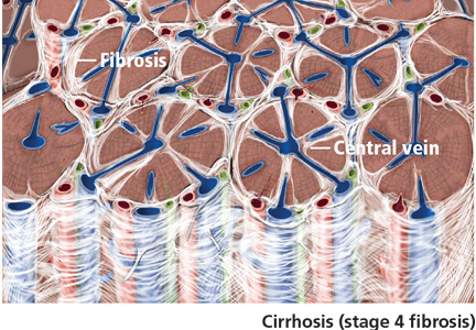
Painful red nodule on the right hand
A 46-year-old healthy man presents with a 15-day history of a tender subcutaneous nodule on the dorsum of the right hand that appeared after cleaning his aquarium. He has no fever or systemic symptoms. For 2 weeks he has been taking amoxicillin-clavulanate (Augmentin) and metronidazole (Flagyl), but without an adequate response.
Q: Which is the most likely diagnosis?
- Tuberculosis verrucosa cutis
- Sporotrichosis
- Nontuberculous mycobacterial infection
- Leishmaniasis
- Nocardiosis
A: The correct answer is nontuberculous mycobacterial infection. Mycobacterium marinum was subsequently isolated from culture (Löwenstein-Jensen medium) of wound drainage.
Tuberculosis verrucosa cutis is an indolent, warty plaque that occurs after direct inoculation of M tuberculosis into the skin of someone previously infected with M tuberculosis. It can result from accidental exposure to tuberculous tissue in high-risk groups such as laboratory workers, physicians, and pathologists.
Sporothrix schenckii is the dimorphic fungus that causes sporotrichosis. Activities associated with acquisition of sporotrichosis include gardening (rose-gardener's disease), landscaping, farming, berry-picking, horticulture, and carpentry. The characteristic infection involves suppurating subcutaneous nodules that progress proximally along lymphatic channels (lymphocutaneous sporotrichosis).
Leishmaniasis is a disease caused by the protozoa of the Leishmania species, which is transmitted by the bite of a female sandfly. Initially, the lesion is a small, red papule up to 2 cm in diameter. Over several weeks, the papule becomes darker, develops a crust in the center, and eventually ulcerates to present a typical appearance of an ulcer with raised edges and surrounding dusky red skin.
Nocardia is a genus of filamentous grampositive bacteria that stains acid-fast, just as M marinum does. Nocardia asteroides primarily affects the lungs and can disseminate systemically. However, Nocardia brasiliensis is often associated with sporotrichoid-spreading subcutaneous nodules.
CLINICAL PRESENTATION AND DIAGNOSIS
M marinum is a genus of nontuberculous mycobacteria that usually causes disease in fish but can cause human skin infection by penetrating through a break in the skin. It can spread to deeper structures, resulting in tenosynovitis, arthritis, or osteomyelitis.1 The disease caused by M marinum is sometimes called “swimming pool granuloma” or “fish-tank granuloma.”2
In this case, the patient probably acquired the infection while cleaning his home aquarium,3 so asking about contact with pet fish is very important for the clinical diagnosis.
Lesions are usually localized to the site of the inoculation, with a predilection for areas predisposed to trauma, such as the hands and the fingers, after an incubation period of 2 to 8 weeks. Lesions are self-limited and usually appear as pruriginous bluish-red papules or pustules that may increase in size to form a verrucous or violaceous plaque or nodule. Superficial lesions often undergo central ulceration. Disseminated infection is rare and occurs mainly in patients who are immunocompromised because of renal transplant, systemic lupus erythematosus, chronic steroid therapy, or anti-tumor necrosis factor alpha therapy.4 In such cases, infection can be life-threatening.
On histopathologic study, a nonspecific inflammation consisting of a mixed infiltrate with lymphocytes, neutrophils, and histiocytes is usually observed. Sometimes a granulomatous inflammatory infiltrate mimicking tuberculoid granuloma may be noted. M marinum is an aerobic, nonmotile acid-fast bacillus. It grows at 30°C to 32°C on Löwenstein-Jensen medium in 2 to 5 weeks, but cultures are rarely positive. Polymerase chain reaction and enzyme-linked immunosorbent assays can help identify the organism. Other diagnoses to be considered are leishmaniasis, tuberculosis verrucosa cutis, blastomycosis, histoplasmosis, and sporotrichosis.
TREATMENT
Treatment of M marinum infection is not based on any specific criteria. Spontaneous resolution in 24 to 36 months has been described. 5 Antibiotics are the first-line therapy, and sometimes surgical treatment may be necessary for deeper infection with necrotic tissue. Cryotherapy, electrode therapy, and irradiation have been reported to be effective.
Few studies have been conducted to determine the first-line antibiotic treatment for M marinum infection. In this case, treatment with sulfamethoxazole-trimethoprim6 (Bactrim) resulted in resolution of the lesions in 6 months. Other antibiotics often used as monotherapy are tetracycline, minocycline (Minocin), and doxycycline (Vibramycin) 100 mg twice daily for 3 months.7 However, treatment failure has been described with many antibiotics.
Multidrug therapy is usually prescribed to minimize the risk of resistance. A combination of clarithromycin (Biaxin), streptomycin, and ethionamide (Trecator) has been prescribed successfully.3 Another combined regimen—clarithromycin 500 mg twice a day, rifampin (Rifadin) 600 mg daily, and ethambutol (Myambutol) 25 mg/kg daily—has been shown to be effective.5 Also, rifampin 600 mg daily and ethambutol 15 to 25 mg/kg/day were effective in a patient with a sporotrichoid M marinum infection associated with infliximab (Remicade). 8 The duration of treatment ranged between 2 and 6 months, or up to 2 months after the disappearance of the cutaneous lesions.
Clinical awareness of M marinum infection is important so that the diagnosis can be made and appropriate therapy can be initiated promptly, because some cases with disseminated disease and serious complications have been described.9
- Lam A, Toma W, Schlesinger N. Mycobacterium marinum arthritis mimicking rheumatoid arthritis. J Rheumatol 2006; 33:817–819.
- Lewis FM, Marsh BJ, von Reyn CF. Fish tank exposure and cutaneous infections due to Mycobacterium marinum: tuberculin skin testing, treatment, and prevention. Clin Infect Dis 2003; 37:390–397.
- Dodiuk-Gad R, Dyachenko P, Ziv M, et al. Nontuberculous mycobacterial infections of the skin: a retrospective study of 25 cases. J Am Acad Dermatol 2007; 57:413–420.
- Streit M, Böhlen LM, Hunziker T, et al. Disseminated Mycobacterium marinum infection with extensive cutaneous eruption and bacteremia in an immunocompromised patient. Eur J Dermatol 2006; 16:79–83.
- Jogi R, Tyring SK. Therapy of nontuberculous mycobacterial infections. Dermatol Ther 2004; 17:491–498.
- Alinovi A, Vecchini F, Bassissi P. Sporothricoid mycobacterial infection. A case report. Acta Derm Venereol 1993; 73:146–147.
- Mahaisavariya P, Chaiprasert A, Khemngern S, et al. Nontuberculous mycobacterial skin infections: clinical and bacteriological studies. J Med Assoc Thai 2003; 86:52–60.
- Rallis E, Koumantaki-Mathioudaki E, Frangoulis E, Chatziolou E, Katsambas A. Severe sporotrichoid fish tank granuloma following infliximab therapy. Am J Clin Dermatol 2007; 8:385–388.
- Imakado S, Kojima Y, Hiyoshi T, Morimoto S. Disseminated Mycobacterium marinum infection in a patient with diabetic nephropathy. Diabetes Res Clin Pract 2009; 83:e35–e36.
A 46-year-old healthy man presents with a 15-day history of a tender subcutaneous nodule on the dorsum of the right hand that appeared after cleaning his aquarium. He has no fever or systemic symptoms. For 2 weeks he has been taking amoxicillin-clavulanate (Augmentin) and metronidazole (Flagyl), but without an adequate response.
Q: Which is the most likely diagnosis?
- Tuberculosis verrucosa cutis
- Sporotrichosis
- Nontuberculous mycobacterial infection
- Leishmaniasis
- Nocardiosis
A: The correct answer is nontuberculous mycobacterial infection. Mycobacterium marinum was subsequently isolated from culture (Löwenstein-Jensen medium) of wound drainage.
Tuberculosis verrucosa cutis is an indolent, warty plaque that occurs after direct inoculation of M tuberculosis into the skin of someone previously infected with M tuberculosis. It can result from accidental exposure to tuberculous tissue in high-risk groups such as laboratory workers, physicians, and pathologists.
Sporothrix schenckii is the dimorphic fungus that causes sporotrichosis. Activities associated with acquisition of sporotrichosis include gardening (rose-gardener's disease), landscaping, farming, berry-picking, horticulture, and carpentry. The characteristic infection involves suppurating subcutaneous nodules that progress proximally along lymphatic channels (lymphocutaneous sporotrichosis).
Leishmaniasis is a disease caused by the protozoa of the Leishmania species, which is transmitted by the bite of a female sandfly. Initially, the lesion is a small, red papule up to 2 cm in diameter. Over several weeks, the papule becomes darker, develops a crust in the center, and eventually ulcerates to present a typical appearance of an ulcer with raised edges and surrounding dusky red skin.
Nocardia is a genus of filamentous grampositive bacteria that stains acid-fast, just as M marinum does. Nocardia asteroides primarily affects the lungs and can disseminate systemically. However, Nocardia brasiliensis is often associated with sporotrichoid-spreading subcutaneous nodules.
CLINICAL PRESENTATION AND DIAGNOSIS
M marinum is a genus of nontuberculous mycobacteria that usually causes disease in fish but can cause human skin infection by penetrating through a break in the skin. It can spread to deeper structures, resulting in tenosynovitis, arthritis, or osteomyelitis.1 The disease caused by M marinum is sometimes called “swimming pool granuloma” or “fish-tank granuloma.”2
In this case, the patient probably acquired the infection while cleaning his home aquarium,3 so asking about contact with pet fish is very important for the clinical diagnosis.
Lesions are usually localized to the site of the inoculation, with a predilection for areas predisposed to trauma, such as the hands and the fingers, after an incubation period of 2 to 8 weeks. Lesions are self-limited and usually appear as pruriginous bluish-red papules or pustules that may increase in size to form a verrucous or violaceous plaque or nodule. Superficial lesions often undergo central ulceration. Disseminated infection is rare and occurs mainly in patients who are immunocompromised because of renal transplant, systemic lupus erythematosus, chronic steroid therapy, or anti-tumor necrosis factor alpha therapy.4 In such cases, infection can be life-threatening.
On histopathologic study, a nonspecific inflammation consisting of a mixed infiltrate with lymphocytes, neutrophils, and histiocytes is usually observed. Sometimes a granulomatous inflammatory infiltrate mimicking tuberculoid granuloma may be noted. M marinum is an aerobic, nonmotile acid-fast bacillus. It grows at 30°C to 32°C on Löwenstein-Jensen medium in 2 to 5 weeks, but cultures are rarely positive. Polymerase chain reaction and enzyme-linked immunosorbent assays can help identify the organism. Other diagnoses to be considered are leishmaniasis, tuberculosis verrucosa cutis, blastomycosis, histoplasmosis, and sporotrichosis.
TREATMENT
Treatment of M marinum infection is not based on any specific criteria. Spontaneous resolution in 24 to 36 months has been described. 5 Antibiotics are the first-line therapy, and sometimes surgical treatment may be necessary for deeper infection with necrotic tissue. Cryotherapy, electrode therapy, and irradiation have been reported to be effective.
Few studies have been conducted to determine the first-line antibiotic treatment for M marinum infection. In this case, treatment with sulfamethoxazole-trimethoprim6 (Bactrim) resulted in resolution of the lesions in 6 months. Other antibiotics often used as monotherapy are tetracycline, minocycline (Minocin), and doxycycline (Vibramycin) 100 mg twice daily for 3 months.7 However, treatment failure has been described with many antibiotics.
Multidrug therapy is usually prescribed to minimize the risk of resistance. A combination of clarithromycin (Biaxin), streptomycin, and ethionamide (Trecator) has been prescribed successfully.3 Another combined regimen—clarithromycin 500 mg twice a day, rifampin (Rifadin) 600 mg daily, and ethambutol (Myambutol) 25 mg/kg daily—has been shown to be effective.5 Also, rifampin 600 mg daily and ethambutol 15 to 25 mg/kg/day were effective in a patient with a sporotrichoid M marinum infection associated with infliximab (Remicade). 8 The duration of treatment ranged between 2 and 6 months, or up to 2 months after the disappearance of the cutaneous lesions.
Clinical awareness of M marinum infection is important so that the diagnosis can be made and appropriate therapy can be initiated promptly, because some cases with disseminated disease and serious complications have been described.9
- Lam A, Toma W, Schlesinger N. Mycobacterium marinum arthritis mimicking rheumatoid arthritis. J Rheumatol 2006; 33:817–819.
- Lewis FM, Marsh BJ, von Reyn CF. Fish tank exposure and cutaneous infections due to Mycobacterium marinum: tuberculin skin testing, treatment, and prevention. Clin Infect Dis 2003; 37:390–397.
- Dodiuk-Gad R, Dyachenko P, Ziv M, et al. Nontuberculous mycobacterial infections of the skin: a retrospective study of 25 cases. J Am Acad Dermatol 2007; 57:413–420.
- Streit M, Böhlen LM, Hunziker T, et al. Disseminated Mycobacterium marinum infection with extensive cutaneous eruption and bacteremia in an immunocompromised patient. Eur J Dermatol 2006; 16:79–83.
- Jogi R, Tyring SK. Therapy of nontuberculous mycobacterial infections. Dermatol Ther 2004; 17:491–498.
- Alinovi A, Vecchini F, Bassissi P. Sporothricoid mycobacterial infection. A case report. Acta Derm Venereol 1993; 73:146–147.
- Mahaisavariya P, Chaiprasert A, Khemngern S, et al. Nontuberculous mycobacterial skin infections: clinical and bacteriological studies. J Med Assoc Thai 2003; 86:52–60.
- Rallis E, Koumantaki-Mathioudaki E, Frangoulis E, Chatziolou E, Katsambas A. Severe sporotrichoid fish tank granuloma following infliximab therapy. Am J Clin Dermatol 2007; 8:385–388.
- Imakado S, Kojima Y, Hiyoshi T, Morimoto S. Disseminated Mycobacterium marinum infection in a patient with diabetic nephropathy. Diabetes Res Clin Pract 2009; 83:e35–e36.

Do incretin drugs for type 2 diabetes increase the risk of acute pancreatitis?
Probably not. Although cases of acute pancreatitis have occurred in patients taking these drugs, cases have been reported in patients taking other drugs as well. Furthermore, the incidence of acute pancreatitis is higher in patients with type 2 diabetes (for which incretin-type drugs are indicated) than in the general population, regardless of treatment.
INCRETINS, A NEW CLASS OF DRUGS FOR TYPE 2 DIABETES
Incretins are hormones secreted by the small intestine in response to glucose in food. Glucagon-like peptide 1 (GLP-1) is an endogenous incretin that stimulates insulin secretion, suppresses glucagon secretion, and delays gastric emptying.
Current incretin-based therapies for type 2 diabetes include two types of agents. First are drugs that mimic the action of native GLP-1, such as the injectable GLP-1 analogues exenatide (Byetta) and liraglutide (Victoza). Second are agents that interfere with the metabolism of native GLP-1, mainly by inhibiting the endogenous enzyme dipeptidyl peptidase 4 (DPP-4), thus extending the life of native GLP-1. Two DPP-4 inhibitors pertinent to this discussion are saxagliptin (Onglyza) and sitagliptin (Januvia), both of which are taken orally.
The question has been raised whether incretin-based therapy causes pancreatitis. The package inserts for exenatide and sitagliptin have been updated to reflect this possibility, thus causing concern to practitioners. Is this concern warranted?
MANY DRUGS ARE ASSOCIATED WITH ACUTE PANCREATIS
In a review published in 2005, Trivedi and Pitchumoni1 reported that, of the top 100 prescribed drugs in the United States, 44 had been associated with acute pancreatitis. These agents included over-the-counter drugs such as acetaminophen (Tylenol), common antibiotics such as trimethoprim-sulfamethoxazole (Bactrim) and erythromycin, and drugs used to treat acquired immunodeficiency syndrome and cancer. No clear pathophysiologic basis connects these agents.
In 2002, Blomgren et al2 suggested that glyburide (Micronase) use might be a risk factor for acute pancreatitis, and that the risk of pancreatitis is higher if the body mass index is 30 kg/m2 or more. In 2008, more concern was raised with a report of hemorrhagic or necrotizing pancreatitis in six patients taking exenatide, two of whom died.3 And more recently, reports of 88 cases of acute pancreatitis (including 2 cases of hemorrhagic or necrotizing pancreatitis) from October 2006 to February 2009 in patients taking sitagliptin or the sitagliptin-metformin combination Janumet4 prompted a revision of the package inserts.
Do these cases represent unexpected toxicities not appreciated in premarket clinical trials, or are they to be expected in the population treated with these agents as greater numbers are exposed?
TYPE 2 DIABETES ALSO POSES A RISK OF PANCREATITIS
A number of comorbidities associated with type 2 diabetes predispose to pancreatitis, particularly hypertriglyceridemia and gallbladder disease.5–7 People with diabetes can also be exposed to alcohol or other drugs reported to be associated with pancreatitis.
What is the risk of pancreatitis in patients with type 2 diabetes? Is there evidence of a greater risk when incretin-based drugs are used to control hyperglycemia rather than other agents?
Pancreatitis appears to be increasingly prevalent in the general population in western countries. Some 60% to 80% of cases are attributed to alcohol or gallstones, but 20% do not have a clear cause.
In 2009, a new cause of acute pancreatitis was introduced when Frulloni et al8 reported that a novel antibody that recognizes epitopes shared with Helicobacter pylori was associated with autoimmune pancreatitis. H pylori is a common gastrointestinal organism, found in diabetic and nondiabetic patients, and it may well account for what has up to now been considered idiopathic pancreatitis.
Type 2 diabetes is associated with obesity and hyperlipidemia, each of which has been considered a putative risk factor for pancreatits.5–7
Noel et al9 examined the risk of pancreatitis in patients with type 2 diabetes in a large insurance database (29,332,477 covered lives). They identified people with type 2 diabetes and those without diabetes eligible for coverage by the plan, using medical and pharmacy claims from January 1, 1999, to December 31, 2005. The authors also used medical claims to identify episodes of acute pancreatitis and gallbladder disease. They found that the risk of acute pancreatitis was 2.8 times higher in the overall diabetic cohort than in the nondiabetic cohort, and five times higher in the youngest diabetic cohort (ages 18 to 44) than in nondiabetic people of the same age. The risk was three times higher in diabetic men than in nondiabetic men, and 2.6 times higher in diabetic women than in nondiabetic women.
The time period examined in this study is fortuitous, since exenatide was approved in June 2005 and had very little market penetration during its first 6 months, corresponding to the last 6 months of the study period. Sitagliptin, the first DPP-4 inhibitor, had not yet reached the market.
Noel et al9 also found that the risk of biliary disease in patients with diabetes was 1.9 times higher than in those without diabetes. The relative risk of gallbladder disease was proportionally greater in a younger population with diabetes than in the population without diabetes, in whom the risk of gallbladder disease increases with age. Cholelithiasis was believed to be the underlying cause in at least 50% of the cases of pancreatitis.
PANCREATITIS AND INCRETIN-BASED THERAPIES
The estimated risk of acute pancreatitis in the population at large is reported as 0.33 to 0.44 events per 1,000 adults per year10; 15% to 20% of cases are considered severe, and 2% to 4% result in death.5,10 A relatively small number (1%–2%) are believed to be drug-induced.10
Exenatide. In the exenatide development program, six cases of acute pancreatitis were observed in about 3,489 subject-years of exposure (1.7 per 1,000 subject-years), compared with one case in about 336 subject-years with placebo (3.0 per 1,000 subject-years) and one case in about 497 subject-years (2.0 per 1,000 subject-years) with insulin.11
Sitagliptin. Dore et al12 examined claims from another database for the period of June 2005 through June 2008 to look specifically at the risk with incretin-based therapies. This database included 27,996 people starting exenatide and 16,276 people starting sitagliptin, matched with people with type 2 diabetes taking metformin (Glucophage) or glyburide. Over a period of 1 year, 0.13% of exenatide users and 0.12% of sitagliptin users suffered acute pancreatitis. The risk of pancreatitis was comparable in each group:
- For exenatide, relative risk (RR) 1.0, 95% confidence interval (CI) 0.6 to 1.7, compared with metformin or glyburide
- For sitagliptin, RR 1.0, 95% CI 0.5 to 2.0.
Saxagliptin. In clinical trials of saxagliptin, the incidence of pancreatitis was 0.2% in 3,422 patients receiving saxagliptin and 0.2% in 1,066 controls,13 similar to the rates for sitagliptin and exenatide.
Liraglutide appeared to be associated with a risk of acute pancreatitis, with seven cases in 3,900 patients receiving liraglutide vs one case in a patient taking another diabetes drug.14 This rate is similar to that reported in exenatide clinical trials, suggesting that pancreatitis has been underreported in the comparator subjects. We need more experience to see if this agent really poses more risk than other antidiabetic therapies.
As new antidiabetic agents enter the market and their use becomes common, it would not be surprising to see rates of pancreatitis similar to those reported by Blomgren et al2 in 2002, when glyburide was becoming a mainstay of therapy for type 2 diabetes.
- Trivedi CD, Pitchumoni CS. Drug-induced pancreatitis: an update. J Clin Gastroenterol 2005; 39:709–716.
- Blomgren KB, Sundström A, Steineck G, Wiholm BE. Obesity and treatment of diabetes with glyburide may both be risk factors for acute pancreatitis. Diabetes Care 2002; 25:298–302.
- US Food and Drug Administration. Information for healthcare professionals: exenatide (marketed as Byetta)—8/2008 update. http://www.fda.gov/Drugs/DrugSafety/PostmarketDrugSafetyInformationforPatientsandProviders/ucm124713.htm. Accessed July 1, 2010.
- US Food and Drug Administration. Information for healthcare professionals—acute pancreatitis and sitagliptin (marketed as Januvia and Janumet). http://www.fda.gov/Drugs/DrugSafety/PostmarketDrugSafetyInformationforPatientsandProviders/DrugSafetyInformationforHeathcareProfessionals/ucm183764.htm. Accessed July 1, 2010.
- Forsmark CE, Baillie J; AGA Institute Clinical Practice and Economics Committee. AGA Institute technical review on acute pancreatitis. Gastroenterology 2007; 132:2022–2044.
- Pagliarulo M, Fornari F, Fraquelli M, et al. Gallstone disease and related risk factors in a large cohort of diabetic patients. Dig Liver Dis 2004; 36:130–134.
- Field AE, Coakley EH, Must A, et al. Impact of overweight on the risk of developing common chronic diseases during a 10-year period. Arch Intern Med 2001; 161:1581–1586.
- Frulloni L, Lunardi C, Simone R, et al. Identification of a novel antibody associated with autoimmune pancreatitis. N Engl J Med 2009; 361:2135–2142.
- Noel RA, Braun DK, Patterson RE, Bloomgren GL. Increased risk of acute pancreatitis and biliary disease observed in patients with type 2 diabetes: a retrospective cohort study. Diabetes Care 2009; 32:834–838.
- Whitcomb DC. Clinical practice. Acute pancreatitis. N Engl J Med 2006; 354:2142–2150.
- Data on file, Amylin Pharmaceuticals, Inc. and Eli Lilly.
- Dore DD, Seeger JD, Arnold Chan K. Use of a claims-based active drug safety surveillance system to assess the risk of acute pancreatitis with exenatide or sitagliptin compared to metformin or glyburide. Curr Med Res Opin 2009; 25:1019–1027.
- US Food and Drug Administration. Controlled Phase 2b/3 Pooled Population—Day 120 Update. http://www.fda.gov/downloads/AdvisoryCommittees/Committees-MeetingMaterials/Drugs/EndocrinologicandMetabolic-DrugsAdvisoryCommittee/UCM149589. Accessed July 4, 2010.
- US Food and Drug Administration. Questions and answers—safety requirements for Victoza (liraglutide). http://www.fda.gov/Drugs/DrugSafety/PostmarketDrug-SafetyInformationforPatientsandProviders/ucm198543.htm. Accessed July 4, 2010.
Probably not. Although cases of acute pancreatitis have occurred in patients taking these drugs, cases have been reported in patients taking other drugs as well. Furthermore, the incidence of acute pancreatitis is higher in patients with type 2 diabetes (for which incretin-type drugs are indicated) than in the general population, regardless of treatment.
INCRETINS, A NEW CLASS OF DRUGS FOR TYPE 2 DIABETES
Incretins are hormones secreted by the small intestine in response to glucose in food. Glucagon-like peptide 1 (GLP-1) is an endogenous incretin that stimulates insulin secretion, suppresses glucagon secretion, and delays gastric emptying.
Current incretin-based therapies for type 2 diabetes include two types of agents. First are drugs that mimic the action of native GLP-1, such as the injectable GLP-1 analogues exenatide (Byetta) and liraglutide (Victoza). Second are agents that interfere with the metabolism of native GLP-1, mainly by inhibiting the endogenous enzyme dipeptidyl peptidase 4 (DPP-4), thus extending the life of native GLP-1. Two DPP-4 inhibitors pertinent to this discussion are saxagliptin (Onglyza) and sitagliptin (Januvia), both of which are taken orally.
The question has been raised whether incretin-based therapy causes pancreatitis. The package inserts for exenatide and sitagliptin have been updated to reflect this possibility, thus causing concern to practitioners. Is this concern warranted?
MANY DRUGS ARE ASSOCIATED WITH ACUTE PANCREATIS
In a review published in 2005, Trivedi and Pitchumoni1 reported that, of the top 100 prescribed drugs in the United States, 44 had been associated with acute pancreatitis. These agents included over-the-counter drugs such as acetaminophen (Tylenol), common antibiotics such as trimethoprim-sulfamethoxazole (Bactrim) and erythromycin, and drugs used to treat acquired immunodeficiency syndrome and cancer. No clear pathophysiologic basis connects these agents.
In 2002, Blomgren et al2 suggested that glyburide (Micronase) use might be a risk factor for acute pancreatitis, and that the risk of pancreatitis is higher if the body mass index is 30 kg/m2 or more. In 2008, more concern was raised with a report of hemorrhagic or necrotizing pancreatitis in six patients taking exenatide, two of whom died.3 And more recently, reports of 88 cases of acute pancreatitis (including 2 cases of hemorrhagic or necrotizing pancreatitis) from October 2006 to February 2009 in patients taking sitagliptin or the sitagliptin-metformin combination Janumet4 prompted a revision of the package inserts.
Do these cases represent unexpected toxicities not appreciated in premarket clinical trials, or are they to be expected in the population treated with these agents as greater numbers are exposed?
TYPE 2 DIABETES ALSO POSES A RISK OF PANCREATITIS
A number of comorbidities associated with type 2 diabetes predispose to pancreatitis, particularly hypertriglyceridemia and gallbladder disease.5–7 People with diabetes can also be exposed to alcohol or other drugs reported to be associated with pancreatitis.
What is the risk of pancreatitis in patients with type 2 diabetes? Is there evidence of a greater risk when incretin-based drugs are used to control hyperglycemia rather than other agents?
Pancreatitis appears to be increasingly prevalent in the general population in western countries. Some 60% to 80% of cases are attributed to alcohol or gallstones, but 20% do not have a clear cause.
In 2009, a new cause of acute pancreatitis was introduced when Frulloni et al8 reported that a novel antibody that recognizes epitopes shared with Helicobacter pylori was associated with autoimmune pancreatitis. H pylori is a common gastrointestinal organism, found in diabetic and nondiabetic patients, and it may well account for what has up to now been considered idiopathic pancreatitis.
Type 2 diabetes is associated with obesity and hyperlipidemia, each of which has been considered a putative risk factor for pancreatits.5–7
Noel et al9 examined the risk of pancreatitis in patients with type 2 diabetes in a large insurance database (29,332,477 covered lives). They identified people with type 2 diabetes and those without diabetes eligible for coverage by the plan, using medical and pharmacy claims from January 1, 1999, to December 31, 2005. The authors also used medical claims to identify episodes of acute pancreatitis and gallbladder disease. They found that the risk of acute pancreatitis was 2.8 times higher in the overall diabetic cohort than in the nondiabetic cohort, and five times higher in the youngest diabetic cohort (ages 18 to 44) than in nondiabetic people of the same age. The risk was three times higher in diabetic men than in nondiabetic men, and 2.6 times higher in diabetic women than in nondiabetic women.
The time period examined in this study is fortuitous, since exenatide was approved in June 2005 and had very little market penetration during its first 6 months, corresponding to the last 6 months of the study period. Sitagliptin, the first DPP-4 inhibitor, had not yet reached the market.
Noel et al9 also found that the risk of biliary disease in patients with diabetes was 1.9 times higher than in those without diabetes. The relative risk of gallbladder disease was proportionally greater in a younger population with diabetes than in the population without diabetes, in whom the risk of gallbladder disease increases with age. Cholelithiasis was believed to be the underlying cause in at least 50% of the cases of pancreatitis.
PANCREATITIS AND INCRETIN-BASED THERAPIES
The estimated risk of acute pancreatitis in the population at large is reported as 0.33 to 0.44 events per 1,000 adults per year10; 15% to 20% of cases are considered severe, and 2% to 4% result in death.5,10 A relatively small number (1%–2%) are believed to be drug-induced.10
Exenatide. In the exenatide development program, six cases of acute pancreatitis were observed in about 3,489 subject-years of exposure (1.7 per 1,000 subject-years), compared with one case in about 336 subject-years with placebo (3.0 per 1,000 subject-years) and one case in about 497 subject-years (2.0 per 1,000 subject-years) with insulin.11
Sitagliptin. Dore et al12 examined claims from another database for the period of June 2005 through June 2008 to look specifically at the risk with incretin-based therapies. This database included 27,996 people starting exenatide and 16,276 people starting sitagliptin, matched with people with type 2 diabetes taking metformin (Glucophage) or glyburide. Over a period of 1 year, 0.13% of exenatide users and 0.12% of sitagliptin users suffered acute pancreatitis. The risk of pancreatitis was comparable in each group:
- For exenatide, relative risk (RR) 1.0, 95% confidence interval (CI) 0.6 to 1.7, compared with metformin or glyburide
- For sitagliptin, RR 1.0, 95% CI 0.5 to 2.0.
Saxagliptin. In clinical trials of saxagliptin, the incidence of pancreatitis was 0.2% in 3,422 patients receiving saxagliptin and 0.2% in 1,066 controls,13 similar to the rates for sitagliptin and exenatide.
Liraglutide appeared to be associated with a risk of acute pancreatitis, with seven cases in 3,900 patients receiving liraglutide vs one case in a patient taking another diabetes drug.14 This rate is similar to that reported in exenatide clinical trials, suggesting that pancreatitis has been underreported in the comparator subjects. We need more experience to see if this agent really poses more risk than other antidiabetic therapies.
As new antidiabetic agents enter the market and their use becomes common, it would not be surprising to see rates of pancreatitis similar to those reported by Blomgren et al2 in 2002, when glyburide was becoming a mainstay of therapy for type 2 diabetes.
- Trivedi CD, Pitchumoni CS. Drug-induced pancreatitis: an update. J Clin Gastroenterol 2005; 39:709–716.
- Blomgren KB, Sundström A, Steineck G, Wiholm BE. Obesity and treatment of diabetes with glyburide may both be risk factors for acute pancreatitis. Diabetes Care 2002; 25:298–302.
- US Food and Drug Administration. Information for healthcare professionals: exenatide (marketed as Byetta)—8/2008 update. http://www.fda.gov/Drugs/DrugSafety/PostmarketDrugSafetyInformationforPatientsandProviders/ucm124713.htm. Accessed July 1, 2010.
- US Food and Drug Administration. Information for healthcare professionals—acute pancreatitis and sitagliptin (marketed as Januvia and Janumet). http://www.fda.gov/Drugs/DrugSafety/PostmarketDrugSafetyInformationforPatientsandProviders/DrugSafetyInformationforHeathcareProfessionals/ucm183764.htm. Accessed July 1, 2010.
- Forsmark CE, Baillie J; AGA Institute Clinical Practice and Economics Committee. AGA Institute technical review on acute pancreatitis. Gastroenterology 2007; 132:2022–2044.
- Pagliarulo M, Fornari F, Fraquelli M, et al. Gallstone disease and related risk factors in a large cohort of diabetic patients. Dig Liver Dis 2004; 36:130–134.
- Field AE, Coakley EH, Must A, et al. Impact of overweight on the risk of developing common chronic diseases during a 10-year period. Arch Intern Med 2001; 161:1581–1586.
- Frulloni L, Lunardi C, Simone R, et al. Identification of a novel antibody associated with autoimmune pancreatitis. N Engl J Med 2009; 361:2135–2142.
- Noel RA, Braun DK, Patterson RE, Bloomgren GL. Increased risk of acute pancreatitis and biliary disease observed in patients with type 2 diabetes: a retrospective cohort study. Diabetes Care 2009; 32:834–838.
- Whitcomb DC. Clinical practice. Acute pancreatitis. N Engl J Med 2006; 354:2142–2150.
- Data on file, Amylin Pharmaceuticals, Inc. and Eli Lilly.
- Dore DD, Seeger JD, Arnold Chan K. Use of a claims-based active drug safety surveillance system to assess the risk of acute pancreatitis with exenatide or sitagliptin compared to metformin or glyburide. Curr Med Res Opin 2009; 25:1019–1027.
- US Food and Drug Administration. Controlled Phase 2b/3 Pooled Population—Day 120 Update. http://www.fda.gov/downloads/AdvisoryCommittees/Committees-MeetingMaterials/Drugs/EndocrinologicandMetabolic-DrugsAdvisoryCommittee/UCM149589. Accessed July 4, 2010.
- US Food and Drug Administration. Questions and answers—safety requirements for Victoza (liraglutide). http://www.fda.gov/Drugs/DrugSafety/PostmarketDrug-SafetyInformationforPatientsandProviders/ucm198543.htm. Accessed July 4, 2010.
Can patients with COPD or asthma take a beta-blocker?
Yes. Treatment with beta-adrenergic receptor blockers decreases the mortality rate in patients with coronary artery disease or heart failure, as well as during the perioperative period in selected patients (eg, those with a history of myocardial infarction, a positive stress test, or current chest pain due to myocardial ischemia). The current evidence supports giving beta-blockers to patients with coronary artery disease and chronic obstructive pulmonary disease (COPD) or asthma, which lowers the 1-year mortality rate to a degree similar to that in patients without COPD or asthma, and without worsening respiratory function.1 However, many clinicians still hesitate to start patients with COPD or asthma on a beta-blocker due to the fear of bronchoconstriction.2
THE RISKS
In patients with reversible airway disease, beta-blockers may increase airway reactivity and bronchospasm, as well as decrease the response to inhaled or oral beta-receptor agonists.3 Even topical ophthalmic nonselective beta-blockers for glaucoma can cause a worsening of pulmonary function.4 However, these data are from small trials in the 1970s and 1980s.
On the other hand, not giving beta-blockers can pose a risk of death. In a retrospective study of more than 200,000 patients with myocardial infarction, Gottlieb et al5 found that beta-blockers were associated with a 40% reduction in mortality rates in patients with conditions often considered a contraindication to beta-blocker therapy, such as congestive heart failure, pulmonary disease, and older age.5
CARDIOSELECTIVE BETA-BLOCKERS
Cardioselective beta-blockers with an affinity for the beta-1 receptor theoretically result in fewer adverse effects on the lungs. They competitively block the response to beta-adrenergic stimulation and selectively block beta-1 receptors with little or no effect on beta-2 receptors, except perhaps at high doses. However, this possible high-dose effect requires further study.
The effect of cardioselective beta-blockers on respiratory function was evaluated in two meta-analyses,6,7 one in patients with mild to moderate reactive airway disease, the other in patients with mild to severe COPD. Patients with reactive airway disease who received a single dose of a beta-blocker had a 7.46% reduction in forced expiratory volume in the first second of expiration (FEV1), an effect that was completely reversed by treatment with a beta-agonist inhaler. The FEV1 increased by a statistically significantly greater amount in response to beta-agonists in patients who received beta-blockers (a single dose or continuous therapy) than in those who did not receive beta-blockers. Patients who received continuous cardioselective beta-blockers experienced no significant drop in FEV1, and no new symptoms developed. These results led the authors to conclude that cardioselective beta-blockers do not cause a significant reduction in pulmonary function in patients with mild to moderate reactive airway disease and COPD and are therefore safe to use. A single dose of a cardioselective beta-blocker may produce a small decrease in FEV1, especially in patients with reactive airway disease, but as therapy is continued over days to weeks, there is no significant change in symptoms or FEV1 and no increase in the need for beta-agonist inhalers.
A major limitation of the two meta-analyses was that the patients were younger than most patients who require beta-blockers: the average age was 40 in patients with reactive airway disease, and 54 in patients with COPD. Also important to consider is that only patients with mild to moderate reactive airway disease were included. Patients with severe asthma, especially those with active bronchospasm, may react differently to even cardioselective beta-blockers.
NONSELECTIVE BETA-BLOCKERS
Recent studies suggest that nonselective beta-blockers can affect respiratory function in patients with COPD, but they have failed to show any harm. For example, propranolol (Inderal) was shown to worsen pulmonary function and to decrease the sensitivity of the airway to the effects of long-acting beta-2-agonists, but the 15 patients included in this study had no increase in respiratory symptoms.8
It has also been suggested that combined nonselective beta- and alpha-receptor blockade—eg, with labetalol (Trandate) or carvedilol (Coreg)—might be better tolerated than nonselective beta-blockers in patients with COPD.9 However, from limited data, Kotlyar et al10 suggested that carvedilol may be less well tolerated in patients with asthma than with COPD. All current evidence on combined nonselective beta-and alpha-blockade is observational, and it is not yet clear whether this class of beta-blockers is better tolerated due to alpha-blockade or merely because nonselective beta-blockers themselves are well tolerated.
OUR RECOMMENDATIONS
- Chen J, Radford MJ, Wang Y, Marciniak TA, Krumholz HM. Effectiveness of beta-blocker therapy after acute myocardial infarction in elderly patients with chronic obstructive pulmonary disease or asthma. J Am Coll Cardiol 2001; 37:1950–1956.
- The sixth report of the Joint National Committee on prevention, detection, evaluation, and treatment of high blood pressure. Arch Intern Med 1997; 157:2413–2446.
- Benson MK, Berrill WT, Cruickshank JM, Sterling GS. A comparison of four beta-adrenoceptor antagonists in patients with asthma. Br J Clin Pharmacol 1978; 5:415–419.
- Fraunfelder FT, Barker AF. Respiratory effects of timolol. N Engl J Med 1984; 311:1441.
- Gottlieb SS, McCarter RJ, Vogel RA. Effect of beta-blockade on mortality among high-risk and low-risk patients after myocardial infarction. N Engl J Med 1998; 339:489–497.
- Salpeter SR, Ormiston TM, Salpeter EE, Poole PJ, Cates CJ. Cardioselective beta-blockers for chronic obstructive pulmonary disease: a meta-analysis. Respir Med 2003; 97:1094–1101.
- Salpeter SR, Ormiston TM, Salpeter EE. Cardioselective beta-blockers in patients with reactive airway disease: a meta-analysis. Ann Intern Med 2002; 137:715–725.
- van der Woude HJ, Zaagsma J, Postma DS, Winter TH, van Hulst M, Aalbers R. Detrimental effects of beta-blockers in COPD: a concern for nonselective beta-blockers. Chest 2005; 127:818–824.
- Sirak TE, Jelic S, Le Jemtel TH. Therapeutic update: non-selective beta- and alpha-adrenergic blockade in patients with coexistent chronic obstructive pulmonary disease and chronic heart failure. J Am Coll Cardiol 2004; 44:497–502.
- Kotlyar E, Keogh AM, Macdonald PS, Arnold RH, McCaffrey DJ, Glanville AR. Tolerability of carvedilol in patients with heart failure and concomitant chronic obstructive pulmonary disease or asthma. J Heart Lung Transplant 2002; 21:1290–1295.
Yes. Treatment with beta-adrenergic receptor blockers decreases the mortality rate in patients with coronary artery disease or heart failure, as well as during the perioperative period in selected patients (eg, those with a history of myocardial infarction, a positive stress test, or current chest pain due to myocardial ischemia). The current evidence supports giving beta-blockers to patients with coronary artery disease and chronic obstructive pulmonary disease (COPD) or asthma, which lowers the 1-year mortality rate to a degree similar to that in patients without COPD or asthma, and without worsening respiratory function.1 However, many clinicians still hesitate to start patients with COPD or asthma on a beta-blocker due to the fear of bronchoconstriction.2
THE RISKS
In patients with reversible airway disease, beta-blockers may increase airway reactivity and bronchospasm, as well as decrease the response to inhaled or oral beta-receptor agonists.3 Even topical ophthalmic nonselective beta-blockers for glaucoma can cause a worsening of pulmonary function.4 However, these data are from small trials in the 1970s and 1980s.
On the other hand, not giving beta-blockers can pose a risk of death. In a retrospective study of more than 200,000 patients with myocardial infarction, Gottlieb et al5 found that beta-blockers were associated with a 40% reduction in mortality rates in patients with conditions often considered a contraindication to beta-blocker therapy, such as congestive heart failure, pulmonary disease, and older age.5
CARDIOSELECTIVE BETA-BLOCKERS
Cardioselective beta-blockers with an affinity for the beta-1 receptor theoretically result in fewer adverse effects on the lungs. They competitively block the response to beta-adrenergic stimulation and selectively block beta-1 receptors with little or no effect on beta-2 receptors, except perhaps at high doses. However, this possible high-dose effect requires further study.
The effect of cardioselective beta-blockers on respiratory function was evaluated in two meta-analyses,6,7 one in patients with mild to moderate reactive airway disease, the other in patients with mild to severe COPD. Patients with reactive airway disease who received a single dose of a beta-blocker had a 7.46% reduction in forced expiratory volume in the first second of expiration (FEV1), an effect that was completely reversed by treatment with a beta-agonist inhaler. The FEV1 increased by a statistically significantly greater amount in response to beta-agonists in patients who received beta-blockers (a single dose or continuous therapy) than in those who did not receive beta-blockers. Patients who received continuous cardioselective beta-blockers experienced no significant drop in FEV1, and no new symptoms developed. These results led the authors to conclude that cardioselective beta-blockers do not cause a significant reduction in pulmonary function in patients with mild to moderate reactive airway disease and COPD and are therefore safe to use. A single dose of a cardioselective beta-blocker may produce a small decrease in FEV1, especially in patients with reactive airway disease, but as therapy is continued over days to weeks, there is no significant change in symptoms or FEV1 and no increase in the need for beta-agonist inhalers.
A major limitation of the two meta-analyses was that the patients were younger than most patients who require beta-blockers: the average age was 40 in patients with reactive airway disease, and 54 in patients with COPD. Also important to consider is that only patients with mild to moderate reactive airway disease were included. Patients with severe asthma, especially those with active bronchospasm, may react differently to even cardioselective beta-blockers.
NONSELECTIVE BETA-BLOCKERS
Recent studies suggest that nonselective beta-blockers can affect respiratory function in patients with COPD, but they have failed to show any harm. For example, propranolol (Inderal) was shown to worsen pulmonary function and to decrease the sensitivity of the airway to the effects of long-acting beta-2-agonists, but the 15 patients included in this study had no increase in respiratory symptoms.8
It has also been suggested that combined nonselective beta- and alpha-receptor blockade—eg, with labetalol (Trandate) or carvedilol (Coreg)—might be better tolerated than nonselective beta-blockers in patients with COPD.9 However, from limited data, Kotlyar et al10 suggested that carvedilol may be less well tolerated in patients with asthma than with COPD. All current evidence on combined nonselective beta-and alpha-blockade is observational, and it is not yet clear whether this class of beta-blockers is better tolerated due to alpha-blockade or merely because nonselective beta-blockers themselves are well tolerated.
OUR RECOMMENDATIONS
- Chen J, Radford MJ, Wang Y, Marciniak TA, Krumholz HM. Effectiveness of beta-blocker therapy after acute myocardial infarction in elderly patients with chronic obstructive pulmonary disease or asthma. J Am Coll Cardiol 2001; 37:1950–1956.
- The sixth report of the Joint National Committee on prevention, detection, evaluation, and treatment of high blood pressure. Arch Intern Med 1997; 157:2413–2446.
- Benson MK, Berrill WT, Cruickshank JM, Sterling GS. A comparison of four beta-adrenoceptor antagonists in patients with asthma. Br J Clin Pharmacol 1978; 5:415–419.
- Fraunfelder FT, Barker AF. Respiratory effects of timolol. N Engl J Med 1984; 311:1441.
- Gottlieb SS, McCarter RJ, Vogel RA. Effect of beta-blockade on mortality among high-risk and low-risk patients after myocardial infarction. N Engl J Med 1998; 339:489–497.
- Salpeter SR, Ormiston TM, Salpeter EE, Poole PJ, Cates CJ. Cardioselective beta-blockers for chronic obstructive pulmonary disease: a meta-analysis. Respir Med 2003; 97:1094–1101.
- Salpeter SR, Ormiston TM, Salpeter EE. Cardioselective beta-blockers in patients with reactive airway disease: a meta-analysis. Ann Intern Med 2002; 137:715–725.
- van der Woude HJ, Zaagsma J, Postma DS, Winter TH, van Hulst M, Aalbers R. Detrimental effects of beta-blockers in COPD: a concern for nonselective beta-blockers. Chest 2005; 127:818–824.
- Sirak TE, Jelic S, Le Jemtel TH. Therapeutic update: non-selective beta- and alpha-adrenergic blockade in patients with coexistent chronic obstructive pulmonary disease and chronic heart failure. J Am Coll Cardiol 2004; 44:497–502.
- Kotlyar E, Keogh AM, Macdonald PS, Arnold RH, McCaffrey DJ, Glanville AR. Tolerability of carvedilol in patients with heart failure and concomitant chronic obstructive pulmonary disease or asthma. J Heart Lung Transplant 2002; 21:1290–1295.
HIV: Just another chronic disease

In subsequent years we learned about HIV—the retrovirus, and the immune system that it cleverly and efficiently disables. For the most part, we matured professionally and moved past the social stigmas of the disease, although that was painful. We developed systems to keep acutely ill patients out of the hospital while providing them with “long-term” (weeks or months of) intravenous antibiotics and humane palliative care.
We learned about AZT and argued about when to use it. But mainly, we watched many, many young men (and some women) die in corner hospital rooms. For me, from the ′80s, there remain heartrending personal images, notes, and cassette tapes voicing thanks for my concern and time spent, but no notes of thanks like those I’ve received from my patients with chronic rheumatoid arthritis who, after years of care, are able to hold their nieces or grandchildren.
A few long-term survivors have raised the hope that immune systems could recover and exist in symbiosis with the virus, and that maybe a drug cocktail or vaccine could provide a cure or remission. Magic Johnson, known to be infected since at least 1991, is likely the most public example of a long-term survivor on highly active antiviral therapy—a hope in the flesh.
But did we ever expect a time when HIV would be viewed as a chronic disease, with patients warranting screening for coronary artery disease in order to decrease long-term coronary complications? Did we ever expect a time that we would be offering organ transplants to HIV-infected patients?
In this issue of the Journal, Drs. Malvestutto and Aberg discuss coronary issues that need to be recognized and managed in HIV-infected patients. This further complicates the management of these patients, and draws cardiologists and primary care providers back into management plans.
I can’t think of a management “complication” of a chronic illness that is more welcome—or more surprising.
Coronary heart disease in people infected with HIV
Widespread use of antiretroviral therapy has caused a remarkable decline in rates of morbidity and death related to acquired immunodeficiency syndrome (AIDS) and has effectively made human immunodeficiency virus (HIV) infection a manageable—although not yet curable— chronic condition. And as the HIV-infected population on antiretroviral therapy ages, the prevalence of chronic conditions (eg, cardiovascular disease, hepatic disease, pulmonary disease, non-AIDS cancers) and deaths attributable to these conditions have also increased.1
Many of the traditional risk factors for cardiovascular disease in the general population, including smoking, dyslipidemia, and diabetes, are common in HIV-infected patients, and HIV infection itself independently increases the risk of coronary heart disease. In addition, different antiretroviral combinations can contribute, in varying degrees, to changes in lipid levels and insulin resistance, further increasing coronary risk.
Ultimately, however, the immunologic benefits of antiretroviral therapy for individual patients far exceed the modest increase in cardiovascular risk associated with certain regimens. In most cases, careful selection of the initial antiretroviral regimen and the addition of lipid-lowering or glucose-controlling medications (with close attention to drug interactions) can effectively manage the metabolic changes associated with antiretroviral therapy and obviate any premature modification of virologically suppressive regimens.
TRADITIONAL CARDIAC RISK FACTORS IN HIV PATIENTS
The risk of coronary heart disease in HIV patients is influenced mostly by traditional factors such as age, smoking, diabetes, and dyslipidemia, including high levels of total cholesterol and low-density lipoprotein cholesterol (LDL-C) and low levels of high-density lipoprotein cholesterol (HDL-C).2
In various large cohorts, HIV-infected men had a higher prevalence of smoking,3 a lower mean HDL-C level,3,4 and a higher mean triglyceride level3,4 than men without HIV infection, placing them at greater risk of coronary heart disease. However, even after adjusting for traditional risk factors, rates of atherosclerosis are still higher in people who are infected with HIV than in those who are not.5
EFFECT OF HIV INFECTION ON CORONARY RISK
HIV infection has been shown to increase coronary risk.
In the Kaiser Permanente database,6 HIV-positive patients had a significantly higher rate of hospitalizations for coronary heart disease than did people who were not infected.
Similarly, in a cohort study of almost 4,000 HIV-infected patients and more than 1 million controls, the risk of acute myocardial infarction was 75% higher for HIV-positive patients than for HIV-negative patients, even after adjusting for sex, race, hypertension, diabetes, and dyslipidemia.5
The Fat Redistribution and Metabolism (FRAM) cross-sectional study7 showed that HIV infection was associated with greater carotid intima media thickness, an established marker of atherosclerosis, independently of traditional risk factors and to virtually the same degree as smoking and male sex.
Other studies of subclinical atherosclerosis in HIV patients have yielded disparate results, likely because of differences in study design, methods of measuring carotid thickness, and characteristics of the study populations (eg, prevalence of cardiovascular risk factors and stage of HIV disease). However, a meta-analysis of six prospective cohort studies, three case-control studies, and four cross-sectional studies confirmed that HIV patients had slightly but statistically significantly greater carotid intima media thickness than HIV-negative people.8
MECHANISMS BY WHICH HIV MAY PROMOTE CORONARY HEART DISEASE
The pathogenesis of coronary heart disease in HIV infection has not been fully elucidated, but the virus appears to contribute directly to the accelerated development of atherosclerosis. It may do so through direct effects on cholesterol processing and transport, attraction of monocytes to the intimal wall, and activation of monocytes to induce an inflammatory response and endothelial proliferation.
Effects on lipids
In early HIV infection, levels of total cholesterol and HDL-C are lower. In more advanced infection, lower CD4+ lymphocyte counts have been associated with lower levels of apolipoprotein B and with smaller LDL-C particles, suggesting that HIV affects lipid processing and delivery to vessel walls.9 HIV infection is also associated with reduced clearance of LDL-C.10 HIV appears to specifically inhibit the compensatory efflux of excess cholesterol from macrophages, thus promoting the formation of foam cells in atherosclerotic plaque.11
Attraction of monocytes to the vessel wall
In vitro studies also suggest that HIV enhances migration of monocytes into the vascular intima during atherosclerotic plaque development by promoting secretion of the chemokine monocyte chemoattractant protein 112 and the expression of endothelial cell adhesion molecules such as intercellular adhesion molecule 1, vascular cell adhesion molecule 1 (VCAM-1), and E-selectin.13
Inflammation
A recent study suggests that chronic inflammation may be a key contributor to the accelerated development of atherosclerosis in HIV patients. Hsue et al14 compared carotid intima media thickness and levels of C-reactive protein (a marker of systemic inflammation) in HIV-positive and HIV-negative patients. The carotid intima media thickness was greater in all groups of HIV patients, irrespective of level of viremia or exposure to antiretroviral therapy, than in healthy controls. In addition, C-reactive protein levels remained elevated in HIV-infected participants regardless of their level of viremia.
These findings suggest not only that HIV-associated atherosclerosis is determined by advanced immunodeficiency, high-level viremia, and exposure to antiretroviral drugs, but also that persistent inflammation due to HIV infection may play an important role in accelerated atherosclerosis.
EFFECT OF ANTIRETROVIRAL THERAPY ON CORONARY RISK
Antiretroviral therapy is associated with a small but significant increase in coronary risk.
Medi-Cal,15 a retrospective study of 28,513 patients, found antiretroviral therapy to be associated with coronary heart disease among patients 18 to 33 years of age (relative risk 2.06, P < .001).
The Data Collection on Adverse Events of Anti-HIV Drugs study16 prospectively followed 23,437 patients for 94,469 person-years. Adjusted for exposure to nonnucleoside reverse transcriptase inhibitors and for hypertension and diabetes, the relative risk of myocardial infarction per year of protease inhibitor exposure was 1.16 (95% confidence interval [CI] 1.10–1.23). The relative risk was lower after adjusting for serum lipid levels but remained significant at 1.10 (95% CI 1.04–1.18).
Reports have been mixed regarding a possible association between myocardial infarction and the nucleoside reverse transcriptase inhibitor abacavir (Ziagen): several studies found a statistically significant association,17–20 and others did not.21–23 Differences in study design (observational cohort studies vs prospective randomized clinical trials), populations studied (differing in age, cardiovascular risk factor prevalence, and whether the patients had already been exposed to treatment), and outcome definition probably contributed to the different conclusions.
On the other hand, several studies have shown that suppression of HIV with antiretroviral therapy actually improves some of the surrogate markers of cardiovascular disease. For example:
- Markers of endothelial function such as flow-mediated vasodilation improve significantly within 4 weeks of a patient’s starting antiretroviral therapy, regardless of the class of antiretroviral drug used.24
- After viral suppression is achieved, levels of the markers of endothelial activation VCAM-1 and P-selectin decline significantly, as do levels of the adipocyte activation marker leptin and the coagulation marker D-dimer.25,26
- Levels of the anti-inflammatory markers adiponectin and interleukin 10 increase. 25,26
Interrupting antiretroviral therapy may increase coronary risk
Not only is uncontrolled viral replication in untreated HIV infection associated with cardiovascular disease, but interrupting antiretroviral therapy may result in a supplementary increase in coronary risk.
In the 5,472-patient Strategies for Management of Antiretroviral Therapy (SMART) trial, the rate of cardiovascular disease events was higher if treatment was interrupted than with continuous treatment, with a hazard ratio of 1.57 (95% CI 1.0–2.46, P = .05).27
This association between treatment interruption and coronary events does not appear to be related to the level of viremia.28 Rather, development of cardiovascular disease in HIV-infected patients who interrupt antiretroviral therapy may be mediated, to a large extent, by chronic inflammation in the setting of viral replication. In the treatment-interruption group, levels of the inflammatory cytokine interleukin 6 (IL-6) and the coagulation marker D-dimer were significantly elevated 1 month after randomization, and these differences were strongly associated with death (odds ratio [OR] 12.6, P < .0001 for IL-6; OR 13.1, P < .0001 for D-dimer). Elevated IL-6 levels were also significantly associated with the development of cardiovascular disease (OR 2.8, P = .03).29
METABOLIC COMPLICATIONS OF ANTIRETROVIRAL THERAPY
Persons with HIV infection may experience metabolic complications that are due to HIV itself or to its treatment.
Cross-sectional studies that included HIV-negative patients as controls have demonstrated changes in lipid processing that are known to promote atherosclerosis. For example, persons with HIV infection have smaller LDL-C particles30 and higher levels of circulating oxidized LDL-C.31
In the Multicenter AIDS Cohort Study (MACS), after HIV seroconversion, nonfasting total cholesterol, LDL-C, and HDL-C levels declined, which is consistent with a chronic inflammatory state. After antiretroviral therapy was started, lipid levels returned to baseline levels or slightly higher except for HDL-C, which remained low.9 These changes may be due to a general “return to health,” or they may be direct medication effects.
Similar patterns were seen in the SMART study.28 Participants randomized to receive intermittent antiretroviral therapy had overall decreases in all lipid levels, with a marked reduction in HDL-C, while those randomized to receive continuous therapy had increased levels of all lipids, including HDL-C, at 12 months. Overall, the ratio of total cholesterol to HDL-C actually increased for participants on episodic therapy, while it decreased in the continuous-treatment group. Along with continued vascular inflammation, the low HDL-C may have contributed to the worse cardiovascular outcomes in patients who received intermittent antiretroviral therapy.
Some lipid changes associated with antiretroviral therapy may actually be beneficial. For example, nonnucleoside reverse transcriptase inhibitors may raise HDL-C levels. However, such increases alone do not necessarily offset the other lipid changes or translate to an observed improvement in coronary risk.32
The degree of dyslipidemia and specific lipid changes differ among the different classes of antiretroviral drugs and even among the individual drugs within each class. Furthermore, the magnitude of the observed lipid changes varies widely among patients on the same antiretroviral regimen, reflecting the likely important role of host genomics.
While the protease inhibitors and nonnucleoside reverse transcriptase inhibitors have well-described effects on lipids (described in greater detail in the following sections), there have been no reported significant changes in lipid profiles or cardiovascular risk associated with the newest classes, ie, fusion inhibitors such as enfuvirtide (Fuzeon), CC chemokine receptor type 5 (CCR5) receptor inhibitors such as maraviroc (Selzentry), or integrase inhibitors such as raltegravir (Isentress).
Impact of protease inhibitors on lipids
Ritonavir (Norvir) and ritonavir-boosted protease inhibitor combinations cause the most significant increases in lipids. Currently, ritonavir is used in low doses to boost the levels of most other protease inhibitors as the standard of care in protease inhibitor-based regimens. However, in most patients, giving ritonavir with protease inhibitors raises lipid levels, particularly triglycerides.
Most boosted protease inhibitor regimens have similar effects on lipid levels, with some exceptions.
Tipranavir (Aptivus) plus ritonavir, for example, markedly raises total cholesterol and triglyceride levels and would not be recommended for patients with dyslipidemia at baseline.33
Atazanavir (Reyataz)34,35 plus ritonavir and darunavir (Prezista)36 plus ritonavir cause more modest lipid changes. Unboosted atazanavir raises lipid levels only minimally, if at all,34,35 but it is no longer a preferred regimen according to US Department of Health and Human Services guidelines.42
Impact of nonnucleoside reverse transcriptase inhibitors on lipids
Efavirenz (Sustiva), a nonnucleoside reverse transcriptase inhibitor, when added to a regimen of two or three nucleoside reverse transcriptase inhibitors, resulted in modest increases in all lipids, including HDL-C (a potentially beneficial change) at 96 weeks compared with a regimen of three nucleoside reverse transcriptase inhibitors only.43
Nevirapine (Viramune), compared with efavirenz, results in a more favorable lipid profile in previously untreated patients, as shown by larger increases in HDL-C and smaller increases in triglycerides at 48 weeks.44
Etravirine (Intelence), the newest nonnucleoside reverse transcriptase inhibitor, does not appear to cause any further increase in lipids when added to a regimen containing darunavir-ritonavir and nucleoside agents.45
Impact of nucleoside reverse transcriptase inhibitors on lipids
As a class, nucleoside reverse transcriptase inhibitors have been associated with mitochondrial toxicity and insulin resistance,46 but the lipid changes associated with them are generally less significant than those caused by protease inhibitors or nonnucleoside reverse transcriptase inhibitors. Nevertheless, within the class, there is considerable variability in lipid changes associated with specific agents.
Stavudine (Zerit), for example, is associated with hypertriglyceridemia.
Tenofovir (Viread), for another example, in combination with emtricitabine (Emtriva) and the nonnucleoside reverse transcriptase inhibitor efavirenz (the three drugs are contained in a formulation called Atripla) was associated with a smaller increase in fasting total cholesterol than with zidovudine-lamivudine and efavirenz at 96 weeks.47
A recent placebo-controlled, crossover, pilot study of 17 HIV-infected patients suggested that tenofovir may actually have independent lipid-lowering properties.48
Abacavir, as discussed above, has been reported to be associated with a higher risk of myocardial infarction, but this is debatable.
MANAGING CORONARY RISK FACTORS IN HIV-INFECTED PATIENTS
Cardiovascular risk assessment
In HIV patients, cardiovascular risk can be assessed using models derived from large epidemiologic studies such as the Framingham Heart Study.49
Current guidelines from the Infectious Diseases Society of America and the AIDS Clinical Trials Group (ACTG) for evaluating and managing dyslipidemia in HIV-infected adults are based on the National Cholesterol Education Program Adult Treatment Panel III.50 They recommend obtaining a fasting lipid profile before starting antiretroviral therapy and within 3 to 6 months after starting a new regimen.
The guidelines also recommend stratifying risk by counting the number of cardiovascular risk factors, as is done for the general population. If the patient has more than two factors, the Framingham equation should be used to calculate the 10-year risk of myocardial infarction or cardiac death. Interventions should be offered for modifiable cardiovascular risk factors such as smoking, hypertension, physical inactivity, and diabetes mellitus. LDL-C goals should be determined, and lipid-lowering drugs should be initiated accordingly. If triglyceride levels are 200 to 500 mg/dL and levels of “non-HDL-C” (total cholesterol minus the HDL-C level) are high, a statin is recommended. If the triglyceride level is higher than 500 mg/dL, a fibrate should be started.51
Dyslipidemia management
In HIV patients, statin and fibrate therapy must be considered cautiously, given the important drug interactions with protease inhibitors and especially ritonavir. Many statins are metabolized by cytochrome P3A4, which protease inhibitors inhibit.
Statins generally considered safe to use with most protease inhibitors:
- Pravastatin (Pravachol)
- Rosuvastatin (Crestor)
- Atorvastatin (Lipitor).
Exceptions and caveats:
- Pravastatin should not be prescribed with boosted darunavir.
- Data for fluvastatin (Lescol) in HIV-infected patients on antiretroviral therapy are limited.
- Lovastatin (Mevacor) and simvastatin (Zocor) are contraindicated with protease inhibitor therapy.52
- In contrast to the increase in statin levels seen with protease inhibitors, efavirenz lowers levels of simvastatin, pravastatin, and atorvastatin.53,54
Ezetimibe (Zetia), which is metabolized independently of the cytochrome P450 system, has been shown to be safe and effective when given to HIV-infected patients on antiretroviral therapy.58
Fenofibrate (Lofibra) is recommended by current guidelines for patients with elevated triglyceride levels (> 500 mg/dL).51 In the ACTG 5087 study, a combination of fenofibrate plus pravastatin was found to be safe and effective in improving lipid profiles.59
Long-acting niacin resulted in significant improvements in triglycerides, total cholesterol, HDL-C, and LDL-C after 48 weeks of use, although insulin sensitivity worsened.60
Fish oil has been shown to be an effective alternative to fibrates, or it can be used in combination with them.61
Switching antiretroviral agents vs adding lipid-lowering agents. In some patients with significant dyslipidemia, switching antiretro viral agents may lower lipid levels without compromising virologic control.62 However, due to the multifactorial nature of dyslipidemia in HIV patients on antiretroviral therapy, switching the HIV therapy alone may not result in sufficient improvement in the lipid profile45 and may be associated with virologic failure, particularly among patients who have underlying treatment-resistant HIV.63
In many cases, adding lipid-lowering agents may be more beneficial than switching the antiretroviral therapy. For example, a randomized trial in HIV-infected patients with hyperlipidemia found that adding a lipid-lowering agent such as pravastatin or bezafibrate to the unchanged antiretroviral regimen resulted in greater improvement in total cholesterol, LDL-C, and triglyceride levels than switching from a protease inhibitor to either nevirapine or efavirenz.64
Given the complexity of prescribing lipid-lowering therapies to patients on antiretroviral therapy, we recommend that providers check with a pharmacist or refer to package inserts and other medical literature if they are unfamiliar with these drug interactions and responses to lipid-lowering therapies.
Managing insulin resistance
Diabetes mellitus is a well-known risk factor for coronary heart disease. The Data Collection on Adverse Events of Anti-HIV Drugs study found a higher incidence of coronary heart disease in HIV-infected patients, with higher rates in those with longer duration of diabetes.65 The prevalence of diabetes in HIV-infected populations varies, depending on demographic characteristics,65,66 prevalence of coinfection with hepatitis C virus,66 and prevalence of exposure to antiretroviral drugs67 in the study population.
Drugs that lessen insulin resistance include the thiazolidinedione rosiglitazone (Avandia) and the biguanide metformin (Glucophage). In a randomized trial, both drugs, alone or in combination, improved insulin sensitivity in HIV-infected patients, but neither lessened the amount of visceral or subcutaneous fat.68
Smoking cessation
Smoking is another well-known modifiable risk factor for coronary heart disease.
The prevalence of smoking is usually higher in HIV patients than in HIV-negative people. For example, a French cohort study reported smoking prevalence rates of 56.6% in HIV-infected men vs 32.7% in HIV-negative men; in women, the rates were 58% vs 28.1%. The 5-year relative risk of coronary heart disease in HIV-infected vs HIV-negative persons was 1.20 for men and 1.59 for women. The estimated attributable risk due to smoking was 65% for men and 29% for women.3
Therefore, smoking cessation should be a top priority in managing cardiovascular risk in HIV-infected patients. In fact, control of modifiable risk factors through lifestyle changes such as smoking cessation, dietary changes, and exercise is likely to have a significant impact on cardiovascular risk in this population.
- Palella FJ, Baker RK, Moorman AC, et al; HIV Outpatient Study Investigators. Mortality in the highly active antiretroviral therapy era: changing causes of death and disease in the HIV outpatient study. J Acquir Immune Defic Syndr 2006; 43:27–34.
- Lichtenstein KA, Armon C, Buchacz K, Moorman AC, Wood KC, Brooks JT; HOPS investigators. Analysis of cardiovascular risk factors in the HIV Outpatient Study (HOPS) cohort. Presented at the 13th Conference on Retroviruses and Opportunistic Infections; Denver, CO; 2006.
- Savès M, Chêne G, Ducimetière P, et al; French WHO MONICA Project and the APROCO (ANRS EP11) Study Group. Risk factors for coronary heart disease in patients treated for human immunodeficiency virus infection compared with the general population. Clin Infect Dis 2003; 37:292–298.
- Kaplan RC, Kingsley LA, Sharrett AR, et al. Ten-year predicted coronary heart disease risk in HIV-infected men and women. Clin Infect Dis 2007; 45:1074–1081.
- Triant VA, Lee H, Hadigan C, Grinspoon SK. Increased acute myocardial infarction rates and cardiovascular risk factors among patients with human immunodeficiency virus disease. J Clin Endocrinol Metab 2007; 92:2506–2512.
- Klein D, Hurley LB, Quesenberry CP, Sidney S. Do protease inhibitors increase the risk for coronary heart disease in patients with HIV-1 infection? J Acquir Immune Defic Syndr 2002; 30:471–477.
- Grunfeld C, Delaney JA, Wanke C, et al. Preclinical atherosclerosis due to HIV infection: carotid intima-medial thickness measurements from the FRAM study. AIDS 2009; 23:1841–1849.
- Hulten E, Mitchell J, Scally J, Gibbs B, Villines TC. HIV positivity, protease inhibitor exposure and subclinical atherosclerosis: a systematic review and meta-analysis of observational studies. Heart 2009; 95:1826–1835.
- Riddler SA, Smit E, Cole SR, et al. Impact of HIV infection and HAART on serum lipids in men. JAMA 2003; 289:2978–2982.
- Shahmanesh M, Das S, Stolinski M, et al. Antiretroviral treatment reduces very-low-density lipoprotein and intermediate-density lipoprotein apolipoprotein B fractional catabolic rate in human immunodeficiency virus-infected patients with mild dyslipidemia. J Clin Endocrinol Metab 2005; 90:755–760.
- Mujawar Z, Rose H, Morrow MP, et al. Human immunodeficiency virus impairs reverse cholesterol transport from macrophages. PLoS Biol 2006; 4:e365.
- Park IW, Wang JF, Groopman JE. HIV-1 Tat promotes monocyte chemoattractant protein-1 secretion followed by transmigration of monocytes. Blood 2001; 97:352–358.
- Fisher SD, Miller TL, Lipshultz SE. Impact of HIV and highly active antiretroviral therapy on leukocyte adhesion molecules, arterial inflammation, dyslipidemia, and atherosclerosis. Atherosclerosis 2006; 185:1–11.
- Hsue PY, Hunt PW, Schnell A, et al. Role of viral replication, antiretroviral therapy, and immunodeficiency in HIV-associated atherosclerosis. AIDS 2009; 23:1059–1067.
- Currier JS, Taylor A, Boyd F, et al. Coronary heart disease in HIV-infected individuals. J Acquir Immune Defic Syndr 2003; 33:506–512.
- DAD Study Group; Friis-Møller N, Reiss P, Sabin CA, et al. Class of antiretroviral drugs and the risk of myocardial infarction. N Engl J Med 2007: 356:1723–1735.
- DAD Study Group; Sabin CA, Worm SW, Weber R, et al. Use of nucleoside reverse transcriptase inhibitors and risk of myocardial infarction in HIV-infected patients enrolled in the D:A:D study: a multi-cohort collaboration. Lancet 2008; 371:1417–1426.
- Durand M, Sheehy O, Baril JG, Lelorier J, Tremblay C; GRUCHUM Research Center (Groupe de Recherche de l’UHRESS du Centre Hospitalier Universitaire de Montréal). Relation between use of nucleoside reverse transcriptase inhibitors (NRTI) and risk of myocardial infarction (MI): a nested case control study using Quebec’s public health insurance database (QPHID). Presented at the 5th IAS Conference on HIV Pathogenesis, Treatment and Prevention in Cape Town, South Africa, July 17–22, 2009.
- Lang S, Mary-Krause M, Cotte L, et al; the Clinical Epi Group of the French Hospital Database on HIV. Impact of specific NRTI and PI exposure on the risk of myocardial infarction: a case-control study nested within FHDH ANRS CO4. Presented at the 16th Conference on Retroviruses and Opportunistic Infections in Montreal, Canada, February 8–11, 2009.
- Strategies for Management of Anti-Retroviral Therapy/INSIGHT. Use of nucleoside reverse transcriptase inhibitors and risk of myocardial infarction in HIV-infected patients. AIDS 2008; 22:F17–F24.
- Bedimo R, Westfall A, Drechsler H, Tebas P. Abacavir use and risk of acute myocardial infarction and cerebrovascular disease in the HAART era. Presented at the 5th IAS Conference on HIV Pathogenesis, Treatment and Prevention in Cape Town, South Africa, July 19–22, 2009.
- Brothers CH, Hernandez JE, Cutrell AG, et al. Risk of myocardial infarction and abacavir therapy: no increased risk across 52 GlaxoSmithKline-sponsored clinical trials in adult subjects. J Acquir Immune Defic Syndr 2009; 51:20–28.
- Benson C, Ribaudo H, Zheng E, et al; the ACTG A5001/ALLRT Protocol Team. No Association of Abacavir Use with Risk of Myocardial Infarction or Severe Cardiovascular Disease Events: Results from ACTG A5001. Presented at the 16th Conference on Retroviruses and Opportunistic Infections in Montreal, Canada, February 8–11, 2009.
- Torriani FJ, Komarow L, Parker RA, et al; ACTG 5152s Study Team. Endothelial function in human immunodeficiency virus-infected antiretroviral-naive subjects before and after starting potent antiretroviral therapy: The ACTG (AIDS Clinical Trials Group) Study 5152s. J Am Coll Cardiol 2008; 52:569–576.
- Calmy A, Gayet-Ageron A, Montecucco F, et al; STACCATO Study Group. HIV increases markers of cardiovascular risk: results from a randomized, treatment interruption trial. AIDS 2009; 23:929–939.
- van Vonderen MG, Hassink EA, van Agtmael MA, et al. Increase in carotid artery intima-media thickness and arterial stiffness but improvement in several markers of endothelial function after initiation of antiretroviral therapy. J Infect Dis 2009; 199:1186–1194.
- Strategies for Management of Antiretroviral Therapy (SMART) Study Group; El-Sadr WM, Lundgren JD, Neaton JD, et al. CD4+ count-guided interruption of antiretroviral treatment. N Engl J Med 2006; 355:2283–2296.
- Phillips AN, Carr A, Neuhaus J, et al. Interruption of antiretroviral therapy and risk of cardiovascular disease in persons with HIV-1 infection: exploratory analyses from the SMART trial. Antivir Ther 2008; 13:177–187.
- Kuller LH, Tracy R, Belloso WINSIGHT SMART Study Group. Inflammatory and coagulation biomarkers and mortality in patients with HIV infection. PLoS Med 2008; 5:e203.
- Badiou S, De Boever CM, Dupuy AM, Baillat V, Cristol JP, Reynes J. Small dense LDL and atherogenic lipid profile in HIV-positive adults: influence of lopinavir/ritonavir-containing regimen. AIDS 2003; 17:772–774.
- Duong M, Petit JM, Martha B, et al. Concentration of circulating oxidized LDL in HIV-infected patients treated with antiretroviral agents: relation to HIV-related lipodystrophy. HIV Clin Trials 2006; 7:41–47.
- Fisac C, Fumero E, Crespo M, et al. Metabolic benefits 24 months after replacing a protease inhibitor with abacavir, efavirenz or nevirapine. AIDS 2005; 19:917–925.
- Hicks CB, Cahn P, Cooper DA, et al; RESIST investigator group. Durable efficacy of tipranavir-ritonavir in combination with an optimised background regimen of antiretroviral drugs for treatmentexperienced HIV-1-infected patients at 48 weeks in the Randomized Evaluation of Strategic Intervention in multi-drug reSistant patients with Tipranavir (RESIST) studies: an analysis of combined data from two randomised open-label trials. Lancet 2006; 368:466–475.
- Malan DR, Krantz E, David N, Wirtz V, Hammond J, McGrath D; 089 Study Group. Efficacy and safety of atazanavir, with or without ritonavir, as part of once-daily highly active antiretroviral therapy regimens in antiretroviral-naive patients. J Acquir Immune Defic Syndr 2008; 47:161–167.
- Anastos K, Lu D, Shi Q, et al. Association of serum lipid levels with HIV serostatus, specific antiretroviral agents, and treatment regimens. J Acquir Immune Defic Syndr 2007; 45:34–42.
- Tomaka F, Lefebvre E, Sekar V, et al. Effects of ritonavir-boosted darunavir vs ritonavir-boosted atazanavir on lipid and glucose parameters in HIV-negative, healthy volunteers. HIV Med 2009; 10:318–327.
- Eron J, Yeni P, Gathe J, et al; KLEAN study team. The KLEAN study of fosamprenavir-ritonavir versus lopinavir-ritonavir, each in combination with abacavir-lamivudine, for initial treatment of HIV infection over 48 weeks: a randomised non-inferiority trial. Lancet 2006; 368:476–482.
- Shafran SD, Mashinter LD, Roberts SE. The effect of low-dose ritonavir monotherapy on fasting serum lipid concentrations. HIV Med 2005; 6:421–425.
- Kumar PN, Rodriguez-French A, Thompson MA, et al; ESS40002 Study Team. A prospective, 96-week study of the impact of trizivir, combivir/nelfinavir, and lamivudine/stavudine/nelfinavir on lipids, metabolic parameters and efficacy in antiretroviral-naive patients: effect of sex and ethnicity. HIV Med 2006; 7:85–98.
- Shafran SD, Mashinter LD, Roberts SE. The effect of low-dose ritonavir monotherapy on fasting serum lipid concentrations. HIV Med 2005; 6:421–425.
- Walmsley S, Avihingsanon A, Slim J, et al. Gemini: a noninferiority study of saquinavir/ritonavir versus lopinavir/ritonavir as initial HIV-1 therapy in adults. J Acquir Immune Defic Syndr 2009; 50:367–374.
- DHHS Panel on Antiretroviral Guidelines for Adults and Adolescents— A Working Group of the Office of AIDS Research Advisory Council (OARAC). Guidelines for the Use of Antiretroviral Agents in HIV-1-Infected Adults and Adolescents. December 1, 1009. http://www.aidsinfo.nih.gov/ContentFiles/AdultandAdolescentGL.pdf. Accessed June 29, 2010.
- Shikuma CM, Yang Y, Glesby MJ, et al. Metabolic effects of protease inhibitor-sparing antiretroviral regimens given as initial treatment of HIV-1 Infection (AIDS Clinical Trials Group Study A5095). J Acquir Immune Defic Syndr 2007; 44:540–550.
- van Leth F, Phanuphak P, Stroes E, et al. Nevirapine and efavirenz elicit different changes in lipid profiles in antiretroviral-therapynaive patients infected with HIV-1. PLoS Med 2004; 1:e19.
- Katlama C, Haubrich R, Lalezari J, et al; DUET-1, DUET-2 study groups. Efficacy and safety of etravirine in treatment-experienced, HIV-1 patients: pooled 48 week analysis of two randomized, controlled trials. AIDS 2009; 23:2289–2300.
- Hammond E, Nolan D, James I, Metcalf C, Mallal S. Reduction of mitochondrial DNA content and respiratory chain activity occurs in adipocytes within 6–12 months of commencing nucleoside reverse transcriptase inhibitor therapy. AIDS 2004; 18:815–817.
- Pozniak AL, Gallant JE, DeJesus E, et al. Tenofovir disoproxil fumarate, emtricitabine, and efavirenz versus fixed-dose zidovudine/lamivudine and efavirenz in antiretroviral-naive patients: virologic, immunologic, and morphologic changes—a 96-week analysis. J Acquir Immune Defic Syndr 2006; 43:535–540.
- Tungsiripat M, Kitch D, Glesby M, et al. A pilot study to determine the effect on dyslipidemia of the addition of tenofovir to stable background ART in HIV-infected subjects: results from the A5206 Study Team. Presented at the 16th Conference on Retroviruses and Opportunistic Infections in Montreal, Canada, February 8–11, 2009.
- Law MG, Friis-Møller N, El-Sadr WM, et al; D:A:D Study Group. The use of the Framingham equation to predict myocardial infarctions in HIV-infected patients: comparison with observed events in the D:A:D Study. HIV Med 2006; 7:218–230.
- Aberg JA. Cardiovascular complications in HIV management: past, present, and future. J Acquir Immune Defic Syndr 2009; 50:54–64.
- Dubé MP, Stein JH, Aberg JA, et al; Adult AIDS Clinical Trials Group Cardiovascular Subcommittee. Guidelines for the evaluation and management of dyslipidemia in human immunodeficiency virus (HIV)-infected adults receiving antiretroviral therapy: recommendations of the HIV Medical Association of the Infectious Disease Society of America and the Adult AIDS Clinical Trials Group. Clin Infect Dis 2003; 37:613–627.
- Fichtenbaum CJ. Metabolic abnormalities associated with HIV infection and antiretroviral therapy. Curr Infect Dis Rep 2009; 11:84–92.
- Gerber JG, Rosenkranz SL, Fichtenbaum CJ, et al; AIDS Clinical Trials Group A5108 Team. Effect of efavirenz on the pharmacokinetics of simvastatin, atorvastatin, and pravastatin: results of AIDS Clinical Trials Group 5108 Study. J Acquir Immune Defic Syndr 2005; 39:307–312.
- Grennan T, Walmsley S. Etravirine for HIV-I: addressing the limitations of the nonnucleoside reverse transcriptase inhibitor class. J Int Assoc Physicians AIDS Care (Chic Ill) 2009; 8:354–363.
- Sekar V S-GS, Marien K. Pharmacokinetic drug-drug interaction between the new HIV protease inhibitor darunavir (TMC114) and the lipid-lowering agent pravastatin. Presented at the 8th International Workshop on Pharmacology of HIV Therapy; Budapest, Hungary, April 16–18, 2007.
- Kiser JJ, Gerber JG, Predhomme JA, Wolfe P, Flynn DM, Hoody DW. Drug/drug interaction between lopinavir/ritonavir and rosuvastatin in healthy volunteers. J Acquir Immune Defic Syndr 2008; 47:570–578.
- Aslangul E, Assoumou L, Bittar R, et al. Rosuvastatin versus pravastatin in dyslipidemic HIV-1-infected patients receiving protease inhibitors: a randomized trial. AIDS 2010; 24:77–83.
- Chow D, Chen H, Glesby MJ, et al. Short-term ezetimibe is well tolerated and effective in combination with statin therapy to treat elevated LDL cholesterol in HIV-infected patients. AIDS 2009; 23:2133–2141.
- Aberg JA, Zackin RA, Brobst SW, et al; ACTG 5087 Study Team. A randomized trial of the efficacy and safety of fenofibrate versus pravastatin in HIV-infected subjects with lipid abnormalities: AIDS Clinical Trials Group Study 5087. AIDS Res Hum Retroviruses 2005; 21:757–767.
- Dubé MP, Wu JW, Aberg JA, et al; AIDS Clinical Trials Group A5148 Study Team. Safety and efficacy of extended-release niacin for the treatment of dyslipidaemia in patients with HIV infection: AIDS Clinical Trials Group Study A5148. Antivir Ther 2006; 11:1081–1089.
- Gerber JG, Kitch DW, Fichtenbaum CJ, et al. Fish oil and fenofibrate for the treatment of hypertriglyceridemia in HIV-infected subjects on antiretroviral therapy: results of ACTG A5186. J Acquir Immune Defic Syndr 2008; 47:459–466.
- Mallolas J, Podzamczer D, Milinkovic A, et al; ATAZIP Study Group. Efficacy and safety of switching from boosted lopinavir to boosted atazanavir in patients with virological suppression receiving a LPV/rcontaining HAART: the ATAZIP study. J Acquir Immune Defic Syndr 2009; 51:29–36.
- Eron J, Andrade J, Zajdenverg R, et al. Switching from stable lopinavir/ritonavir-based to raltegravir-based combination ART resulted in a superior lipid profile at week 12 but did not demonstrate noninferior virologic efficacy at week 24. Presented at the 16th Conference on Retroviruses and Opportunistic Infections in Montreal, Canada, February 8–11, 2009.
- Calza L, Manfredi R, Colangeli V, et al. Substitution of nevirapine or efavirenz for protease inhibitor versus lipid-lowering therapy for the management of dyslipidaemia. AIDS 2005; 19:1051–1058.
- Worm SW, De Wit S, Weber R, et al. Diabetes mellitus, preexisting coronary heart disease, and the risk of subsequent coronary heart disease events in patients infected with human immunodeficiency virus: the Data Collection on Adverse Events of Anti-HIV Drugs (D:A:D Study). Circulation 2009; 119:805–811.
- Brown TT, Cole SR, Li X, et al. Antiretroviral therapy and the prevalence and incidence of diabetes mellitus in the multicenter AIDS cohort study. Arch Intern Med 2005; 165:1179–1184.
- Butt AA, McGinnis K, Rodriguez-Barradas MC, et al; Veterans Aging Cohort Study. HIV infection and the risk of diabetes mellitus. AIDS 2009; 23:1227–1234.
- Mulligan K, Yang Y, Wininger DA, et al. Effects of metformin and rosiglitazone in HIV-infected patients with hyperinsulinemia and elevated waist/hip ratio. AIDS 2007; 21:47–57.
Widespread use of antiretroviral therapy has caused a remarkable decline in rates of morbidity and death related to acquired immunodeficiency syndrome (AIDS) and has effectively made human immunodeficiency virus (HIV) infection a manageable—although not yet curable— chronic condition. And as the HIV-infected population on antiretroviral therapy ages, the prevalence of chronic conditions (eg, cardiovascular disease, hepatic disease, pulmonary disease, non-AIDS cancers) and deaths attributable to these conditions have also increased.1
Many of the traditional risk factors for cardiovascular disease in the general population, including smoking, dyslipidemia, and diabetes, are common in HIV-infected patients, and HIV infection itself independently increases the risk of coronary heart disease. In addition, different antiretroviral combinations can contribute, in varying degrees, to changes in lipid levels and insulin resistance, further increasing coronary risk.
Ultimately, however, the immunologic benefits of antiretroviral therapy for individual patients far exceed the modest increase in cardiovascular risk associated with certain regimens. In most cases, careful selection of the initial antiretroviral regimen and the addition of lipid-lowering or glucose-controlling medications (with close attention to drug interactions) can effectively manage the metabolic changes associated with antiretroviral therapy and obviate any premature modification of virologically suppressive regimens.
TRADITIONAL CARDIAC RISK FACTORS IN HIV PATIENTS
The risk of coronary heart disease in HIV patients is influenced mostly by traditional factors such as age, smoking, diabetes, and dyslipidemia, including high levels of total cholesterol and low-density lipoprotein cholesterol (LDL-C) and low levels of high-density lipoprotein cholesterol (HDL-C).2
In various large cohorts, HIV-infected men had a higher prevalence of smoking,3 a lower mean HDL-C level,3,4 and a higher mean triglyceride level3,4 than men without HIV infection, placing them at greater risk of coronary heart disease. However, even after adjusting for traditional risk factors, rates of atherosclerosis are still higher in people who are infected with HIV than in those who are not.5
EFFECT OF HIV INFECTION ON CORONARY RISK
HIV infection has been shown to increase coronary risk.
In the Kaiser Permanente database,6 HIV-positive patients had a significantly higher rate of hospitalizations for coronary heart disease than did people who were not infected.
Similarly, in a cohort study of almost 4,000 HIV-infected patients and more than 1 million controls, the risk of acute myocardial infarction was 75% higher for HIV-positive patients than for HIV-negative patients, even after adjusting for sex, race, hypertension, diabetes, and dyslipidemia.5
The Fat Redistribution and Metabolism (FRAM) cross-sectional study7 showed that HIV infection was associated with greater carotid intima media thickness, an established marker of atherosclerosis, independently of traditional risk factors and to virtually the same degree as smoking and male sex.
Other studies of subclinical atherosclerosis in HIV patients have yielded disparate results, likely because of differences in study design, methods of measuring carotid thickness, and characteristics of the study populations (eg, prevalence of cardiovascular risk factors and stage of HIV disease). However, a meta-analysis of six prospective cohort studies, three case-control studies, and four cross-sectional studies confirmed that HIV patients had slightly but statistically significantly greater carotid intima media thickness than HIV-negative people.8
MECHANISMS BY WHICH HIV MAY PROMOTE CORONARY HEART DISEASE
The pathogenesis of coronary heart disease in HIV infection has not been fully elucidated, but the virus appears to contribute directly to the accelerated development of atherosclerosis. It may do so through direct effects on cholesterol processing and transport, attraction of monocytes to the intimal wall, and activation of monocytes to induce an inflammatory response and endothelial proliferation.
Effects on lipids
In early HIV infection, levels of total cholesterol and HDL-C are lower. In more advanced infection, lower CD4+ lymphocyte counts have been associated with lower levels of apolipoprotein B and with smaller LDL-C particles, suggesting that HIV affects lipid processing and delivery to vessel walls.9 HIV infection is also associated with reduced clearance of LDL-C.10 HIV appears to specifically inhibit the compensatory efflux of excess cholesterol from macrophages, thus promoting the formation of foam cells in atherosclerotic plaque.11
Attraction of monocytes to the vessel wall
In vitro studies also suggest that HIV enhances migration of monocytes into the vascular intima during atherosclerotic plaque development by promoting secretion of the chemokine monocyte chemoattractant protein 112 and the expression of endothelial cell adhesion molecules such as intercellular adhesion molecule 1, vascular cell adhesion molecule 1 (VCAM-1), and E-selectin.13
Inflammation
A recent study suggests that chronic inflammation may be a key contributor to the accelerated development of atherosclerosis in HIV patients. Hsue et al14 compared carotid intima media thickness and levels of C-reactive protein (a marker of systemic inflammation) in HIV-positive and HIV-negative patients. The carotid intima media thickness was greater in all groups of HIV patients, irrespective of level of viremia or exposure to antiretroviral therapy, than in healthy controls. In addition, C-reactive protein levels remained elevated in HIV-infected participants regardless of their level of viremia.
These findings suggest not only that HIV-associated atherosclerosis is determined by advanced immunodeficiency, high-level viremia, and exposure to antiretroviral drugs, but also that persistent inflammation due to HIV infection may play an important role in accelerated atherosclerosis.
EFFECT OF ANTIRETROVIRAL THERAPY ON CORONARY RISK
Antiretroviral therapy is associated with a small but significant increase in coronary risk.
Medi-Cal,15 a retrospective study of 28,513 patients, found antiretroviral therapy to be associated with coronary heart disease among patients 18 to 33 years of age (relative risk 2.06, P < .001).
The Data Collection on Adverse Events of Anti-HIV Drugs study16 prospectively followed 23,437 patients for 94,469 person-years. Adjusted for exposure to nonnucleoside reverse transcriptase inhibitors and for hypertension and diabetes, the relative risk of myocardial infarction per year of protease inhibitor exposure was 1.16 (95% confidence interval [CI] 1.10–1.23). The relative risk was lower after adjusting for serum lipid levels but remained significant at 1.10 (95% CI 1.04–1.18).
Reports have been mixed regarding a possible association between myocardial infarction and the nucleoside reverse transcriptase inhibitor abacavir (Ziagen): several studies found a statistically significant association,17–20 and others did not.21–23 Differences in study design (observational cohort studies vs prospective randomized clinical trials), populations studied (differing in age, cardiovascular risk factor prevalence, and whether the patients had already been exposed to treatment), and outcome definition probably contributed to the different conclusions.
On the other hand, several studies have shown that suppression of HIV with antiretroviral therapy actually improves some of the surrogate markers of cardiovascular disease. For example:
- Markers of endothelial function such as flow-mediated vasodilation improve significantly within 4 weeks of a patient’s starting antiretroviral therapy, regardless of the class of antiretroviral drug used.24
- After viral suppression is achieved, levels of the markers of endothelial activation VCAM-1 and P-selectin decline significantly, as do levels of the adipocyte activation marker leptin and the coagulation marker D-dimer.25,26
- Levels of the anti-inflammatory markers adiponectin and interleukin 10 increase. 25,26
Interrupting antiretroviral therapy may increase coronary risk
Not only is uncontrolled viral replication in untreated HIV infection associated with cardiovascular disease, but interrupting antiretroviral therapy may result in a supplementary increase in coronary risk.
In the 5,472-patient Strategies for Management of Antiretroviral Therapy (SMART) trial, the rate of cardiovascular disease events was higher if treatment was interrupted than with continuous treatment, with a hazard ratio of 1.57 (95% CI 1.0–2.46, P = .05).27
This association between treatment interruption and coronary events does not appear to be related to the level of viremia.28 Rather, development of cardiovascular disease in HIV-infected patients who interrupt antiretroviral therapy may be mediated, to a large extent, by chronic inflammation in the setting of viral replication. In the treatment-interruption group, levels of the inflammatory cytokine interleukin 6 (IL-6) and the coagulation marker D-dimer were significantly elevated 1 month after randomization, and these differences were strongly associated with death (odds ratio [OR] 12.6, P < .0001 for IL-6; OR 13.1, P < .0001 for D-dimer). Elevated IL-6 levels were also significantly associated with the development of cardiovascular disease (OR 2.8, P = .03).29
METABOLIC COMPLICATIONS OF ANTIRETROVIRAL THERAPY
Persons with HIV infection may experience metabolic complications that are due to HIV itself or to its treatment.
Cross-sectional studies that included HIV-negative patients as controls have demonstrated changes in lipid processing that are known to promote atherosclerosis. For example, persons with HIV infection have smaller LDL-C particles30 and higher levels of circulating oxidized LDL-C.31
In the Multicenter AIDS Cohort Study (MACS), after HIV seroconversion, nonfasting total cholesterol, LDL-C, and HDL-C levels declined, which is consistent with a chronic inflammatory state. After antiretroviral therapy was started, lipid levels returned to baseline levels or slightly higher except for HDL-C, which remained low.9 These changes may be due to a general “return to health,” or they may be direct medication effects.
Similar patterns were seen in the SMART study.28 Participants randomized to receive intermittent antiretroviral therapy had overall decreases in all lipid levels, with a marked reduction in HDL-C, while those randomized to receive continuous therapy had increased levels of all lipids, including HDL-C, at 12 months. Overall, the ratio of total cholesterol to HDL-C actually increased for participants on episodic therapy, while it decreased in the continuous-treatment group. Along with continued vascular inflammation, the low HDL-C may have contributed to the worse cardiovascular outcomes in patients who received intermittent antiretroviral therapy.
Some lipid changes associated with antiretroviral therapy may actually be beneficial. For example, nonnucleoside reverse transcriptase inhibitors may raise HDL-C levels. However, such increases alone do not necessarily offset the other lipid changes or translate to an observed improvement in coronary risk.32
The degree of dyslipidemia and specific lipid changes differ among the different classes of antiretroviral drugs and even among the individual drugs within each class. Furthermore, the magnitude of the observed lipid changes varies widely among patients on the same antiretroviral regimen, reflecting the likely important role of host genomics.
While the protease inhibitors and nonnucleoside reverse transcriptase inhibitors have well-described effects on lipids (described in greater detail in the following sections), there have been no reported significant changes in lipid profiles or cardiovascular risk associated with the newest classes, ie, fusion inhibitors such as enfuvirtide (Fuzeon), CC chemokine receptor type 5 (CCR5) receptor inhibitors such as maraviroc (Selzentry), or integrase inhibitors such as raltegravir (Isentress).
Impact of protease inhibitors on lipids
Ritonavir (Norvir) and ritonavir-boosted protease inhibitor combinations cause the most significant increases in lipids. Currently, ritonavir is used in low doses to boost the levels of most other protease inhibitors as the standard of care in protease inhibitor-based regimens. However, in most patients, giving ritonavir with protease inhibitors raises lipid levels, particularly triglycerides.
Most boosted protease inhibitor regimens have similar effects on lipid levels, with some exceptions.
Tipranavir (Aptivus) plus ritonavir, for example, markedly raises total cholesterol and triglyceride levels and would not be recommended for patients with dyslipidemia at baseline.33
Atazanavir (Reyataz)34,35 plus ritonavir and darunavir (Prezista)36 plus ritonavir cause more modest lipid changes. Unboosted atazanavir raises lipid levels only minimally, if at all,34,35 but it is no longer a preferred regimen according to US Department of Health and Human Services guidelines.42
Impact of nonnucleoside reverse transcriptase inhibitors on lipids
Efavirenz (Sustiva), a nonnucleoside reverse transcriptase inhibitor, when added to a regimen of two or three nucleoside reverse transcriptase inhibitors, resulted in modest increases in all lipids, including HDL-C (a potentially beneficial change) at 96 weeks compared with a regimen of three nucleoside reverse transcriptase inhibitors only.43
Nevirapine (Viramune), compared with efavirenz, results in a more favorable lipid profile in previously untreated patients, as shown by larger increases in HDL-C and smaller increases in triglycerides at 48 weeks.44
Etravirine (Intelence), the newest nonnucleoside reverse transcriptase inhibitor, does not appear to cause any further increase in lipids when added to a regimen containing darunavir-ritonavir and nucleoside agents.45
Impact of nucleoside reverse transcriptase inhibitors on lipids
As a class, nucleoside reverse transcriptase inhibitors have been associated with mitochondrial toxicity and insulin resistance,46 but the lipid changes associated with them are generally less significant than those caused by protease inhibitors or nonnucleoside reverse transcriptase inhibitors. Nevertheless, within the class, there is considerable variability in lipid changes associated with specific agents.
Stavudine (Zerit), for example, is associated with hypertriglyceridemia.
Tenofovir (Viread), for another example, in combination with emtricitabine (Emtriva) and the nonnucleoside reverse transcriptase inhibitor efavirenz (the three drugs are contained in a formulation called Atripla) was associated with a smaller increase in fasting total cholesterol than with zidovudine-lamivudine and efavirenz at 96 weeks.47
A recent placebo-controlled, crossover, pilot study of 17 HIV-infected patients suggested that tenofovir may actually have independent lipid-lowering properties.48
Abacavir, as discussed above, has been reported to be associated with a higher risk of myocardial infarction, but this is debatable.
MANAGING CORONARY RISK FACTORS IN HIV-INFECTED PATIENTS
Cardiovascular risk assessment
In HIV patients, cardiovascular risk can be assessed using models derived from large epidemiologic studies such as the Framingham Heart Study.49
Current guidelines from the Infectious Diseases Society of America and the AIDS Clinical Trials Group (ACTG) for evaluating and managing dyslipidemia in HIV-infected adults are based on the National Cholesterol Education Program Adult Treatment Panel III.50 They recommend obtaining a fasting lipid profile before starting antiretroviral therapy and within 3 to 6 months after starting a new regimen.
The guidelines also recommend stratifying risk by counting the number of cardiovascular risk factors, as is done for the general population. If the patient has more than two factors, the Framingham equation should be used to calculate the 10-year risk of myocardial infarction or cardiac death. Interventions should be offered for modifiable cardiovascular risk factors such as smoking, hypertension, physical inactivity, and diabetes mellitus. LDL-C goals should be determined, and lipid-lowering drugs should be initiated accordingly. If triglyceride levels are 200 to 500 mg/dL and levels of “non-HDL-C” (total cholesterol minus the HDL-C level) are high, a statin is recommended. If the triglyceride level is higher than 500 mg/dL, a fibrate should be started.51
Dyslipidemia management
In HIV patients, statin and fibrate therapy must be considered cautiously, given the important drug interactions with protease inhibitors and especially ritonavir. Many statins are metabolized by cytochrome P3A4, which protease inhibitors inhibit.
Statins generally considered safe to use with most protease inhibitors:
- Pravastatin (Pravachol)
- Rosuvastatin (Crestor)
- Atorvastatin (Lipitor).
Exceptions and caveats:
- Pravastatin should not be prescribed with boosted darunavir.
- Data for fluvastatin (Lescol) in HIV-infected patients on antiretroviral therapy are limited.
- Lovastatin (Mevacor) and simvastatin (Zocor) are contraindicated with protease inhibitor therapy.52
- In contrast to the increase in statin levels seen with protease inhibitors, efavirenz lowers levels of simvastatin, pravastatin, and atorvastatin.53,54
Ezetimibe (Zetia), which is metabolized independently of the cytochrome P450 system, has been shown to be safe and effective when given to HIV-infected patients on antiretroviral therapy.58
Fenofibrate (Lofibra) is recommended by current guidelines for patients with elevated triglyceride levels (> 500 mg/dL).51 In the ACTG 5087 study, a combination of fenofibrate plus pravastatin was found to be safe and effective in improving lipid profiles.59
Long-acting niacin resulted in significant improvements in triglycerides, total cholesterol, HDL-C, and LDL-C after 48 weeks of use, although insulin sensitivity worsened.60
Fish oil has been shown to be an effective alternative to fibrates, or it can be used in combination with them.61
Switching antiretroviral agents vs adding lipid-lowering agents. In some patients with significant dyslipidemia, switching antiretro viral agents may lower lipid levels without compromising virologic control.62 However, due to the multifactorial nature of dyslipidemia in HIV patients on antiretroviral therapy, switching the HIV therapy alone may not result in sufficient improvement in the lipid profile45 and may be associated with virologic failure, particularly among patients who have underlying treatment-resistant HIV.63
In many cases, adding lipid-lowering agents may be more beneficial than switching the antiretroviral therapy. For example, a randomized trial in HIV-infected patients with hyperlipidemia found that adding a lipid-lowering agent such as pravastatin or bezafibrate to the unchanged antiretroviral regimen resulted in greater improvement in total cholesterol, LDL-C, and triglyceride levels than switching from a protease inhibitor to either nevirapine or efavirenz.64
Given the complexity of prescribing lipid-lowering therapies to patients on antiretroviral therapy, we recommend that providers check with a pharmacist or refer to package inserts and other medical literature if they are unfamiliar with these drug interactions and responses to lipid-lowering therapies.
Managing insulin resistance
Diabetes mellitus is a well-known risk factor for coronary heart disease. The Data Collection on Adverse Events of Anti-HIV Drugs study found a higher incidence of coronary heart disease in HIV-infected patients, with higher rates in those with longer duration of diabetes.65 The prevalence of diabetes in HIV-infected populations varies, depending on demographic characteristics,65,66 prevalence of coinfection with hepatitis C virus,66 and prevalence of exposure to antiretroviral drugs67 in the study population.
Drugs that lessen insulin resistance include the thiazolidinedione rosiglitazone (Avandia) and the biguanide metformin (Glucophage). In a randomized trial, both drugs, alone or in combination, improved insulin sensitivity in HIV-infected patients, but neither lessened the amount of visceral or subcutaneous fat.68
Smoking cessation
Smoking is another well-known modifiable risk factor for coronary heart disease.
The prevalence of smoking is usually higher in HIV patients than in HIV-negative people. For example, a French cohort study reported smoking prevalence rates of 56.6% in HIV-infected men vs 32.7% in HIV-negative men; in women, the rates were 58% vs 28.1%. The 5-year relative risk of coronary heart disease in HIV-infected vs HIV-negative persons was 1.20 for men and 1.59 for women. The estimated attributable risk due to smoking was 65% for men and 29% for women.3
Therefore, smoking cessation should be a top priority in managing cardiovascular risk in HIV-infected patients. In fact, control of modifiable risk factors through lifestyle changes such as smoking cessation, dietary changes, and exercise is likely to have a significant impact on cardiovascular risk in this population.
- Palella FJ, Baker RK, Moorman AC, et al; HIV Outpatient Study Investigators. Mortality in the highly active antiretroviral therapy era: changing causes of death and disease in the HIV outpatient study. J Acquir Immune Defic Syndr 2006; 43:27–34.
- Lichtenstein KA, Armon C, Buchacz K, Moorman AC, Wood KC, Brooks JT; HOPS investigators. Analysis of cardiovascular risk factors in the HIV Outpatient Study (HOPS) cohort. Presented at the 13th Conference on Retroviruses and Opportunistic Infections; Denver, CO; 2006.
- Savès M, Chêne G, Ducimetière P, et al; French WHO MONICA Project and the APROCO (ANRS EP11) Study Group. Risk factors for coronary heart disease in patients treated for human immunodeficiency virus infection compared with the general population. Clin Infect Dis 2003; 37:292–298.
- Kaplan RC, Kingsley LA, Sharrett AR, et al. Ten-year predicted coronary heart disease risk in HIV-infected men and women. Clin Infect Dis 2007; 45:1074–1081.
- Triant VA, Lee H, Hadigan C, Grinspoon SK. Increased acute myocardial infarction rates and cardiovascular risk factors among patients with human immunodeficiency virus disease. J Clin Endocrinol Metab 2007; 92:2506–2512.
- Klein D, Hurley LB, Quesenberry CP, Sidney S. Do protease inhibitors increase the risk for coronary heart disease in patients with HIV-1 infection? J Acquir Immune Defic Syndr 2002; 30:471–477.
- Grunfeld C, Delaney JA, Wanke C, et al. Preclinical atherosclerosis due to HIV infection: carotid intima-medial thickness measurements from the FRAM study. AIDS 2009; 23:1841–1849.
- Hulten E, Mitchell J, Scally J, Gibbs B, Villines TC. HIV positivity, protease inhibitor exposure and subclinical atherosclerosis: a systematic review and meta-analysis of observational studies. Heart 2009; 95:1826–1835.
- Riddler SA, Smit E, Cole SR, et al. Impact of HIV infection and HAART on serum lipids in men. JAMA 2003; 289:2978–2982.
- Shahmanesh M, Das S, Stolinski M, et al. Antiretroviral treatment reduces very-low-density lipoprotein and intermediate-density lipoprotein apolipoprotein B fractional catabolic rate in human immunodeficiency virus-infected patients with mild dyslipidemia. J Clin Endocrinol Metab 2005; 90:755–760.
- Mujawar Z, Rose H, Morrow MP, et al. Human immunodeficiency virus impairs reverse cholesterol transport from macrophages. PLoS Biol 2006; 4:e365.
- Park IW, Wang JF, Groopman JE. HIV-1 Tat promotes monocyte chemoattractant protein-1 secretion followed by transmigration of monocytes. Blood 2001; 97:352–358.
- Fisher SD, Miller TL, Lipshultz SE. Impact of HIV and highly active antiretroviral therapy on leukocyte adhesion molecules, arterial inflammation, dyslipidemia, and atherosclerosis. Atherosclerosis 2006; 185:1–11.
- Hsue PY, Hunt PW, Schnell A, et al. Role of viral replication, antiretroviral therapy, and immunodeficiency in HIV-associated atherosclerosis. AIDS 2009; 23:1059–1067.
- Currier JS, Taylor A, Boyd F, et al. Coronary heart disease in HIV-infected individuals. J Acquir Immune Defic Syndr 2003; 33:506–512.
- DAD Study Group; Friis-Møller N, Reiss P, Sabin CA, et al. Class of antiretroviral drugs and the risk of myocardial infarction. N Engl J Med 2007: 356:1723–1735.
- DAD Study Group; Sabin CA, Worm SW, Weber R, et al. Use of nucleoside reverse transcriptase inhibitors and risk of myocardial infarction in HIV-infected patients enrolled in the D:A:D study: a multi-cohort collaboration. Lancet 2008; 371:1417–1426.
- Durand M, Sheehy O, Baril JG, Lelorier J, Tremblay C; GRUCHUM Research Center (Groupe de Recherche de l’UHRESS du Centre Hospitalier Universitaire de Montréal). Relation between use of nucleoside reverse transcriptase inhibitors (NRTI) and risk of myocardial infarction (MI): a nested case control study using Quebec’s public health insurance database (QPHID). Presented at the 5th IAS Conference on HIV Pathogenesis, Treatment and Prevention in Cape Town, South Africa, July 17–22, 2009.
- Lang S, Mary-Krause M, Cotte L, et al; the Clinical Epi Group of the French Hospital Database on HIV. Impact of specific NRTI and PI exposure on the risk of myocardial infarction: a case-control study nested within FHDH ANRS CO4. Presented at the 16th Conference on Retroviruses and Opportunistic Infections in Montreal, Canada, February 8–11, 2009.
- Strategies for Management of Anti-Retroviral Therapy/INSIGHT. Use of nucleoside reverse transcriptase inhibitors and risk of myocardial infarction in HIV-infected patients. AIDS 2008; 22:F17–F24.
- Bedimo R, Westfall A, Drechsler H, Tebas P. Abacavir use and risk of acute myocardial infarction and cerebrovascular disease in the HAART era. Presented at the 5th IAS Conference on HIV Pathogenesis, Treatment and Prevention in Cape Town, South Africa, July 19–22, 2009.
- Brothers CH, Hernandez JE, Cutrell AG, et al. Risk of myocardial infarction and abacavir therapy: no increased risk across 52 GlaxoSmithKline-sponsored clinical trials in adult subjects. J Acquir Immune Defic Syndr 2009; 51:20–28.
- Benson C, Ribaudo H, Zheng E, et al; the ACTG A5001/ALLRT Protocol Team. No Association of Abacavir Use with Risk of Myocardial Infarction or Severe Cardiovascular Disease Events: Results from ACTG A5001. Presented at the 16th Conference on Retroviruses and Opportunistic Infections in Montreal, Canada, February 8–11, 2009.
- Torriani FJ, Komarow L, Parker RA, et al; ACTG 5152s Study Team. Endothelial function in human immunodeficiency virus-infected antiretroviral-naive subjects before and after starting potent antiretroviral therapy: The ACTG (AIDS Clinical Trials Group) Study 5152s. J Am Coll Cardiol 2008; 52:569–576.
- Calmy A, Gayet-Ageron A, Montecucco F, et al; STACCATO Study Group. HIV increases markers of cardiovascular risk: results from a randomized, treatment interruption trial. AIDS 2009; 23:929–939.
- van Vonderen MG, Hassink EA, van Agtmael MA, et al. Increase in carotid artery intima-media thickness and arterial stiffness but improvement in several markers of endothelial function after initiation of antiretroviral therapy. J Infect Dis 2009; 199:1186–1194.
- Strategies for Management of Antiretroviral Therapy (SMART) Study Group; El-Sadr WM, Lundgren JD, Neaton JD, et al. CD4+ count-guided interruption of antiretroviral treatment. N Engl J Med 2006; 355:2283–2296.
- Phillips AN, Carr A, Neuhaus J, et al. Interruption of antiretroviral therapy and risk of cardiovascular disease in persons with HIV-1 infection: exploratory analyses from the SMART trial. Antivir Ther 2008; 13:177–187.
- Kuller LH, Tracy R, Belloso WINSIGHT SMART Study Group. Inflammatory and coagulation biomarkers and mortality in patients with HIV infection. PLoS Med 2008; 5:e203.
- Badiou S, De Boever CM, Dupuy AM, Baillat V, Cristol JP, Reynes J. Small dense LDL and atherogenic lipid profile in HIV-positive adults: influence of lopinavir/ritonavir-containing regimen. AIDS 2003; 17:772–774.
- Duong M, Petit JM, Martha B, et al. Concentration of circulating oxidized LDL in HIV-infected patients treated with antiretroviral agents: relation to HIV-related lipodystrophy. HIV Clin Trials 2006; 7:41–47.
- Fisac C, Fumero E, Crespo M, et al. Metabolic benefits 24 months after replacing a protease inhibitor with abacavir, efavirenz or nevirapine. AIDS 2005; 19:917–925.
- Hicks CB, Cahn P, Cooper DA, et al; RESIST investigator group. Durable efficacy of tipranavir-ritonavir in combination with an optimised background regimen of antiretroviral drugs for treatmentexperienced HIV-1-infected patients at 48 weeks in the Randomized Evaluation of Strategic Intervention in multi-drug reSistant patients with Tipranavir (RESIST) studies: an analysis of combined data from two randomised open-label trials. Lancet 2006; 368:466–475.
- Malan DR, Krantz E, David N, Wirtz V, Hammond J, McGrath D; 089 Study Group. Efficacy and safety of atazanavir, with or without ritonavir, as part of once-daily highly active antiretroviral therapy regimens in antiretroviral-naive patients. J Acquir Immune Defic Syndr 2008; 47:161–167.
- Anastos K, Lu D, Shi Q, et al. Association of serum lipid levels with HIV serostatus, specific antiretroviral agents, and treatment regimens. J Acquir Immune Defic Syndr 2007; 45:34–42.
- Tomaka F, Lefebvre E, Sekar V, et al. Effects of ritonavir-boosted darunavir vs ritonavir-boosted atazanavir on lipid and glucose parameters in HIV-negative, healthy volunteers. HIV Med 2009; 10:318–327.
- Eron J, Yeni P, Gathe J, et al; KLEAN study team. The KLEAN study of fosamprenavir-ritonavir versus lopinavir-ritonavir, each in combination with abacavir-lamivudine, for initial treatment of HIV infection over 48 weeks: a randomised non-inferiority trial. Lancet 2006; 368:476–482.
- Shafran SD, Mashinter LD, Roberts SE. The effect of low-dose ritonavir monotherapy on fasting serum lipid concentrations. HIV Med 2005; 6:421–425.
- Kumar PN, Rodriguez-French A, Thompson MA, et al; ESS40002 Study Team. A prospective, 96-week study of the impact of trizivir, combivir/nelfinavir, and lamivudine/stavudine/nelfinavir on lipids, metabolic parameters and efficacy in antiretroviral-naive patients: effect of sex and ethnicity. HIV Med 2006; 7:85–98.
- Shafran SD, Mashinter LD, Roberts SE. The effect of low-dose ritonavir monotherapy on fasting serum lipid concentrations. HIV Med 2005; 6:421–425.
- Walmsley S, Avihingsanon A, Slim J, et al. Gemini: a noninferiority study of saquinavir/ritonavir versus lopinavir/ritonavir as initial HIV-1 therapy in adults. J Acquir Immune Defic Syndr 2009; 50:367–374.
- DHHS Panel on Antiretroviral Guidelines for Adults and Adolescents— A Working Group of the Office of AIDS Research Advisory Council (OARAC). Guidelines for the Use of Antiretroviral Agents in HIV-1-Infected Adults and Adolescents. December 1, 1009. http://www.aidsinfo.nih.gov/ContentFiles/AdultandAdolescentGL.pdf. Accessed June 29, 2010.
- Shikuma CM, Yang Y, Glesby MJ, et al. Metabolic effects of protease inhibitor-sparing antiretroviral regimens given as initial treatment of HIV-1 Infection (AIDS Clinical Trials Group Study A5095). J Acquir Immune Defic Syndr 2007; 44:540–550.
- van Leth F, Phanuphak P, Stroes E, et al. Nevirapine and efavirenz elicit different changes in lipid profiles in antiretroviral-therapynaive patients infected with HIV-1. PLoS Med 2004; 1:e19.
- Katlama C, Haubrich R, Lalezari J, et al; DUET-1, DUET-2 study groups. Efficacy and safety of etravirine in treatment-experienced, HIV-1 patients: pooled 48 week analysis of two randomized, controlled trials. AIDS 2009; 23:2289–2300.
- Hammond E, Nolan D, James I, Metcalf C, Mallal S. Reduction of mitochondrial DNA content and respiratory chain activity occurs in adipocytes within 6–12 months of commencing nucleoside reverse transcriptase inhibitor therapy. AIDS 2004; 18:815–817.
- Pozniak AL, Gallant JE, DeJesus E, et al. Tenofovir disoproxil fumarate, emtricitabine, and efavirenz versus fixed-dose zidovudine/lamivudine and efavirenz in antiretroviral-naive patients: virologic, immunologic, and morphologic changes—a 96-week analysis. J Acquir Immune Defic Syndr 2006; 43:535–540.
- Tungsiripat M, Kitch D, Glesby M, et al. A pilot study to determine the effect on dyslipidemia of the addition of tenofovir to stable background ART in HIV-infected subjects: results from the A5206 Study Team. Presented at the 16th Conference on Retroviruses and Opportunistic Infections in Montreal, Canada, February 8–11, 2009.
- Law MG, Friis-Møller N, El-Sadr WM, et al; D:A:D Study Group. The use of the Framingham equation to predict myocardial infarctions in HIV-infected patients: comparison with observed events in the D:A:D Study. HIV Med 2006; 7:218–230.
- Aberg JA. Cardiovascular complications in HIV management: past, present, and future. J Acquir Immune Defic Syndr 2009; 50:54–64.
- Dubé MP, Stein JH, Aberg JA, et al; Adult AIDS Clinical Trials Group Cardiovascular Subcommittee. Guidelines for the evaluation and management of dyslipidemia in human immunodeficiency virus (HIV)-infected adults receiving antiretroviral therapy: recommendations of the HIV Medical Association of the Infectious Disease Society of America and the Adult AIDS Clinical Trials Group. Clin Infect Dis 2003; 37:613–627.
- Fichtenbaum CJ. Metabolic abnormalities associated with HIV infection and antiretroviral therapy. Curr Infect Dis Rep 2009; 11:84–92.
- Gerber JG, Rosenkranz SL, Fichtenbaum CJ, et al; AIDS Clinical Trials Group A5108 Team. Effect of efavirenz on the pharmacokinetics of simvastatin, atorvastatin, and pravastatin: results of AIDS Clinical Trials Group 5108 Study. J Acquir Immune Defic Syndr 2005; 39:307–312.
- Grennan T, Walmsley S. Etravirine for HIV-I: addressing the limitations of the nonnucleoside reverse transcriptase inhibitor class. J Int Assoc Physicians AIDS Care (Chic Ill) 2009; 8:354–363.
- Sekar V S-GS, Marien K. Pharmacokinetic drug-drug interaction between the new HIV protease inhibitor darunavir (TMC114) and the lipid-lowering agent pravastatin. Presented at the 8th International Workshop on Pharmacology of HIV Therapy; Budapest, Hungary, April 16–18, 2007.
- Kiser JJ, Gerber JG, Predhomme JA, Wolfe P, Flynn DM, Hoody DW. Drug/drug interaction between lopinavir/ritonavir and rosuvastatin in healthy volunteers. J Acquir Immune Defic Syndr 2008; 47:570–578.
- Aslangul E, Assoumou L, Bittar R, et al. Rosuvastatin versus pravastatin in dyslipidemic HIV-1-infected patients receiving protease inhibitors: a randomized trial. AIDS 2010; 24:77–83.
- Chow D, Chen H, Glesby MJ, et al. Short-term ezetimibe is well tolerated and effective in combination with statin therapy to treat elevated LDL cholesterol in HIV-infected patients. AIDS 2009; 23:2133–2141.
- Aberg JA, Zackin RA, Brobst SW, et al; ACTG 5087 Study Team. A randomized trial of the efficacy and safety of fenofibrate versus pravastatin in HIV-infected subjects with lipid abnormalities: AIDS Clinical Trials Group Study 5087. AIDS Res Hum Retroviruses 2005; 21:757–767.
- Dubé MP, Wu JW, Aberg JA, et al; AIDS Clinical Trials Group A5148 Study Team. Safety and efficacy of extended-release niacin for the treatment of dyslipidaemia in patients with HIV infection: AIDS Clinical Trials Group Study A5148. Antivir Ther 2006; 11:1081–1089.
- Gerber JG, Kitch DW, Fichtenbaum CJ, et al. Fish oil and fenofibrate for the treatment of hypertriglyceridemia in HIV-infected subjects on antiretroviral therapy: results of ACTG A5186. J Acquir Immune Defic Syndr 2008; 47:459–466.
- Mallolas J, Podzamczer D, Milinkovic A, et al; ATAZIP Study Group. Efficacy and safety of switching from boosted lopinavir to boosted atazanavir in patients with virological suppression receiving a LPV/rcontaining HAART: the ATAZIP study. J Acquir Immune Defic Syndr 2009; 51:29–36.
- Eron J, Andrade J, Zajdenverg R, et al. Switching from stable lopinavir/ritonavir-based to raltegravir-based combination ART resulted in a superior lipid profile at week 12 but did not demonstrate noninferior virologic efficacy at week 24. Presented at the 16th Conference on Retroviruses and Opportunistic Infections in Montreal, Canada, February 8–11, 2009.
- Calza L, Manfredi R, Colangeli V, et al. Substitution of nevirapine or efavirenz for protease inhibitor versus lipid-lowering therapy for the management of dyslipidaemia. AIDS 2005; 19:1051–1058.
- Worm SW, De Wit S, Weber R, et al. Diabetes mellitus, preexisting coronary heart disease, and the risk of subsequent coronary heart disease events in patients infected with human immunodeficiency virus: the Data Collection on Adverse Events of Anti-HIV Drugs (D:A:D Study). Circulation 2009; 119:805–811.
- Brown TT, Cole SR, Li X, et al. Antiretroviral therapy and the prevalence and incidence of diabetes mellitus in the multicenter AIDS cohort study. Arch Intern Med 2005; 165:1179–1184.
- Butt AA, McGinnis K, Rodriguez-Barradas MC, et al; Veterans Aging Cohort Study. HIV infection and the risk of diabetes mellitus. AIDS 2009; 23:1227–1234.
- Mulligan K, Yang Y, Wininger DA, et al. Effects of metformin and rosiglitazone in HIV-infected patients with hyperinsulinemia and elevated waist/hip ratio. AIDS 2007; 21:47–57.
KEY POINTS
- Traditional risk factors are the main contributors to cardiovascular disease in this population, although HIV infection is independently associated with increased cardiovascular risk.
- Antiretroviral therapy contributes modestly to the risk of coronary heart disease. Antiretroviral combinations that include protease inhibitors cause the most substantial deleterious changes in lipid levels.
- Most changes in lipids and insulin resistance can be managed by adding lipid-lowering and antiglycemic agents and may not require changes to the antiretroviral regimen.
- Close attention to drug interactions is important when selecting lipid-lowering medications for patients on antiretroviral therapy to avoid dangerous increases in the levels of certain statins.
- Addressing modifiable risk factors such as smoking, obesity, and sedentary lifestyle can have a far greater impact on cardiovascular risk than changes in antiretroviral therapy.
How to prevent glucocorticoid-induced osteoporosis
Although glucocorticoid drugs such as prednisone, methylprednisolone, and dexamethasone have many benefits, they are the number-one cause of secondary osteoporosis. 1 When prescribing them for long-term therapy, physicians should take steps to prevent bone loss and fractures.
Being inexpensive and potent anti-inflammatory drugs, glucocorticoids are widely used to treat many diseases affecting millions of Americans, such as dermatologic conditions, inflammatory bowel disease, pulmonary diseases (eg, asthma, chronic obstructive pulmonary disease, interstitial lung disease), renal diseases (eg, glomerulonephritis), rheumatologic disorders (eg, rheumatoid arthritis, lupus, vasculitis, polymyalgia rheumatica), and transplant rejection.
This article discusses the mechanisms of glucocorticoid-induced bone loss and guidelines for preventing and treating it.
GLUCOCORTICOIDS PROMOTE BONE LOSS DIRECTLY AND INDIRECTLY
The pathophysiology of glucocorticoid-induced osteoporosis is much more complicated than was previously thought.
The older view was that these drugs mostly affect bone indirectly by inhibiting calcium absorption, causing secondary hyperparathyroidism. Indeed, they do inhibit calcium absorption from the gastrointestinal tract and induce renal calcium loss. However, most patients do not have elevated levels of parathyroid hormone.
Now, reduced bone formation rather than increased bone resorption is thought to be the predominant effect of glucocorticoids on bone turnover, as these drugs suppress the number and the activity of osteoblasts.
Direct effects on bone
Glucocorticoids directly affect bone cells in a number of ways—eg, by stimulating osteoclastogenesis, decreasing osteoblast function and life span, increasing osteoblast apoptosis, and impairing preosteoblast formation.2
Glucocorticoids also increase osteocyte apoptosis.3 Osteocytes, the most numerous bone cells, are thought to be an integral part of the “nervous system” of bone, directing bone-remodeling units to locations where repair of bone microfractures or removal of bone is needed. Osteocyte apoptosis caused by glucocorticoids may disrupt the signaling process, resulting in increased osteoclast activity in an area of apoptotic osteocytes and the inability to directly repair bone, thus impairing the bone’s ability to preserve its strength and architecture. Such disruption may affect bone quality and increase the risk of fracture independent of any decrease in bone mineral density. 4
Direct molecular effects
Glucocorticoids have been found to:
- Block the stimulatory effect of insulin-like growth factor 1 on bone formation5
- Oppose Wnt/beta-catenin signaling, resulting in decreased bone formation6
- Affect stromal cell differentiation, shunting cell formation towards more adipocyte formation so that fewer osteoblasts and chondrocytes are formed, resulting in less bone formation
- Increase levels of receptor activator of nuclear factor kappa (RANK) ligand and macrophage colony-stimulating factor and decrease levels of osteoprotegerin, resulting in increased osteoclastogenesis and increased bone resorption7
- Decrease estrogen, testosterone, and adrenal androgen levels, which also have adverse effects on bone cells.8
Inflammatory diseases also affect bone
Furthermore, many patients taking glucocorticoids are already at risk of osteoporosis because many of the diseases that require these drugs for treatment are associated with bone loss due to their inflammatory nature. In rheumatoid arthritis, RANK ligand, one of the cytokines involved in inflammation, causes bony erosions and also causes localized osteopenia. The malabsorption of calcium and vitamin D in inflammatory bowel disease is a cause of secondary osteoporosis.
Trabecular bone is affected first
The degree of bone loss in patients receiving glucocorticoids can vary markedly, depending on the skeletal site. Initially, these drugs affect trabecular bone because of its higher metabolic activity, but with prolonged use cortical bone is also affected.2 Greater trabecular thinning is seen in glucocorticoid-induced osteoporosis than in postmenopausal osteoporosis, in which more trabecular perforations are seen.9
Bone loss occurs rapidly during the first few months of glucocorticoid therapy, followed by a slower but continued loss with ongoing use.
FRACTURE RISK INCREASES RAPIDLY
With this decrease in bone mass comes a rapid increase in fracture risk, which correlates with the dose of glucocorticoids and the duration of use.10 Vertebral fractures resulting from prolonged cortisone use were first described in 1954.11
A dosage of 5 mg or more of prednisolone or its equivalent per day decreases bone mineral density and rapidly increases the risk of fracture over 3 to 6 months. The relative risks12:
- Any fracture—1.33 to 1.91
- Hip fracture—1.61 to 2.01
- Vertebral fracture—2.60 to 2.86
- Forearm fracture—1.09 to 1.13.
These risks are independent of age, sex, and underlying disease.12
Patients receiving glucocorticoids may suffer vertebral and hip fractures at higher bone mineral density values than patients with postmenopausal osteoporosis. In 2003, van Staa et al13 reported that, at any given bone mineral density, the incidence of new vertebral fracture in postmenopausal women receiving glucocorticoids was higher than in nonusers. This suggests that glucocorticoids have both a qualitative and a quantitative effect on bone.
Glucocorticoids also cause a form of myopathy, which increases the propensity to fall, further increasing the risk of fractures.
Fracture risk declines after oral glucocorticoids are stopped, reaching a relative risk of 1 approximately 2 years later.12 However, keep in mind that the underlying conditions being treated by the glucocorticoids also increase the patient’s fracture risk. Therefore, the patient’s risk of fracture needs to be evaluated even after stopping the glucocorticoid.
INHALED STEROIDS IN HIGH DOSES MAY ALSO INCREASE RISK
Although inhaled glucocorticoids are generally believed not to affect bone, some evidence suggests that in high doses (> 2,000 μg/day) they may result in significant osteoporosis over several years.14,15
In a retrospective cohort study, van Staa et al15 compared the risk of fracture in 171,000 patients taking the inhaled glucocorticoids fluticasone (Flovent), budesonide (Pulmicort), or beclomethasone (Beconase); 109,000 patients taking inhaled nonglucocorticoid bronchodilators; and 171,000 controls not using inhalers. They found no differences between the inhaled glucocorticoid and nonglucocorticoid bronchodilator groups in the risk of nonvertebral fracture. Users of inhaled glucocorticoids had a higher risk of fracture, particularly of the hip and spine, than did controls, but this may have been related more to the severity of the underlying respiratory disease than to the inhaled glucocorticoids.
Weldon et al16 suggested preventive measures to prevent glucocorticoid-induced effects on bone metabolism when prescribing inhaled glucocorticoids to children. They stated that prophylaxis against osteoporosis requires suspicion, assessment of bone density, supplemental calcium and vitamin D, and, if indicated, bisphosphonates to prevent bone fractures that could compromise the patient’s quality of life.
PREVENTING AND TREATING BONE LOSS DUE TO GLUCOCORTICOIDS
Effective options are available to prevent the deleterious effects of glucocorticoids on bone.
A plethora of guidelines offer direction on how to reduce fracture risk—ie, how to maintain bone mineral density while preventing additional bone loss, alleviating pain associated with existing fractures, maintaining and increasing muscle strength, and initiating lifestyle changes as needed.17,18 Guidelines from the American College of Rheumatology (ACR),17 published in 2001, are being updated. United Kingdom (UK) guidelines,18 published in December 2002, differ slightly from those of the ACR.
Limit exposure to glucocorticoids
Oral glucocorticoids should be given in the lowest effective dose for the shortest possible time. However, there is no safe oral glucocorticoid dose with respect to bone. Alternate-day dosing suppresses the adrenal axis less but has the same effect as daily dosing with regard to bone.
Recommend lifestyle measures from day 1
All guidelines recommend that as soon as a patient is prescribed a glucocorticoid, the clinician should prescribe certain preventive measures, including:
- Smoking cessation
- Weight-bearing and strength-building exercises
- Calcium intake of 1,000 to 1,500 mg per day
- Vitamin D 800 to 1,000 IU per day.
Calcium and vitamin D for all
The Cochrane Database of Systematic Reviews19 evaluated the data supporting the recommendation to use calcium and vitamin D as preventive therapy in patients receiving glucocorticoids. Five trials with 274 patients were included in the meta-analysis. At 2 years after starting calcium and vitamin D, there was a significant weighted mean difference of 2.6% (95% confidence interval [CI] 0.7–4.5) between the treatment and control groups in lumbar spine bone mineral density.
The authors concluded that because calcium and vitamin D have low toxicity and are inexpensive, all patients starting glucocorticoids should also take a calcium and a vitamin D supplement prophylactically.
Bisphosphonates are effective and recommended
The ACR17 and UK18 guidelines said that bisphosphonates are effective for preventing and treating bone loss in patients receiving glucocorticoids.
More recently, Stoch et al20 evaluated the efficacy and safety of alendronate (Fosamax) 70 mg weekly for preventing and treating bone loss in patients on glucocorticoid therapy. At 12 months, bone mineral density in the lumbar spine, trochanter, and total hip had increased from baseline in the alendronate group and was significantly higher than in the placebo group. At the same time, levels of biochemical markers of bone remodeling were significantly lower than at baseline in the alendronate group.
For premenopausal women, postmenopausal women on estrogen replacement therapy, and men, the ACR17 recommends risedronate (Actonel) 5 mg per day or alendronate 5 mg per day; for postmenopausal women not on estrogen, risedronate 5 mg per day or alendronate 10 mg per day is recommended.
Who should receive a bisphosphonate?
In men and postmenopausal women, the ACR17 recommends a bisphosphonate for patients starting long-term glucocorticoid treatment (ie, expected to last 3 months or more) in doses of 5 mg or more per day of prednisone or its equivalent, irrespective of bone mineral density values.
In patients already taking glucocorticoids, a bisphosphonate should be started if the bone mineral density is below a certain threshold. The rationale for using bone mineral thresholds instead of giving bisphosphonates to all is that these drugs have potentially significant side effects and so should not be prescribed if not needed. The appropriate threshold at which intervention should be considered in glucocorticoid-treated patients is a matter of controversy. Based on evidence that fractures occur at a higher bone mineral density in glucocorticoid-treated patients than in postmenopausal women, the UK guidelines18 recommend starting a bisphosphonate if the T score is less than −1.5 at the spine or hip, but the ACR17 guidelines propose a T-score cutoff of −1.0. Whichever cutoff is chosen, its significance in terms of absolute fracture risk will differ according to the age of the patient. Therefore, use of T scores as an intervention threshold is not advisable.
The ACR and the UK guidelines both recommend measuring the bone mineral density by dual-energy x-ray absorptiometry at baseline (even though preventive therapy is not based on this value) and repeating it 6 months later and then yearly.
In premenopausal women, bisphosphonates should be used with caution, as they cross the placenta and are teratogenic in animals. Nevertheless, the ACR guidelines17 state they can be given after appropriate counseling and instruction about contraception.
The UK guidelines18 note that in the large clinical trials of alendronate and risedronate, the incidence of vertebral fractures was low in premenopausal women, indicating a very low fracture risk. Therefore, the UK guidelines state that bone-active drugs should be reserved for premenopausal women who have very low bone mineral density or who suffer fragility fractures or who have other strong risk factors for fracture.
In children and adolescents, the data are insufficient to produce evidence-based guidelines for the prevention and treatment of glucocorticoid-induced osteoporosis. General measures include using the lowest effective dose of glucocorticoids for the shortest period of time, and considering alternate therapies, calcium and vitamin D supplementation, weight-bearing exercise, and proper nutrition.
Bisphosphonates are recommended when bone mineral density is falling despite these general measures and when “high-dose” glucocorticoids are likely to be used for a “prolonged” time, or in patients who have already had a fracture.21
Weekly doses may improve compliance
Risedronate is approved by the US Food and Drug Administration (FDA) for the prevention of glucocorticoid-induced osteoporosis, and both risedronate and alendronate are approved for its treatment.
The ACR guidelines recommend the FDA-approved (ie, daily) doses of alendronate and risedronate for glucocorticoid-induced osteoporosis. Most patients, however, are pre-scribed weekly doses of these two agents, as compliance is much greater with this schedule of administration.
Estrogen is being used more selectively
The 2001 ACR guidelines said that, although there were no randomized controlled trials of hormone replacement (or testosterone) therapy to prevent glucocorticoid-induced bone loss, patients receiving long-term glucocorticoid therapy who are hypogonadal should be offered hormone replacement therapy.17
In 2002, the principal results of the Women’s Health Initiative22 showed that hormone replacement therapy with estrogen and progesterone was associated with a higher risk of breast cancer. Since then, the consensus has been that hormone replacement therapy should be restricted to women with menopausal symptoms or to older women who cannot tolerate other therapies or who express a strong preference for hormone replacement therapy despite being informed about potential adverse events.23
A role for testosterone?
Since a daily dose of more than 5 to 7.5 mg of prednisone increases the risk of gonadotropin and testosterone suppression,24 testosterone replacement therapy has been used to treat glucocorticoid-induced osteoporosis in men.
In two placebo-controlled trials in men receiving glucocorticoid therapy for bronchial asthma or chronic obstructive pulmonary disease, testosterone therapy was associated with a significant 4% increase (95% CI 2–7) in bone mineral density in the lumbar spine.25,26
While these studies cannot be considered conclusive in view of their small size and the lack of fracture data, the Endocrine Society currently recommends that men with chronic obstructive pulmonary disease who are receiving glucocorticoids, are hypogonadal, and have no contraindications to androgen replacement therapy (eg, prostate cancer) be offered testosterone therapy to preserve lean body mass and bone mineral density.27
Calcitonin is not a first-line therapy
Neither the ACR nor the UK guidelines recommended calcitonin as first-line therapy.
A Cochrane systematic review28 evaluated the data on the use of calcitonin to prevent and treat glucocorticoid-induced osteoporosis. Nine trials met the inclusion criteria, and included 221 patients randomized to receive calcitonin and 220 patients who received placebo. Calcitonin was more effective than placebo in preserving bone density in the lumbar spine, with a weighted mean difference of 2.8% (95% CI 1.4–4.3) at 6 months and 3.2% (95% CI 0.3–6.1) at 12 months. However, at 24 months, the lumbar spine bone mineral density was not statistically different between groups, nor was the relative risk of fractures. Calcitonin was given subcutaneously in one trial, in which it showed a substantially greater degree of prevention of bone loss than in the other trials, in which it was given nasally.
NEWLY APPROVED AND INVESTIGATIONAL AGENTS
Zoledronic acid once a year
Zoledronic acid (Reclast), a bisphosphonate given intravenously once a year, was approved for glucocorticoid-induced osteoporosis after the ACR and UK guidelines were published.
Zoledronic acid underwent a randomized multicenter, double-blind, active control trial29 in 833 men and women, age range 18 to 85 years, who had glucocorticoid-induced osteoporosis (they had been treated with 7.5 mg per day or more of prednisone or its equivalent). Of these patients, 416 received a single infusion of 5 mg of zoledronic acid and daily oral placebo, and 417 received a single placebo infusion and daily oral risedronate 5 mg as an active control. All patients also received 1,000 mg of calcium and 400 to 1,000 IU of vitamin D per day. The study duration was 1 year.
Of those who had received a glucocorticoid for more than 3 months, those who received zoledronic acid had a significantly greater mean increase in lumbar spine bone mineral density compared with those in the oral risedronate group: 4.1% vs 2.7%, an absolute difference of 1.4% (P < .0001).
In those who had received a glucocorticoid for 3 months or less, those who received zoledronic acid also had a significantly greater mean increase in lumbar spine bone mineral density compared with those in the risedronate group at 1 year: 2.6% vs 0.6%, a treatment difference of 2% (P < .0001).
Bone biopsy specimens were obtained from 23 patients, 12 in the zoledronic acid group and 11 in the risedronate group.30 Qualitative assessment showed normal bone architecture and quality without mineralization defects. Apparent reductions in activation frequency and remodeling rates were seen when compared with the histomorphometric results in the zoledronic acid postmenopausal osteoporosis population.31 The long-term consequences of this degree of suppression of bone remodeling in the glucocorticoid-treated patients are unknown.
The overall safety and tolerability of zoledronic acid in the glucocorticoid-induced osteoporosis population was similar to that in the postmenopausal osteoporosis clinical trial.29,31 Adverse reactions reported in at least 2% of patients that were either not reported in the postmenopausal osteoporosis trial or were reported more frequently in the glucocorticoid-induced trial included the following: abdominal pain, musculoskeletal pain, nausea, and dyspepsia. The incidence of serious adverse events was similar in the zoledronic acid and the active control groups. In the zoledronic acid group, 2.2% of the patients withdrew from the study due to adverse events vs 1.4% in the active control group.
Teriparatide, a parathyroid hormone drug
Teriparatide (Forteo) consists of a fragment of the human parathyroid hormone molecule. It is given once daily by subcutaneous injection. It was also approved for treating glucocorticoid-induced osteoporosis after the current guidelines were written.
Teriparatide was compared with alendronate in a randomized, double-blind trial in patients with glucocorticoid-induced osteoporosis. 32 Entry criteria were treatment with at least 5 mg of prednisone per day for at least 3 months before screening and a T score of −2.0 or less in the lumbar spine, total hip, or femoral neck, or −1.0 or less plus one or more fragility fractures.
Eighty-three men and 345 women ages 21 or older were enrolled and randomized to receive injectable teriparatide 20 μg per day plus oral placebo or oral alendronate 10 mg per day plus injectable placebo. All of them also received calcium 1,000 mg per day and vitamin D 800 IU per day.
At 18 months, the bone mineral density had increased significantly more in the teriparatide group than in the alendronate group in the lumbar spine (P < .001) and in the total hip (P < .01). As expected, markers of bone turnover were suppressed in the alendronate group but were increased in the teriparatide group.
New vertebral fractures were found on radiography in 10 of 165 patients in the alendronate group vs 1 of 171 patients in the teriparatide group (P = .004). Clinical vertebral fractures occurred in 3 of 165 patients treated with alendronate but in none of the teriparatide-treated patients (P = .07). Nonvertebral fractures occurred in 8 of 214 patients treated with alendronate and 12 of 214 patients treated with teriparatide (P = .362). Three of 214 patients treated with alendronate suffered nonvertebral fragility fractures, compared with 5 of 214 patients treated with teriparatide (P = .455).
Denosumab, an antibody to RANK ligand
Denosumab (Prolia) is a fully human monoclonal antibody to RANK ligand. (Recall that glucocorticoids are associated with increases in RANK ligand and decreases in osteoprotegerin.) Denosumab is given subcutaneously in a dosage of 60 mg every 6 months. It was recently approved for the treatment of postmenopausal osteoporosis.
In a phase 2 study of denosumab33 in men and women with rheumatoid arthritis (an independent risk factor for bone loss), the bone mineral density of the lumbar spine increased irrespective of whether the patients were treated with bisphosphonates and glucocorticoids.
ADHERENCE TO GUIDELINES IS POOR
Unfortunately, prevention and treatment in actual clinical practice still lag behind what is recommended in the current guidelines, even though multiple therapies are available.
In 2005, Blalock et al34 expressed concerns about patients’ knowledge, beliefs, and behavior and the prevention and treatment of glucocorticoid-induced osteoporosis. They found that most patients taking oral glucocorticoids are not adequately educated about the prevention of osteoporosis, stating that “patients either are not being counseled or they are being counseled in a manner that is not sufficient to promote subsequent recall and behavior change.”34 They concluded that research is needed to develop effective ways to educate patients about how to prevent glucocorticoid-induced osteoporosis.
Also in 2005, Curtis et al35 reviewed the records of managed-care patients taking glucocorticoids, comparing the prescription of antiresorptive therapy and the use of over-the-counter calcium or vitamin D or both in the periods 2001 to 2003 vs 1995 to 1998. The frequency of bone mineral density measurement in 2001 to 2003 had increased threefold compared with 1995 to 1998, and the use of a prescription antiresorptive drug had increased approximately twofold. However, only 42% of the patients underwent bone mineral density testing or were prescribed bone-protective medicine. The rates were lowest for men, at 25%.
A CALL TO ACTION
Evidenced-based guidelines exist to guide the clinician in an attempt to prevent the deleterious effects of glucocorticoids on bone. Physicians, physician assistants, nurse practitioners, and pharmacists need to coordinate their effects to ensure that adherence to these guidelines improves. Only then will the bone health of patients treated with glucocorticoids improve.
- Bouvard B, Legrand E, Audran M, Chappard D. Glucocorticoid-induced osteoporosis: a review. Clin Rev Bone Miner Metab 2010; 8:15–26.
- Yao W, Cheng Z, Busse C, Pham A, Nakamura MC, Lane NE. Glucocorticoid excess in mice results in early activation of osteoclastogenesis and adipogenesis and prolonged suppression of osteogenesis: a longitudinal study of gene expression in bone tissue from glucocorticoid-treated mice. Arthritis Rheum 2008; 58:1674–1686.
- Manolagas SC. Corticosteroids and fractures: a close encounter of the third cell kind. J Bone Miner Res 2000; 15:1001–1005.
- Manolagas SC, Weinstein RS. New developments in the pathogenesis and treatment of steroid-induced osteoporosis. J Bone Miner Res 1999; 14:1061–1066.
- Canalis E, Bilezikian JP, Angeli A, Giustina A. Perspectives on glucocorticoid-induced osteoporosis. Bone 2004; 34:593–598.
- Ohnaka K, Tanabe M, Kawate H, Nawata H, Takayanagi R. Glucocorticoid suppresses the canonical Wnt signal in cultured human osteoblasts. Biochem Biophys Res Commun 2005; 329:177–181.
- Deal C. Potential new drug targets for osteoporosis. Nat Clin Pract Rheumatol 2009; 5:20–27.
- Lane NE, Lukert B. The science and therapy of glucocorticoid-induced bone loss. Endocrinol Metab Clin North Am 1998; 27:465–483.
- Dalle Carbonare L, Arlot ME, Chavassieux PM, Roux JP, Portero NR, Meunier PJ. Comparison of trabecular bone microarchitecture and remodeling in glucocorticoid-induced and postmenopausal osteoporosis. J Bone Miner Res 2001; 16:97–103.
- van Staa TP, Leufkens HG, Abenhaim L, Begaud B, Zhang B, Cooper C. Use of oral corticosteroids in the United Kingdom. QJM 2000; 93:105–111.
- Curtiss PH, Clark WS, Herndon CH. Vertebral fractures resulting from prolonged cortisone and corticotropin therapy. J Am Med Assoc 1954; 156:467–469.
- van Staa TP, Leufkens HG, Cooper C. The epidemiology of corticosteroid-induced osteoporosis: a meta-analysis. Osteoporos Int 2002; 13:777–787.
- van Staa TP, Laan RF, Barton IP, Cohen S, Reid DM, Cooper C. Bone density threshold and other predictors of vertebral fracture in patients receiving oral glucocorticoid therapy. Arthritis Rheum 2003; 48:3224–3229.
- Wong CA, Walsh LJ, Smith CJ, et al. Inhaled corticosteroid use and bone-mineral density in patients with asthma. Lancet 2000; 355:1399–1403.
- van Staa TP, Leufkens HG, Cooper C. Use of inhaled corticosteroids and risk of fractures. J Bone Miner Res 2001; 16:581–588.
- Weldon D. The effects of corticosteroids on bone growth and bone density. Ann Allergy Asthma Immunol 2009; 103:3–11.
- American College of Rheumatology Ad Hoc Committee on Glucocorticoid-Induced Osteoporosis. Recommendations for the prevention and treatment of glucocorticoid-induced osteoporosis: 2001 update. Arthritis Rheum 2001; 44:1496–1503.
- Compston J, Barlow D, Brown P, et al. Glucocorticoid-induced osteoporosis. Guidelines for prevention and treatment. London: Royal College of Physicians; 2002. http://www.rcplondon.ac.uk/pubs/books/glucocorticoid/Glucocorticoid.pdf. Accessed 5/20/2010.
- Homik J, Suarez-Almazor ME, Shea B, Cranney A, Wells G, Tugwell P. Calcium and vitamin D for corticosteroid-induced osteoporosis. Cochrane Database Syst Rev 2000; ( 2):CD000952.
- Stoch SA, Saag KG, Greenwald M, et al. Once-weekly oral alendronate 70 mg in patients with glucocorticoid-induced bone loss: a 12-month randomized, placebocontrolled clinical trial. J Rheumatol 2009; 36:1705–1714.
- Bianchi ML. Glucorticoids and bone: some general remarks and some special observations in pediatric patients. Calcif Tissue Int 2002; 70:384–390.
- Writing Group for the Women’s Health Initiative Investigators. Risks and benefits of estrogen plus progestin in healthy postmenopausal women. Principal results from the Women’s Health Initiative Randomized Controlled Trial. JAMA 2002; 288:321–333.
- Compston JE. The risks and benefits of HRT. J Musculoskelet Neuronal Interact 2004; 4:187–190.
- Reid IR, Ibbertson HK, France JT, Pybus J. Plasma testosterone concentrations in asthmatic men treated with glucocorticoids. Br Med J (Clin Res Ed) 1985; 291:574.
- Reid IR, Wattie DJ, Evans MC, Stapleton JP. Testosterone therapy in glucocorticoid-treated men. Arch Intern Med 1996; 156:1173–1177.
- Crawford BA, Liu PY, Kean MT, Bleasel JF, Handelsman DJ. Randomized placebo-controlled trial of androgen effects on muscle and bone in men requiring long-term systemic glucocorticoid treatment. J Clin Endocrinol Metab 2003; 88:3167–3176.
- Bhasin S, Cunningham GR, Hayes FJ, et al. Testosterone therapy in adult men with androgen deficiency syndromes: an Endocrine Society clinical practice guideline. J Clin Endocrinol Metab 2006; 91:1995–2010.
- Cranney A, Welch V, Adachi J, et al. Calcitonin for the treatment and prevention of corticosteroid-induced osteoporosis. Cochrane Database Syst Rev 2000; ( 2):CD0019830.
- Reid DM, Devogelaer JP, Saag K, et al; HORIZON investigators. Zoledronic acid and risedronate in the prevention and treatment of glucocorticoid-induced osteoporosis (HORIZON): a multicentre, double-blind, double-dummy, randomised controlled trial. Lancet 2009; 373:1253–1263.
- Recker RR, Delmas PD, Halse J, et al. Effects of intravenous zoledronic acid once yearly on bone remodeling and bone structure. J Bone Miner Res 2008; 23:6–16.
- Black DM, Delmas PD, Eastell R, et al. Once-yearly zoledronic acid for treatment of postmenopausal osteoporosis. N Engl J Med 2007; 356:1809–1822.
- Saag KG, Shane E, Boonen S, et al. Teriparatide or alendronate in glucocorticoid-induced osteoporosis. N Engl J Med 2007; 357:2028–2039.
- Dore RK, Cohen SB, Lane NE, et al; Denosumab RA Study Group. Effects of denosumab on bone mineral density and bone turnover in patients with rheumatoid arthritis receiving concurrent glucocorticoids or bisphosphonates. Ann Rheum Dis 2010; 69:872–875.
- Blalock SJ, Norton LL, Patel RA, Dooley MA. Patient knowledge, beliefs, and behavior concerning the prevention and treatment of glucocorticoid-induced osteoporosis. Arthritis Rheum 2005; 53:732–739.
- Curtis JR, Westfall AO, Allison JJ, et al. Longitudinal patterns in the prevention of osteoporosis in glucocorticoid-treated patients. Arthritis Rheum 2005; 52:2485–2494.
Although glucocorticoid drugs such as prednisone, methylprednisolone, and dexamethasone have many benefits, they are the number-one cause of secondary osteoporosis. 1 When prescribing them for long-term therapy, physicians should take steps to prevent bone loss and fractures.
Being inexpensive and potent anti-inflammatory drugs, glucocorticoids are widely used to treat many diseases affecting millions of Americans, such as dermatologic conditions, inflammatory bowel disease, pulmonary diseases (eg, asthma, chronic obstructive pulmonary disease, interstitial lung disease), renal diseases (eg, glomerulonephritis), rheumatologic disorders (eg, rheumatoid arthritis, lupus, vasculitis, polymyalgia rheumatica), and transplant rejection.
This article discusses the mechanisms of glucocorticoid-induced bone loss and guidelines for preventing and treating it.
GLUCOCORTICOIDS PROMOTE BONE LOSS DIRECTLY AND INDIRECTLY
The pathophysiology of glucocorticoid-induced osteoporosis is much more complicated than was previously thought.
The older view was that these drugs mostly affect bone indirectly by inhibiting calcium absorption, causing secondary hyperparathyroidism. Indeed, they do inhibit calcium absorption from the gastrointestinal tract and induce renal calcium loss. However, most patients do not have elevated levels of parathyroid hormone.
Now, reduced bone formation rather than increased bone resorption is thought to be the predominant effect of glucocorticoids on bone turnover, as these drugs suppress the number and the activity of osteoblasts.
Direct effects on bone
Glucocorticoids directly affect bone cells in a number of ways—eg, by stimulating osteoclastogenesis, decreasing osteoblast function and life span, increasing osteoblast apoptosis, and impairing preosteoblast formation.2
Glucocorticoids also increase osteocyte apoptosis.3 Osteocytes, the most numerous bone cells, are thought to be an integral part of the “nervous system” of bone, directing bone-remodeling units to locations where repair of bone microfractures or removal of bone is needed. Osteocyte apoptosis caused by glucocorticoids may disrupt the signaling process, resulting in increased osteoclast activity in an area of apoptotic osteocytes and the inability to directly repair bone, thus impairing the bone’s ability to preserve its strength and architecture. Such disruption may affect bone quality and increase the risk of fracture independent of any decrease in bone mineral density. 4
Direct molecular effects
Glucocorticoids have been found to:
- Block the stimulatory effect of insulin-like growth factor 1 on bone formation5
- Oppose Wnt/beta-catenin signaling, resulting in decreased bone formation6
- Affect stromal cell differentiation, shunting cell formation towards more adipocyte formation so that fewer osteoblasts and chondrocytes are formed, resulting in less bone formation
- Increase levels of receptor activator of nuclear factor kappa (RANK) ligand and macrophage colony-stimulating factor and decrease levels of osteoprotegerin, resulting in increased osteoclastogenesis and increased bone resorption7
- Decrease estrogen, testosterone, and adrenal androgen levels, which also have adverse effects on bone cells.8
Inflammatory diseases also affect bone
Furthermore, many patients taking glucocorticoids are already at risk of osteoporosis because many of the diseases that require these drugs for treatment are associated with bone loss due to their inflammatory nature. In rheumatoid arthritis, RANK ligand, one of the cytokines involved in inflammation, causes bony erosions and also causes localized osteopenia. The malabsorption of calcium and vitamin D in inflammatory bowel disease is a cause of secondary osteoporosis.
Trabecular bone is affected first
The degree of bone loss in patients receiving glucocorticoids can vary markedly, depending on the skeletal site. Initially, these drugs affect trabecular bone because of its higher metabolic activity, but with prolonged use cortical bone is also affected.2 Greater trabecular thinning is seen in glucocorticoid-induced osteoporosis than in postmenopausal osteoporosis, in which more trabecular perforations are seen.9
Bone loss occurs rapidly during the first few months of glucocorticoid therapy, followed by a slower but continued loss with ongoing use.
FRACTURE RISK INCREASES RAPIDLY
With this decrease in bone mass comes a rapid increase in fracture risk, which correlates with the dose of glucocorticoids and the duration of use.10 Vertebral fractures resulting from prolonged cortisone use were first described in 1954.11
A dosage of 5 mg or more of prednisolone or its equivalent per day decreases bone mineral density and rapidly increases the risk of fracture over 3 to 6 months. The relative risks12:
- Any fracture—1.33 to 1.91
- Hip fracture—1.61 to 2.01
- Vertebral fracture—2.60 to 2.86
- Forearm fracture—1.09 to 1.13.
These risks are independent of age, sex, and underlying disease.12
Patients receiving glucocorticoids may suffer vertebral and hip fractures at higher bone mineral density values than patients with postmenopausal osteoporosis. In 2003, van Staa et al13 reported that, at any given bone mineral density, the incidence of new vertebral fracture in postmenopausal women receiving glucocorticoids was higher than in nonusers. This suggests that glucocorticoids have both a qualitative and a quantitative effect on bone.
Glucocorticoids also cause a form of myopathy, which increases the propensity to fall, further increasing the risk of fractures.
Fracture risk declines after oral glucocorticoids are stopped, reaching a relative risk of 1 approximately 2 years later.12 However, keep in mind that the underlying conditions being treated by the glucocorticoids also increase the patient’s fracture risk. Therefore, the patient’s risk of fracture needs to be evaluated even after stopping the glucocorticoid.
INHALED STEROIDS IN HIGH DOSES MAY ALSO INCREASE RISK
Although inhaled glucocorticoids are generally believed not to affect bone, some evidence suggests that in high doses (> 2,000 μg/day) they may result in significant osteoporosis over several years.14,15
In a retrospective cohort study, van Staa et al15 compared the risk of fracture in 171,000 patients taking the inhaled glucocorticoids fluticasone (Flovent), budesonide (Pulmicort), or beclomethasone (Beconase); 109,000 patients taking inhaled nonglucocorticoid bronchodilators; and 171,000 controls not using inhalers. They found no differences between the inhaled glucocorticoid and nonglucocorticoid bronchodilator groups in the risk of nonvertebral fracture. Users of inhaled glucocorticoids had a higher risk of fracture, particularly of the hip and spine, than did controls, but this may have been related more to the severity of the underlying respiratory disease than to the inhaled glucocorticoids.
Weldon et al16 suggested preventive measures to prevent glucocorticoid-induced effects on bone metabolism when prescribing inhaled glucocorticoids to children. They stated that prophylaxis against osteoporosis requires suspicion, assessment of bone density, supplemental calcium and vitamin D, and, if indicated, bisphosphonates to prevent bone fractures that could compromise the patient’s quality of life.
PREVENTING AND TREATING BONE LOSS DUE TO GLUCOCORTICOIDS
Effective options are available to prevent the deleterious effects of glucocorticoids on bone.
A plethora of guidelines offer direction on how to reduce fracture risk—ie, how to maintain bone mineral density while preventing additional bone loss, alleviating pain associated with existing fractures, maintaining and increasing muscle strength, and initiating lifestyle changes as needed.17,18 Guidelines from the American College of Rheumatology (ACR),17 published in 2001, are being updated. United Kingdom (UK) guidelines,18 published in December 2002, differ slightly from those of the ACR.
Limit exposure to glucocorticoids
Oral glucocorticoids should be given in the lowest effective dose for the shortest possible time. However, there is no safe oral glucocorticoid dose with respect to bone. Alternate-day dosing suppresses the adrenal axis less but has the same effect as daily dosing with regard to bone.
Recommend lifestyle measures from day 1
All guidelines recommend that as soon as a patient is prescribed a glucocorticoid, the clinician should prescribe certain preventive measures, including:
- Smoking cessation
- Weight-bearing and strength-building exercises
- Calcium intake of 1,000 to 1,500 mg per day
- Vitamin D 800 to 1,000 IU per day.
Calcium and vitamin D for all
The Cochrane Database of Systematic Reviews19 evaluated the data supporting the recommendation to use calcium and vitamin D as preventive therapy in patients receiving glucocorticoids. Five trials with 274 patients were included in the meta-analysis. At 2 years after starting calcium and vitamin D, there was a significant weighted mean difference of 2.6% (95% confidence interval [CI] 0.7–4.5) between the treatment and control groups in lumbar spine bone mineral density.
The authors concluded that because calcium and vitamin D have low toxicity and are inexpensive, all patients starting glucocorticoids should also take a calcium and a vitamin D supplement prophylactically.
Bisphosphonates are effective and recommended
The ACR17 and UK18 guidelines said that bisphosphonates are effective for preventing and treating bone loss in patients receiving glucocorticoids.
More recently, Stoch et al20 evaluated the efficacy and safety of alendronate (Fosamax) 70 mg weekly for preventing and treating bone loss in patients on glucocorticoid therapy. At 12 months, bone mineral density in the lumbar spine, trochanter, and total hip had increased from baseline in the alendronate group and was significantly higher than in the placebo group. At the same time, levels of biochemical markers of bone remodeling were significantly lower than at baseline in the alendronate group.
For premenopausal women, postmenopausal women on estrogen replacement therapy, and men, the ACR17 recommends risedronate (Actonel) 5 mg per day or alendronate 5 mg per day; for postmenopausal women not on estrogen, risedronate 5 mg per day or alendronate 10 mg per day is recommended.
Who should receive a bisphosphonate?
In men and postmenopausal women, the ACR17 recommends a bisphosphonate for patients starting long-term glucocorticoid treatment (ie, expected to last 3 months or more) in doses of 5 mg or more per day of prednisone or its equivalent, irrespective of bone mineral density values.
In patients already taking glucocorticoids, a bisphosphonate should be started if the bone mineral density is below a certain threshold. The rationale for using bone mineral thresholds instead of giving bisphosphonates to all is that these drugs have potentially significant side effects and so should not be prescribed if not needed. The appropriate threshold at which intervention should be considered in glucocorticoid-treated patients is a matter of controversy. Based on evidence that fractures occur at a higher bone mineral density in glucocorticoid-treated patients than in postmenopausal women, the UK guidelines18 recommend starting a bisphosphonate if the T score is less than −1.5 at the spine or hip, but the ACR17 guidelines propose a T-score cutoff of −1.0. Whichever cutoff is chosen, its significance in terms of absolute fracture risk will differ according to the age of the patient. Therefore, use of T scores as an intervention threshold is not advisable.
The ACR and the UK guidelines both recommend measuring the bone mineral density by dual-energy x-ray absorptiometry at baseline (even though preventive therapy is not based on this value) and repeating it 6 months later and then yearly.
In premenopausal women, bisphosphonates should be used with caution, as they cross the placenta and are teratogenic in animals. Nevertheless, the ACR guidelines17 state they can be given after appropriate counseling and instruction about contraception.
The UK guidelines18 note that in the large clinical trials of alendronate and risedronate, the incidence of vertebral fractures was low in premenopausal women, indicating a very low fracture risk. Therefore, the UK guidelines state that bone-active drugs should be reserved for premenopausal women who have very low bone mineral density or who suffer fragility fractures or who have other strong risk factors for fracture.
In children and adolescents, the data are insufficient to produce evidence-based guidelines for the prevention and treatment of glucocorticoid-induced osteoporosis. General measures include using the lowest effective dose of glucocorticoids for the shortest period of time, and considering alternate therapies, calcium and vitamin D supplementation, weight-bearing exercise, and proper nutrition.
Bisphosphonates are recommended when bone mineral density is falling despite these general measures and when “high-dose” glucocorticoids are likely to be used for a “prolonged” time, or in patients who have already had a fracture.21
Weekly doses may improve compliance
Risedronate is approved by the US Food and Drug Administration (FDA) for the prevention of glucocorticoid-induced osteoporosis, and both risedronate and alendronate are approved for its treatment.
The ACR guidelines recommend the FDA-approved (ie, daily) doses of alendronate and risedronate for glucocorticoid-induced osteoporosis. Most patients, however, are pre-scribed weekly doses of these two agents, as compliance is much greater with this schedule of administration.
Estrogen is being used more selectively
The 2001 ACR guidelines said that, although there were no randomized controlled trials of hormone replacement (or testosterone) therapy to prevent glucocorticoid-induced bone loss, patients receiving long-term glucocorticoid therapy who are hypogonadal should be offered hormone replacement therapy.17
In 2002, the principal results of the Women’s Health Initiative22 showed that hormone replacement therapy with estrogen and progesterone was associated with a higher risk of breast cancer. Since then, the consensus has been that hormone replacement therapy should be restricted to women with menopausal symptoms or to older women who cannot tolerate other therapies or who express a strong preference for hormone replacement therapy despite being informed about potential adverse events.23
A role for testosterone?
Since a daily dose of more than 5 to 7.5 mg of prednisone increases the risk of gonadotropin and testosterone suppression,24 testosterone replacement therapy has been used to treat glucocorticoid-induced osteoporosis in men.
In two placebo-controlled trials in men receiving glucocorticoid therapy for bronchial asthma or chronic obstructive pulmonary disease, testosterone therapy was associated with a significant 4% increase (95% CI 2–7) in bone mineral density in the lumbar spine.25,26
While these studies cannot be considered conclusive in view of their small size and the lack of fracture data, the Endocrine Society currently recommends that men with chronic obstructive pulmonary disease who are receiving glucocorticoids, are hypogonadal, and have no contraindications to androgen replacement therapy (eg, prostate cancer) be offered testosterone therapy to preserve lean body mass and bone mineral density.27
Calcitonin is not a first-line therapy
Neither the ACR nor the UK guidelines recommended calcitonin as first-line therapy.
A Cochrane systematic review28 evaluated the data on the use of calcitonin to prevent and treat glucocorticoid-induced osteoporosis. Nine trials met the inclusion criteria, and included 221 patients randomized to receive calcitonin and 220 patients who received placebo. Calcitonin was more effective than placebo in preserving bone density in the lumbar spine, with a weighted mean difference of 2.8% (95% CI 1.4–4.3) at 6 months and 3.2% (95% CI 0.3–6.1) at 12 months. However, at 24 months, the lumbar spine bone mineral density was not statistically different between groups, nor was the relative risk of fractures. Calcitonin was given subcutaneously in one trial, in which it showed a substantially greater degree of prevention of bone loss than in the other trials, in which it was given nasally.
NEWLY APPROVED AND INVESTIGATIONAL AGENTS
Zoledronic acid once a year
Zoledronic acid (Reclast), a bisphosphonate given intravenously once a year, was approved for glucocorticoid-induced osteoporosis after the ACR and UK guidelines were published.
Zoledronic acid underwent a randomized multicenter, double-blind, active control trial29 in 833 men and women, age range 18 to 85 years, who had glucocorticoid-induced osteoporosis (they had been treated with 7.5 mg per day or more of prednisone or its equivalent). Of these patients, 416 received a single infusion of 5 mg of zoledronic acid and daily oral placebo, and 417 received a single placebo infusion and daily oral risedronate 5 mg as an active control. All patients also received 1,000 mg of calcium and 400 to 1,000 IU of vitamin D per day. The study duration was 1 year.
Of those who had received a glucocorticoid for more than 3 months, those who received zoledronic acid had a significantly greater mean increase in lumbar spine bone mineral density compared with those in the oral risedronate group: 4.1% vs 2.7%, an absolute difference of 1.4% (P < .0001).
In those who had received a glucocorticoid for 3 months or less, those who received zoledronic acid also had a significantly greater mean increase in lumbar spine bone mineral density compared with those in the risedronate group at 1 year: 2.6% vs 0.6%, a treatment difference of 2% (P < .0001).
Bone biopsy specimens were obtained from 23 patients, 12 in the zoledronic acid group and 11 in the risedronate group.30 Qualitative assessment showed normal bone architecture and quality without mineralization defects. Apparent reductions in activation frequency and remodeling rates were seen when compared with the histomorphometric results in the zoledronic acid postmenopausal osteoporosis population.31 The long-term consequences of this degree of suppression of bone remodeling in the glucocorticoid-treated patients are unknown.
The overall safety and tolerability of zoledronic acid in the glucocorticoid-induced osteoporosis population was similar to that in the postmenopausal osteoporosis clinical trial.29,31 Adverse reactions reported in at least 2% of patients that were either not reported in the postmenopausal osteoporosis trial or were reported more frequently in the glucocorticoid-induced trial included the following: abdominal pain, musculoskeletal pain, nausea, and dyspepsia. The incidence of serious adverse events was similar in the zoledronic acid and the active control groups. In the zoledronic acid group, 2.2% of the patients withdrew from the study due to adverse events vs 1.4% in the active control group.
Teriparatide, a parathyroid hormone drug
Teriparatide (Forteo) consists of a fragment of the human parathyroid hormone molecule. It is given once daily by subcutaneous injection. It was also approved for treating glucocorticoid-induced osteoporosis after the current guidelines were written.
Teriparatide was compared with alendronate in a randomized, double-blind trial in patients with glucocorticoid-induced osteoporosis. 32 Entry criteria were treatment with at least 5 mg of prednisone per day for at least 3 months before screening and a T score of −2.0 or less in the lumbar spine, total hip, or femoral neck, or −1.0 or less plus one or more fragility fractures.
Eighty-three men and 345 women ages 21 or older were enrolled and randomized to receive injectable teriparatide 20 μg per day plus oral placebo or oral alendronate 10 mg per day plus injectable placebo. All of them also received calcium 1,000 mg per day and vitamin D 800 IU per day.
At 18 months, the bone mineral density had increased significantly more in the teriparatide group than in the alendronate group in the lumbar spine (P < .001) and in the total hip (P < .01). As expected, markers of bone turnover were suppressed in the alendronate group but were increased in the teriparatide group.
New vertebral fractures were found on radiography in 10 of 165 patients in the alendronate group vs 1 of 171 patients in the teriparatide group (P = .004). Clinical vertebral fractures occurred in 3 of 165 patients treated with alendronate but in none of the teriparatide-treated patients (P = .07). Nonvertebral fractures occurred in 8 of 214 patients treated with alendronate and 12 of 214 patients treated with teriparatide (P = .362). Three of 214 patients treated with alendronate suffered nonvertebral fragility fractures, compared with 5 of 214 patients treated with teriparatide (P = .455).
Denosumab, an antibody to RANK ligand
Denosumab (Prolia) is a fully human monoclonal antibody to RANK ligand. (Recall that glucocorticoids are associated with increases in RANK ligand and decreases in osteoprotegerin.) Denosumab is given subcutaneously in a dosage of 60 mg every 6 months. It was recently approved for the treatment of postmenopausal osteoporosis.
In a phase 2 study of denosumab33 in men and women with rheumatoid arthritis (an independent risk factor for bone loss), the bone mineral density of the lumbar spine increased irrespective of whether the patients were treated with bisphosphonates and glucocorticoids.
ADHERENCE TO GUIDELINES IS POOR
Unfortunately, prevention and treatment in actual clinical practice still lag behind what is recommended in the current guidelines, even though multiple therapies are available.
In 2005, Blalock et al34 expressed concerns about patients’ knowledge, beliefs, and behavior and the prevention and treatment of glucocorticoid-induced osteoporosis. They found that most patients taking oral glucocorticoids are not adequately educated about the prevention of osteoporosis, stating that “patients either are not being counseled or they are being counseled in a manner that is not sufficient to promote subsequent recall and behavior change.”34 They concluded that research is needed to develop effective ways to educate patients about how to prevent glucocorticoid-induced osteoporosis.
Also in 2005, Curtis et al35 reviewed the records of managed-care patients taking glucocorticoids, comparing the prescription of antiresorptive therapy and the use of over-the-counter calcium or vitamin D or both in the periods 2001 to 2003 vs 1995 to 1998. The frequency of bone mineral density measurement in 2001 to 2003 had increased threefold compared with 1995 to 1998, and the use of a prescription antiresorptive drug had increased approximately twofold. However, only 42% of the patients underwent bone mineral density testing or were prescribed bone-protective medicine. The rates were lowest for men, at 25%.
A CALL TO ACTION
Evidenced-based guidelines exist to guide the clinician in an attempt to prevent the deleterious effects of glucocorticoids on bone. Physicians, physician assistants, nurse practitioners, and pharmacists need to coordinate their effects to ensure that adherence to these guidelines improves. Only then will the bone health of patients treated with glucocorticoids improve.
- Bouvard B, Legrand E, Audran M, Chappard D. Glucocorticoid-induced osteoporosis: a review. Clin Rev Bone Miner Metab 2010; 8:15–26.
- Yao W, Cheng Z, Busse C, Pham A, Nakamura MC, Lane NE. Glucocorticoid excess in mice results in early activation of osteoclastogenesis and adipogenesis and prolonged suppression of osteogenesis: a longitudinal study of gene expression in bone tissue from glucocorticoid-treated mice. Arthritis Rheum 2008; 58:1674–1686.
- Manolagas SC. Corticosteroids and fractures: a close encounter of the third cell kind. J Bone Miner Res 2000; 15:1001–1005.
- Manolagas SC, Weinstein RS. New developments in the pathogenesis and treatment of steroid-induced osteoporosis. J Bone Miner Res 1999; 14:1061–1066.
- Canalis E, Bilezikian JP, Angeli A, Giustina A. Perspectives on glucocorticoid-induced osteoporosis. Bone 2004; 34:593–598.
- Ohnaka K, Tanabe M, Kawate H, Nawata H, Takayanagi R. Glucocorticoid suppresses the canonical Wnt signal in cultured human osteoblasts. Biochem Biophys Res Commun 2005; 329:177–181.
- Deal C. Potential new drug targets for osteoporosis. Nat Clin Pract Rheumatol 2009; 5:20–27.
- Lane NE, Lukert B. The science and therapy of glucocorticoid-induced bone loss. Endocrinol Metab Clin North Am 1998; 27:465–483.
- Dalle Carbonare L, Arlot ME, Chavassieux PM, Roux JP, Portero NR, Meunier PJ. Comparison of trabecular bone microarchitecture and remodeling in glucocorticoid-induced and postmenopausal osteoporosis. J Bone Miner Res 2001; 16:97–103.
- van Staa TP, Leufkens HG, Abenhaim L, Begaud B, Zhang B, Cooper C. Use of oral corticosteroids in the United Kingdom. QJM 2000; 93:105–111.
- Curtiss PH, Clark WS, Herndon CH. Vertebral fractures resulting from prolonged cortisone and corticotropin therapy. J Am Med Assoc 1954; 156:467–469.
- van Staa TP, Leufkens HG, Cooper C. The epidemiology of corticosteroid-induced osteoporosis: a meta-analysis. Osteoporos Int 2002; 13:777–787.
- van Staa TP, Laan RF, Barton IP, Cohen S, Reid DM, Cooper C. Bone density threshold and other predictors of vertebral fracture in patients receiving oral glucocorticoid therapy. Arthritis Rheum 2003; 48:3224–3229.
- Wong CA, Walsh LJ, Smith CJ, et al. Inhaled corticosteroid use and bone-mineral density in patients with asthma. Lancet 2000; 355:1399–1403.
- van Staa TP, Leufkens HG, Cooper C. Use of inhaled corticosteroids and risk of fractures. J Bone Miner Res 2001; 16:581–588.
- Weldon D. The effects of corticosteroids on bone growth and bone density. Ann Allergy Asthma Immunol 2009; 103:3–11.
- American College of Rheumatology Ad Hoc Committee on Glucocorticoid-Induced Osteoporosis. Recommendations for the prevention and treatment of glucocorticoid-induced osteoporosis: 2001 update. Arthritis Rheum 2001; 44:1496–1503.
- Compston J, Barlow D, Brown P, et al. Glucocorticoid-induced osteoporosis. Guidelines for prevention and treatment. London: Royal College of Physicians; 2002. http://www.rcplondon.ac.uk/pubs/books/glucocorticoid/Glucocorticoid.pdf. Accessed 5/20/2010.
- Homik J, Suarez-Almazor ME, Shea B, Cranney A, Wells G, Tugwell P. Calcium and vitamin D for corticosteroid-induced osteoporosis. Cochrane Database Syst Rev 2000; ( 2):CD000952.
- Stoch SA, Saag KG, Greenwald M, et al. Once-weekly oral alendronate 70 mg in patients with glucocorticoid-induced bone loss: a 12-month randomized, placebocontrolled clinical trial. J Rheumatol 2009; 36:1705–1714.
- Bianchi ML. Glucorticoids and bone: some general remarks and some special observations in pediatric patients. Calcif Tissue Int 2002; 70:384–390.
- Writing Group for the Women’s Health Initiative Investigators. Risks and benefits of estrogen plus progestin in healthy postmenopausal women. Principal results from the Women’s Health Initiative Randomized Controlled Trial. JAMA 2002; 288:321–333.
- Compston JE. The risks and benefits of HRT. J Musculoskelet Neuronal Interact 2004; 4:187–190.
- Reid IR, Ibbertson HK, France JT, Pybus J. Plasma testosterone concentrations in asthmatic men treated with glucocorticoids. Br Med J (Clin Res Ed) 1985; 291:574.
- Reid IR, Wattie DJ, Evans MC, Stapleton JP. Testosterone therapy in glucocorticoid-treated men. Arch Intern Med 1996; 156:1173–1177.
- Crawford BA, Liu PY, Kean MT, Bleasel JF, Handelsman DJ. Randomized placebo-controlled trial of androgen effects on muscle and bone in men requiring long-term systemic glucocorticoid treatment. J Clin Endocrinol Metab 2003; 88:3167–3176.
- Bhasin S, Cunningham GR, Hayes FJ, et al. Testosterone therapy in adult men with androgen deficiency syndromes: an Endocrine Society clinical practice guideline. J Clin Endocrinol Metab 2006; 91:1995–2010.
- Cranney A, Welch V, Adachi J, et al. Calcitonin for the treatment and prevention of corticosteroid-induced osteoporosis. Cochrane Database Syst Rev 2000; ( 2):CD0019830.
- Reid DM, Devogelaer JP, Saag K, et al; HORIZON investigators. Zoledronic acid and risedronate in the prevention and treatment of glucocorticoid-induced osteoporosis (HORIZON): a multicentre, double-blind, double-dummy, randomised controlled trial. Lancet 2009; 373:1253–1263.
- Recker RR, Delmas PD, Halse J, et al. Effects of intravenous zoledronic acid once yearly on bone remodeling and bone structure. J Bone Miner Res 2008; 23:6–16.
- Black DM, Delmas PD, Eastell R, et al. Once-yearly zoledronic acid for treatment of postmenopausal osteoporosis. N Engl J Med 2007; 356:1809–1822.
- Saag KG, Shane E, Boonen S, et al. Teriparatide or alendronate in glucocorticoid-induced osteoporosis. N Engl J Med 2007; 357:2028–2039.
- Dore RK, Cohen SB, Lane NE, et al; Denosumab RA Study Group. Effects of denosumab on bone mineral density and bone turnover in patients with rheumatoid arthritis receiving concurrent glucocorticoids or bisphosphonates. Ann Rheum Dis 2010; 69:872–875.
- Blalock SJ, Norton LL, Patel RA, Dooley MA. Patient knowledge, beliefs, and behavior concerning the prevention and treatment of glucocorticoid-induced osteoporosis. Arthritis Rheum 2005; 53:732–739.
- Curtis JR, Westfall AO, Allison JJ, et al. Longitudinal patterns in the prevention of osteoporosis in glucocorticoid-treated patients. Arthritis Rheum 2005; 52:2485–2494.
KEY POINTS
- Glucocorticoids have both direct and indirect effects on bone cells, and they both suppress bone formation and promote resorption.
- Patients who need glucocorticoids should receive the lowest effective dose for the shortest possible time. They should also be advised to undertake general health measures, including stopping smoking, reducing alcohol intake, exercising daily, and taking in adequate amounts of calcium and vitamin D.
- Bisphosphonates and teriparatide (Forteo) are approved for treating glucocorticoid-induced osteoporosis, but adherence to guidelines for managing this condition is far from optimal.
Proceedings of the 2009 Heart-Brain Summit
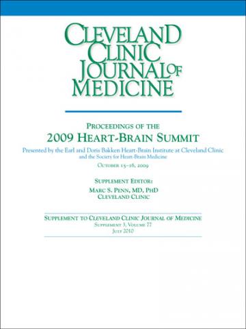
Supplement Editor:
Marc S. Penn, MD, PhD
Contents
Introduction: Heart-brain medicine: Update 2009
Marc S. Penn, MD, PhD, and Earl E. Bakken, MD (HonC), DSc (3 Hon), DHL (2 Hon)
Depression and Heart Disease
Depression and heart failure: An overview of what we know and don't know
Marc A. Silver, MD
The American Heart Association science advisory on depression and coronary heart disease: An exploration of the issues raised
J. Thomas Bigger, MD, and Alexander H. Glassman, MD
Depression and cardiovascular disease: Selected findings, controversies, and clinical implications from 2009
Karina W. Davidson, PhD, and Maya Rom Korin, PhD
Pioneer Lecture
Recovery of consciousness after severe brain injury: The role of arousal regulation mechanisms and some speculation on the heart-brain interface
Nicholas D. Schiff, MD
Heart-Brain Medicine Publications: The Year in Review
Neuroscience and heart-brain medicine: The year in review
David S. Goldstein, MD, PhD
Pathophysiologic mechanisms linking impaired cardiovascular health and neurologic dysfunction: The year in review
Ki E. Park, MD, and Carl J. Pepine, MD
Biomedical engineering in heart-brain medicine: A review
Peter G. Katona, ScD
Novel Findings in Heart-Brain Medicine
Sudden death in epilepsy, surgery, and seizure outcomes: The interface between heart and brain
Lara Jehi, MD
Biofeedback in the Treatment of Disease
Biofeedback in the treatment of heart failure
Michael G. McKee, PhD, and Christine S. Moravec, PhD
Biofeedback in the treatment of epilepsy
M. Barry Sterman, PhD
The effects of biofeedback in diabetes and essential hypertension
Angele McGrady, PhD, MEd, LPCC
Biofeedback in headache: An overview of approaches and evidence
Frank Andrasik, PhD
Device-Based Therapies
Use of deep brain stimulation in treatment-resistant depression
Donald A. Malone, Jr, MD
Poster Abstracts
Abstract 1: Potential role of the cardiac protease corin in energy metabolism
Jingjing Jiang, Yujie Cui, Wei Wang, and Qingyu Wu
Abstract 2: Anxiety and type D personality in ICD patients: Impact of shocks
Mina K. Chung, MD; Melanie Panko, RN; Tina Gupta; Scott Bea, PhD; Karen Broer, PhD; Diana Bauer; Denise Kosty-Sweeney, RN; Betty Ching, RN; Suzanne Pedersen, PhD; Sam Sears, PhD; and Leo Pozuelo, MD
Abstract 3: Microglia activation and neuroprotection during CNS preconditioning
Walid Jalabi, Ranjan Dutta, Yongming Jin, Gerson Criste, Xinghua Yin, Grahame J. Kidd, and Bruce D. Trapp
Abstract 4: Brain MRI correlates of atrial fibrillation
Stephen E. Jones, MD, PhD; Thomas Callahan, MD; Kamal Chémali, MD; Michael Phillips, MD; David Van Wagoner, PhD; and Walid Saliba, MD
Abstract 5: Identification and characterization of autonomic dysfunction in migraineurs with and without auras: Phase I
Mark Stillman, MD
Abstract 6: Sudden unexpected death in epilepsy: Finding the missing cardiac links
Lara Jehi, MD; Kanjana Unnongswe, MD; Thomas Callahan, MD; Liang Li, PhD; and Imad Najm, MD
Abstract 7: Cardiomyopathy after subarachnoid hemorrhage is mediated by neutrophils
J. Javier Provencio, Shari Moore, and Saksith Smithason
Abstract 8: Mindfulness, yoga, and cardiovascular disease
Didier Allexandre, Emily Fox, Mladen Golubic, Tom Morledge, and Joan E.B. Fox
Abstract 9: Multidisciplinary research in biofeedback
Christine S. Moravec, PhD; Michael G. McKee, PhD; James B. Young, MD; Betul Hatipoglu, MD; Leopoldo Pozuelo, MD; Leslie Cho, MD; Gordon Blackburn, MD; Francois Bethoux, MD; Mary Rensel, MD; Katherine Hoercher, RN; J. Javier Provencio, MD; and Marc S. Penn, MD
Abstract 10: Complex regional pain syndrome (CRPS I): A systemic disease of the autonomic nervous system
Kamal Chemali, MD; Robert Shields, MD; Lan Zhou, MD, PhD; Salim Hayek, MD, PhD; and Thomas Chelimsky, MD
Abstract 11: Biofeedback in the treatment of heart failure
Dana L. Frank, BA; Lamees Khorshid, PsyD; Jerome Kiffer, MA; Christine S. Moravec, PhD; and Michael G. McKee, PhD
Abstract 12: Change in depressive symptom status predicts health-related quality of life in patients with heart failure
Rebecca L. Dekker, MSN, RN, PhD candidate; Terry A. Lennie, PhD, RN; Nancy Albert, PhD, CCNS; Barbara Riegel, DNSc, RN; Misook L. Chung, PhD, RN; Seongkum Heo, PhD, RN; Eun Kyeung Song, PhD, RN; Jia-Rong Wu, PhD, RN; and Debra K. Moser, DNSc, RN
Abstract 13: Entropy of EKG time series distinguishes epileptic from nonepileptic patients
Rebecca O’Dwyer, Ulrich Zurcher, Brian Vyhnalek, Miron Kaufman, and Richard Burgess
Abstract 14: Evaluation of cardiac autonomic balance in major depression treated with different antidepressant therapies: A study with heart rate variability measures
K. Udupa, K.R. Kishore, J. Thirthalli, B.N. Gangadhar, T.R. Raju, and T.N. Sathyaprabha
Abstract 15: Proinflammatory status in major depression: Effects of escitalopram
John Piletz, PhD; Angelos Halaris, MD; Erin Tobin, MS; Edwin Meresh, MD; Jawed Fareed, PhD; Omer Iqbal, MD; Debra Hoppenstead, PhD; and James Sinacore, PhD
Abstract 16: Heart rate variability in depression: Effect of escitalopram
Angelos Halaris, MD; John Piletz, PhD; Erin Tobin, MA; Edwin Meresh, MD; James Sinacore, PhD; and Christopher Lowden
Abstract 17: Effects of omega-3/6 dietary ratio variation after a myocardial infarction in a rat model
Guy Rousseau, Isabelle Rondeau, Sandrine Picard, Thierno Madjou Bah, Louis Roy, and Roger Godbout
Abstract 18: The effects of tai chi on the heart and the brain
Qian Luo, Xi Cheng, and Xi Zha
Abstract 19: A randomized controlled trial of the effect of hostility reduction on cardiac autonomic regulation
Richard P. Sloan, PhD; Peter A. Shapiro, MD; Ethan E. Gorenstein, PhD; Felice A. Tager, PhD; Catherine E. Monk, PhD; Paula S. McKinley, PhD; Michael M. Myers, PhD; Emilia Bagiella, PhD; Ivy Chen, MST; Richard Steinman, BA; and J. Thomas Bigger, Jr., MD
Heart-brain medicine: Update 2009
Last October, the 2009 Heart-Brain Summit—the fourth annual summit of this type presented by the Bakken Heart-Brain Institute—was held in Chicago and built on the the first three summits’ tradition of open-minded discussion, out-of-the-box thinking, scholarly activity, and engagement of attendees from varied backgrounds.
DEPRESSION AND HEART DISEASE: A WATERSHED YEAR, OR JUMPING THE GUN?
The year leading up to the 2009 summit may be remembered as a watershed period for the field of heart-brain medicine, in light of the American Heart Association’s (AHA’s) inclusion of the recommendation to screen patients with coronary artery disease (CAD) for depression in its science advisory on depression and CAD.1 As has been discussed at prior Heart-Brain Summits, there is incontrovertible evidence in the literature that CAD patients with depression have a worse prognosis than do their counterparts without depression.2–6 While the link is clear, the etiology or mechanism behind depression’s association with worse CAD outcomes is debated. Possible reasons for the association range from greater nonadherence with medical therapy7 to increased systemic inflammation related to the decreased vagal tone associated with depression.8 Furthermore, there is clear evidence that patients with depression and CAD can be treated for their depression safely with cognitive and pharmacologic therapy.5,9 What is lacking, however, is convincing data that the treatment of depression in patients with CAD leads to improved outcomes.10
The topic for the first half of the opening day of the 2009 summit was whether the AHA has gotten ahead of itself in its science advisory1 and whether we should require demonstrable benefits from the treatment of depression in CAD patients before screening for depression is recommended in all patients with CAD. This is a critically important question for the field as well as for the Bakken Heart-Brain Institute, which under our leadership has been advocating for a clinical trial to address this very issue. Cardiologists addressing this question were well reminded that logical therapeutic targets without proven end points have failed us in the past. For instance, it was a rational concept that the suppression of premature ventricular contractions in patients with a history of acute myocardial infarction would lead to decreased ventricular tachycardia and death. Unfortunately, when this concept was put to the test in a randomized clinical trial, increased death was observed in the treatment group.11 More recent examples—and perhaps more applicable to depression, given its chronic nature—come from recent clinical trials demonstrating that tight blood sugar control is associated with higher mortality than moderate blood sugar control in critically ill patients12 and that intensive blood pressure control does not yield greater reductions in cardiovascular events compared with moderate blood pressure control in patients with type 2 diabetes.13
So we are faced with a chronic disease state—depression—that is clearly linked to adverse outcomes and death in patients with CAD. In the context of this association, we also know the following:
- The AHA science advisory recommends that we screen all CAD patients for depression.
- Treating depression in heart disease patients is safe.
- There is no clear proof that treating depression will reverse the increased risk associated with depression in patients with CAD.
- There is a community of physicians who treat CAD patients who are skeptical about therapies that do not have outcomes data.
The summit’s first morning concluded with a debate on whether now is the time for a large-scale multicenter randomized trial, which raised several important issues:
- The limited effectiveness of treatment for depression (approximately 30% to 40%)
- The ethics of randomizing a patient with depression to placebo
- The required size of the trial, given the efficacy of antidepressant therapy
- Measures to define response to therapy
- The utility of surrogate markers for adverse events in CAD versus a mortality end point.
The discussion and presentations were excellent and animated. In the end, each attendee was left to reach his or her own conclusion. Personally, one of us (M.S.P.) was surprised to be left with the conclusion that we are not ready for a definitive clinical trial.
In the cardiovascular medicine literature we were faced with a similar situation regarding the management of patients with atrial fibrillation. In the AFFIRM trial, patients were randomized to conservative treatment (rate control and warfarin) or aggressive treatment (rate control, warfarin, and any and all therapies to convert to and maintain normal sinus rhythm).14 Ultimately there was no difference between the groups, with a trend toward improved outcomes in the conservatively treated patients. What we really learned was that our therapies to convert to and maintain normal sinus rhythm were inadequate, and that in the case of atrial fibrillation at least we could clearly identify which patients did not respond to therapy.14 These findings ultimately may have led the field astray, as we still do not know if we have efficacious therapies for the treatment of atrial fibrillation and whether patients would benefit.
STRATEGIES FOR MODULATING HEART-BRAIN INTERACTIONS
In line with the need for more effective strategies to modulate heart-brain interactions, the summit went on to review and discuss the role of biofeedback. If the effects of depression, post-traumatic stress disorder, and other psychological modulators of vagal tone are the mechanism of action for adverse outcomes in these patient populations, then methods to directly modulate vagal tone may prove efficacious.15 Within the Bakken Heart-Brain Institute we recently committed half a million dollars to fund a biofeedback program. The program’s goal is to investigate the efficacy of biofeedback in improving outcomes within and across several states of cardiovascular disease and chronic disease. We believe that rigorous and standardized delivery and quantification of the effects of biofeedback are critical in order to robustly determine the role of biofeedback in the treatment of patients with chronic disease.
The group of experts assembled at this year’s summit presented further evidence of the potential importance of biofeedback for the control and treatment of multiple disorders, including heart failure, epilepsy, and chronic headache. As the mechanisms underlying brain interactions with end-organ innervations and systemic inflammation are dissected, it is clear that this field of medicine will have greater impact on the outcomes of many patient populations.
CROSS-FERTILIZATION OF TREATMENT APPROACHES
The summit abounded with evidence and examples of how neurology, cardiology, and psychiatry continue to cross-fertilize one another and foster interdisciplinary innovation. We were fortunate to have Brian Litt, MD, from the University of Pennsylvania return for the 2009 summit to update us on the progress of detecting, mapping, and extinguishing early seizure activity before there is clinical evidence of a seizure. The lessons learned and clinical advancement of internal cardiac defibrillators offer insights and great hope for this potentially important advancement in the treatment of seizure disorders. Similarly, Irving Zucker, PhD, from the University of Nebraska reviewed how neuromodulation through the baroreceptors can be targeted to modulate arterial blood pressure. Clearly there is great potential for device-based therapies to augment the treatment of chronic hypertension and improve outcomes in clinical populations at risk.
A LOOK AHEAD
Many of the topics reviewed above are discussed in detail in the proceedings supplement that follows. We continue to be excited and gratified by the progress being made in the field of heart-brain medicine. The continuing commitment to the rigorous multidisciplinary approach that has served this field well to date will continue to advance our understanding of disease and improve outcomes in our patients. We hope you will join us September 23–24, 2010, at the Lou Ruvo Center for Brain Health in Las Vegas, Nevada, for the 2010 Heart-Brain Summit, our fifth annual gathering.
- Lichtman JH, Bigger JT, Blumenthal JA, et al Depression and coronary heart disease: recommendations for screening, referral, and treatment: a science advisory from the American Heart Association Prevention Committee of the Council on Cardiovascular Nursing, Council on Clinical Cardiology, Council on Epidemiology and Prevention, and Interdisciplinary Council on Quality of Care and Outcomes Research: endorsed by the American Psychiatric Association. Circulation 2008; 118:1768–1775.
- Frazier L, Vaughn WK, Willerson JT, Ballantyne CM, Boerwinkle E. Inflammatory protein levels and depression screening after coronary stenting predict major adverse coronary events. Biol Res Nurs 2009; 11:163–173.
- Connerney I, Shapiro PA, McLaughlin JS, Bagiella E, Sloan RP. Relation between depression after coronary artery bypass surgery and 12-month outcome: a prospective study. Lancet 2001; 358:1766–1771.
- Davidson KW, Schwartz JE, Kirkland SA, et al Relation of inflammation to depression and incident coronary heart disease (from the Canadian Nova Scotia Health Survey [NSHS95] Prospective Population Study). Am J Cardiol 2009; 103:755–761.
- Summers KM, Martin KE, Watson K. Impact and clinical management of depression in patients with coronary artery disease. Pharmacotherapy 2010; 30:304–322.
- Kendler KS, Gardner CO, Fiske A, Gatz M. Major depression and coronary artery disease in the Swedish Twin Registry. Arch Gen Psychiatry 2009; 66:857–863.
- Albert NM, Fonarow GC, Abraham WT, et al Depression and clinical outcomes in heart failure: an OPTIMIZE-HF analysis. Am J Med 2009; 122:366–373.
- Khawaja IS, Westermeyer JJ, Gajwani P, Feinstein RE. Depression and coronary artery disease: the association, mechanisms, and therapeutic implications. Psychiatry (Edgmont) 2009; 6:38–51.
- Glassman AH, O’Connor CM, Califf RM, et al Sertraline treatment of major depression in patients with acute MI or unstable angina. Sertraline Antidepressant Heart Attack Randomized Trial (SADHART) Group. JAMA 2002; 288:701–709.
- Shapiro PA. Depression in coronary artery disease: does treatment help? Cleve Clin J Med 2008; 75( suppl 2):S5–S9.
- Echt DS, Liebson PR, Mitchell LB, et al Mortality and morbidity in patients receiving encainide, flecainide, or placebo: the Cardiac Arrhythmia Suppression Trial. N Engl J Med 1991; 324:781–788.
- Finfer S, Chittock DR, Su SY, et al Intensive versus conventional glucose control in critically ill patients. N Engl J Med 2009; 360:1283–1297.
- Cushman WC, Evans GW, Byington RP, et al Effects of intensive blood-pressure control in type 2 diabetes mellitus. N Engl J Med 2010; 362:1575–1585.
- Wyse DG, Waldo AL, DiMarco JP, et al A comparison of rate control and rhythm control in patients with atrial fibrillation. N Engl J Med 2002; 347:1825–1833.
- Penn MS, Bakken EE. Heart-brain medicine: update 2008. Cleve Clin J Med 2009; 76( suppl 2):S5–S7.
Last October, the 2009 Heart-Brain Summit—the fourth annual summit of this type presented by the Bakken Heart-Brain Institute—was held in Chicago and built on the the first three summits’ tradition of open-minded discussion, out-of-the-box thinking, scholarly activity, and engagement of attendees from varied backgrounds.
DEPRESSION AND HEART DISEASE: A WATERSHED YEAR, OR JUMPING THE GUN?
The year leading up to the 2009 summit may be remembered as a watershed period for the field of heart-brain medicine, in light of the American Heart Association’s (AHA’s) inclusion of the recommendation to screen patients with coronary artery disease (CAD) for depression in its science advisory on depression and CAD.1 As has been discussed at prior Heart-Brain Summits, there is incontrovertible evidence in the literature that CAD patients with depression have a worse prognosis than do their counterparts without depression.2–6 While the link is clear, the etiology or mechanism behind depression’s association with worse CAD outcomes is debated. Possible reasons for the association range from greater nonadherence with medical therapy7 to increased systemic inflammation related to the decreased vagal tone associated with depression.8 Furthermore, there is clear evidence that patients with depression and CAD can be treated for their depression safely with cognitive and pharmacologic therapy.5,9 What is lacking, however, is convincing data that the treatment of depression in patients with CAD leads to improved outcomes.10
The topic for the first half of the opening day of the 2009 summit was whether the AHA has gotten ahead of itself in its science advisory1 and whether we should require demonstrable benefits from the treatment of depression in CAD patients before screening for depression is recommended in all patients with CAD. This is a critically important question for the field as well as for the Bakken Heart-Brain Institute, which under our leadership has been advocating for a clinical trial to address this very issue. Cardiologists addressing this question were well reminded that logical therapeutic targets without proven end points have failed us in the past. For instance, it was a rational concept that the suppression of premature ventricular contractions in patients with a history of acute myocardial infarction would lead to decreased ventricular tachycardia and death. Unfortunately, when this concept was put to the test in a randomized clinical trial, increased death was observed in the treatment group.11 More recent examples—and perhaps more applicable to depression, given its chronic nature—come from recent clinical trials demonstrating that tight blood sugar control is associated with higher mortality than moderate blood sugar control in critically ill patients12 and that intensive blood pressure control does not yield greater reductions in cardiovascular events compared with moderate blood pressure control in patients with type 2 diabetes.13
So we are faced with a chronic disease state—depression—that is clearly linked to adverse outcomes and death in patients with CAD. In the context of this association, we also know the following:
- The AHA science advisory recommends that we screen all CAD patients for depression.
- Treating depression in heart disease patients is safe.
- There is no clear proof that treating depression will reverse the increased risk associated with depression in patients with CAD.
- There is a community of physicians who treat CAD patients who are skeptical about therapies that do not have outcomes data.
The summit’s first morning concluded with a debate on whether now is the time for a large-scale multicenter randomized trial, which raised several important issues:
- The limited effectiveness of treatment for depression (approximately 30% to 40%)
- The ethics of randomizing a patient with depression to placebo
- The required size of the trial, given the efficacy of antidepressant therapy
- Measures to define response to therapy
- The utility of surrogate markers for adverse events in CAD versus a mortality end point.
The discussion and presentations were excellent and animated. In the end, each attendee was left to reach his or her own conclusion. Personally, one of us (M.S.P.) was surprised to be left with the conclusion that we are not ready for a definitive clinical trial.
In the cardiovascular medicine literature we were faced with a similar situation regarding the management of patients with atrial fibrillation. In the AFFIRM trial, patients were randomized to conservative treatment (rate control and warfarin) or aggressive treatment (rate control, warfarin, and any and all therapies to convert to and maintain normal sinus rhythm).14 Ultimately there was no difference between the groups, with a trend toward improved outcomes in the conservatively treated patients. What we really learned was that our therapies to convert to and maintain normal sinus rhythm were inadequate, and that in the case of atrial fibrillation at least we could clearly identify which patients did not respond to therapy.14 These findings ultimately may have led the field astray, as we still do not know if we have efficacious therapies for the treatment of atrial fibrillation and whether patients would benefit.
STRATEGIES FOR MODULATING HEART-BRAIN INTERACTIONS
In line with the need for more effective strategies to modulate heart-brain interactions, the summit went on to review and discuss the role of biofeedback. If the effects of depression, post-traumatic stress disorder, and other psychological modulators of vagal tone are the mechanism of action for adverse outcomes in these patient populations, then methods to directly modulate vagal tone may prove efficacious.15 Within the Bakken Heart-Brain Institute we recently committed half a million dollars to fund a biofeedback program. The program’s goal is to investigate the efficacy of biofeedback in improving outcomes within and across several states of cardiovascular disease and chronic disease. We believe that rigorous and standardized delivery and quantification of the effects of biofeedback are critical in order to robustly determine the role of biofeedback in the treatment of patients with chronic disease.
The group of experts assembled at this year’s summit presented further evidence of the potential importance of biofeedback for the control and treatment of multiple disorders, including heart failure, epilepsy, and chronic headache. As the mechanisms underlying brain interactions with end-organ innervations and systemic inflammation are dissected, it is clear that this field of medicine will have greater impact on the outcomes of many patient populations.
CROSS-FERTILIZATION OF TREATMENT APPROACHES
The summit abounded with evidence and examples of how neurology, cardiology, and psychiatry continue to cross-fertilize one another and foster interdisciplinary innovation. We were fortunate to have Brian Litt, MD, from the University of Pennsylvania return for the 2009 summit to update us on the progress of detecting, mapping, and extinguishing early seizure activity before there is clinical evidence of a seizure. The lessons learned and clinical advancement of internal cardiac defibrillators offer insights and great hope for this potentially important advancement in the treatment of seizure disorders. Similarly, Irving Zucker, PhD, from the University of Nebraska reviewed how neuromodulation through the baroreceptors can be targeted to modulate arterial blood pressure. Clearly there is great potential for device-based therapies to augment the treatment of chronic hypertension and improve outcomes in clinical populations at risk.
A LOOK AHEAD
Many of the topics reviewed above are discussed in detail in the proceedings supplement that follows. We continue to be excited and gratified by the progress being made in the field of heart-brain medicine. The continuing commitment to the rigorous multidisciplinary approach that has served this field well to date will continue to advance our understanding of disease and improve outcomes in our patients. We hope you will join us September 23–24, 2010, at the Lou Ruvo Center for Brain Health in Las Vegas, Nevada, for the 2010 Heart-Brain Summit, our fifth annual gathering.
- Lichtman JH, Bigger JT, Blumenthal JA, et al Depression and coronary heart disease: recommendations for screening, referral, and treatment: a science advisory from the American Heart Association Prevention Committee of the Council on Cardiovascular Nursing, Council on Clinical Cardiology, Council on Epidemiology and Prevention, and Interdisciplinary Council on Quality of Care and Outcomes Research: endorsed by the American Psychiatric Association. Circulation 2008; 118:1768–1775.
- Frazier L, Vaughn WK, Willerson JT, Ballantyne CM, Boerwinkle E. Inflammatory protein levels and depression screening after coronary stenting predict major adverse coronary events. Biol Res Nurs 2009; 11:163–173.
- Connerney I, Shapiro PA, McLaughlin JS, Bagiella E, Sloan RP. Relation between depression after coronary artery bypass surgery and 12-month outcome: a prospective study. Lancet 2001; 358:1766–1771.
- Davidson KW, Schwartz JE, Kirkland SA, et al Relation of inflammation to depression and incident coronary heart disease (from the Canadian Nova Scotia Health Survey [NSHS95] Prospective Population Study). Am J Cardiol 2009; 103:755–761.
- Summers KM, Martin KE, Watson K. Impact and clinical management of depression in patients with coronary artery disease. Pharmacotherapy 2010; 30:304–322.
- Kendler KS, Gardner CO, Fiske A, Gatz M. Major depression and coronary artery disease in the Swedish Twin Registry. Arch Gen Psychiatry 2009; 66:857–863.
- Albert NM, Fonarow GC, Abraham WT, et al Depression and clinical outcomes in heart failure: an OPTIMIZE-HF analysis. Am J Med 2009; 122:366–373.
- Khawaja IS, Westermeyer JJ, Gajwani P, Feinstein RE. Depression and coronary artery disease: the association, mechanisms, and therapeutic implications. Psychiatry (Edgmont) 2009; 6:38–51.
- Glassman AH, O’Connor CM, Califf RM, et al Sertraline treatment of major depression in patients with acute MI or unstable angina. Sertraline Antidepressant Heart Attack Randomized Trial (SADHART) Group. JAMA 2002; 288:701–709.
- Shapiro PA. Depression in coronary artery disease: does treatment help? Cleve Clin J Med 2008; 75( suppl 2):S5–S9.
- Echt DS, Liebson PR, Mitchell LB, et al Mortality and morbidity in patients receiving encainide, flecainide, or placebo: the Cardiac Arrhythmia Suppression Trial. N Engl J Med 1991; 324:781–788.
- Finfer S, Chittock DR, Su SY, et al Intensive versus conventional glucose control in critically ill patients. N Engl J Med 2009; 360:1283–1297.
- Cushman WC, Evans GW, Byington RP, et al Effects of intensive blood-pressure control in type 2 diabetes mellitus. N Engl J Med 2010; 362:1575–1585.
- Wyse DG, Waldo AL, DiMarco JP, et al A comparison of rate control and rhythm control in patients with atrial fibrillation. N Engl J Med 2002; 347:1825–1833.
- Penn MS, Bakken EE. Heart-brain medicine: update 2008. Cleve Clin J Med 2009; 76( suppl 2):S5–S7.
Depression and heart failure: An overview of what we know and don’t know
The relationship between depression and heart failure is not as obvious as the one between depression and coronary artery disease. Myriad questions on the subject are open to research:
- Does depression occur in heart failure at higher-than-expected rates?
- Does heart failure severity influence depression?
- Is depression a risk factor for heart failure? If so, why?
- How should we screen for depression in patients with heart failure?
- Do we have an evidence-based approach to treatment?
Part of the challenge in clarifying the relationship between depression and heart failure is that heart failure is a disease of chronically ill, elderly patients—a population in which adjustment disorder with depressed mood and major depression are also common diagnoses, with rates recently found to be 22.3% and 13.3%, respectively.1 Nevertheless, interest in examining the relationship between heart failure and depression is long-standing, and many clinical studies have examined this relationship.2–16 Unfortunately, measures are not standardized, so comparisons between studies are difficult.
DEPRESSION IS COMMON IN PATIENTS WITH HEART FAILURE
Many studies show that rates of depression among patients with heart failure are higher than expected among other elderly, chronically ill patients. Furthermore, depression has been linked to more severe heart failure symptoms and worse outcomes in some studies.
In a 2001 study, Jiang et al screened 374 hospitalized patients with heart failure using the Beck Depression Inventory score and found that 35% had scores of 10 or higher (indicative of at least mild depression).12 Further testing showed that 14% met criteria for major depression.
A 2006 meta-analysis of 27 studies by Rutledge et al found a 21% incidence of clinically significant depression in patients with heart failure.17 Rates of depression depended heavily on the rigor of screening criteria for classifying participants as depressed: rates were as high as 38% with the use of liberal criteria and as low as 14% with strict criteria. New York Heart Association (NYHA) functional status correlated strongly with the prevalence of depression, which increased steadily from 11% in patients with NYHA class I (mild) heart failure to 20% in those with class II, 38% in those with class III, and 42% in those with class IV (severe) heart failure.
In one of the studies included in the meta-analysis, Freedland et al found that the prevalence of major depression was strongly associated with age and functional status in hospitalized patients with heart failure.18 In patients younger than age 60 years, rates of major depression rose particularly sharply as heart failure symptoms worsened.
The Psychosocial Factors Outcome Study found that the prevalence of depression in patients with heart failure who participated in the community-based Sudden Cardiac Death in Heart Failure Trial (SCD-HeFT) was 36%, based on a score of 13 or greater on the Beck Depression Inventory-II.19 Similarly, unpublished preliminary data from the Heart Failure Adherence and Retention Trial (HART) show that about one-third of community-based patients with heart failure have depression.20
DEPRESSION AND HEART FAILURE ARE CLOSELY LINKED PHYSIOLOGICALLY
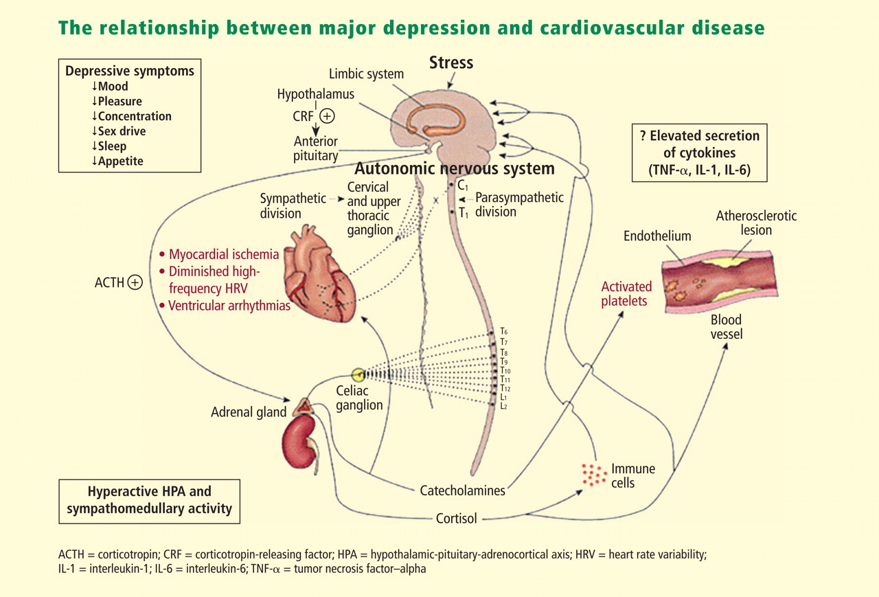
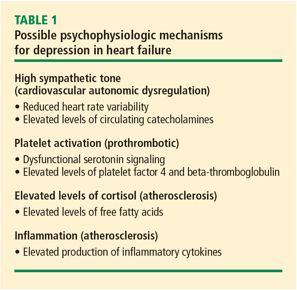
The evidence, therefore, strongly suggests a common pathway between heart failure and depression, and this evidence of a relationship favors a new paradigm that integrates the treatment of the two conditions.
BETA-BLOCKERS DO NOT RAISE RISK OF DEPRESSION
Pharmacotherapy is often implicated as a related factor in the development or exacerbation of depression in patients with heart failure, especially in those taking beta-blockers. Meta-analyses of the incidence of depression associated with various beta-blockers have been conducted in patients with hypertension, heart failure, and recent myocardial infarction. A 2002 meta-analysis of eight trials that randomized patients to treatment with a beta-blocker or placebo found no difference in the incidence of depressive symptoms between the active treatment and placebo groups, or between patients in the two groups who withdrew from the trial, presumably because of depression or other symptoms.24 An additional study not included in that meta-analysis found no differences in rates of depression among hypertensive patients according to the type of antihypertensive medication they were taking (ie, beta-blockers, diuretics, reserpine, or no drug therapy).25
Based on the evidence, I see no reason to avoid a trial of beta-blockers in patients who have depression at baseline or to be overly concerned that patients without depression will develop it as a result of beta-blocker treatment.
DEPRESSION LEADS TO WORSE OUTCOMES, HIGHER COSTS IN HEART FAILURE
Not only is depression prevalent in patients with heart failure, but depression adversely affects heart failure outcomes. One study of hospitalized patients older than 70 years found readmission rates to be 67% among heart failure patients with depression versus 44% among heart failure patients without depression.26 Patients in this study were three times as likely to die if they had heart failure than if they did not have heart failure, and they were twice as likely to die if they had depression than if they did not have depression. The mortality rate was 21% for patients with both heart failure and depression versus 15% for patients with heart failure without depression.26
Depressive symptoms also correlate with poorer quality of life in patients with heart failure. Gottlieb et al found that quality-of-life scores were significantly worse in heart failure patients if they had a diagnosis of depression on the basis of the Beck Depression Index.27
The 27-study meta-analysis by Rutledge et al discussed earlier found that the presence of depression in a patient with heart failure predicts worse outcomes in terms of hospital readmission rates, functional status, and walk times.17 This analysis also found twice the rate of death in heart failure patients with depression compared with heart failure patients without depression.
In a large community-based trial involving more than 48,000 patients with heart failure, Macchia et al found that survival was markedly reduced in patients who had a history of depression.28 This study also showed that depressed patients were less likely to adhere to medication regimens with angiotensin-converting enzyme inhibitors and beta-blockers, which may offer a potential explanation for the reduction in survival among depressed patients.
In a study of patients with coronary artery disease who had no diagnosis of depression or heart failure, May et al demonstrated that those who subsequently developed depression had more than four times the risk of also developing heart failure.29
The combination of heart failure and depression also is costly. In a 3-year retrospective study of community-based patients following a first hospitalization for heart failure, Sullivan et al found that annualized adjusted total costs were nearly 30% greater in patients diagnosed with depression and that inpatient and outpatient service utilization was also greater in those with depression.5
DIAGNOSIS AND TREATMENT
One simple question can effectively screen for depression
Numerous tools are available for the diagnosis of depression, but developing a tool that is readily useful to a busy clinician is challenging. Simply asking the single question, “Are you depressed?” has fairly high sensitivity (55%) and specificity (74%) for diagnosing depression in palliative care patients, a population even more seriously ill than heart failure patients.30 A variation on the question from British studies—“Do you feel that your life is empty?”—is considered to be a better screening question for elderly patients.31,32
Effect of depression therapy on heart failure still unclear
Unfortunately, evidence for the best treatment for depression in patients with heart failure is lacking. Some guidance may be gleaned from studies in patients with coronary artery disease. The Sertraline Antidepressant Heart Attack Randomized Trial (SADHART) was a multicenter, randomized, placebo-controlled study of the safety and efficacy of treating major depressive disorder with sertraline for 24 weeks in patients hospitalized for acute coronary syndrome.33,34 No significant differences were found between treatment groups in left ventricular ejection fraction, blood pressure, resting electrocardiogram, and cardiac arrhythmias. Although the trial was not powered to detect an effect of treatment on mortality, there were fewer deaths and severe cardiovascular adverse events in the active treatment group.
A later study was designed to evaluate 12 weeks of treatment with sertraline in patients with major depression and heart failure.35 Although symptoms of depression improved with treatment, no beneficial effect on heart failure was found.36 A nursing intervention that was included for both the treatment and placebo groups may have served to limit the impact of sertraline on heart failure surrogate end points.
The abovementioned HART study randomized patients with systolic or diastolic dysfunction and NYHA class II or III functional status to receive either heart failure education (comprising 18 American Heart Association tip sheets and 18 phone calls) or heart failure education plus self-management strategies (comprising the tip sheets, 18 group sessions, and problem-solving and self-management skills) following hospital discharge.37 Over 3 years, no difference between the two groups was found in the rates and timing of deaths or heart failure hospitalizations.
The best treatment strategies for depression in heart failure are still unclear, and more research is needed. Although guidelines exist for the management of depression in patients with coronary heart disease,38 no such guidelines have been issued for the management of depression in heart failure.
CONCLUSIONS
Although evidence is strong that treatment with medication or cognitive therapies improves symptoms of depression, evidence is lacking for a significant effect of such interventions on cardiac outcomes.39 Because depression and heart failure are so closely linked and appear to share a genetic and pathophysiologic basis, greater understanding of the relationship between these diseases across the stages of heart failure should be pursued.
Any patient with heart failure who is symptomatic has advanced disease, and is therefore closer to death than to health. The same is probably true of depression. Patients with heart failure and depression must be identified early, and interventions must be tried at these early stages of disease. Better depression screening tools and heightened awareness of the relationship between heart failure and depression are essential.
- Yazgan IC, Kuscu MK, Fistikci N, Keyvan A, Topcuoglu V. Geriatric psychiatry consultations in a Turkish university hospital. Int Psychogeriatr 2006; 18:327–333.
- Abramson J, Berger A, Krumholz HM, Vaccarino V. Depression and risk of heart failure among older persons with isolated systolic hypertension. Arch Intern Med 2001; 161:1725–1730.
- Williams SA, Kasl SV, Heiat A, Abramson JL, Krumholz HM, Vaccarino V. Depression and risk of heart failure among the elderly: a prospective community-based study. Psychosom Med 2002; 64:6–12.
- Himelhoch S, Weller WE, Wu AW, Anderson GF, Cooper LA. Chronic medical illness, depression, and use of acute medical services among Medicare beneficiaries. Med Care 2004; 42:512–521.
- Sullivan M, Simon G, Spertus J, Russo J. Depression-related costs in heart failure care. Arch Intern Med 2002; 162:1860–1866.
- Fulop G, Strain JJ, Stettin G. Congestive heart failure and depression in older adults: clinical course and health services use 6 months after hospitalization. Psychosomatics 2003; 44:367–373.
- Koenig HG. Depression in hospitalized older patients with congestive heart failure. Gen Hosp Psychiatry 1998; 20:29–43.
- Rumsfeld JS, Havranek E, Masoudi FA, et al Depressive symptoms are the strongest predictors of short-term declines in health status in patients with heart failure. J Am Coll Cardiol 2003; 42:1811–1817.
- de Denus S, Spinler SA, Jessup M, Kao A. History of depression as a predictor of adverse outcomes in patients hospitalized for decompensated heart failure. Pharmacotherapy 2004; 24:1306–1310.
- Faris R, Purcell H, Henein MY, Coats AJ. Clinical depression is common and significantly associated with reduced survival in patients with non-ischaemic heart failure. Eur J Heart Fail 2002; 4:541–551.
- Freedland KE, Carney RM, Rich MW, et al Depression in elderly patients with congestive heart failure. J Geriatr Psychiatry 1991; 24:59–71.
- Jiang W, Alexander J, Christopher E, et al Relationship of depression to increased risk of mortality and rehospitalization in patients with congestive heart failure. Arch Intern Med 2001; 161:1849–1856.
- Jünger J, Schellberg D, Müller-Tasch T, et al Depression increasingly predicts mortality in the course of congestive heart failure. Eur J Heart Fail 2005; 7:261–267.
- Murberg TA, Bru E, Svebak S, Tveterås R, Aarsland T. Depressed mood and subjective health symptoms as predictors of mortality in patients with congestive heart failure: a two-years follow-up study. Int J Psychiatry Med 1999; 29:311–326.
- Sullivan MD, Levy WC, Crane BA, Russo JE, Spertus JA. Usefulness of depression to predict time to combined end point of transplant or death for outpatients with advanced heart failure. Am J Cardiol 2004; 94:1577–1580.
- Vaccarino V, Kasl SV, Abramson J, Krumholz HM. Depressive symptoms and risk of functional decline and death in patients with heart failure. J Am Coll Cardiol 2001; 38:199–205.
- Rutledge T, Reis VA, Linke SE, Greenberg BH, Mills PJ. Depression in heart failure: a meta-analytic review of prevalence, intervention effects, and associations with clinical outcomes. J Am Coll Cardiol 2006; 48:1527–1537.
- Freedland KE, Rich MW, Skala JA, Carney RM, Dávila-Román VG, Jaffe AS. Prevalence of depression in hospitalized patients with congestive heart failure. Psychosom Med 2003; 65:119–128.
- Friedmann E, Thomas SA, Liu F, et al Relationship of depression, anxiety, and social isolation to chronic heart failure outpatient mortality. Am Heart J 2006; 152:940.e1–940.e8.
- de Leon CF, Grady KL, Eaton C, et al Quality of life in a diverse population of patients with heart failure: baseline findings from the Heart Failure Adherence and Retention trial (HART). J Cardiopulm Rehabil Prev 2009; 29:171–178.
- Musselman DL, Evans DL, Nemeroff CB. The relationship of depression to cardiovascular disease: epidemiology, biology, and treatment. Arch Gen Psychiatry 1998; 55:580–592.
- Caspi A, Sugden K, Moffitt TE, et al Influence of life stress on depression: moderation by a polymorphism in the 5-HTT gene. Science 2003; 301:386–389.
- Cohn JN, Levine TB, Olivari MT, et al Plasma norepinephrine as a guide to prognosis in patients with chronic congestive heart failure. N Engl J Med 1984; 311:819–823.
- Ko DT, Hebert PR, Coffey CS, Sedrakyan A, Curtis JP, Krumholz HM. β-blocker therapy and symptoms of depression, fatigue, and sexual dysfunction. JAMA 2002; 288:351–357.
- Prisant LM, Spruill WJ, Fincham JE, Wade WE, Carr AA, Adams MA. Depression associated with antihypertensive drugs. J Fam Pract 1991; 33:481–485.
- Rozzini R, Sabatini T, Frisoni GB, et al Depression and major outcomes in older patients with heart failure [letter]. Arch Intern Med 2002; 162:362–364.
- Gottlieb SS, Khatta M, Friedmann E, et al The influence of age, gender, and race on the prevalence of depression in heart failure patients. J Am Coll Cardiol 2004; 43:1542–1549.
- Macchia A, Monte S, Pellegrini F, et al Depression worsens outcomes in elderly patients with heart failure: an analysis of 48,117 patients in a community setting. Eur J Heart Fail 2008; 10:714–721.
- May HT, Horne BD, Carlquist JF, Sheng X, Joy E, Catinella AP. Depression after coronary artery disease is associated with heart failure. J Am Coll Cardiol 2009; 53:1440–1447.
- Lloyd-Williams M, Dennis M, Taylor F, Baker I. Is asking patients in palliative care, “Are you depressed?” appropriate? Prospective study. BMJ 2003; 327:372–373.
- Whelan PJ, Gaughran F, Walwyn R, Chatterton K, Macdonald A. ‘Do you feel that your life is empty?’ The clinical utility of a one-off question for detecting depression in elderly care home residents. Age Ageing 2008; 37:475–478.
- D’Ath P, Katona P, Mullan E, Evans S, Katona C. Screening, detection and management of depression in elderly primary care attenders. I: The acceptability and performance of the 15 item Geriatric Depression Scale (GDS15) and the development of short versions. Fam Pract 1994; 11:260–266.
- Glassman AH, O’Connor CM, Califf RM, et al., Sertraline Antidepressant Heart Attack Randomized Trial (SADHART) Group. Sertraline treatment of major depression in patients with acute MI or unstable angina. JAMA 2002; 288:701–709.
- Carney RM, Jaffe AS. Treatment of depression following acute myocardial infarction. JAMA 2002; 288:750–751.
- Jiang W, O’Connor C, Silva SG, et al., SADHART-CHF Investigators. Safety and efficacy of sertraline for depression in patients with CHF (SADHART-CHF): a randomized, double-blind, placebo-controlled trial of sertraline for major depression with congestive heart failure. Am Heart J 2008; 156:437–444.
- O’Connor CM, Wei J, Silva SG, et al., SADHART-CHF investigators. Safety and efficacy of sertraline plus nurse facilitated supportive intervention (NFSI) versus placebo plus nurse facilitated supportive intervention for depression in patients with CHF (SADHART-CHF). Presented at: 2008 Scientific Sessions of the Heart Failure Society of America; September 20–23, 2008; Toronto, ON, Canada.
- Powell LH, Calvin JE, Mendes de Leon CF, et al Impact of patient self-management skills training on death and hospitalization in patients with heart failure: results from the Heart Failure Adherence and Retention Trial. Circulation 2007; 116:2629. Abstract.
- Lichtman JH, Bigger JT, Blumenthal JA, et al Depression and coronary heart disease: recommendations for screening, referral, and treatment: a science advisory from the American Heart Association Prevention Committee of the Council on Cardiovascular Nursing, Council on Clinical Cardiology, Council on Epidemiology and Prevention, and Interdisciplinary Council on Quality of Care and Outcomes Research: endorsed by the American Psychiatric Association. Circulation 2008; 118:1768–1775.
- Thombs BD, de Jonge P, Coyne JC, et al Depression screening and patient outcomes in cardiovascular care: a systematic review. JAMA 2008; 300:2161–2171.
The relationship between depression and heart failure is not as obvious as the one between depression and coronary artery disease. Myriad questions on the subject are open to research:
- Does depression occur in heart failure at higher-than-expected rates?
- Does heart failure severity influence depression?
- Is depression a risk factor for heart failure? If so, why?
- How should we screen for depression in patients with heart failure?
- Do we have an evidence-based approach to treatment?
Part of the challenge in clarifying the relationship between depression and heart failure is that heart failure is a disease of chronically ill, elderly patients—a population in which adjustment disorder with depressed mood and major depression are also common diagnoses, with rates recently found to be 22.3% and 13.3%, respectively.1 Nevertheless, interest in examining the relationship between heart failure and depression is long-standing, and many clinical studies have examined this relationship.2–16 Unfortunately, measures are not standardized, so comparisons between studies are difficult.
DEPRESSION IS COMMON IN PATIENTS WITH HEART FAILURE
Many studies show that rates of depression among patients with heart failure are higher than expected among other elderly, chronically ill patients. Furthermore, depression has been linked to more severe heart failure symptoms and worse outcomes in some studies.
In a 2001 study, Jiang et al screened 374 hospitalized patients with heart failure using the Beck Depression Inventory score and found that 35% had scores of 10 or higher (indicative of at least mild depression).12 Further testing showed that 14% met criteria for major depression.
A 2006 meta-analysis of 27 studies by Rutledge et al found a 21% incidence of clinically significant depression in patients with heart failure.17 Rates of depression depended heavily on the rigor of screening criteria for classifying participants as depressed: rates were as high as 38% with the use of liberal criteria and as low as 14% with strict criteria. New York Heart Association (NYHA) functional status correlated strongly with the prevalence of depression, which increased steadily from 11% in patients with NYHA class I (mild) heart failure to 20% in those with class II, 38% in those with class III, and 42% in those with class IV (severe) heart failure.
In one of the studies included in the meta-analysis, Freedland et al found that the prevalence of major depression was strongly associated with age and functional status in hospitalized patients with heart failure.18 In patients younger than age 60 years, rates of major depression rose particularly sharply as heart failure symptoms worsened.
The Psychosocial Factors Outcome Study found that the prevalence of depression in patients with heart failure who participated in the community-based Sudden Cardiac Death in Heart Failure Trial (SCD-HeFT) was 36%, based on a score of 13 or greater on the Beck Depression Inventory-II.19 Similarly, unpublished preliminary data from the Heart Failure Adherence and Retention Trial (HART) show that about one-third of community-based patients with heart failure have depression.20
DEPRESSION AND HEART FAILURE ARE CLOSELY LINKED PHYSIOLOGICALLY
The evidence, therefore, strongly suggests a common pathway between heart failure and depression, and this evidence of a relationship favors a new paradigm that integrates the treatment of the two conditions.
BETA-BLOCKERS DO NOT RAISE RISK OF DEPRESSION
Pharmacotherapy is often implicated as a related factor in the development or exacerbation of depression in patients with heart failure, especially in those taking beta-blockers. Meta-analyses of the incidence of depression associated with various beta-blockers have been conducted in patients with hypertension, heart failure, and recent myocardial infarction. A 2002 meta-analysis of eight trials that randomized patients to treatment with a beta-blocker or placebo found no difference in the incidence of depressive symptoms between the active treatment and placebo groups, or between patients in the two groups who withdrew from the trial, presumably because of depression or other symptoms.24 An additional study not included in that meta-analysis found no differences in rates of depression among hypertensive patients according to the type of antihypertensive medication they were taking (ie, beta-blockers, diuretics, reserpine, or no drug therapy).25
Based on the evidence, I see no reason to avoid a trial of beta-blockers in patients who have depression at baseline or to be overly concerned that patients without depression will develop it as a result of beta-blocker treatment.
DEPRESSION LEADS TO WORSE OUTCOMES, HIGHER COSTS IN HEART FAILURE
Not only is depression prevalent in patients with heart failure, but depression adversely affects heart failure outcomes. One study of hospitalized patients older than 70 years found readmission rates to be 67% among heart failure patients with depression versus 44% among heart failure patients without depression.26 Patients in this study were three times as likely to die if they had heart failure than if they did not have heart failure, and they were twice as likely to die if they had depression than if they did not have depression. The mortality rate was 21% for patients with both heart failure and depression versus 15% for patients with heart failure without depression.26
Depressive symptoms also correlate with poorer quality of life in patients with heart failure. Gottlieb et al found that quality-of-life scores were significantly worse in heart failure patients if they had a diagnosis of depression on the basis of the Beck Depression Index.27
The 27-study meta-analysis by Rutledge et al discussed earlier found that the presence of depression in a patient with heart failure predicts worse outcomes in terms of hospital readmission rates, functional status, and walk times.17 This analysis also found twice the rate of death in heart failure patients with depression compared with heart failure patients without depression.
In a large community-based trial involving more than 48,000 patients with heart failure, Macchia et al found that survival was markedly reduced in patients who had a history of depression.28 This study also showed that depressed patients were less likely to adhere to medication regimens with angiotensin-converting enzyme inhibitors and beta-blockers, which may offer a potential explanation for the reduction in survival among depressed patients.
In a study of patients with coronary artery disease who had no diagnosis of depression or heart failure, May et al demonstrated that those who subsequently developed depression had more than four times the risk of also developing heart failure.29
The combination of heart failure and depression also is costly. In a 3-year retrospective study of community-based patients following a first hospitalization for heart failure, Sullivan et al found that annualized adjusted total costs were nearly 30% greater in patients diagnosed with depression and that inpatient and outpatient service utilization was also greater in those with depression.5
DIAGNOSIS AND TREATMENT
One simple question can effectively screen for depression
Numerous tools are available for the diagnosis of depression, but developing a tool that is readily useful to a busy clinician is challenging. Simply asking the single question, “Are you depressed?” has fairly high sensitivity (55%) and specificity (74%) for diagnosing depression in palliative care patients, a population even more seriously ill than heart failure patients.30 A variation on the question from British studies—“Do you feel that your life is empty?”—is considered to be a better screening question for elderly patients.31,32
Effect of depression therapy on heart failure still unclear
Unfortunately, evidence for the best treatment for depression in patients with heart failure is lacking. Some guidance may be gleaned from studies in patients with coronary artery disease. The Sertraline Antidepressant Heart Attack Randomized Trial (SADHART) was a multicenter, randomized, placebo-controlled study of the safety and efficacy of treating major depressive disorder with sertraline for 24 weeks in patients hospitalized for acute coronary syndrome.33,34 No significant differences were found between treatment groups in left ventricular ejection fraction, blood pressure, resting electrocardiogram, and cardiac arrhythmias. Although the trial was not powered to detect an effect of treatment on mortality, there were fewer deaths and severe cardiovascular adverse events in the active treatment group.
A later study was designed to evaluate 12 weeks of treatment with sertraline in patients with major depression and heart failure.35 Although symptoms of depression improved with treatment, no beneficial effect on heart failure was found.36 A nursing intervention that was included for both the treatment and placebo groups may have served to limit the impact of sertraline on heart failure surrogate end points.
The abovementioned HART study randomized patients with systolic or diastolic dysfunction and NYHA class II or III functional status to receive either heart failure education (comprising 18 American Heart Association tip sheets and 18 phone calls) or heart failure education plus self-management strategies (comprising the tip sheets, 18 group sessions, and problem-solving and self-management skills) following hospital discharge.37 Over 3 years, no difference between the two groups was found in the rates and timing of deaths or heart failure hospitalizations.
The best treatment strategies for depression in heart failure are still unclear, and more research is needed. Although guidelines exist for the management of depression in patients with coronary heart disease,38 no such guidelines have been issued for the management of depression in heart failure.
CONCLUSIONS
Although evidence is strong that treatment with medication or cognitive therapies improves symptoms of depression, evidence is lacking for a significant effect of such interventions on cardiac outcomes.39 Because depression and heart failure are so closely linked and appear to share a genetic and pathophysiologic basis, greater understanding of the relationship between these diseases across the stages of heart failure should be pursued.
Any patient with heart failure who is symptomatic has advanced disease, and is therefore closer to death than to health. The same is probably true of depression. Patients with heart failure and depression must be identified early, and interventions must be tried at these early stages of disease. Better depression screening tools and heightened awareness of the relationship between heart failure and depression are essential.
- Yazgan IC, Kuscu MK, Fistikci N, Keyvan A, Topcuoglu V. Geriatric psychiatry consultations in a Turkish university hospital. Int Psychogeriatr 2006; 18:327–333.
- Abramson J, Berger A, Krumholz HM, Vaccarino V. Depression and risk of heart failure among older persons with isolated systolic hypertension. Arch Intern Med 2001; 161:1725–1730.
- Williams SA, Kasl SV, Heiat A, Abramson JL, Krumholz HM, Vaccarino V. Depression and risk of heart failure among the elderly: a prospective community-based study. Psychosom Med 2002; 64:6–12.
- Himelhoch S, Weller WE, Wu AW, Anderson GF, Cooper LA. Chronic medical illness, depression, and use of acute medical services among Medicare beneficiaries. Med Care 2004; 42:512–521.
- Sullivan M, Simon G, Spertus J, Russo J. Depression-related costs in heart failure care. Arch Intern Med 2002; 162:1860–1866.
- Fulop G, Strain JJ, Stettin G. Congestive heart failure and depression in older adults: clinical course and health services use 6 months after hospitalization. Psychosomatics 2003; 44:367–373.
- Koenig HG. Depression in hospitalized older patients with congestive heart failure. Gen Hosp Psychiatry 1998; 20:29–43.
- Rumsfeld JS, Havranek E, Masoudi FA, et al Depressive symptoms are the strongest predictors of short-term declines in health status in patients with heart failure. J Am Coll Cardiol 2003; 42:1811–1817.
- de Denus S, Spinler SA, Jessup M, Kao A. History of depression as a predictor of adverse outcomes in patients hospitalized for decompensated heart failure. Pharmacotherapy 2004; 24:1306–1310.
- Faris R, Purcell H, Henein MY, Coats AJ. Clinical depression is common and significantly associated with reduced survival in patients with non-ischaemic heart failure. Eur J Heart Fail 2002; 4:541–551.
- Freedland KE, Carney RM, Rich MW, et al Depression in elderly patients with congestive heart failure. J Geriatr Psychiatry 1991; 24:59–71.
- Jiang W, Alexander J, Christopher E, et al Relationship of depression to increased risk of mortality and rehospitalization in patients with congestive heart failure. Arch Intern Med 2001; 161:1849–1856.
- Jünger J, Schellberg D, Müller-Tasch T, et al Depression increasingly predicts mortality in the course of congestive heart failure. Eur J Heart Fail 2005; 7:261–267.
- Murberg TA, Bru E, Svebak S, Tveterås R, Aarsland T. Depressed mood and subjective health symptoms as predictors of mortality in patients with congestive heart failure: a two-years follow-up study. Int J Psychiatry Med 1999; 29:311–326.
- Sullivan MD, Levy WC, Crane BA, Russo JE, Spertus JA. Usefulness of depression to predict time to combined end point of transplant or death for outpatients with advanced heart failure. Am J Cardiol 2004; 94:1577–1580.
- Vaccarino V, Kasl SV, Abramson J, Krumholz HM. Depressive symptoms and risk of functional decline and death in patients with heart failure. J Am Coll Cardiol 2001; 38:199–205.
- Rutledge T, Reis VA, Linke SE, Greenberg BH, Mills PJ. Depression in heart failure: a meta-analytic review of prevalence, intervention effects, and associations with clinical outcomes. J Am Coll Cardiol 2006; 48:1527–1537.
- Freedland KE, Rich MW, Skala JA, Carney RM, Dávila-Román VG, Jaffe AS. Prevalence of depression in hospitalized patients with congestive heart failure. Psychosom Med 2003; 65:119–128.
- Friedmann E, Thomas SA, Liu F, et al Relationship of depression, anxiety, and social isolation to chronic heart failure outpatient mortality. Am Heart J 2006; 152:940.e1–940.e8.
- de Leon CF, Grady KL, Eaton C, et al Quality of life in a diverse population of patients with heart failure: baseline findings from the Heart Failure Adherence and Retention trial (HART). J Cardiopulm Rehabil Prev 2009; 29:171–178.
- Musselman DL, Evans DL, Nemeroff CB. The relationship of depression to cardiovascular disease: epidemiology, biology, and treatment. Arch Gen Psychiatry 1998; 55:580–592.
- Caspi A, Sugden K, Moffitt TE, et al Influence of life stress on depression: moderation by a polymorphism in the 5-HTT gene. Science 2003; 301:386–389.
- Cohn JN, Levine TB, Olivari MT, et al Plasma norepinephrine as a guide to prognosis in patients with chronic congestive heart failure. N Engl J Med 1984; 311:819–823.
- Ko DT, Hebert PR, Coffey CS, Sedrakyan A, Curtis JP, Krumholz HM. β-blocker therapy and symptoms of depression, fatigue, and sexual dysfunction. JAMA 2002; 288:351–357.
- Prisant LM, Spruill WJ, Fincham JE, Wade WE, Carr AA, Adams MA. Depression associated with antihypertensive drugs. J Fam Pract 1991; 33:481–485.
- Rozzini R, Sabatini T, Frisoni GB, et al Depression and major outcomes in older patients with heart failure [letter]. Arch Intern Med 2002; 162:362–364.
- Gottlieb SS, Khatta M, Friedmann E, et al The influence of age, gender, and race on the prevalence of depression in heart failure patients. J Am Coll Cardiol 2004; 43:1542–1549.
- Macchia A, Monte S, Pellegrini F, et al Depression worsens outcomes in elderly patients with heart failure: an analysis of 48,117 patients in a community setting. Eur J Heart Fail 2008; 10:714–721.
- May HT, Horne BD, Carlquist JF, Sheng X, Joy E, Catinella AP. Depression after coronary artery disease is associated with heart failure. J Am Coll Cardiol 2009; 53:1440–1447.
- Lloyd-Williams M, Dennis M, Taylor F, Baker I. Is asking patients in palliative care, “Are you depressed?” appropriate? Prospective study. BMJ 2003; 327:372–373.
- Whelan PJ, Gaughran F, Walwyn R, Chatterton K, Macdonald A. ‘Do you feel that your life is empty?’ The clinical utility of a one-off question for detecting depression in elderly care home residents. Age Ageing 2008; 37:475–478.
- D’Ath P, Katona P, Mullan E, Evans S, Katona C. Screening, detection and management of depression in elderly primary care attenders. I: The acceptability and performance of the 15 item Geriatric Depression Scale (GDS15) and the development of short versions. Fam Pract 1994; 11:260–266.
- Glassman AH, O’Connor CM, Califf RM, et al., Sertraline Antidepressant Heart Attack Randomized Trial (SADHART) Group. Sertraline treatment of major depression in patients with acute MI or unstable angina. JAMA 2002; 288:701–709.
- Carney RM, Jaffe AS. Treatment of depression following acute myocardial infarction. JAMA 2002; 288:750–751.
- Jiang W, O’Connor C, Silva SG, et al., SADHART-CHF Investigators. Safety and efficacy of sertraline for depression in patients with CHF (SADHART-CHF): a randomized, double-blind, placebo-controlled trial of sertraline for major depression with congestive heart failure. Am Heart J 2008; 156:437–444.
- O’Connor CM, Wei J, Silva SG, et al., SADHART-CHF investigators. Safety and efficacy of sertraline plus nurse facilitated supportive intervention (NFSI) versus placebo plus nurse facilitated supportive intervention for depression in patients with CHF (SADHART-CHF). Presented at: 2008 Scientific Sessions of the Heart Failure Society of America; September 20–23, 2008; Toronto, ON, Canada.
- Powell LH, Calvin JE, Mendes de Leon CF, et al Impact of patient self-management skills training on death and hospitalization in patients with heart failure: results from the Heart Failure Adherence and Retention Trial. Circulation 2007; 116:2629. Abstract.
- Lichtman JH, Bigger JT, Blumenthal JA, et al Depression and coronary heart disease: recommendations for screening, referral, and treatment: a science advisory from the American Heart Association Prevention Committee of the Council on Cardiovascular Nursing, Council on Clinical Cardiology, Council on Epidemiology and Prevention, and Interdisciplinary Council on Quality of Care and Outcomes Research: endorsed by the American Psychiatric Association. Circulation 2008; 118:1768–1775.
- Thombs BD, de Jonge P, Coyne JC, et al Depression screening and patient outcomes in cardiovascular care: a systematic review. JAMA 2008; 300:2161–2171.