User login
News and Views that Matter to Physicians
Frailty stratifies pediatric liver disease severity
MONTREAL – A newly devised measurement of frailty in children effectively determined the severity of liver disease in pediatric patients and might serve as a useful, independent predictor of outcomes following liver transplantations in children and adolescents.
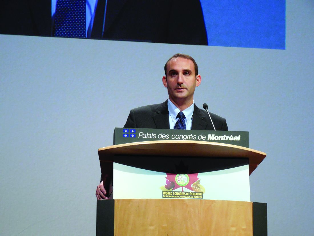
The adapted pediatric frailty assessment formula is a “very valid, feasible, and valuable tool” for assessing children with chronic liver disease, Eberhard Lurz, MD, said at the World Congress of Pediatric Gastroenterology, Hepatology and Nutrition. “Frailty captures an additional marker of ill health that is independent of the MELD-Na [Model for End-Stage Liver Disease–Na] and PELD,” [Pediatric End-Stage Liver Disease] said Dr. Lurz, a pediatric gastroenterologist at the Hospital for Sick Children in Toronto.
The idea of frailty assessment of children with liver disease sprang from a 2014 report that showed a five-item frailty index could predict mortality in adults with liver disease who were listed for liver transplantation and that this predictive power was independent of the patients’ MELD scores (Am J Transplant. 2014 Aug;14[8]:1870-9). That study used a five-item frailty index developed for adults (J Gerontol A Biol Sci Med Sci. 2001;56[3]:M146-57).
Dr. Lurz came up with a pediatric version of this frailty score using pediatric-oriented measures for each of the five items. To measure exhaustion he used the PedsQL (Pediatric Quality of Life Inventory) Multidimensional Fatigue Scale; for slowness he used a 6-minute walk test; for weakness he measured grip strength; for shrinkage he measured triceps skinfold thickness; and for diminished activity he used an age-appropriate physical activity questionnaire. He prespecified that a patient’s scores for each of these five measures are calculated by comparing their test results against age-specific norms. A patient with a value that fell more than one standard deviation below the normal range scores one point for the item and those with values more than two standard deviations below the normal range score two points. Hence the maximum score for all five items is 10.
Researchers at the collaborating centers completed full assessments for 71 of 85 pediatric patients with chronic liver disease in their clinics, and each full assessment took a median of 60 minutes. The patients ranged from 8-16 years old, with an average age of 13. The cohort included 36 patients with compensated chronic liver disease (CCLD) and 35 with end-stage liver disease (ESLD) who were listed for liver transplantation.
The median frailty score of the CCLD patients was 3 and the median score for those with ESLD was 5, a statistically significant difference that was largely driven by between-group differences in fatigue scores and physical activity scores. A receiver operating characteristic curve analysis by area under the curve showed that the frailty score accounted for 83% of the difference between patients with CCLD and ESLD, comparable to the distinguishing power of the MELD-Na score. Using a cutoff on the score of 6 or greater identified patients with ESLD with 47% sensitivity and 98% specificity, and this diagnostic capability was independent of a patient’s MELD-Na or PELD score.
The five elements that contribute to this pediatric frailty score could be the focus for targeted interventions to improve the outcomes of patients scheduled to undergo liver transplantation, Dr. Lurz said.
Dr. Lurz had no relevant financial disclosures.
[email protected]
On Twitter @mitchelzoler
AT WCPGHAN 2016
Key clinical point:
Major finding: The pediatric frailty score identified patients with end-stage liver disease with sensitivity of 47% and specificity of 98%.
Data source: A series of 71 pediatric patients with liver disease compiled from 17 U.S. and Canadian centers.
Disclosures: Dr. Lurz had no relevant financial disclosures.
Vedolizumab shows safety, efficacy for pediatric IBD
MONTREAL – Vedolizumab, 2 years out from its entry onto the U.S. market as an option for treating adults with inflammatory bowel disease, also has shown early safety and efficacy in 52 pediatric patients with ulcerative colitis or Crohn’s disease.
Retrospective review of a patients series assembled from three U.S. centers showed 13 (76%) ulcerative colitis patients in remission among 17 treated and followed for 14 weeks, the study’s primary outcome. Among 24 Crohn’s disease patients treated and followed for 14 weeks, 10 (42%) had remissions, Namita Singh, MD, said at the World Congress of Pediatric Gastroenterology, Hepatology, and Nutrition.
Treatment with vedolizumab (Entyvio), an anti-integrin with highly gut-specific activity that limits its systemic effects, was especially potent for the five patients in the series who were naive to treatment with an anti–tumor necrosis factor (TNF) agent. All five patients were in remission at 14 weeks, said Dr. Singh, a pediatric gastroenterologist at Cedars-Sinai Medical Center, Los Angeles.
“From this and other data it seems like there is a potential role for vedolizumab as first-line treatment” for selected patients, Dr. Singh said in an interview. “The attraction of vedolizumab is its gut selectivity.”
The American Gastroenterological Association lists vedolizumab as an equal alternative to an anti-TNF agent in its recommended algorithm for treating ulcerative colitis, but vedolizumab is not mentioned in the association’s posted guidance for treating Crohn’s disease. ”Prospective studies are needed” to generate more definitive evidence on how to use vedolizumab, she acknowledged, but added that pediatric gastroenterological societies “need to come up with guidelines on where to place vedolizumab.”Dr. Singh said she discusses with patients and their families the potential risks and benefits of the various treatment options available when inflammatory bowel disease (IBD) doesn’t respond to mesalamine: an anti-TNF agent, an immunomodulator like methotrexate or azathioprine, or vedolizumab. One important issue when deciding which drug class to try first is the cost of treatment and whether it will be covered by insurance.
The combined series included pediatric IBD patients less than 18 years old, with an actual median age of just under 15 years. They had been diagnosed with ulcerative colitis or Crohn’s disease for a median of 3 years. The 30 Crohn’s disease patients had received a median of two anti-TNF agents prior to starting vedolizumab, while the ulcerative colitis patients had received a median of one anti-TNF drug before vedolizumab. Three-quarters of the patients received the adult dose of 300 mg per infusion. A fifth of the patients received a dosage of 6 mg/kg, and the remaining patients received 5 mg/kg.
Although the 24 Crohn’s disease patients followed through 14 weeks of therapy had a 42% remission rate, the remission rate jumped sharply to more than 70% among the 11 patients followed to 30 weeks.
One limitation to vedolizumab is its relatively slow onset of action, slower than anti-TNF agents, which makes vedolizumab less suitable for patients with acute, severe colitis, although short-term improvement of acute colitis can be achieved with a corticosteroid when starting a patient on vedolizumab, Dr. Singh said. A trial currently underway is collecting prospective data on vedolizumab in pediatric IBD patients that could result in pediatric labeling for the drug, she added.
Until those data are available, current experience suggests vedolizumab “is good for ulcerative colitis patients,” she said. “Our data encourage us to continue” offering vedolizumab to selected pediatric IBD patients.
Dr. Singh has been a consultant to Janssen and Prometheus and received grant support from Janssen.
[email protected]
On Twitter @mitchelzoler
AT WCPGHAN 2016
Key clinical point:
Major finding: Fourteen weeks of treatment with vedolizumab produced remission in 76% of ulcerative colitis patients and 42% of Crohn’s disease patients.
Data source: Retrospective review of 52 pediatric patients with inflammatory bowel disease at seen at any of three U.S. centers.
Disclosures: Dr. Singh has been a consultant to Janssen and Prometheus and received grant support from Janssen.
Pulmonary embolism common in patients hospitalized for syncope
When specifically looked for, pulmonary embolism was identified in approximately 17% of adults hospitalized for a first episode of syncope, according to a report published in the New England Journal of Medicine.
Most medical textbooks include pulmonary embolism (PE) in the differential diagnosis of syncope, but “current international guidelines, including those from the European Society of Cardiology and the American Heart Association, pay little attention to establishing a diagnostic workup for PE in these patients. Hence, when a patient is admitted to a hospital for an episode of syncope, PE – a potentially fatal disease that can be effectively treated – is rarely considered as a possible cause,” said Paolo Prandoni, MD, PhD, of the vascular medicine unit, University of Padua (Italy), and his associates in the PESY (Prevalence of Pulmonary Embolism in Patients With Syncope) trial.
The investigators used a systematic diagnostic work-up to determine the prevalence of PE in a cross-sectional study involving 560 adults hospitalized for syncope at 11 medical centers across Italy during a 2.5-year period. Most of these patients were elderly (mean age, 76 years), and most had clinical evidence indicating that a factor other than PE had caused their fainting. For this study, syncope was defined as a transient loss of consciousness with rapid onset, short duration (less than 1 minute), and spontaneous resolution, with obvious causes ruled out (such as epileptic seizure, stroke, or head trauma).
The “unexpectedly high” prevalence of PE was 17.3% overall, and it was consistent, ranging from 15% to 20%, across all 11 hospitals. The prevalence was even higher, at 25.4%, in the subgroup of 205 patients who had syncope of undetermined origin, as well as in 12.7% of the subgroup of 355 patients considered to have an alternative explanation for the disorder, Dr. Prandoni and his associates wrote (N Engl J Med. 2016 Oct 20. doi: 10.1056/NEJMoa1602172).
The researchers noted that this study likely underestimates the actual prevalence of PE among patients with syncope because it did not include patients who were not hospitalized, such as those who received only ambulatory care and those who presented to an emergency department but were not admitted.
The study was supported by the University of Padua. Dr. Prandoni and his associates reported having no relevant financial disclosures.
FROM THE NEW ENGLAND JOURNAL OF MEDICINE
Key clinical point: When specifically looked for, pulmonary embolism was identified in approximately 17% of adults hospitalized for a first episode of syncope.
Major finding: The “unexpectedly high” prevalence of PE was 17.3% overall, and it was consistent, ranging from 15% to 20%, across all 11 hospitals in the study.
Data source: A cross-sectional study involving 560 adults hospitalized for syncope at 11 Italian medical centers during a 2.5 year period.
Disclosures: This study was supported by the University of Padua (Italy). Dr. Prandoni and his associates reported having no relevant financial disclosures.
Navigating the ambiguity around whether to admit or discharge
In my first week of intern year, I learned the criteria for admission to an inpatient psychiatric unit: imminent harm to self, imminent harm to others, or the inability to care for self.1 The standard risk assessment. In residency training, each patient encounter trains us in the challenging practice of risk assessment in potentially dangerous situations. One quickly learns the anxiety a moderate risk patient will cause.
Less discussed than the risk assessment, but certainly, a frequent challenge facing psychiatry residents is whether an admission is “good” or “bad.” The bad admission reflects the type of patient and situation, when you, the psychiatrist, basically know that inpatient admission is likely inappropriate.2 Usually, these occur when your hands are tied by structural and systemic pressures. Perhaps the patient is a known “high utilizer” whose admission is primarily motivated by homelessness or lack of community mental health resources.3 Or maybe the bad admission is a patient with a personality disorder who is consistently readmitted by each resident in the program with seemingly little improvement after each admission.4 Bad admissions are the type of patient who, as the overnight resident, you feel a touch embarrassed signing out to the fresh resident there to relieve you in the morning. In these cases, I find myself making various justifications: It was a busy night; there was no collateral; no family; no friends; no outpatient support. I tell myself, I just couldn’t manage a safe enough discharge. I couldn’t mitigate the risk enough.
If the situation allows in the midst of the tumult, I create an intimate space to interview my patient. I pull up a chair and lean in to listen. To maintain an empathetic stance, I must recognize and control my biases toward high utilizers, drug use, homelessness, noncompliance, and the other host of factors that may influence my judgment. Between the hours of 1 a.m. and 6 a.m., fatigue, in particular, will breed negative countertransference. Before I sit down to listen, I repeat my mantra to myself: The least I can offer is kindness. Then the assessment and the decision-making process that leads to an admission or a discharge begins.
When I’m on call, I am torn by my obligations: to the patient, their safety and well-being; to the health care system and distributive justice; to making the “right” decision to admit or discharge; to my nursing staff and their safety; to my supervisors and my fellow residents who will judge and must deal with my clinical decision to admit or discharge. Some of these obligations that I struggle to balance are outlined as core competencies by the Accreditation Council for Graduate Medical Education and the American Board of Psychiatry and Neurology, and called the Psychiatry Milestone Project.5 I am supposed to be thinking about these issues as I work up a patient, evaluating, and making purposeful trade-offs. The educational language of these core competencies does not capture the tensions of these complicated on-call experiences.
The most useful thing I have learned this year is that the decision to admit or discharge is not a binary decision. The very act of assessment through an interview and making a plan with the patient is valuable in itself as risk mitigation. Only recently as a second-year resident have I fully realized how my presence could have therapeutic effects. Even a brief interview in the emergency department can be generative. I try to bring calm to the chaos around the patient. I listen, elicit protective factors and coping skills, and try to mobilize hope and internal strength building capabilities just as we are taught in my residency program.
However, I admittedly continue to dread my overnight calls. As a second-year resident, I am still uncomfortable with the ambiguity in some decisions to admit or discharge. Nonetheless, I recognize these experiences are only helping me become a better psychiatrist with every night I spend running between the ED and the psychiatric unit. To get through this process of residency, I have formulated another mantra: Every call and every patient is a learning experience.
References
1. BMC Health Serv Res. 2006;6:150.
2. Health Policy. 2000 Oct;53(3):157-84.
3. Adm Policy Ment Health. 2012 May;39(3):200-9.
4. Psychiatr Serv. 2015 Jan 1;66(1):15-20.
5. “The Psychiatry Milestone Project”: Assessment Tools. A Joint Initiative of the Accreditation Council for Graduate Medical Education and the American Board of Psychiatry and Neurology.
Dr. Posada is a second-year resident in the psychiatry & behavioral sciences department at George Washington University, Washington. She completed a bachelor’s degree at the George Washington University. For 2 years after her undergraduate education, she worked at the National Institutes of Allergy and Infectious Diseases studying HIV pathogenesis. Dr. Posada completed her medical degree at the University of Texas Medical Branch in Galveston. Her interests include public psychiatry, health care policy, health disparities, and psychosomatic medicine.
VIDEO: Pre–gastric bypass antibiotics alter gut microbiome
WASHINGTON – Antibiotics given in advance of gastric bypass surgery preferentially alter the microbiome, nudging it toward a more “lean” physiologic profile.
Given before a sleeve gastrectomy, vancomycin, which has little gut penetration, barely shifted the high ratio of Firmicutes to Bacteroidetes, a profile typically associated with obesity and insulin resistance. But cefazolin, which has much higher gut penetration, suppressed the presence of Firmicutes, which metabolize fat, and allowed the expansion of carbohydrate-loving Bacteroidetes – a profile generally seen in lean people.
Cyrus Jahansouz, MD, of the University of Minnesota, Minneapolis, and his colleagues wanted to examine whether a shift in preoperative antibiotics might affect the way the microbiome re-establishes itself in the wake of vertical sleeve gastrectomy. They enrolled 32 patients who were candidates for the procedure. None had undergone prior gastrointestinal surgery, and none had been exposed to antibiotics in the 3 months prior to bariatric surgery. They were similar in age, weight, body mass index, and fasting glucose. The mean HbA1c was about 6%.
Patients were randomized to three groups: maximal diet therapy (800 calories per day) without surgery; vertical sleeve gastrectomy with the usual preoperative antibiotic cefazolin and the postsurgical diet; and vertical sleeve gastrectomy with preoperative vancomycin and the postsurgical diet. All patients gave a fecal sample immediately before surgery and another one 6 days after surgery.
Preoperative cluster analysis of bacterial DNA showed that all of the samples had a similar composition, predominated by Firmicutes species (60%-70%). Bacteroidetes species made up about 20%-30%, with Proteobacteriae, Actinobacteriae, Verrucomicrobia, and other phyla comprising the remainder of the microbiome.
At the second sampling, the diet-only group showed no microbiome changes at all. The vancomycin group showed a very small but not significant expansion of Bacteroidetes and reduction of Firmicutes.
Patients in the cefazolin group showed a significant shift in the ratio – and it was quite striking, Dr. Jahansouz said. Among these patients, Firmicutes had decreased from 70% to 40% of the community. Bacteroidetes showed a corresponding shift, increasing from 20% of the community to 45%. The findings are quite surprising, he noted, considering that only one dose of antibiotic was associated with the changes and that they were evident within just a few days.
Although “a little hard to interpret” because of its small size and short follow-up, the study suggests that antibiotic choice might contribute to the success of weight-loss surgery, Dr. Jahansouz said at the annual clinical congress of the American College of Surgeons.
“There are still several factors in the perioperative period that we have to study to be able to identify what other things might have also influenced the shift,” he said in an interview. “But I do think that, in the future, these changes can be manipulated to benefit metabolic outcomes.”
Two phyla – Bacteroidetes and Firmicutes – dominate the human gut microbiome in a dynamic ratio that is highly associated with the way energy is extracted from food. Bacteroidetes species specialize in carbohydrate digestion and Firmicutes in fat digestion. “In a lean, insulin-sensitive state, Bacteroidetes dominates the human gut microbiome,” Dr. Jahansouz said. “With the progression of obesity and insulin resistance, there is a subsequent shift in the microbiome phenotype, favoring the growth of Firmicutes at the expense and reduction of Bacteroidetes. This is a significant change, because this obesity-associated phenotype has an increased capacity to harvest energy. It’s not the same for a lean person to consume 1,000 calories as it is for an obese person to consume them.”
Bariatric surgery has been shown to alter the gut microbiome, shifting it toward this more “lean” profile (Cell Metab. 2015 Aug 4;22[2]:228-38). This shift may be an important component of the still not fully elucidated mechanisms by which bariatric surgery causes weight loss and normalizes insulin signaling, Dr. Jahansouz said.
Dr. Jahansouz is following this group of patients to explore whether there are differences in weight loss and insulin signaling. He also will track whether the microbiome stabilizes at its early postsurgical profile, or continues to shift, either toward an even higher Bacteroidetes to Firmicutes ratio, or back to a more “obese” profile.
He and his colleagues are also investigating the effect of antibiotics and gastric bypass surgery in mouse models. “I can say that antibiotics seem to have a remarkable impact on the effect of mouse sleeve gastrectomy. We’re not quite there yet with humans,” but the data are compelling.
Dr. Jahansouz said that he had no financial disclosures.
The video associated with this article is no longer available on this site. Please view all of our videos on the MDedge YouTube channel
[email protected]
On Twitter @Alz_Gal
EXPERT ANALYSIS FROM THE ACS CLINICAL CONGRESS
ACOS under-recognized, under-treated
Exacerbations in bronchodilator-responsive asthma–chronic obstructive pulmonary disease overlap syndrome (ACOS) were more frequent and severe than in COPD with emphysema, but only a minority of patients were treated to prevent them, in a review of 1,005 patients from the Annals of the American Thoracic Society.
All the subjects were current or former smokers culled from the COPDGene Study, a multicenter observational study looking for the genetic roots of COPD susceptibility; 385 patients met the investigators’ criteria for ACOS with bronchodilator response (ACOS-BDR), which included a history of asthma or hay fever, airway obstruction with significant bronchodilator responsiveness, and less then 15% emphysema on chest CT.
Another 620 subjects met criteria for COPD with emphysema, including airway obstruction without bronchodilator reversibility, and more than 15% emphysema on chest CT (Ann Am Thorac Soc. 2016 Sep;13(9):1483-9).
Although the ACOS patients had better lung function, they had similar severity and frequency of exacerbations, compared with the COPD group. After adjustment for forced expiratory volume in 1 second percent predicted and other factors, the patients with ACOS-BDR were actually more likely to have severe and frequent exacerbations. Possible explanations for this are that they were more likely to smoke and have gastroesophageal reflux disease and obstructive sleep apnea, all of which increase the risk of exacerbations.
Even so, ACOS-BDR patients were less likely to be on a long-acting beta-agonist (6.8% versus 13.9%); a long-acting muscarinic antagonist (20% versus 60.8%); or a combination long-acting beta-agonist/inhaled corticosteroid (29.9% versus 55.6%).
“Only a small percentage of them were being treated ... Early and aggressive treatment with combination therapy may help alleviate symptoms and decrease exacerbations,” said investigators led by James Cosentino, DO, of Temple University School of Medicine, Philadelphia. Patients with ACOS “are a particularly high-risk group.” They deserve “special attention, and practitioners need to be diligent in evaluation of them.”
ACOS is being increasingly recognized as a distinct clinical entity with perhaps a worse prognosis than either asthma or COPD alone. The goal of the study was to better characterize the disease.
To that end, the team found four features that seemed to distinguish ACOS-BDR from COPD with emphysema: ACOS-BDR patients were younger (60.6 versus 65.9 years old); heavier (body mass index 29.6 versus 25.1 kg/m2; more likely to be African American (26.8% versus 14.4%); and more likely to be current smokers (50.9% versus 20.7%).
It’s “likely that current smoking in subjects with ACOS, coupled with the long duration of asthma, leads to inflammation and small airway remodeling with development of symptoms earlier in the disease course than that seen in those with COPD with emphysema,” the investigators said.
“Early and aggressive treatment with combination therapy may help alleviate symptoms and decrease exacerbations. Recognition and treatment of comorbidities and aggressive smoking cessation may also play a key role in preventing exacerbations and alleviating the morbidity associated with ACOS; however, future studies on the treatment of ACOS are needed,” they said.
The majority of subjects with ACOS-BDR met criteria for Global Initiative for Chronic Obstructive Lung Disease grade B, indicating a high degree of symptoms despite less severe airflow obstruction.
The funding source wasn’t reported. Dr. Cosentino had no conflicts. Other authors disclosed personal fees from Concert Pharmaceuticals, CSA Medical, CSL Behring, Gala Therapeutics, and Novartis.
The importance of this study is that it used readily available metrics to define ACOS in a COPD population. Although diffusion capacity was not reported, quantification of emphysema on chest CT scans combined with history and spirometry provide a reasonable approach to distinguishing ACOS-BDR from COPD with emphysema.
Although subjects with COPD had smoked more heavily as measured by cigarette pack-years, subjects with ACOS were much more likely to be current smokers... Subjects with ACOS also had a higher prevalence of comorbidities such as sleep apnea, diabetes mellitus, hypertension, and hypercholesterolemia as compared with patients with COPD. Having a higher BMI, to near obesity, and a greater prevalence of gastroesophageal reflux disease raises questions related to diet, lifestyle, and nutrition as potential contributors to ACOS pathophysiology.
In the future, the use of diagnostic terms such as “asthma,” “COPD,” and “ACOS,” will likely give way to the more unifying diagnosis of obstructive airway disease (OAD)... OAD would be further delineated on the basis of molecular phenotyping, genomic, and systems biology approaches, in combination with more traditional clinical and physiological parameters... This new mindset can help us solve the problem of obstructive airway disease taxonomy and develop not only better treatments, but eventually invent lasting cures – if we are so lucky.
Amir Zeki, MD , is an assistant professor in the Division of Pulmonary, Critical Care, and Sleep Medicine at the University of California, Davis. Nizar Jarjour, MD , is a professor of medicine and head of the allergy, pulmonary, and critical care division at the University of Wisconsin, Madison. Dr. Zeki had no disclosures. Dr. Jarjour reported consulting fees from AstraZeneca, Daiichi Sankyo, and Teva. They made their comments in an editorial ( Ann Am Thorac Soc. 2016 Sep;13(9):1440-2 ).
Exacerbations in bronchodilator-responsive asthma–chronic obstructive pulmonary disease overlap syndrome (ACOS) were more frequent and severe than in COPD with emphysema, but only a minority of patients were treated to prevent them, in a review of 1,005 patients from the Annals of the American Thoracic Society.
All the subjects were current or former smokers culled from the COPDGene Study, a multicenter observational study looking for the genetic roots of COPD susceptibility; 385 patients met the investigators’ criteria for ACOS with bronchodilator response (ACOS-BDR), which included a history of asthma or hay fever, airway obstruction with significant bronchodilator responsiveness, and less then 15% emphysema on chest CT.
Another 620 subjects met criteria for COPD with emphysema, including airway obstruction without bronchodilator reversibility, and more than 15% emphysema on chest CT (Ann Am Thorac Soc. 2016 Sep;13(9):1483-9).
Although the ACOS patients had better lung function, they had similar severity and frequency of exacerbations, compared with the COPD group. After adjustment for forced expiratory volume in 1 second percent predicted and other factors, the patients with ACOS-BDR were actually more likely to have severe and frequent exacerbations. Possible explanations for this are that they were more likely to smoke and have gastroesophageal reflux disease and obstructive sleep apnea, all of which increase the risk of exacerbations.
Even so, ACOS-BDR patients were less likely to be on a long-acting beta-agonist (6.8% versus 13.9%); a long-acting muscarinic antagonist (20% versus 60.8%); or a combination long-acting beta-agonist/inhaled corticosteroid (29.9% versus 55.6%).
“Only a small percentage of them were being treated ... Early and aggressive treatment with combination therapy may help alleviate symptoms and decrease exacerbations,” said investigators led by James Cosentino, DO, of Temple University School of Medicine, Philadelphia. Patients with ACOS “are a particularly high-risk group.” They deserve “special attention, and practitioners need to be diligent in evaluation of them.”
ACOS is being increasingly recognized as a distinct clinical entity with perhaps a worse prognosis than either asthma or COPD alone. The goal of the study was to better characterize the disease.
To that end, the team found four features that seemed to distinguish ACOS-BDR from COPD with emphysema: ACOS-BDR patients were younger (60.6 versus 65.9 years old); heavier (body mass index 29.6 versus 25.1 kg/m2; more likely to be African American (26.8% versus 14.4%); and more likely to be current smokers (50.9% versus 20.7%).
It’s “likely that current smoking in subjects with ACOS, coupled with the long duration of asthma, leads to inflammation and small airway remodeling with development of symptoms earlier in the disease course than that seen in those with COPD with emphysema,” the investigators said.
“Early and aggressive treatment with combination therapy may help alleviate symptoms and decrease exacerbations. Recognition and treatment of comorbidities and aggressive smoking cessation may also play a key role in preventing exacerbations and alleviating the morbidity associated with ACOS; however, future studies on the treatment of ACOS are needed,” they said.
The majority of subjects with ACOS-BDR met criteria for Global Initiative for Chronic Obstructive Lung Disease grade B, indicating a high degree of symptoms despite less severe airflow obstruction.
The funding source wasn’t reported. Dr. Cosentino had no conflicts. Other authors disclosed personal fees from Concert Pharmaceuticals, CSA Medical, CSL Behring, Gala Therapeutics, and Novartis.
FROM THE ANNALS OF THE AMERICAN THORACIC SOCIETY
Key clinical point:
Major finding: ACOS-BDR patients were less likely to be on a long-acting beta-agonist (6.8% versus 13.9%); a long-acting muscarinic antagonist (20% versus 60.8%); or a combination long-acting beta-agonist/inhaled corticosteroid (29.9% versus 55.6%).
Data source: 1,005 patients from the COPDGene study.
Disclosures: The funding source wasn’t reported. Authors disclosed personal fees from Concert Pharmaceuticals, CSA Medical, CSL Behring, Gala Therapeutics, and Novartis.
Unvaccinated patients rack up billions in preventable costs
Adult patients who avoid vaccines cost the health care system $7 billion in preventable illness in 2015, according to a meta-analysis.
Sachiko Ozawa, PhD., of the University of North Carolina at Chapel Hill and her colleagues estimated the annual economic burden of diseases associated with 10 adult vaccines recommended by the Centers for Disease Control and Prevention that protect against 14 pathogens by looking at studies with U.S. cost data for adult age groups and using cost-of-illness modeling (Health Affairs 2016 Oct. doi:10.1377/hlthaff.2016.0462).
The cost of outpatient care ranged between $108 and $457 per patient, while the cost of medication ranged from $0 per patient for diseases that do not have curative drug treatments to $605 per patients treated for tetanus, investigators found. When it came to inpatient care, costs ranged from $5,770 per patient for those hospitalized for influenza to $15,600 for those hospitalized for invasive meningococcal disease.
Outpatient productivity loss per patient ranged from $29 for patients requiring a single outpatient visit to $154 for patients diagnosed with HPV-related cancers. Inpatient productivity loss per person ranged from $122 for patients with mumps to $580 for patients with tetanus.
The results underscore the need for improved uptake of vaccines among adults and the need for patients to better appreciate the value of vaccines, Dr. Ozawa said in an interview.
“If these individuals were to be vaccinated, than $7 billion in costs would be eliminated every year from the U.S. economy,” she said. “That’s pretty big. That’s the high-level takeaway.”
Dr. Ozawa said that she hopes the study will spur some creative policy solutions to increase vaccine usage, while preserving the autonomy of patients to make more informed choices.
[email protected]
On Twitter @legal_med
Pyuria is an important tool in diagnosing UTI in infants
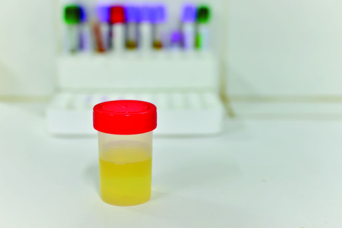
Diagnosing urinary tract infections can be achieved by determining the white blood cell concentration of the patient’s urine, according to a new study.
“Previously recommended pyuria thresholds for the presumptive diagnosis of UTI in young infants were based on manual microscopy of centrifuged urine [but] test performance has not been studied in newer automated systems that analyze uncentrifuged urine,” wrote Pradip P. Chaudhari, MD, and his associates at Harvard University in Boston.
Of these 2,700 infants with a median age of 1.7 months, 211 (7.8%) had a urine culture come back positive for UTI. Likelihood ratio (LR) positive and negative were calculated to determine the microscopic pyuria thresholds at which UTIs became more likely in both dilute and concentrated urine. A white blood cell to high-power field (WBC/HPF) count of 3 yielded an LR-positive of 9.9 and LR-negative of just 0.15, making it the cutoff for dilute urine samples. For concentrated urine samples, 6 WBC/HPF had an LR-positive of 10.1 and LR-negative of 0.17, making it the cutoff for those samples. Leukocyte esterase (LE) thresholds also were determined for dipstick testing, with investigators finding that any positive result on the dipstick was a strong indicator of UTI.
“The optimal diagnostic threshold for microscopic pyuria varies by urine concentration,” the authors concluded. “For young infants, urine concentration should be incorporated into the interpretation of uncentrifuged urine analyzed by automated microscopic urinalysis systems.”
There was no external funding for this study. Dr. Chaudhari and his coauthors did not report any relevant financial disclosures.
In this issue of Pediatrics, Chaudhari et al. share the results of a study of the impact of urine concentration on the optimal threshold in the new era of automated urinalysis. Centrifugation of urine specimens has long been standard laboratory practice, presumably performed to concentrate sediment and facilitate the detection of cellular elements and bacteria.
However, for the many sites that do not have machines for automated urinalyses (virtually all office practices, for example), the most important finding in this study may well be how well LE [leukocyte esterase] performs regardless of urine concentration. The optimal threshold for LE is not clear, however. The authors use “small” as their threshold for LE. At any threshold, can a negative urinalysis be relied on to exclude the diagnosis of UTI? A “positive” culture without inflammation evident in the urine is likely due to contamination, very early infection (rare), or asymptomatic bacteriuria (positive urine cultures in febrile children can still represent asymptomatic bacteriuria, because the fever may be due to a source other than the urinary tract).
If there are, in fact, some true UTIs without evidence of inflammation from the urinalysis, are they as harmful as those with “pyuria”?
Animal data demonstrate it is the inflammatory response, not the presence of organisms, that causes renal damage in the form of scarring. So the role of using evidence of inflammation in the urine to screen for who needs a culture seems justified on the basis not only of practicality at point of care and likelihood of UTI, but also sparing individuals at low to no risk of scarring from invasive urine collection. Moreover, using the urinalysis as a screen permits selecting individuals for antimicrobial treatment 24 hours sooner than if clinicians were to wait for culture results before treating. The urinalysis provides a practical window for clinicians to render prompt treatment. And Chaudhari et al. provide valuable assistance for interpreting the results of automated urinalyses.
Kenneth B. Roberts, MD , is a professor of therapeutic radiology at Yale University, New Haven, Conn. He did not report any relevant financial disclosures. These comments are excerpted from a commentary that accompanied Dr. Chaudhari and his associates’ study ( Pediatrics. 2016;138(5):e20162877 ).
Diagnosing urinary tract infections can be achieved by determining the white blood cell concentration of the patient’s urine, according to a new study.
“Previously recommended pyuria thresholds for the presumptive diagnosis of UTI in young infants were based on manual microscopy of centrifuged urine [but] test performance has not been studied in newer automated systems that analyze uncentrifuged urine,” wrote Pradip P. Chaudhari, MD, and his associates at Harvard University in Boston.
Of these 2,700 infants with a median age of 1.7 months, 211 (7.8%) had a urine culture come back positive for UTI. Likelihood ratio (LR) positive and negative were calculated to determine the microscopic pyuria thresholds at which UTIs became more likely in both dilute and concentrated urine. A white blood cell to high-power field (WBC/HPF) count of 3 yielded an LR-positive of 9.9 and LR-negative of just 0.15, making it the cutoff for dilute urine samples. For concentrated urine samples, 6 WBC/HPF had an LR-positive of 10.1 and LR-negative of 0.17, making it the cutoff for those samples. Leukocyte esterase (LE) thresholds also were determined for dipstick testing, with investigators finding that any positive result on the dipstick was a strong indicator of UTI.
“The optimal diagnostic threshold for microscopic pyuria varies by urine concentration,” the authors concluded. “For young infants, urine concentration should be incorporated into the interpretation of uncentrifuged urine analyzed by automated microscopic urinalysis systems.”
There was no external funding for this study. Dr. Chaudhari and his coauthors did not report any relevant financial disclosures.
FROM PEDIATRICS
Key clinical point:
Major finding: UTIs can be safely diagnosed if the patient has a pyuria threshold of at least 3 WBC/HPF in dilute urine and 6 WBC/HPF in concentrated urine.
Data source: Retrospective cross-sectional study of 2,700 infants younger than 3 months between May 2009 and December 2014.
Disclosures: No external funding for this study; authors did not report any relevant financial disclosures.
Myth of the Month: Does nitroglycerin response predict coronary artery disease?
A 55-year-old man presents to the emergency department with substernal chest pain. The pain has occurred off and on over the past 2 hours. He has no family history of coronary artery disease. He has no history of diabetes, hypertension, or cigarette smoking. His most recent total cholesterol was 220 mg/dL (HDL, 40; LDL, 155). Blood pressure is 130/70. An ECG obtained on arrival is unremarkable. When he reached the ED, he received a nitroglycerin tablet with resolution of his pain within 4 minutes.
What is the most accurate statement?
A. The chance of CAD in this man over the next 10 years was 8% before his symptoms and is now greater than 20%.
B. The chance of CAD in this man over the next 10 years was 8% and is still 8%.
C. The chance of CAD in this man over the next 10 years was 15% before his symptoms and is now close to 100%.
D. The chance of CAD in this man over the next 10 years was 15% before his symptoms and is now close to 50%.
For years, giving nitroglycerin to patients who present with chest pain has been considered a good therapy, and the response to the medication has been considered a sign that the pain was likely due to cardiac ischemia. Is there evidence that this is true?
The study was a retrospective review of 223 patients who presented to the ED over a 5-month period with ongoing chest pain. They looked at patients who had ongoing chest pain in the ED, received nitroglycerin, and did not receive any therapy other than aspirin within 10 minutes of receiving nitroglycerin. Nitroglycerin response was compared with the final diagnosis of cardiac versus noncardiac chest pain.
Of the patients with a final determination of cardiac chest pain, 88% had a nitroglycerin response, whereas 92% of the patients with noncardiac chest pain had a nitroglycerin response (P = .50).
Deborah B. Diercks, MD, and her colleagues looked at improvement in chest pain scores in the ED in patients treated with nitroglycerin and whether it correlated with a cardiac etiology of chest pain.2 The study was a prospective, observational study of 664 patients in an urban tertiary care ED over a 16-month period. An 11-point numeric chest pain scale was assessed and recorded by research assistants before and 5 minutes after receiving nitroglycerin. The scale ranged from 0 (no pain) to 10 (worst pain imaginable).
A final diagnosis of a cardiac etiology for chest pain was found in 18% of the patients in the study. Of the patients who had cardiac-related chest pain, 20% had no reduction in pain with nitroglycerin, compared with 19% of the patients without cardiac-related chest pain. Complete or significant reduction in chest pain occurred with nitroglycerin in 31% of patients with cardiac chest pain and 27% of the patients without cardiac chest pain (P = .76).
Two other studies with similar designs showed similar results. Robert Steele, MD, and his colleagues studied 270 patients in a prospective observational cohort study of patients with chest pain presenting to an urban ED.3 Patients presenting to the ED with active chest pain who received nitroglycerin were enrolled.
The sensitivity in this study for nitroglycerin relief determining cardiac chest pain was 72%, and the specificity was 37%, with a positive likelihood ratio for coronary artery disease if nitroglycerin response of 1.1 (0.96-1.34).
In another prospective, observational cohort study, 459 patients who presented to an ED with chest pain were evaluated for response to nitroglycerin as a marker for ischemic cardiac disease.4 In this study, presence of ischemic cardiac disease was defined as diagnosis in the ED or during a 4-month follow-up period. Nitroglycerin relieved chest pain in 35% of patients who had coronary disease, whereas 41% of patients without coronary disease had a nitroglycerin response. This study had a much lower overall nitroglycerin response rate than any of the other studies.
Katherine Grailey, MD, and Paul Glasziou, MD, PhD, published a meta-analysis of nitroglycerin use for the diagnosis of chest pain, using the above referenced studies. They concluded that in the acute setting, nitroglycerin is not a reliable test of treatment for use in diagnosis of coronary artery disease.5
High response rate for nitroglycerin in the noncoronary artery groups in the studies may be due to a strong placebo effect and/or that nitroglycerin may help with pain caused by esophageal spasm. The lack of specificity in the pain relief response for nitroglycerin makes it not a helpful test. Note that all the studies have been in the acute, ED setting for chest pain. In the case presented at the beginning of the article, the response the patient had to nitroglycerin would not change the probability that he has coronary artery disease.
References
1. Am J Cardiol. 2002 Dec 1;90(11):1264-6.
2. Ann Emerg Med. 2005 Jun;45(6):581-5.
3. CJEM. 2006 May;8(3):164-9.
4. Ann Intern Med. 2003 Dec 16;139(12):979-86.
5. Emerg Med J. 2012 Mar;29(3):173-6.
Dr. Paauw is professor of medicine in the division of general internal medicine at the University of Washington, Seattle, and he serves as third-year medical student clerkship director at the University of Washington. Contact Dr. Paauw at [email protected] .
CD64 validated as biomarker for pediatric Crohn’s disease
MONTREAL – Blood levels of a neutrophil receptor protein, CD64, proved to be a reliable, noninvasive marker of both Crohn’s disease activity and the risk for relapse from remission in children and adolescents in a pair of single-center studies with a total of 140 patients.
An elevation in blood levels of CD64, a marker for inflammation, in asymptomatic patients with Crohn’s disease “is a significant risk factor for treatment failure or complications during infliximab maintenance,” Phillip Minar, MD, said at the World Congress of Pediatric Gastroenterology, Hepatology and Nutrition. Although Dr. Minar acknowledged that larger validation studies are still needed, neutrophil CD64 levels can potentially serve as a “treat-to-target” biomarker of disease status in selected pediatric Crohn’s disease patients.
Dr. Minar cautioned that in some pediatric patients with Crohn’s disease CD64 is not an effective marker for inflammation and a change in their Crohn’s disease status. In his study, the sensitivity of an elevated CD64 level was 64% as a surrogate marker for mucosal damage seen with endoscopy.
“I get a CD64 level at the time we diagnose Crohn’s disease. If it is elevated, then I will follow it; if it is not elevated, then I won’t use it for that patient. It’s patient specific,” he explained in an interview.
Dr. Minar and his associates first established the prognostic value of elevated CD64 levels in patients with Crohn’s disease in a study with 208 pediatric patients with inflammatory bowel disease and 43 controls (Inflam Bowel Dis. 2014, Jun;20[6]:1037-48). His new validation study included 105 pediatric patients with Crohn’s disease, of whom 54 were newly diagnosed. Among the 51 previously diagnosed patients, 18 had inactive disease. The patients averaged 14 years old, and all 105 underwent endoscopy to directly examine their Crohn’s disease activity.
The results showed clear and statistically significant correlations among the average CD64 levels in the patients and the blinded endoscopic evaluations that categorized the patients as having inactive Crohn’s disease, mild disease, or moderate to severe disease. The results also suggested that a useful dichotomous cut point for CD64 was an index of 1. Among patients with a level above 1, diagnostic sensitivity for mucosal damage was 64% and specificity was 100%, he reported. In these studies as well as their routine practice, Dr. Minar and his associates use a commercially available immunoassay for quantifying blood levels of CD64.
The second study he reported on assessed the ability of CD64 levels to predict a patient’s status on infliximab (Remicade) maintenance treatment. This study enrolled 35 pediatric patients, who averaged about 15 years old, had been diagnosed with Crohn’s disease for an average of about 2 years and were in remission after having received at least four serial infliximab doses. During 1 year of follow-up, 15 patients relapsed and 21 remained in remission.
The researchers measured CD64 levels at baseline and found that, during the next year, those who had a CD64 index of less than 1 at baseline had a relapse rate of less than 40% during follow-up, while those with a CD64 index of 1 or greater at baseline had a relapse rate of more than 70% during follow-up, a statistically significant difference between the two subgroups. The analysis also showed that lower CD64 levels linked with higher trough levels of infliximab.
A multivariate analysis showed that a CD64 index level of 1 or greater at baseline linked with a statistically significant, 4.5-fold increased risk for relapse, compared with patients with a baseline CD64 level below 1. This analysis identified three additional significant correlates of an elevated risk for relapse: nonwhite race, a baseline serum albumin level of less than 3.9 g/dL, and a baseline infliximab serum level of less than 5 mcg/mL.
The CD64 test that his group has been using typically has a work week turnaround time of about an hour, and costs less than $100 per test per patient. Blood levels of CD64 are stable for 48 hours in the refrigerator, so specimens can sit over a weekend without compromising results. The Cincinnati group is planning to soon change to an in-house test that will cost about $10-$20 per test per patient, Dr. Minar said.
Dr. Minar had no relevant financial disclosures.
[email protected]
On Twitter @mitchelzoler
We are in desperate need of more reliable biomarkers of disease activity in patients with Crohn’s disease. Identifying effective noninvasive biomarkers has been a holy grail that we have pursued for many years because what we currently have is imperfect. CD64 appears to be a very reliable and specific biomarker of disease activity.
I think pediatric gastroenterologists will pay attention to Dr. Minar’s report. The entire community is very interested in this and will be watching the evolution of the science behind CD64 assessment.
John A. Barnard, MD , is chief of pediatrics at Nationwide Children’s Hospital and professor and chairman of pediatrics at Ohio State University, both in Columbus. He had no relevant disclosures. He made these comments in an interview.
MONTREAL – Blood levels of a neutrophil receptor protein, CD64, proved to be a reliable, noninvasive marker of both Crohn’s disease activity and the risk for relapse from remission in children and adolescents in a pair of single-center studies with a total of 140 patients.
An elevation in blood levels of CD64, a marker for inflammation, in asymptomatic patients with Crohn’s disease “is a significant risk factor for treatment failure or complications during infliximab maintenance,” Phillip Minar, MD, said at the World Congress of Pediatric Gastroenterology, Hepatology and Nutrition. Although Dr. Minar acknowledged that larger validation studies are still needed, neutrophil CD64 levels can potentially serve as a “treat-to-target” biomarker of disease status in selected pediatric Crohn’s disease patients.
Dr. Minar cautioned that in some pediatric patients with Crohn’s disease CD64 is not an effective marker for inflammation and a change in their Crohn’s disease status. In his study, the sensitivity of an elevated CD64 level was 64% as a surrogate marker for mucosal damage seen with endoscopy.
“I get a CD64 level at the time we diagnose Crohn’s disease. If it is elevated, then I will follow it; if it is not elevated, then I won’t use it for that patient. It’s patient specific,” he explained in an interview.
Dr. Minar and his associates first established the prognostic value of elevated CD64 levels in patients with Crohn’s disease in a study with 208 pediatric patients with inflammatory bowel disease and 43 controls (Inflam Bowel Dis. 2014, Jun;20[6]:1037-48). His new validation study included 105 pediatric patients with Crohn’s disease, of whom 54 were newly diagnosed. Among the 51 previously diagnosed patients, 18 had inactive disease. The patients averaged 14 years old, and all 105 underwent endoscopy to directly examine their Crohn’s disease activity.
The results showed clear and statistically significant correlations among the average CD64 levels in the patients and the blinded endoscopic evaluations that categorized the patients as having inactive Crohn’s disease, mild disease, or moderate to severe disease. The results also suggested that a useful dichotomous cut point for CD64 was an index of 1. Among patients with a level above 1, diagnostic sensitivity for mucosal damage was 64% and specificity was 100%, he reported. In these studies as well as their routine practice, Dr. Minar and his associates use a commercially available immunoassay for quantifying blood levels of CD64.
The second study he reported on assessed the ability of CD64 levels to predict a patient’s status on infliximab (Remicade) maintenance treatment. This study enrolled 35 pediatric patients, who averaged about 15 years old, had been diagnosed with Crohn’s disease for an average of about 2 years and were in remission after having received at least four serial infliximab doses. During 1 year of follow-up, 15 patients relapsed and 21 remained in remission.
The researchers measured CD64 levels at baseline and found that, during the next year, those who had a CD64 index of less than 1 at baseline had a relapse rate of less than 40% during follow-up, while those with a CD64 index of 1 or greater at baseline had a relapse rate of more than 70% during follow-up, a statistically significant difference between the two subgroups. The analysis also showed that lower CD64 levels linked with higher trough levels of infliximab.
A multivariate analysis showed that a CD64 index level of 1 or greater at baseline linked with a statistically significant, 4.5-fold increased risk for relapse, compared with patients with a baseline CD64 level below 1. This analysis identified three additional significant correlates of an elevated risk for relapse: nonwhite race, a baseline serum albumin level of less than 3.9 g/dL, and a baseline infliximab serum level of less than 5 mcg/mL.
The CD64 test that his group has been using typically has a work week turnaround time of about an hour, and costs less than $100 per test per patient. Blood levels of CD64 are stable for 48 hours in the refrigerator, so specimens can sit over a weekend without compromising results. The Cincinnati group is planning to soon change to an in-house test that will cost about $10-$20 per test per patient, Dr. Minar said.
Dr. Minar had no relevant financial disclosures.
[email protected]
On Twitter @mitchelzoler
AT WCPGHAN 2016
Key clinical point: Results from two studies further validated neutrophil CD64 as a highly specific biomarker for Crohn’s disease severity in children and adolescents and suggested that CD64 could serve as a treat-to-target guide for infliximab treatment.
Major finding: During infliximab maintenance, relapses occurred in fewer than 40% of pediatric Crohn’s disease patients with low CD64 and in more than 70% with high CD64.
Data source: A single-center study of 105 pediatric patients with Crohn’s disease to assess disease severity correlates, and 35 patients in remission on infliximab to assess predicted efficacy.
Disclosures: Dr. Minar had no relevant financial disclosures.


