User login
It’s time for universal HCV screening in the ED
SAN FRANCISCO – Emergency departments are the ideal place to screen for hepatitis C infection, according to investigators from Vanderbilt University, Nashville, Tenn.
Current recommendations call for screening baby boomers born from 1945 to 1965 and patients with risk factors, especially injection drug use. The problem is that the guidelines don’t say, exactly, how and where that should be done, so uptake has been spotty. Also, people aren’t exactly forthcoming when it comes to admitting IV drug use.
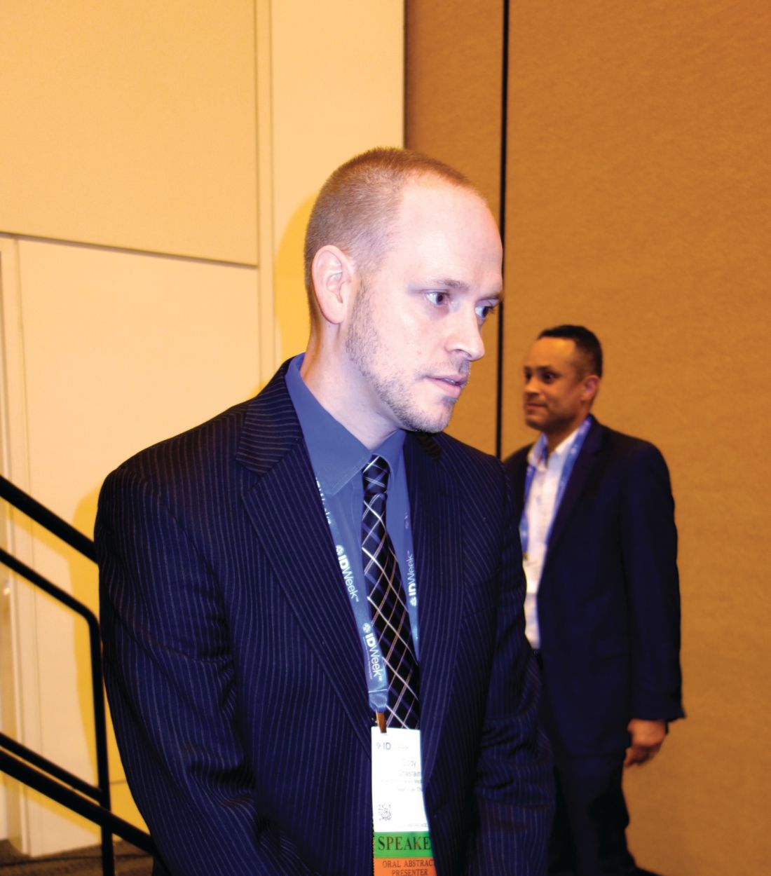
Enter universal screening in the ED. Vanderbilt is one of several academic centers that have adopted the approach, and others are following suit. Across the board, they’ve found that HCV infection is more common than projections based on baby boomer and risk factor demographics suggest, and, even more importantly, the boomer/risk factor strategy misses a large number of active cases, said Cody A. Chastain, MD, assistant professor of infectious diseases at Vanderbilt, who led the ED screening initiative.
In short, universal screening in the ED would keep people from falling through the cracks.
From April 2017 to March 2018, every adult who had blood drawn at Vanderbilt’s tertiary care ED was asked by a nurse if they’d also like to be checked for HCV, so long as they were alert enough for the conversation. If they agreed, an additional phlebotomy tube was added to the draw, and sent off for testing. Fewer than 5% of patients opted out.
Antibody positive samples were automatically screened for active disease by HCV RNA. Results were entered into the medical record and shared with patients at discharge. Active cases were counseled and offered linkage to care, regardless of insurance status.
The initiative screened 11,637 patients; 1,008 (8.7%) were antibody positive, of whom 488 (48%) were RNA positive. Thirty-seven percent of the active cases were in non–baby boomers – most born after 1965 – with no known injection drug use. The baby boomer/risk factor model would have missed most of them.
Also, spontaneous clearance – antibody positive, RNA negative without HCV treatment – “is dramatically higher” than what’s thought. “The historic estimate of 20% clearly is not reflected” in the Vanderbilt results, nor in similar universal screening studies; “spontaneous clearance is about 50% or so,” Dr. Chastain said.
Even so, “virtually every study published in this space finds more cases of infection than traditional screening would find. [Our work] is just one more piece of data” to indicate the usefulness of the approach. “Emergency departments [are] ideal for hepatitis C screening,” he said at IDWeek, an annual scientific meeting on infectious diseases, where he presented the findings.
“This is well trodden territory; we’ve already addressed it with HIV. We recognized that HIV screening had a stigma and was a challenge, [so we] moved to universal screening” of all adults, at least once. It “drastically improved screening rates. I don’t see a rational reason” not to do this for hepatitis C. “There are very well-meaning people who engage in the cost effectiveness side of this discussion, but I don’t think it helps us in our efforts to control this epidemic from a public health standpoint,” Dr. Chastain said.
Vanderbilt continues to screen for HCV in the ED; the next step is to see how well efforts to link active cases with care are working. Many times during the study, Dr. Chastain said positive patients eventually revealed that they already knew they had HCV, but had been told there was nothing they could do about it, so they didn’t get care. Maybe they were told that because they didn’t have insurance.
Vanderbilt has dropped screening ED patients born before 1945 because the odds of picking up an unknown HCV infection proved to be tiny, and, in any case, patients are generally too comorbid for treatment. It’s made screening more efficient.
Dr. Chastain reported that he had no personal disclosures. The study was funded by Vanderbilt, which receives grants from pharmaceutical companies.
SOURCE: Chastain C et al. 2018 ID Week, Abstract 932.
SAN FRANCISCO – Emergency departments are the ideal place to screen for hepatitis C infection, according to investigators from Vanderbilt University, Nashville, Tenn.
Current recommendations call for screening baby boomers born from 1945 to 1965 and patients with risk factors, especially injection drug use. The problem is that the guidelines don’t say, exactly, how and where that should be done, so uptake has been spotty. Also, people aren’t exactly forthcoming when it comes to admitting IV drug use.
Enter universal screening in the ED. Vanderbilt is one of several academic centers that have adopted the approach, and others are following suit. Across the board, they’ve found that HCV infection is more common than projections based on baby boomer and risk factor demographics suggest, and, even more importantly, the boomer/risk factor strategy misses a large number of active cases, said Cody A. Chastain, MD, assistant professor of infectious diseases at Vanderbilt, who led the ED screening initiative.
In short, universal screening in the ED would keep people from falling through the cracks.
From April 2017 to March 2018, every adult who had blood drawn at Vanderbilt’s tertiary care ED was asked by a nurse if they’d also like to be checked for HCV, so long as they were alert enough for the conversation. If they agreed, an additional phlebotomy tube was added to the draw, and sent off for testing. Fewer than 5% of patients opted out.
Antibody positive samples were automatically screened for active disease by HCV RNA. Results were entered into the medical record and shared with patients at discharge. Active cases were counseled and offered linkage to care, regardless of insurance status.
The initiative screened 11,637 patients; 1,008 (8.7%) were antibody positive, of whom 488 (48%) were RNA positive. Thirty-seven percent of the active cases were in non–baby boomers – most born after 1965 – with no known injection drug use. The baby boomer/risk factor model would have missed most of them.
Also, spontaneous clearance – antibody positive, RNA negative without HCV treatment – “is dramatically higher” than what’s thought. “The historic estimate of 20% clearly is not reflected” in the Vanderbilt results, nor in similar universal screening studies; “spontaneous clearance is about 50% or so,” Dr. Chastain said.
Even so, “virtually every study published in this space finds more cases of infection than traditional screening would find. [Our work] is just one more piece of data” to indicate the usefulness of the approach. “Emergency departments [are] ideal for hepatitis C screening,” he said at IDWeek, an annual scientific meeting on infectious diseases, where he presented the findings.
“This is well trodden territory; we’ve already addressed it with HIV. We recognized that HIV screening had a stigma and was a challenge, [so we] moved to universal screening” of all adults, at least once. It “drastically improved screening rates. I don’t see a rational reason” not to do this for hepatitis C. “There are very well-meaning people who engage in the cost effectiveness side of this discussion, but I don’t think it helps us in our efforts to control this epidemic from a public health standpoint,” Dr. Chastain said.
Vanderbilt continues to screen for HCV in the ED; the next step is to see how well efforts to link active cases with care are working. Many times during the study, Dr. Chastain said positive patients eventually revealed that they already knew they had HCV, but had been told there was nothing they could do about it, so they didn’t get care. Maybe they were told that because they didn’t have insurance.
Vanderbilt has dropped screening ED patients born before 1945 because the odds of picking up an unknown HCV infection proved to be tiny, and, in any case, patients are generally too comorbid for treatment. It’s made screening more efficient.
Dr. Chastain reported that he had no personal disclosures. The study was funded by Vanderbilt, which receives grants from pharmaceutical companies.
SOURCE: Chastain C et al. 2018 ID Week, Abstract 932.
REPORTING FROM IDWEEK 2018
Key clinical point: HCV infection is more common than traditionally thought; screening in the ED will keep people from falling through the cracks.
Major finding: Of patients screened; 8.7% were antibody positive, with 48% of these RNA positive; 37% of active cases were in non–baby boomers with no known injection drug use. Spontaneous remission appeared to top 50%.
Study details: Quality improvement initiative in Vanderbilt University’s tertiary care ED.
Disclosures: Dr. Chastain reported that he had no disclosures. The study was funded by Vanderbilt, which receives grants from pharmaceutical companies.
Source: Chastain C et al. 2018 ID Week, Abstract 932.
Combined novel risk factors raise coronary event rates
CHICAGO – Individuals with both elevated lipoprotein(a) levels and a family history of coronary heart disease are at a considerably higher long-term risk of atherosclerotic cardiovascular disease and coronary heart disease events than those with one risk factor alone, according to results from a large clinical study presented at the American Heart Association scientific sessions.
“Elevated lipoprotein(a) levels or a positive family history of coronary heart disease each is independently associated with cardiovascular disease risk,” said Anurag Mehta, MD, of Emory University School of Medicine, Atlanta. “This study showed that the presence of both an elevated Lp(a) level and a positive family history has an additive joint association with long-term cardiovascular risk.”
Dr. Mehta reported on an analysis of 12,149 individuals participating in the Atherosclerosis Risk in Communities (ARIC) study. All study participants were free of cardiovascular disease at the time of enrollment. The researchers measured Lp(a) levels and ascertained family history by self-report. Forty-four percent of the study participants had a family history of coronary heart disease (CHD), and 23% were black.
Median follow-up of study participants was 21 years, over which time 3,114 atherosclerotic cardiovascular disease (ASCVD) and 2,283 CHD events occurred.
Black participants had a significantly higher average plasma Lp(a) concentration than white persons, at 16.7 mg/dL vs. 5.7 mg/dL. However, plasma Lp(a) levels between participants with either a positive or a negative family history of CHD were similar on average, 7.6 mg/dL and 7.8 mg/dL, respectively.
The study pooled black and white ARIC participants by race-specific Lp(a) levels (quintiles) and stratified them into four different groups: 1. positive family history and an elevated race-specific Lp(a) level (quintile 5); 2. positive family history and nonelevated race-specific Lp(a) level (quintiles 1-4); 3. negative family history and elevated race-specific Lp(a) level; and 4. negative family history and nonelevated race-specific Lp(a) level. “There was an increase in the proportion of participants with a family history of CHD across race-specific Lp(a) quintiles, highlighting the fact that family history is associated with race-specific Lp(a) levels,” Dr. Mehta said.
“We observed that the ASCVD incidence was higher among participants with an elevated Lp(a) level or a family history of CHD as compared with participants with nonelevated Lp(a) levels and no family history,” Dr. Mehta said. “The highest ASCVD incidence was noted among participants with an elevated Lp(a) level as well as a positive family history.” Among those patients, the cumulative incidence of ASCVD events was nearly 25%, compared with 22% for those with a positive family history and nonelevated Lp(a) levels (group 2) or those with a negative family history but elevated Lp(a) levels (group 3), and 18% for those with negative family history and nonelevated Lp(a) levels.
Results for the cumulative incidence of coronary events trended similarly, Dr. Mehta noted: around 22% for group 1, 19% for group 2, 17% for group 3, and 14% for group 4.
“Having an elevated Lp(a) level as well as a family history of CHD was associated with a higher adjusted hazard for ASCVD and coronary events,” he said. Group 1 patients had a 43% greater risk for ASCVD and 68% greater risk for CHD, respectively, compared with a 16% and 30% greater risk for group 2, and 20% and 27% greater risk for group 3.
“Our findings indicate that these easily measurable nontraditional risk markers can help identify those at an elevated long-term CVD risk and may be useful for informing CVD prevention strategies among asymptomatic individuals,” Dr. Mehta said.
Dr. Mehta had no financial relationships to disclose.
SOURCE: Mehta A et al. AHA scientific sessions, Abstract AT.AOS.03 119.
REPORTING FROM THE AHA SCIENTIFIC SESSIONS
Key clinical point: .
Major finding: Patients with both risk factors had a 68% greater risk for coronary events.
Study details: Analysis of 12,149 asymptomatic participants enrolled in the ARIC study.
Disclosures: Dr. Mehta has no financial relationships to report.
Source: Mehta A et al. AHA scientific sessions, Abstract AT.AOS.03 119.
How does caring affect the placebo effect?
How thorough are you when you prescribe medication? You check the patient’s list of allergies and current medications. You make sure that the dose is appropriate for the patient’s weight. Hopefully, you spend a minute or 2 describing the most common side effects. You prescribe the correct amount of medication and an appropriate number of refills. If you think you can distill it into one or two sentences, you also explain the medication’s mechanism of action. That is if you understand it yourself.

What about placebos? How often do you believe that your patient has gotten better because of the placebo effect? Do you ever intentionally recommend or prescribe a placebo? Do you share with the patient that there is no current explanation of why the treatment you are recommending should work? Or, do you just play dumb?
Whether you admit to being a frequent prescriber of placebos or not you should take the 20 minutes it will take to read a New York Times article titled “What if the Placebo Effect Isn’t a Trick” (Gary Greenberg, Nov 7, 2018). You will learn a bit about the history of the placebo effect including some recent functional MRI studies that have uncovered consistent brain activity patterns in subjects that respond to placebos.
You will read about some exciting research indicating that certain people with a genomic variant of an enzyme that has been shown to affect the response to painkillers generally have the weakest response to placebo. While in some studies the association between the patient’s response and the level of the enzyme is the reverse, Kathryn Hall, PhD, the molecular biologist overseeing these studies, feels that at this point in her research the fact that there is an association that varies with genotype is a critical finding. She suspects that the placebo effect and the drug operate on the same biochemical highway that includes this enzyme and that “clinician warmth” is particularly effective in patients with a certain genotype.
Ted Kaptchuk, who heads up Harvard Medical School’s Program in Placebo Studies and the Therapeutic Encounter and has collaborated with Dr. Hall, hypothesizes “that the placebo effect is a biological response to an act of caring.” Is Dr. Hall’s work the first step in defining that response?
What does all of this new information mean for us as care dispensers? I think it means that caring is important and can make a critical difference if we have chosen a patient with the favorable genome. Of course, how are we to know whether we are working with such a patient? All the caring in the world may not change the outcome if we have selected incorrectly.
And then there is the other side of the practitioner-patient relationship and the definition and quantification of “caring.” Are there practitioners who are so inept and/or devoid of caring that even patients with the most favorable genome are not going to respond to their attempts at dispensing placebos?
Are there some practitioners who are born with a knack for caring? Can it be taught? Do we select for the quality of caring with the Medical College Admission Test (MCAT)? Do we weed out those who obviously don’t have it during their training?

Is caring a finite resource that can be exhausted? Is it affected by sleep deprivation or marital troubles at home? Or hours sitting in front of a computer screen? I suspect I know the answers to some of these questions. But what I do know for sure is that the placebo effect is real and is just another example that practicing medicine is more of an art than a science.
Dr. Wilkoff practiced primary care pediatrics in Brunswick, Maine, for nearly 40 years. He has authored several books on behavioral pediatrics, including “How to Say No to Your Toddler.” Email him at [email protected].
How thorough are you when you prescribe medication? You check the patient’s list of allergies and current medications. You make sure that the dose is appropriate for the patient’s weight. Hopefully, you spend a minute or 2 describing the most common side effects. You prescribe the correct amount of medication and an appropriate number of refills. If you think you can distill it into one or two sentences, you also explain the medication’s mechanism of action. That is if you understand it yourself.
What about placebos? How often do you believe that your patient has gotten better because of the placebo effect? Do you ever intentionally recommend or prescribe a placebo? Do you share with the patient that there is no current explanation of why the treatment you are recommending should work? Or, do you just play dumb?
Whether you admit to being a frequent prescriber of placebos or not you should take the 20 minutes it will take to read a New York Times article titled “What if the Placebo Effect Isn’t a Trick” (Gary Greenberg, Nov 7, 2018). You will learn a bit about the history of the placebo effect including some recent functional MRI studies that have uncovered consistent brain activity patterns in subjects that respond to placebos.
You will read about some exciting research indicating that certain people with a genomic variant of an enzyme that has been shown to affect the response to painkillers generally have the weakest response to placebo. While in some studies the association between the patient’s response and the level of the enzyme is the reverse, Kathryn Hall, PhD, the molecular biologist overseeing these studies, feels that at this point in her research the fact that there is an association that varies with genotype is a critical finding. She suspects that the placebo effect and the drug operate on the same biochemical highway that includes this enzyme and that “clinician warmth” is particularly effective in patients with a certain genotype.
Ted Kaptchuk, who heads up Harvard Medical School’s Program in Placebo Studies and the Therapeutic Encounter and has collaborated with Dr. Hall, hypothesizes “that the placebo effect is a biological response to an act of caring.” Is Dr. Hall’s work the first step in defining that response?
What does all of this new information mean for us as care dispensers? I think it means that caring is important and can make a critical difference if we have chosen a patient with the favorable genome. Of course, how are we to know whether we are working with such a patient? All the caring in the world may not change the outcome if we have selected incorrectly.
And then there is the other side of the practitioner-patient relationship and the definition and quantification of “caring.” Are there practitioners who are so inept and/or devoid of caring that even patients with the most favorable genome are not going to respond to their attempts at dispensing placebos?
Are there some practitioners who are born with a knack for caring? Can it be taught? Do we select for the quality of caring with the Medical College Admission Test (MCAT)? Do we weed out those who obviously don’t have it during their training?
Is caring a finite resource that can be exhausted? Is it affected by sleep deprivation or marital troubles at home? Or hours sitting in front of a computer screen? I suspect I know the answers to some of these questions. But what I do know for sure is that the placebo effect is real and is just another example that practicing medicine is more of an art than a science.
Dr. Wilkoff practiced primary care pediatrics in Brunswick, Maine, for nearly 40 years. He has authored several books on behavioral pediatrics, including “How to Say No to Your Toddler.” Email him at [email protected].

An iPledge Halloween
It was a dark and stormy night.

OK, it was a warm and sunny afternoon. But Halloween was approaching. Strange things happen. Plus, the patient’s name was Ichabod ...
OK, his name was Jerry. Jerry came to Boston from Chula Taco, Calif., to study at CIT, the famed Boston Chipotle Institute of Technology. He’d finished 4 months of isotretinoin and needed one more.
I asked him to call iPledge to request a transfer to me. He called back later to say that This seemed odd, since his pills had only run out 3 days before.
Having confirmed his name, address, telephone number, and the last four digits of his social security number, I tried enrolling him on iPledge at 5:30 p.m. (Cue: thunder and lightning), expecting to get a request for an override code. Instead the screen just asked for his iPledge number (you have to use the old one, you know). I called iPledge (my favorite pastime), identified myself by the usual means (Full name. iPledge ID number. Date of personal significance. Office telephone. Thank you. How can I help you?).
I explained my dilemma. The representative asked that I verify Jerry’s identity. I gave her his name, date of birth, and the last four of his social.
“We have his name and date of birth,” she said, “but the social security digits don’t match.” She asked for his phone number, but his Boston number didn’t match what she had. “Do you have his address?” she asked. I did not, since he’d given me his Boston address, not his California one.
I left her on hold and called Jerry on my cell. He confirmed that the social security digits he’d given me were correct. He gave me his mother’s cell phone number, but that also turned out not to be what iPledge had on file.
“What other identifying information can I give you?” I asked the iPledge rep. “How about his home address?” she said. Back to my cell: “Jerry, what’s your home address?” “It’s 2470 Chalupa Drive, Chula Taco, California 9090909-090909,” he said.
I repeated that to the iPledge representative. “Please hold a moment,” she said.
She was back. “The street address is correct,” she said, “The ZIP is correct. But the town is wrong.”
The town is wrong? If Jerry didn’t know either the last four of his social or his town, how did he get Amazon deliveries? Was this identity theft by an Accutane seeker? Maybe Jerry was really a Russian spy with dry lips posing as an acne patient! (Cue: screeches, howls, more thunder.)
“Can you tell me which town you have listed for him?” I asked iPledge.
“No,” she said, “because you haven’t identified him properly yet,” (emphasis added).
Back to the cell: “Jerry, are you sure you know what town you live in?” He insisted he did. (But then, so would a spy, wouldn’t he?)
In near despair, I returned to the iPledge rep. “I really want to get this patient his medication, “I said. “And I really want to go home. Can you help either of us?”
“Let me get my supervisor,” she said. “This may take a few minutes.” I hung up on Jerry and, in a blaze of multitasking, filled out three Prior Authorization forms for clindamycin gel.
“I found your patient,” said the rep, returning at last. “Not only that, I was able to reregister him in the iPledge program. Want to know his iPledge number?
Of course!
“Now that he’s registered,” I said, “could you give me the name of the town you have him listed as living in on Chalupa Drive?”
“Sure,” she said, “We have him in Rancho Carmen Miranda. Can help you with anything else today?”
“No, thanks ...”

“Would you be willing to take a 2-minute survey ...?”
“No, but thank you very much!” I said, hanging up in triumph. (Cue: sunshine, violins.)
Back to the cell: “Jerry, you’re in! Here’s your iPledge number.”
“Thanks, Doc.”
“By the way, Jerry, iPledge has you living in the town of Rancho Carmen Miranda. Do you live there?”
“No,” said Jerry. “I don’t.”
“Well, Jerry, for 1 more month, for federal purposes, you do!”
I’m sure there’s a good explanation for all this. I just don’t want to know it. Just pass the candy corn.
Dr. Rockoff practices dermatology in Brookline, Mass., and is a longtime contributor to Dermatology News. He serves on the clinical faculty at Tufts University, Boston, and has taught senior medical students and other trainees for 30 years. His second book, “Act Like a Doctor, Think Like a Patient,” is available at amazon.com and barnesandnoble.com. Write to him at [email protected].
It was a dark and stormy night.
OK, it was a warm and sunny afternoon. But Halloween was approaching. Strange things happen. Plus, the patient’s name was Ichabod ...
OK, his name was Jerry. Jerry came to Boston from Chula Taco, Calif., to study at CIT, the famed Boston Chipotle Institute of Technology. He’d finished 4 months of isotretinoin and needed one more.
I asked him to call iPledge to request a transfer to me. He called back later to say that This seemed odd, since his pills had only run out 3 days before.
Having confirmed his name, address, telephone number, and the last four digits of his social security number, I tried enrolling him on iPledge at 5:30 p.m. (Cue: thunder and lightning), expecting to get a request for an override code. Instead the screen just asked for his iPledge number (you have to use the old one, you know). I called iPledge (my favorite pastime), identified myself by the usual means (Full name. iPledge ID number. Date of personal significance. Office telephone. Thank you. How can I help you?).
I explained my dilemma. The representative asked that I verify Jerry’s identity. I gave her his name, date of birth, and the last four of his social.
“We have his name and date of birth,” she said, “but the social security digits don’t match.” She asked for his phone number, but his Boston number didn’t match what she had. “Do you have his address?” she asked. I did not, since he’d given me his Boston address, not his California one.
I left her on hold and called Jerry on my cell. He confirmed that the social security digits he’d given me were correct. He gave me his mother’s cell phone number, but that also turned out not to be what iPledge had on file.
“What other identifying information can I give you?” I asked the iPledge rep. “How about his home address?” she said. Back to my cell: “Jerry, what’s your home address?” “It’s 2470 Chalupa Drive, Chula Taco, California 9090909-090909,” he said.
I repeated that to the iPledge representative. “Please hold a moment,” she said.
She was back. “The street address is correct,” she said, “The ZIP is correct. But the town is wrong.”
The town is wrong? If Jerry didn’t know either the last four of his social or his town, how did he get Amazon deliveries? Was this identity theft by an Accutane seeker? Maybe Jerry was really a Russian spy with dry lips posing as an acne patient! (Cue: screeches, howls, more thunder.)
“Can you tell me which town you have listed for him?” I asked iPledge.
“No,” she said, “because you haven’t identified him properly yet,” (emphasis added).
Back to the cell: “Jerry, are you sure you know what town you live in?” He insisted he did. (But then, so would a spy, wouldn’t he?)
In near despair, I returned to the iPledge rep. “I really want to get this patient his medication, “I said. “And I really want to go home. Can you help either of us?”
“Let me get my supervisor,” she said. “This may take a few minutes.” I hung up on Jerry and, in a blaze of multitasking, filled out three Prior Authorization forms for clindamycin gel.
“I found your patient,” said the rep, returning at last. “Not only that, I was able to reregister him in the iPledge program. Want to know his iPledge number?
Of course!
“Now that he’s registered,” I said, “could you give me the name of the town you have him listed as living in on Chalupa Drive?”
“Sure,” she said, “We have him in Rancho Carmen Miranda. Can help you with anything else today?”
“No, thanks ...”
“Would you be willing to take a 2-minute survey ...?”
“No, but thank you very much!” I said, hanging up in triumph. (Cue: sunshine, violins.)
Back to the cell: “Jerry, you’re in! Here’s your iPledge number.”
“Thanks, Doc.”
“By the way, Jerry, iPledge has you living in the town of Rancho Carmen Miranda. Do you live there?”
“No,” said Jerry. “I don’t.”
“Well, Jerry, for 1 more month, for federal purposes, you do!”
I’m sure there’s a good explanation for all this. I just don’t want to know it. Just pass the candy corn.
Dr. Rockoff practices dermatology in Brookline, Mass., and is a longtime contributor to Dermatology News. He serves on the clinical faculty at Tufts University, Boston, and has taught senior medical students and other trainees for 30 years. His second book, “Act Like a Doctor, Think Like a Patient,” is available at amazon.com and barnesandnoble.com. Write to him at [email protected].
AVERT: Apixaban reduced thromboembolism risk in cancer patients
Cancer patients treated with the oral anticoagulant apixaban (Eliquis) had a lower rate of venous thromboembolism but a higher rate of major bleeding, according to data from the AVERT study.
In the placebo-controlled, double-blind trial, 574 ambulatory cancer patients who were at moderate to high risk of thromboembolism (Khorana risk score of 2 or more) and were starting chemotherapy were randomized to either apixaban 2.5 mg twice daily or to placebo for 180 days. Over the 210-day study period, 12 patients (4.2%) in the apixaban group experienced a venous thromboembolism as did 28 patients (10.2%) in the placebo group, an adjusted 61% reduction in risk associated with anticoagulant therapy. The number needed to treat to prevent one venous thromboembolism was 17, Marc Carrier, MD, of the University of Ottawa, and his coauthors reported in the Dec. 4 edition of the New England Journal of Medicine.
“The treatment of venous thromboembolism with therapeutic anticoagulation is challenging in patients with cancer, because it often involves daily injections of low-molecular-weight heparin and is associated with a high risk of thromboembolism recurrence and serious bleeding complications,” they wrote. As an oral agent, apixaban offers a more convenient alternative.
The authors added that their study found more favorable benefits from anticoagulant therapy than had been seen in previous studies and suggested that this may be the result of using a different agent and a twice-daily dosing regimen.
In the AVERT study, the lower incidence of thromboembolism in the treatment arm was largely because of a reduction in pulmonary embolisms; there were 5 cases in the apixaban group, compared with 16 in the placebo group. The apixaban group experienced 7 cases of deep-vein thrombosis, and the placebo group experienced 12 cases.
During the treatment period, the placebo group had 20 venous thrombembolisms and the apixaban group had 3.
However the incidence of major bleeding was twice as high in the apixaban group: 10 patients (3.5%), compared with 5 (1.8%) in the placebo group (P = .046). The difference between the two groups was mostly based on an increased incidence of gastrointestinal bleeding, hematuria, and gynecologic bleeding among patients treated with apixaban.
None of the major bleeds affected critical organs in any patients. Most were category 2 bleeds, and three cases were judged to be clinical emergencies.
There were 62 deaths overall in the study – 35 in the apixaban group and 27 in the placebo group – and 87% of these deaths were related to the cancer.
Many patients in the study had advanced cancer, which was also the most common cause of death, the authors said. However, there was one death from pulmonary embolism in the placebo group. The dominant cancer types in the study participants were lymphoma, gynecologic, pancreatic, and lung cancers. Two-thirds of the patients in each group had a Khorana risk score of 2, and one patient in each group had a score of 5.
A different trial design and larger study would be needed to examine the impact of treatment on mortality and outcomes related to specific tumor types and chemotherapy regimens, the authors said.
They stressed that only 5.9% of patients in the study had renal dysfunction, so the study results cannot necessarily be applied to these patients more generally, especially as they are known to be at higher risk of bleeding.
The study was supported by the Canadian Institutes of Health Research and Bristol-Myers Squibb–Pfizer Alliance. Thirteen authors declared honoraria, grants or personal fees from the pharmaceutical industry unrelated to the study. Two declared grants from the study funders for the study; ten authors had no conflicts of interest to declare.
SOURCE: Carrier M et al. N Engl J Med. 2018 Dec 4. doi: 10.1056/NEJMoa1814468
FROM NEW ENGLAND JOURNAL OF MEDICINE
Key clinical point: Apixaban lowered the rate of venous thromboembolism to 4.2% in patients with cancer, half the rate seen in similar patients given placebo.
Major finding: The number needed to treat to prevent 1 venous thromboembolism was 17.
Study details: A placebo-controlled, double-blind, randomized trial in 574 cancer patients.
Disclosures: The study was supported by the Canadian Institutes of Health Research and Bristol-Myers Squibb–Pfizer Alliance. Thirteen authors declared honoraria, grants, or personal fees from the pharmaceutical industry unrelated to the study. Two declared grants from the study funders for the study; ten authors had no conflicts of interest to declare.
Source: Carrier M et al. N Engl J Med. 2018 Dec 4. doi: 10.1056/NEJMoa1814468.
The end of aspirin for primary prevention
Just when I thought that all the nonsense about the benefits of aspirin for the prevention of heart attack had been obliterated with a series of reports on its danger and lack of benefit, more fake news arrived in the form of a proposal by the Centers for Disease Control and Prevention.
It proclaimed that there were “213 million opportunities to improve cardiovascular risks in America,” one of which would be to change behavior so that 9 million Americans would take a daily baby aspirin (MMWR 2018 Sep 7;67[35];983–91). Although the proposed benefits of aspirin have been around for a long time, the major contemporary emphasis resulted from the report of the Physicians’ Heart Study in healthy men in 1989 that there was a decrease in fatal and nonfatal MIs as a result of taking daily aspirin. However, listed in a separate endpoint analysis, sudden death, for some reason not considered to be a fatal event, was twice as frequent in the aspirin treated group compared to the placebo, and when included with fatal events wiped out most of the benefit of aspirin (N Engl J Med. 1989 Jul 20;321:129-35). Ask your friends like I do, if they take a baby aspirin daily and they all smile innocently and complacently and say, “Yes.”
Over the intervening years, there has been a general jousting around the benefits of aspirin with conflicting data and little convincing evidence. With the recent publications, there seems to be at least some glimmer hope that the public and medicine may come to their collective senses. In a series of articles published from the ASPREE and ASCEND trials, which included 19,114 and 15,480 healthy and diabetic patients, respectively, there is little benefit and significant risks to taking aspirin, not the least of which is severe bleeding and cancer.
In the ASPREE trial of healthy elderly men over 70, aspirin has no effect on both total all-cause mortality and cardiovascular mortality and is associated with increased major bleeding. ASPREE also reported that, in healthy elderly men, aspirin was associated with an increase in mortality because of increased cancer incidence (N Engl J Med. 2018 Oct 18;379:1499-529).
The ASCEND trial, which included in the patients with diabetes and without evidence of coronary vascular disease, aspirin use as primary prevention was associated with a decrease in cardiovascular mortality, which was erased by an increase in serious bleeding (N Engl J Med. 2018 Oct 18; 379:1529-39). In a companion review, Paul M. Ridker, MD, of Brigham and Women’s Hospital, Boston, concludes that “the best strategy for the use of aspirin in the prevention of cardiovascular disease may simply be to prescribe a statin instead” (N Engl J Med 2018 Oct. 18; 379:1572-4)
Someone needs to call the CDC.
Dr. Goldstein, medical editor of Cardiology News, is professor of medicine at Wayne State University and division head emeritus of cardiovascular medicine at Henry Ford Hospital, both in Detroit. He is on data safety monitoring committees for the National Institutes of Health and several pharmaceutical companies.
Autistic youth face higher risks from online child pornography
Prevention efforts include advising adolescent patients about puberty and sex.
AUSTIN, TEX. – It is important to understand the legislative and social lay of the land for child pornography and related issues, such as sexting and revenge porn, according to Nicole Sussman, MD.

Dr. Sussman of Cambridge (Mass.) Health Alliance provided an overview of the history of child pornography legislation before discussing the current landscape and the unique challenges and risks it presents to autistic youth at the annual meeting of the American Academy of Psychiatry and the Law.
History of U.S. child pornography laws
The Protection of Children Against Sexual Exploitation Act, passed in 1977, criminalized the act of forcing a child to engage in sexual activity. But it wasn’t widely cited. Little awareness existed around the issue until New York v. Ferber in 1982, which upheld a New York statute that outlawed distribution of material depicting children under 16 years of age engaged in sexual acts. The U.S. Supreme Court linked child porn to sexual abuse of a child and determined that the only way to control production of child pornography was to regulate distribution of it.
Shortly thereafter, the Child Protection Act of 1984 limited the production, distribution, and possession of “materials involving the sexual exploitation of minors even if the material is not found to be ‘obscene.’ ” The law also raised the age of a minor for the law’s purposes to anyone younger than age 18 years, removed the requirement that the materials be sold (free distribution was now also regulated), and authorized interception of communications to investigate offenses.
Two years later, the Child Sexual Abuse and Pornography Act and the Child Abuse Victims’ Rights Act strengthened child pornography laws; the first made it a federal offense to advertise “any product depicting sexually explicit conduct with a minor or the opportunity to engage in such conduct with a minor.”
More regulation followed with the Child Protection and Obscenity Enforcement Act of 1988, which added regulation of child pornography on computers, and the Child Pornography Prevention Act of 1996, which regulates all forms of online/virtual child pornography.
The first weakening of these laws came with Ashcroft v. Free Speech Coalition in 2002, which held that the 1996 law was overly broad, with the potential to violate free speech, since prohibition of images that “appear to be” or “convey the impression” of child pornography might not necessarily have actually involved child exploitation.
Finally, the Adam Walsh Child Protection and Safety Act of 2006 established the national sex offender registry and mandated convicted offender requirements for reporting their whereabouts based on the “tier” of their crime.
Today’s landscape: Internet use and pornography
With all that legislation as a backdrop, the intersection of growing use of mobile technology, online pornography and sexting can become thorny.
Recent data show that 95% of teens aged 13-17 years have access to a smartphone – independent of their race, sex, ethnicity, or socioeconomic status. Nearly half of teens (45%) report that they are online nearly constantly, Dr. Sussman said.
And pornography is free and easy to find online. A 2006 survey of New Hampshire college students found that 72% of them had seen porn before age 18 years – and that’s decade-old data.
A 2013-2014 survey of 16- and 17-year-olds in Boston found that about half (51%) reported watching porn at least weekly, and 54% watched porn to learn how to do something. Further, 30% of youth in that survey said porn was their primary source of sexual education, followed by parents, cited by 21%.
Put these realities together, and you encounter sexting, the act of sharing “sexually explicit images, videos, or messages through electronic media.” Research on the prevalence of sexting varies widely, with estimates up to 60% of teens. Though prevalence estimates depend on definitions, recent studies suggest that one in four teens send “sexts” and one in seven teens receive them, Dr. Sussman said.
But these figures should be considered alongside an understanding normal sexual development among adolescents. Sexting might simply represent a normal emerging component of sexual development within the context of today’s society, Dr. Sussman said. Sexting often is viewed by youth as a way to initiate and maintain relationships, she said.
Nevertheless, teens might not be able to fully appreciate the risks associated with sending or receiving sexually explicit texts. One in eight teens report being involved in nonconsensual sexting, whether as recipient of an unsolicited sext or as the subject of one.
Sexting also can take the form of “revenge porn” and “sextortion,” in which sexually explicit electronic images are distributed as a form of revenge or are threatened to be distributed.
Early legislation related to sexting has led to litigation, such as the case of 16-year-old A.H., who was charged with producing child pornography after she emailed her 17-year-old boyfriend images of the two of them engaged in sexual activity. She argued she had a right to privacy. But the court disagreed, finding the state had a compelling interest “in protecting children from sexual exploitation,” regardless of “whether the person sexually exploiting the child is an adult or a minor.”
By 2008-2009, about 4,000 cases involving minors sexting were making their way through the courts, demonstrating a “need for laws to evolve and to consider developmental context,” Dr. Sussman said. Punishment could be severe, including requirements for youth to register in the national sex offender registry. Today, however, 25 states have laws differentiating sexting from child pornography.
Child pornography and autistic youth
Teens with autism spectrum disorder might be particularly at higher risk for accessing child pornography and subsequent conviction. Autistic youth’s weaknesses in social skills make it difficult for them to understand the unwritten rules and subjectivity of dating. While their bodies and hormones are changing, their mental age might lag, and their weak interpersonal skills limit their ability to move a relationship in a romantic direction.
Meanwhile, autistic youth might feel more comfortable interacting with others on their computers. Paired with a difficulty in judging others’ age and a limited awareness or understanding of the potential outcomes of their actions, autistic youth can easily fall into a trap of accessing child pornography.
Porn might become a substitute for human interaction, and the accessibility of porn online makes it easy to discover child pornography whose “mere existence implies legality,” Dr. Sussman said. Further, youth are drawn toward images depicting people they personally identify with in terms of their social or emotional age.
Given that pornography typically is not discussed by parents or in sex education, “there have been some cases where people who have autism spectrum disorders have gotten in trouble,” Dr. Sussman said. Autistic youth also might struggle to make the connection between what’s wrong in real life versus what might appear abstract and more acceptable on a computer.
The realities of this special population have several implications courts should consider, Dr. Sussman said. For one, their actions may be misinterpreted as criminal when they might not pose the same level of danger to society as someone else who accesses child pornography. In general, criminal behavior is statistically lower among autistic individuals, but victimization of them is higher than average.
Yet it might be difficult for courts to perceive deficits in individuals with stronger (“high-functioning”) skills in some areas. Courts also should consider how an autistic person might fare in a correctional facility, where inability to understand and adhere to the prison environment’s social structure could prove fatal.
Autistic individuals might be more inclined to report those who break rules and might have an eagerness to please that makes them easily manipulated. Prison staff might misinterpret their behavior, and autistic inmates might be at risk for higher rates of isolation for their own protection.
Dr. Sussman said. Physicians should provide anticipatory guidance when it comes to puberty, sex, romantic interests, and masturbation, she said, and parents can us parental controls.
Youth, especially autistic youth, should be taught the difference between acceptable (“good”) touch, versus unacceptable (“bad”) touch, respect of personal space, and the difference between public and private behavior. Discussions of reality vs. fantasy – especially considering how unrealistic online porn often is – and the definition of consent are also vital preventive strategies.
Dr. Sussman had no conflicts of interest.
Prevention efforts include advising adolescent patients about puberty and sex.
Prevention efforts include advising adolescent patients about puberty and sex.
AUSTIN, TEX. – It is important to understand the legislative and social lay of the land for child pornography and related issues, such as sexting and revenge porn, according to Nicole Sussman, MD.
Dr. Sussman of Cambridge (Mass.) Health Alliance provided an overview of the history of child pornography legislation before discussing the current landscape and the unique challenges and risks it presents to autistic youth at the annual meeting of the American Academy of Psychiatry and the Law.
History of U.S. child pornography laws
The Protection of Children Against Sexual Exploitation Act, passed in 1977, criminalized the act of forcing a child to engage in sexual activity. But it wasn’t widely cited. Little awareness existed around the issue until New York v. Ferber in 1982, which upheld a New York statute that outlawed distribution of material depicting children under 16 years of age engaged in sexual acts. The U.S. Supreme Court linked child porn to sexual abuse of a child and determined that the only way to control production of child pornography was to regulate distribution of it.
Shortly thereafter, the Child Protection Act of 1984 limited the production, distribution, and possession of “materials involving the sexual exploitation of minors even if the material is not found to be ‘obscene.’ ” The law also raised the age of a minor for the law’s purposes to anyone younger than age 18 years, removed the requirement that the materials be sold (free distribution was now also regulated), and authorized interception of communications to investigate offenses.
Two years later, the Child Sexual Abuse and Pornography Act and the Child Abuse Victims’ Rights Act strengthened child pornography laws; the first made it a federal offense to advertise “any product depicting sexually explicit conduct with a minor or the opportunity to engage in such conduct with a minor.”
More regulation followed with the Child Protection and Obscenity Enforcement Act of 1988, which added regulation of child pornography on computers, and the Child Pornography Prevention Act of 1996, which regulates all forms of online/virtual child pornography.
The first weakening of these laws came with Ashcroft v. Free Speech Coalition in 2002, which held that the 1996 law was overly broad, with the potential to violate free speech, since prohibition of images that “appear to be” or “convey the impression” of child pornography might not necessarily have actually involved child exploitation.
Finally, the Adam Walsh Child Protection and Safety Act of 2006 established the national sex offender registry and mandated convicted offender requirements for reporting their whereabouts based on the “tier” of their crime.
Today’s landscape: Internet use and pornography
With all that legislation as a backdrop, the intersection of growing use of mobile technology, online pornography and sexting can become thorny.
Recent data show that 95% of teens aged 13-17 years have access to a smartphone – independent of their race, sex, ethnicity, or socioeconomic status. Nearly half of teens (45%) report that they are online nearly constantly, Dr. Sussman said.
And pornography is free and easy to find online. A 2006 survey of New Hampshire college students found that 72% of them had seen porn before age 18 years – and that’s decade-old data.
A 2013-2014 survey of 16- and 17-year-olds in Boston found that about half (51%) reported watching porn at least weekly, and 54% watched porn to learn how to do something. Further, 30% of youth in that survey said porn was their primary source of sexual education, followed by parents, cited by 21%.
Put these realities together, and you encounter sexting, the act of sharing “sexually explicit images, videos, or messages through electronic media.” Research on the prevalence of sexting varies widely, with estimates up to 60% of teens. Though prevalence estimates depend on definitions, recent studies suggest that one in four teens send “sexts” and one in seven teens receive them, Dr. Sussman said.
But these figures should be considered alongside an understanding normal sexual development among adolescents. Sexting might simply represent a normal emerging component of sexual development within the context of today’s society, Dr. Sussman said. Sexting often is viewed by youth as a way to initiate and maintain relationships, she said.
Nevertheless, teens might not be able to fully appreciate the risks associated with sending or receiving sexually explicit texts. One in eight teens report being involved in nonconsensual sexting, whether as recipient of an unsolicited sext or as the subject of one.
Sexting also can take the form of “revenge porn” and “sextortion,” in which sexually explicit electronic images are distributed as a form of revenge or are threatened to be distributed.
Early legislation related to sexting has led to litigation, such as the case of 16-year-old A.H., who was charged with producing child pornography after she emailed her 17-year-old boyfriend images of the two of them engaged in sexual activity. She argued she had a right to privacy. But the court disagreed, finding the state had a compelling interest “in protecting children from sexual exploitation,” regardless of “whether the person sexually exploiting the child is an adult or a minor.”
By 2008-2009, about 4,000 cases involving minors sexting were making their way through the courts, demonstrating a “need for laws to evolve and to consider developmental context,” Dr. Sussman said. Punishment could be severe, including requirements for youth to register in the national sex offender registry. Today, however, 25 states have laws differentiating sexting from child pornography.
Child pornography and autistic youth
Teens with autism spectrum disorder might be particularly at higher risk for accessing child pornography and subsequent conviction. Autistic youth’s weaknesses in social skills make it difficult for them to understand the unwritten rules and subjectivity of dating. While their bodies and hormones are changing, their mental age might lag, and their weak interpersonal skills limit their ability to move a relationship in a romantic direction.
Meanwhile, autistic youth might feel more comfortable interacting with others on their computers. Paired with a difficulty in judging others’ age and a limited awareness or understanding of the potential outcomes of their actions, autistic youth can easily fall into a trap of accessing child pornography.
Porn might become a substitute for human interaction, and the accessibility of porn online makes it easy to discover child pornography whose “mere existence implies legality,” Dr. Sussman said. Further, youth are drawn toward images depicting people they personally identify with in terms of their social or emotional age.
Given that pornography typically is not discussed by parents or in sex education, “there have been some cases where people who have autism spectrum disorders have gotten in trouble,” Dr. Sussman said. Autistic youth also might struggle to make the connection between what’s wrong in real life versus what might appear abstract and more acceptable on a computer.
The realities of this special population have several implications courts should consider, Dr. Sussman said. For one, their actions may be misinterpreted as criminal when they might not pose the same level of danger to society as someone else who accesses child pornography. In general, criminal behavior is statistically lower among autistic individuals, but victimization of them is higher than average.
Yet it might be difficult for courts to perceive deficits in individuals with stronger (“high-functioning”) skills in some areas. Courts also should consider how an autistic person might fare in a correctional facility, where inability to understand and adhere to the prison environment’s social structure could prove fatal.
Autistic individuals might be more inclined to report those who break rules and might have an eagerness to please that makes them easily manipulated. Prison staff might misinterpret their behavior, and autistic inmates might be at risk for higher rates of isolation for their own protection.
Dr. Sussman said. Physicians should provide anticipatory guidance when it comes to puberty, sex, romantic interests, and masturbation, she said, and parents can us parental controls.
Youth, especially autistic youth, should be taught the difference between acceptable (“good”) touch, versus unacceptable (“bad”) touch, respect of personal space, and the difference between public and private behavior. Discussions of reality vs. fantasy – especially considering how unrealistic online porn often is – and the definition of consent are also vital preventive strategies.
Dr. Sussman had no conflicts of interest.
REPORTING FROM THE AAPL ANNUAL MEETING
Empowering women through self-managed abortion
Consider Ashley, a 22-year-old G3P2, 8 weeks pregnant, on Medicaid and living in rural Arkansas. The victim of intimate partner violence, she just broke up with her boyfriend and feels she does not have the financial or emotional resources to raise another child; she has no family in town to turn to and wants to be the best parent she can be to her 10-month-old and 3-year old.
In Arkansas, as in many other states and the District of Columbia, Medicaid covers abortion only for rape, incest, or danger to the woman’s life. Arkansas, as well as many other states, requires women to wait 48 hours following counseling before they can proceed with abortion. Waiting periods exacerbate Ashley’s tenuous situation. Will her boss give her time off from work? How will she get to the clinic? Who will watch her children? And lost wages and greater expenses are not the only problems she faces. Arkansas requires a legal contract between the abortion provider and a physician with hospital admitting privileges to provide medical abortion. The result: Only one clinic in Arkansas can legally provide medical abortion for its entire female population. For our impoverished young mother of two, the best choice is the most difficult. And she is far from alone.
Since 2010, many states have passed numerous laws restricting access to safe abortion. As geography plays a growing role in determining access, women and health care providers actively seek ways to circumvent barriers. Telemedicine, initially designed to expedite primary care for patients whose access was hampered by Boston traffic, now brings quality health care to areas lacking providers.1 Telemedicine works for a variety of medical services, from prescribing antibiotics to performing neurosurgery; reproductive health care is part of this digital revolution.2 In 2008, Iowa’s Planned Parenthood of the Heartland began using telemedicine to offer medical abortion.3
As approved by the Food and Drug Administration, medical abortion is the termination of a pregnancy of up to 10 weeks’ gestation using a combination of mifepristone and misoprostol, the former taken to block progesterone receptors, the latter to cause expulsion of the pregnancy. Today, about a third of all abortions in the United States are medical abortions. Because current FDA regulations require that mifepristone be dispensed by a physician, patients usually receive the medications after an in-person evaluation by a health care provider in a clinic.
Two models of telemedicine could improve access for Ashley.
In the first, like the Iowa Planned Parenthood model, remote clinic staff evaluate patients with history and physical examination, ultrasonography, and hemoglobin measurement; the information is forwarded to an off-site physician who has a video discussion with the patient and remotely dispenses the medication for eligible candidates. Between 2008 and 2015, Iowa Planned Parenthood provided 8,765 medical abortions using this model.3 Clinically adverse events, such as hospital admission, surgery, blood transfusion, and death occurred in 16 (0.18%) with no ectopic pregnancies or death.3 For comparison, the rate of severe maternal morbidity in the United States is 1.4%, approximately 10 times the rate with this model of medical abortion.4
In the second model of fully self-managed telemedicine abortion, patients complete a checklist that is reviewed by a provider who sends the medications through the mail. For safety, women must be able to determine their eligibility through the checklist, manage the medications, and self-assess for abortion completion. The World Health Organization endorses self-managed abortion as an option when there is “a source of accurate information and access to a health care provider should they need or want it at any stage of the process.”5 Women on Web, an organization that has provided telemedicine abortion to women globally, has recently begun providing services to the United States after sweeping restrictions vastly increased the number of requests from U.S. women. The U.S. service, Aid Access, operates similarly and for $95 provides online consultation, shipping of the medications, and Skype or phone calls for questions.6
Self-managed abortion has a bad reputation, in part from anti-abortion activists who seek to punish women who attempt to end their pregnancies themselves, but also because of its association with pre–Roe v. Wade “back alley” unsafe abortions. Neither perspective recognizes the benefits of safe self-managed abortion. Some states have criminalized self-induced abortion; both the American College of Obstetricians and Gynecologists and the American Medical Association have voiced opposition to such laws to ensure that women do not fear prosecution for seeking medical care for complications.
Given the landscape of abortion access in the United States, where legal constraints, lack of insurance, and a dearth of providers may create insurmountable barriers, we support self-managed abortion for the following reasons:
- Access barriers: The complexity and number of legal restrictions to abortion care have made it unavailable/unaffordable through traditional clinic visits in many parts of the United States. With the addition of Justice Brett M. Kavanaugh to the Supreme Court, restrictions are likely to increase.
- Safety: The evidence-based assessment of the World Health Organization is that in-person clinical evaluation is unnecessary if the appropriate checklists, educational information, and access to a provider are available.
- Autonomy and equity: Even without the barriers mentioned above, self-managed telemedicine abortion remains a patient-centered option. Often more accessible and less expensive, inherently more private, it is bound to appeal to many women.

This decade has seen unprecedented challenges to comprehensive safe reproductive health care, with no relief in sight. In the decades prior to Roe v. Wade, illegal abortions were responsible for 20% of all maternal mortality in the United States. As government, national medical organizations, and the public become more aware of our intolerably high maternal mortality rate, these actors are increasingly driven to bring our maternal health to parity with our industrialized peers. Restricting access to safe abortion runs counter to that goal. Two hundred forty years of American history teach us that legal restrictions do not prevent abortions, because they do not eliminate the reasons for which women seek abortion. Legal restrictions do, however, prevent women from ending pregnancies in the safest manner possible. The inability to obtain safe abortions invariably leads to dead women – our mothers, daughters, sisters, and wives. In this country’s harsh political climate, we must protect a woman’s right to choose. By advocating for innovative approaches to protect women’s reproductive choices, we empower women and save lives.
Dr. Anwar is an obstetrician/gynecologist at Michigan State University in Flint and Dr. Espey is professor and chair of obstetrics and gynecology at the University of New Mexico, Albuquerque. Neither of them have conflicts of interest. Email them at [email protected].
References
1. “How a ‘Stupid Idea’ Gave Birth to Telemedicine,” MedPageToday. Dec 15, .
2. J Neurosurg Pediatr. 2016 Dec;25(6):753-7.
3. Obstet Gynecol. 2017 Oct;130(4):778-82.
4. Centers for Disease Control and Prevention. Severe Maternal Morbidity in the United States.
5. Guttmacher Rep Public Policy. 2018;21:41-7.
6. “International ‘safe abortions by mail’ service can now ship to women in US,” The Hill, Nov 7, 2018.
Consider Ashley, a 22-year-old G3P2, 8 weeks pregnant, on Medicaid and living in rural Arkansas. The victim of intimate partner violence, she just broke up with her boyfriend and feels she does not have the financial or emotional resources to raise another child; she has no family in town to turn to and wants to be the best parent she can be to her 10-month-old and 3-year old.
In Arkansas, as in many other states and the District of Columbia, Medicaid covers abortion only for rape, incest, or danger to the woman’s life. Arkansas, as well as many other states, requires women to wait 48 hours following counseling before they can proceed with abortion. Waiting periods exacerbate Ashley’s tenuous situation. Will her boss give her time off from work? How will she get to the clinic? Who will watch her children? And lost wages and greater expenses are not the only problems she faces. Arkansas requires a legal contract between the abortion provider and a physician with hospital admitting privileges to provide medical abortion. The result: Only one clinic in Arkansas can legally provide medical abortion for its entire female population. For our impoverished young mother of two, the best choice is the most difficult. And she is far from alone.
Since 2010, many states have passed numerous laws restricting access to safe abortion. As geography plays a growing role in determining access, women and health care providers actively seek ways to circumvent barriers. Telemedicine, initially designed to expedite primary care for patients whose access was hampered by Boston traffic, now brings quality health care to areas lacking providers.1 Telemedicine works for a variety of medical services, from prescribing antibiotics to performing neurosurgery; reproductive health care is part of this digital revolution.2 In 2008, Iowa’s Planned Parenthood of the Heartland began using telemedicine to offer medical abortion.3
As approved by the Food and Drug Administration, medical abortion is the termination of a pregnancy of up to 10 weeks’ gestation using a combination of mifepristone and misoprostol, the former taken to block progesterone receptors, the latter to cause expulsion of the pregnancy. Today, about a third of all abortions in the United States are medical abortions. Because current FDA regulations require that mifepristone be dispensed by a physician, patients usually receive the medications after an in-person evaluation by a health care provider in a clinic.
Two models of telemedicine could improve access for Ashley.
In the first, like the Iowa Planned Parenthood model, remote clinic staff evaluate patients with history and physical examination, ultrasonography, and hemoglobin measurement; the information is forwarded to an off-site physician who has a video discussion with the patient and remotely dispenses the medication for eligible candidates. Between 2008 and 2015, Iowa Planned Parenthood provided 8,765 medical abortions using this model.3 Clinically adverse events, such as hospital admission, surgery, blood transfusion, and death occurred in 16 (0.18%) with no ectopic pregnancies or death.3 For comparison, the rate of severe maternal morbidity in the United States is 1.4%, approximately 10 times the rate with this model of medical abortion.4
In the second model of fully self-managed telemedicine abortion, patients complete a checklist that is reviewed by a provider who sends the medications through the mail. For safety, women must be able to determine their eligibility through the checklist, manage the medications, and self-assess for abortion completion. The World Health Organization endorses self-managed abortion as an option when there is “a source of accurate information and access to a health care provider should they need or want it at any stage of the process.”5 Women on Web, an organization that has provided telemedicine abortion to women globally, has recently begun providing services to the United States after sweeping restrictions vastly increased the number of requests from U.S. women. The U.S. service, Aid Access, operates similarly and for $95 provides online consultation, shipping of the medications, and Skype or phone calls for questions.6
Self-managed abortion has a bad reputation, in part from anti-abortion activists who seek to punish women who attempt to end their pregnancies themselves, but also because of its association with pre–Roe v. Wade “back alley” unsafe abortions. Neither perspective recognizes the benefits of safe self-managed abortion. Some states have criminalized self-induced abortion; both the American College of Obstetricians and Gynecologists and the American Medical Association have voiced opposition to such laws to ensure that women do not fear prosecution for seeking medical care for complications.
Given the landscape of abortion access in the United States, where legal constraints, lack of insurance, and a dearth of providers may create insurmountable barriers, we support self-managed abortion for the following reasons:
- Access barriers: The complexity and number of legal restrictions to abortion care have made it unavailable/unaffordable through traditional clinic visits in many parts of the United States. With the addition of Justice Brett M. Kavanaugh to the Supreme Court, restrictions are likely to increase.
- Safety: The evidence-based assessment of the World Health Organization is that in-person clinical evaluation is unnecessary if the appropriate checklists, educational information, and access to a provider are available.
- Autonomy and equity: Even without the barriers mentioned above, self-managed telemedicine abortion remains a patient-centered option. Often more accessible and less expensive, inherently more private, it is bound to appeal to many women.
This decade has seen unprecedented challenges to comprehensive safe reproductive health care, with no relief in sight. In the decades prior to Roe v. Wade, illegal abortions were responsible for 20% of all maternal mortality in the United States. As government, national medical organizations, and the public become more aware of our intolerably high maternal mortality rate, these actors are increasingly driven to bring our maternal health to parity with our industrialized peers. Restricting access to safe abortion runs counter to that goal. Two hundred forty years of American history teach us that legal restrictions do not prevent abortions, because they do not eliminate the reasons for which women seek abortion. Legal restrictions do, however, prevent women from ending pregnancies in the safest manner possible. The inability to obtain safe abortions invariably leads to dead women – our mothers, daughters, sisters, and wives. In this country’s harsh political climate, we must protect a woman’s right to choose. By advocating for innovative approaches to protect women’s reproductive choices, we empower women and save lives.
Dr. Anwar is an obstetrician/gynecologist at Michigan State University in Flint and Dr. Espey is professor and chair of obstetrics and gynecology at the University of New Mexico, Albuquerque. Neither of them have conflicts of interest. Email them at [email protected].
References
1. “How a ‘Stupid Idea’ Gave Birth to Telemedicine,” MedPageToday. Dec 15, .
2. J Neurosurg Pediatr. 2016 Dec;25(6):753-7.
3. Obstet Gynecol. 2017 Oct;130(4):778-82.
4. Centers for Disease Control and Prevention. Severe Maternal Morbidity in the United States.
5. Guttmacher Rep Public Policy. 2018;21:41-7.
6. “International ‘safe abortions by mail’ service can now ship to women in US,” The Hill, Nov 7, 2018.
RAS inhibitors improve outcomes in TAVR patients
After transcatheter aortic valve replacement (TAVR), treatment with a renin-angiotensin system (RAS) inhibitor at hospital discharge is associated with lower risk of mortality and heart failure–related readmission, according to an analysis of Medicare patients.

RAS inhibitors may reverse left ventricular remodeling and improve function, which could explain the association, noted lead investigator Taku Inohara, MD.
The researchers analyzed data from consecutive Medicare patients who underwent TAVR, drawn from the Society of Thoracic Surgeons/American College of Cardiology TVT Registry. They included 15,896 propensity-matched patients from 417 U.S. centers.
At 1 year, a RAS inhibitor prescription at discharge was linked to a statistically significant relative 18% reduction in all-cause mortality (12.5% vs. 14.9%) and a 16% drop in heart failure readmissions (12.0% vs. 13.8%).
The researchers conducted a propensity-scored analysis of 12,942 patients with preserved left ventricular ejection fraction (greater than 40%) and 2,954 with LVEF up to 40%. In the preserved LVEF group, RAS inhibitor prescription was associated with a significant 22% reduction in mortality (11.1% vs. 13.9%), but there was no statistically significant association in patients with reduced LVEF (18.8% vs. 19.5%).
There was no clinically meaningful difference in quality of life between those who received a RAS inhibitor and those who did not, but the subgroup analysis could be performed on only 30% of the overall cohort, Dr. Inohara and his colleagues at the Duke Clinical Research Center in Durham, N.C., wrote.
They added that most patients undergoing TAVR are eligible for RAS inhibitors because of frequently comorbid hypertension, coronary artery disease, and renal dysfunction.
The study was funded by the American College of Cardiology Foundation’s National Cardiovascular Data Registry and the Society of Thoracic Surgeons. Study authors have wide-ranging financial relationships with pharmaceutical companies.
SOURCE: Inohara T et al. JAMA. 2018 Dec 4;320(21):2231-41.
After transcatheter aortic valve replacement (TAVR), treatment with a renin-angiotensin system (RAS) inhibitor at hospital discharge is associated with lower risk of mortality and heart failure–related readmission, according to an analysis of Medicare patients.
RAS inhibitors may reverse left ventricular remodeling and improve function, which could explain the association, noted lead investigator Taku Inohara, MD.
The researchers analyzed data from consecutive Medicare patients who underwent TAVR, drawn from the Society of Thoracic Surgeons/American College of Cardiology TVT Registry. They included 15,896 propensity-matched patients from 417 U.S. centers.
At 1 year, a RAS inhibitor prescription at discharge was linked to a statistically significant relative 18% reduction in all-cause mortality (12.5% vs. 14.9%) and a 16% drop in heart failure readmissions (12.0% vs. 13.8%).
The researchers conducted a propensity-scored analysis of 12,942 patients with preserved left ventricular ejection fraction (greater than 40%) and 2,954 with LVEF up to 40%. In the preserved LVEF group, RAS inhibitor prescription was associated with a significant 22% reduction in mortality (11.1% vs. 13.9%), but there was no statistically significant association in patients with reduced LVEF (18.8% vs. 19.5%).
There was no clinically meaningful difference in quality of life between those who received a RAS inhibitor and those who did not, but the subgroup analysis could be performed on only 30% of the overall cohort, Dr. Inohara and his colleagues at the Duke Clinical Research Center in Durham, N.C., wrote.
They added that most patients undergoing TAVR are eligible for RAS inhibitors because of frequently comorbid hypertension, coronary artery disease, and renal dysfunction.
The study was funded by the American College of Cardiology Foundation’s National Cardiovascular Data Registry and the Society of Thoracic Surgeons. Study authors have wide-ranging financial relationships with pharmaceutical companies.
SOURCE: Inohara T et al. JAMA. 2018 Dec 4;320(21):2231-41.
REPORTING FROM JAMA
Key clinical point:
Major finding: Treatment was linked to reductions in 1-year mortality and heart failure of 18% and 16%, respectively.
Study details: Propensity matched, retrospective analysis of 15,896 Medicare patients.
Disclosures: The study was funded by the American College of Cardiology Foundation’s National Cardiovascular Data Registry and the Society of Thoracic Surgeons. Study authors have wide-ranging financial relationships with pharmaceutical companies.
Source: Inohara T et al. JAMA. 2018 Dec 4;320(21):2231-41.

Comorbid TBI & PTSD raise the risk for sleep disturbances, pain
Veterans living with comorbid traumatic brain injury (TBI) and posttraumatic stress disorder were at increased risk for worse pain and sleep disturbances, reported Nadir M. Balba and colleagues at the VA Portland (Ore.) Health Care System.

The authors conducted a retrospective review of medical records at the VA Portland Health Care System (VAPORHCS) that evaluated 639 veterans who were referred to the VAPORHCS Sleep Disorders Clinic between May 2015 and November 2016. They wrote, “The purpose of this study was to determine whether Veterans with comorbid TBI and PTSD exhibit a higher prevalence of sleep disturbances (determined via self-report and objective polysomnography) and pain compared to Veterans with only TBI or PTSD.”
Patients were recruited to participate in the cross-sectional study, which included participation in an overnight sleep clinic as well as patient self-reported sleep quality, pain, and TBI and PTSD symptom severity. Sleep disturbances included insomnia, nightmares, sleep fragmentation, obstructive sleep apnea, and parasomnias. The survey tools used in the study included the Rivermead Post Concussion Questionnaire (RPCQ), the PTSD Checklist DSM-5 (PSTD-5), the Insomnia Severity Index (ISI), and the Functional Outcomes of Sleep Questionnaire-10 (FOSQ-10). Sleep studies were recorded using Polysmith version 9.0 and sleep staging was performed by a certified sleep technician and verified by a board-certified sleep medicine physician.
Patients were grouped into one of four trauma exposure classifications based on their prior history of trauma, including neither (n = 383), TBI (n = 67), PTSD (n = 126), and TBI+PTSD (n = 63).
Self-reported sleep disturbance, which was the worst among those with PTSD and those with comorbid TBI and PTSD, indicated that PTSD plays a more significant role in the occurrence of disturbed sleep than TBI, the researchers noted. “Participants in the TBI+PTSD and PTSD groups had significantly worse ISI scores (i.e., higher scores) compared to both the TBI and neither groups (P less than .001). Furthermore, participants in the TBI+PTSD and PTSD groups had significantly worse FOSQ-10 scores (ie, lower scores) compared to both the TBI and neither groups (P less than .001),” they wrote.
In terms of pain, patients with comorbid TBI and PTSD reported the greatest severity of pain, including more frequent headaches and worse photo and phono sensitivities. The TBI and PTSD groups, however, both scored significantly higher in their pain reports than those in the neither group, which suggests “that each of these conditions independently contributes to increased pain,” the authors observed. Ultimately, they cited multiple linear regression models, which attributed sleep disturbances and TBI symptom severity as the primary contributors to pain presentation.
“It is well established that sleep disturbances and pain are inextricably linked,” they said. The results of this study serve to validate that connection “but also suggest this link may be even stronger in those with comorbid TBI and PTSD,” they added.
The researchers cited self-report data as a possible study limitation. They also conceded that comorbid depression and substance use disorder could both play a role in further exacerbating sleep disturbance and pain.
Future research should evaluate how TBI and PTSD, along with other unidentified comorbid conditions, may work together in exacerbating symptoms so that more effective treatment interventions can be developed to address sleep and pain disturbance following multiple traumas.
The authors had no relevant financial disclosures to report.
SOURCE: Balba N et al. J Clin Sleep Med. 2018;14(11):1865-78.
Veterans living with comorbid traumatic brain injury (TBI) and posttraumatic stress disorder were at increased risk for worse pain and sleep disturbances, reported Nadir M. Balba and colleagues at the VA Portland (Ore.) Health Care System.
The authors conducted a retrospective review of medical records at the VA Portland Health Care System (VAPORHCS) that evaluated 639 veterans who were referred to the VAPORHCS Sleep Disorders Clinic between May 2015 and November 2016. They wrote, “The purpose of this study was to determine whether Veterans with comorbid TBI and PTSD exhibit a higher prevalence of sleep disturbances (determined via self-report and objective polysomnography) and pain compared to Veterans with only TBI or PTSD.”
Patients were recruited to participate in the cross-sectional study, which included participation in an overnight sleep clinic as well as patient self-reported sleep quality, pain, and TBI and PTSD symptom severity. Sleep disturbances included insomnia, nightmares, sleep fragmentation, obstructive sleep apnea, and parasomnias. The survey tools used in the study included the Rivermead Post Concussion Questionnaire (RPCQ), the PTSD Checklist DSM-5 (PSTD-5), the Insomnia Severity Index (ISI), and the Functional Outcomes of Sleep Questionnaire-10 (FOSQ-10). Sleep studies were recorded using Polysmith version 9.0 and sleep staging was performed by a certified sleep technician and verified by a board-certified sleep medicine physician.
Patients were grouped into one of four trauma exposure classifications based on their prior history of trauma, including neither (n = 383), TBI (n = 67), PTSD (n = 126), and TBI+PTSD (n = 63).
Self-reported sleep disturbance, which was the worst among those with PTSD and those with comorbid TBI and PTSD, indicated that PTSD plays a more significant role in the occurrence of disturbed sleep than TBI, the researchers noted. “Participants in the TBI+PTSD and PTSD groups had significantly worse ISI scores (i.e., higher scores) compared to both the TBI and neither groups (P less than .001). Furthermore, participants in the TBI+PTSD and PTSD groups had significantly worse FOSQ-10 scores (ie, lower scores) compared to both the TBI and neither groups (P less than .001),” they wrote.
In terms of pain, patients with comorbid TBI and PTSD reported the greatest severity of pain, including more frequent headaches and worse photo and phono sensitivities. The TBI and PTSD groups, however, both scored significantly higher in their pain reports than those in the neither group, which suggests “that each of these conditions independently contributes to increased pain,” the authors observed. Ultimately, they cited multiple linear regression models, which attributed sleep disturbances and TBI symptom severity as the primary contributors to pain presentation.
“It is well established that sleep disturbances and pain are inextricably linked,” they said. The results of this study serve to validate that connection “but also suggest this link may be even stronger in those with comorbid TBI and PTSD,” they added.
The researchers cited self-report data as a possible study limitation. They also conceded that comorbid depression and substance use disorder could both play a role in further exacerbating sleep disturbance and pain.
Future research should evaluate how TBI and PTSD, along with other unidentified comorbid conditions, may work together in exacerbating symptoms so that more effective treatment interventions can be developed to address sleep and pain disturbance following multiple traumas.
The authors had no relevant financial disclosures to report.
SOURCE: Balba N et al. J Clin Sleep Med. 2018;14(11):1865-78.
FROM THE JOURNAL OF CLINICAL SLEEP MEDICINE
Key clinical point: The combination of traumatic brain injury and posttraumatic stress disorder is linked to a higher risk for sleep disturbances, pain, and light and noise sensitivities.
Major finding: Participants with comorbid TBI and PTSD and PTSD only had higher Insomnia Severity Index scores than those with TBI only or neither condition (P less than .001).
Study details: Retrospective medical record review.
Disclosures: The authors noted no relevant financial disclosures.
Source: Balba N et al. J Clin Sleep Med. 2018;14(11):1865-78.