User login
Hepatitis C debrief: Therapy has matured, access issues remain
SAN FRANCISCO – Hepatitis C therapy has matured and now offers excellent sustained viral response (SVR) in the vast majority of cases, but key challenges remain in getting the therapy to those who need it.
“Unfortunately, we’re not making some of the progress we might have hoped to see, particularly in North America,” said Jordan Feld, MD, MPH, who gave a debrief of hepatitis C abstracts during a wrap-up session at the annual meeting of the American Association for the Study of Liver Diseases.
The problem is particularly acute in young adults aged 18-39 years – only about 9% of those who tested positive for HCV RNA saw a specialist, and about 23% of those who saw a specialist went on to receive treatment, according to an analysis of over 17 million patients in the United States (abstract 1567). The numbers were better for older adults but still far from optimal, with 23% who tested positive seeing a specialist, and just 32% of those patients getting treatment.
Another study (abstract 0147) looked state by state at the percentage of Medicaid patients who received a prescription for direct-acting antiviral (DAA) medication and then went on to fill the prescription. The rates ranged from 0% in Alaska to 96% in Connecticut. Eight states were higher than 70%, six were between 50% and 70%, and 15 states were below 50%.
“Despite our efforts, there continue to be major access barriers across the U.S., particularly for Medicaid individuals,” said Dr. Feld, who is a clinician-scientist at the Toronto Western Hospital Liver Clinic and the McLaughlin-Rotman Centre for Global Health.
A study examining the Chronic Hepatitis (CHeCS) cohort (abstract 0585) described a big spike in treatment uptake shortly after approvals of the new HCV regimens, but by 2016, only about one-third of individuals who required treatment actually began treatment. Factors associated with nontreatment largely reflected marginalization, including low income, being on Medicaid, and lack of long-term follow-up.
Even as health systems struggle to get treatment to those who need it, new studies are showing how to expand existing treatments into new populations.
Results from the EXPEDITION 8 study (abstract LB-7) showed efficacy of an 8-week regimen of the glecaprevir/pibrentasvir combination in patients with compensated cirrhosis. It looked at genotypes 1, 2, and 4-6. In an intention-to-treat analysis, 98% attained SVR and there were no viral failures or safety concerns. A follow-up trial is ongoing that includes patients with genotype 3. “This is exciting to be able to shorten therapy in patients with cirrhosis,” said Dr. Feld.
Although first-line DAAs are extremely effective, there are a few patients who do not achieve a cure. One study (abstract 0227) examined the combination of sofosbuvir, velpatasvir, and voxilaprevir in retreatment of these patients. The drugs resulted in SVR rates similar to those in registration trials, but the regimen was somewhat less effective in patients previously treated with sofosbuvir and velpatasvir. “I think we need to investigate that further,” said Dr. Feld.
The combination of glecaprevir and pibrentasvir also proved effective for retreatment in patients with genotype 1/1A who had failed treatment with an NS5A inhibitor plus sofosbuvir with or without ribavirin (abstract 226). SVR rates at 16 weeks were quite good, but lower in genotype 1a patients at 12 weeks (87% week 12 versus 94% week 16).”I think this is a really good regimen for genotype 1b. For 1a, serum definitely needs 16 weeks [to clear],” said Dr. Feld.
Other abstracts presented at the meeting detailed some of the benefits of SVR, not all of which are broadly appreciated. An analysis of the Hepatitis Testers Cohort in British Columbia (abstract 145), which includes over 7,000 patients who were followed for a median of 2 years (DAA) or 9.5 years (interferon-based), showed survival advantages to SVR in both cirrhotic (adjusted hazard ratio, 0.14) and noncirrhotic patients (aHR, 0.13). Other benefits include lower risk of diabetes (aHR, 0.53), chronic kidney disease/endstage renal disease (aHR, 0.48), stroke (aHR, 0.67), and mood and anxiety disorders (aHR, 0.53) (abstract 148).
As is generally accepted, SVR reduces the risk of hepatocellular cancer (HCC), according to analyses of VA and Gilead data (abstract 635), with a benefit in both cirrhotic and noncirrhotic patients. The risk almost disappears in patients without cirrhosis (incidence rate 0.07 per 100 person-years and is curbed in cirrhotic patients (incidence rate 1.30 in compensated, 4.05 in decompensated cirrhosis).
“There is really very significantly high incidence in cancer in decompensated cirrhosis, which just highlights that these patients continue to need ongoing surveillance. Although there have been efforts at developing strategies to risk stratify patients with cirrhosis, at least for now we’re stuck with surveillance, but I think for patients without cirrhosis there are now enough data showing a low enough incidence of primary HCC that we can probably avoid surveillance in that group,” said Dr. Feld.
Injectable drug users represent a special challenge in hepatitis C treatment, but new studies show cause for optimism in this population. These patients are harder to reach, and they may be less medication compliant, but one study (abstract 1632) found that imperfect adherence doesn’t necessarily undermine results – in a 12-week regimen, patients who didn’t finish until 14 weeks had no significant difference in SVR rates.
“So these therapies have a bit of forgiveness. We probably shouldn’t tell that to the patients, but it’s reassuring that we can use these therapies even in tough-to-reach populations,” said Dr. Feld.
REPORTING FROM THE LIVER MEETING 2018
Age-related gene expression may affect responses to RCC therapy
Older patients with ccRCC may respond better than younger patients to phosphoinositide 3-kinase (PI3K) or checkpoint inhibition because of age-related changes in gene expression, according to investigators.
This possibility was raised by in silico results from a broader study of gene expression patterns in clear cell renal carcinoma (ccRCC) and normal kidney tissues, reported lead author, Lara Feulner, MD, of the department of human genetics at McGill University and Genome Quebec Innovation Centre in Montreal.
“Several factors could contribute to the interindividual diversity among cancer patients,” the investigators wrote in a report published in Urologic Oncology.
“Their disease course could be affected not only by cell-intrinsic factors, but also by age-related changes impacting the vasculature, immune system and stroma. Little is known in this regard about ccRCC, a disease which affects adults across a wide age spectrum. Whether and how aging and comorbidities such as atherosclerosis may affect the biology and therapy of ccRCC has scarcely been considered,” they wrote.
The investigators explored this territory by analyzing datasets from The Cancer Genome Atlas (TCGA) and the Cancer Genomics of the Kidney (CAGEKID) program of the International Cancer Genome Consortium. Using regression, pathway enrichment, and connectivity mapping analyses, they were able to determine associations between age and gene expression, cellular processes, and drug treatment responses, respectively.
The investigators reported that age-related gene expression patterns occurred commonly in both normal and tumor tissues. Associations were reproducible between TCGA and CAGEKID datasets for both classes of tissue (tumor samples, R equal to 0.416, P less than 2.2 x 10⁻¹⁶; normal samples, R equal to 0.403, P less than 2.2 x 10⁻¹⁶). Out of the top 1,000 age-associated genes in tumor samples from each dataset, 383 were commonly downregulated with age and 294 were commonly upregulated with age in both datasets (P less than 2.2 x 10⁻¹⁶).
Among cellular pathways, the investigators found opposite age-relationship patterns. For example, normal tissues upregulated extracellular matrix and cell adhesion pathways with age, whereas tumor tissues downregulated the same pathways. Similar patterns of opposition were found in metabolism and oxidation pathways. Other age-related patterns were noted in some immune pathways, such as upregulation of toll-like receptor and tumor necrosis factor 2 noncanonical NF-kappa-B signaling in tumors, which became more common with age. A closer look showed that upregulation of tumor necrosis factor signaling was more common in female patients, who also downregulated Notch pathways more often than men.
Analysis of treatment responses showed possible relationships with age-dependent gene expression and immunotherapy. Specifically, of 532 genes tied to programmed cell death protein 1 (PD-1) resistance, 69 were among the 383 genes downregulated in older patients with ccRCC (P less than 2.2 x 10⁻¹⁶; 4.05 fold-enrichment), suggesting that older patients may respond better to anti-PD-1 therapy than younger patients. Similarly, connectivity map analysis showed that age-dependent gene expression may improve candidacy of older ccRCC patients for PI3K inhibition.
“We now have evidence that there are notable differences in tumor-associated pathway regulation between younger and older ccRCC patients, which may be therapeutically actionable,” the authors concluded.
The study was funded by the Cancer Research Society operation grant, a Canadian Cancer Society Research Institute Innovation-to-Impact grant, and a Canadian Institutes of Health Research Foundation grant. The authors reported no conflicts of interest.
SOURCE: Feulner et al. Urol Onc. 2018 Nov 23. doi: 10.1016/j.urolonc.2018.11.006.
FROM UROLOGIC ONCOLOGY
Key clinical point: Older patients with ccRCC may respond better than younger patients to phosphoinositide 3-kinase or checkpoint inhibition due to age-related changes in gene expression.
Major finding: Out of the top 1,000 age-associated genes in tumor samples, 383 were commonly downregulated with age and 294 were commonly upregulated with age in two large data sets (P less than 2.2 x 10⁻¹⁶).
Study details: An analysis of data from The Cancer Genome Atlas (TCGA) and the Cancer Genomics of the Kidney (CAGEKID) program of the International Cancer Genome Consortium.
Disclosures: The study was funded by the Cancer Research Society operation grant, a Canadian Cancer Society Research Institute Innovation-to-Impact grant, and a Canadian Institutes of Health Research Foundation grant.
Source: Feulner et al. Urol Oncol. 2018 Nov 23. doi: 10.1016/j.urolonc.2018.11.006.
DOAC pause yields favorable outcomes for AF patients
San Diego – In patients with atrial fibrillation who had direct oral anticoagulant (DOAC) interruption for an elective surgery, a simple and standardized management strategy yielded low rates of bleeding and thromboembolism, according to results of a prospective study of more than 3,000 patients.
Rates of major bleeding were less than 2% and rates of arterial thromboembolism were less than 1% in patients managed in accordance with the strategy, which foregoes heparin bridging and preoperative coagulation testing, according to investigator James D. Douketis, MD, of St. Joseph’s Healthcare and McMaster University, Hamilton, Ont.
“This is the first study to demonstrate the safety of a standardized perioperative management approach in a patients with atrial fibrillation who are taking a DOAC, and we hope will establish a standard and will have an effect on our clinical practice guidelines,” Dr. Douketis said during a press briefing at the annual meeting of the American Society of Hematology.
This trial offers the “most definitive evidence to date” that atrial fibrillation patients can – in an organized fashion based on bleeding risk – safely stop taking DOACs, said Mark Crowther, MD, chair and professor of medicine at McMaster University.
“This study will almost instantaneously establish a treatment practice and a treatment standard for the vast number of patients in North America and around the world who take these drugs,” added Dr. Crowther, who moderated the press briefing.
The PAUSE study included three parallel cohorts of atrial fibrillation patients taking DOACs (apixaban, dabigatran, or rivaroxaban) who required anticoagulant interruption for an elective surgery or procedure.
The DOAC interruptions were done using standardized protocols based on the pharmacokinetic properties of each DOAC, procedure-associated bleeding risk, and creatinine clearance, the investigators reported.
The interruptions occurred 1 day before and after low bleeding risk surgeries, and 2 days before and after high bleeding risk surgeries, while longer interruptions were used in patients receiving dabigatran who had a creatinine clearance below 50 mL/min.
A total of 3,007 patients at 23 sites in Canada, the United States, and Europe were managed by this approach in the PAUSE study – 1,257 patients receiving apixaban, 668 receiving dabigatran, and 1,082 receiving rivaroxaban – and were evaluated weekly for 30 days post-procedure.
PAUSE is the largest study to date that addresses how to manage the common problem of perioperative DOAC management. It is likely to have a practice-changing impact and will inform future practice guidelines in perioperative care.
The 30-day postoperative rate of major bleeding was low, according to investigators, at 1.35% (95% confidence interval, 0-2.00%) for apixaban, 0.90% (95% CI, 0-1.73%) for dabigatran, and 1.85% (95% CI, 0-2.65%) for rivaroxaban, Dr. Douketis reported.
Likewise, the rate of arterial thromboembolism was low at 0.16% (95% CI, 0-0.48%) for apixaban, 0.6% (95% CI, 0-1.33%) for dabigatran, and 0.37% (95% CI, 0-0.82%) for rivaroxaban, he said.
Most patients (greater than 90%) had minimal to no residual DOAC levels at the time of surgery, the investigator added.
The study was funded by the Canadian Institutes of Health Research and the H&S Foundation of Canada. Dr. Douketis reported disclosures related to Janssen, which makes rivaroxaban; Boehringer-Ingelheim, which makes dabigatran; and other companies. Dr. Crowther reported financial relationships with Bristol-Myers Squibb and other companies.
SOURCE: Douketis J et al. ASH 2018, Abstract LBA-5.
REPORTING FROM ASH 2018
Key clinical point:
Major finding: The 30-day postoperative rate of major bleeding was 1.35% (95% CI, 0-2.00%) for apixaban, 0.90% (95% CI, 0-1.73%) for dabigatran, and 1.85% (95% CI, 0-2.65%) for rivaroxaban.
Study details: A prospective study of more than 3,000 subjects with atrial fibrillation who underwent DOAC interruption due to an elective surgery or procedure.
Disclosures: The study was funded by the Canadian Institutes of Health Research and H&S Foundation of Canada. Dr. Douketis reported disclosures related to Janssen, which makes rivaroxaban; Boehringer-Ingelheim, which makes dabigatran; and other companies.
Source: Douketis J et al. ASH 2018, Abstract LBA-5.
Clear cell RCC with papillary features is still ccRCC
Clear cell renal cell carcinoma (ccRCC) with predominant papillary features is best classified as a rare morphologic variant of ccRCC, not MiT family translocation or papillary RCC, according to an analysis of 23 tumors.
Areas of papillary pattern made up anywhere from 40% to 100% of the 23 tumors. Even so, cytology and immunohistochemical and genetic testing was most consistent with ccRCC, and tumors were negative for TFE3 protein, which ruled out MiT family translocation RCC, reported Reza Alaghehbandan, MD, and his associates. The report is in Annals of Diagnostic Pathology.
The findings help clarify where ccRCC fits in the diagnostic tree when there’s significant papillary morphology, something that hasn’t been clear until now. The proper classification matters because it carries treatment implications; for instance, ccRCC generally responds well to immunotherapy and targeted therapy, but papillary RCC does not, so doctors often recommend treatment through a clinical trial.
“The presence of mixed morphologic components in renal neoplasms in general and in ccRCCs in particular can be puzzling in routine practice, which can potentially lead to misdiagnosis.” Getting it right is “crucial” to ensure the best treatment, said Dr. Alaghehbandan, of the University of British Columbia, Vancouver, and his associates.
Cytokeratin 7 (CK7) staining was negative in the nonpapillary areas of 20 tumors (87%), and only focally positive in three (13%). In contrast, clear cell papillary RCC is strongly and diffusely positive for CK7.
In papillary areas, Alpha-methyl CoA racemase was positive or focally positive in 17 tumors (73.9%); in nonpapillary areas, it was positive or focally positive in 22 (95.6%). Carbonic anhydrase IX was mainly negative in both nonpapillary and papillary areas, while vimentin and CD10 were positive or focally positive in both.
Patients were a mean of 65.2 years old, and 19 were men. The median tumor size was 6.5 cm. At a median follow-up of 2.5 years, two patients had died of their disease, and two had developed metastasis.
There was no industry funding, and the investigators didn’t have any disclosures.
SOURCE: Alaghehbandan R et al. Ann Diagn Pathol. 2018 Nov 22. doi: 10.1016/j.anndiagpath.2018.11.004.
FROM ANNALS OF DIAGNOSTIC PATHOLOGY
Key clinical point: Clear cell renal cell carcinoma (ccRCC) with predominant papillary features is best classified as a rare morphologic variant of ccRCC, not MiT family translocation or papillary RCC. The distinction has treatment implications.
Major finding: Areas of papillary pattern made up anywhere from 40% to 100% of the 23 tumors. Even so, cytology and immunohistochemical and genetic testing was most consistent with ccRCC, and tumors were negative for TFE3 protein, which ruled out MiT family translocation RCC.
Study details: Analysis of 23 tumors.
Disclosures: There was no industry funding, and the investigators didn’t have any disclosures.
Source: Alaghehbandan R et al. Ann Diagn Pathol. 2018 Nov 22. doi: 10.1016/j.anndiagpath.2018.11.004.
Obesity linked to 10% of childhood asthma
Around one-quarter of new asthma cases in children with obesity may be attributable to their obesity, according to research published in Pediatrics.
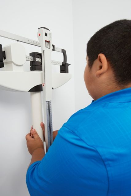
Jason E. Lang, MD, MPH, of Duke University, Durham, N.C., and his coauthors used the PEDSnet clinical data research network to conduct a retrospective cohort study of 507,496 children aged 2-17 years from 2009-2015, comparing the incidence of asthma in overweight and obese children to the incidence in healthy weight children.
The overall rate of new diagnoses of asthma was 2.4 per 1,000 patient years among normal-weight children and 3.2 per 1,000 patient years among obese children.
After adjustment for factors such as age, ethnicity, insurance status, sex, allergic rhinitis, food allergy, and proton pump inhibitor use, overweight children had a 17% higher risk of incident asthma, and obese children had a 26% higher risk of asthma, compared with children of normal weight. The relative risk of spirometry-confirmed asthma was 29% higher in obese children compared with normal-weight children, and the association between obesity and asthma persisted even when a second asthma encounter was required for the diagnosis.
Overall, the authors estimated that 23%-25% of clinically diagnosed asthma in children with obesity could be specifically attributed to obesity, and that in the overall population of children 10% of asthma was attributable to obesity.
“Currently, there are few known preventable risk factors that can be used to reduce childhood asthma,” wrote Dr. Lang and his coauthors. “With these data, it is suggested that reducing the onset of obesity in childhood would significantly reduce the public health burden of asthma in children.”
They noted that with current estimates of U.S. pediatric asthma prevalence being around 6-8 million cases, obesity could therefore account for up to 1 million of these cases.
The study was funded by the Patient-Centered Outcomes Research Institute, the Nemours Children’s Hospital and Nemours Children’s Health System. One author declared advisory board positions and consultancies with the pharmaceutical industry. The remaining researchers said they had no conflicts of interest.
SOURCE: Lang J et al. Pediatrics. 2018 Dec;142(6):e20182119.
While there has long been recognition of an association between childhood obesity and childhood asthma, the incidence of pediatric obesity–related asthma has not been well known. This study therefore addresses that gap in knowledge, and does so among children with a range of racial and ethnic backgrounds and while addressing potential confounders such as comorbidities and sociodemographic variables.
The findings show how significant a contribution obesity makes to the burden of childhood asthma, and also points to the potential increase in childhood asthma incidence that may arise with the increase in childhood obesity.
However, the good news is that this offers what may be the first modifiable risk factor for childhood asthma, which presents an opportunity for primary prevention of this common childhood condition.
Deepa Rastogi, MBBS, MS, is from the department of pediatrics at the Albert Einstein College of Medicine, New York. These comments are taken from an editorial (Pediatrics. 2018 Dec;142(6):e20182979.). No conflicts of interest were declared.
Around one-quarter of new asthma cases in children with obesity may be attributable to their obesity, according to research published in Pediatrics.
Jason E. Lang, MD, MPH, of Duke University, Durham, N.C., and his coauthors used the PEDSnet clinical data research network to conduct a retrospective cohort study of 507,496 children aged 2-17 years from 2009-2015, comparing the incidence of asthma in overweight and obese children to the incidence in healthy weight children.
The overall rate of new diagnoses of asthma was 2.4 per 1,000 patient years among normal-weight children and 3.2 per 1,000 patient years among obese children.
After adjustment for factors such as age, ethnicity, insurance status, sex, allergic rhinitis, food allergy, and proton pump inhibitor use, overweight children had a 17% higher risk of incident asthma, and obese children had a 26% higher risk of asthma, compared with children of normal weight. The relative risk of spirometry-confirmed asthma was 29% higher in obese children compared with normal-weight children, and the association between obesity and asthma persisted even when a second asthma encounter was required for the diagnosis.
Overall, the authors estimated that 23%-25% of clinically diagnosed asthma in children with obesity could be specifically attributed to obesity, and that in the overall population of children 10% of asthma was attributable to obesity.
“Currently, there are few known preventable risk factors that can be used to reduce childhood asthma,” wrote Dr. Lang and his coauthors. “With these data, it is suggested that reducing the onset of obesity in childhood would significantly reduce the public health burden of asthma in children.”
They noted that with current estimates of U.S. pediatric asthma prevalence being around 6-8 million cases, obesity could therefore account for up to 1 million of these cases.
The study was funded by the Patient-Centered Outcomes Research Institute, the Nemours Children’s Hospital and Nemours Children’s Health System. One author declared advisory board positions and consultancies with the pharmaceutical industry. The remaining researchers said they had no conflicts of interest.
SOURCE: Lang J et al. Pediatrics. 2018 Dec;142(6):e20182119.
FROM PEDIATRICS
Key clinical point: Obesity may be responsible for around 10% of childhood asthma.
Major finding: Obesity in children is associated with a 26% higher risk of asthma compared with normal-weight children.
Study details: A retrospective cohort study of 507,496 children.
Disclosures: The study was funded by the Patient-Centered Outcomes Research Institute and the Nemours Children’s Hospital and Nemours Children’s Health System. One author declared advisory board positions and consultancies with the pharmaceutical industry.
Source: Lang J et al. Pediatrics. 2018 Dec;142(6):e20182119. doi: 10.1542/peds.2018- 2119
Early intervention initiative cut oncology patient hospitalizations
, according to a report from a large, independent, community-based oncology practice.
The program saved nearly $3.2 million in Medicare costs over the course of the year, said Molly Mendenhall, RN, of Oncology Hematology Care in Cincinnati.
“By keeping those patients out of the hospital, we were able to improve patient quality of life, and increase patient satisfaction by treating them in their home clinic instead of the hospital,” Ms. Mendenhall said at a symposium on quality care sponsored by the American Society of Clinical Oncology.
The campaign was developed in anticipation of participating in the Oncology Care Model (OCM), a program focused on providing coordinated and high-quality care for Medicare oncology patients at the same or lower cost.
Prior to participating in OCM, Ms. Mendenhall and her colleagues set up an after-hours phone triage system, proactive chemotherapy follow-up calls, and a Saturday-Sunday urgent care clinic designed to help avoid any unnecessary hospitalizations over the weekend.
They also set up a 2-hour structured OCM treatment planning visit to prioritize shared decision making between the patient and the clinical team regarding diagnosis, symptom management, financial assistance, and other aspects of care.
The most influential part of the initiative, according to Ms. Mendenhall, was a patient-directed “Call Us Early – Call Us First” campaign that included symptom management teaching sheets, a 34-page teaching book, and branded buttons, pens, and magnets all designed to emphasize the patient responsibility to use the phone.
“All of those previous steps really wouldn’t make a difference if the patients still weren’t calling us,” Ms. Mendenhall explained.
Over the first year of participation in the OCM program, the oncology practice saw a 16% statistically significant reduction in hospital admissions (P = .005). The number of inpatient admissions per 100 patients dropped from 26.8 at baseline to 22.6 at the most recent follow-up in a report published simultaneously in the Journal of Oncology Practice.
Reduced admissions translated into a drop of $798,000 in inpatient costs per quarter over 1,600 patients, or $3.129 million in savings for the Centers for Medicare & Medicaid Services over the first year of participation in OCM, according to the researchers.
Patient satisfaction scores trended positively over the course of that year based on a review of blinded surveys that asked patients to rate clinical care, communication, access, information exchange, and other aspects of their experience.
Scores on those surveys were 8.03 on a scale of 0-10 (low to high) during the baseline period of January to September 2016, the researchers said. Scores were 8.29 and 8.26 for two follow-up surveys.
Ms. Mendenhall had no disclosures to report. Coauthors on the study provided disclosures related to Janssen Oncology, Pfizer, Amgen, Abbvie, Merck, Pharmacyclics, Bristol-Myers Squibb, Celgene, Genentech/Roche, AZTherapies, and Lilly. Two coauthors reported leadership, stock, or other ownership interests in Oncology Hematology Care/US Oncology.
SOURCE: Mendenhall M et al. Quality Care Symposium, Abstract 30.
REPORTING FROM THE QUALITY CARE SYMPOSIUM
Key clinical point: An initiative designed to reduce avoidable emergency room visits and hospitalizations reduced both admissions and inpatient costs.
Major finding: The program cut admissions by 16% and saved nearly $3.2 million in Medicare costs savings over the course of a year.
Study details: Analysis of first-year experience including 1,600 patients per quarter for a large, independent, community-based oncology practice participating in the Oncology Care Model (OCM) of the Centers for Medicare and Medicaid Services.
Disclosures: Authors on the study provided disclosures related to Janssen Oncology, Pfizer, Amgen, Abbvie, Merck, Pharmacyclics, Bristol-Myers Squibb, Celgene, Genentech/Roche, AZTherapies, Lilly, and Oncology Hematology Care/US Oncology.
Source: Mendenhall M et al. Quality Care Symposium, Abstract 30.
Case shows clinical assessment supersedes psychological screening tools
AUSTIN, TEX. – Using psychological screenings for law enforcement employment decisions can be a worthwhile supplement to more traditional hiring procedures, but such tools should be used with caution, a recent case study suggests.
“Pre-employment psychological evaluations for police officers are increasingly utilizing self-reported personality assessments to identify attributes in candidates that have shown to correlate with job performance outcomes,” Ann Marie Mckenzie Cassidy, DO, of Icahn School of Medicine at Mount Sinai, New York, and her colleagues wrote in an abstract presented at the annual meeting of the American Academy of Psychiatry and the Law.
“As research supporting the predictive power of written self-reported measurements expands, the call for this empirically validated data to be weighted over clinician judgment is becoming more substantive,” the researchers wrote. “The following case exemplifies a psychological evaluation where test results were either inconclusive or strongly conflicted with the clinical picture of the candidate.”
The applicant was a 34-year-old male Army veteran who received an honorable discharge after three deployments. Though he had no relevant medical or formal psychiatric history or drug use, he said he did drink alcohol heavily for a short time after joining the Army. He also had four speeding citations and one drag racing citation.
His personal history revealed several problems, including a military write-up for yelling at a subordinate and a history of difficulties working with his supervisor.
“While working as a car mechanic, he was unable to resolve a conflict with a difficult customer” and quit his job without notice, leading his employer to say he would not hire the applicant again. Yet, the applicant “denied interpersonal issues at work” and said he did not recall the yelling incident. He also said the situation where he quit with only 2 hours’ notice was unfair.
The applicant reported stress, “feeling down and having a diminished interest in activities” following his deployment in Iraq, but he turned down treatment for his stress. He also “used unprofessional language during the examination, and, when asked to refrain from cursing, he did not express concern about this conduct.”
His psychological test results on the Minnesota Multiphasic Personality Inventory-2 (MMPI-2), however, suggested “a pattern of positive impression management and defensiveness that is not likely to accurately represent existing psychopathology,” the researchers reported. “Closer review suggests the applicant is apt to see himself as having high moral standards and not having aggressive impulses,” they wrote. Similarly, the applicant’s Sixteen Personality Factor Questionnaire results “reflected an individual who is tough-minded, with low anxiety, who is emotionally stable, deferential, and relaxed.”
These two tools’ findings conflicted with the applicant’s history and presentation, and the examiner deemed him “psychologically unsuitable for hire.”
The researchers said this case reinforces the importance of investigating how empirical data – even with tools such as the MMPI-2, whose predictive power has been validated in several studies – are weighted and used with clinical psychological assessments.
the researchers concluded. “Until there is greater exploration of divergent or complementary testing findings and clinical judgment, test data should not be weighted over clinical judgment in psychological evaluations.”
REPORTING FROM THE AAPL ANNUAL MEETING
Cervical bupivacaine blocks pain after laparoscopic hysterectomy
LAS VEGAS – according to a small trial at the University of Tennessee, Chattanooga.

Twenty-one women were randomized to 0.5% bupivacaine, 5 mL injected into the cervix at the 3 o’clock position, and 5 mL injected into at the 9 o’clock position to a depth of 3 cm, after anesthesia induction but before insertion of the uterine manipulator. A control group of 20 women received 5 mL of 0.9% saline injected into the same positions. Surgeons were blinded to the randomization.
A stopwatch was started at extubation, and the women were asked to rate their pain on a 10-point visual analogue scale exactly at 30 and 60 minutes.
The bupivacaine group had less pain at both 30 minutes (3.2 versus 5.7 points, P = .01) and 60 minutes (2.3 versus 5.9 points, P less than .001); 71% of women in the bupivacaine group had an average score of 4 or less, indicating adequate pain control, versus just 25% in the control arm (P = .003)
“This is something we should be considering” routinely for laparoscopic hysterectomy, an audience member said after hearing the presentation at a meeting sponsored by AAGL.
Another audience member was concerned about urinary retention, but there was no increase in the treatment arm, said lead investigator Steven Radtke, MD, a former ob.gyn. surgery fellow at the university, but now at Texas Tech University, El Paso.
There have been many prior attempts to reduce pain after laparoscopic hysterectomy, such as infiltrating port sites with local anesthetic, but the results have been marginal at best, and almost all of them have focused on the abdominal wall as the source of pain.
The investigators thought that pain was more related to perimetrium dissection, colpotomy, and other parts of the operation. There also have been good studies showing that agents injected into the cervix infuse throughout the area. The team decided to try bupivacaine because it’s inexpensive and has a good duration of action, about 8 hours.
There were no significant demographic or intraoperative differences between the groups. On average, women were in their mid-40s, with a body mass index of about 31 kg/m2. The operations took about 2 hours, and were for benign indications, such as fibroids. Oophorectomy was the only concomitant procedure allowed.
The investigators are interested in repeating their investigation with liposomal bupivacaine (Exparel), which has a duration of action past 24 hours. It’s much more expensive, but the strong trial results justify the cost, Dr. Radtke said.
There was no external funding, and Dr. Radtke didn’t have any disclosures.
SOURCE: Radtke S et al. 2018 AAGL Global Congress, Abstract 130.
LAS VEGAS – according to a small trial at the University of Tennessee, Chattanooga.
Twenty-one women were randomized to 0.5% bupivacaine, 5 mL injected into the cervix at the 3 o’clock position, and 5 mL injected into at the 9 o’clock position to a depth of 3 cm, after anesthesia induction but before insertion of the uterine manipulator. A control group of 20 women received 5 mL of 0.9% saline injected into the same positions. Surgeons were blinded to the randomization.
A stopwatch was started at extubation, and the women were asked to rate their pain on a 10-point visual analogue scale exactly at 30 and 60 minutes.
The bupivacaine group had less pain at both 30 minutes (3.2 versus 5.7 points, P = .01) and 60 minutes (2.3 versus 5.9 points, P less than .001); 71% of women in the bupivacaine group had an average score of 4 or less, indicating adequate pain control, versus just 25% in the control arm (P = .003)
“This is something we should be considering” routinely for laparoscopic hysterectomy, an audience member said after hearing the presentation at a meeting sponsored by AAGL.
Another audience member was concerned about urinary retention, but there was no increase in the treatment arm, said lead investigator Steven Radtke, MD, a former ob.gyn. surgery fellow at the university, but now at Texas Tech University, El Paso.
There have been many prior attempts to reduce pain after laparoscopic hysterectomy, such as infiltrating port sites with local anesthetic, but the results have been marginal at best, and almost all of them have focused on the abdominal wall as the source of pain.
The investigators thought that pain was more related to perimetrium dissection, colpotomy, and other parts of the operation. There also have been good studies showing that agents injected into the cervix infuse throughout the area. The team decided to try bupivacaine because it’s inexpensive and has a good duration of action, about 8 hours.
There were no significant demographic or intraoperative differences between the groups. On average, women were in their mid-40s, with a body mass index of about 31 kg/m2. The operations took about 2 hours, and were for benign indications, such as fibroids. Oophorectomy was the only concomitant procedure allowed.
The investigators are interested in repeating their investigation with liposomal bupivacaine (Exparel), which has a duration of action past 24 hours. It’s much more expensive, but the strong trial results justify the cost, Dr. Radtke said.
There was no external funding, and Dr. Radtke didn’t have any disclosures.
SOURCE: Radtke S et al. 2018 AAGL Global Congress, Abstract 130.
REPORTING FROM AAGL GLOBAL CONGRESS
Key clinical point: Postoperative pain can be significantly reduced by injecting the cervix with bupivacaine prior to laparoscopic hysterectomy.
Major finding: The bupivacaine group had less pain at both 30 minutes (3.2 versus 5.7 points, P = .01) and 60 minutes (2.3 versus 5.9 points, P less than .001).
Study details: In a randomized study, 21 women received bupivacaine anesthesia and 20 control women were injected with saline.
Disclosures: There was no external funding, and Dr. Radtke didn’t have any disclosures.
Source: Radtke S et al. 2018 AAGL Global Congress, Abstract 130.
Sickle cell disease guidelines release set for early 2019
, according to Robert Liem, MD, chair of the American Society of Hematology coordination panel for the initiative.
The new clinical practice recommendations will expand on 2014 guidelines published by the National Heart, Lung, and Blood Institute in a way that will help both hematologists and nonhematologists who take care of patients with sickle cell disease, Dr. Liem said in a video interview at the annual meeting of the American Society of Hematology.
Five different guidelines are under development to cover different aspects of acute and chronic complications of sickle cell disease, including pain, cardiopulmonary and kidney disease, cerebrovascular disease, transfusion support, and stem cell transplantation.
Watch the video to learn more about the guideline effort from the perspective of Dr. Liem, who is also the director of the Comprehensive Sickle Cell Program at the Ann & Robert H. Lurie Children’s Hospital of Chicago.
FROM ASH 2018
Nocturnal hypoxemia predicts incident atrial fibrillation in sleep apnea patients
Researchers have found a significant independent association between nocturnal hypoxemia and risk of incident atrial fibrillation (AF) in patients with obstructive sleep apnea (OSA), which they believe may help prevent the development of AF in this population, according to recent research published in the journal CHEST®.
“These findings are consistent with those of previous studies suggesting that these groups, who are typically at somewhat lower risk of [atrial fibrillation], might be especially vulnerable to the effects of [obstructive sleep apnea] and hypoxemia,” Tetyana Kendzerska, MD, PhD, of the University of Ottawa in Ottawa, and her colleagues wrote in their study.
They performed an analysis of 8,256 patients with data linked to a provincial health administrative database who had suspected OSA who underwent a sleep study at a large academic hospital between 1994 and 2010. The patients were median 47 years old; 62% of the cohort were men, 28% had an apnea-hypopnea index (AHI) of greater than 30 events per hour, and 6% spent more than 30% of the time during sleep with less than 90% oxygen saturation.
Overall, 173 of 8,256 patients (2.1%) developed AF during the study period. In patients with suspected OSA and who were arrhythmia free, nocturnal hypoxemia significantly increased the risk of incident hospitalized AF (hazard ratio, 2.47; 95% confidence interval, 1.64-3.71) over median 10 years of follow-up (interquartile range, 7-13 years) after the researchers controlled for age, sex, chronic obstructive pulmonary disease, history of heart failure, smoking status, nocturnal hypoxemia, and pulmonary embolism, and this association remained significant after adjustment for body mass index and hypertension (HR, 1.77; 95% CI, 1.15-2.74).
“These findings support a relationship between OSA, chronic nocturnal hypoxemia, and the development of [atrial fibrillation], and may be used to identify those patients with OSA who are at greatest risk of developing AF.”
Researchers cited the observational design, retrospective data collection, and examining hospitalized AF only as limitations in the study. However, they noted that clinical data was collected prospectively and the inability to adjust to positive airway pressure would push results to the null with regard to unstudied confounders.
The authors reported no relevant conflicts of interest.
SOURCE: Kendzerska T et al. CHEST. 2018;doi:10.1016/j.chest.2018.08.1075.
FROM CHEST
Key clinical point: In patients with suspected obstructive sleep apnea and who were arrhythmia free, nocturnal hypoxemia increased the risk of incident hospitalized atrial fibrillation by 77%.
Major finding: Nocturnal hypoxemia significantly predicted atrial fibrillation (hazard ratio, 2.47; 95% confidence interval, 1.64-3.71) and remained significant after adjustment for body mass index and hypertension (HR, 1.77; 95% CI, 1.15-2.74).
Study details: An observational study of 8,256 arrhythmia-free patients with suspected obstructive sleep apnea who underwent a sleep study at a large academic hospital between 1994 and 2010.
Disclosures: The authors report no relevant conflicts of interest.
Source: Kendzerska T et al. CHEST. 2018;doi:10.1016/j.chest.2018.08.1075.