User login
ADHD and the role of wellness
ADHD is a very common disorder with several medication treatment options. There also are wellness and parenting strategies that can address aspects of the challenges of ADHD that are not perfectly covered by medication, such as excess symptoms, times of day that are not covered, or oppositional behavior that often develops secondarily.
Case summary
James is a 6-year-old boy who has been an active, high-energy child since preschool. He has had difficulty with wiggling around in kindergarten and preschool, talking excessively, and being unable to follow directions and pay attention. He is impulsive, disruptive, and frequently doesn’t listen to what his parents tell him to do. Parents and teachers rank him in the clinical range for hyperactivity, impulsivity, and attention problems on standardized rating scales.
Discussion
When we first discuss a new diagnosis with a family, we have the opportunity to shape the family’s expectations about that diagnosis and how it should be addressed. When I discuss ADHD with a new family, I want them to understand the symptoms of inattention, hyperactivity, and impulsiveness, and that these symptoms are not the child’s fault, but rather related to the way his brain is connected. At the same time, I also emphasize that these connections are not entirely fixed, that they mature over time, and that they are affected by experiences in life. In particular, I stress that positive experiences and wellness activities can influence the brain in a positive way. While, of course, I discuss the range of medications that can address these issues, I also deal with wellness in the treatment plan.

Exercise
Studies in humans and animals have provided background evidence that exercise increases the release of neurotransmitters such as dopamine and norepinephrine that are important in the pathophysiology of ADHD. Cerillo-Urbina et al. did a meta-analysis in 2015 of randomized controlled trials and found medium to large effect sizes for a variety of physical activity programs with respect to attention, hyperactivity, and impulsivity, although the study quality was generally low.1 Clearly we need additional more rigorous studies, but given the positive direction of outcomes, the lack of side effects, and the many other positive effects of exercise, it does not seem too soon to add exercise as a prescription for our patients with ADHD. I review this evidence with families, ask them about physical activity they like, and ask if they are willing to work toward an hour of exercise a day.
Sleep
Many children with ADHD have problems with sleep even before they start on stimulant medications, which can further affect sleep. Addressing sleep early on can improve ADHD symptoms, as well as help parents change or avoid patterns like having children fall asleep to the sound of a television. Brief sleep hygiene and cognitive-behavioral therapy interventions over three visits were demonstrated in a randomized controlled trial by Hisock et al. to improve ADHD symptoms and behavioral function.2 These psychosocial interventions clearly are the first line in addressing sleep problems in ADHD, and can benefit even sleep problems connected to medication.
Parent training
Treatment plan
1. Have the child exercise 1 hour every day. Have fun!
2. Establish a nightly bedtime routine, with a bath at 7:30 p.m., brushing of teeth, a story, and lights out at 8 with no TV in the room.
3. Check out the CHADD website for Parent to Parent.
4. Start a trial of stimulant medication.
5. Return in 2 weeks to monitor these interventions, adjust goals, and adjust medications.
When to refer
Many parents will be able to put such a plan in motion with your support and that of other parents. If they are struggling, therapists, psychologists, and psychiatrists trained in motivational and behavioral methods can provide more individualized parent training. Also consider whether the parents themselves may have ADHD and could use referral and treatment.
Dr. Hall is assistant professor of psychiatry and pediatrics at the University of Vermont, Burlington. She said she had no relevant financial disclosures.
References
1. Child Care Health Dev. 2015 Nov;41(6):779-88.
2. BMJ. 2015. doi: 10.1136/bmj.h68.
ADHD is a very common disorder with several medication treatment options. There also are wellness and parenting strategies that can address aspects of the challenges of ADHD that are not perfectly covered by medication, such as excess symptoms, times of day that are not covered, or oppositional behavior that often develops secondarily.
Case summary
James is a 6-year-old boy who has been an active, high-energy child since preschool. He has had difficulty with wiggling around in kindergarten and preschool, talking excessively, and being unable to follow directions and pay attention. He is impulsive, disruptive, and frequently doesn’t listen to what his parents tell him to do. Parents and teachers rank him in the clinical range for hyperactivity, impulsivity, and attention problems on standardized rating scales.
Discussion
When we first discuss a new diagnosis with a family, we have the opportunity to shape the family’s expectations about that diagnosis and how it should be addressed. When I discuss ADHD with a new family, I want them to understand the symptoms of inattention, hyperactivity, and impulsiveness, and that these symptoms are not the child’s fault, but rather related to the way his brain is connected. At the same time, I also emphasize that these connections are not entirely fixed, that they mature over time, and that they are affected by experiences in life. In particular, I stress that positive experiences and wellness activities can influence the brain in a positive way. While, of course, I discuss the range of medications that can address these issues, I also deal with wellness in the treatment plan.
Exercise
Studies in humans and animals have provided background evidence that exercise increases the release of neurotransmitters such as dopamine and norepinephrine that are important in the pathophysiology of ADHD. Cerillo-Urbina et al. did a meta-analysis in 2015 of randomized controlled trials and found medium to large effect sizes for a variety of physical activity programs with respect to attention, hyperactivity, and impulsivity, although the study quality was generally low.1 Clearly we need additional more rigorous studies, but given the positive direction of outcomes, the lack of side effects, and the many other positive effects of exercise, it does not seem too soon to add exercise as a prescription for our patients with ADHD. I review this evidence with families, ask them about physical activity they like, and ask if they are willing to work toward an hour of exercise a day.
Sleep
Many children with ADHD have problems with sleep even before they start on stimulant medications, which can further affect sleep. Addressing sleep early on can improve ADHD symptoms, as well as help parents change or avoid patterns like having children fall asleep to the sound of a television. Brief sleep hygiene and cognitive-behavioral therapy interventions over three visits were demonstrated in a randomized controlled trial by Hisock et al. to improve ADHD symptoms and behavioral function.2 These psychosocial interventions clearly are the first line in addressing sleep problems in ADHD, and can benefit even sleep problems connected to medication.
Parent training
Treatment plan
1. Have the child exercise 1 hour every day. Have fun!
2. Establish a nightly bedtime routine, with a bath at 7:30 p.m., brushing of teeth, a story, and lights out at 8 with no TV in the room.
3. Check out the CHADD website for Parent to Parent.
4. Start a trial of stimulant medication.
5. Return in 2 weeks to monitor these interventions, adjust goals, and adjust medications.
When to refer
Many parents will be able to put such a plan in motion with your support and that of other parents. If they are struggling, therapists, psychologists, and psychiatrists trained in motivational and behavioral methods can provide more individualized parent training. Also consider whether the parents themselves may have ADHD and could use referral and treatment.
Dr. Hall is assistant professor of psychiatry and pediatrics at the University of Vermont, Burlington. She said she had no relevant financial disclosures.
References
1. Child Care Health Dev. 2015 Nov;41(6):779-88.
2. BMJ. 2015. doi: 10.1136/bmj.h68.
Emerging sickle cell agents target new pathways
CONCORD, N.C. – Approved treatments for sickle cell disease have been extremely limited, but there are several therapies in the research pipeline that use new pathways to target the disease.
“We do have much better understanding of the pathophysiology, which is getting us a few more targets to aim at,” Julie Kanter, MD, director of sickle cell research at the Medical University of South Carolina, Charleston, said at Sickle Cell Disease Symposium held by Carolinas Health Care System. These targets include influencing how cells interact with the vascular endothelium, inhibiting platelets, and preventing cell sickling and inflammation.

“I’m waiting to see who’s willing to take it just because it is a lot of powder that the patient has to mix and drink twice a day, but it does look promising to reduce inflammation,” Dr. Kanter said.
SCD pipeline
Deeper in the sickle cell pipeline is a class of antisickling agents known as hemoglobin modifiers. “We’re tying to change the way hemoglobin binds to oxygen, and if we can keep hemoglobin binding to oxygen longer, it actually decreases the risk of hemoglobin sickling and polymerizing in the cell,” Dr. Kanter explained.
One hemoglobin modifier is voxelotor (previously called GBT440), a once-daily oral agent that Global Blood Therapeutics has in development.
Another category of antisickling agents that researchers are looking at is anti-inflammatory moderators, Dr. Kanter said. These include nitric oxide donors like sildenafil, which did not “quite work” in SCD, and arginine and glutamine, which increase the amount of nitric oxide once in the body “and hopefully reduce the risk of sickling,” she said. “If we can improve inflammation, we might be able to improve the risk of crisis.”
Cell adhesion modifiers are a drug class that aims to prevent cells from binding to each other. These include platelet inhibitors and endothelial blockers. “There are several antiplatelet agents that are approved really for stroke prevention or heart attack prevention, and we’re trying to see if we can repurpose these in sickle cell disease in a specific pathway that allows the platelet to stick to the endothelium, but if we only inhibit one pathway it should not increase the risk of bleeding,” Dr. Kanter said.
One platelet inhibition pathway that researchers are focused on is the P2Y12 adenosine diphosphate blockade, which the platelet inhibitor prasugrel (Effient) acts on. A 2016 study of this pathway in SCD “wasn’t successful,” Dr. Kanter said, “but it had some interesting results” – namely that the drug may be most effective in adolescents (N Engl J Med. 2016 Feb 18;374[7]:625-35).
Selectin-blocking medications are a drug class that act on white blood cell adhesion to, and movement through, the endothelium, Dr. Kanter said. “Neutrophils can instigate a sickle cell crisis, so if we can interrupt some of this rolling or sticking, could we decrease the risk of a sickle cell crisis?” The drug GMI-1070 is currently being studied in a phase III trial and so far has shown “a significant decrease in the amount of opioids used by those individuals who received the study drug,” she said.
Crizanlizumab (also known as SelG1) is a humanized monoclonal antibody with an affinity to P-selectin and is the subject of the phase II SUSTAIN trial, which included a cohort that also was taking hydroxyurea. Treatment with high-dose crizanlizumab resulted in an annual rate of sickle cell–related pain crises that was more than 45% lower than with placebo (N Engl J Med. 2017 Feb 2;376[5]:429-39).
Stem cell transplants
Besides drugs, stem cell transplants to treat SCD have advanced in recent years to the point where cure rates are exceeding 90%, Dr. Kanter noted. “However, the real issue with stem cells is that patients still don’t have enough donors,” she said.
SCD is also potentially amenable to gene therapy, Dr. Kanter said, noting that SCD gene therapy trials in progress are looking at harvesting patients’ own bone marrow, using the lentivirus viral vector, inserting a gene to increase production of nonsickle hemoglobin, and using myeloablative chemotherapy to remove old marrow and replace it with new, manipulated bone marrow.
Several programs are investigating using a gene editing technique, known as CRISPR/Cas9, to alter the BCL11A gene to maintain fetal hemoglobin production.
“We all make fetal hemoglobin at birth, and then over 6 months to 1 year of life, our bodies convert fetal hemoglobin to adult hemoglobin,” she said. “In sickle cell disease, it converts to sickle hemoglobin. What if we could prevent that conversion and keep the fetal hemoglobin turned on?” That could potentially eradicate the complications of SCD starting at an early age, Dr. Kanter said.
Dr. Kanter, who has been involved in several of the SCD trials, reported relationships with Pfizer, AstraZeneca, Bluebird Bio, Global Blood Therapeutics, Novartis, Guidepoint, GLG, ApoPharma, and Purdue Pharma.
CONCORD, N.C. – Approved treatments for sickle cell disease have been extremely limited, but there are several therapies in the research pipeline that use new pathways to target the disease.
“We do have much better understanding of the pathophysiology, which is getting us a few more targets to aim at,” Julie Kanter, MD, director of sickle cell research at the Medical University of South Carolina, Charleston, said at Sickle Cell Disease Symposium held by Carolinas Health Care System. These targets include influencing how cells interact with the vascular endothelium, inhibiting platelets, and preventing cell sickling and inflammation.
“I’m waiting to see who’s willing to take it just because it is a lot of powder that the patient has to mix and drink twice a day, but it does look promising to reduce inflammation,” Dr. Kanter said.
SCD pipeline
Deeper in the sickle cell pipeline is a class of antisickling agents known as hemoglobin modifiers. “We’re tying to change the way hemoglobin binds to oxygen, and if we can keep hemoglobin binding to oxygen longer, it actually decreases the risk of hemoglobin sickling and polymerizing in the cell,” Dr. Kanter explained.
One hemoglobin modifier is voxelotor (previously called GBT440), a once-daily oral agent that Global Blood Therapeutics has in development.
Another category of antisickling agents that researchers are looking at is anti-inflammatory moderators, Dr. Kanter said. These include nitric oxide donors like sildenafil, which did not “quite work” in SCD, and arginine and glutamine, which increase the amount of nitric oxide once in the body “and hopefully reduce the risk of sickling,” she said. “If we can improve inflammation, we might be able to improve the risk of crisis.”
Cell adhesion modifiers are a drug class that aims to prevent cells from binding to each other. These include platelet inhibitors and endothelial blockers. “There are several antiplatelet agents that are approved really for stroke prevention or heart attack prevention, and we’re trying to see if we can repurpose these in sickle cell disease in a specific pathway that allows the platelet to stick to the endothelium, but if we only inhibit one pathway it should not increase the risk of bleeding,” Dr. Kanter said.
One platelet inhibition pathway that researchers are focused on is the P2Y12 adenosine diphosphate blockade, which the platelet inhibitor prasugrel (Effient) acts on. A 2016 study of this pathway in SCD “wasn’t successful,” Dr. Kanter said, “but it had some interesting results” – namely that the drug may be most effective in adolescents (N Engl J Med. 2016 Feb 18;374[7]:625-35).
Selectin-blocking medications are a drug class that act on white blood cell adhesion to, and movement through, the endothelium, Dr. Kanter said. “Neutrophils can instigate a sickle cell crisis, so if we can interrupt some of this rolling or sticking, could we decrease the risk of a sickle cell crisis?” The drug GMI-1070 is currently being studied in a phase III trial and so far has shown “a significant decrease in the amount of opioids used by those individuals who received the study drug,” she said.
Crizanlizumab (also known as SelG1) is a humanized monoclonal antibody with an affinity to P-selectin and is the subject of the phase II SUSTAIN trial, which included a cohort that also was taking hydroxyurea. Treatment with high-dose crizanlizumab resulted in an annual rate of sickle cell–related pain crises that was more than 45% lower than with placebo (N Engl J Med. 2017 Feb 2;376[5]:429-39).
Stem cell transplants
Besides drugs, stem cell transplants to treat SCD have advanced in recent years to the point where cure rates are exceeding 90%, Dr. Kanter noted. “However, the real issue with stem cells is that patients still don’t have enough donors,” she said.
SCD is also potentially amenable to gene therapy, Dr. Kanter said, noting that SCD gene therapy trials in progress are looking at harvesting patients’ own bone marrow, using the lentivirus viral vector, inserting a gene to increase production of nonsickle hemoglobin, and using myeloablative chemotherapy to remove old marrow and replace it with new, manipulated bone marrow.
Several programs are investigating using a gene editing technique, known as CRISPR/Cas9, to alter the BCL11A gene to maintain fetal hemoglobin production.
“We all make fetal hemoglobin at birth, and then over 6 months to 1 year of life, our bodies convert fetal hemoglobin to adult hemoglobin,” she said. “In sickle cell disease, it converts to sickle hemoglobin. What if we could prevent that conversion and keep the fetal hemoglobin turned on?” That could potentially eradicate the complications of SCD starting at an early age, Dr. Kanter said.
Dr. Kanter, who has been involved in several of the SCD trials, reported relationships with Pfizer, AstraZeneca, Bluebird Bio, Global Blood Therapeutics, Novartis, Guidepoint, GLG, ApoPharma, and Purdue Pharma.
EXPERT ANALYSIS FROM A MEETING ON SICKLE CELL DISEASE
ACC survey: Burnout pervasive in cardiologists
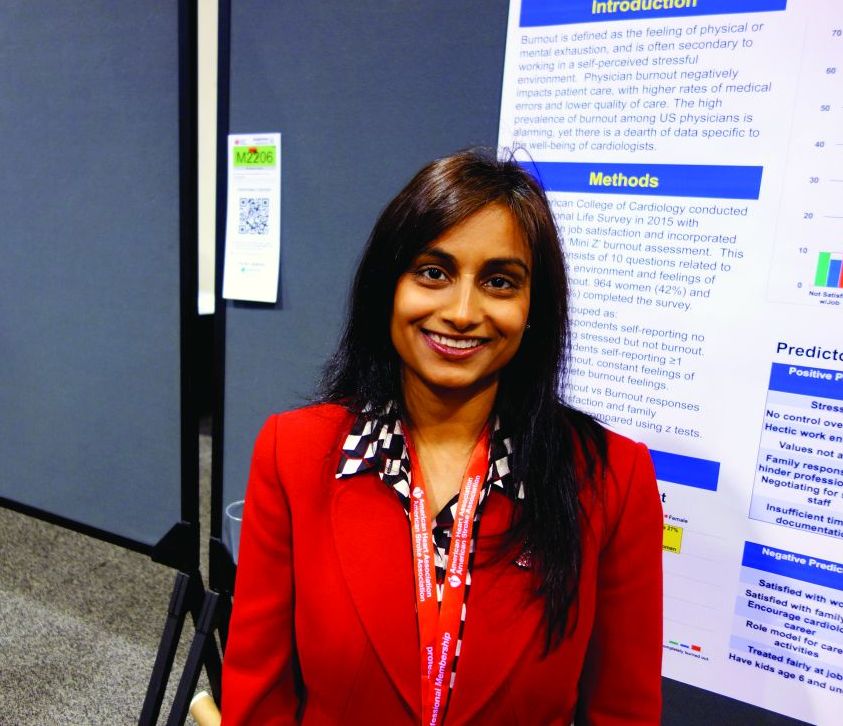
ANAHEIM, CALIF. – A disturbingly high 27% of U.S. cardiologists reported currently feeling burnout in the American College of Cardiology’s third Professional Life Survey, Laxmi S. Mehta, MD, said at the American Heart Association scientific sessions.
A gender gap existed: The prevalence of burnout was 29% greater among female cardiologists than their male counterparts, by a margin of 31%-24%.
“These are the doctors who are taking care of people’s hearts, and we know that when you’re burned out, there are higher rates of medical errors and the quality of care is poorer. So this is problematic,” she said in an interview.
Burnout had a negative effect on career satisfaction: While 94% of cardiologists in the nonburnout group professed they were satisfied with their career, that was the case for only 74% of cardiologists who felt burnout. Just 56% percent of the burnout group said they would recommend cardiology as a career, compared with 80% of the practitioners who felt no burnout.
The 2015 ACC survey was completed by 2,313 U.S. cardiologists, 964 of whom were women. The first round of results, which focused on career satisfaction and racial and gender discrimination in the workplace, have been published (J Am Coll Cardiol. 2017 Jan 31;69[4]:452-62). The survey included the validated 10-question Mini Z burnout assessment, the results of which were the focus of the new analysis.
The 27% of cardiologists in the burnout group fell into three subcategories, the largest of which comprised those who reported feeling at least one burnout symptom of physical or mental exhaustion. Those who said their burnout symptoms don’t go away and that they think about their work frustrations frequently made up a smaller group. Just a few percent of survey participants fell into the completely burned out category.
Only 51% of the burnout group were satisfied with their financial compensation, compared with 68% of the nonburnout group. Sixty-one percent of the burnout group felt they were treated fairly at their job, as did 86% of the cardiologists who felt burnout. Half of the cardiologists with burnout reported experiencing past discrimination, compared with 37% of the nonburnout group. And 40% of the burnout group felt their family responsibilities hindered career advancement, a sentiment expressed by 22% of the nonburnout group.
EMR “pajama time” cited as a major burden
Two-thirds of cardiologists with constant burnout symptoms or complete burnout cited excessive time spent completing their electronic medical records as a significant contributing factor.
“The electronic medical record ends up taking over our personal time,” according to Dr. Mehta. “We call it ‘pajama time’ because many of us are doing the charts or responding to patients at midnight, on vacation, at meetings like this. There is no separation, and that’s a problem.”
What can be done to reduce burnout
The 2015 Professional Life Survey was the third one in 20 years. Compared with the earlier two, the most recent survey painted a picture of an aging workforce that is less likely to be in private practice. The survey – the first one to assess burnout within the specialty – was carried out by the ACC Women in Cardiology Leadership Council. Armed with the survey results, the ACC leadership is now in the process of redefining the organization’s mission statement to incorporate a new emphasis on providing for physician health and well-being in addition to the more traditional goals of improving the quality and reducing the cost of care.
“Many cardiologists are working a lot harder than they used to, with less personal time. We need to work on mechanisms to reduce burnout by reducing the burdens put on them. The survey data help because they show the cardiology profession, and hopefully hospital administrators, the importance of making a better work environment. That’s the hope,” Dr. Mehta said.
She reported having no financial conflicts of interest regarding the survey.
AT THE AHA SCIENTIFIC SESSIONS
Key clinical point:
Major finding: There is a noticeable gender gap in U.S. cardiologist burnout.
Data source: This survey of 2,313 U.S. cardiologists addressed burnout and career satisfaction within the profession.
Disclosures: The presenter reported having no financial conflicts of interest regarding the survey, conducted by the American College of Cardiology.
The ‘Virtual Radiology Resident’—Coming to a Computer Near You
Researchers around the world may be able to teach computers how to better detect and diagnose disease, thanks to > 100,000 chest x-ray images and corresponding data recently released by the NIH Clinical Center.
Reading and diagnosing chest x-rays requires careful observation, as well as knowledge of anatomy, physiology, and pathology. When that is combined with the need to consider all common thoracic diseases, it becomes hard to automate a consistent technique for reading images, the NIH says. With the free dataset, the hope is that academic and research institution staff will be able to teach their computers to read and process enormous amounts of scans, to confirm radiologists’ results, and potentially identify anything that may have been overlooked.
The NIH says in addition to being a “virtual radiology resident,” advanced computer technology has other potential benefits: For instance, it could identify slow changes occurring over the course of multiple chest x-rays that might otherwise be overlooked. The technology also would be useful in poor countries that lack radiologists. And in the future, the “resident” might be taught to read more complex images, such as CT and MRI.
The dataset, compiled from scans from > 30,000 patients, including many with advanced lung disease, was scrubbed of private information before release. The images are available via Box at https://nihcc.app.box.com/v/ChestXray-NIHCC.
Nilotinib approved to treat kids with CML in EU
The European Commission has approved nilotinib (Tasigna®) for the treatment of pediatric patients.
The drug is now approved to treat children age 2 and older with newly diagnosed, Philadelphia chromosome-positive (Ph+), chronic phase (CP) chronic myeloid leukemia (CML) or Ph+ CP-CML with resistance or intolerance to prior therapy, including imatinib.
Nilotinib is the only second-generation tyrosine kinase inhibitor currently approved in the European Union (EU) for the treatment of Ph+ CP-CML in children. The approval applies to all EU member states.
According to Novartis, the expanded indication for nilotinib is based on 2 prospective studies of the drug in children with Ph+ CP-CML, which were part of a formal “pediatric investigation plan” agreed upon with the European Medicines Agency.
The company said 69 patients received nilotinib in these studies. The patients ranged in age from 2 to 18. They had either newly diagnosed Ph+ CP-CML or Ph+ CP-CML with resistance or intolerance to prior therapy, including imatinib.
In the newly diagnosed patients, the major molecular response (MMR) rate was 60.0% (95% CI: 38.7, 78.9) at 12 cycles, with 15 patients achieving MMR.
In patients with resistance or intolerance to prior therapy, the MMR rate was 40.9% (95% CI: 26.3, 56.8) at 12 cycles, with 18 patients being in MMR.
In newly diagnosed patients, the cumulative MMR rate was 64.0% by cycle 12. In patients with resistance or intolerance to prior therapy, the cumulative MMR rate was 47.7% by cycle 12.
Adverse events were generally consistent with those observed in adults, with the exception of hyperbilirubinemia and transaminase elevation, which were reported at a higher frequency than in adults.
The rate of grade 3/4 hyperbilirubinemia was 13.0%, the rate of grade 3/4 AST elevation was 1.4%, and the rate of grade 3/4 ALT elevation was 8.7%.
There were no deaths on treatment or after treatment discontinuation. ![]()
CD22-CAR therapy shows activity in rel/ref B-ALL

Researchers say they have reported the first results demonstrating clinical activity of a CD22-directed chimeric antigen receptor (CAR) T-cell therapy in B-cell acute lymphoblastic leukemia (B-ALL).
The team conducted a phase 1 study of the therapy in 21 children and adults with relapsed/refractory B-ALL.
Twelve patients achieved a complete response (CR) to the treatment, with 3 patients still in CR at last follow-up.
Sixteen patients developed cytokine release syndrome (CRS), all grade 1 or 2.
Crystal Mackall, MD, of Stanford University in California, and her colleagues reported these results in Nature Medicine.*
“This is the first time that we’ve seen response rates anything like we achieved when we were first testing the CD19 CAR T therapy,” Dr Mackall said.
“We were all a little worried that we wouldn’t find anything comparable, but this study gives hope to the idea that there may be another similar, very potent treatment.”
Patients
Dr Mackall and her colleagues studied the CD22-CAR T-cell therapy in 21 patients with relapsed/refractory B-ALL. They had a median age of 19 (range, 7 to 30).
All of the patients had received a hematopoietic stem cell transplant at least once, and 2 patients had 2 prior transplants each. Seventeen patients had received prior CD19-directed immunotherapy. Fifteen had received CD19-directed CAR T-cell therapy, and 2 had received blinatumomab.
Lymphoblasts were CD19− or CD19dim in 10 patients (9 who had received a CD19-CAR and 1 treated with blinatumomab).
The median CD22 site density was 2839 molecules per cell (range, 613 to 13,452).
Dosing and DLTs
Patients received the CD22-CAR T-cell therapy at 1 of 3 dose levels:
- 0.3 × 10⁶ CD22-CAR T cells per kg body weight (n=6)
- 1 × 10⁶ cells per kg (n=13)
- 3 × 10⁶ cells per kg (n=2).
There was 1 dose-limiting toxicity (DLT) at the first dose level. It was grade 3, self-limited, noninfectious diarrhea that occurred during CRS and resolved with supportive care.
The other DLT occurred in a patient who received treatment at the third dose level. This patient had grade 4 hypoxia that was associated with rapid disease progression. The patient required brief intubation, and the hypoxia was resolved within 24 hours of starting steroid treatment.
Based on these results, the second dose level became the recommended phase 2 dose.
Other adverse events
The researchers said the primary toxicity was CRS, which occurred in 16 patients. Nine patients had grade 1 CRS, and 7 had grade 2.
There were no cases of irreversible neurotoxicity or seizure reported. Among the first 16 patients with complete assessments, there were cases of transient visual hallucinations (n=2), mild unresponsiveness (n=1), mild disorientation (n=1), and mild to moderate pain (n=2). However, these incidents resolved by day 28.
One patient died from gram-negative rod sepsis that developed after the resolution of CRS and neutrophil count recovery to >1000 cells/μL blood. The patient had a history of multi-organ failure due to sepsis.
Response
Twelve patients (57%) had a CR, and 9 of them were minimal residual disease negative.
One CR occurred at the lowest dose of therapy, 1 occurred at the highest dose, and the remaining 10 CRs occurred in patients who received dose level 2.
The researchers said there was no evidence to suggest that previous CD19-directed immunotherapy or diminished surface expression of CD19 impacted response to the CD22-CAR T-cell therapy.
Of the 9 patients who did not respond, 4 progressed and 5 had stable disease.
The researchers said 4 non-responders had “very high disease burden with rapid disease progression.” And 2 non-responders expressed diminished or partial CD22 on leukemic blasts at the time of enrollment.
The median duration of response was 6 months (range, 1.5 to 21+ months). Three patients are still in CR at 6, 9, and 21 months of follow-up.
“The take-home message is that we’ve found another CAR T-cell therapy that displays high-level activity in this phase 1 trial,” Dr Mackall said. “But the relapse rate was also high. So this forces the field to get even more sophisticated. How much of a target is needed for successful, long-lasting treatment? What happens if we target both CD19 and CD22 simultaneously?”
The researchers are already tackling the last question by testing a CAR T-cell therapy that recognizes both CD19 and CD22. They’ve confirmed this therapy can kill cancer cells in vitro and in vivo. Now, they’re testing it in a clinical trial that has opened at Stanford University and will open soon at the National Cancer Institute. ![]()
*This research was supported, in part, by the Intramural Research Program, National Cancer Institute and NIH Clinical Center, National Institutes of Health; by a Stand Up to Cancer–St. Baldrick’s Pediatric Dream Team translational research grant; and by a St. Baldrick’s Foundation Scholar Award.
NOACs may do less harm to kidneys than warfarin

New research suggests warfarin may produce worse renal outcomes than non-vitamin K antagonist oral anticoagulants (NOACs) in patients with atrial fibrillation.
In a study of close to 10,000 patients, dabigatran and rivaroxaban were associated with lower risks of adverse renal outcomes than warfarin.
However, risks with warfarin and apixaban were not significantly different.
This research was published in the Journal of the American College of Cardiology.*
“Kidney function decline in patients taking oral anticoagulant drugs is an important topic that has been overlooked in previous clinical trials,” said study author Xiaoxi Yao, PhD, of Mayo Clinic in Rochester, Minnesota.
“Even our past work at Mayo Clinic has been primarily focused on risks for stroke or bleeding.”
For the current study, Dr Yao and her colleagues examined the de-identified records of 9769 patients from the OptumLabs Data Warehouse.
The patients had atrial fibrillation and started taking oral anticoagulants—apixaban, dabigatran, rivaroxaban, or warfarin—between Oct. 1, 2010, and April 30, 2016.
The researchers looked at 4 indicators of kidney function in these patients:
- A 30% or greater decline in estimated glomerular filtration rate (eGFR)
- Doubled serum creatinine level
- Acute kidney injury (AKI)
- Kidney failure.
For the entire study population, the cumulative risk of each event occurring within 2 years of beginning anticoagulation was as follows:
- 24.4% for ≥30% eGFR
- 4.0% for doubled creatinine level
- 14.8% for AKI
- 1.7% for kidney failure.
“Our study demonstrated that renal function decline is very common among atrial fibrillation patients on blood thinners,” Dr Yao said. “About 1 in 4 patients had significantly reduced kidney function within 2 years of being on any of these medications, and 1 in 7 patients had acute kidney injury.”
“In general, patients with atrial fibrillation taking blood-thinning medications tend to have declining kidney function over time,” added study author Peter Noseworthy, MD, of Mayo Clinic in Rochester, Minnesota.
“However, our findings indicate that the non-vitamin K antagonist oral anticoagulants, as a group, are associated with less injury to kidneys than warfarin.”
When the researchers compared all 3 NOACs to warfarin, they found NOAC use was associated with a reduced risk of:
- ≥30% decline in eGFR—hazard ratio (HR)=0.77 (P<0.001)
- Doubling of creatinine—HR=0.62 (P=0.03)
- AKI—HR=0.68 (P<0.001).
However, results differed in one-to-one comparisons.
There was no significant difference between warfarin and apixaban for any of the renal endpoints measured. The HRs (for apixaban vs warfarin) were:
- 0.88 for ≥30% decline in eGFR (P=0.25)
- 0.80 for doubling of creatinine (P=0.51)
- 0.84 for AKI (P=0.16)
- 1.02 for kidney failure (P=0.95).
Dabigatran, on the other hand, was associated with lower risks of ≥30% decline in eGFR and AKI than warfarin. The HRs were as follows:
- 0.72 for ≥30% decline in eGFR (P=0.01)
- 0.64 for doubling of creatinine (P=0.24)
- 0.55 for AKI (P<0.001)
- 0.45 for kidney failure (P=0.21).
Rivaroxaban was associated with lower risks of ≥30% decline in eGFR, doubling of creatinine, and AKI. The HRs were as follows:
- 0.73 for ≥30% decline in eGFR (P<0.001)
- 0.46 for doubling of creatinine (P<0.01)
- 0.69 for AKI (P<0.001)
- 0.63 for kidney failure (P=0.13).
“Patients with atrial fibrillation already face a high risk of kidney disease, perhaps because many such patients have risk factors, such as advanced age, diabetes, and hypertension,” Dr Yao said. “Many drugs these patients are taking rely on kidney function for drug elimination. Therefore, it is particularly important for these patients to choose a drug that minimizes the impact on kidneys.”
“Since non-vitamin K antagonist oral anticoagulants have a different drug mechanism than warfarin, researchers have hypothesized that non-vitamin K antagonist oral anticoagulants may be related to better renal outcomes. Our study is among the first few studies confirming this hypothesis.” ![]()
*This study was funded by the Mayo Clinic Robert D. and Patricia E. Kern Center for the Science of Health Care Delivery, which receives no industry funding. However, study authors did declare industry relationships.
Mixed results for rheumatologists on Medicare quality measures
SAN DIEGO – As quality measures are poised to become crucial to U.S. physician incomes in 2019, an analysis of a nationwide registry sample finds that rheumatologists overall have decent scores on several fronts. But they still need to boost their record on preventive measures that are not geared specifically to their specialty.
“Rheumatologists are performing well, so far, on aspects of care coordination, such as communication with the health care team post osteoporotic fracture and on several patient safety measures, such as avoiding high-risk drugs in the elderly,” said Jinoos Yazdany, MD, of the University of California, San Francisco. However, “in some preventive care measures, such as tobacco screening, there is room for improvement compared to benchmarks that the Centers for Medicare & Medicaid Services has set across providers.”
The ACR created the registry “to help rheumatology harness the power of electronic health record data to advance our understanding of the natural history, treatment, and outcomes of rheumatic disease,” Dr. Yazdany said. “Another important goal was to harness the power of a national registry to measure and improve quality of care and outcomes. Practices can use RISE to see where they are performing well and where there is room for improvement.”
Since 2016, the registry has passively collected data on 2.5 million patients and 13.7 million encounters.
“The quality measures in RISE serve several purposes,” Dr. Yazdany said. “First, they fulfill reporting requirements to CMS through the Merit-Based Incentive Payment System [MIPS]. Second, the measures provide information to practices that can be used to track quality improvement and population health management. Finally, the measures create unprecedented opportunities to learn from practices that are excelling and to adapt successful work flows to improve care for our patients.”
These measures matter. In 2019, payments for many physicians under Medicare Part B will be adjusted based on their performance in these areas in previous years. Most rheumatologists will take part, Dr. Yazdany said.
“Rather than focusing on a single measure, the key number in 2017 for the MIPS program is 70 points across the three domains of Quality, Advancing Care Information, and Improvement Activities,” she said. “Above that threshold, rheumatologists will qualify for an ‘exceptional performance bonus.’ That means they will get a minimum of an additional 0.5% on their Medicare billing.”
She added that “there is no reason that most rheumatologists should not cross the 70-point threshold. Proactively monitoring their progress in RISE will help them succeed.”
The ACR session focused on a registry sample of 225 practices and 750 rheumatologists. The analysis measured their performance from January to September 2017 in the Quality, Advancing Care Information, and Improvement Activities areas.
In terms of meeting benchmarks, the rheumatologists in the sample performed especially well in several areas.
On the drug safety front, across elderly patients, an average of just 3.6% were prescribed one or more high-risk medications, and 0.2% were prescribed two or more.
On rheumatoid arthritis measures, 52% of patients had documentation of tuberculosis screening before biologics, and 46.3% underwent functional status assessments. And in the care coordination and documentation measure, 92.9% documented current medications in the EHR.
Rheumatologists lagged in terms of preventive care, compared with other physicians nationally: The average performance across patients was 77.2% for tobacco screening and counseling, 42.7% for body mass index screening and counseling, and 60.2% for blood pressure management.
Why are these preventive care measures being tracked in rheumatology instead of more rheumatology-specific measures? “CMS requires that physicians submit an outcome measure. Unfortunately, we don’t have validated outcome measures in rheumatology, so we had to adopt outcome measures like controlling blood pressure,” Dr. Yazdany said. “Also, many preventive care measures are designated ‘high priority,’ which enables physicians to get bonus points. We wanted rheumatologists to have access to these extra points and therefore included these measures in RISE.”
The ACR is working on developing outcome measures, she said, “and hopefully we’ll have outcomes to put in the registry in coming years.”
What are the chances that rheumatologists will do well? “Our analyses show that most rheumatologists participating in RISE are well positioned to succeed. If they complete their improvement activities (15% of MIPS), and advancing care information (25% of MIPS) modules, that gets them to 40 points. That means they only need 30 additional points in the quality domain to get to the exceptional performance threshold and qualify for a bonus,” she said. “All 15 of the rheumatologists who have completed all three MIPS categories have reached 70 points, and we anticipate that many others will by the end of the year.”
Even just participating in RISE will boost points, she said. “It is clear that CMS is encouraging the large-scale development of quality improvement registries like RISE.”
In the big picture, she said, “the key point is that rheumatologists should be proactive. They need to understand their performance on measures, pick areas to focus on, including areas where they can easily improve their scores.”
Dr. Yazdany reported no relevant disclosures. The study was funded by ACR.
AT ACR 2017
Here’s what’s trending at SHM – Nov. 2017
Recognizing American Diabetes Month, COPD Awareness Month, and CDC’s Get Smart Week with QI Solutions
- There’s no better time than during American Diabetes Month to learn more about SHM’s Glycemic Control programs. Find out how your institution can submit point-of-care data to SHM’s Data Center, generate monthly reports and be included in the national glucometrics benchmark report. Hospital systems are also encouraged to subscribe to track and compare their individual as well as overall performance. Be one of the 100-plus hospitals nationwide that are supported by SHM’s respected Glycemic Control Programs. Contact Sara Platt for a free demo at [email protected] or by phone at 267-702-2672. For additional information, visit hospitalmedicine.org/gc.
- November marks Chronic Obstructive Pulmonary Disease (COPD) Month, and it is critical that hospitals begin to direct QI resources to improving care for COPD patients. SHM developed a free guide to help you make changes to COPD care at both the individual patient and the institutional levels. Whether you are a clinician, medical director, VP of quality or chief medical officer, these resources can help you. Visit hospitalmedicine.org/COPD to download the guide.
- And, in conjunction with the Centers for Disease Control & Prevention’s (CDC’s) Get Smart Week, SHM is committed to promoting improved antibiotic prescribing behaviors among U.S. hospitalists. Through the Fight the Resistance campaign, SHM has developed many antimicrobial stewardship resources, including an implementation guide, four educational modules, and posters to hang in your hospital. Learn more at hospitalmedicine.org/abx.
Present your abstract in front of a national audience at HM18
SHM is accepting submissions for the Research, Innovations, and Clinical Vignettes (RIV) Competition at Hospital Medicine 2018 (HM18). Based on past experience, the RIV Competition is likely to be one of the most popular events at HM18, enabling hospitalists from across the country to discuss emerging science and clinical cases, share feedback, and make valuable professional connections.
Many of the cutting-edge abstracts that are first presented at SHM’s RIV sessions go on to be published in highly respected medical journals. The competition also includes a special Trainee Award category for resident and student authors.
SHM is excited to launch the Resident Travel Grant for 10 residents to receive funding to help cover the costs of travel and accommodations to attend SHM’s annual conference. See full details on how to apply and the selection process at shmannualconference.org/riv.
The submission deadline is Sunday, Dec. 3, 2017.
Distinguish yourself as a Class of 2018 Fellow in Hospital Medicine
SHM’s Fellows designation is a prestigious way to differentiate yourself in the rapidly growing profession of hospital medicine. There are currently over 2,000 hospitalists who have earned the Fellow in Hospital Medicine (FHM) or Senior Fellow in Hospital Medicine (SFHM) designation by demonstrating the core values of leadership, teamwork, and quality improvement.
Apply now and learn how you can join this prestigious group of hospitalists at hospitalmedicine.org/fellows. Applications officially close on Nov. 30, 2017.
The hospital observation care problem: Perspectives and solutions from SHM
Hospitalists provide the majority of observation care to Medicare beneficiaries and are often the primary points of contact for patients as they navigate the impact of inpatient and observation care determinations during and after their hospitalizations.
In 2017, SHM re-surveyed members to understand the state of hospital observation care after several legislative and regulatory changes. Through this new survey, hospitalists reported on their experience with the two-midnight rule and the impact of the recent Notification of Observation Treatment and Implication of Care Eligibility (NOTICE) Act, which requires hospitals to inform patients through the Medicare Outpatient Observation Notice (MOON) form that they are hospitalized under observation. Read the white paper to get perspectives and solutions from SHM at hospitalmedicine.org/advocacy.
Introducing ‘Ultrasonography: Essentials in Critical Care’
Brought to you by SHM and CHEST®, the Ultrasonography: Essentials in Critical Care course will be held Dec. 1-3, 2017, at the CHEST Innovation, Simulation, and Training Center in Glenview, Ill.
Enhance your point-of-care ultrasonography skills through hands-on training by experts in the field. Discover key elements of critical care ultrasonography and practice image acquisition with human models using high-quality ultrasound machines in this intensive 3-day course. Participants will earn 20.50 AMA PRA Category 1 Credits™ and MOC points.
Topics include:
- Vascular Access
- Vascular Diagnostic
- Echocardiography: Techniques and Standard Views
- Basic Critical Care and Echocardiography Overview
- Common Clinical Application of Ultrasonography to Guide Management of the Critically Ill
Learn more and register at livelearning.chestnet.org/ultrasonography.
Not a member? Join the movement today
Over 15,000 members have joined SHM to show their commitment to transforming health care and revolutionizing patient care. As an SHM member you will be connected to a wealth of opportunities designed to help you grow professionally, network with colleagues nationwide, and shape the practice of hospital medicine. See a full list of SHM member benefits or become a member today by visiting hospitalmedicine.org/join.
Mr. Radler is marketing communications manager at the Society of Hospital Medicine.
Technique for apical suspension at the time of total laparoscopic hysterectomy
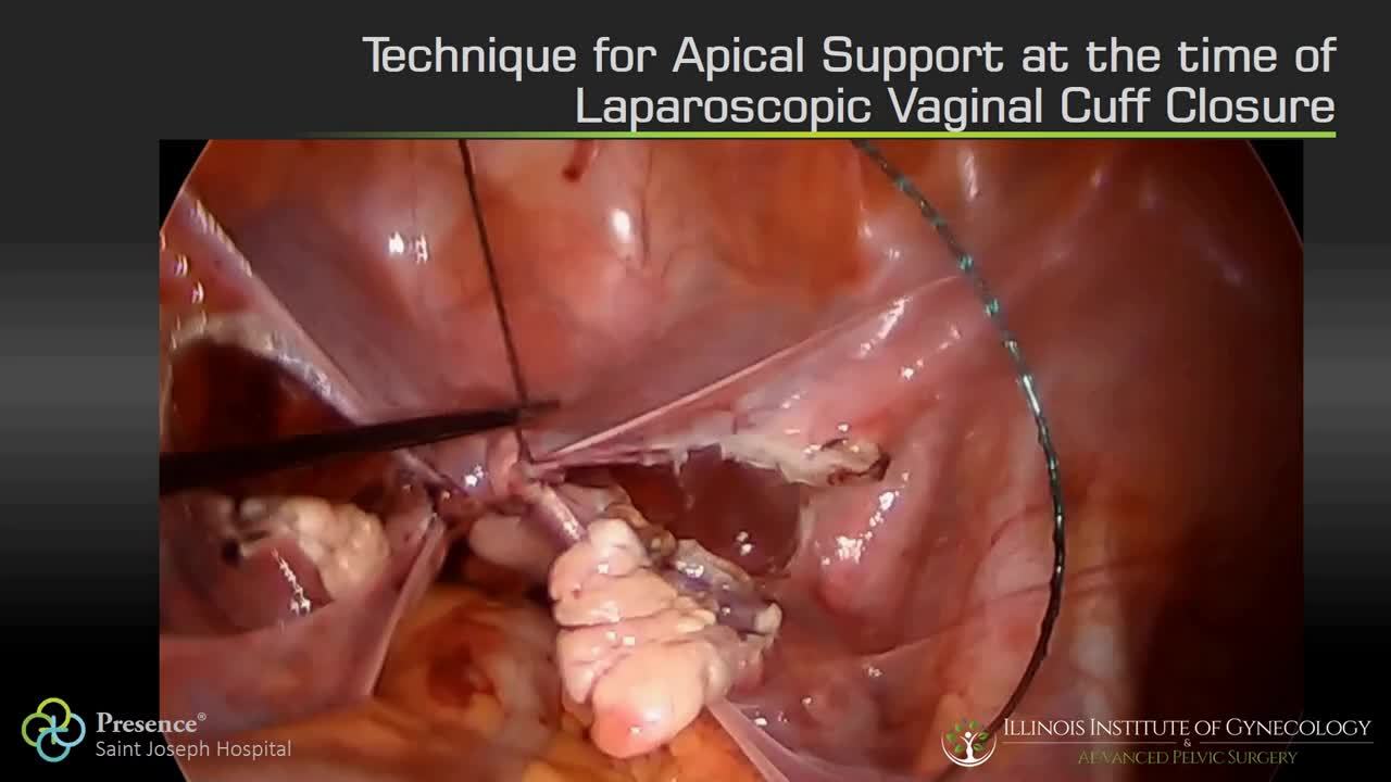
Visit the Society of Gynecologic Surgeons online: sgsonline.org
Additional videos from SGS are available here, including these recent offerings:
Visit the Society of Gynecologic Surgeons online: sgsonline.org
Additional videos from SGS are available here, including these recent offerings:
Visit the Society of Gynecologic Surgeons online: sgsonline.org
Additional videos from SGS are available here, including these recent offerings:
This video is brought to you by![]()