User login
BP accuracy is the ghost in the machine
SAN FRANCISCO – Amid all the talk about subgroup blood pressure targets and tiny differences in drug regimens at a recent hypertension meeting, there was an elephant in the room that attendees refused to ignore.
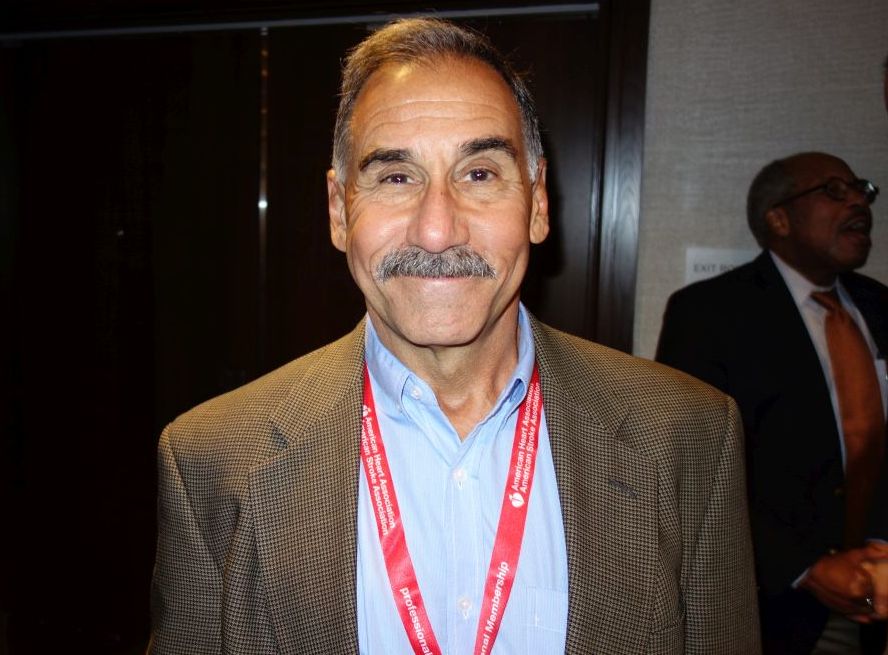
“We do it wrong,” said Dr. Steven Yarows, a primary care physician in Chelsea, Mich., who estimated he’s taken 44,000 blood pressures in his 36 years of practice.
Everyone in medicine is taught that people should rest a bit and not talk while their blood pressure is taken; that the last measurement matters more than the first; and that most Americans need a large-sized cuff. Current guidelines are based on patients sitting for 5-10 minutes alone in a quiet room while an automatic machine averages their last 3-5 blood pressures.
But when Dr. Yarows asked his 300 or so audience members – hypertension physicians who paid to come to the meeting – how many actually followed those rules, four hands went up. It’s not good enough; “if you are going to make a diagnosis that lasts a lifetime, you have to be accurate,” he said at the joint scientific sessions of the American Heart Association Council on Hypertension, AHA Council on Kidney Cardiovascular Disease, and American Society of Hypertension.
There’s resistance. No one has a room set aside for blood pressure; staff don’t want to deal with it; and at a time when primary care doctors are nickel and dimed for everything they do, insurers haven’t stepped up to pay to make accurate blood pressure a priority.
To do it right, you have to ask patients to come in 10 minutes early and have a room set up for them where they can sit alone with a large oscillometric cuff to average a few blood pressures at rest, Dr. Yarows said. They also need at least one 24-hour monitoring.
“Most of the time, the patient walks over from the waiting room, they get on the scale which automatically elevates the blood pressure tremendously, and then they sit down and talk about their family while their blood pressure is being taken.” Even in normotensive patients, that alone could raise systolic pressure 20 mm Hg or more, he said. It makes one-time blood pressure pretty much meaningless.
The biggest problem is that blood pressure is hugely variable, so it’s hard to know what matters. In one of Dr. Yarows’ normotensive patients, BP varied 44 mm Hg systolic and 37 mm Hg diastolic over 24 hours. In a hypertensive patient, systolic pressure varied 62 mm Hg and diastolic 48 mm Hg over 24 hours. Another patient was 114/85 mm Hg at noon, and 159/73 mm Hg an hour later. “That’s a huge spread,” he said.
Twenty-four hour monitoring is the only way to really know if patients are hypertensive and need treatment. “Any person you suspect of having hypertension, before you place them on medicine, you should have 24 hour blood pressure monitoring. This is the most effective way to determine if they do have high blood pressure,” and how much it needs to be lowered, he said.
Dr. Yarows had no disclosures.
SAN FRANCISCO – Amid all the talk about subgroup blood pressure targets and tiny differences in drug regimens at a recent hypertension meeting, there was an elephant in the room that attendees refused to ignore.
“We do it wrong,” said Dr. Steven Yarows, a primary care physician in Chelsea, Mich., who estimated he’s taken 44,000 blood pressures in his 36 years of practice.
Everyone in medicine is taught that people should rest a bit and not talk while their blood pressure is taken; that the last measurement matters more than the first; and that most Americans need a large-sized cuff. Current guidelines are based on patients sitting for 5-10 minutes alone in a quiet room while an automatic machine averages their last 3-5 blood pressures.
But when Dr. Yarows asked his 300 or so audience members – hypertension physicians who paid to come to the meeting – how many actually followed those rules, four hands went up. It’s not good enough; “if you are going to make a diagnosis that lasts a lifetime, you have to be accurate,” he said at the joint scientific sessions of the American Heart Association Council on Hypertension, AHA Council on Kidney Cardiovascular Disease, and American Society of Hypertension.
There’s resistance. No one has a room set aside for blood pressure; staff don’t want to deal with it; and at a time when primary care doctors are nickel and dimed for everything they do, insurers haven’t stepped up to pay to make accurate blood pressure a priority.
To do it right, you have to ask patients to come in 10 minutes early and have a room set up for them where they can sit alone with a large oscillometric cuff to average a few blood pressures at rest, Dr. Yarows said. They also need at least one 24-hour monitoring.
“Most of the time, the patient walks over from the waiting room, they get on the scale which automatically elevates the blood pressure tremendously, and then they sit down and talk about their family while their blood pressure is being taken.” Even in normotensive patients, that alone could raise systolic pressure 20 mm Hg or more, he said. It makes one-time blood pressure pretty much meaningless.
The biggest problem is that blood pressure is hugely variable, so it’s hard to know what matters. In one of Dr. Yarows’ normotensive patients, BP varied 44 mm Hg systolic and 37 mm Hg diastolic over 24 hours. In a hypertensive patient, systolic pressure varied 62 mm Hg and diastolic 48 mm Hg over 24 hours. Another patient was 114/85 mm Hg at noon, and 159/73 mm Hg an hour later. “That’s a huge spread,” he said.
Twenty-four hour monitoring is the only way to really know if patients are hypertensive and need treatment. “Any person you suspect of having hypertension, before you place them on medicine, you should have 24 hour blood pressure monitoring. This is the most effective way to determine if they do have high blood pressure,” and how much it needs to be lowered, he said.
Dr. Yarows had no disclosures.
EXPERT ANALYSIS FROM JOINT HYPERTENSION 2017
Obesity paradox slings its weight around in atrial fibrillation
BARCELONA – The obesity paradox is alive and well in the rapidly growing population with atrial fibrillation, Samuel Z. Goldhaber, MD, reported at the annual congress of the European Society of Cardiology.
In an analysis of 22,541 participants in the international registry known as GARFIELD-AF (Global Anticoagulation Registry in the Field-Atrial Fibrillation) – the largest ongoing prospective AF registry in the world – all-cause mortality during the first 2 years after diagnosis of the arrhythmia paradoxically decreased as body mass index increased all the way up to a ceiling of 40 kg/m2, according to Dr. Goldhaber, professor of medicine at Harvard Medical School and section head of vascular medicine and director of the thrombosis research group at Brigham and Women’s Hospital, Boston.

“It’s pretty impressive. This degree of mortality reduction with overweight and obesity is of about the same magnitude you might get with thrombolytic therapy for an acute MI, or with beta blocker therapy or statins post-MI,” the cardiologist said in an interview.
This newly described obesity paradox in AF is all the more baffling in light of the heavy cardiovascular risk factor burden that accompanied increased BMI. For example, the prevalence of type 2 diabetes rose from 14.1% among normal-weight individuals with AF, to 19.7% in the overweight, 27.5% in the obese, 34.6% in those with a BMI of 35 to less than 40 kg/m2, and 41.6% in patients with a BMI of 40 kg/m2 or more.
“It’s perplexing,” Dr. Goldhaber confessed. “I think there’s some hidden message here that we haven’t decoded yet.”
Patients with AF who were at the low extreme of the BMI spectrum -- the 3.3% who were underweight, with a BMI below 20 kg/m2 – fared particularly poorly. Their 2-year all-cause mortality rate of 8.71 per 100 person-years was by far the highest of any BMI class in the study. But that’s relatively straightforward to understand, as it’s likely that many underweight patients with AF were frail and/or had cancer. Indeed, only 49% of deaths in the underweight group were due to cardiovascular disease, in contrast to 64% of deaths in persons whose BMI was 40 kg/m2 or more.
Interestingly, only 5%-6% of all deaths in GARFIELD-AF were due to stroke; deaths from heart failure were far more common.
Overall, 71% of patients who presented with newly diagnosed AF in GARFIELD-AF were overweight or obese. The only contributory factor to the obesity paradox that Dr. Goldhaber could spot was that the heavier patients were younger at diagnosis of AF. But this age disparity seems unlikely to overcome their massive burden of multiple other cardiovascular risk factors. So he solicited theories from his audience. One physician argued that morbidly obese individuals are sedentary, so they don’t leave the house as much as leaner AF patients.
“You stay at home because you cannot move. You don’t get into fatal car accidents. You don’t get into conflicts and get murdered,” the physician postulated.
Dr. Goldhaber wasn’t convinced.
“When I go out to bars or dancing, I look around at people, and I estimate that a lot of them have a BMI of 35,” he commented.
Another proposed theory was that overweight and obese AF patients exercise less, so they inhale less of the airborne toxic fine particulates present in the urban environment. The adverse health impact of air pollution is a particularly hot topic of late among European cardiologists, but the notion that obese AF patients fare better because they don’t exercise runs contrary to a wealth of data supporting the health benefits of working out.
The GARFIELD-AF registry is funded by Bayer AG. Dr. Goldhaber reported receiving research grants and/or serving as a consultant to Bayer and numerous other entities, including the National Heart, Lung and Blood Institute.
BARCELONA – The obesity paradox is alive and well in the rapidly growing population with atrial fibrillation, Samuel Z. Goldhaber, MD, reported at the annual congress of the European Society of Cardiology.
In an analysis of 22,541 participants in the international registry known as GARFIELD-AF (Global Anticoagulation Registry in the Field-Atrial Fibrillation) – the largest ongoing prospective AF registry in the world – all-cause mortality during the first 2 years after diagnosis of the arrhythmia paradoxically decreased as body mass index increased all the way up to a ceiling of 40 kg/m2, according to Dr. Goldhaber, professor of medicine at Harvard Medical School and section head of vascular medicine and director of the thrombosis research group at Brigham and Women’s Hospital, Boston.
“It’s pretty impressive. This degree of mortality reduction with overweight and obesity is of about the same magnitude you might get with thrombolytic therapy for an acute MI, or with beta blocker therapy or statins post-MI,” the cardiologist said in an interview.
This newly described obesity paradox in AF is all the more baffling in light of the heavy cardiovascular risk factor burden that accompanied increased BMI. For example, the prevalence of type 2 diabetes rose from 14.1% among normal-weight individuals with AF, to 19.7% in the overweight, 27.5% in the obese, 34.6% in those with a BMI of 35 to less than 40 kg/m2, and 41.6% in patients with a BMI of 40 kg/m2 or more.
“It’s perplexing,” Dr. Goldhaber confessed. “I think there’s some hidden message here that we haven’t decoded yet.”
Patients with AF who were at the low extreme of the BMI spectrum -- the 3.3% who were underweight, with a BMI below 20 kg/m2 – fared particularly poorly. Their 2-year all-cause mortality rate of 8.71 per 100 person-years was by far the highest of any BMI class in the study. But that’s relatively straightforward to understand, as it’s likely that many underweight patients with AF were frail and/or had cancer. Indeed, only 49% of deaths in the underweight group were due to cardiovascular disease, in contrast to 64% of deaths in persons whose BMI was 40 kg/m2 or more.
Interestingly, only 5%-6% of all deaths in GARFIELD-AF were due to stroke; deaths from heart failure were far more common.
Overall, 71% of patients who presented with newly diagnosed AF in GARFIELD-AF were overweight or obese. The only contributory factor to the obesity paradox that Dr. Goldhaber could spot was that the heavier patients were younger at diagnosis of AF. But this age disparity seems unlikely to overcome their massive burden of multiple other cardiovascular risk factors. So he solicited theories from his audience. One physician argued that morbidly obese individuals are sedentary, so they don’t leave the house as much as leaner AF patients.
“You stay at home because you cannot move. You don’t get into fatal car accidents. You don’t get into conflicts and get murdered,” the physician postulated.
Dr. Goldhaber wasn’t convinced.
“When I go out to bars or dancing, I look around at people, and I estimate that a lot of them have a BMI of 35,” he commented.
Another proposed theory was that overweight and obese AF patients exercise less, so they inhale less of the airborne toxic fine particulates present in the urban environment. The adverse health impact of air pollution is a particularly hot topic of late among European cardiologists, but the notion that obese AF patients fare better because they don’t exercise runs contrary to a wealth of data supporting the health benefits of working out.
The GARFIELD-AF registry is funded by Bayer AG. Dr. Goldhaber reported receiving research grants and/or serving as a consultant to Bayer and numerous other entities, including the National Heart, Lung and Blood Institute.
AT THE ESC CONGRESS 2017
Key clinical point:
Major finding: Two-year all-cause mortality following diagnosis of atrial fibrillation fell substantially in stepwise fashion with increasing body mass index, for reasons unknown.
Data source: GARFIELD-AF is an ongoing enormous international prospective registry of patients newly diagnosed with atrial fibrillation.
Disclosures: The GARFIELD-AF registry is funded by Bayer AG. The presenter reported receiving research grants and/or consultant fees from that company and numerous others.
Public health intervention helps stem tuberculosis transmission
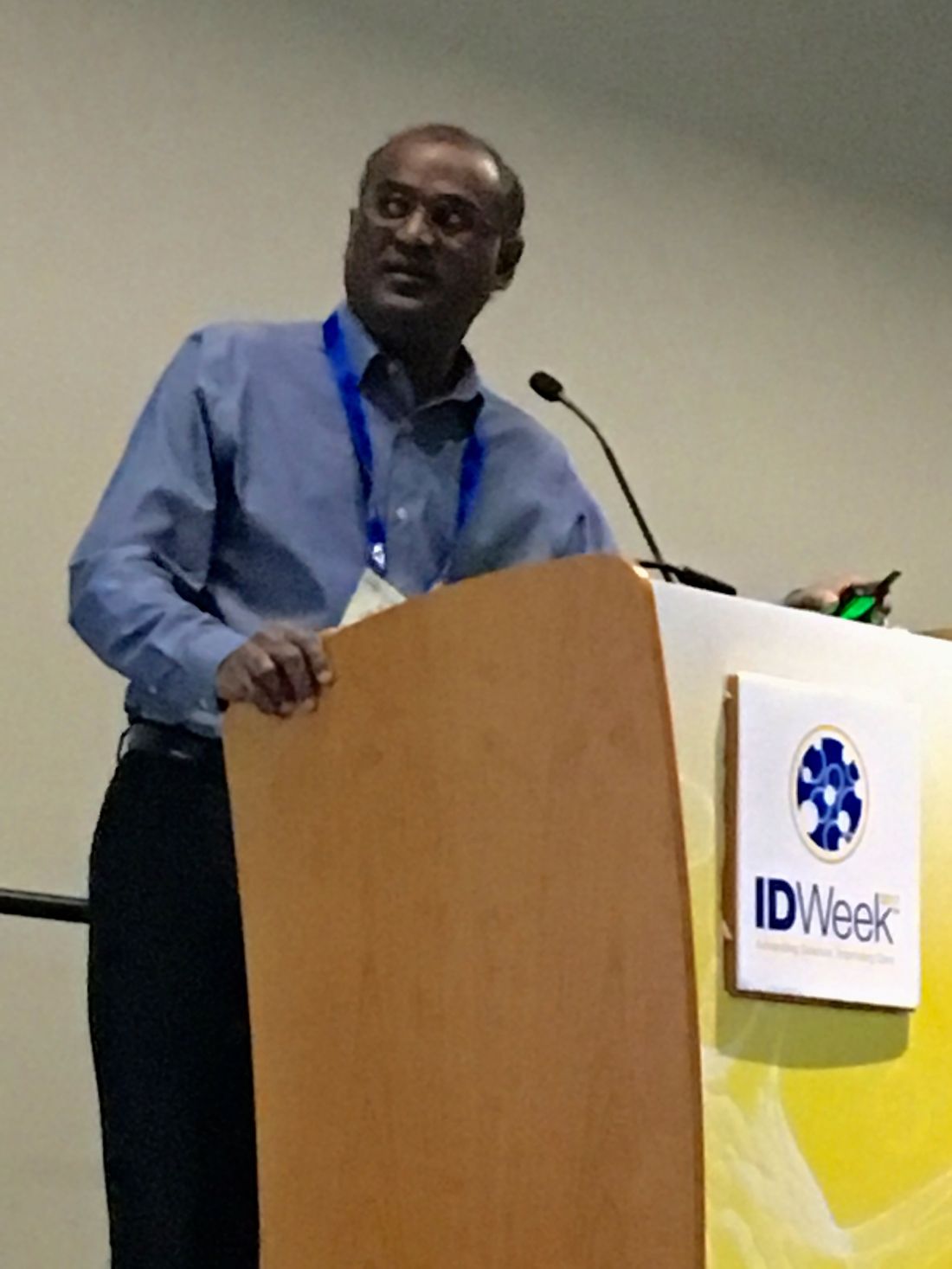
SAN DIEGO – Using an on-site multi-disciplinary team to approach patients who are unwilling to receive tuberculosis treatment can improve patient cooperation, according to a case study presented ID Week 2017, an infectious disease meeting.
Such public health interventions may be able to improve disease control and interrupt the transmission cycle among patients who are not adhering to treatment, according to Aisha Haynie, MD, MPA.
“We started the patient on treatment and she actually decided to move into a motel,” said Dr. Haynie. “After that, she moved back into the family home and said she wasn’t having any contact [with residents], which we didn’t really believe.”
While the patient did reluctantly give Dr. Haynie and her team a list of five family members to test, the patient told Dr. Haynie that she and her team were not allowed into the patient’s home.
When the five members were tested, the new patients showed similar signs of reluctance and mistrust of the health system.
“The family members came to our clinic and two of them were TB positive,” recounted Dr. Haynie. “So they came back, but while they were sitting in the waiting room, they signed papers and they left.”
Despite continuous attempts to reach the family and address the growing concern from health professionals of the danger of TB transmission to other members of the family, patients continue not to adhere to procedures beyond pharmaceutical intervention.
In an attempt to directly contact the patient’s family, Dr. Haynie lead a team consisting of a local health authority physician, a TB nurse practitioner, a TB contact analyst, a nurse case manager, a county attorney, and an interpreter for unscheduled visits to family members individually.
“We had specific requests [including] keeping appointments, getting labs, and we added a deadline,” said Dr. Haynie, Chief of Disease Control and Medical Epidemiology for the Harris County Public Health & Environmental Services. “By the next Friday [we told them that] we were filing court papers with or without the information and we [would] be contacting child protective services as well.”
Following the intervention, the patient and her family adhered to testing procedures. This revealed an infant with active TB and 8 other family members with TB Infection. Isolation breaches were also discovered. Most importantly, the TB transmission cycle was interrupted, according to Dr. Haynie.
A key aspect of the team’s successful approach was to address cultural and economic barriers that hindered successful interaction with the family, TB misconceptions corrected in order for a trusting relationship to develop.
The investigators developed this intervention in Harris County, Tx, which at 4.3 million residents is the 3rd most populous U.S. county, and has reported a TB case rate of 7.6 cases per 100,000, which is approximately double that of the U.S. average, according to Dr. Haynie. Of those cases in Harris County, 73% are foreign-born, compared with the average rate of 59% in Texas.
Dr. Haynie and fellow investigators asserted part of the reason patients were so reluctant to receive treatment from the Harris County Public Health department was a combination of mistrust in the system and a number of false ideas patients have regarding TB, a sign of further educational tools being needed.
Since the first use of the intervention, Dr. Haynie and her team have implemented this approach with other non-adherent patients with relative success.
“This is something that we now do and we have not been back to court [to enforce compliance] since,” said Dr. Haynie.
[email protected]
On Twitter @eaztweets
ID Week 2017
Inappropriate C. diff. testing reduced with ‘Hard Stop’ protocol
SAN DIEGO – After other strategies to reduce inappropriate testing for Clostridium difficile infection did not provide adequate reductions in inappropriate orders, a “hard stop” protocol, which prevents testing if specific conditions are not met, reduced the rate of testing by 42% without any incidence of delayed diagnosis, according to data presented at ID Week 2017.
“Testing stewardship is really critical to minimize the frequency of false-positive [Clostridium difficile infection] cases, which of course lead to harm. They lead to inappropriate treatment, prolonged length of stay, and patient dissatisfaction,” reported Marci L. Drees, MD, Infection Prevention Officer and Hospital Epidemiologist, Christiana Care Health System, Newark, Del.

“Of course, we did, at the time, educate our clinicians about this new testing algorithm, explaining how it is so much more sensitive and that they need to be smarter about who they are testing. But as we saw the cases roll in, we noticed that many of the [colonization] cases were in patients with a history of recent laxative use,” Dr. Drees recounted.
This led initially to a “soft stop” protocol in which clinicians ordering C. difficile–infection testing received an alert if laxatives had been ordered within the previous 24 hours. The alert provided details about the laxatives and suggested that the order for C. difficile–infection testing be reconsidered. This alert was no more than a suggestion, but it did reduce C. difficile–infection testing orders by 25% at first.
“The problem was that the alert lost effect over time as people got used to seeing it and clicked right through it,” Dr. Drees explained.
Despite the diminishing effect of the soft stop, no further steps were planned until a new set of data alerted the infectious disease department that C. difficile–infection rates were higher than benchmarks. This prompted concern among the hospital leadership and led to creation of a multidisciplinary team that was given the task to dig deeper for the source of problems and develop strategies to lower rates of inappropriate testing.
When that team did their initial analysis, what really came to the front was that about half the cases that had been identified as hospital-onset C. difficile were likely only C. difficile colonizations. They either had received laxatives prior to their test or they did not have significant diarrhea,” Dr. Drees said.
It was this finding that prompted a hard stop protocol to be built into C. difficile–infection test orders. In this protocol, which is executed only in patients who have been hospitalized for at least 36 hours, two conditions must be met sequentially for the testing order to proceed. The first is there must be documentation of at least two episodes of diarrhea in the prior 24 hours. If this condition is met, then the ordering system asks for verification that the patient has not received a laxative in the prior 24 hours.
“If either of these criteria are not met, the order can only proceed if the clinician calls the lab for an override,” Dr. Drees explained. Although the person answering the phone in the laboratory does not require any additional documentation or justification, the person ordering the test must enter the name of the person in the laboratory with whom they spoke. If this field is not filled in, the order will not proceed.
Prior to the hard stop, there was an average of 12 C. difficile–infection tests ordered per day. After the hard stop, the average fell to 7 per day, a 42% decline, according to Dr. Drees. C. difficile-infection cases did not increase, and there were no incidences of delayed diagnosis resulting in adverse clinical consequences.
Of the 157 overrides on the hard stop, representing 15% of all orders, only 11% produced a positive C. difficile-infection result. It was noted that almost one third of the overrides did not follow the protocol.
“What I mean is that they did not actually call the lab. They put in their own name [for the field requiring the name of a lab worker] or they put in Mickey Mouse or they put in something, so the order would proceed,” Dr. Drees said. After followup in those cases, “we have not had repeat offenders.”
Even a hard stop is likely to have diminishing efficacy over time as clinicians identify work-arounds, Dr. Drees acknowledged. For example, there is concern that therapy for CDI will be ordered without testing, although this has not yet been detected. However, the hard stop has been more effective and shown a longer duration of effect than the soft stop, according to Dr. Drees. Although Dr. Drees acknowledged that there have been exceptions, the hard stop has “generally been well accepted.”
Dr. Drees reported that she has no financial relationships relevant to this topic.
SAN DIEGO – After other strategies to reduce inappropriate testing for Clostridium difficile infection did not provide adequate reductions in inappropriate orders, a “hard stop” protocol, which prevents testing if specific conditions are not met, reduced the rate of testing by 42% without any incidence of delayed diagnosis, according to data presented at ID Week 2017.
“Testing stewardship is really critical to minimize the frequency of false-positive [Clostridium difficile infection] cases, which of course lead to harm. They lead to inappropriate treatment, prolonged length of stay, and patient dissatisfaction,” reported Marci L. Drees, MD, Infection Prevention Officer and Hospital Epidemiologist, Christiana Care Health System, Newark, Del.
“Of course, we did, at the time, educate our clinicians about this new testing algorithm, explaining how it is so much more sensitive and that they need to be smarter about who they are testing. But as we saw the cases roll in, we noticed that many of the [colonization] cases were in patients with a history of recent laxative use,” Dr. Drees recounted.
This led initially to a “soft stop” protocol in which clinicians ordering C. difficile–infection testing received an alert if laxatives had been ordered within the previous 24 hours. The alert provided details about the laxatives and suggested that the order for C. difficile–infection testing be reconsidered. This alert was no more than a suggestion, but it did reduce C. difficile–infection testing orders by 25% at first.
“The problem was that the alert lost effect over time as people got used to seeing it and clicked right through it,” Dr. Drees explained.
Despite the diminishing effect of the soft stop, no further steps were planned until a new set of data alerted the infectious disease department that C. difficile–infection rates were higher than benchmarks. This prompted concern among the hospital leadership and led to creation of a multidisciplinary team that was given the task to dig deeper for the source of problems and develop strategies to lower rates of inappropriate testing.
When that team did their initial analysis, what really came to the front was that about half the cases that had been identified as hospital-onset C. difficile were likely only C. difficile colonizations. They either had received laxatives prior to their test or they did not have significant diarrhea,” Dr. Drees said.
It was this finding that prompted a hard stop protocol to be built into C. difficile–infection test orders. In this protocol, which is executed only in patients who have been hospitalized for at least 36 hours, two conditions must be met sequentially for the testing order to proceed. The first is there must be documentation of at least two episodes of diarrhea in the prior 24 hours. If this condition is met, then the ordering system asks for verification that the patient has not received a laxative in the prior 24 hours.
“If either of these criteria are not met, the order can only proceed if the clinician calls the lab for an override,” Dr. Drees explained. Although the person answering the phone in the laboratory does not require any additional documentation or justification, the person ordering the test must enter the name of the person in the laboratory with whom they spoke. If this field is not filled in, the order will not proceed.
Prior to the hard stop, there was an average of 12 C. difficile–infection tests ordered per day. After the hard stop, the average fell to 7 per day, a 42% decline, according to Dr. Drees. C. difficile-infection cases did not increase, and there were no incidences of delayed diagnosis resulting in adverse clinical consequences.
Of the 157 overrides on the hard stop, representing 15% of all orders, only 11% produced a positive C. difficile-infection result. It was noted that almost one third of the overrides did not follow the protocol.
“What I mean is that they did not actually call the lab. They put in their own name [for the field requiring the name of a lab worker] or they put in Mickey Mouse or they put in something, so the order would proceed,” Dr. Drees said. After followup in those cases, “we have not had repeat offenders.”
Even a hard stop is likely to have diminishing efficacy over time as clinicians identify work-arounds, Dr. Drees acknowledged. For example, there is concern that therapy for CDI will be ordered without testing, although this has not yet been detected. However, the hard stop has been more effective and shown a longer duration of effect than the soft stop, according to Dr. Drees. Although Dr. Drees acknowledged that there have been exceptions, the hard stop has “generally been well accepted.”
Dr. Drees reported that she has no financial relationships relevant to this topic.
ID WEEK 2017
Key clinical point:
Major finding: After a hard stop protocol was implemented, the average per day rate of C. diff testing at a tertiary medical center was reduced 42%.
Data source: Prospective performance improvement project.
Disclosures: Dr. Drees reported that she has no financial relationships relevant to this topic.
Faster multiplex PCR improved management of pediatric acute respiratory illness
SAN DIEGO – Switching to a faster, more comprehensive multiplex PCR viral respiratory assay enabled a hospital to discharge young children with acute respiratory illnesses sooner, prescribe oseltamivir more often, and curtail the use of antibiotics and thoracic radiography, Rangaraj Selvarangan, PhD, reported at an annual meeting on infectious disease.
The study shows how rapid multiplex PCR testing can facilitate antimicrobial stewardship, said Dr. Selvarangan, who is a professor at the University of Missouri Kansas City School of Medicine and director of the microbiology laboratory at Children’s Mercy Kansas City. “Our antimicrobial stewardship programs monitors these test results daily, add notes, and make recommendations on antibiotic choices,” he said.
For the study, the researchers compared hospital records from December 2008 through May 2012, when Children’s Mercy hospital used the Luminex xTAG Respiratory Viral Panel, with records from August 2012 through June 2015, after the hospital had switched over to the Biofire FilmArray Respiratory Panel. FilmArray targets the same 17 viral pathogens as the Luminex panel, but also targets Bordetella pertussis, Chlamydia pneumoniae, and Mycoplasma pneumoniae, seasonal influenza A, parainfluenza type 4, and four coronaviruses.
The study included children aged up to 2 years who were not on immunosuppressive medications, in the NICU, or hospitalized for more than 7 days. For this population, the two panels yielded similar rates of positivity overall (about 60%) and for individual viruses, Dr. Selvarangan said. A total of 810 patients tested positive for at least one virus on the Luminex panel, and 2,096 patients tested positive on FilmArray. Results for FilmArray were available within a median of 4 hours, versus 29 hours for Luminex (P less than .001). The prevalence of empiric antibiotic therapy was 44% during the Luminex era and 28% after the hospital switched to FilmArray (P less than .001). Rates of antibiotic discontinuation rose from 16% with Luminex to 23% with FilmArray (P less than .01). Strikingly, oseltamivir prescriptions rose five-fold (from 17% to 85%; P less than .001) after the hospital began using FilmArray, which covers seasonal influenza. Finally, use of chest radiography fell significantly in both infants and older children after the hospital began using FilmArray instead of Luminex.
Clinicians might have become more comfortable interpreting molecular test results over time, which could have altered their clinical decision-making and thereby biased the study results, Dr. Selvarangan noted. Although at least four other single-center retrospective studies have linked multiplex PCR assays to better antibiotic stewardship, this study is one of the first to directly compare clinical outcomes between two assays, he added. More studies are needed to help guide choice of multiplex assays, whose cost remains a major barrier to widespread use, he concluded.
Dr. Selvarangan disclosed grant support from both Biofire Diagnostics and Luminex corporation, and an advisory relationship with BioFire.
AT IDWEEK 2017
Key clinical point: Faster multiplex PCR respiratory panel results helped guide management of young children hospitalized with acute respiratory illness.
Major finding: Empiric antibiotic therapy, thoracic radiography, and median length of stay all decreased.
Data source: A descriptive single-center retrospective study of 2,905 children up to 24 months old who were hospitalized with acute respiratory illness and tested positive for at least one virus on a multiplex PCR.
Disclosures: Dr. Selvarangan disclosed grant support from both Biofire Diagnostics and Luminex corporation, and an advisory relationship with BioFire.
Study eyes factors that may trigger breakthrough bacteremia
SAN DIEGO – , according to a detailed study of six isolates.
“Both patient and microbe factors can contribute to treatment failure, even involving pan-susceptible isolates,” Andrew Berti, PharmD, PhD, said in an interview prior to an annual scientific meeting on infectious diseases.

The researchers presented the case of a patient who experienced multiple episodes of breakthrough Staphylococcus aureus bacteremia over a period of 5 years in the setting of appropriately dosed antimicrobial suppressive therapy. They recovered six clinical bloodstream isolates from the patient during distinct episodes of methicillin-susceptible S. aureus (MSSA) bacteremia, and it was determined that a central line infection was the source for each episode. Isolates recovered were susceptible to the individual therapies received, which included oxacillin, daptomycin, and dalbavancin. The researchers used Illumina technology to collect bacterial whole genome sequence data. “Relatively little is known about bacterial evolution during human infection as these tend to be acute, rapidly resolved events,” Dr. Berti said. “This case was unique in that we could observe changes to a pathogen over a more than 6 years period as it adapted to survive host defenses and multiple antibiotic interventions.”
The researchers discovered that the first two isolates (ST256) and the last four isolates (ST5) “represent distinct populations and suggest that a distinct MSSA strain displaced the previous population between bacteremia episodes 2 and 3,” they wrote in their abstract. “Of note, all of these strains were able to survive and establish breakthrough bacteremias despite favorable susceptibility profiles to the agents used as suppressive therapy. Although the MICs remain low and in the susceptible range to oxacillin, daptomycin, and dalbavancin, these isolates progressively developed significant antimicrobial tolerance phenotypes, which coincided with mutations in walK (yycG), htrA2, ftsW, ebh, and ileS that may be advantageous to survival under antibiotic pressure.”
Dr. Berti acknowledged certain limitations of the analysis, including the inability to test for heteroresistance to some antibiotics, which may also contribute to treatment failure. “Specific therapeutic interventions that coincide with bacterial population changes are not necessarily causative of those changes,” he said. One of the study authors, Warren Rose, PharmD, disclosed having received research grants, speaker honoraria, and/or consulting fees from Theravance, Merck, The Medicines Company and Visante, Inc. All other authors reported having no financial disclosures.
SAN DIEGO – , according to a detailed study of six isolates.
“Both patient and microbe factors can contribute to treatment failure, even involving pan-susceptible isolates,” Andrew Berti, PharmD, PhD, said in an interview prior to an annual scientific meeting on infectious diseases.
The researchers presented the case of a patient who experienced multiple episodes of breakthrough Staphylococcus aureus bacteremia over a period of 5 years in the setting of appropriately dosed antimicrobial suppressive therapy. They recovered six clinical bloodstream isolates from the patient during distinct episodes of methicillin-susceptible S. aureus (MSSA) bacteremia, and it was determined that a central line infection was the source for each episode. Isolates recovered were susceptible to the individual therapies received, which included oxacillin, daptomycin, and dalbavancin. The researchers used Illumina technology to collect bacterial whole genome sequence data. “Relatively little is known about bacterial evolution during human infection as these tend to be acute, rapidly resolved events,” Dr. Berti said. “This case was unique in that we could observe changes to a pathogen over a more than 6 years period as it adapted to survive host defenses and multiple antibiotic interventions.”
The researchers discovered that the first two isolates (ST256) and the last four isolates (ST5) “represent distinct populations and suggest that a distinct MSSA strain displaced the previous population between bacteremia episodes 2 and 3,” they wrote in their abstract. “Of note, all of these strains were able to survive and establish breakthrough bacteremias despite favorable susceptibility profiles to the agents used as suppressive therapy. Although the MICs remain low and in the susceptible range to oxacillin, daptomycin, and dalbavancin, these isolates progressively developed significant antimicrobial tolerance phenotypes, which coincided with mutations in walK (yycG), htrA2, ftsW, ebh, and ileS that may be advantageous to survival under antibiotic pressure.”
Dr. Berti acknowledged certain limitations of the analysis, including the inability to test for heteroresistance to some antibiotics, which may also contribute to treatment failure. “Specific therapeutic interventions that coincide with bacterial population changes are not necessarily causative of those changes,” he said. One of the study authors, Warren Rose, PharmD, disclosed having received research grants, speaker honoraria, and/or consulting fees from Theravance, Merck, The Medicines Company and Visante, Inc. All other authors reported having no financial disclosures.
REPORTING FROM ID WEEK 2017
Key clinical point: Both patient and microbe factors can contribute to antibitoic treatment failure.
Major finding: Two distinct Staphylococcus aureus lineages were islolated over a period of 5 years, and each was able to persist despite appropriate antibitoic interventions.
Study details: Clinical analysis of six bloodstream isolates from a singles patient over a 5-year period.
Disclosures: One of the study authors, Warren Rose, PharmD, disclosed having received research grants, speaker honoraria and/or consulting fees from Theravance, Merck, The Medicines Company and Visante. All other authors reported having no financial disclosures.
New data dissect global burden of scabies
Scabies hits hardest in tropical parts of the world, and children and teens are most affected by the condition, based on data from a global epidemiological assessment.
“,” wrote Chante Karimkhani, MD, of the University of Colorado, Aurora, and her colleagues. To estimate the global impact of scabies, the researchers used the Global Burden of Disease (GBD) Study 2015, which included literature searches between 1980 and 2014 in both English and Spanish.
Overall, the prevalence of scabies worldwide was 204,151,715, and the age-standardized DALYs was 71/100,000 people. The highest DALYs in terms of world regions were reported in East Asia (136/100,000), Southeast Asia (135/100,000) the region described as Oceania (120/100,000) (which included American Samoa, Fiji, Guam, Marshall Islands, Northern Mariana Islands, Papua New Guinea, Samoa, Solomon Islands, and others), tropical Latin America (100/100,000), and South Asia (69/100,000). Scabies caused 0.21% of DALYs from all the conditions that are studied by GBD 2015 globally. The mean percent change in age-standardized DALY from 1990 to 2015 increased 22% in North America, but the age-standardized DALY still remained less than 5/100,000.
In addition, global DALYs from scabies skin infection were highest in children aged 1-4 years (116/100,000 individuals) and adolescents aged 15-19 years (102/100,000). DALYs were not significantly different between men and women across all age groups, Dr. Karimkhani and her associates reported.
“As a worldwide epidemiological assessment, GBD 2015 provides broad and frequently updated measures of scabies burden in terms of skin effects. These global data might help guide research protocols and prioritization efforts and focus scabies treatment and control measures,” the researchers said.
Of note is that these analyses do not include information about complications of scabies such as impetigo, local and systemic bacterial infections, glomerulonephritis, and rheumatic fever.
The study was funded by the Bill and Melinda Gates Foundation. The researchers had no financial conflicts to disclose.
Read the full study here: (Lancet Infect. Dis. 2017. doi: 10.1016/S1473-3099[17]30483-8).
FROM THE LANCET INFECTIOUS DISEASES
Behavioral approach to appropriate antimicrobial prescribing in hospitals: The DUMAS study
Clinical question: How effective is an antimicrobial stewardship approach grounded in behavioral theory and focused on preserving prescriber autonomy and participation in improving appropriateness of antimicrobial prescribing in hospitals?
Background: Antimicrobial stewardship programs aim to achieve the goal of improving antimicrobial prescribing. This leads to significant benefits, including decreased antimicrobial resistance, improved clinical outcomes (lower morbidity and mortality), and lower health care costs. Stewardship programs do not often focus on the human behavior element of the prescribing physicians. Changing antimicrobial prescribing is a complex behavioral process, and there is a known persistent resistance between prescribers and the stewardship team. In a simple sense, this resistance is generated by tension created when prescribers do not have autonomy.
Previous studies that used interventions based on behavioral theory found promising results, but none of them were in a hospital setting. Rather, most of these studies had a narrow focus of respiratory tract infections in outpatient clinics.

Setting: Seven clinical departments (two medical, three surgical, and two pediatric) in a tertiary care medical center and a general teaching hospital, both in Amsterdam. The first hospital was a 700-bed tertiary care center with salaried specialists, while the second hospital was a 550-bed general medical center with self-employed specialists.
Synopsis: During a baseline period of 16 months and an intervention period of 12 months, physicians who prescribed systemic antimicrobial drugs for any indication were included in the study. In all, 1,121 patient cases with 700 prescriptions were studied during the baseline period and 882 patient cases with 531 prescriptions were studied during the intervention period. The intervention was as follows: Prescribers were offered a free choice of how to improve their antimicrobial prescribing. They were stimulated to choose interventions with higher potential for success based on a root cause analysis of inappropriate prescribing. The study was inspired by the participatory action research paradigm, which focuses on collaboration and empowerment of the stakeholders in the change process. In this study, prescribers were given reports of root cause analysis of their prior prescribing patterns. Then, they were invited to choose and codevelop one or more interventions that were tailored and individualized to improve their own prescribing. This approach draws on the following three behavioral principles: 1) respect for the prescriber’s autonomy to avoid feelings of resistance; 2) the inclination that people have to value a product higher and feel more ownership of it if they made it themselves (the IKEA effect); 3) the tendency of people to follow up on an active and public commitment.
The primary outcome was antimicrobial appropriateness, measured with a validated appropriateness assessment instrument. One of three infectious disease specialists assessed the adult prescriptions, and one of three infectious disease/immunology pediatricians assessed the pediatric prescriptions for appropriateness. Appropriateness criteria were as follows: indication, choice of antimicrobial, dosage, route, and duration. A secondary outcome was antimicrobial consumption, reported as days of therapy per 100 admissions per month. Other outcomes were changes in specific appropriateness categories, intravenous antimicrobial consumption, consumption of specific antimicrobial subgroups, and length of hospital stay.
The mean antimicrobial appropriateness increased from 64.1% at intervention start to 77.4% at 12-month follow-up (+13.3%; relative risk, 1.17; 95% CI, 1.04-1.27), without a change in slope. Antimicrobial consumption remained the same during both study periods. Length of hospital stay did not change relative to the start of the intervention approach.
This is the first study of its kind, as a hospital antimicrobial stewardship program study grounded in behavioral science, with the key element being the free choice allowed to the prescribers, who made their own autonomous decisions about how to improve their prescribing. The authors hypothesize that the prescribers felt relatively nonthreatened by their approach since the prescribers maintained their free will to change their own behavior if so desired. The prescribers were given a free intervention choice. For example, they could have just chosen “education” as an easy out; however, the root cause analysis seemed to be an impetus for the prescribers to choose interventions that would be more effective. A prior study in a nursing home setting was unsuccessful; aside from other differences, that study used a predetermined set of interventions, thus lacking the autonomy and IKEA effect seen in this study.
Bottom line: Use of a behavioral approach that preserves prescriber autonomy resulted in an increase in antimicrobial appropriateness sustained for at least 12 months. The intervention is effective, inexpensive, and transferable to various health care settings.
Citation: Sikkens JJ, van Agtmael MA, Peters EJG, et al. Behavioral approach to appropriate antimicrobial prescribing in hospitals: The dutch unique method for antimicrobial stewardship (DUMAS) participatoryintervention study. JAMA Intern Med. Published online May 1, 2017. doi: 10.1001/jamainternmed.2017.0946.
Dr. Ramee is a hospitalist at Ochsner Health System, New Orleans.
Clinical question: How effective is an antimicrobial stewardship approach grounded in behavioral theory and focused on preserving prescriber autonomy and participation in improving appropriateness of antimicrobial prescribing in hospitals?
Background: Antimicrobial stewardship programs aim to achieve the goal of improving antimicrobial prescribing. This leads to significant benefits, including decreased antimicrobial resistance, improved clinical outcomes (lower morbidity and mortality), and lower health care costs. Stewardship programs do not often focus on the human behavior element of the prescribing physicians. Changing antimicrobial prescribing is a complex behavioral process, and there is a known persistent resistance between prescribers and the stewardship team. In a simple sense, this resistance is generated by tension created when prescribers do not have autonomy.
Previous studies that used interventions based on behavioral theory found promising results, but none of them were in a hospital setting. Rather, most of these studies had a narrow focus of respiratory tract infections in outpatient clinics.
Setting: Seven clinical departments (two medical, three surgical, and two pediatric) in a tertiary care medical center and a general teaching hospital, both in Amsterdam. The first hospital was a 700-bed tertiary care center with salaried specialists, while the second hospital was a 550-bed general medical center with self-employed specialists.
Synopsis: During a baseline period of 16 months and an intervention period of 12 months, physicians who prescribed systemic antimicrobial drugs for any indication were included in the study. In all, 1,121 patient cases with 700 prescriptions were studied during the baseline period and 882 patient cases with 531 prescriptions were studied during the intervention period. The intervention was as follows: Prescribers were offered a free choice of how to improve their antimicrobial prescribing. They were stimulated to choose interventions with higher potential for success based on a root cause analysis of inappropriate prescribing. The study was inspired by the participatory action research paradigm, which focuses on collaboration and empowerment of the stakeholders in the change process. In this study, prescribers were given reports of root cause analysis of their prior prescribing patterns. Then, they were invited to choose and codevelop one or more interventions that were tailored and individualized to improve their own prescribing. This approach draws on the following three behavioral principles: 1) respect for the prescriber’s autonomy to avoid feelings of resistance; 2) the inclination that people have to value a product higher and feel more ownership of it if they made it themselves (the IKEA effect); 3) the tendency of people to follow up on an active and public commitment.
The primary outcome was antimicrobial appropriateness, measured with a validated appropriateness assessment instrument. One of three infectious disease specialists assessed the adult prescriptions, and one of three infectious disease/immunology pediatricians assessed the pediatric prescriptions for appropriateness. Appropriateness criteria were as follows: indication, choice of antimicrobial, dosage, route, and duration. A secondary outcome was antimicrobial consumption, reported as days of therapy per 100 admissions per month. Other outcomes were changes in specific appropriateness categories, intravenous antimicrobial consumption, consumption of specific antimicrobial subgroups, and length of hospital stay.
The mean antimicrobial appropriateness increased from 64.1% at intervention start to 77.4% at 12-month follow-up (+13.3%; relative risk, 1.17; 95% CI, 1.04-1.27), without a change in slope. Antimicrobial consumption remained the same during both study periods. Length of hospital stay did not change relative to the start of the intervention approach.
This is the first study of its kind, as a hospital antimicrobial stewardship program study grounded in behavioral science, with the key element being the free choice allowed to the prescribers, who made their own autonomous decisions about how to improve their prescribing. The authors hypothesize that the prescribers felt relatively nonthreatened by their approach since the prescribers maintained their free will to change their own behavior if so desired. The prescribers were given a free intervention choice. For example, they could have just chosen “education” as an easy out; however, the root cause analysis seemed to be an impetus for the prescribers to choose interventions that would be more effective. A prior study in a nursing home setting was unsuccessful; aside from other differences, that study used a predetermined set of interventions, thus lacking the autonomy and IKEA effect seen in this study.
Bottom line: Use of a behavioral approach that preserves prescriber autonomy resulted in an increase in antimicrobial appropriateness sustained for at least 12 months. The intervention is effective, inexpensive, and transferable to various health care settings.
Citation: Sikkens JJ, van Agtmael MA, Peters EJG, et al. Behavioral approach to appropriate antimicrobial prescribing in hospitals: The dutch unique method for antimicrobial stewardship (DUMAS) participatoryintervention study. JAMA Intern Med. Published online May 1, 2017. doi: 10.1001/jamainternmed.2017.0946.
Dr. Ramee is a hospitalist at Ochsner Health System, New Orleans.
FDA grants drug orphan designation to treat cGVHD

The US Food and Drug Administration (FDA) has granted orphan drug designation to KD025 for the treatment of chronic graft-versus-host disease (cGVHD).
KD025 is a Rho-associated coiled-coil kinase (ROCK) inhibitor being developed by Kadmon Holdings, Inc.
The drug is currently under investigation in a phase 2 trial of adults with steroid-dependent or steroid-refractory cGVHD and active disease.
This dose-finding study has enrolled 48 patients divided into 3 cohorts in which patients receive KD025 at 200 mg once daily, 200 mg twice daily, or 400 mg once daily. The patients were enrolled sequentially following a safety assessment of each cohort.
In July, Kadmon Holdings, Inc. released a preliminary analysis of data from the lowest-dose cohort (n=17, 200 mg once daily).
The overall response rate in this cohort was 71% (12/17). Of responders remaining on KD025 through week 24, 89% (8/9) sustained responses. Sixty-seven percent (8/12) of responders saw an improvement in symptoms, as measured by the Lee cGVHD Symptom Scale score.
In addition to the 12 responders, 3 patients had stable disease and were still on KD025 through week 24. So an overall clinical benefit (response and stable disease) occurred in 88% (15/17) of patients.
Sixty-seven percent (8/12) of responders had a reduction in steroid doses, and 67% (4/6) of responders on tacrolimus had a reduction in tacrolimus doses.
There were no drug-related serious adverse events or drug-related elevations in liver function tests reported.
Kadmon Holdings, Inc. plans to present additional data from this study toward the end of this year.
About orphan designation
The FDA grants orphan designation to products intended to treat, diagnose, or prevent diseases/disorders that affect fewer than 200,000 people in the US.
The designation provides incentives for sponsors to develop products for rare diseases. This may include tax credits toward the cost of clinical trials, prescription drug user fee waivers, and 7 years of market exclusivity if the product is approved. ![]()
Duty vs. confidentiality
Question: During an office visit, a patient confided in his primary care provider his intention to go on a shooting spree. When pressed, he admitted he owned a firearm, but did not truly intend to carry out the act. He denied having a specific target location or victim in mind. The primary care provider (PCP) opted to counsel the patient, and continued him on his antidepressant medication. The PCP did not make a referral to a psychiatrist. Several weeks later, the patient put his threat into action, shooting and injuring several innocent shoppers at a mall.
In a lawsuit against the PCP, which of the following is best?
A. It is unethical for the PCP to report this threat, because it violates the principle of confidentiality.
B. Failure to warn under the circumstances definitely makes the physician liable.
C. The PCP is liable under the Tarasoff doctrine.
D. Without a named victim, there is no one to warn.
E. Whether the PCP is liable may turn on the issue of foreseeability, and an immediate referral to a psychiatrist would have been a more prudent course of action.
Answer: E. One of the physician’s sacred promises is to maintain patient confidentiality. It is more than an unspoken contractual agreement; it’s a central component of trust, without which the doctor-patient relationship cannot exist. Indeed, Hippocrates had cautioned, “whatever in connection with my professional practice, or not in connection with it, I see or hear, in the life of men, which ought not to be spoken of abroad, I will not divulge as reckoning that all should be kept secret.”

Thus, the recent expansive decision handed down by the Washington state Supreme Court stunned many mental health professionals.1 In Volk v. DeMeerleer, a clinic patient had confided in his psychiatrist over 9 years of therapy his suicidal and homicidal ideations. He eventually went on to kill his former girlfriend and one of her sons, and then committed suicide. The psychiatrist did not have full control of this clinic patient, nor had the patient named a specific victim. Nevertheless, the Volk court held that the doctor’s duty extended to any foreseeable harm by the patient, leaving it up to the jury to decide on the issue of foreseeability.
The court held that the psychiatrist had a “special relationship” with the victims, because he had a duty to exercise reasonable care to act, consistent with the standards of the mental health profession, in order to protect the foreseeable victims.
The court reasoned that some ability to control the patient’s conduct is sufficient for the “special relationship” and the consequent duty of care to exist, writing: “We find that absolute control is unnecessary, and the actions available to mental health professionals, even in the outpatient setting, weigh in favor of imposing a duty.” The court also noted that a psychiatrist’s obligation to protect third parties against patients’ violence would normally set aside confidentiality and yield to greater societal interests.
The vexing issue of compromising confidentiality in the name of protecting public safety had its genesis in the famous Tarasoff v. Regents of University of California case.2
In Tarasoff, the California court imposed a legal duty on a college psychologist to warn an intended victim of harm, even though that meant breaching confidentiality in a professional relationship. A jilted patient had confided in the university psychologist his intention to kill his ex-girlfriend. The information, though shared with campus security, was not released to the intended victim, whom the patient stabbed to death 2 months later.
The court reasoned that the protection of public safety was more important than the sanctity of the doctor-patient confidentiality relationship, pointing out: “We recognize the public interest in supporting effective treatment of mental illness and in protecting the rights of patients to privacy and the consequent public importance of safeguarding the confidential character of psychotherapeutic communication. Against this interest, however, we must weigh the public interest in safety from violent assault. ... In this risk-infested society, we can hardly tolerate the further exposure to danger that would result from a concealed knowledge of the therapist that his patient was lethal.”
Following Tarasoff, it is generally accepted that there is no affirmative duty to warn where there is no readily identifiable victim. In a subsequent case, the same court explained that “the duty to warn depends upon and arises from the existence of a prior threat to a specific identifiable victim. In those instances in which the released offender poses a predictable threat of harm to a named or readily identifiable victim or group of victims who can be effectively warned of the danger, a releasing agent may well be liable for failure to warn such persons. Despite the tragic events underlying the present complaint, plaintiffs’ decedent was not a known, identifiable victim, but rather a member of a large, amorphous public group of potential targets. Under these circumstances, we hold that County had no affirmative duty to warn plaintiffs, the police, the mother of the juvenile offender, or other local parents.”3
Commentators have suggested that such post-Tarasoff cases, coupled with the advent of state statutes that codify Tarasoff, have witnessed a retreat from imposing on clinicians a duty to warn.4 By dispensing with the control and “readily identifiable victim” elements, Volk appears to contradict this perceived trend. Foreseeability may turn out to be the dispositive issue.
Some have speculated whether Volk’s expanded view will apply to other professionals such as a family practitioner or even a school counselor. Physicians who are not experienced in treating mental health patients will do well to promptly refer their unstable patients to a specialist psychiatrist.
Volk can also be read as offering judicial insight into patients who pose a health hazard by insisting on operating a vehicle against medical advice. In its 2012 Code of Medical Ethics, the American Medical Association had this to say: “In situations where clear evidence of substantial driving impairment implies a strong threat to patient and public safety, and where the physician’s advice to discontinue driving privileges is ignored, it is desirable and ethical to notify the department of motor vehicles.”5
That is now replaced in the AMA code with a broader version covering the duty to disclose: “Physicians may disclose personal health information without the specific consent of the patient ... to other third parties situated to mitigate the threat when in the physician’s judgment there is a reasonable probability that: (i) the patient will seriously harm him/herself; or (ii) the patient will inflict serious physical harm on an identifiable individual or individuals.”6
Under Volk, physicians are now more likely to forsake confidentiality in the name of public safety – even in the absence of a readily identifiable potential victim.
Dr. Tan is emeritus professor of medicine and former adjunct professor of law at the University of Hawaii, Honolulu. This article is meant to be educational and does not constitute medical, ethical, or legal advice. Some of the materials have been taken from earlier columns in Internal Medicine News. For additional information, readers may contact the author at [email protected].
References
1. Volk v. DeMeerleer, 386 P.3d 254 (Wa. 2016).
2. Tarasoff v. Regents of University of California, 551 P.2d 334 (Cal. 1976).
3. Thompson v. County of Alameda, 167 Cal. Rptr. 70, 76 (1980).
4. Behav Sci Law. 2001;19(3):325-43.
5. AMA Code of Medical Ethics [2.24 (3), 2012-2013 ed].
6. AMA Code of Medical Ethics, 2017 ed. [3.2.1 Confidentiality].
Question: During an office visit, a patient confided in his primary care provider his intention to go on a shooting spree. When pressed, he admitted he owned a firearm, but did not truly intend to carry out the act. He denied having a specific target location or victim in mind. The primary care provider (PCP) opted to counsel the patient, and continued him on his antidepressant medication. The PCP did not make a referral to a psychiatrist. Several weeks later, the patient put his threat into action, shooting and injuring several innocent shoppers at a mall.
In a lawsuit against the PCP, which of the following is best?
A. It is unethical for the PCP to report this threat, because it violates the principle of confidentiality.
B. Failure to warn under the circumstances definitely makes the physician liable.
C. The PCP is liable under the Tarasoff doctrine.
D. Without a named victim, there is no one to warn.
E. Whether the PCP is liable may turn on the issue of foreseeability, and an immediate referral to a psychiatrist would have been a more prudent course of action.
Answer: E. One of the physician’s sacred promises is to maintain patient confidentiality. It is more than an unspoken contractual agreement; it’s a central component of trust, without which the doctor-patient relationship cannot exist. Indeed, Hippocrates had cautioned, “whatever in connection with my professional practice, or not in connection with it, I see or hear, in the life of men, which ought not to be spoken of abroad, I will not divulge as reckoning that all should be kept secret.”
Thus, the recent expansive decision handed down by the Washington state Supreme Court stunned many mental health professionals.1 In Volk v. DeMeerleer, a clinic patient had confided in his psychiatrist over 9 years of therapy his suicidal and homicidal ideations. He eventually went on to kill his former girlfriend and one of her sons, and then committed suicide. The psychiatrist did not have full control of this clinic patient, nor had the patient named a specific victim. Nevertheless, the Volk court held that the doctor’s duty extended to any foreseeable harm by the patient, leaving it up to the jury to decide on the issue of foreseeability.
The court held that the psychiatrist had a “special relationship” with the victims, because he had a duty to exercise reasonable care to act, consistent with the standards of the mental health profession, in order to protect the foreseeable victims.
The court reasoned that some ability to control the patient’s conduct is sufficient for the “special relationship” and the consequent duty of care to exist, writing: “We find that absolute control is unnecessary, and the actions available to mental health professionals, even in the outpatient setting, weigh in favor of imposing a duty.” The court also noted that a psychiatrist’s obligation to protect third parties against patients’ violence would normally set aside confidentiality and yield to greater societal interests.
The vexing issue of compromising confidentiality in the name of protecting public safety had its genesis in the famous Tarasoff v. Regents of University of California case.2
In Tarasoff, the California court imposed a legal duty on a college psychologist to warn an intended victim of harm, even though that meant breaching confidentiality in a professional relationship. A jilted patient had confided in the university psychologist his intention to kill his ex-girlfriend. The information, though shared with campus security, was not released to the intended victim, whom the patient stabbed to death 2 months later.
The court reasoned that the protection of public safety was more important than the sanctity of the doctor-patient confidentiality relationship, pointing out: “We recognize the public interest in supporting effective treatment of mental illness and in protecting the rights of patients to privacy and the consequent public importance of safeguarding the confidential character of psychotherapeutic communication. Against this interest, however, we must weigh the public interest in safety from violent assault. ... In this risk-infested society, we can hardly tolerate the further exposure to danger that would result from a concealed knowledge of the therapist that his patient was lethal.”
Following Tarasoff, it is generally accepted that there is no affirmative duty to warn where there is no readily identifiable victim. In a subsequent case, the same court explained that “the duty to warn depends upon and arises from the existence of a prior threat to a specific identifiable victim. In those instances in which the released offender poses a predictable threat of harm to a named or readily identifiable victim or group of victims who can be effectively warned of the danger, a releasing agent may well be liable for failure to warn such persons. Despite the tragic events underlying the present complaint, plaintiffs’ decedent was not a known, identifiable victim, but rather a member of a large, amorphous public group of potential targets. Under these circumstances, we hold that County had no affirmative duty to warn plaintiffs, the police, the mother of the juvenile offender, or other local parents.”3
Commentators have suggested that such post-Tarasoff cases, coupled with the advent of state statutes that codify Tarasoff, have witnessed a retreat from imposing on clinicians a duty to warn.4 By dispensing with the control and “readily identifiable victim” elements, Volk appears to contradict this perceived trend. Foreseeability may turn out to be the dispositive issue.
Some have speculated whether Volk’s expanded view will apply to other professionals such as a family practitioner or even a school counselor. Physicians who are not experienced in treating mental health patients will do well to promptly refer their unstable patients to a specialist psychiatrist.
Volk can also be read as offering judicial insight into patients who pose a health hazard by insisting on operating a vehicle against medical advice. In its 2012 Code of Medical Ethics, the American Medical Association had this to say: “In situations where clear evidence of substantial driving impairment implies a strong threat to patient and public safety, and where the physician’s advice to discontinue driving privileges is ignored, it is desirable and ethical to notify the department of motor vehicles.”5
That is now replaced in the AMA code with a broader version covering the duty to disclose: “Physicians may disclose personal health information without the specific consent of the patient ... to other third parties situated to mitigate the threat when in the physician’s judgment there is a reasonable probability that: (i) the patient will seriously harm him/herself; or (ii) the patient will inflict serious physical harm on an identifiable individual or individuals.”6
Under Volk, physicians are now more likely to forsake confidentiality in the name of public safety – even in the absence of a readily identifiable potential victim.
Dr. Tan is emeritus professor of medicine and former adjunct professor of law at the University of Hawaii, Honolulu. This article is meant to be educational and does not constitute medical, ethical, or legal advice. Some of the materials have been taken from earlier columns in Internal Medicine News. For additional information, readers may contact the author at [email protected].
References
1. Volk v. DeMeerleer, 386 P.3d 254 (Wa. 2016).
2. Tarasoff v. Regents of University of California, 551 P.2d 334 (Cal. 1976).
3. Thompson v. County of Alameda, 167 Cal. Rptr. 70, 76 (1980).
4. Behav Sci Law. 2001;19(3):325-43.
5. AMA Code of Medical Ethics [2.24 (3), 2012-2013 ed].
6. AMA Code of Medical Ethics, 2017 ed. [3.2.1 Confidentiality].