User login
One meal increases odds of doctors prescribing drugs
Doctors who accepted a single, industry-sponsored meal worth less than $20 were significantly more likely to prescribe the featured drug to Medicare patients, based on data from a cross-sectional study of 279,669 physicians. The findings were published online June 20 in JAMA Internal Medicine (JAMA Intern. Med. 2016 Jun. doi: 10.1001/jamainternmed.2016.2765).
In a multivariate analysis, physicians who received a single meal related to the promoted drug were 1.8 times more likely to prescribe rosuvastatin than other statins, 1.7 times more likely to prescribe nebivolol than other beta-blockers, 1.5 times more likely to prescribe olmesartan than other ACE inhibitors or ARBs, and more than twice as likely to prescribe desvenlafaxine than other SSRIs and serotonin norepinephrine reuptake inhibitors (odds ratio, 2.18).

These differences remained significant after controlling for factors including prescribing volume, physician specialty, practice setting, and demographics. Prescribing rates increased with additional meals and more expensive meals.
Previous studies have identified associations between increased prescribing and industry payments to physicians, wrote Colette DeJong, a research fellow at the University of California, San Francisco, Center for Healthcare Value, and her colleagues. However, “It is not known whether much smaller payments, such as sponsored meals, are associated with increased prescribing of the promoted brand-name drug over therapeutic alternatives,” they noted.
The researchers reviewed data from the federal Open Payments program from Aug. 1 through Dec. 31, 2013, as well as Medicare Part D prescribing data for 2013.
The results were limited by the cross-sectional nature of the study and the limitations of data from the Open Payments program, and the data reflect an association, not a cause-and-effect relationship, the researchers noted. However, “Our results are consistent with recent analyses that linked federal or state-level physician payment records with Medicare Part D prescribing data,” and with smaller studies, they said. “Future research could compare industry-sponsored meals and other methods for disseminating drug information, such as academic detailing and independent drug bulletins, with respect to the cost and quality of prescribing,” they added. The researchers had no financial conflicts to disclose.
Read the full study here: http://archinte.jamanetwork.com/article.aspx?doi=10.1001/jamainternmed.2016.2765.
“Is it necessary to prove a causal relationship between industry payments to physicians and the prescribing of brand-name medications?” asked Dr. Robert Steinbrook in an editor’s note accompanying the study. “Other than research support, product development, and bona fide consulting related to specific research programs and projects, it is already evident that there are few reasons for physicians to have financial associations with industry,” he wrote. “Although the association between industry payments to physicians and higher rates of prescribing of brand-name medications is not in dispute, none of these studies have established a cause-and-effect relationship,” he noted. One possible reason for the association: “Physicians may choose to attend industry events where information is provided about drugs that they already prefer,” he said. However, if drug companies and device manufacturers spent less money on physician activities and more on independent safety, effectiveness, and affordability studies, “our patients and the health care system would be better off,” he said (JAMA Intern. Med. 2016, published online June 20).
Dr. Steinbrook is editor at large for JAMA Internal Medicine. He had no relevant financial conflicts to disclose.
Doctors who accepted a single, industry-sponsored meal worth less than $20 were significantly more likely to prescribe the featured drug to Medicare patients, based on data from a cross-sectional study of 279,669 physicians. The findings were published online June 20 in JAMA Internal Medicine (JAMA Intern. Med. 2016 Jun. doi: 10.1001/jamainternmed.2016.2765).
In a multivariate analysis, physicians who received a single meal related to the promoted drug were 1.8 times more likely to prescribe rosuvastatin than other statins, 1.7 times more likely to prescribe nebivolol than other beta-blockers, 1.5 times more likely to prescribe olmesartan than other ACE inhibitors or ARBs, and more than twice as likely to prescribe desvenlafaxine than other SSRIs and serotonin norepinephrine reuptake inhibitors (odds ratio, 2.18).
These differences remained significant after controlling for factors including prescribing volume, physician specialty, practice setting, and demographics. Prescribing rates increased with additional meals and more expensive meals.
Previous studies have identified associations between increased prescribing and industry payments to physicians, wrote Colette DeJong, a research fellow at the University of California, San Francisco, Center for Healthcare Value, and her colleagues. However, “It is not known whether much smaller payments, such as sponsored meals, are associated with increased prescribing of the promoted brand-name drug over therapeutic alternatives,” they noted.
The researchers reviewed data from the federal Open Payments program from Aug. 1 through Dec. 31, 2013, as well as Medicare Part D prescribing data for 2013.
The results were limited by the cross-sectional nature of the study and the limitations of data from the Open Payments program, and the data reflect an association, not a cause-and-effect relationship, the researchers noted. However, “Our results are consistent with recent analyses that linked federal or state-level physician payment records with Medicare Part D prescribing data,” and with smaller studies, they said. “Future research could compare industry-sponsored meals and other methods for disseminating drug information, such as academic detailing and independent drug bulletins, with respect to the cost and quality of prescribing,” they added. The researchers had no financial conflicts to disclose.
Read the full study here: http://archinte.jamanetwork.com/article.aspx?doi=10.1001/jamainternmed.2016.2765.
FROM JAMA INTERNAL MEDICINE
Key clinical point: Industry-sponsored meals were associated with higher prescribing rates of the featured drug to Medicare patients.
Major finding: Doctors were significantly more likely to prescribe rosuvastatin, nebivolol, and olmesartan, and more than twice as likely to prescribe desvenlafaxine (OR, 2.18) over those drugs’ competitors if they received a $20 or less complimentary meal from the company promoting the drug of interest.
Data source: A cross-sectional study of 279,669 physicians.
Disclosures: The researchers had no financial conflicts to disclose.

Sweet spot found for LDL-C reduction to minimize risk
Researchers have identified a “sweet spot” target for reducing low-density lipoprotein cholesterol levels with statin therapy that is associated with the lowest risk of adverse cardiac outcomes.
A study published online June 20 in JAMA Internal Medicine analyzed data from an Israeli health care organization between 2009-2013 and identified 31,619 patients with ischemic heart disease, aged 30-84 years, who were being treated with statins and were at least 80% adherent to the medication regime.

The analysis showed individuals who achieved a “moderate” LDL-C level – between 70.1-100.0 mg/dL – after 1 year of statin treatment had an 11% lower incidence of major adverse cardiac events, compared with those with a “high” LDL-C level – between 100.1-130.0 mg/dL (95% confidence interval, 0.84-0.94; P less than .001).
However researchers observed no significant difference in the risk of adverse cardiac events between the moderate group and those who achieved a “low” LDL-C equal to or less than 70.0mg/dL (hazard ratio, 1.02; 95% CI, 0.97-1.07; P = .54).
In a further analysis that included 54,884 individuals who were at least 50% adherent to their statin therapy, there was a slightly increased risk of major cardiac events in patients who achieved a low LDL-C, compared with those in the moderate LDL-C group, and a significant 13% reduction in risk in the moderate LDL-C group, compared with the high LDL-C group.
“Whereas this may reflect clinical risk of low levels of LDL-C, these results further support the main findings of this study that achieving a level below 70 mg/dL is not beneficial for all patients,” wrote Dr. Morton Leibowitz, from the Clalit Research Institute, Tel Aviv, and his coauthors.
Adjustment for age did not significantly alter the interaction between LDL-C levels and the risk of adverse cardiac events (JAMA Intern Med. 2016 Jun 20. doi: 10.1001/jamainternmed.2016.2751).
“The question of the association between achieved LDL-C levels and major adverse cardiac events for secondary prevention has become highly relevant, particularly in the real-world context of patients excluded from [randomized, controlled trials],” the authors wrote, suggesting that their data only partially support recent claims that lower LDL-C is better for cardiac outcomes.
“Our findings of significantly lower risk of MACEs associated with achieved LDL-C level of less than 100.0 mg/dL but not with achieved LDL-C of less than 70.0 mg/dL suggest a target for long-term statin treatment.”
The study was funded by the Clalit Research Institute. No conflicts of interest were reported.
Researchers have identified a “sweet spot” target for reducing low-density lipoprotein cholesterol levels with statin therapy that is associated with the lowest risk of adverse cardiac outcomes.
A study published online June 20 in JAMA Internal Medicine analyzed data from an Israeli health care organization between 2009-2013 and identified 31,619 patients with ischemic heart disease, aged 30-84 years, who were being treated with statins and were at least 80% adherent to the medication regime.
The analysis showed individuals who achieved a “moderate” LDL-C level – between 70.1-100.0 mg/dL – after 1 year of statin treatment had an 11% lower incidence of major adverse cardiac events, compared with those with a “high” LDL-C level – between 100.1-130.0 mg/dL (95% confidence interval, 0.84-0.94; P less than .001).
However researchers observed no significant difference in the risk of adverse cardiac events between the moderate group and those who achieved a “low” LDL-C equal to or less than 70.0mg/dL (hazard ratio, 1.02; 95% CI, 0.97-1.07; P = .54).
In a further analysis that included 54,884 individuals who were at least 50% adherent to their statin therapy, there was a slightly increased risk of major cardiac events in patients who achieved a low LDL-C, compared with those in the moderate LDL-C group, and a significant 13% reduction in risk in the moderate LDL-C group, compared with the high LDL-C group.
“Whereas this may reflect clinical risk of low levels of LDL-C, these results further support the main findings of this study that achieving a level below 70 mg/dL is not beneficial for all patients,” wrote Dr. Morton Leibowitz, from the Clalit Research Institute, Tel Aviv, and his coauthors.
Adjustment for age did not significantly alter the interaction between LDL-C levels and the risk of adverse cardiac events (JAMA Intern Med. 2016 Jun 20. doi: 10.1001/jamainternmed.2016.2751).
“The question of the association between achieved LDL-C levels and major adverse cardiac events for secondary prevention has become highly relevant, particularly in the real-world context of patients excluded from [randomized, controlled trials],” the authors wrote, suggesting that their data only partially support recent claims that lower LDL-C is better for cardiac outcomes.
“Our findings of significantly lower risk of MACEs associated with achieved LDL-C level of less than 100.0 mg/dL but not with achieved LDL-C of less than 70.0 mg/dL suggest a target for long-term statin treatment.”
The study was funded by the Clalit Research Institute. No conflicts of interest were reported.
FROM JAMA INTERNAL MEDICINE
Key clinical point: A moderate target for LDL-C reduction achieves the greatest reduction in the risk of adverse events, compared with low or high targets.
Major finding: Achieving an LDL-C of 70.1-100.0 mg/dL is associated with an 11% lower incidence of major adverse cardiac events, compared with an LDL-C between 100.1-130.0 mg/dL.
Data source: A population-based cohort study tracked 31,619 patients with ischemic heart disease who were being treated with statins and were at least 80% adherent to the medication regime.
Disclosures: The study was funded by the Clalit Research Institute. No conflicts of interest were reported.

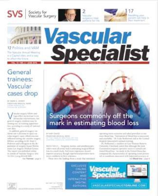
The June issue of Vascular Specialist is now online
Check out the June issue of Vascular Specialist now online in our enhanced digital edition and standard PDF. This month includes stories on vascular trainee case loads, the difficulties surgeons have at estimating blood loss, and an editorial by Dr. Russell Samson asking “Why be a vascular surgeon?”

Our Point/Counterpoint features a debate on the importance of the angiosome concept in revascularization for limb salvage between Dr. Richard Neville and Dr. Bauer Sumpio, and our Medicolegal Issues column examines the liability issues raised by the new move to value-based care.
Check out the June issue of Vascular Specialist now online in our enhanced digital edition and standard PDF. This month includes stories on vascular trainee case loads, the difficulties surgeons have at estimating blood loss, and an editorial by Dr. Russell Samson asking “Why be a vascular surgeon?”
Our Point/Counterpoint features a debate on the importance of the angiosome concept in revascularization for limb salvage between Dr. Richard Neville and Dr. Bauer Sumpio, and our Medicolegal Issues column examines the liability issues raised by the new move to value-based care.
Red Alert: Can Topical Skin Care Products Promote Melanoma Metastasis?

Le Gal et al (Sci Transl Med. 2015;7:308re8) discovered that antioxidant administration in mice not only increased lymph node metastases but also increased the migration and invasive properties of human melanoma cells. However, the antioxidant N-acetylcysteine (NAC) had no impact on the number and size of the primary tumors (in mice), and neither NAC nor Trolox (6-hydroxy-2,5,7,8-tetramethylchroman-2-carboxylic acid), a structurally unrelated antioxidant and soluble vitamin E analogue, affected the proliferation of human melanoma cells. Hence, the progression of malignant melanoma (MM), a cancer that is sensitive to changes in reduction-oxidation status, may be influenced by exposure to antioxidants and vitamin E.
What’s the issue?
Healthy individuals and oncology patients commonly use supplements containing antioxidants to prevent cancer and fight malignancy, respectively. However, animal studies and human clinical trials have shown that antioxidants increase cancer risk and accelerate the progression of primary lung tumors. Le Gal et al’s study regarding progression of melanoma metastases following exposure to antioxidants extends the observations demonstrated for lung neoplasms. N-acetylcysteine was added to the drinking water of mice, whereas NAC and Trolox were added to a panel of human MM cell lines. N-acetylcysteine increased lymph node metastases in the endogenous mouse model of MM, and both NAC and Trolox markedly increased the migrations and invasive properties of human MM cells.
Cancers may be caused or exacerbated by free radicals. It has been assumed that antioxidants may protect against malignancy by destroying free radicals. Although prior studies have concluded that antioxidants prevent healthy cells from transforming into cancer after exposure to free radicals, Le Gal et al’s research suggests that antioxidants may not only protect but also enhance tumor progression once a cancer has developed.
If one extends the results of animal and tissue culture studies to humans, exposure to antioxidants may potentially influence the course of metastatic disease in patients who have already developed melanoma. In addition to systemic exposure after receiving oral antioxidants, melanoma patients also can be topically exposed to antioxidants. For example, nonprescription skin care products such as cutaneous rejuvenation treatments, emollients, and sunscreens can contain β-carotene, vitamin E, and other antioxidants. It remains to be determined whether topical exposure to antioxidants can cause the same observations that have occurred following systemic absorption in mice or tissue culture studies in human cell lines. Should we caution our melanoma patients with regards to what they apply to their skin?

Member Decisions Coming July 1
We understand membership applicants are anxious for a decision!
Check your mailbox. All those who submitted applications to be members of SVS will receive a letter about the membership decision around July 1.
We understand membership applicants are anxious for a decision!
Check your mailbox. All those who submitted applications to be members of SVS will receive a letter about the membership decision around July 1.
We understand membership applicants are anxious for a decision!
Check your mailbox. All those who submitted applications to be members of SVS will receive a letter about the membership decision around July 1.

New SVS Officers Elected at Annual Meeting
At the Vascular Annual Meeting last week, members voted on new officers and approved selection of new members of the Board of Directors.
Officers elected are:
• Vice president, Dr. Michel Makaroun
• Secretary, Dr. Ali AbuRahma
• Treasurer, Dr. Kim Hodgson
New members of the Board of Directors are:
Dr. Frank Pomposelli, Clinical Practice Council chair
Dr. Michael Golden, Eastern Vascular Society
Dr. Thomas Forbes, Canadian Society for Vascular Surgery
Dr. Donald Jacobs, Midwestern Vascular Surgical Society
Dr. William Marston, Southern Association for Vascular Surgery
Dr. Thomas Bower, Society for Clinical Vascular Surgery
Dr. Darrin Clouse, Vascular and Endovascular Surgery Society
Dr. William Jordan, Association of Program Directors in Vascular Surgery.

Better Reporting Needed to Accurately Estimate Medical Error as Cause of Death in U.S.
Clinical question: What is the contribution of hospital-based medical errors to national mortality in the U.S. compared to other causes listed by the Centers for Disease Control and Prevention (CDC)?

Background: Medical error can contribute to patient mortality. Currently, the annual list of the most common causes of death in the U.S. is compiled by the CDC using the International Classification of Diseases (ICD) codes on death certificates. Deaths caused by errors are unmeasured because medical errors are not included in the death certificate.
Study design: Analysis of existing literature on medical errors.
Setting: U.S. hospitals.
Synopsis: Findings of four studies on U.S. death rates from medical errors published between 2000 and 2008 were synthesized and extrapolated to the total number of U.S. hospital admissions in 2013. This resulted in a mean rate of death from medical errors of 251,454 per year, which is much higher than the annual incidence of 44,000–98,000 deaths published in the 1999 Institute of Medicine report. Comparing these data to the CDC ranking makes medical errors the third-leading cause of death in the U.S.
Although the accuracy of this result is limited to inpatient deaths and as the authors extrapolated the data from other studies, the number is still staggering and highlights the need for systematic measurement of the problem. One simple solution for this could be to have an extra field on the death certificate asking whether a preventable complication stemming from the patient’s medical care contributed to the death.
Bottom line: Medical error as the estimated third-leading cause of the death in the U.S. remains under-recognized, underappreciated, and highly unmeasured.
Citation: Makary MA, Daniel M. Medical error-the third leading cause of death in the US. BMJ. 2016;353:i2139.
Short Take
Isolating C. Difficile Carriers Decreases Hospital-Acquired C. Difficile Infections
In a nonblinded time-series analysis, screening all patients for asymptomatic C. diff carrier status and isolating carriers reduced rates of hospital-acquired C. diff, preventing 62.4% of expected cases.
Citation: Longtin Y, Paquet-Bolduc B, Gilca R, et al. Effect of detecting and isolating Clostridium difficile carriers at hospital admission on the incidence of C difficile infections: a quasi-experimental controlled study. JAMA Inter Med. 2016;176(6):796¬-804.
Clinical question: What is the contribution of hospital-based medical errors to national mortality in the U.S. compared to other causes listed by the Centers for Disease Control and Prevention (CDC)?
Background: Medical error can contribute to patient mortality. Currently, the annual list of the most common causes of death in the U.S. is compiled by the CDC using the International Classification of Diseases (ICD) codes on death certificates. Deaths caused by errors are unmeasured because medical errors are not included in the death certificate.
Study design: Analysis of existing literature on medical errors.
Setting: U.S. hospitals.
Synopsis: Findings of four studies on U.S. death rates from medical errors published between 2000 and 2008 were synthesized and extrapolated to the total number of U.S. hospital admissions in 2013. This resulted in a mean rate of death from medical errors of 251,454 per year, which is much higher than the annual incidence of 44,000–98,000 deaths published in the 1999 Institute of Medicine report. Comparing these data to the CDC ranking makes medical errors the third-leading cause of death in the U.S.
Although the accuracy of this result is limited to inpatient deaths and as the authors extrapolated the data from other studies, the number is still staggering and highlights the need for systematic measurement of the problem. One simple solution for this could be to have an extra field on the death certificate asking whether a preventable complication stemming from the patient’s medical care contributed to the death.
Bottom line: Medical error as the estimated third-leading cause of the death in the U.S. remains under-recognized, underappreciated, and highly unmeasured.
Citation: Makary MA, Daniel M. Medical error-the third leading cause of death in the US. BMJ. 2016;353:i2139.
Short Take
Isolating C. Difficile Carriers Decreases Hospital-Acquired C. Difficile Infections
In a nonblinded time-series analysis, screening all patients for asymptomatic C. diff carrier status and isolating carriers reduced rates of hospital-acquired C. diff, preventing 62.4% of expected cases.
Citation: Longtin Y, Paquet-Bolduc B, Gilca R, et al. Effect of detecting and isolating Clostridium difficile carriers at hospital admission on the incidence of C difficile infections: a quasi-experimental controlled study. JAMA Inter Med. 2016;176(6):796¬-804.

Hospital Medicine's Movers and Shakers – July 2016

Several prominent hospitalist leaders have been named to Modern Healthcare magazine’s “50 Most Influential Physician Executives and Leaders” for 2016. Among them are Patrick Conway, MD, MSc, MHM, a pediatric hospitalist as well as CMO and deputy administrator for innovation and quality at the Centers for Medicare & Medicaid Services (CMS); Vivek Murthy, MD, MBA, a hospitalist and the current U.S. Surgeon General; Lynn Massingale, MD, co-founder and executive chairman of the hospitalist staffing firm TeamHealth; and Robert M. Wachter, MD, MHM, a national hospitalist leader, professor, and interim chairman of the Department of Medicine at the University of California, San Francisco (UCSF), and a founder of the hospitalist movement.

Susan George, MD, SFHM, recently received the Katharine F. Erskine Award from the YWCA in Worcester, Mass. Dr. George served as an internal medicine physician at Saint Vincent Hospital in Worcester for a total of 20 years and as hospitalist medical director there from 2007 until this year, when she left to go into private practice. Dr. George still teaches at the University of Massachusetts Medical School as an associate professor of medicine. The award is named for Katharine F. Erskine, a former YWCA president and women’s advocate since before the turn of the 20th century.
Alanna Small, MD, was recently named deputy chief of staff for Physician Services at Samuel Simmonds Memorial Hospital in Barrow, Alaska. Prior to this role, Dr. Small served as a hospitalist at the Alaska Native Medical Center in Anchorage.
Business Moves


Envision also announced its planned acquisition of Emergency Physicians Medical Group (EPMG), based in Ann Arbor, Mich., a private emergency and hospital medicine staffing firm serving the Midwestern United States since 1976.


Michael O’Neal is a freelance writer in New York City.
Several prominent hospitalist leaders have been named to Modern Healthcare magazine’s “50 Most Influential Physician Executives and Leaders” for 2016. Among them are Patrick Conway, MD, MSc, MHM, a pediatric hospitalist as well as CMO and deputy administrator for innovation and quality at the Centers for Medicare & Medicaid Services (CMS); Vivek Murthy, MD, MBA, a hospitalist and the current U.S. Surgeon General; Lynn Massingale, MD, co-founder and executive chairman of the hospitalist staffing firm TeamHealth; and Robert M. Wachter, MD, MHM, a national hospitalist leader, professor, and interim chairman of the Department of Medicine at the University of California, San Francisco (UCSF), and a founder of the hospitalist movement.
Susan George, MD, SFHM, recently received the Katharine F. Erskine Award from the YWCA in Worcester, Mass. Dr. George served as an internal medicine physician at Saint Vincent Hospital in Worcester for a total of 20 years and as hospitalist medical director there from 2007 until this year, when she left to go into private practice. Dr. George still teaches at the University of Massachusetts Medical School as an associate professor of medicine. The award is named for Katharine F. Erskine, a former YWCA president and women’s advocate since before the turn of the 20th century.
Alanna Small, MD, was recently named deputy chief of staff for Physician Services at Samuel Simmonds Memorial Hospital in Barrow, Alaska. Prior to this role, Dr. Small served as a hospitalist at the Alaska Native Medical Center in Anchorage.
Business Moves
Envision also announced its planned acquisition of Emergency Physicians Medical Group (EPMG), based in Ann Arbor, Mich., a private emergency and hospital medicine staffing firm serving the Midwestern United States since 1976.
Michael O’Neal is a freelance writer in New York City.
Hospital Admission, Stroke Clinic Follow-up Improve Outcomes for Patients with Transient Ischemic Attack, Minor Ischemic Stroke
Clinical question: How do guideline-based care and outcomes of patients with transient ischemic attack (TIA) and minor ischemic stroke differ among patients admitted to the hospital and discharged from the ED, as well as in those referred versus not referred to stroke prevention clinics following discharge?
Background: Previous research demonstrated that urgent outpatient management strategies for patients with TIA and minor ischemic stroke are superior to standard outpatient care. However, there is less known about how outpatient stroke care compares to inpatient care in terms of outcomes, rapid risk factor identification/modification, and initiation of antithrombotic therapy.
Study design: Retrospective cohort study.
Setting: EDs of acute-care hospitals in Ontario, Canada.
Synopsis: Using the Ontario Stroke Registry, 8,540 patients seen in the ED with TIA or minor ischemic stroke were identified. The use of guideline-based interventions was highest in admitted patients, followed by patients discharged from the ED with stroke clinic follow-up, followed by patients discharged without follow-up. There was no significant difference in one-year mortality between admitted and discharged patients when adjusted for age, sex, and comorbid conditions (adjusted hazard ratio, 1.11; 95% CI, 0.92–1.34). However, stroke clinic referral was associated with a lower risk of one-year mortality compared with those discharged without follow-up (adjusted hazard ratio, 0.49; 95% CI, 0.38–0.64).
Limitations of this study include that it was carried out only in Ontario, where there is a universal healthcare system, which may limit the generalizability of the findings. Additionally, patient information was limited to what was available through the registry, which may mean there were other unmeasurable differences among groups.
Bottom line: Admitted patients with TIA or minor ischemic stroke are more likely to receive guideline-based therapy, and among patients discharged from the ED, referral to stroke clinic improves outcomes, including one-year mortality.
Citation: Kapral MK, Hall R, Fang J, et al. Association between hospitalization and care after transient ischemic attack or minor stroke. Neurology. 2016;86(17):1582-1589.
Platelet transfusion deemed ‘harmful’ in patient group

Congress of the European
Hematology Association
COPENHAGEN—Results of the phase 3 PATCH trial suggest that platelet transfusions are not beneficial and can actually cause harm in patients with intracerebral hemorrhage (ICH) associated with antiplatelet therapy.
The study showed that the odds of death or dependence at 3 months were significantly higher among patients who received a platelet transfusion plus standard care than among patients who received standard care alone.
Patients who received a platelet transfusion also had a higher incidence of severe adverse events (SAEs) during hospital admission.
“PATCH shows that platelet transfusion seems harmful for patients with antiplatelet-associated ICH,” said study investigator Maria Koopman, PhD, of Sanquin Bloodbank in Amsterdam, The Netherlands.
“Pending further evidence, we think that platelet transfusions should not be used in this patient group.”
Dr Koopman presented results of the PATCH trial at the 21st Congress of the European Hematology Association (abstract LB2234). The study was also recently published in The Lancet.
Dr Koopman noted that, worldwide, there are 2 million cases of ICH each year, and more than 25% of these patients are taking antiplatelet therapy at ICH onset.
She and her colleagues theorized that because antiplatelet therapy can lead to hematoma growth and poor outcomes, platelet transfusions might modify the effect of antiplatelet therapy and improve patient outcomes.
To test this theory, the team studied ICH patients treated at 60 hospitals in The Netherlands, Scotland, and France.
Patients and treatment
The study included patients with spontaneous supratentorial ICH who were 18 years of age or older, had been using antiplatelet therapy for at least 7 days preceding ICH, and had a Glasgow Coma Scale score of 8 or higher.
The patients were randomized 1:1 to receive either standard care alone or standard care plus a platelet transfusion within 6 hours of the start of symptoms and within 90 minutes of diagnostic brain imaging.
In all, 190 patients were randomized—97 to the transfusion arm and 93 to the standard care arm. Ultimately, 93 patients actually received a platelet transfusion, and 91 received standard care alone.
Patient characteristics were generally balanced between the treatment arms. The mean age was 74.2 in the transfusion arm and 73.5 in the standard care arm. Median scores on the Glasgow Coma Scale were 14 (range, 13-15) and 15 (range, 13-15), respectively.
Comorbidities included cerebral infarction (38.4% and 40%, respectively), hypertension (72.3% and 72.8%, respectively), diabetes (15.5% and 18.9%, respectively), myocardial infarction (23.6% and 24.4%, respectively), and peripheral artery disease (16.5% and 4.4%, respectively).
Antiplatelet therapy consisted of a cyclooxygenase (COX) inhibitor (73.2% and 83.9%, respectively), a COX inhibitor plus dipyridamole (18.6% and 14%, respectively), an adenosine diphosphate (ADP) receptor inhibitor (4.1% and 1.1%, respectively), and an ADP inhibitor plus a COX inhibitor (3.1% and 1.1%, respectively).
Efficacy
The study’s primary outcome was a shift toward death or dependence, rated on the modified Rankin Scale (mRS), at 3 months after ICH, adjusted for hospital, antiplatelet therapy, and ICH score. The primary analysis was done in the intention-to-treat population (n=97 in the transfusion arm and 93 in the standard care arm).
The investigators found the odds of death or dependence at 3 months were higher in the platelet transfusion arm than the standard care arm. The adjusted common odds ratio (OR) was 2.05 (95% CI 1.18-3.56; P=0.0114).
The study’s secondary endpoints were survival at 3 months, poor outcome (dichotomous mRS 4-6 or 3-6) at 3 months, and hematoma growth on imaging within 24 hours.
The survival rate was lower in the transfusion arm than the standard care arm, although the difference was not statistically significant—68% and 77.4%, respectively (OR=0.62; 95% CI, 0.33-1.19; P=0.15).
Poor outcome was more likely in the transfusion arm than the standard care arm, with 72.2% and 55.9% of patients, respectively, having an mRS score of 4-6 (OR=2.04; 95% CI, 1.12-3.74; P=0.0195) and 88.7% and 81.7%, respectively, having an mRS score of 3-6 (OR=1.75; 95% CI, 0.77-3.97; P=0.18).
And there was no significant difference between the arms when it came to hematoma growth, with a median of 2.01 mL (interquartile range, 0.32-9.34) in the transfusion arm and 1.16 mL (interquartile range, 0.03-4.42) in the standard care arm (P=0.81).
Safety
Safety endpoints included thromboembolic events, transfusion reactions, and SAEs during hospital admission. The safety analysis was done in the intention-to-treat population (n=97 in the transfusion arm and 93 in the standard care arm) and the as-treated population (n=93 and 91, respectively).
In the as-treated population, the incidence of SAEs was 42% in the transfusion arm and 29% in the standard care arm (OR=1.74; 95% CI, 0.96-3.17). The incidence of any fatal SAE was 24% and 17%, respectively (OR=1.58; 95% CI, 1.77-3.22).
The incidence of SAEs due to ICH was 25% and 14%, respectively (OR=2.13; 95% CI, 1.01-4.50). The incidence of SAEs due to thromboembolism was 4% and 1%, respectively (OR=4.13; 95% CI, 0.45-37.67). And there was 1 SAE due to transfusion (OR=3.03; 95% CI, 0.12-75.37). ![]()