User login
AATS Focus on Thoracic Surgery: Current and Future Challenges
View Preliminary Program & Register
October 28-29, 2016
Westin Boston Waterfront Hotel
Boston, MA, USA
Program Directors
G. Alexander Patterson
David J. Sugarbaker
Program Committee
Thomas A. D’Amico
Shaf Keshavjee
James D. Luketich
Bryan F. Meyers
Scott J. Swanson
Overview
Currently practicing surgeons will be able to improve patient outcomes by enriching their knowledge and technical skills in the definition, diagnosis and resolution of thoracic surgical difficulties and post-operative complications. Expert faculty will provide state-of-the-art solutions to challenges in the field. Attendees will augment their overall understanding of thoracic diseases, upgrade their competency and ability to formulate new clinical strategies, and enhance their diagnosis and surgical treatment of thoracic diseases.
Prep-free capsule uses low-dose X-rays to image colonic polyps
SAN DIEGO – A colonoscopy capsule that requires no bowel preparation safely detected pedunculated and sessile polyps as small as 7 mm in a feasibility study of 54 volunteers.
The capsule scans the colon with very-low-dose X-rays, and the images are converted to high-resolution three-dimensional reconstructions that physicians can review remotely in about 10 minutes, said Dr. Nadir Arber of the Integrated Cancer Prevention Center at Tel Aviv Sourasky Medical Center. “All 54 capsules were excreted naturally, without discomfort or side effects,” he reported at the annual Digestive Disease Week.
Bowel preparation is the most common reason for skipping a colonoscopy, and poor preparation leads to missed adenomas on conventional screens, Dr. Arber noted. “A colorectal cancer screening method that would generate structural data of the colon, without cathartic cleansing or diet restrictions, would offer an attractive alternative for many patients,” he said.
This capsule is no larger than others that have been approved, but it uses low-dose X-rays to circumvent the need for bowel preparation. Patients swallow the capsules and go about their day while wearing tracking patches on their lower backs that transmit (nonionizing) radiofrequency data to a localization system. This system determines when a capsule has reached the colon. At this point, the capsule is activated and begins emitting X-rays, but only when it is moving, Dr. Arber said. Because the capsule usually is not moving, radiation exposure is minimized. Also, an axis-positioning system reconciles the location of the capsule with the colonic wall to prevent movement artifacts.
The 54 volunteers in the study were 45-75 years old. The capsules took an average of 66 hours to move through the entire alimentary tract, with a standard deviation of 37 hours. If swallowed before the first meal of the day, the transit time averaged 51 hours, with a standard deviation of 25 hours. The total radiation dose per procedure averaged 0.03 mSv (standard deviation, 0.0007 mSv), which is equivalent to one dental or chest X-ray, Dr. Arber said.
Examples of reconstructed lesions included a 12 × 4 mm flat sessile polyp, a double-headed polyp with heads measuring 7 and 15 mm, and a 25-mm polyp in the sigmoid colon. “In humans, 7-mm polyps are well within the detection capability of the system,” he said.
He and his associates are planning multicenter studies to compare adenoma detection by the capsule with traditional colonoscopy. Reconstructing long pedunculated polyps might be difficult because of image distortion, Dr. Arber acknowledged.
He reported that his spouse or partner has received consulting fees from Check-Cap Ltd., the maker of the capsule.
AT DDW® 2016
Key clinical point: A new colonoscopy capsule uses very-low-dose X-rays to generate high-resolution three-dimensional reconstructions without the need for bowel preparation.
Major finding: The method detected pedunculated and sessile polyps in various locations ranging from 7 to 50 mm, without known adverse effects. All capsules were excreted intact.
Data source: A single-center feasibility study of 54 volunteers aged 45-75 years.
Disclosures: Dr. Arber reported that his spouse or partner has received consulting fees from Check-Cap Ltd., the maker of the capsule.

Registration and Housing Now Open: AATS Clinical Trials Methods Course 2016
October 20-22, 2016
Hyatt Regency O’Hare
Chicago, IL
Program Directors
David H. Harpole, Jr.
Marco A. Zenati
This course will provide an intensive and interactive training program for cardiothoracic surgeons across all subspecialties to acquire the critical skills required in effective clinical trial design and implementation. This course is particularly suited to help professionals who are considering applying for clinical trial funding to better understand the complex nature of preparing and submitting clinical trial proposals.
Invited faculty includes currently funded clinical trial leading investigators and experts in the field of biostatistics and health sciences research. The program will offer a process in which the clinical trial protocol development can be streamlined. Interactive features will include hands-on focus groups and mock study sessions.
Register Today! Space available for only 40 participants.
Register Today: Inaugural Patient Safety Course
June 24-25, 2016
Renaissance Boston Waterfront Hotel
Boston, MA
Co-Directors
Thoralf M. Sundt, III
Steven Yule
Program Committee
David J. Bunnell (APACVS)
David C. Fitzgerald (AMSECT)
Jake Jaquiss
M. Blair Marshall
Shannon Pengel
Kenneth Shann (AMSECT)
Marco Zenati
Patient Safety is essential for successful surgical care — in cardiothoracic surgery — and all disciplines. In the more than 10 years since the Institute of Medicine made patient safety a priority, little progress has been made in changing culture and practice on the front lines.
This two-day course — including team training and simulation experiences — will make a difference, offering surgical team members practical safety behaviors for improving patient outcomes.
Special Benefits for Team Registration!
Housing Closes June 2nd.
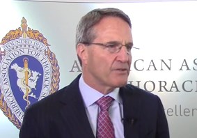
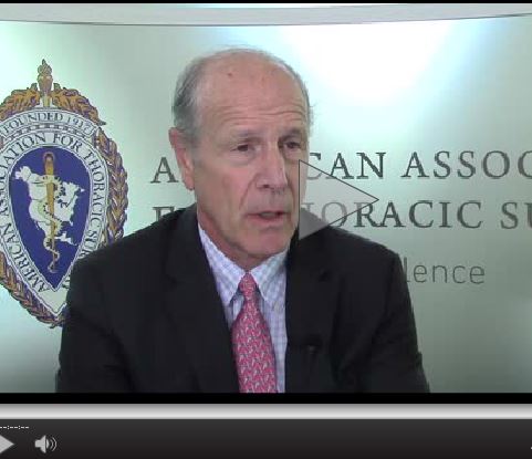
VIDEO: TAVR may soon eclipse surgery for aortic stenosis patients
BALTIMORE – A study comparing the causes and timing of death in high-risk patients with severe, symptomatic aortic stenosis suggests that transcatheter aortic valve replacement (TAVR) may have significant advantages over surgical aortic valve replacement (SAVR).
The research team, presenting their findings at the annual meeting of the American Association for Thoracic Surgery, revealed that death rates in the TAVR and SAVR cohorts in the CoreValve US Pivotal Trial High Risk Study were similar at 0-30 days and 91-365 days post procedure, but found that more SAVR patients died from day 31-90 related to bleeding, frailty, and comorbid diseases. The investigators concluded that this finding reflected the impact of the added strain of surgery over intravascular intervention for aortic valve replacement.
Dr. Craig Smith, chair of the department of surgery at NewYork–Presbyterian Hospital/Columbia University Medical Center, and a discussant on the paper, said this study is the only randomized clinical trial to show inferiority of surgery to TAVR.
“The difference seems to reside in the recovery phase mortality, which they showed very well,” Dr. Smith said in a video interview. He predicted that the trial results would add to the momentum making TAVR the first choice for any patient who is high risk and at least an equivalent choice for patients who are considered intermediate risk.
“The big question on the horizon is what is the best thing to recommend to patients who we classify as low risk,” Dr. Smith said. “Unless something happens to the impressive results that TAVR is racking up, in a few years … approval will be granted for those who are low risk.”
Dr. Smith reported no relevant financial disclosures.
The video associated with this article is no longer available on this site. Please view all of our videos on the MDedge YouTube channel
On Twitter @richpizzi
AT THE AATS ANNUAL MEETING
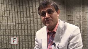
VIDEO: EVLP may extend lung preservation, quality for transplants
BALTIMORE – The use of ex vivo lung perfusion (EVLP) may allow for the safe transplantation of lungs preserved for more than 12 hours, according to a study presented at the annual meeting of the American Association for Thoracic Surgery.
A research team at the University of Toronto evaluated the outcomes of transplant patients who received a lung with a preservation time of over 12 hours between January 2006 and April 2015 and compared them to the general lung transplant population. Median hospital and ICU length of stay were similar between the two groups, and Kaplan-Meier survival curves between the two groups did not show any difference. Preservation time, donor PO2, and use of EVLP were not significant variables affecting survival.
Dr. Bartley P. Griffith, chief of cardiac surgery at the University of Maryland, Baltimore, and a discussant on the paper at the meeting, said that the findings of the study open up the possibility of a more “planned” approach to transplantation.
“Anything that not only extends preservation time, but perhaps even improves quality of preservation, would be a godsend,” Dr. Griffith said in a video interview. He cautioned that the “devil is in the details,” and that the data had to be examined closely. Nevertheless, Dr. Griffith said transplant surgeons should be grateful for the important work done by the University of Toronto team.
Dr. Griffith reported no relevant financial disclosures.
The video associated with this article is no longer available on this site. Please view all of our videos on the MDedge YouTube channel
On Twitter @richpizzi
AT THE AATS ANNUAL MEETING
VIDEO: CBT interventions abound for psychosis
ATLANTA – Now that cognitive-behavioral therapy (CBT) for psychosis is an established intervention, what are the novel ways it is being used, either alone or combined with cognitive remediation therapy, to help outpatients with mild to moderate symptoms?
In this video interview, Dr. Farooq Naeem, an associate professor of psychiatry at Queen’s University in Kingston, Ont., discusses how a variety of CBT therapies are proving cost effective with good patient outcomes. Dr. Naeem covers what is known as brief CBT, as well as online self-help CBT, and CBT combination therapies.
He pointed to success with brief CBT for psychosis that is delivered in 6 to 10 sessions, compared with the standard, which is 12 to 20 sessions.
Dr. Naeem also offered perspective on where CBT for psychosis is being delivered across the globe. “In England, possibly, you have the best coverage,” Dr. Naeem said at the annual meeting of the American Psychiatric Association. Australia and New Zealand have had some success with CBT, he said, “but Canada and [the] U.S. are far behind.”
Dr. Naeem did not have any relevant disclosures.
The video associated with this article is no longer available on this site. Please view all of our videos on the MDedge YouTube channel
On Twitter @whitneymcknight
EXPERT ANALYSIS FROM the APA ANNUAL MEETING
CVS MinuteClinics: A Cure for Long Wait Times at Veterans Affairs?
Struggling with long wait times, the Veterans Affairs Health Care System is trying something new: a partnership with the CVS Pharmacy chain to offer urgent care services to more than 65,000 veterans.
The experiment begins today at the VA’s operations in Palo Alto, California.
Veterans can visit 14 “MinuteClinics” operated by CVS in the San Francisco Bay area and Sacramento, where staff will treat them for conditions such as respiratory infections, order lab tests and prescribe medications, which can be filled at CVS pharmacies.
The care will be free for veterans, and the VA will reimburse CVS for the treatment and medications. Whether the partnership will spread to other VA locales isn’t yet clear.
The collaboration comes amid renewed scrutiny of the nation’s troubled VA health system, which has tried without much success to improve long wait times for veterans needing health care.
Despite a $10 billion “Veterans Choice” program allowing veterans to receive care outside the closed VA system, vets nationwide wait for an appointment even longer than they did before the program started in 2014, according to a federal audit.
The MinuteClinic partnership is not part of the Veterans Choice program.
“The concern has always been, how do we make sure veterans get the care they need in a timely way and in a way that works for the veteran?” said Dr. Stephen Ezeji-Okoye, the Palo Alto VA’s deputy chief of staff. The deal indicates that the VA is willing to try outside partnerships to meet veterans’ needs, he said. “We want to have not just timely access but geographic access to care.”
Sarah Russell, the Palo Alto VA’s chief medical informatics officer, came up with the idea, said Ezeji-Okoye.
The VA will integrate MinuteClinics’ patient records with its own electronic health records to provide consistency of care, Ezeji-Okoye said.
The Palo Alto VA fares better than some other facilities nationwide in providing timely care to veterans, according to VA data, and Ezeji-Okoye said most patients with urgent care needs are seen quickly.
But the system was so busy in the past year that about 11 percent of appointments at its network of hospitals and clinics — which stretch south from Sonora to Monterey — could not be scheduled within 30 or fewer days, which is considered an acceptable timeframe, VA data show. That includes appointments that would require urgent care.
More than 5,000 appointments system-wide were scheduled more than 30 days out, but each hospital and clinic’s performance varied widely. At a Fremont clinic, less than 2 percent of appointment requests could not be scheduled within 30 days. At the VA’s rural Modesto clinic, by contrast, more than 17 percent of requests were not be scheduled within 30 days.
Once the MinuteClinic operation is well underway, Ezeji-Okoye anticipates that between 10 and 15 veterans — from among the estimated 150 who call the Palo Alto VA’s advice nurse hotline daily — will be treated at the retail clinics on any given day.
About 95,000 veterans are eligible to use the Palo Alto system, one of the VA’s largest in the Western United States. About 65,000 use it every year.
The $330,000 pilot project will be evaluated after one year. CVS’ MinuteClinic president, Dr. Andrew Sussman, hopes it can be rolled out nationally if it succeeds. CVS is by far the biggest player in retail pharmacy clinics, operating 1,135 of them in 35 states.
“We’d love to have that opportunity to expand after we go through this phase,” Sussman said. “We’re well suited to help because of our large footprint and ability to see people on a quick basis.”
It is unclear, however, what the VA’s nationwide plans are. The Veterans Health Administration office did not respond to Kaiser Health News’ request for comment.
Blake Schindler, a retired Army major who lives in Santa Clara near one of the participating MinuteClinics, was intrigued, but cautious about the MinuteClinics. He counts himself lucky because unlike some other veterans, he has access to the U.S. military’s TRICARE health insurance program for active and some retired service members.
“It could make a big difference, but how much access are the veterans going to have? That was the big problem with the Veterans Choice program; it didn’t end up the way it was supposed to,” said Schindler, 58.
“I’m always hopeful when I hear about these things; I keep an open mind until I have experience with it,” he added.
Interested veterans served by the VA Palo Alto can call its advice nurse line at 800-455-0057.
This story was produced by Kaiser Health News, which publishes California Healthline, a service of the California Health Care Foundation.
This story also ran on NPR.
Repeat SICU admissions should trigger palliative care consult
ICU readmission was most predictive of the need for palliative care among patients in the surgical intensive care unit, based on a study of six potential trigger criteria associated with in-hospital death or discharge to hospice.
To facilitate proactive case findings of patients who would benefit from a palliative care consult, a team of surgical ICU and palliative care clinicians at the Icahn School of Medicine at Mount Sinai, N.Y., developed and tested a system of palliative care triggers. The study was published online in the Journal of Critical Care (http://dx.doi.org/10.1016/j.jcrc.2016.04.010).
Based on a literature review, the researchers created a six-item list of potential triggers for palliative care: length of stay over 10 days, ICU readmission, intensivist referral, status post cardiac arrest, metastatic cancer, and a match of two or more on a set of secondary criteria.

Data were collected for the period from Sept. 4, 2013, through May 30, 2014, at the surgical ICU of a 1,170-bed tertiary medical center. Patients who received a palliative care consultation were compared with those who did not, and the trigger list was tested for accuracy in predicting patient outcomes. The primary outcomes were hospital death, hospice discharge, and a combined endpoint of these two outcomes. Patients who died in the hospital or were released to hospice care were assumed to be those most in need of a palliative care consult.
Bivariate analysis was done to calculate the unadjusted odds ratios of individual triggers to each of these outcomes. Then, the team used logistic regression analysis to calculate the adjusted odds ratios of triggers to outcomes.
Of the 512 patients admitted to the SICU in the study period, those not discharged by the end of the study were excluded, leaving 492 patients in the study.
Bivariate analysis found that all of the triggers were significantly associated with in-hospital death. With the multivariate analysis and adjusted odds ratios, SICU readmission, status post cardiac arrest, metastatic cancer, and secondary triggers were significantly associated with hospital death.
For the combined outcome of hospital death or release to hospice care, the relationships were stronger. In particular, repeat SICU readmissions and metastatic cancer triggers were strongly associated with the combined outcome (odds ratio, 19.41, CI 5.81-54.86 and OR, 16.40, CI 4.69-57.36, respectively). The secondary triggers did not show the same strength of association, although they were associated significantly with the combined outcome (OR, 4.41, CI 2.05-9.53).
The most prominent finding is the strength of repeat SICU admissions with the hospital death or release to hospice. The strong relationship between repeat SICU admission and outcomes led the researchers to conclude “that one might consider adapting this clinical criterion as a standalone criterion. This would require all patients who are readmitted to the SICU to be seen by palliative care to assess their overall goals of care and understanding of their serious illness. This approach may be particularly useful for smaller palliative care teams that do not have the resources to screen daily with a series of triggers.”
The American Federation of Aging Research and the National Institute on Aging funded the study.
ICU readmission was most predictive of the need for palliative care among patients in the surgical intensive care unit, based on a study of six potential trigger criteria associated with in-hospital death or discharge to hospice.
To facilitate proactive case findings of patients who would benefit from a palliative care consult, a team of surgical ICU and palliative care clinicians at the Icahn School of Medicine at Mount Sinai, N.Y., developed and tested a system of palliative care triggers. The study was published online in the Journal of Critical Care (http://dx.doi.org/10.1016/j.jcrc.2016.04.010).
Based on a literature review, the researchers created a six-item list of potential triggers for palliative care: length of stay over 10 days, ICU readmission, intensivist referral, status post cardiac arrest, metastatic cancer, and a match of two or more on a set of secondary criteria.
Data were collected for the period from Sept. 4, 2013, through May 30, 2014, at the surgical ICU of a 1,170-bed tertiary medical center. Patients who received a palliative care consultation were compared with those who did not, and the trigger list was tested for accuracy in predicting patient outcomes. The primary outcomes were hospital death, hospice discharge, and a combined endpoint of these two outcomes. Patients who died in the hospital or were released to hospice care were assumed to be those most in need of a palliative care consult.
Bivariate analysis was done to calculate the unadjusted odds ratios of individual triggers to each of these outcomes. Then, the team used logistic regression analysis to calculate the adjusted odds ratios of triggers to outcomes.
Of the 512 patients admitted to the SICU in the study period, those not discharged by the end of the study were excluded, leaving 492 patients in the study.
Bivariate analysis found that all of the triggers were significantly associated with in-hospital death. With the multivariate analysis and adjusted odds ratios, SICU readmission, status post cardiac arrest, metastatic cancer, and secondary triggers were significantly associated with hospital death.
For the combined outcome of hospital death or release to hospice care, the relationships were stronger. In particular, repeat SICU readmissions and metastatic cancer triggers were strongly associated with the combined outcome (odds ratio, 19.41, CI 5.81-54.86 and OR, 16.40, CI 4.69-57.36, respectively). The secondary triggers did not show the same strength of association, although they were associated significantly with the combined outcome (OR, 4.41, CI 2.05-9.53).
The most prominent finding is the strength of repeat SICU admissions with the hospital death or release to hospice. The strong relationship between repeat SICU admission and outcomes led the researchers to conclude “that one might consider adapting this clinical criterion as a standalone criterion. This would require all patients who are readmitted to the SICU to be seen by palliative care to assess their overall goals of care and understanding of their serious illness. This approach may be particularly useful for smaller palliative care teams that do not have the resources to screen daily with a series of triggers.”
The American Federation of Aging Research and the National Institute on Aging funded the study.
FROM THE JOURNAL OF CRITICAL CARE
Key clinical point: A list of tested triggers can predict the need of surgical ICU patients for a palliative care consultation.
Major finding: Readmission to the surgical ICU was strongly associated with the study endpoint of hospital death or release to hospice (odds ratio 19.41, CI 5.81-54.86).
Data source: A case review of all 492 patients admitted to the surgical intensive care facility at a 1,170-bed, tertiary care medical center.
Disclosures: The American Federation of Aging Research and the National Institute on Aging funded the study.

SSRIs don’t boost risk in patients with bleeding peptic ulcers
SAN DIEGO – Taking selective serotonin reuptake inhibitors (SSRIs) was not associated with an increased risk of endoscopy-refractory bleeding, rebleeding, or in-hospital mortality among 14,343 consecutive patients admitted with bleeding peptic ulcer in Denmark.
The study suggests that patients with bleeding peptic ulcers can “continue SSRI treatment if the treatment is well indicated,” and that these patients can otherwise receive treatment similar to that of other patients with peptic ulcer bleeding, Dr. Stig B. Laursen reported at the annual Digestive Disease Week.
Observational research has linked use of SSRI antidepressants to an approximately 1.5-fold increase in upper GI bleeding in peptic ulcer bleeding cases, said Dr. Laursen of Odense (Denmark) University Hospital. Studies, mostly in vitro ones, indicate SSRIs decrease the function of thrombocytes and fibrin formation via lowered levels of serotonin. Therefore, it has been suggested that temporarily discontinuing SSRIs may benefit patients with bleeding peptic ulcers.
However, sudden cessation of SSRIs can be associated with withdrawal symptoms, which have been reported in up to one-third of such patients.
Dr. Laursen and his associates prospectively analyzed data for 14,343 consecutive patients admitted with bleeding peptic ulcers in Denmark from 2006 to 2014. They investigated associations between SSRI use and several outcomes, adjusted for confounding factors including age, sex, comorbidity, anemia, medication use, circulatory failure at hospital admission, location of ulcer, stigmata of bleeding, and weekend admission.
After adjustment for confounding risk factors, SSRIs were not associated with increased risk of endoscopy-refractory bleeding (odds ratio [OR] 95% confidence interval [CI]: 1.01 [0.78-1.30]), rebleeding (OR [95% CI]: 0.94 [0.81-1.10]), or in-hospital mortality (hazard ratio [95% CI]: 0.84 [0.68-1.05]).
“Patients taking SSRIs have the same outcomes following peptic ulcer bleeding as non-SSRI users. This suggests that there is no clinically significant impairment of hemostatic function,” Dr. Laursen said. “Therefore, it seems safe to continue SSRI treatment in patients developing peptic ulcer bleeding and thereby avoid development of withdrawal symptoms.”
Dr. Laursen noted that the study has several limitations. It’s not a randomized controlled trial, he said, and there’s no information about the types of SSRIs that the patients were taking. Also, there’s a lack of information about possible discontinuation of SSRIs during hospitalization. However, he said, “that doesn’t seem to be a major confounder.”
During the question-and-answer session following his presentation, Dr. Laursen said the increased risk of poor outcome is quite low, but “there may be a small risk that we haven’t found yet. But if it’s small, maybe it doesn’t have an impact in day-to-day practice.”
The research is hospital-funded. Dr. Laursen had no financial disclosures to report.
AT DDW 2016
Key clinical point: Patients with bleeding peptic ulcers who are on SSRIs may be able to avoid cessation and withdrawal symptoms because outcomes aren’t affected despite worsened bleeding.
Major finding: After adjustment for confounding factors, SSRIs didn’t significantly worsen endoscopy-refractory bleeding (OR 1.01), rebleeding (OR 0.94) or in-hospital mortality (HR 0.84).
Data source: Analysis of 14,343 consecutive patients with bleeding peptic ulcers in Denmark from 2006 to 2014.
Disclosures: The research is hospital funded. Dr. Laursen had no financial disclosures to report.
