User login
Oxandrolone, propranolol combo increases growth in severely burned children
CHICAGO – Combination therapy with oxandrolone and propranolol can attenuate burn-induced growth arrest and increase growth rate in severely burned children, according to findings from a prospective, randomized clinical trial.
Of 612 children with burns over at least 30% of their total body surface area (average of more than 50%), 103 were randomized to receive treatment with both oxandrolone and propranolol, 67 received oxandrolone alone, 194 received propranolol alone, and 248 served as controls. After a minimum of 1 year of treatment, the average growth rate was 5.9 cm in the control group and 7.6 cm in the group receiving combination therapy, Dr. David N. Herndon of the University of Texas Medical Branch at Galveston reported at the annual meeting of the American Surgical Association.
“The rate of growth with combination therapy was significantly greater than with either of the individual drugs alone,” Dr. Herndon said.
Further, the period of growth arrest was significantly shorter – by 84 days – among those in the combination-treatment group, compared with those in the control group.
Study subjects were children treated at Shriners Hospitals for Children – Galveston from 1997 to 2015. Boys aged 6 months to 14 years and girls aged 6 months to 12 years were included to eliminate the variable onset of postpubescent growth delay. About two-thirds in each group were boys, and the ages in the patient groups were similar. Mortality was low and was similar across the groups, as was hospital length of stay.
Dr. Herndon and his colleagues controlled for heterogeneous burn distribution between the groups in the course of their analyses, as well as age.
In children with severe, extensive burn injury, the hypercatabolic response is mediated by increased production of catecholamines and corticosteroids, coupled with decreased production of testosterone. This contributes to growth arrest and to decreased strength for up to 2 years after burn injury, he explained. Children with burns over 50% of their total body surface routinely survive acute hospitalization but, at 3 months post injury, are thin, have difficulty walking, and require occupational and physical therapy to help them perform even the simplest activities of daily living.
At 1 year, a raised inflammatory mass covers their wounds, they experience itching, and they have, in large part, stunted growth; there is severe loss of lean body mass and strength, and fracture risk is increased, Dr. Herndon said.
In previous work, he and his colleagues showed that administration of propranolol at an average dose of 4 mg/kg per day for 1 year decreased cardiac work and resting energy expenditure while increasing peripheral lean mass. Further, they found that the testosterone analog oxandrolone, given at 0.1 mg/kg twice per day for 1 year, improved lean body mass accretion and bone mineral content.
The current study was conducted to test the effects of administering both agents in combination.
“The combined use of oxandrolone and propranolol in severely burned children confers an additional benefit on growth over either treatment alone,” Dr. Herndon said, adding that the additive effects of combination therapy may be due to the effects of oxandrolone on bone growth and the anti-inflammatory effects of propranolol.
“The additional benefits point out mechanistic changes that may be eventful in the treatment of hypermetabolism generally and in inflammatory states,” he concluded.
Dr. Herndon reported having no disclosures.
AT THE ASA ANNUAL MEETING
Key clinical point: Combination therapy with oxandrolone and propranolol can attenuate burn-induced growth arrest and increase growth rate in severely burned children, according to findings from a prospective, randomized clinical trial.
Major finding: After a minimum of 1 year of treatment, the average growth rate was 5.9 cm in the control group and 7.6 cm in the group receiving combination therapy.
Data source: A prospective, randomized clinical trial involving 612 children.
Disclosures: Dr. Herndon reported having no disclosures.
Materials help families find support for children with serious illnesses
Materials to support the families of children with serious illnesses have been developed by the National Institute of Nursing Research, which is part of the National Institutes of Health. The materials are associated with the NINR’s “Palliative Care: Conversations Matter” campaign.
“Palliative care is often associated with end of life, making it difficult for patients and their families – and even for healthcare providers – to start conversations around the subject. However, palliative care can be incredibly helpful for patients and their families at any stage during an illness. We hope these materials will improve patient and family understanding of pediatric palliative care and facilitate discussion with healthcare teams,” NINR Director Patricia A. Grady said in a written statement.
The resources, which include a fact sheet, a resource card to help families find support, and a series of family stories, are available in both Spanish and English. The NINR developed these materials with feedback from parents of seriously ill children. To learn more, click here
Things Hospitalists Want Hospital Administrators to Know
I think it is really cool that this publication has a series of articles on “What Cardiologists [or infection disease specialists, nephrologists, etc.] Want Hospitalists to Know.” I’m always interested to see which clinical topics made the list and which I’m already reasonably familiar with versus know little about. I’ve added this series to my list of things that are always worth the time to read, along with the “What’s New” section in UpToDate, review articles in major journals, and the Cleveland Clinic Journal of Medicine.
Not long ago, I worked with a hospitalist group that had agreed to cardiologists’ request that new hospitalists round with a cardiologist for something like three days as part of their orientation. This seems like they’ve taken the idea of “What Cardiologists Want Hospitalists to Know” a lot further than I had ever considered. I’m sure it would have value on many levels, including positioning the new hospitalist to work more effectively with the cardiologists, but I’m not sure it’s worth the cost. And I’m really concerned it sends a signal that the relationship is one way—that is, the hospitalists need to understand what the cardiologists do and want from them and not the reverse. For many reasons, I think this should be a reciprocal relationship, and it seems reasonable that new cardiologists should orient by rounding with hospitalists.
Same goes for the “… Want Hospitalists to Know” series. I’d like to see articles enumerating what hospitalists want doctors in other fields to know either in this magazine or its counterpart in the other specialty. What follows is the first of these. It is my take on non-clinical topics hospitalists want hospital leaders to know, and I’ll leave it to others to write about clinical topics.
We Aren’t on ‘Vacation’ Every Other Week
If you always think of our days off as a vacation, as in, “Those hospitalists get 26 weeks of vacation a year,” you’re making a mistake. A significant portion of our weekdays off are just like your weekends; they’re days to take a breather.
And you’re likely forgetting how many weekends we work.
And maybe lots of nights also.
You probably work more hours annually, but having more days for a breather are one offset for our weekends and nights.
Insisting Hospitalists Work an Entire Shift (12 Hours) Doesn’t Make a Lot of Sense on Slow Days
Staying around after completing clinical work yields no value. Too often, the time is spent watching YouTube or similar activities. And it means the doctor will be much more frustrated, and more likely to lobby for overtime compensation, when needing to stay beyond the scheduled end of the shift on busy days.
Avoid measuring work effort in hours. And in many cases, it is best to avoid precise determinations of when a day shift ends. At most hospitals, you do need at least one daytime doctor to stay on duty until the next shift arrives, but it rarely makes sense to have all of the hospitalists stay.
Your hospitalists need to be professional enough not to dash out the door the minute they’ve put notes on every patient’s chart. Instead, rather than leaving at the first opportunity on slow days, they could do all of the discharge preparation (med rec, discharge summary, etc.) for patients likely ready for discharge the next day; this can help a lot to discharge patients early the next day. Or they could make “secondary” rounds focused on patient satisfaction, etc.
Obs Patients Usually Are No Less Complicated—or Labor-Intensive—to Care For
It’s best to think of observation as solely a payor classification and not a good indicator of risk, complexity, or work required. Unfortunately “observation” is often thought of as shorthand for simple, not sick, easy to manage, etc. While true for a small subset of observation patients, such as younger people with a single problem such as atypical chest pain, it isn’t true for older (Medicare) patients with multiple chronic illnesses, on multiple medications, and with complex social situations.
Shouldn’t We Measure Length of Stay for All Patients in Hours Rather Than Days?
Then we could better understand throughput issues such as whether afternoon discharges for inpatients are late discharges or really very early discharges that weren’t held until the next morning.
Even High-Performing Hospitalist Groups Are Likely to Have Patient Satisfaction Scores on the Lower End of Doctors at Your Hospital
Don’t decide that just because they have much lower scores than the orthopedists, cardiologists, obstetricians, and other specialties, it is the hospitalists who are falling furthest below their potential. It may be the cardiologists who have a long way to go to achieve great scores for their specialty.
This isn’t an excuse. Just about every hospitalist group can do better and should work to make it happen. And because in nearly every hospital more HCAHPS surveys are attributed to hospitalists than any other specialty by a wide margin, our scores have a huge impact on the overall hospital averages. But you should keep in mind that, for a variety of reasons, hospitalists everywhere have physician communication scores that are lower than many or most other specialties.
To my knowledge, there isn’t a data set that provides patient satisfaction scores by specialty. And scores seem to vary a lot by geographic region, e.g., they’re nearly always higher in the South than other parts of the country. So there isn’t a good way to control for all the variables and know you’re setting appropriate improvement goals for each specialty. But your hospitalists will appreciate it if you acknowledge it may be unreasonable to set the same goals across specialties.
We’d Love Your Help Getting Rid of Pagers
Secure text messaging between all caregivers seems to be the way to go, and we will look to the hospital to make an investment in technology to make it possible and train users to ensure that by making messaging easier the volume of messages (interruptions) doesn’t just skyrocket. We, the hospitalists at your hospital, are happy to help with all of this, from vendor selection to plans for implementation. Please ask! TH
FDA authorizes use of new Zika test

Photo by Juan D. Alfonso
The US Food and Drug Administration (FDA) has granted Emergency Use Authorization (EUA) for another test designed to detect Zika virus infection.
The RealStar® Zika Virus RT-PCR Kit U.S. is a product of altona Diagnostics GmbH.
Under the EUA, the test can be used as a molecular diagnostic tool for the in vitro qualitative detection of RNA from the Zika virus in human serum or urine (collected alongside a patient-matched serum specimen).
The RealStar® Zika Virus RT-PCR Kit U.S. is intended for use in individuals meeting US Centers for Disease Control and Prevention (CDC) Zika virus clinical criteria (eg, clinical signs and symptoms associated with Zika virus infection) and/or CDC Zika virus epidemiological criteria (eg, history of residence in or travel to a geographic region with active Zika virus transmission at the time of travel).
The test is authorized for use only under the EUA in CLIA-certified High Complexity Laboratories in the US or by similarly qualified non-US laboratories, by clinical laboratory personnel specifically trained in the techniques of real-time PCR and in vitro diagnostic procedures.
The RealStar® Zika Virus RT-PCR Kit U.S. is authorized for a workflow consisting of nucleic acid extraction using the QIAamp® Viral RNA Mini Kit (QIAGEN), followed by the amplification and detection of Zika-virus-specific RNA using the RealStar® Zika Virus RT-PCR Kit U.S. on an ABI Prism® 7500 SDS/Fast SDS (Applied Biosystems), CFX96™ Real-Time PCR Detection System or CFX96™ Deep Well Real-Time PCR Detection System (both from BIO-RAD), LightCycler® 480 Instrument II (Roche), Rotor-Gene® 6000 (Corbett Research), or Rotor-Gene® Q 5/6 plex/MDxPlatform (QIAGEN).
About the EUA
The RealStar® Zika Virus RT-PCR Kit U.S. has not been FDA-cleared or approved. An EUA allows for the use of unapproved medical products or unapproved uses of approved medical products in an emergency.
The products must be used to diagnose, treat, or prevent serious or life-threatening conditions caused by chemical, biological, radiological, or nuclear threat agents, when there are no adequate alternatives.
The RealStar® Zika Virus RT-PCR Kit U.S. is only authorized as long as circumstances exist to justify the authorization of the emergency use of in vitro diagnostics for the detection of Zika virus under section 564(b)(1) of the Federal Food, Drug & Cosmetic Act, 21 U.S.C.§360bbb-3(b)(1), unless the authorization is terminated or revoked sooner.
About Zika testing
The FDA has granted EUAs for 3 other tests designed to detect Zika virus:
- Zika Virus RNA Qualitative Real-Time RT-PCR test (Focus Diagnostics)
- Trioplex Real-time RT-PCR Assay (CDC)
- Zika IgM Antibody Capture Enzyme-Linked Immunosorbent Assay (CDC).
The FDA has also authorized use of the cobas® Zika test (Roche) to screen blood donations for Zika virus. The test may be used under an investigational new drug application for screening donated blood in areas with active mosquito-borne transmission of the virus. ![]()
Phototherapy may increase risk of pediatric cancers
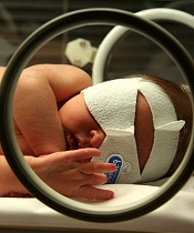
phototherapy to treat
neonatal jaundice
Photo by Martin Pot
Two new studies raise questions about a possible link between childhood cancer and phototherapy for newborn jaundice.
One study showed a significant positive association between phototherapy and 2 cancer types—myeloid leukemia and kidney cancer—but the other study did not.
Although these results are inconclusive, researchers say clinicians should exercise caution in prescribing phototherapy for infants whose jaundice is likely to resolve on its own.
However, the findings should not discourage the use of phototherapy in infants who otherwise would be at risk of brain damage or hearing loss.
The studies were published in Pediatrics alongside a related editorial.
“Phototherapy has been perceived by most as causing minimal risk to the infant,” said editorial author A. Lindsay Frazier, MD, of the Dana-Farber Cancer Institute in Boston, Massachusetts.
“Although these studies are inconclusive and do not prove a relationship between phototherapy and cancer, they should give us pause. That being said, however, the brain damage and hearing loss from high bilirubin levels are real and well-documented, and the suggested risk of cancer from these new studies is both unclear and very small.”
Kaiser Permanente study
Thomas B. Newman, MD, of University of California, San Francisco, and his colleagues conducted a study of children born in Kaiser Permanente Northern California hospitals from 1995 to 2011. The researchers analyzed data on 499,621 children born at 35 weeks’ gestation or later.
There were 60 cases of cancer among the 39,403 children exposed to phototherapy and 651 cases of cancer among the 460,218 children who were not exposed to phototherapy. That translates to 25 per 100,000 person-years and 18 per 100,000 person-years, respectively, for an incidence rate ratio (IRR) of 1.4 (P=0.01).
In an unadjusted analysis, phototherapy was associated with increased rates of any leukemia (IRR=2.1, P=0.0007), nonlymphocytic leukemia (IRR=4.0, P=0.0004), and liver cancer (IRR=5.2, P=0.04).
However, when the researchers adjusted for bilirubin levels, chromosomal disorders, congenital anomalies, and other covariates, these associations were no longer statistically significant.
Study of California hospitals
Andrea C. Wickremasinghe, MD, of Kaiser Permanente Northern California in Santa Clara, and her colleagues conducted a study of children born in California hospitals from 1998 to 2007.
The team analyzed data from the California Office of Statewide Health Planning and Development, which included 5,144,849 infants born at 35 weeks’ gestation or later.
There were 58 cases of cancer among the 178,017 infants exposed to phototherapy and 1042 cancer cases among the 4,966,832 infants who were not exposed to phototherapy. That translated to 32.6 per 100,000 and 21.0 per 100,000, respectively, for a relative risk of 1.6 (95% confidence interval [CI], 1.2–2.0; P=0.002).
In propensity-adjusted analyses, there were significant positive associations between phototherapy and overall cancer (adjusted odds ratio [aOR]=1.4; 95% CI, 1.1–1.9; P=0.007), myeloid leukemia (aOR=2.6; 95% CI, 1.3–5.0; P=0.005), and kidney cancer (aOR=2.5; 95% CI, 1.2–5.1; P=0.02).
Dr Frazier noted that these studies come at a time when the number of infants receiving phototherapy is increasing, perhaps because of the availability of light therapy units that can be used in the home. In the Kaiser Permanente study, 15.9% of infants received phototherapy in 2011, compared to 2.7% in 1995.
“What is concerning is the fact that, at least in the Kaiser Permanente Northern California healthcare system, the number of children receiving phototherapy has dramatically increased,” Dr Frazier said.
“The risks associated with such a prevalent exposure require close scrutiny. If I were the one prescribing phototherapy today, I would want to be sure it was indicated.” ![]()
Health Canada approves pathogen inactivation system for plasma

Photo by Cristina Granados
Health Canada has approved the INTERCEPT Blood System for plasma, a pathogen inactivation system used to reduce the risk of transfusion-transmitted infections.
The system can be used to reduce pathogens in plasma derived from whole blood or obtained by apheresis.
The system inactivates pathogens through a photochemical process involving controlled exposure to ultraviolet light and the chemical amotosalen.
The plasma is then purified to remove the chemical and its byproducts.
Plasma, platelets, and red blood cells do not require functional DNA or RNA for therapeutic efficacy, but pathogens and white blood cells do require these nucleic acids in order to replicate.
The INTERCEPT Blood System targets this basic biological difference. Ultraviolet light is used to activate amotosalen, which binds to DNA and RNA, thereby preventing nucleic acid replication and rendering pathogens inactive.
In studies, the INTERCEPT Blood System for plasma has proven effective in reducing a broad spectrum of viruses, bacteria, spirochetes, and parasites.
However, no pathogen inactivation process has been shown to eliminate all pathogens. Certain viruses (eg, human parvovirus B19) and spores formed by certain bacteria are known to be resistant to the INTERCEPT process.
The INTERCEPT Blood System for plasma, which is marketed by Cerus Corporation, has been approved for use in Europe since 2002 and in the US since 2014.
The safety and efficacy of plasma prepared with the INTERCEPT Blood System has been evaluated in 8 clinical studies including more than 700 patients.
For more information on these studies and the system itself, see the package insert, which is available at http://intercept-canada.com. ![]()
EC grants drug conditional approval to treat MM

Photo courtesy of Janssen
The European Commission (EC) has granted conditional marketing authorization for daratumumab (Darzalex), a monoclonal antibody targeting CD38.
The conditional approval is for daratumumab as monotherapy in adults with relapsed and refractory multiple myeloma (MM) who progressed on their last therapy and have received treatment with a proteasome inhibitor and an immunomodulatory agent.
Daratumumab is the first human CD38 monoclonal antibody approved for use in Europe.
About conditional marketing authorization
A product may receive conditional marketing authorization if the EC finds that, although comprehensive clinical data on the safety and efficacy of the product are not available, all of the following requirements are met:
- The risk-benefit balance of the product is positive
- The company developing the product will likely be in a position to provide comprehensive clinical data in the future
- Unmet medical needs will be fulfilled
- The benefit to public health of the immediate availability of the product outweighs the risk inherent in the fact that additional data are still required.
Conditional marketing authorizations are valid for 1 year, on a renewable basis. The holder will be required to complete ongoing studies or to conduct new studies with a view to confirming that the benefit-risk balance of a product is positive. In addition, specific obligations may be imposed in relation to the collection of pharmacovigilance data.
About daratumumab
Daratumumab works by binding to CD38, a signaling molecule highly expressed on the surface of MM cells. In binding to CD38, daratumumab triggers the patient’s own immune system to attack the cancer cells, resulting in tumor cell death through multiple, immune-mediated mechanisms of action and through immunomodulatory effects, in addition to direct tumor cell death via apoptosis.
The EC’s decision to grant conditional marketing authorization for daratumumab was based on a review of data from the phase 2 SIRIUS study, the phase 1/2 GEN501 study, and 3 additional supportive studies.
The GEN501 study enrolled 102 patients with relapsed MM or relapsed MM that was refractory to 2 or more prior lines of therapy. The patients received daratumumab at a range of doses and on a number of different schedules.
The results suggested daratumumab is most effective at a dose of 16 mg/kg. At this dose, the overall response rate was 36%. Most adverse events in this study were grade 1 or 2, although serious events did occur.
The SIRIUS study enrolled 124 MM patients who had received 3 or more prior lines of therapy. They received daratumumab at different doses and on different schedules, but 106 patients received the drug at 16 mg/kg.
Twenty-nine percent of the 106 patients responded to treatment, and the median duration of response was 7 months. Thirty percent of patients experienced serious adverse events.
Findings from a combined efficacy analysis of the GEN501 and SIRIUS trials demonstrated that, after a mean follow-up of 14.8 months, the estimated median overall survival for patients who received single-agent daratumumab at 16 mg/kg was 20 months.
Five phase 3 clinical studies of daratumumab in MM patients—in relapsed and frontline settings—are ongoing. Additional studies are ongoing or planned to assess the drug’s potential in other malignant and pre-malignant diseases in which CD38 is expressed.
Janssen Biotech, Inc., has exclusive worldwide rights to the development, manufacturing, and commercialization of daratumumab for all potential indications. Janssen licensed daratumumab from Genmab A/S in August 2012. ![]()
Low-residue diet enables clean colonoscopy
There is good news for people who avoid colonoscopies because of the day-long preparation with a clear liquid cleansing diet. New research shows that consuming small portions of low-residue (low-fiber) solid foods on the day before colonoscopy led to improved colonoscopies, compared with a clear liquid diet.
In addition, people who consumed low-residue foods had more energy on the day before and the day of the procedure, compared with those on a clear liquid diet.
“Colonoscopies prevent death from colorectal cancer, yet millions of people avoid this screening. Many cite dietary restrictions as a deterrent. The clear liquid diet is standard of care [for colonoscopy preparation] in the U.S. We found that low-residue foods led to a better colonoscopy compared to a liquid diet. Also, patients were more comfortable, less hungry, and less fatigued on the morning of colonoscopy,” said lead author Dr. Jason Samarasena, associate professor at the University of California, Irvine. He presented this research at a teleconference at the annual Digestive Disease Week.
In this single-center, randomized trial, people assigned to the low-residue diet could eat small portions of protein, carbohydrate, and fat at three meals on the day before colonoscopy. The group assigned to the clear liquid diet could drink only broth, black coffee, tea, and other clear liquids. Both groups drank standard bowel-cleansing liquid on the night before and the morning of the procedure.
Dr. Samarasena explained that low-residue foods, such as eggs, yogurt, cheese, bread, cottage cheese, chicken nuggets, and macaroni and cheese, are easily broken down in the stomach and cleaned out by the bowel preparation.
“High-residue foods, such as fruit, nuts, or vegetables, don’t break down as much and make it more difficult to visualize the colon,” he said.
The study included 83 patients who underwent colonoscopies at two sites over a 1-year period from 2014 to 2015. An adequate bowel preparation was defined as a Boston Bowel Preparation Scale (BBPS) score greater than 6. All endoscopists were blinded and all colonoscopies were video recorded.
In an interim blinded analysis, the low-residue diet allowed a significantly higher number of adequate bowel preparations, compared with the clear liquid diet group: mean BBPS was 7.98 for the low-residue diet group and 7.54 for the clear liquid diet group (P = .05).
People assigned to the low-residue diet expressed a much higher level of satisfaction for the diet: 97% versus 46% who rated the clear liquid diet satisfactory.
In addition, people assigned to the low-residue group had significantly lower hunger scores on a scale from 1 to 10, with 10 being most hungry on the evening before the colonoscopy (3.5 for the low-residue diet versus 6.9 for the clear liquid diet, P = .001), and lower fatigue scores (3.5 versus 6, respectively, P = .01) on a 10-point scale on the morning of the procedure, compared with the clear liquid diet group.
There was no significant difference between the two groups for mean symptom scores for nausea, vomiting, bloating, abdominal cramping, and overall discomfort.
“Patients slated to undergo colonoscopy often have to miss at least a day of work. A low-residue diet may allow patients to work on the day before the procedure,” Dr. Samarasena noted.
Dr. Samarasena and his coinvestigators plan to enroll more patients for a larger sample size to compare both diets.
“This will be one of the largest studies in the U.S. to evaluate a low-residue diet. We hope this will encourage gastroenterologists in the U.S. to use a low-residue diet and that more patients will then undergo colonoscopy screening,“ he said.
FROM DDW® 2016
Key clinical point: A low-residue diet led to improved preparation for colonoscopy, compared with a clear liquid diet.
Major finding: A low-residue diet allowed a significantly higher number of adequate bowel preparations, compared with the clear liquid diet group: mean BBPS was 7.98 for the low-residue diet group and 7.54 for the clear liquid diet group (P = .05).
Data source: Interim analysis of a multicenter, single-center, randomized, single blinded trial of 83 patients.
Disclosures: Dr. Samarasena received funding from Medtronic, Medivators, Olympus, and Pentax.
Naldemedine improves opioid-induced constipation
SAN DIEGO – The investigational oral drug, naldemedine, was found effective in treating opioid-induced constipation (OIC) in patients with noncancer chronic pain, according to results of two identically designed phase III trials (COMPOSE I and II). Naldemedine achieved durable efficacy and consistent safety, with a low incidence of gastrointestinal side effects, and importantly did not compromise or interfere with the analgesic effect of opioids.
In both studies, the drug met the primary endpoint and significantly improved all secondary endpoints. Improved frequency of spontaneous bowel movements (SBMs) was observed for at least 9 weeks out of the 12 weeks (the primary endpoint) in 47.6% of naldemedine-treated patients, compared with 34.6% of placebo patients in COMPOSE I. In COMPOSE II, 52.5% versus 33.6%, respectively, had improved frequency of SMB for at least 9 out of 12 weeks.
“Improvement with naldemedine was seen early and was durable, with no evidence of opiate withdrawal. This is good news for patients with OIC, given the significant impact this condition can have and the difficulty they can have in finding a safe and effective treatment,” said presenting author Dr. Martin Hale, an orthopedic surgeon and pain management specialist. He presented results of both trials at the annual Digestive Disease Week.
Naldemedine is a naltrexone-based, once-daily, oral, peripherally acting mu-opioid–receptor antagonist being developed by Shionogi. The fact that it is not centrally acting suggests that its side effect profile would be acceptable. Opiates work on mu receptors, reducing GI motility and fluid secretion, resulting in OIC.
“Laxatives have limited efficacy for OIC and do not address the underlying problem,” Dr. Hale of Gold Coast Research, Plantation, Fla., said. Thus, better treatments are needed.
Study details
Both studies were 12-week, multicenter, randomized, double-blind, placebo-controlled, parallel-group studies. COMPOSE I included 547 patients, and COMPOSE II, 553 patients. To be included, patients had to be on opioid therapy for at least 3 months and on a stable dose of opioids for at least 4 weeks. They had to have noncancer pain accompanied by OIC.
Patients were randomized in a 1:1 ratio to oral naldemedine 0.2 mg with or without food versus placebo. “Being able to take the drug with food makes it more acceptable to patients,” Dr. Hale noted.
Responses were observed early in the course of the 12-week trial and remained durable. Naldemedine treatment significantly improved the frequency of SBMs per week, compared with placebo (P less than .0001 in both studies), and SBMs without straining per week from baseline to the last 2 weeks of the study (P = .0003 in COMPOSE I and P = .0011 in COMPOSE II).
Adverse events were similar across all four arms of both studies. Treatment-related adverse events were reported in about 48% of patients across both trials. Major adverse cardiovascular events (MACE) of concern occurred in 1 patient in treated with naldemedine and 1 in the placebo group. “MACE is not relevant with this drug,” Dr Hale told listeners.
Abdominal pain and diarrhea were the only treatment-related adverse events reported in greater than 5% of patients treated with naldemedine versus placebo. Abdominal pain was reported in 6.3% of the treated groups in COMPOSE I and 5.2% of the treated group in COMPOSE II versus 1.8% and 2.9%, respectively, in the placebo groups. Diarrhea was reported in 6.6% of the naldemedine patients in COMPOSE I and in 8.9% in COMPOSE II, compared with 2.9% and 1.8%, respectively, on placebo. Nausea was reported in 4.8% of naldemedine-treated patients in both trials, compared with 2.6% and 3.3%, respectively, in the placebo arms.
“Most treatment-emergent adverse events were mild or moderate. Back pain and flatulence were rare,” he commented.
Two scales were used to measure opiate withdrawal: Clinical Opiate Withdrawal Scale (COWS) and Subjective Opiate Withdrawal Score (SOWS). The graph curves were superimposable for naldemedine and placebo at all time points, showing that naldemedine did not interfere with the analgesic effect of opioids.
The study was funded by Shionogi, Florham Park, N.J.
AT DDW® 2016
Key clinical point: Naldemedine was safe and effective in opioid-induced constipation in patients with chronic noncancer pain.
Major finding: Naldemedine improved frequency of spontaneous bowel movements for at least 9 weeks out of the 12 weeks (the primary endpoint) in 47.6% of patients, compared with 34.6% of placebo patients in COMPOSE I. In COMPOSE II, 52.5% versus 33.6%, respectively, had improved frequency of SMB for at least 9 out of 12 weeks.
Data source: Two identically designed phase III, randomized, controlled, double-blind clinical trials that included 547 and 553 patients, respectively.
Disclosures: The study was funded by Shionogi, Florham Park, N.J.
Methylated DNA markers hold promise for Lynch syndrome
SAN DIEGO – It has been challenging to identify reliable biomarkers for Lynch syndrome colorectal neoplasms, but that may be about to change. Researchers have identified 10 discriminant methylated DNA markers (MDMs) for detection of Lynch syndrome colorectal neoplasia.
In particular, OPLAH was the most discriminant MDM across all Lynch syndrome and sporadic colorectal neoplasias, according to a study reported at the annual Digestive Disease Week.
OPLAH had 100% specificity for both Lynch neoplasms and sporadic neoplasms, and sensitivity was 85% for Lynch syndrome neoplasms and 97% in sporadic patients. For adenocarcinoma, specificity and sensitivity were 100% with OPLAH.
“We found some highly discriminating and promising markers for detection of Lynch-associated neoplasms, which could complement current approaches for colorectal cancer,” said presenting author Dr. Veroushka Ballester-Vargas.
Lynch syndrome is the most common hereditary colorectal cancer (CRC), accounting for up to 5% of all CRC. Detection of Lynch neoplasms is currently challenging for several reasons, she explained.
Although there are at least three different sets of criteria for Lynch neoplasms, up to one-third of patients don’t fulfill those criteria. Moreover, 60% to 80% of Lynch tumors arise in the right colon, which is challenging to visualize on colonoscopy. Cologuard is a good test for sporadic CRC, but suboptimal for Lynch syndrome neoplasms.
Dr. Ballester-Vargas, an assistant professor at Columbia University, New York, and her colleagues have previously shown that MDMs may be advantageous as biomarkers. “These markers have one target site per gene, are highly informative and easy to assay,” she said. “Specific discovery of MDMs for Lynch-colorectal neoplasms has not been previously reported.”
Most biomarker discovery efforts have focused on sporadic colorectal neoplasia.
For discovery, the investigators evaluated 54 paraffin-embedded tissues for Lynch syndrome patients (18 normal mucosa, 18 adenoma larger than 1 cm, and 18 adenocarcinoma). Unbiased whole methylome method identified 21 top candidate MDMs from differentially methylated regions. Subsequent biological validation was performed for these 21 top candidates on 218 independent paraffin-embedded samples, 103 Lynch and 115 sporadic.
From these, the 10 most discriminant markers were selected and their performance was compared with sporadic MDMs.
“These MDMs were highly discriminant for sporadic adenoma, sporadic adenocarcinoma, and Lynch syndrome adenocarcinoma, but many were less so for Lynch,” she noted.
“Of the 10 MDMs, the key observation was that OPLAH had nearly perfect discrimination for both Lynch and sporadic neoplasms, with an area under the curve [AUC] approaching 1 – meaning 100% sensitivity and specificity for both adenoma and cancer,” she reported.
The remaining 9 MDMs were equally discriminant for adenoma and cancer but less discriminant for Lynch neoplasms.
“A closer look at OPLAH alone showed it was a highly discriminant marker, with specificity of 97% for sporadic colorectal cancer and 96% for Lynch neoplasms,” Dr. Ballester-Vargas stated.
“This is an early study. We are discussing the next steps, which will include studying MDMs in blood and stool,” she noted. “The other MDMs we identified are highly discriminant for adenomas and cancers.”
The paucity of Lynch tissues was a limitation of the study.
AT DDW® 2016
Key clinical point: Methylated DNA markers (MDMs) may prove helpful in identifying Lynch syndrome colorectal neoplasms.
Major finding: OPLAH had 100% specificity for both Lynch neoplasms and sporadic neoplasms, and sensitivity was 85% for Lynch syndrome neoplasms and 97% for sporadic neoplasm.
Data source: Analysis of paraffin-embedded tissues from 54 Lynch patients for discovery and 218 tissue samples (103 Lynch, 115 sporadic) for validation.
Disclosures: The study was funded by the Mayo Clinic and Exact Sciences.
