User login
Individualizing treatment of menopausal symptoms
Menopause experts Andrew M. Kaunitz, MD, and JoAnn E. Manson, MD, DrPH, provide a comprehensive review of various treatments for menopausal symptoms in an article recently published ahead of print in Obstetrics and Gynecology.1 They discuss hormonal and nonhormonal options to treat vasomotor symptoms, genitourinary syndrome of menopause (GSM), and considerations for the use of hormone therapy in special populations: women with early menopause, women with a history of breast cancer and those who carry the BRCA gene mutation, and women with a history of venous thrombosis.1
The authors write that, “given the lower rates of adverse events on HT among women close to menopause onset and at lower baseline risk of cardiovascular disease, risk stratification and personalized risk assessment appear to represent a sound strategy for optimizing the benefit–risk profile and safety of HT.”1 They suggest that instead of stopping systemic HT at age 65 years, the length of treatment be individualized based on a woman’s risk profile and preferences. The authors encourage gynecologists and other clinicians to use benefit–risk profile tools for both hormonal and nonhormonal options to help women make sound decisions on treating menopausal symptoms.1
Readthe full Clinical Expert Series here.
Reference
- Kaunitz AM, Manson JE. Management of menopausal symptoms [published online ahead of print September 3, 2015]. Obstet Gynecol. doi: 10.1097/AOG.0000000000001058. Accessed September 18, 2015.
Menopause experts Andrew M. Kaunitz, MD, and JoAnn E. Manson, MD, DrPH, provide a comprehensive review of various treatments for menopausal symptoms in an article recently published ahead of print in Obstetrics and Gynecology.1 They discuss hormonal and nonhormonal options to treat vasomotor symptoms, genitourinary syndrome of menopause (GSM), and considerations for the use of hormone therapy in special populations: women with early menopause, women with a history of breast cancer and those who carry the BRCA gene mutation, and women with a history of venous thrombosis.1
The authors write that, “given the lower rates of adverse events on HT among women close to menopause onset and at lower baseline risk of cardiovascular disease, risk stratification and personalized risk assessment appear to represent a sound strategy for optimizing the benefit–risk profile and safety of HT.”1 They suggest that instead of stopping systemic HT at age 65 years, the length of treatment be individualized based on a woman’s risk profile and preferences. The authors encourage gynecologists and other clinicians to use benefit–risk profile tools for both hormonal and nonhormonal options to help women make sound decisions on treating menopausal symptoms.1
Readthe full Clinical Expert Series here.
Reference
- Kaunitz AM, Manson JE. Management of menopausal symptoms [published online ahead of print September 3, 2015]. Obstet Gynecol. doi: 10.1097/AOG.0000000000001058. Accessed September 18, 2015.
Reference
- Kaunitz AM, Manson JE. Management of menopausal symptoms [published online ahead of print September 3, 2015]. Obstet Gynecol. doi: 10.1097/AOG.0000000000001058. Accessed September 18, 2015.
Vegetative Sacral Plaque in a Patient With Human Immunodeficiency Virus
The Diagnosis: Herpes Simplex Vegetans
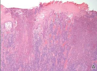
Histopathologic examination using hematoxylin and eosin stain demonstrated marked pseudoepitheliomatous hyperplasia with granulation tissue, ulceration, and abundant exudate joined by a dense mixed inflammatory cell infiltrate that included a myriad of eosinophils (Figure, A). At higher power (Figure, B), many single and multinucleate acantholytic keratinocytes showed ground-glass nuclei and peripheral margination of chromatin within zones of ulceration and crust. Viral culture and direct fluorescent antibody assay identified herpes simplex virus (HSV) type 2. Based on the clinical and histopathologic findings, the patient was diagnosed with herpes simplex vegetans. He was initially treated with oral acyclovir and then oral famciclovir but showed minimal improvement. Eventually, he was referred to surgery and the mass was totally excised with clear margins and no evidence of underlying malignancy.

|
| Histopathology revealed marked pseudoepitheliomatous hyperplasia, ulceration, and a dense mixed inflammatory cell infiltrate (A)(H&E, original magnification ×20). Many multinucleate acantholytic keratinocytes with ground-glass nuclei and peripheral margination of chromatin were shown (B)(H&E, original magnification ×400). |
Herpes simplex virus is one of the most common sexually transmitted infections, with a notably increased incidence and prevalence among individuals with human immunodeficiency virus (HIV) infection.1 Although typical HSV manifestation in immunocompetent patients includes clustered vesicles and/or ulcerations, immunocompromised patients may have unusual presentations, such as persistent and extensive ulcerations or nodular hyperkeratotic lesions.2,3 Herpes vegetans, a term used to describe these atypical exophytic lesions, rarely has been reported in literature, but its presence should raise suspicion for possible underlying immunocompromise. The pathogenesis behind the hypertrophic nature of these lesions is not well understood, but it is postulated that the immune dysregulation from concomitant HIV and HSV infection plays a role.2 Overproduction of tumor necrosis factor and IL-6 by HIV-infected dermal dendritic cells causes an increase in antiapoptotic factors within the epidermis, resulting in enhanced keratinocyte proliferation and clinical hyperkeratosis.2,4
The differential diagnosis for herpes vegetans is somewhat broad, owing to the verrucous and often eroded appearance of the lesions. Biopsy and cultures can be obtained to differentiate from condyloma acuminatum, condyloma latum (secondary syphilis), pyoderma vegetans, pemphigus vegetans, granuloma inguinale, extraintestinal Crohn disease, deep fungal infections, cutaneous tuberculosis, and malignancy.2-4 Histopathology shows epithelial hyperplasia and ulceration with scattered multinucleate keratinocytes, usually at the periphery of the ulcer, and intranuclear inclusions typical of HSV. In addition, a dense dermal infiltrate of lymphocytes, histiocytes, plasma cells, and eosinophils is usually present beneath the base of the ulcer.2,4
Treatment options for herpes vegetans are limited due to the high prevalence of acyclovir-resistant (ACV-R) HSV-2 strains in HIV patients. Valacyclovir and penciclovir have been largely ineffective against ACV-R HSV due to their dependence on the same enzyme—thymidine kinase—involved in the mechanism of acyclovir resistance. Intravenous foscarnet and cidofovir have shown efficacy against ACV-R virus, but concerns of nephrotoxicity have limited their use over prolonged intervals.5 Castelo-Soccio et al6 reported promising results with intralesional cidofovir. This route of administration provides the advantage of increased bioavailability with reduced risk for nephrotoxicity.6 Finally, surgical resection may be considered for refractory lesions to circumvent the toxicity from systemically administered drugs.3
- Severson JL, Tyring SK. Relation between herpes simplex viruses and human immunodeficiency virus infections. Arch Dermatol. 1999;135:1393-1397.
- Patel AB, Rosen T. Herpes vegetans as a sign of HIV infection. Dermatol Online J. 2008;14:6.
- Chung VQ, Parker DC, Parker SR. Surgical excision for vegetative herpes simplex virus infection. Dermatol Surg. 2007;33:1374-1379.
- Beasley KL, Cooley GE, Kao GF, et al. Herpes simplex vegetans: atypical genital herpes infection in a patient with common variable immunodeficiency. J Am Acad Dermatol. 1997;37(5, pt 2):860-863.
- Chilukuri S, Rosen T. Management of acyclovir-resistant herpes simplex virus. Dermatol Clin. 2003;21:311-320.
- Castelo-Soccio L, Bernardin R, Stern J, et al. Successful treatment of acyclovir-resistant herpes simplex virus with intralesional cidofovir. Arch Dermatol. 2010;146:124-126.
The Diagnosis: Herpes Simplex Vegetans
Histopathologic examination using hematoxylin and eosin stain demonstrated marked pseudoepitheliomatous hyperplasia with granulation tissue, ulceration, and abundant exudate joined by a dense mixed inflammatory cell infiltrate that included a myriad of eosinophils (Figure, A). At higher power (Figure, B), many single and multinucleate acantholytic keratinocytes showed ground-glass nuclei and peripheral margination of chromatin within zones of ulceration and crust. Viral culture and direct fluorescent antibody assay identified herpes simplex virus (HSV) type 2. Based on the clinical and histopathologic findings, the patient was diagnosed with herpes simplex vegetans. He was initially treated with oral acyclovir and then oral famciclovir but showed minimal improvement. Eventually, he was referred to surgery and the mass was totally excised with clear margins and no evidence of underlying malignancy.
|
|
| Histopathology revealed marked pseudoepitheliomatous hyperplasia, ulceration, and a dense mixed inflammatory cell infiltrate (A)(H&E, original magnification ×20). Many multinucleate acantholytic keratinocytes with ground-glass nuclei and peripheral margination of chromatin were shown (B)(H&E, original magnification ×400). |
Herpes simplex virus is one of the most common sexually transmitted infections, with a notably increased incidence and prevalence among individuals with human immunodeficiency virus (HIV) infection.1 Although typical HSV manifestation in immunocompetent patients includes clustered vesicles and/or ulcerations, immunocompromised patients may have unusual presentations, such as persistent and extensive ulcerations or nodular hyperkeratotic lesions.2,3 Herpes vegetans, a term used to describe these atypical exophytic lesions, rarely has been reported in literature, but its presence should raise suspicion for possible underlying immunocompromise. The pathogenesis behind the hypertrophic nature of these lesions is not well understood, but it is postulated that the immune dysregulation from concomitant HIV and HSV infection plays a role.2 Overproduction of tumor necrosis factor and IL-6 by HIV-infected dermal dendritic cells causes an increase in antiapoptotic factors within the epidermis, resulting in enhanced keratinocyte proliferation and clinical hyperkeratosis.2,4
The differential diagnosis for herpes vegetans is somewhat broad, owing to the verrucous and often eroded appearance of the lesions. Biopsy and cultures can be obtained to differentiate from condyloma acuminatum, condyloma latum (secondary syphilis), pyoderma vegetans, pemphigus vegetans, granuloma inguinale, extraintestinal Crohn disease, deep fungal infections, cutaneous tuberculosis, and malignancy.2-4 Histopathology shows epithelial hyperplasia and ulceration with scattered multinucleate keratinocytes, usually at the periphery of the ulcer, and intranuclear inclusions typical of HSV. In addition, a dense dermal infiltrate of lymphocytes, histiocytes, plasma cells, and eosinophils is usually present beneath the base of the ulcer.2,4
Treatment options for herpes vegetans are limited due to the high prevalence of acyclovir-resistant (ACV-R) HSV-2 strains in HIV patients. Valacyclovir and penciclovir have been largely ineffective against ACV-R HSV due to their dependence on the same enzyme—thymidine kinase—involved in the mechanism of acyclovir resistance. Intravenous foscarnet and cidofovir have shown efficacy against ACV-R virus, but concerns of nephrotoxicity have limited their use over prolonged intervals.5 Castelo-Soccio et al6 reported promising results with intralesional cidofovir. This route of administration provides the advantage of increased bioavailability with reduced risk for nephrotoxicity.6 Finally, surgical resection may be considered for refractory lesions to circumvent the toxicity from systemically administered drugs.3
- Severson JL, Tyring SK. Relation between herpes simplex viruses and human immunodeficiency virus infections. Arch Dermatol. 1999;135:1393-1397.
- Patel AB, Rosen T. Herpes vegetans as a sign of HIV infection. Dermatol Online J. 2008;14:6.
- Chung VQ, Parker DC, Parker SR. Surgical excision for vegetative herpes simplex virus infection. Dermatol Surg. 2007;33:1374-1379.
- Beasley KL, Cooley GE, Kao GF, et al. Herpes simplex vegetans: atypical genital herpes infection in a patient with common variable immunodeficiency. J Am Acad Dermatol. 1997;37(5, pt 2):860-863.
- Chilukuri S, Rosen T. Management of acyclovir-resistant herpes simplex virus. Dermatol Clin. 2003;21:311-320.
- Castelo-Soccio L, Bernardin R, Stern J, et al. Successful treatment of acyclovir-resistant herpes simplex virus with intralesional cidofovir. Arch Dermatol. 2010;146:124-126.
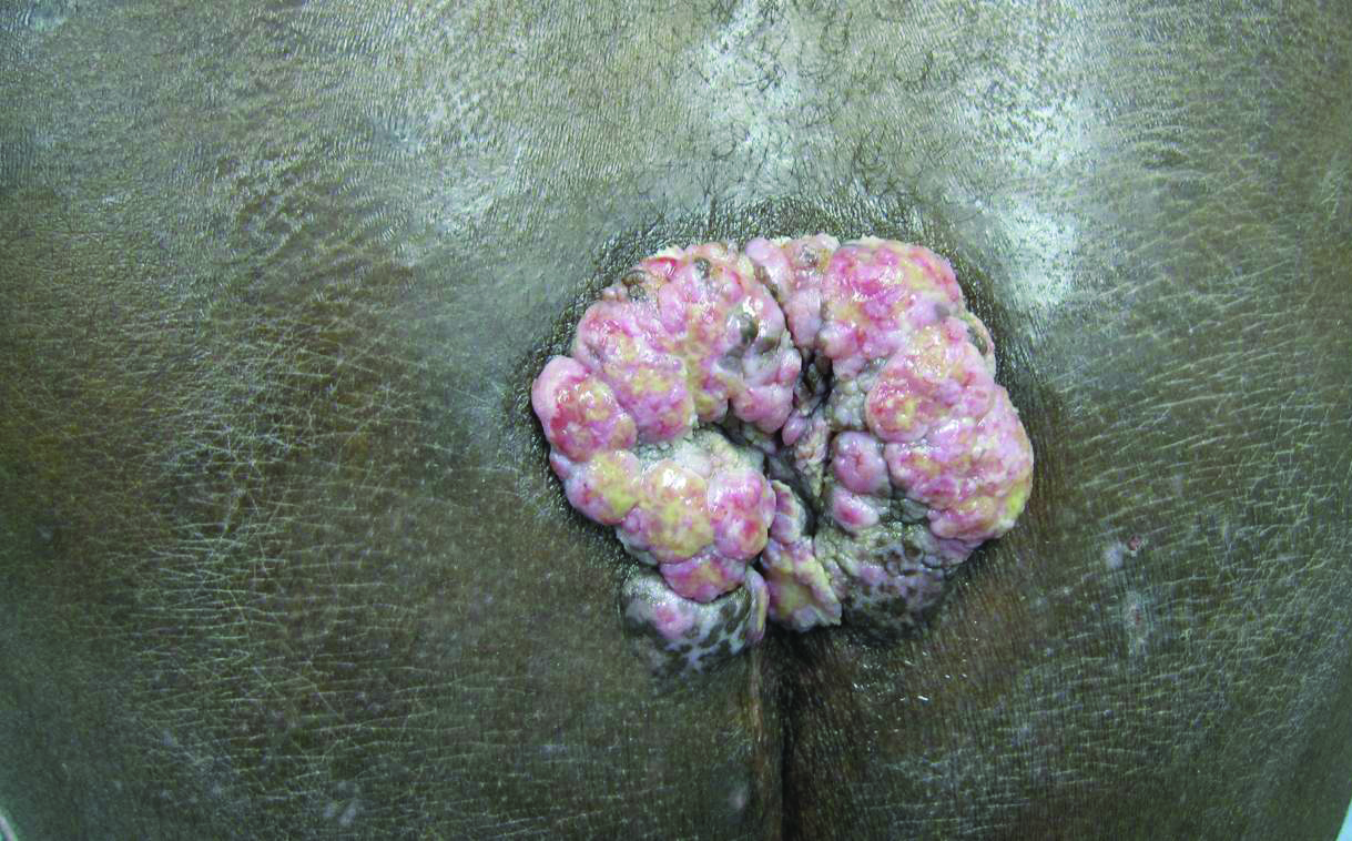
A 53-year-old man presented to our clinic with a sacral mass that had progressively enlarged over 2 years. The patient reported occasional oozing from the mass as well as pain when laying flat but denied fever or other symptoms. His medical history was remarkable for human immunodeficiency virus infection with variable adherence to a highly active antiretroviral therapy regimen. At the time of presentation, the patient had a CD4 lymphocyte count of 78 cells/mm3 (reference range, 500–1400 cells/mm3) and a viral load of 290 copies/mL (reference range, 0 copies/mL). Physical examination revealed a 10-cm discrete, moist and pink, exophytic plaque on the sacrum with superficial erosions. The plaque was nontender and without associated lymphadenopathy. The skin and mucous membranes were otherwise clear. A cutaneous biopsy specimen was obtained from the tumor and sent for histopathologic analysis.

Readmissions rise with endovascular lower limb procedures
CHICAGO – Endovascular lower-extremity procedures were not associated with lower 30-day readmission rates compared with open surgery in a retrospective review of 7,089 patients.
All-cause, 30-day readmissions were actually higher with an endovascular approach at 12.3% vs. 9.6% for open procedures (Relative risk, 1.28; P = .0003).
Among all patients, an index diagnosis of gangrene was most predictive of readmission (RR, 1.89; P less than .0001), Dr. Todd R. Vogel said at the annual meeting of the Midwestern Vascular Surgical Society.
The data were compiled from 7,089 patients in the Cerner Health Facts database who were admitted for peripheral artery disease and elective lower extremity procedures (3,615 open; 3,474 endo) between September 2008 and March 2014. Their average age was 67.7 years, 44.7% were aged 70 years or older, 60% were men, and 21% were African American.

Older patients and men were significantly more likely to receive endovascular procedures (P less than .0001), said Dr. Vogel, chief of vascular surgery, University of Missouri Health System in Columbia.
Overall, 767 patients (11%) were readmitted (344 open; 423 endo), with gangrene accounting for 21.7% of readmissions.
Other index diagnoses associated with higher 30-day readmissions for all lower extremity procedures were fluid and electrolyte disorders, chronic anemia, lower extremity infection, heart failure, chronic kidney disease, and chronic pulmonary disease.
When stratified by procedure type, the reasons for readmission were very different within the same population of patients based on procedure type, Dr. Vogel said.
Patients who underwent an open procedure were more likely to be readmitted if they had heart failure (RR, 1.78; P less than .0001) or posthemorrhagic anemia (RR, 1.54: P = .006).
Infections – be they lower extremity infection, other infection, postoperative infection, or sepsis – were not predictive of readmission when documented at the index admission for the open cohort.
In contrast, chronic conditions were the major predictors of readmission for patients undergoing endovascular procedures, he said. They included chronic anemia (RR, 1.58; P less than .0001), chronic airway obstruction (RR, 1.36; P = .0095), chronic heart disease (RR, 1.33; P = .0019), chronic kidney disease (RR, 1.37; P = .0013), diabetes (RR, 1.34; P = .0012), and hypertension (RR, 1.27; P = .023).
Fluid and electrolyte disorders (RR, 1.65, P less than .0001) and lower extremity infections (RR, 1.57, P = .0016) were also significant predictors of readmission in the endovascular group.
To ensure there were no disparities between index and readmission diagnoses, a final analysis was performed by procedure type in the 767 readmissions. It confirmed that for the endovascular procedures, chronic problems are bringing patients back to the hospital and not necessarily complications from the procedure, whereas infections, device complications, and hemorrhage are the reasons open surgery patients return, Dr. Vogel said.
“The question is are chronic conditions associated with readmissions the fault of the intervention? As physicians can we hope to curb this in patients who have chronic problems and are then readmitted?” he said.
Some audience members argued that no matter if the patient had a chronic condition or not preoperatively, the responsibility rests with the surgeon because he or she opted to put the patient through an elective endovascular procedure and now they’re returning with chronic heart failure, for example.
Dr. Vogel said this was the first pass at the data and trying to understand what drives readmissions and that it’s possible an endovascular procedure could exacerbate a chronic condition, but that surgeons should take steps to mitigate readmission risk in those with known chronic conditions.
Other attendees questioned how many of the readmissions were planned, hinting that the readmissions may not be directly related to the endovascular technique.
Dr. Vogel said it was difficult using only the ICD-9 codes in the database to determine exactly how many readmissions were planned, but noted that further analyses are intended.
“Reasons for readmission can be exacerbation of chronic patient issues, as seen in the endovascular group, or may be secondary to later complications of the procedure such as wound infections and device complications, as seen after open bypass procedures,” he said in an interview. “Identifying patients with increased risk for readmission after vascular procedures may lead to more effective and higher quality care during the index hospitalization. Our future studies will focus on a more detailed, granular evaluation of these high-risk diagnoses groups through use of the electronic medical record.”
Dr. Vogel reported having no financial disclosures.
On Twitter @pwendl
CHICAGO – Endovascular lower-extremity procedures were not associated with lower 30-day readmission rates compared with open surgery in a retrospective review of 7,089 patients.
All-cause, 30-day readmissions were actually higher with an endovascular approach at 12.3% vs. 9.6% for open procedures (Relative risk, 1.28; P = .0003).
Among all patients, an index diagnosis of gangrene was most predictive of readmission (RR, 1.89; P less than .0001), Dr. Todd R. Vogel said at the annual meeting of the Midwestern Vascular Surgical Society.
The data were compiled from 7,089 patients in the Cerner Health Facts database who were admitted for peripheral artery disease and elective lower extremity procedures (3,615 open; 3,474 endo) between September 2008 and March 2014. Their average age was 67.7 years, 44.7% were aged 70 years or older, 60% were men, and 21% were African American.
Older patients and men were significantly more likely to receive endovascular procedures (P less than .0001), said Dr. Vogel, chief of vascular surgery, University of Missouri Health System in Columbia.
Overall, 767 patients (11%) were readmitted (344 open; 423 endo), with gangrene accounting for 21.7% of readmissions.
Other index diagnoses associated with higher 30-day readmissions for all lower extremity procedures were fluid and electrolyte disorders, chronic anemia, lower extremity infection, heart failure, chronic kidney disease, and chronic pulmonary disease.
When stratified by procedure type, the reasons for readmission were very different within the same population of patients based on procedure type, Dr. Vogel said.
Patients who underwent an open procedure were more likely to be readmitted if they had heart failure (RR, 1.78; P less than .0001) or posthemorrhagic anemia (RR, 1.54: P = .006).
Infections – be they lower extremity infection, other infection, postoperative infection, or sepsis – were not predictive of readmission when documented at the index admission for the open cohort.
In contrast, chronic conditions were the major predictors of readmission for patients undergoing endovascular procedures, he said. They included chronic anemia (RR, 1.58; P less than .0001), chronic airway obstruction (RR, 1.36; P = .0095), chronic heart disease (RR, 1.33; P = .0019), chronic kidney disease (RR, 1.37; P = .0013), diabetes (RR, 1.34; P = .0012), and hypertension (RR, 1.27; P = .023).
Fluid and electrolyte disorders (RR, 1.65, P less than .0001) and lower extremity infections (RR, 1.57, P = .0016) were also significant predictors of readmission in the endovascular group.
To ensure there were no disparities between index and readmission diagnoses, a final analysis was performed by procedure type in the 767 readmissions. It confirmed that for the endovascular procedures, chronic problems are bringing patients back to the hospital and not necessarily complications from the procedure, whereas infections, device complications, and hemorrhage are the reasons open surgery patients return, Dr. Vogel said.
“The question is are chronic conditions associated with readmissions the fault of the intervention? As physicians can we hope to curb this in patients who have chronic problems and are then readmitted?” he said.
Some audience members argued that no matter if the patient had a chronic condition or not preoperatively, the responsibility rests with the surgeon because he or she opted to put the patient through an elective endovascular procedure and now they’re returning with chronic heart failure, for example.
Dr. Vogel said this was the first pass at the data and trying to understand what drives readmissions and that it’s possible an endovascular procedure could exacerbate a chronic condition, but that surgeons should take steps to mitigate readmission risk in those with known chronic conditions.
Other attendees questioned how many of the readmissions were planned, hinting that the readmissions may not be directly related to the endovascular technique.
Dr. Vogel said it was difficult using only the ICD-9 codes in the database to determine exactly how many readmissions were planned, but noted that further analyses are intended.
“Reasons for readmission can be exacerbation of chronic patient issues, as seen in the endovascular group, or may be secondary to later complications of the procedure such as wound infections and device complications, as seen after open bypass procedures,” he said in an interview. “Identifying patients with increased risk for readmission after vascular procedures may lead to more effective and higher quality care during the index hospitalization. Our future studies will focus on a more detailed, granular evaluation of these high-risk diagnoses groups through use of the electronic medical record.”
Dr. Vogel reported having no financial disclosures.
On Twitter @pwendl
AT MIDWESTERN VASCULAR 2015
Key clinical point: Endovascular procedures were not superior to open surgery in reducing 30-day readmissions in patients undergoing lower extremity procedures.
Major finding: All-cause 30-day readmissions were 12.3% for endovascular and 9.6% for open (P = .0003).
Data source: Retrospective study in 7,089 patients undergoing elective lower extremity procedures.
Disclosures: The research was supported by an award from the Agency for Healthcare Research and Quality. Dr. Vogel reported having no conflicts of interest.
MM drugs granted priority review

multiple myeloma
The US Food and Drug Administration (FDA) has granted priority review for 4 drugs intended to treat multiple myeloma (MM): elotuzumab (Empliciti), daratumumab, ixazomib (MLN9708), and carfilzomib (Kyprolis).
The FDA grants priority review to investigational therapies that, if approved, may offer significant improvements in the treatment, prevention, or diagnosis of a serious condition.
The designation shortens the review period from 10 months to 6 months.
Carfilzomib
Carfilzomib, an injectable proteasome inhibitor, is currently under review for use in combination with dexamethasone to treat patients with relapsed MM who have received at least 1 prior therapy.
Carfilzomib is already FDA-approved for use in combination with lenalidomide and dexamethasone to treat patients with relapsed MM who have received 1 to 3 prior lines of therapy.
Carfilzomib also has accelerated approval from the FDA as monotherapy for MM patients who have received at least 2 prior therapies, including bortezomib and an immunomodulatory agent (IMiD), and have demonstrated disease progression on or within 60 days of completing their last treatment.
The new regulatory submission for carfilzomib is based on data from the phase 3 ENDEAVOR trial, which were presented at ASCO 2015. In this trial, relapsed MM patients who received carfilzomib and low-dose dexamethasone had significantly longer progression-free survival (PFS) than patients who received bortezomib and low-dose dexamethasone.
Carfilzomib is under development by Onyx Pharmaceuticals, Inc., an Amgen subsidiary.
Ixazomib
Ixazomib, an oral proteasome inhibitor, is under review for the treatment of relapsed and/or refractory MM. Ixazomib has orphan drug designation from the FDA for this patient population.
The regulatory submission for ixazomib is primarily based on results of the first interim analysis of the phase 3 TOURMALINE-MM1 trial.
In this trial, patients with relapsed and/or refractory MM who received ixazomib plus lenalidomide and dexamethasone had superior PFS to patients who received placebo plus lenalidomide and dexamethasone, according to Takeda Pharmaceutical Company Limited, the company developing ixazomib. Detailed data from this study have not yet been released.
Ixazomib is currently under investigation in 3 other phase 3 trials of MM patients:
- TOURMALINE-MM2, investigating ixazomib vs placebo, both in combination with lenalidomide and dexamethasone in patients with newly diagnosed MM
- TOURMALINE-MM3, investigating ixazomib vs placebo as maintenance therapy in patients with newly diagnosed MM following induction therapy and autologous stem cell transplant
- TOURMALINE-MM4, investigating ixazomib vs placebo as maintenance therapy in patients with newly diagnosed MM who have not undergone autologous stem cell transplant.
Daratumumab
Daratumumab, an investigational human anti-CD38 monoclonal antibody, is under review as monotherapy for MM patients who are refractory to both a proteasome inhibitor and an IMiD, or who have received 3 or more prior lines of therapy, including a proteasome inhibitor and an IMiD.
Daratumumab already has fast track, breakthrough therapy, and orphan drug designations from the FDA.
The regulatory submission for daratumumab is primarily supported by data from the phase 2 MMY2002 (SIRIUS) monotherapy study, which were presented at ASCO 2015. Results from this study indicated that daratumumab can produce responses in heavily pretreated MM patients.
Additional data from 4 other studies, including the phase 1/2 GEN501 monotherapy study, which was recently published in NEJM, also support the submission.
Daratumumab is under development by Janssen Research & Development, LLC.
Elotuzumab
Elotuzumab, a signaling lymphocyte activation molecule (SLAMF7)-directed immunostimulatory antibody, is under review for use in combination with lenalidomide and dexamethasone to treat MM patients who have received at least 1 prior treatment.
The FDA has already granted elotuzumab breakthrough therapy and orphan drug designations.
The regulatory submission for elotuzumab is primarily supported by data from the ELOQUENT-2 trial, which were presented at ASCO 2015. In this phase 3 study, researchers evaluated elotuzumab in combination with lenalidomide and dexamethasone, compared to lenalidomide and dexamethasone alone.
The submission is also supported by data from the CA204-009 trial, which were presented at EHA 2015. In this phase 2 study, researchers evaluated elotuzumab in combination with bortezomib and dexamethasone compared to bortezomib and dexamethasone alone.
Results from both trials suggested that adding elotuzumab to combination treatment can prolong PFS in MM patients.
Bristol-Myers Squibb and AbbVie are co-developing elotuzumab, with Bristol-Myers Squibb solely responsible for commercial activities. ![]()
Aspirin, hydrochlorothiazide okay in gout
It’s okay in most cases for gout patients to be on thiazide diuretics or low-dose aspirin, so long as hypouricemic therapy is adjusted as needed, according to rheumatologist and Cleveland Clinic professor Dr. Brian F. Mandell.
Coronary artery disease is common in gout, so patients benefit from inexpensive, well-tolerated, and effective treatments like thiazides for hypertension and 81 mg aspirin daily for cardioprotection, Dr. Mandell said at the annual Perspectives in Rheumatic Diseases conference held by Global Academy for Medical Education.

The concern, however, is that both can elevate serum uric acid. Observational and survey data suggest that the drugs may be associated with an increase in attacks, so some shy away from them in gout.
To make sense of the issue, the first thing to remember is that the drugs cause only “minimal elevations in serum uric acid. Serum urate can still be lowered to less than 6 mg/dL with appropriate therapy without stopping them,” Dr. Mandell said.
Low-dose aspirin raises serum urate by about 0.3 mg/dL. At 12.5 or 25 mg once a day – common hypertension doses – hydrochlorothiazide increases serum urate by 0.8 mg/dL or less in patients with normal renal function (Arthritis Rheum. 2012 Jan;64[1]:121-9).
“In patients with chronic gout treated with a xanthine oxidase inhibitor (allopurinol or febuxostat) to lower the serum urate” to recommended target levels below 6.0 mg/dL, “the small elevation in serum urate is unlikely to negate the clinical efficacy of these drugs when dosing is optimized,” Dr. Mandell wrote in an article that expands upon his presentation points (Cleve Clin J Med. 2014 Feb;81[2]:83-6).
Based on those considerations, “my practice in most patients is to use a thiazide if it helps to control the blood pressure and to adjust the dose of the hypouricemic therapy as needed to reduce the serum urate to the desired level. ... Continuing thiazide therapy and, if necessary, adjusting hypouricemic therapy will not worsen the control of the serum urate level or gouty arthritis, and in most patients will not complicate the management of gout.” In general, “when I add a thiazide to a patient’s regimen, I do not usually need to increase the dose of allopurinol significantly to keep the serum urate level below the desired target,” he said.
The approach is similar with low-dose aspirin. Because of its negligible effect on serum uric acid levels, it does not need to be stopped in hyperuricemia or gout. “Since patients with gout have a higher risk of having cardiovascular disease, metabolic syndrome, and chronic kidney disease, many will benefit from low-dose aspirin therapy,” he said.
Occasionally, it makes sense to switch from a thiazide to another hypertensive, such as losartan, in chronic, hypertensive gout patients with serum urate levels marginally above the precipitation threshold of 6.7 mg/dL. “Losartan is a weak uricosuric and can lower the serum urate level slightly, possibly making the addition of another hypouricemic agent unnecessary, while still controlling the blood pressure with a single pill,” Dr. Mandell said.
The decision “must be individualized, taking into consideration the efficacy and cost of the alternative antihypertensive drug, as well as the potential but as yet unproven cardiovascular and renal benefits of lowering the serum urate with a more potent hypouricemic to a degree not likely to be attained with losartan alone,” he said.
Dr. Mandell has been a consultant for Savient/Crealta, AstraZeneca, Regeneron, and Novartis. Global Academy for Medical Education and this news organization are owned by the same parent company.
It’s okay in most cases for gout patients to be on thiazide diuretics or low-dose aspirin, so long as hypouricemic therapy is adjusted as needed, according to rheumatologist and Cleveland Clinic professor Dr. Brian F. Mandell.
Coronary artery disease is common in gout, so patients benefit from inexpensive, well-tolerated, and effective treatments like thiazides for hypertension and 81 mg aspirin daily for cardioprotection, Dr. Mandell said at the annual Perspectives in Rheumatic Diseases conference held by Global Academy for Medical Education.
The concern, however, is that both can elevate serum uric acid. Observational and survey data suggest that the drugs may be associated with an increase in attacks, so some shy away from them in gout.
To make sense of the issue, the first thing to remember is that the drugs cause only “minimal elevations in serum uric acid. Serum urate can still be lowered to less than 6 mg/dL with appropriate therapy without stopping them,” Dr. Mandell said.
Low-dose aspirin raises serum urate by about 0.3 mg/dL. At 12.5 or 25 mg once a day – common hypertension doses – hydrochlorothiazide increases serum urate by 0.8 mg/dL or less in patients with normal renal function (Arthritis Rheum. 2012 Jan;64[1]:121-9).
“In patients with chronic gout treated with a xanthine oxidase inhibitor (allopurinol or febuxostat) to lower the serum urate” to recommended target levels below 6.0 mg/dL, “the small elevation in serum urate is unlikely to negate the clinical efficacy of these drugs when dosing is optimized,” Dr. Mandell wrote in an article that expands upon his presentation points (Cleve Clin J Med. 2014 Feb;81[2]:83-6).
Based on those considerations, “my practice in most patients is to use a thiazide if it helps to control the blood pressure and to adjust the dose of the hypouricemic therapy as needed to reduce the serum urate to the desired level. ... Continuing thiazide therapy and, if necessary, adjusting hypouricemic therapy will not worsen the control of the serum urate level or gouty arthritis, and in most patients will not complicate the management of gout.” In general, “when I add a thiazide to a patient’s regimen, I do not usually need to increase the dose of allopurinol significantly to keep the serum urate level below the desired target,” he said.
The approach is similar with low-dose aspirin. Because of its negligible effect on serum uric acid levels, it does not need to be stopped in hyperuricemia or gout. “Since patients with gout have a higher risk of having cardiovascular disease, metabolic syndrome, and chronic kidney disease, many will benefit from low-dose aspirin therapy,” he said.
Occasionally, it makes sense to switch from a thiazide to another hypertensive, such as losartan, in chronic, hypertensive gout patients with serum urate levels marginally above the precipitation threshold of 6.7 mg/dL. “Losartan is a weak uricosuric and can lower the serum urate level slightly, possibly making the addition of another hypouricemic agent unnecessary, while still controlling the blood pressure with a single pill,” Dr. Mandell said.
The decision “must be individualized, taking into consideration the efficacy and cost of the alternative antihypertensive drug, as well as the potential but as yet unproven cardiovascular and renal benefits of lowering the serum urate with a more potent hypouricemic to a degree not likely to be attained with losartan alone,” he said.
Dr. Mandell has been a consultant for Savient/Crealta, AstraZeneca, Regeneron, and Novartis. Global Academy for Medical Education and this news organization are owned by the same parent company.
EXPERT ANALYSIS FROM THE ANNUAL PERSPECTIVES IN RHEUMATIC DISEASES

Traffic-related pollution linked to AML, not ALL, in kids

A French study has revealed an increased incidence of acute myeloid leukemia (AML) among children living close to heavily used roads.
The incidence of AML was 30% higher among children who lived within 150 m of heavily used roads and where the combined length of road sections
within this radius exceeded 260 m.
The researchers believe the association between AML and road proximity may be driven by traffic-related benzene exposure.
Previous research has shown an increased risk of leukemia among adults with a history of occupational exposure to benzene.
The current study did not suggest an increased risk of acute lymphoblastic leukemia (ALL) among children living closed to heavily used roads.
Jacqueline Clavel, MD, PhD, of INSERM in Paris, France, and her colleagues reported these findings in the American Journal of Epidemiology.
The team analyzed 2760 cases of leukemia diagnosed in children younger than 15 years of age in metropolitan France between 2002 and 2007, including 418 cases of AML and 2275 cases of ALL.
The researchers compared these cases to a contemporary sample of 30,000 control children representative of the metropolitan population.
The data showed that neither distance from the nearest major road(s) nor the length of major roads within 150 m of a child’s residence was associated with ALL.
However, there was an association for AML. For children whose home was less than 150 m from the nearest major road(s), the odds ratio (OR) was 1.2.
When the total length of major road(s) within 150 m from the child’s residence was 257-308 m (second tertile) or 309 m or greater (third tertile), the OR was 1.3. When the total length of major roads was 1-256 m (first tertile), the OR was 0.90.
The researchers noted that traffic-related nitrogen dioxide concentration was not associated with ALL or AML. But their data indicated that benzene concentration was associated with AML.
To assess this potential association, the team studied the Île-de-France region of Paris, the most urbanized region, for which the mean annual concentration of benzene, mainly from road traffic, was estimated in the vicinity of each residence.
The median estimated benzene concentration for controls living in the Île-de-France region was 1.3 μg/m3 (range, 0.3 to 8.5 μg/m3). And the length of major roads within 150 m of a child’s residence was positively and significantly correlated with log benzene concentration (r=0.3, P<0.001).
So it followed that exposure to an estimated benzene concentration greater than the median was associated with AML (OR=1.6).
The researchers also used a composite variable based on the estimated benzene concentration and the length of major roads around a child’s residence.
The association with AML was largest among children with at least 309 m of major roads within 150 m of their residence and estimated benzene concentrations of 1.3 μg/m3 or greater (OR=2.2).
The researchers said these results support a role for traffic-related benzene exposure in the etiology of childhood AML. ![]()
CHADS2 Variant Calculates Stroke Risk in Heart Failure Patients
NEW YORK - A variant of the CHADS2 score that's used to estimate ischemic stroke risk in patients with atrial fibrillation (AF) is also modestly accurate in heart failure patients, even in those without AF, researchers say. The variant, CHA2DS2-VASc, calculates stroke risk based on 10 possible points with higher scores indicating higher risk.
Line Melgaard from Aalborg University in Denmark and colleagues used three Danish nationwide registries to investigate whether the CHA2DS2-VASc score could predict ischemic stroke, thromboembolism, and death in patients with heart failure without AF as effectively as it does in patients with AF.
Patients with heart failure had a high risk of all three outcomes, whether or not AF was present, and the CHA2DS2-VASc score modestly predicted these endpoints at one-year and five-year follow-up (C statistics, 0.67 and 0.69, respectively).
Heart failure patients without AF whose CHA2DS2-VASc score was 4 or higher had increased risks of ischemic stroke, thromboembolism, and death in a manner comparable to patients with AF, according to the August 30 JAMA online report.
The negative predictive value (NPV) was around 90% at one-year follow-up for all three outcomes, although NPVs were strongly attenuated by the five-year follow-up.
"In our study, one of our principal findings was that the absolute risk of ischemic stroke among patients without AF was about 1.5% per year or higher with CHA2DS2-VASc scores of 2 or higher, with associated five-year absolute ischemic stroke risks in excess of 4% or more," the researchers noted. This risk level would be sufficient to prompt initiation of long-term anticoagulation in patients with AF, they say.
"The poor prognosis of atrial fibrillation for ischemic stroke and death in patients with heart failure was evident in our study and expected," Melgaard said. "But the observation that additional risk factors in patients with heart failure are particularly significant among those without atrial fibrillation is an important and (to some extent) unexpected result."
"I hope physicians will recognize that patients with heart failure and sinus rhythm have an increased risk of ischemic stroke, and that some subgroups within this population most likely need thromboprophylaxis," Melgaard concluded. "Especially patients with multiple comorbidities (high CHA2DS2-VASc score) need attention in the clinic."
Low-Dose Screening CT for High-Risk NSCLC/SCLC: The Providence VA’s Experience
Background: There are about 200,000 new cases of lung cancer each year in the U.S. and about 150,000 deaths. Lung cancer is the second most common malignancy but the most common cause of cancer deaths. In 2010, the National Lung Screening Trial described a 20.3% reduction in lung cancer mortality in high-risk patients who were screened by CT scan. In 2012, guidelines were set forth by multiple groups. The National Comprehensive Cancer Network encouraged low-dose CT screening for patients aged 55 to 75 years, ≥ 30 pack-years of smoking, or smoking cessation within 15 years.
Methods: In late 2013, screening was implemented at the Providence VA Medical Center (PVAMC); a rise in the incidence of early stage lung cancer was anticipated. We reviewed the number and stages of cases of non-small cell (NSCLC) and small cell (SCLC) lung cancer in 2012, 2013 (mostly pre-screening), 2014 (screening), and 2015 (4 months of screening).
Results: In 2012 there were 23 cases of NSCLC (5 stage I; 5 stage II; 8 stage III; 2 stage IV; 3 unknown) and 4 cases of SCLC (extensive). In 2013 there were 42 cases of NSCLC (9 stage I; 2 stage II; 10 stage III [1 screened]; 17 stage IV; 4 unknown [1 screened]) and 9 cases of SCLC (1 lim-ited, 8 extensive). In 2014, there were 54 cases of NSCLC (15 stage I [5 screened]; 10 stage II [2 screened]; 11 stage III [3 screened]; 14 stage IV [1 screened]; and 4 unknown) and 5 cases of SCLC (5 extensive). In the first 7 months of 2015, there were 36 cases of NSCLC (13 stage I [10 screened]; 5 stage II [3 screened]; 7 stage III [3 screened]; 8 stage IV [1 screened]; and 3 unknown [1 screened]) and 1 SCLC (extensive) not screened and 1 SCLC (limited) screened.
Discussion: The total number of lung cancer cases has increased with screening (36 in 2012; 51 in 2013; 59 in 2014; 38 in 7 months of 2015). With screening in 2014 there were more cases of late stage disease (NSCLC stage III/IV) than anticipated. In the first 7 months of 2015 72% of screened patients were stage I/II, 22% were stage III/IV, and 6% were unknown. As time progresses and the same patients are screened yearly, the percentage of early stage cases should increase and late stages decrease. It will be worthwhile to reevaluate these data over the next several years.
Gauging the Use of Single Fraction Radiotherapy for Painful Bone Metastases: The Experience of a Single Veterans Administration Radiation Oncology Center
Background: In addition to patient and/or family convenience, there is mounting category 1 evidence of the effectiveness of single fraction radiotherapy (SFRT) and its utility in the treatment of uncomplicated painful bone metastases. Surveys indicated that physicians in the U.S. still prefer multiple fractions radiotherapy (MFRT) for uncomplicated bone metastases compared with their Canadian or European colleagues. This report used code tracking to determine the scope of use of SFRT for painful malignant bone lesions compared with MFRT.
Methods: We reviewed encounters logged as international classification of disease (ICD) code 198.5 (secondary malignancy of bone and or bone marrow) and current procedural terminology (CPT) codes: 77427 (physician weekly visit for 3 to 5 treatments); and 77431 (physician weekly visit for 1 to 2 treatments) from 2002 to 2008 and 2009 to 2014.
Results: Data limitations/assumptions: The ICD 198.5 diagnosis code was pulled from either the primary or secondary position. By merging CPT/ICD codes to track use patterns, we noted a 43% increase in the use of SFRT for painful bone lesions at James J. Peters VA Medical Center. The CPT code 77431 was used on a yearly average of 2.45% of bone metastases cases from periods 2002 to 2008, and 5.67% (8.50% ex-cluding years 2009/2010) from 2009 to 2014. Of note, from the period years of 2003, 2008, and 2009 to 2010, CPT code 77431 was not used at all.
Conclusions: We saw an increased use of SFRT for bone metastases over the time period covered, and that tracking the encounters by ICD and CPT codes served, in part, as a useful tool in providing a snapshot view (if proper code is used) of SFRT usage. Physician education is a requisite for the proper use of CPT 77431 to capture the true rate of usage of SFRT in clinical practice.
Trabectedin superior for advanced liposarcoma, leiomyosarcoma
Trabectedin proved superior to standard dacarbazine therapy by numerous measures but not by overall survival in an industry-sponsored phase III clinical trial reported online Sept. 14 in Journal of Clinical Oncology.
Trabectedin, which has been used extensively in Europe for a decade but has not been approved in the U.S., has a complex mechanism of action that affects several critical cell biology processes within and surrounding tumor cells. It exhibited activity against metastatic soft tissue sarcomas in several phase II trials, said Dr. George D. Demetri of the Ludwig Center at Harvard Medical School and the Center for Sarcoma and Bone Oncology, Dana-Farber Cancer Institute, both in Boston, and his associates.
In this study, trabectedin was assessed in 518 patients aged 15 years and older who had heavily pretreated and rapidly progressing advanced or metastatic liposarcoma or leiomyosarcoma and were treated at 85 sites in four countries. These participants were randomly assigned to receive either trabectedin (345 patients) or dacarbazine (173 patients) via central intravenous infusion in 3-week cycles.
Compared with dacarbazine, trabectedin reduced the risk of disease progression by 45% (hazard ratio, 0.55), and superior disease control was discernible at the first patient assessment at 6 weeks. Median progression-free survival was significantly longer with trabectedin (4.2 months) than with dacarbazine (1.5 months), a benefit that was consistent across all 19 subgroups of patients assessed in sensitivity analyses, regardless of disease histology, previous therapies, or any clinical characteristics. Trabectedin also bested dacarbazine with regard to objective response rate (9.9% vs. 6.9%), median duration of response (6.5 months vs. 4.2 months), achievement of stable disease (51% vs. 35%), and achievement of durable stable disease (34% vs. 19%).
Trabectedin showed only a nonsignificant 13% reduction in overall survival, which was the primary endpoint of this study. However, several previous studies have demonstrated that it can be extremely difficult to prolong overall survival despite robust improvements in progression-free survival in patients with advanced sarcomas. Given this “historical difficulty in demonstrating overall survival improvement,” the documentation of disease control such as that achieved in this study may be considered a measure of clinically relevant efficacy in this setting, Dr. Demetri and his associates wrote (J. Clin. Oncol. 2015 Sep 14 [doi:10.1200/JCO.2015.62.4734]).
Adverse events in this study population “were consistent with the well-characterized safety and toxicity profiles of both study drugs.” Toxicity was more common with trabectedin, and deaths considered to be treatment-related occurred only in the trabectedin group: three cases of sepsis/septic shock and one each of rhabdomyolysis/sepsis, renal failure, cardiac arrest, and multiorgan failure, for a treatment-related mortality of 2.1%.
This study confirms what we originally heard a decade ago from European investigators: Trabectedin shows clinical activity against liposarcomas and leiomyosarcomas. But the benefits demonstrated here still seem small, and it is not yet clear whether the data are now sufficient to make trabectedin the standard of care for all patients who have these sarcomas.
The primary end point – improving overall survival to a greater degree than dacarbazine – was not met. However, in the investigators’ defense, this was partly because the dacarbazine group survived much longer than expected. This, in turn, may be because more patients who had disease progression with dacarbazine (56%) than with trabectedin (47%) crossed over to other anticancer drugs, including the receptor tyrosine kinase inhibitor pazopanib, which was approved during the course of this study.
Dr. Gary K. Schwartz is at the Herbert Irving Comprehensive Cancer Center and Columbia University, New York. He reported ties to Novartis, AstraZeneca, and Boehringer Ingelheim. Dr. Schwartz made these remarks in an editorial accompanying Dr. Demetri’s report (J. Clin. Oncol. 2015 Sep 14 [doi:10.1200/JCO.2015.63.5938]).
Trabectedin proved superior to standard dacarbazine therapy by numerous measures but not by overall survival in an industry-sponsored phase III clinical trial reported online Sept. 14 in Journal of Clinical Oncology.
Trabectedin, which has been used extensively in Europe for a decade but has not been approved in the U.S., has a complex mechanism of action that affects several critical cell biology processes within and surrounding tumor cells. It exhibited activity against metastatic soft tissue sarcomas in several phase II trials, said Dr. George D. Demetri of the Ludwig Center at Harvard Medical School and the Center for Sarcoma and Bone Oncology, Dana-Farber Cancer Institute, both in Boston, and his associates.
In this study, trabectedin was assessed in 518 patients aged 15 years and older who had heavily pretreated and rapidly progressing advanced or metastatic liposarcoma or leiomyosarcoma and were treated at 85 sites in four countries. These participants were randomly assigned to receive either trabectedin (345 patients) or dacarbazine (173 patients) via central intravenous infusion in 3-week cycles.
Compared with dacarbazine, trabectedin reduced the risk of disease progression by 45% (hazard ratio, 0.55), and superior disease control was discernible at the first patient assessment at 6 weeks. Median progression-free survival was significantly longer with trabectedin (4.2 months) than with dacarbazine (1.5 months), a benefit that was consistent across all 19 subgroups of patients assessed in sensitivity analyses, regardless of disease histology, previous therapies, or any clinical characteristics. Trabectedin also bested dacarbazine with regard to objective response rate (9.9% vs. 6.9%), median duration of response (6.5 months vs. 4.2 months), achievement of stable disease (51% vs. 35%), and achievement of durable stable disease (34% vs. 19%).
Trabectedin showed only a nonsignificant 13% reduction in overall survival, which was the primary endpoint of this study. However, several previous studies have demonstrated that it can be extremely difficult to prolong overall survival despite robust improvements in progression-free survival in patients with advanced sarcomas. Given this “historical difficulty in demonstrating overall survival improvement,” the documentation of disease control such as that achieved in this study may be considered a measure of clinically relevant efficacy in this setting, Dr. Demetri and his associates wrote (J. Clin. Oncol. 2015 Sep 14 [doi:10.1200/JCO.2015.62.4734]).
Adverse events in this study population “were consistent with the well-characterized safety and toxicity profiles of both study drugs.” Toxicity was more common with trabectedin, and deaths considered to be treatment-related occurred only in the trabectedin group: three cases of sepsis/septic shock and one each of rhabdomyolysis/sepsis, renal failure, cardiac arrest, and multiorgan failure, for a treatment-related mortality of 2.1%.
FROM JOURNAL OF CLINICAL ONCOLOGY
Key clinical point: Trabectedin was superior to standard dacarbazine by most measures in advanced, refractory liposarcoma and leiomyosarcoma, though not for overall survival.
Major finding: Trabectedin reduced the risk of disease progression by 45% (HR, 0.55).
Data source: An international industry-sponsored open-label randomized phase III clinical trial involving 518 patients.
Disclosures: This study was supported by Janssen Pharmaceuticals and the Adelson Medical Research Foundation. Dr. Demetri and his associates reported ties to numerous industry sources.
