User login
Law & Medicine: class-action lawsuits
Question: Choose the single best answer to complete the statement, “A class-action lawsuit ...”
A. Is a legal cause of action for a wrongful act perpetrated by a class of wrongdoers.
B. Requires prior certification by a judge before it can go forward.
C. Is appropriate where the claims of a few injured victims can be joined together against a common wrongdoer.
D. Fulfills twin criteria of numerosity and commonality.
E. Can only be filed where there are substantial monetary damages at issue.
Answer: B. A class-action lawsuit describes a legal cause of action where a representative plaintiff(s), known as the lead plaintiff, asserts claims on behalf of a large class of similarly injured members, who then give up their rights to pursue an individual lawsuit. Class-action suits are distinguished from the usual lawsuit, which characteristically affects only one or a few plaintiffs.

Four prerequisites govern an action before it can be deemed a class action: numerosity, commonality, typicality, and adequacy. These are words of art, with specific definitions.
Numerosity denotes a large class membership, usually exceeding 30-40, and sometimes numbering in the thousands, such that the usual joining together of a few injured plaintiffs is not practical. Commonality speaks to common questions of fact and law, typicality requires the claim(s) of the representative plaintiff to match that of the class members, and adequacy demands that the representation of the class members be adequate.
These rules, as well as other important technicalities governing class action, are enumerated in the Federal Rules of Civil Procedure under Rule 23.12Wal-Mart Stores, Inc. v. Dukes et al., where Wal-Mart was alleged to have violated Title VII of the Civil Rights Act of 1964 by having disparate wages and promotion reflecting gender discrimination.3 The plaintiffs sought an injunction against the alleged practice, as well as monetary damages. An earlier ruling of the U.S. Court of Appeals for the Ninth Circuit gave the green light to this sprawling nationwide class action.
It had started out with Betty Dukes and five other women employees, but grew to more than 1.5 million female Wal-Mart employees. However, on appeal, the U.S. Supreme Court reversed, holding that the certification of the plaintiff class was not consistent with Rule 23(a), as the prerequisite of commonality was not fulfilled. The Supreme Court also disallowed the claim for monetary relief, as it was not incidental or secondary to the injunction sought.
Class-action suits are commonly encountered in mass torts, where a product or accident causes injury to numerous individuals. Other situations include environmental pollution, securities fraud, and improper employment practices.
A favorite target is in the health care industry, where manufacturers of drugs and medical devices face allegations that defective products have been allowed to enter the market, causing harm to end users.
An example is the silicon breast implant litigation of the 1990s. The Food and Drug Administration had placed breast implants in the category of medical products and learned that the Dow Corning Corporation, which manufactured the silicon implants, had withheld safety information. The FDA severely restricted the use of these implants, which predictably led to widespread litigation with billions of dollars of judgment awards and settlements.45 and the FDA subsequently required the revision of the package insert of Lipitor and other statins to warn of this association. In 2013, a large Canadian study confirmed the increased incidence of new-onset diabetes in patients taking atorvastatin (hazard ratio, 1.22) or simvastatin.6
However, the outcome of the trial may turn on whether the clear health benefit of lowering serum cholesterol outweighs any purported safety concern.
A fast-growing trend is the filing of class-action suits against nursing homes for providing inadequate care. This is not unexpected, because large numbers of nursing home residents have a common concern over any breach of safety, hygiene, or other statutorily mandated standards of nursing home care. This important topic will be taken up in a subsequent column.
References
1. Available at www.classactionlitigation.com/rule23.html.
2. Mullenix LS. Ending class actions as we know them: rethinking the American class action. Emory Law J. 2014;64:399-449.
3. Wal-Mart Stores, Inc. v. Dukes et al.131 S. Ct. 2541 (2011).
4. Snyder JW. Silicon breast implants. Can emerging medical, legal and scientific concepts be reconciled? J. Leg. Med. 1997;18:133-220.
5. Sattar N, et al. Statins and risk of incident diabetes: a collaborative meta-analysis of randomised statin trials. Lancet 2010;375:735-42.
6. Carter AC, et al. Risk of incident diabetes among patients treated with statins: population based study. BMJ 2013;346:f2610.
Dr. Tan is emeritus professor of medicine and former adjunct professor of law at the University of Hawaii, and currently directs the St. Francis International Center for Healthcare Ethics in Honolulu. This article is meant to be educational and does not constitute medical, ethical, or legal advice. Some of the articles in this series are adapted from the author’s 2006 book, “Medical Malpractice: Understanding the Law, Managing the Risk,” and his 2012 Halsbury treatise, “Medical Negligence and Professional Misconduct.” For additional information, readers may contact the author at [email protected].
Question: Choose the single best answer to complete the statement, “A class-action lawsuit ...”
A. Is a legal cause of action for a wrongful act perpetrated by a class of wrongdoers.
B. Requires prior certification by a judge before it can go forward.
C. Is appropriate where the claims of a few injured victims can be joined together against a common wrongdoer.
D. Fulfills twin criteria of numerosity and commonality.
E. Can only be filed where there are substantial monetary damages at issue.
Answer: B. A class-action lawsuit describes a legal cause of action where a representative plaintiff(s), known as the lead plaintiff, asserts claims on behalf of a large class of similarly injured members, who then give up their rights to pursue an individual lawsuit. Class-action suits are distinguished from the usual lawsuit, which characteristically affects only one or a few plaintiffs.
Four prerequisites govern an action before it can be deemed a class action: numerosity, commonality, typicality, and adequacy. These are words of art, with specific definitions.
Numerosity denotes a large class membership, usually exceeding 30-40, and sometimes numbering in the thousands, such that the usual joining together of a few injured plaintiffs is not practical. Commonality speaks to common questions of fact and law, typicality requires the claim(s) of the representative plaintiff to match that of the class members, and adequacy demands that the representation of the class members be adequate.
These rules, as well as other important technicalities governing class action, are enumerated in the Federal Rules of Civil Procedure under Rule 23.12Wal-Mart Stores, Inc. v. Dukes et al., where Wal-Mart was alleged to have violated Title VII of the Civil Rights Act of 1964 by having disparate wages and promotion reflecting gender discrimination.3 The plaintiffs sought an injunction against the alleged practice, as well as monetary damages. An earlier ruling of the U.S. Court of Appeals for the Ninth Circuit gave the green light to this sprawling nationwide class action.
It had started out with Betty Dukes and five other women employees, but grew to more than 1.5 million female Wal-Mart employees. However, on appeal, the U.S. Supreme Court reversed, holding that the certification of the plaintiff class was not consistent with Rule 23(a), as the prerequisite of commonality was not fulfilled. The Supreme Court also disallowed the claim for monetary relief, as it was not incidental or secondary to the injunction sought.
Class-action suits are commonly encountered in mass torts, where a product or accident causes injury to numerous individuals. Other situations include environmental pollution, securities fraud, and improper employment practices.
A favorite target is in the health care industry, where manufacturers of drugs and medical devices face allegations that defective products have been allowed to enter the market, causing harm to end users.
An example is the silicon breast implant litigation of the 1990s. The Food and Drug Administration had placed breast implants in the category of medical products and learned that the Dow Corning Corporation, which manufactured the silicon implants, had withheld safety information. The FDA severely restricted the use of these implants, which predictably led to widespread litigation with billions of dollars of judgment awards and settlements.45 and the FDA subsequently required the revision of the package insert of Lipitor and other statins to warn of this association. In 2013, a large Canadian study confirmed the increased incidence of new-onset diabetes in patients taking atorvastatin (hazard ratio, 1.22) or simvastatin.6
However, the outcome of the trial may turn on whether the clear health benefit of lowering serum cholesterol outweighs any purported safety concern.
A fast-growing trend is the filing of class-action suits against nursing homes for providing inadequate care. This is not unexpected, because large numbers of nursing home residents have a common concern over any breach of safety, hygiene, or other statutorily mandated standards of nursing home care. This important topic will be taken up in a subsequent column.
References
1. Available at www.classactionlitigation.com/rule23.html.
2. Mullenix LS. Ending class actions as we know them: rethinking the American class action. Emory Law J. 2014;64:399-449.
3. Wal-Mart Stores, Inc. v. Dukes et al.131 S. Ct. 2541 (2011).
4. Snyder JW. Silicon breast implants. Can emerging medical, legal and scientific concepts be reconciled? J. Leg. Med. 1997;18:133-220.
5. Sattar N, et al. Statins and risk of incident diabetes: a collaborative meta-analysis of randomised statin trials. Lancet 2010;375:735-42.
6. Carter AC, et al. Risk of incident diabetes among patients treated with statins: population based study. BMJ 2013;346:f2610.
Dr. Tan is emeritus professor of medicine and former adjunct professor of law at the University of Hawaii, and currently directs the St. Francis International Center for Healthcare Ethics in Honolulu. This article is meant to be educational and does not constitute medical, ethical, or legal advice. Some of the articles in this series are adapted from the author’s 2006 book, “Medical Malpractice: Understanding the Law, Managing the Risk,” and his 2012 Halsbury treatise, “Medical Negligence and Professional Misconduct.” For additional information, readers may contact the author at [email protected].

Consider these medications to help patients stay sober
Consider prescribing oral naltrexone (50 mg/d) for patients with alcohol use disorder who wish to maintain abstinence after a brief period of detoxification.1
Strength of recommendation
A: Based on a meta-analysis of 95 randomized controlled trials.
Jonas DE, Amick HR, Feltner C, et al. Pharmacotherapy for adults with alcohol use disorders in outpatient settings: a systematic review and meta-analysis. JAMA. 2014;311:1889-1900.
Illustrative case
Your patient, a 42-year-old man with alcohol use disorder (AUD), detoxifies from alcohol during a recent hospitalization. He doesn’t want to resume drinking, but reports frequent cravings. Are there any medications you can prescribe to help prevent relapse?
Excessive alcohol consumption is responsible for 1 of every 10 deaths among US adults ages 20 to 64 years.2 Twenty percent to 36% of patients seen in a primary care office have AUD.3 Up to 70% of people who quit with psychosocial support alone will relapse.3
The US Preventive Services Task Force gives a grade B recommendation to screening all adults for AUD, indicating that physicians should provide this service.4 For patients with AUD who wish to abstain but struggle with cravings and relapse, the National Institute on Alcohol Abuse and Alcoholism (NIAAA) recommends considering medication as an adjunct to brief behavioral counseling.5
STUDY SUMMARY: Evidence shows naltrexone can prevent a return to drinking
In a meta-analysis, Jonas et al1 reviewed 123 studies (N=22,803) of pharmacotherapy for AUD. After excluding 28 studies (7 were the only study of a given drug, one was a prospective cohort, and 20 had insufficient data), 95 randomized control trials were included in the analysis. Twenty-two were placebo-controlled for acamprosate (1000-3000 mg/d), 44 for naltrexone (50 mg/d oral, 100 mg/d oral, or injectable) and 4 compared the 2 drugs. Additional studies evaluated disulfiram as well as 23 other off-label medications such as valproic acid and topiramate.
Two investigators independently reviewed the studies, checking for completeness and accuracy. Studies were also analyzed for bias using predefined criteria; those with high or unclear risk of bias were excluded from the main analysis but included in the sensitivity analysis. Funnel plots showed no evidence of publication bias.
Participants were primarily recruited as inpatients and in most studies the mean age was in the 40s. Most patients were diagnosed with alcohol dependence based on criteria in the Diagnostic and Statistical Manual of Mental Disorders, 4th edition, text revision (DSM-IV-TR); this diagnosis translates to likely moderate to severe AUD in DSM-5. Prior to starting medications, participants underwent detoxification or achieved at least 3 days of sobriety. Most studies included psychosocial intervention in addition to medication, but the types of interventions varied. The duration of the trials ranged from 12 to 52 weeks.
Researchers analyzed 5 drinking outcomes—return to any drinking, return to heavy drinking (defined as ≥4 drinks/d for women and ≥5 drinks/d for men), number of drinking days, number of heavy drinking days, and drinks per drinking day. They also evaluated health outcomes (accidents, injuries, quality of life, function, and mortality) and adverse effects.
Acamprosate and oral naltrexone (50 mg/d) significantly decreased return to any drinking, with a number needed to treat (NNT) of 12 (95% confidence interval [CI], 8-26) for acamprosate and 20 (95% CI, 11-500) for naltrexone. Oral naltrexone (50 mg/d) also decreased return to heavy drinking (NNT=12; 95% CI, 8-26), while acamprosate did not. Neither medication showed a decrease in heavy drinking days. In a post hoc subgroup analysis of acamprosate for return to any drinking, the drug appeared to be more effective in studies with a higher risk of bias and less effective in studies with a lower risk of bias; the 2 studies with the lowest risk of bias found no significant effect.
Disulfiram had no effect on any of the drinking outcomes analyzed.
Of the off-label medications, topiramate showed a decrease in drinking days (weighted mean difference [WMD]=-6.5%; 95% CI, -12.0% to -1.0%), heavy drinking days (WMD=-9.0%; 95% CI, -15.3% to -2.7%), and drinks per drinking day (WMD=-1.0; 95% CI, -1.6 to -0.48).
There were no significant differences in health outcomes for any of the medications. Adverse events were greater in treatment groups than placebo groups. Acamprosate was associated with increased risk of diarrhea (number needed to harm [NNH]=11; 95% CI, 6-34), vomiting (NNH=42; 95% CI, 24-143), and anxiety (NNH=7; 95% CI, 5-11). Naltrexone was associated with increased risk of nausea (NNH=9; 95% CI, 7-14), vomiting (NNH=24; 95% CI, 17-44), and dizziness (NNH 16; 95% CI, 12-28).
WHAT'S NEW: Consider prescribing naltrexone to prevent relapse
While previous studies suggested that pharmacotherapy could help patients with AUD remain abstinent, this methodologically rigorous meta-analysis compared the efficacy of several commonly used medications and found clear evidence favoring oral naltrexone. Prescribe oral naltrexone 50 mg/d to help patients with moderate to severe AUD avoid returning to any drinking or heavy drinking after alcohol detoxification. Acamprosate may also decrease return to drinking, although the evidence is not as strong (the studies with low bias showed no effect).
CAVEATS: Medication should be used with psychosocial treatments
Pharmacotherapy for AUD should be reserved for patients who want to quit drinking and used in conjunction with psychosocial intervention.3 Only one of the studies analyzed by Jonas et al1 was conducted in primary care. That said, many of the psychosocial interventions—such as regular follow-up visits to encourage adherence and monitor for adverse effects, in conjunction with attendance at Alcoholics Anonymous meetings—could be done in primary care settings.
Comorbidities may limit therapy options. Naltrexone is contraindicated in acute hepatitis and liver failure, and in combination with opioids.5 Acamprosate is contraindicated in renal disease.5
CHALLENGES TO IMPLEMENTATION: Cost, adherence may be factors for some patients
Perhaps the greatest hurdle in pharmacotherapy for AUD in primary care is a lack of familiarity with these medications. For physicians who are comfortable with prescribing these medications, implementation may be hindered by a lack of available psychosocial resources for successful abstinence.
Additionally, the medications are expensive. The branded version of naltrexone 50 mg costs approximately $118 for a 30-day supply,6 and the branded version of acamprosate costs approximately $284 for a 30-day supply.7
As is the case with any chronic medical condition, medication adherence is a challenge. Naltrexone is taken once daily, while acamprosate is taken 3 times a day. The risk of relapse is high until 6 to 12 months of sobriety and then wanes over several years.5 The NIAAA recommends treatment for a minimum of 3 months.5
ACKNOWLEDGEMENT
The PURLs Surveillance System was supported in part by Grant Number UL1RR024999 from the National Center For Research Resources, a Clinical Translational Science Award to the University of Chicago. The content is solely the responsibility of the authors and does not necessarily represent the official views of the National Center For Research Resources or the National Institutes of Health.
1. Jonas DE, Amick HR, Feltner C, et al. Pharmacotherapy for adults with alcohol use disorders in outpatient settings: a systematic review and meta-analysis. JAMA. 2014;311:1889-1900.
2. Centers for Disease Control and Prevention. Fact sheets - Alcohol use and your health. Centers for Disease Control and Prevention Web site. Available at: http://www.cdc.gov/alcohol/fact-sheets/alcohol-use.htm. Updated November 7, 2014. Accessed January 6, 2015.
3. Johnson BA. Pharmacotherapy for alcohol use disorder. UpTo-Date Web site. Available at: http://www.uptodate.com/contents/pharmacotherapy-for-alcohol-use-disorder. Accessed January 6, 2015.
4. US Preventive Services Task Force. Final recommendation statement: Alcohol misuse: Screening and behavioral counseling interventions in primary care. US Preventive Services Task Force Web site. Available at: http://www.uspreventiveservicestaskforce.org/Page/Document/RecommendationStatementFinal/alcohol-misuse-screening-and-behavioral-counseling-interventions-in-primary-care. Accessed October 2, 2014.
5. US Department of Health and Human Services; National Institutes of Health; National Institute on Alcohol Abuse and Alcoholism. Excerpt from Helping Patients Who Drink Too Much: A Clinician’s Guide. National Institute on Alcohol Abuse and Alcoholism Web site. Available at: http://pubs.niaaa.nih.gov/publications/Practitioner/CliniciansGuide2005/PrescribingMeds.pdf. Updated October 2008. Accessed January 6, 2015.
6. Drugs.com. Revia prices, coupons and patient assistance programs. Drugs.com Web site. Available at: http://www.drugs.com/price-guide/revia. Accessed February 18, 2015.
7. Drugs.com. Campral prices, coupons and patient assistance programs. Drugs.com Web site. Available at: http://www.drugs.com/price-guide/campral. Accessed February 18, 2015.
Consider prescribing oral naltrexone (50 mg/d) for patients with alcohol use disorder who wish to maintain abstinence after a brief period of detoxification.1
Strength of recommendation
A: Based on a meta-analysis of 95 randomized controlled trials.
Jonas DE, Amick HR, Feltner C, et al. Pharmacotherapy for adults with alcohol use disorders in outpatient settings: a systematic review and meta-analysis. JAMA. 2014;311:1889-1900.
Illustrative case
Your patient, a 42-year-old man with alcohol use disorder (AUD), detoxifies from alcohol during a recent hospitalization. He doesn’t want to resume drinking, but reports frequent cravings. Are there any medications you can prescribe to help prevent relapse?
Excessive alcohol consumption is responsible for 1 of every 10 deaths among US adults ages 20 to 64 years.2 Twenty percent to 36% of patients seen in a primary care office have AUD.3 Up to 70% of people who quit with psychosocial support alone will relapse.3
The US Preventive Services Task Force gives a grade B recommendation to screening all adults for AUD, indicating that physicians should provide this service.4 For patients with AUD who wish to abstain but struggle with cravings and relapse, the National Institute on Alcohol Abuse and Alcoholism (NIAAA) recommends considering medication as an adjunct to brief behavioral counseling.5
STUDY SUMMARY: Evidence shows naltrexone can prevent a return to drinking
In a meta-analysis, Jonas et al1 reviewed 123 studies (N=22,803) of pharmacotherapy for AUD. After excluding 28 studies (7 were the only study of a given drug, one was a prospective cohort, and 20 had insufficient data), 95 randomized control trials were included in the analysis. Twenty-two were placebo-controlled for acamprosate (1000-3000 mg/d), 44 for naltrexone (50 mg/d oral, 100 mg/d oral, or injectable) and 4 compared the 2 drugs. Additional studies evaluated disulfiram as well as 23 other off-label medications such as valproic acid and topiramate.
Two investigators independently reviewed the studies, checking for completeness and accuracy. Studies were also analyzed for bias using predefined criteria; those with high or unclear risk of bias were excluded from the main analysis but included in the sensitivity analysis. Funnel plots showed no evidence of publication bias.
Participants were primarily recruited as inpatients and in most studies the mean age was in the 40s. Most patients were diagnosed with alcohol dependence based on criteria in the Diagnostic and Statistical Manual of Mental Disorders, 4th edition, text revision (DSM-IV-TR); this diagnosis translates to likely moderate to severe AUD in DSM-5. Prior to starting medications, participants underwent detoxification or achieved at least 3 days of sobriety. Most studies included psychosocial intervention in addition to medication, but the types of interventions varied. The duration of the trials ranged from 12 to 52 weeks.
Researchers analyzed 5 drinking outcomes—return to any drinking, return to heavy drinking (defined as ≥4 drinks/d for women and ≥5 drinks/d for men), number of drinking days, number of heavy drinking days, and drinks per drinking day. They also evaluated health outcomes (accidents, injuries, quality of life, function, and mortality) and adverse effects.
Acamprosate and oral naltrexone (50 mg/d) significantly decreased return to any drinking, with a number needed to treat (NNT) of 12 (95% confidence interval [CI], 8-26) for acamprosate and 20 (95% CI, 11-500) for naltrexone. Oral naltrexone (50 mg/d) also decreased return to heavy drinking (NNT=12; 95% CI, 8-26), while acamprosate did not. Neither medication showed a decrease in heavy drinking days. In a post hoc subgroup analysis of acamprosate for return to any drinking, the drug appeared to be more effective in studies with a higher risk of bias and less effective in studies with a lower risk of bias; the 2 studies with the lowest risk of bias found no significant effect.
Disulfiram had no effect on any of the drinking outcomes analyzed.
Of the off-label medications, topiramate showed a decrease in drinking days (weighted mean difference [WMD]=-6.5%; 95% CI, -12.0% to -1.0%), heavy drinking days (WMD=-9.0%; 95% CI, -15.3% to -2.7%), and drinks per drinking day (WMD=-1.0; 95% CI, -1.6 to -0.48).
There were no significant differences in health outcomes for any of the medications. Adverse events were greater in treatment groups than placebo groups. Acamprosate was associated with increased risk of diarrhea (number needed to harm [NNH]=11; 95% CI, 6-34), vomiting (NNH=42; 95% CI, 24-143), and anxiety (NNH=7; 95% CI, 5-11). Naltrexone was associated with increased risk of nausea (NNH=9; 95% CI, 7-14), vomiting (NNH=24; 95% CI, 17-44), and dizziness (NNH 16; 95% CI, 12-28).
WHAT'S NEW: Consider prescribing naltrexone to prevent relapse
While previous studies suggested that pharmacotherapy could help patients with AUD remain abstinent, this methodologically rigorous meta-analysis compared the efficacy of several commonly used medications and found clear evidence favoring oral naltrexone. Prescribe oral naltrexone 50 mg/d to help patients with moderate to severe AUD avoid returning to any drinking or heavy drinking after alcohol detoxification. Acamprosate may also decrease return to drinking, although the evidence is not as strong (the studies with low bias showed no effect).
CAVEATS: Medication should be used with psychosocial treatments
Pharmacotherapy for AUD should be reserved for patients who want to quit drinking and used in conjunction with psychosocial intervention.3 Only one of the studies analyzed by Jonas et al1 was conducted in primary care. That said, many of the psychosocial interventions—such as regular follow-up visits to encourage adherence and monitor for adverse effects, in conjunction with attendance at Alcoholics Anonymous meetings—could be done in primary care settings.
Comorbidities may limit therapy options. Naltrexone is contraindicated in acute hepatitis and liver failure, and in combination with opioids.5 Acamprosate is contraindicated in renal disease.5
CHALLENGES TO IMPLEMENTATION: Cost, adherence may be factors for some patients
Perhaps the greatest hurdle in pharmacotherapy for AUD in primary care is a lack of familiarity with these medications. For physicians who are comfortable with prescribing these medications, implementation may be hindered by a lack of available psychosocial resources for successful abstinence.
Additionally, the medications are expensive. The branded version of naltrexone 50 mg costs approximately $118 for a 30-day supply,6 and the branded version of acamprosate costs approximately $284 for a 30-day supply.7
As is the case with any chronic medical condition, medication adherence is a challenge. Naltrexone is taken once daily, while acamprosate is taken 3 times a day. The risk of relapse is high until 6 to 12 months of sobriety and then wanes over several years.5 The NIAAA recommends treatment for a minimum of 3 months.5
ACKNOWLEDGEMENT
The PURLs Surveillance System was supported in part by Grant Number UL1RR024999 from the National Center For Research Resources, a Clinical Translational Science Award to the University of Chicago. The content is solely the responsibility of the authors and does not necessarily represent the official views of the National Center For Research Resources or the National Institutes of Health.
1. Jonas DE, Amick HR, Feltner C, et al. Pharmacotherapy for adults with alcohol use disorders in outpatient settings: a systematic review and meta-analysis. JAMA. 2014;311:1889-1900.
2. Centers for Disease Control and Prevention. Fact sheets - Alcohol use and your health. Centers for Disease Control and Prevention Web site. Available at: http://www.cdc.gov/alcohol/fact-sheets/alcohol-use.htm. Updated November 7, 2014. Accessed January 6, 2015.
3. Johnson BA. Pharmacotherapy for alcohol use disorder. UpTo-Date Web site. Available at: http://www.uptodate.com/contents/pharmacotherapy-for-alcohol-use-disorder. Accessed January 6, 2015.
4. US Preventive Services Task Force. Final recommendation statement: Alcohol misuse: Screening and behavioral counseling interventions in primary care. US Preventive Services Task Force Web site. Available at: http://www.uspreventiveservicestaskforce.org/Page/Document/RecommendationStatementFinal/alcohol-misuse-screening-and-behavioral-counseling-interventions-in-primary-care. Accessed October 2, 2014.
5. US Department of Health and Human Services; National Institutes of Health; National Institute on Alcohol Abuse and Alcoholism. Excerpt from Helping Patients Who Drink Too Much: A Clinician’s Guide. National Institute on Alcohol Abuse and Alcoholism Web site. Available at: http://pubs.niaaa.nih.gov/publications/Practitioner/CliniciansGuide2005/PrescribingMeds.pdf. Updated October 2008. Accessed January 6, 2015.
6. Drugs.com. Revia prices, coupons and patient assistance programs. Drugs.com Web site. Available at: http://www.drugs.com/price-guide/revia. Accessed February 18, 2015.
7. Drugs.com. Campral prices, coupons and patient assistance programs. Drugs.com Web site. Available at: http://www.drugs.com/price-guide/campral. Accessed February 18, 2015.
Copyright © 2015 Family Physicians Inquiries Network. All rights reserved.
Failure to recognize impending MI has tragic consequences ... More
Failure to recognize impending MI has tragic consequences
A 55-YEAR-OLD WOMAN WENT TO HER MEDICAL CLINIC because she had heartburn and bilateral arm pain with numbness and tingling in her forearms. She said she’d had intermittent arm pain over the previous 7 to 10 days. A physician’s assistant diagnosed gastroesophageal reflux disease, gave the patient an antacid medication, and instructed her to return in 2 to 3 weeks. The patient came back to the clinic 10 days later with increased heartburn and continued arm pain with tingling. Because no clinicians were available to see her at that time, a prescription for ranitidine was called in and the patient was sent home. That evening, the patient died of a myocardial infarction (MI).
PLAINTIFF’S CLAIM There were specific, objective signs of an impending MI that were not recognized.
The patient should have been seen by a medical provider on the day of her death or referred to an emergency department.
THE DEFENSE No information about the defense is available.
VERDICT $275,000 California settlement.
COMMENT There was clearly an opportunity to make the correct diagnosis for this woman, especially when she returned a second time. The one lesson I have learned from reviewing malpractice cases for 15 years is that if a patient returns unimproved, you must up the ante with the evaluation. Start all over again and think through the entire history very carefully; you are likely to find a clue to the correct diagnosis.
Pulmonary embolism mistaken for respiratory infection
A 40-YEAR-OLD MAN SOUGHT TREATMENT FOR SYMPTOMS OF A COLD. He also complained of shortness of breath, dizziness, and pain in his left calf. His family physician (FP) treated him for a respiratory infection. Three days later, the patient returned to the office with continued shortness of breath. The FP scheduled a cardiac work-up. Two days before the work-up, the patient died from a pulmonary embolism (PE).
PLAINTIFF'S CLAIM No information about the plaintiff’s claim is available.
THE DEFENSE No information about the defense is available.
VERDICT $1.1 million Virginia settlement.
COMMENT PE has clearly unseated syphilis as “The Great Masquerader.” We cannot tell from this short synopsis how significant the patient’s calf pain was and whether or not there were any physical findings of deep vein thrombosis. However, when the patient returned 3 days later with increasing shortness of breath, PE should have been toward the top of the differential diagnosis.
Back spasms—or something far more serious?
A 47-YEAR-OLD WOMAN WENT TO THE EMERGENCY DEPARTMENT (ED) seeking treatment for severe back and abdominal pain. The patient had previously undergone gastric bypass surgery. The ED physician diagnosed back spasms, but admitted her to the hospital for observation. The next day, the patient died from a bowel obstruction.
PLAINTIFF'S CLAIM The ED physician failed to order testing and consult with a specialist to diagnose bowel obstruction, which is a known complication of gastric bypass surgery.
THE DEFENSE No information about the defense is available.
VERDICT $2.4 million Illinois verdict.
COMMENT Bowel obstruction with back pain only? And dead the next day from bowel obstruction? I can only presume the history was inadequate, which led to a failure to do an abdominal exam.
VIDEO: Nanodermatology Society roundtable tackles tough questions on photoprotection
SAN FRANCISCO – What role does nanotechnology play in photoprotection? How is the current regulatory climate helping and hurting innovation and product development? What are the real and perceived safety issues? These and other questions were addressed in a roundtable discussion hosted by the Nanodermatology Society at the annual meeting of the American Academy of Dermatology. Participants included dermatology practitioners and researchers, along with representatives of industry and the media.
“We have good data on a lot of the filters that are out there,” emphasized Dr. Adnan Nasir, president of the Nanodermatology Society and moderator of the roundtable.
Given the epidemic of skin cancer in the United States, “I hope that we can target our message as a public health message” and increase the confidence of physicians in discussing photoprotection and the confidence of patients in their doctors’ opinions on this important topic, Dr. Nasir said.
The video associated with this article is no longer available on this site. Please view all of our videos on the MDedge YouTube channel
AT THE AAD ANNUAL MEETING

Anxiety tied to fear of falling • fatigue • difficulty concentrating • Dx?
THE CASE
A 21-year-old college student was referred to us by the counseling center at our university for a psychiatric evaluation after 11 psychotherapy sessions over 3 months had failed to reduce her feelings of anxiety and panic.
During our evaluation, the patient described feeling “not quite right” for many months. She had been experiencing mental fogginess, fatigue, and worsening concentration/ memory. Her anxiety, which had been gradually increasing, was the result of being unsure about her gait. She first noticed this while walking down some bleachers; she felt dizzy, was afraid of falling, and couldn’t walk down without assistance. All episodes of “panic” occurred in situations where she experienced disequilibrium, unsteady gait, and fear of falling. She grew fearful of driving or going anywhere without assistance.
The patient had celiac disease that was well controlled with a gluten-free diet. She had no personal or family psychiatric history and no history of substance abuse.
THE DIAGNOSIS
Physical exam and lab studies, including a complete blood count, comprehensive metabolic panel, and thyrotropin and folate levels, were normal. Her homocysteine level was 11.8 μmol/L (reference range, 5.4-11.9 μmol/L) and vitamin B12 level was 292 pg/mL (reference range, 200-1100 pg/mL). Her lab report included a note that read, “Although the reference range for vitamin B12 is 200 to 1100 pg/mL, it has been reported that between 5% and 10% of patients with values between 200 and 400 pg/mL may experience neuropsychiatric and hematologic abnormalities due to occult B12 deficiency; <1% of patients with values >400 pg/mL will have symptoms.”
Based on this vitamin B12 level, the patient’s symptoms, and her borderline high homocysteine level, we diagnosed vitamin B12 deficiency.
DISCUSSION
There are no recommendations by the US Preventive Services Task Force or any other major US medical society for routine vitamin B12 screening.1 In Canada, the Medical Services Commission of the British Columbia Ministry of Health recommends B12 screening for patients who present with macrocytic anemia or unexplained neurologic symptoms (eg, paresthesia, numbness, poor motor coordination, memory lapses, or cognitive or personality changes).2
Vitamin B12 deficiency can be caused by numerous conditions, including those that cause malabsorption (such as gastric bypass). It can also be caused by diseases such as human immunodeficiency virus infection or Crohn’s disease, long-term adherence to a vegetarian or vegan diet, or by any other lack of dietary intake.1 The condition can cause hematologic-related signs and symptoms such as megaloblastic anemia, fatigue, and syncope. It also can have neurologic manifestations, including paresthesia, weakness, motor disturbances (including gait abnormalities), vision loss, and a wide range of cognitive and behavioral changes.1 Anemia is uncommon because since 1998, the US Food and Drug Administration has required fortification of all enriched grain and cereal products with folic acid; thus, vitamin B12 deficiency may proceed without anemia revealing its presence.1
A controversial topic. Vitamin B12 deficiency is a complicated and controversial subject. Specifically, there is uncertainty about the clinical importance of lower serum levels of vitamin B12 (200-400 pg/mL), their impact on well-being, and the need for treatment. In addition to measuring a patient’s serum B12 level, testing a second biomarker (such as homocysteine or methylmalonic acid) can be helpful in establishing a diagnosis of B12 deficiency.1 Levels of each of these are elevated in patients with B12 deficiency.1
Although vitamin B12 deficiency has been well studied in older patients,3 little has been published about the condition in young adults. National Health and Nutrition Examination Survey (NHANES) data from 2000 to 2004 shows that almost 40% of people ages 19 to 30 years have a B12 level <400 pg/mL.1 How many of these individuals are at risk of complications of B12 deficiency is unknown.
B12 supplementation might improve depression, anxiety
B12 supplementation is inexpensive and has no significant adverse effects.1 It can be administered orally, parenterally (intramuscularly or subcutaneously), or intranasally.1 A common oral regimen is 1 mg/d; parental regimens vary widely, but might include a 1-mg injection once a week for 8 weeks, then once a month for life.1
Some evidence suggests B12 supplementation may improve symptoms of depression and anxiety. A Pakistani study randomized 73 patients with depression and “low normal” B12 levels (190-300 pg/mL) to an antidepressant only (equivalent to imipramine 100-250 mg/d or fluoxetine 20-40 mg/d) or an antidepressant plus parenteral B12 (1000 mcg once a week).4 At 3 months follow-up, 100% of the treatment group showed at least a 20% reduction in their Hamilton Depression Rating Scale (HAM-D) score, compared to 69% in the control arm (P<.001).4
A Swedish study analyzed the effects of several B vitamins, including 0.5 mg/d of B12 vs placebo on mood in 65 celiac patients on a gluten-free diet who had borderline/low normal B12 levels (>191 pg/mL).5 Patients who scored low on a measure of psychological well-being at the beginning of the study and who received B12 experienced significant improvements in anxiety and depressed mood compared to those who received placebo.
Our patient
Because neurologic and psychiatric symptoms require assured compliance and urgent treatment, our patient received vitamin B12 parenterally as cyanocobalamin 1 mg/mL. She was given this dosage intramuscularly once a day for 5 days, then once a week for 4 weeks. She will continue to receive it once a month indefinitely.
The patient was advised that if she wished to switch to oral therapy, she could do so after several months of parenteral treatment, as long as she had close follow-up with frequent B12 measurements to assure that she was absorbing oral therapy. Her anxiety and mood symptoms resolved within one month, and her disequilibrium was almost entirely resolved within 3 months of treatment.
THE TAKEAWAY
Although more common in older patients, vitamin B12 deficiency can also affect younger patients. “Low normal” B12 levels (200-400 pg/mL) may affect psychological well-being.
Consider testing serum B12 and a second biomarker—such as homocysteine or methylmalonic acid, if indicated—in patients who present with depressed mood, anxiety, cognitive symptoms, and/or fatigue. Vitamin B12 supplementation can be administered orally and has no major adverse effects.
1. Centers for Disease Control and Prevention. Why vitamin B12 deficiency should be on your radar screen. Centers for Disease Control and Prevention Web site. Available at: http://www.cdc.gov/ncbddd/b12/. Accessed February 26, 2015.
2. Guidelines and Protocols Advisory Committee, Medical Services Commission, British Columbia Ministry of Health. Cobalamin (vitamin B12) Deficiency - Investigation & Management. British Columbia Ministry of Health Web site. Available at: http://www.bcguidelines.ca/guideline_cobalamin.html. Accessed March 13, 2015.
3. Pennypacker LC, Allen RH, Kelly JP, et al. High prevalence of cobalamin deficiency in elderly outpatients. J Am Geriatr Soc. 1992;40:1197-1204.
4. Syed EU, Wasay M, Awan S. Vitamin B12 supplementation in treating major depressive disorder: a randomized controlled trial. Open Neurol J. 2013;7:44-48
5. Hallert C, Svensson M, Tholstrup J, et al. Clinical trial: B vitamins improve health in patients with coeliac disease living on a gluten-free diet. Ailment Pharmacol Ther. 2009;29:811-816.
THE CASE
A 21-year-old college student was referred to us by the counseling center at our university for a psychiatric evaluation after 11 psychotherapy sessions over 3 months had failed to reduce her feelings of anxiety and panic.
During our evaluation, the patient described feeling “not quite right” for many months. She had been experiencing mental fogginess, fatigue, and worsening concentration/ memory. Her anxiety, which had been gradually increasing, was the result of being unsure about her gait. She first noticed this while walking down some bleachers; she felt dizzy, was afraid of falling, and couldn’t walk down without assistance. All episodes of “panic” occurred in situations where she experienced disequilibrium, unsteady gait, and fear of falling. She grew fearful of driving or going anywhere without assistance.
The patient had celiac disease that was well controlled with a gluten-free diet. She had no personal or family psychiatric history and no history of substance abuse.
THE DIAGNOSIS
Physical exam and lab studies, including a complete blood count, comprehensive metabolic panel, and thyrotropin and folate levels, were normal. Her homocysteine level was 11.8 μmol/L (reference range, 5.4-11.9 μmol/L) and vitamin B12 level was 292 pg/mL (reference range, 200-1100 pg/mL). Her lab report included a note that read, “Although the reference range for vitamin B12 is 200 to 1100 pg/mL, it has been reported that between 5% and 10% of patients with values between 200 and 400 pg/mL may experience neuropsychiatric and hematologic abnormalities due to occult B12 deficiency; <1% of patients with values >400 pg/mL will have symptoms.”
Based on this vitamin B12 level, the patient’s symptoms, and her borderline high homocysteine level, we diagnosed vitamin B12 deficiency.
DISCUSSION
There are no recommendations by the US Preventive Services Task Force or any other major US medical society for routine vitamin B12 screening.1 In Canada, the Medical Services Commission of the British Columbia Ministry of Health recommends B12 screening for patients who present with macrocytic anemia or unexplained neurologic symptoms (eg, paresthesia, numbness, poor motor coordination, memory lapses, or cognitive or personality changes).2
Vitamin B12 deficiency can be caused by numerous conditions, including those that cause malabsorption (such as gastric bypass). It can also be caused by diseases such as human immunodeficiency virus infection or Crohn’s disease, long-term adherence to a vegetarian or vegan diet, or by any other lack of dietary intake.1 The condition can cause hematologic-related signs and symptoms such as megaloblastic anemia, fatigue, and syncope. It also can have neurologic manifestations, including paresthesia, weakness, motor disturbances (including gait abnormalities), vision loss, and a wide range of cognitive and behavioral changes.1 Anemia is uncommon because since 1998, the US Food and Drug Administration has required fortification of all enriched grain and cereal products with folic acid; thus, vitamin B12 deficiency may proceed without anemia revealing its presence.1
A controversial topic. Vitamin B12 deficiency is a complicated and controversial subject. Specifically, there is uncertainty about the clinical importance of lower serum levels of vitamin B12 (200-400 pg/mL), their impact on well-being, and the need for treatment. In addition to measuring a patient’s serum B12 level, testing a second biomarker (such as homocysteine or methylmalonic acid) can be helpful in establishing a diagnosis of B12 deficiency.1 Levels of each of these are elevated in patients with B12 deficiency.1
Although vitamin B12 deficiency has been well studied in older patients,3 little has been published about the condition in young adults. National Health and Nutrition Examination Survey (NHANES) data from 2000 to 2004 shows that almost 40% of people ages 19 to 30 years have a B12 level <400 pg/mL.1 How many of these individuals are at risk of complications of B12 deficiency is unknown.
B12 supplementation might improve depression, anxiety
B12 supplementation is inexpensive and has no significant adverse effects.1 It can be administered orally, parenterally (intramuscularly or subcutaneously), or intranasally.1 A common oral regimen is 1 mg/d; parental regimens vary widely, but might include a 1-mg injection once a week for 8 weeks, then once a month for life.1
Some evidence suggests B12 supplementation may improve symptoms of depression and anxiety. A Pakistani study randomized 73 patients with depression and “low normal” B12 levels (190-300 pg/mL) to an antidepressant only (equivalent to imipramine 100-250 mg/d or fluoxetine 20-40 mg/d) or an antidepressant plus parenteral B12 (1000 mcg once a week).4 At 3 months follow-up, 100% of the treatment group showed at least a 20% reduction in their Hamilton Depression Rating Scale (HAM-D) score, compared to 69% in the control arm (P<.001).4
A Swedish study analyzed the effects of several B vitamins, including 0.5 mg/d of B12 vs placebo on mood in 65 celiac patients on a gluten-free diet who had borderline/low normal B12 levels (>191 pg/mL).5 Patients who scored low on a measure of psychological well-being at the beginning of the study and who received B12 experienced significant improvements in anxiety and depressed mood compared to those who received placebo.
Our patient
Because neurologic and psychiatric symptoms require assured compliance and urgent treatment, our patient received vitamin B12 parenterally as cyanocobalamin 1 mg/mL. She was given this dosage intramuscularly once a day for 5 days, then once a week for 4 weeks. She will continue to receive it once a month indefinitely.
The patient was advised that if she wished to switch to oral therapy, she could do so after several months of parenteral treatment, as long as she had close follow-up with frequent B12 measurements to assure that she was absorbing oral therapy. Her anxiety and mood symptoms resolved within one month, and her disequilibrium was almost entirely resolved within 3 months of treatment.
THE TAKEAWAY
Although more common in older patients, vitamin B12 deficiency can also affect younger patients. “Low normal” B12 levels (200-400 pg/mL) may affect psychological well-being.
Consider testing serum B12 and a second biomarker—such as homocysteine or methylmalonic acid, if indicated—in patients who present with depressed mood, anxiety, cognitive symptoms, and/or fatigue. Vitamin B12 supplementation can be administered orally and has no major adverse effects.
1. Centers for Disease Control and Prevention. Why vitamin B12 deficiency should be on your radar screen. Centers for Disease Control and Prevention Web site. Available at: http://www.cdc.gov/ncbddd/b12/. Accessed February 26, 2015.
2. Guidelines and Protocols Advisory Committee, Medical Services Commission, British Columbia Ministry of Health. Cobalamin (vitamin B12) Deficiency - Investigation & Management. British Columbia Ministry of Health Web site. Available at: http://www.bcguidelines.ca/guideline_cobalamin.html. Accessed March 13, 2015.
3. Pennypacker LC, Allen RH, Kelly JP, et al. High prevalence of cobalamin deficiency in elderly outpatients. J Am Geriatr Soc. 1992;40:1197-1204.
4. Syed EU, Wasay M, Awan S. Vitamin B12 supplementation in treating major depressive disorder: a randomized controlled trial. Open Neurol J. 2013;7:44-48
5. Hallert C, Svensson M, Tholstrup J, et al. Clinical trial: B vitamins improve health in patients with coeliac disease living on a gluten-free diet. Ailment Pharmacol Ther. 2009;29:811-816.
Do statins increase the risk of developing diabetes?
Yes. Statin therapy produces a small increase in the incidence of diabetes: one additional case per 255 patients taking statins over 4 years (strength of recommendation [SOR]: A, meta-analysis). Intensive statin therapy, compared with moderate therapy, produces an additional 2 cases of diabetes per 1000 patient years (SOR: B, meta-analysis with significant heterogeneity among trials).
EVIDENCE SUMMARY
A meta-analysis of 13 randomized, placebo or standard of care-controlled statin trials (113,148 patients, 81% without diabetes at enrollment, mean ages 55-76 years) found that statin therapy increased the incidence of diabetes by 9% over 4 years (odds ratio [OR]=1.09; 95% confidence interval [CI], 1.02-1.17), or one additional case per 255 patients.1 The increased risk was similar for lipophilic (pravastatin, rosuvastatin) and hydrophilic (atorvastatin, simvastatin, lovastatin) statins, although the analysis wasn’t adjusted for doses used.
In a meta-regression analysis, baseline body mass index or percentage change in low-density lipoprotein cholesterol didn’t appear to confer additional risk. The risk of diabetes with statins was generally higher in studies with older patients (data given graphically).
Higher statin doses mean higher risk
A meta-analysis of 5 placebo and standard-of-care randomized controlled trials (39,612 patients, 83% without diabetes at enrollment, mean age 58-64 years) found that the risk of diabetes was higher with higher-dose statins.2 Therapy with atorvastatin 80 mg or simvastatin 40 to 80 mg was defined as intensive. Treatment with simvastatin 20 to 40 mg, atorvastatin 10 mg, or pravastatin 40 mg was defined as moderate.
At a mean follow-up of 4.9 years, intensive statin therapy was associated with a higher risk of developing diabetes than moderate therapy (OR=1.12; 95% CI, 1.04-1.22) with 2 additional cases of diabetes per 1000 patient-years in the intensive therapy group. The authors noted significant heterogeneity between trials with regard to major cardiovascular events.
Similar results were found in a subsequent population-based cohort study of 471,250 nondiabetic patients older than 66 years who were newly prescribed a statin.3 The study authors used the incidence of new diabetes in patients taking pravastatin as the baseline, since it had been associated with reduced rates of diabetes in a large cardiovascular prevention trial.4 Without adjusting for dose, patients were at significantly higher risk of diabetes if prescribed atorvastatin (hazard ratio [HR]=1.22; 95% CI, 1.15-1.29), rosuvastatin (HR=1.18; 95% CI, 1.10-1.26), or simvastatin (HR=1.10; 95% CI, 1.04-1.17) compared with pravastatin. The risk with fluvastatin and lovastatin was similar to pravastatin.
A subanalysis that compared moderate- and high-dose statin therapy with low-dose therapy (atorvastatin <20 mg, rosuvastatin <10 mg, simvastatin <80 mg, or any dose of fluvastatin, lovastatin, or pravastatin) found a 22% increased risk of diabetes (HR=1.22; 95% CI, 1.19-1.26) for moderate-dose therapy (atorvastatin 20-79 mg, rosuvastatin 10-39 mg, or simvastatin >80 mg) and a 30% increased risk (HR=1.3; 95% CI, 1.2-1.4) for high-dose therapy (atorvastatin ≥80 mg or rosuvastatin ≥40 mg).
A cohort trial also shows increased diabetes risk
A smaller subsequent cohort trial based on data from Taiwan National Health Insurance records compared 8412 nondiabetic adult patients (mean age 63 years) taking statins with 33,648 age- and risk-matched controls not taking statins over a mean duration of 7.2 years.5 Statin use was associated with a 15% higher risk of developing diabetes (HR=1.15; 95% CI, 1.08-1.22).
RECOMMENDATIONS
The 2013 American College of Cardiology/American Heart Association guidelines for lipid-lowering therapy recommend that patients taking statins be screened for diabetes according to current screening recommendations.6 The guidelines advise encouraging patients who develop diabetes while on statin therapy to adhere to a heart-healthy dietary pattern, engage in physical activity, achieve and maintain a healthy body weight, cease tobacco use, and continue statin therapy to reduce the risk of cardiovascular events.
1. Sattar N, Preiss D, Murray HM, et al. Statins and risk of incident diabetes: a collaborative meta-analysis of randomised statin trials. Lancet. 2010;375:735-742.
2. Preiss D, Seshasai SR, Welsh P, et al. Risk of incident diabetes with intensive-dose compared with moderatedose statin therapy: a meta-analysis. JAMA. 2011;305:2556-2564.
3. Carter AA, Gomes T, Camacho X, et al. Risk of incident diabetes among patients treated with statins: population-based study. BMJ. 2013;346:f2610.
4. Freeman DJ, Morrie J, Sattar N, et al. Pravastatin and the development of diabetes mellitus: evidence for a protective treatment effect in the West of Scotland Coronary Prevention Study. Circulation. 2001;103:357-362.
5. Wang KL, Liu CJ, Chao TF, et al. Statins, risk of diabetes and implications on outcomes in the general population. J Am Coll Cardiol. 2012;60:1231-1238.
6. Stone NJ, Robinson JG, Lichtenstein AH, et al; American College of Cardiology/American Heart Association Task Force on Practice Guidelines. 2013 ACC/AHA guideline on the treatment of blood cholesterol to reduce atherosclerotic cardiovascular risk in adults: a report of the American College of Cardiology/American Heart Association Task Force on Practice Guidelines. Circulation. 2014;129:S1-S45.
Yes. Statin therapy produces a small increase in the incidence of diabetes: one additional case per 255 patients taking statins over 4 years (strength of recommendation [SOR]: A, meta-analysis). Intensive statin therapy, compared with moderate therapy, produces an additional 2 cases of diabetes per 1000 patient years (SOR: B, meta-analysis with significant heterogeneity among trials).
EVIDENCE SUMMARY
A meta-analysis of 13 randomized, placebo or standard of care-controlled statin trials (113,148 patients, 81% without diabetes at enrollment, mean ages 55-76 years) found that statin therapy increased the incidence of diabetes by 9% over 4 years (odds ratio [OR]=1.09; 95% confidence interval [CI], 1.02-1.17), or one additional case per 255 patients.1 The increased risk was similar for lipophilic (pravastatin, rosuvastatin) and hydrophilic (atorvastatin, simvastatin, lovastatin) statins, although the analysis wasn’t adjusted for doses used.
In a meta-regression analysis, baseline body mass index or percentage change in low-density lipoprotein cholesterol didn’t appear to confer additional risk. The risk of diabetes with statins was generally higher in studies with older patients (data given graphically).
Higher statin doses mean higher risk
A meta-analysis of 5 placebo and standard-of-care randomized controlled trials (39,612 patients, 83% without diabetes at enrollment, mean age 58-64 years) found that the risk of diabetes was higher with higher-dose statins.2 Therapy with atorvastatin 80 mg or simvastatin 40 to 80 mg was defined as intensive. Treatment with simvastatin 20 to 40 mg, atorvastatin 10 mg, or pravastatin 40 mg was defined as moderate.
At a mean follow-up of 4.9 years, intensive statin therapy was associated with a higher risk of developing diabetes than moderate therapy (OR=1.12; 95% CI, 1.04-1.22) with 2 additional cases of diabetes per 1000 patient-years in the intensive therapy group. The authors noted significant heterogeneity between trials with regard to major cardiovascular events.
Similar results were found in a subsequent population-based cohort study of 471,250 nondiabetic patients older than 66 years who were newly prescribed a statin.3 The study authors used the incidence of new diabetes in patients taking pravastatin as the baseline, since it had been associated with reduced rates of diabetes in a large cardiovascular prevention trial.4 Without adjusting for dose, patients were at significantly higher risk of diabetes if prescribed atorvastatin (hazard ratio [HR]=1.22; 95% CI, 1.15-1.29), rosuvastatin (HR=1.18; 95% CI, 1.10-1.26), or simvastatin (HR=1.10; 95% CI, 1.04-1.17) compared with pravastatin. The risk with fluvastatin and lovastatin was similar to pravastatin.
A subanalysis that compared moderate- and high-dose statin therapy with low-dose therapy (atorvastatin <20 mg, rosuvastatin <10 mg, simvastatin <80 mg, or any dose of fluvastatin, lovastatin, or pravastatin) found a 22% increased risk of diabetes (HR=1.22; 95% CI, 1.19-1.26) for moderate-dose therapy (atorvastatin 20-79 mg, rosuvastatin 10-39 mg, or simvastatin >80 mg) and a 30% increased risk (HR=1.3; 95% CI, 1.2-1.4) for high-dose therapy (atorvastatin ≥80 mg or rosuvastatin ≥40 mg).
A cohort trial also shows increased diabetes risk
A smaller subsequent cohort trial based on data from Taiwan National Health Insurance records compared 8412 nondiabetic adult patients (mean age 63 years) taking statins with 33,648 age- and risk-matched controls not taking statins over a mean duration of 7.2 years.5 Statin use was associated with a 15% higher risk of developing diabetes (HR=1.15; 95% CI, 1.08-1.22).
RECOMMENDATIONS
The 2013 American College of Cardiology/American Heart Association guidelines for lipid-lowering therapy recommend that patients taking statins be screened for diabetes according to current screening recommendations.6 The guidelines advise encouraging patients who develop diabetes while on statin therapy to adhere to a heart-healthy dietary pattern, engage in physical activity, achieve and maintain a healthy body weight, cease tobacco use, and continue statin therapy to reduce the risk of cardiovascular events.
1. Sattar N, Preiss D, Murray HM, et al. Statins and risk of incident diabetes: a collaborative meta-analysis of randomised statin trials. Lancet. 2010;375:735-742.
2. Preiss D, Seshasai SR, Welsh P, et al. Risk of incident diabetes with intensive-dose compared with moderatedose statin therapy: a meta-analysis. JAMA. 2011;305:2556-2564.
3. Carter AA, Gomes T, Camacho X, et al. Risk of incident diabetes among patients treated with statins: population-based study. BMJ. 2013;346:f2610.
4. Freeman DJ, Morrie J, Sattar N, et al. Pravastatin and the development of diabetes mellitus: evidence for a protective treatment effect in the West of Scotland Coronary Prevention Study. Circulation. 2001;103:357-362.
5. Wang KL, Liu CJ, Chao TF, et al. Statins, risk of diabetes and implications on outcomes in the general population. J Am Coll Cardiol. 2012;60:1231-1238.
6. Stone NJ, Robinson JG, Lichtenstein AH, et al; American College of Cardiology/American Heart Association Task Force on Practice Guidelines. 2013 ACC/AHA guideline on the treatment of blood cholesterol to reduce atherosclerotic cardiovascular risk in adults: a report of the American College of Cardiology/American Heart Association Task Force on Practice Guidelines. Circulation. 2014;129:S1-S45.
Evidence-based answers from the Family Physicians Inquiries Network
Headache • fatigue • blurred vision • Dx?
THE CASE
One month after moving into her mother’s apartment, a 27-year-old woman sought care at our clinic for fatigue, headache, blurred vision, nausea, and morning vomiting. She had weakness and difficulty sleeping, but denied any fever, rashes, neck stiffness, recent travel, trauma, or tobacco or illicit drug use. She did, however, have a 6-year history of migraines. Her physical exam was normal. She was sent home with a prescription for tramadol 50 mg bid for her headaches.
The patient subsequently went to the emergency department 3 times for the same complaints; none of the treatments she received there (mostly acetaminophen with codeine) relieved her symptoms. Three weeks later she returned to our clinic. She was distressed that the symptoms hadn’t gone away, and noted that her family was now experiencing similar symptoms.
Her temperature was 98.1°F (36.7°C), blood pressure was 131/88 mm Hg, pulse was 85 beats/min, and respiratory rate was 18 breaths/min. Physical and neurologic exams were normal.
THE DIAGNOSIS
Although most of the patient’s lab test results were within normal ranges, her carboxyhemoglobin (COHb) level was 4.2%. COHb levels of >2% to 3% in nonsmokers or >9% to 10% in smokers suggest carbon monoxide (CO) poisoning.1,2 Based on this finding and our patient’s symptoms, we diagnosed unintentional CO poisoning. We recommended that she and her mother vacate the apartment and have it inspected.
DISCUSSION

CO is the leading cause of poisoning mortality in the United States, and causes half of all fatal poisonings worldwide.1,3,4 It is a colorless, odorless, and tasteless gas that is produced by the incomplete combustion of carbon-based products, such as coal or gas.5,6 Exposure can occur from car exhaust fumes, faulty room heaters, and other sources (TABLE 1).6 The incidence of CO poisoning is higher during the winter months and after natural disasters. Individuals who have a lowered oxygen capacity, such as older adults, pregnant women (and their fetuses), infants, and patients with anemia, cardiovascular disease, or cerebrovascular disease, are more susceptible to CO poisoning.5,6
COHb, a stable complex of CO that forms in red blood cells when CO is inhaled, impairs oxygen delivery and peripheral utilization, resulting in cellular hypoxia.1 Signs and symptoms of CO poisoning are nonspecific and require a high degree of clinical suspicion for early diagnosis and treatment. Although cherry-red lips, peripheral cyanosis, and retinal hemorrhages are often described as “classic” symptoms of CO poisoning, these are rarely seen.6 The most common symptoms are actually headache (90%), dizziness (82%), and weakness (53%).7 Other symptoms include nausea, vomiting, confusion, visual disturbances, loss of consciousness, angina, seizure, and fatigue.6,7 Symptoms of chronic CO poisoning may differ from those of acute poisoning and can include chronic fatigue, neuropathy, and memory deficit.8
The differential diagnosis for CO poisoning includes flu-like syndrome/influenza/other viral illnesses, migraine or tension headaches, depression, transient ischemic attack, encephalitis, coronary artery disease, gastroenteritis or food poisoning, seizures, and dysrhythmias.1,4 Lab testing for COHb can help narrow the diagnosis. CO poisoning can be classified as mild, moderate, or severe based on COHb levels and the patient’s signs and symptoms (TABLE 2).6 However, COHb level is a poor predictor of clinical presentation and should not be used to dictate management.2,7
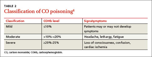
Oxygen therapy is the recommended treatment
Early treatment with supplemental oxygen is recommended to reduce the length of time red blood cells are exposed to CO.1 A COHb level >25% is the criterion for hyperbaric oxygen therapy.1,3 Patients should receive treatment until their symptoms become less intense.
Delayed neuropsychiatric sequelae (DNS) can occur in up to one-third of patients with acute CO poisoning more than a month after apparent recovery.1,6,9 DNS symptoms include cognitive changes, emotional lability, visual disturbances, disorientation, depression, dementia, psychotic behavior, parkinsonism, amnesia, and incontinence.1,6,9 Approximately 50% to 75% of patients with DNS recover spontaneously within a year with symptomatic treatment.1,6,9
Our patient
After recommending that our patient (and her mother) leave the apartment and have it inspected, we later learned that the fire department was unable to determine the source of the CO. A CO detector was installed and our patient was advised to keep the windows in the apartment open to allow for adequate oxygen flow. One month later she returned to our clinic and reported that her symptoms resolved; serum COHb was negative upon repeat lab tests.
THE TAKEAWAY
Patients who present with headaches, dizziness and/or fatigue should be evaluated for CO poisoning. The patient’s environmental history should be reviewed carefully, especially because CO poisoning is more common during the winter months. Oxygen therapy is the mainstay of treatment. Up to one-third of patients with acute poisoning may develop delayed neuropsychiatric sequelae, including cognitive changes, emotional lability, visual disturbances, disorientation, and depression, that may resolve within one year.
1. Nikkanen H, Skolnik A. Diagnosis and management of carbon monoxide poisoning in the emergency department. Emerg Med Pract. 2011;13:1-14.
2. Hampson NB, Hauff NM. Carboxyhemoglobin levels in carbon monoxide poisoning: do they correlate with the clinical picture? Am J Emerg Med. 2008;26:665-669.
3. Kao LW, Nañagas KA. Toxicity associated with carbon monoxide. Clin Lab Med. 2006;26:99-125.
4. Varon J, Marik PE, Fromm RE Jr, et al. Carbon monoxide poisoning: a review for clinicians. J Emerg Med. 1999;17:87-93.
5. Harper A, Croft-Baker J. Carbon monoxide poisoning: undetected by both patients and their doctors. Age Ageing. 2004;33:105-109.
6. Smollin C, Olson K. Carbon monoxide poisoning (acute). BMJ Clin Evid. 2010;2010. pii:2103.
7. Wright J. Chronic and occult carbon monoxide poisoning: we don’t know what we’re missing. Emerg Med J. 2002;19:366-390.
8. Weaver LK. Clinical practice. Carbon monoxide poisoning. N Engl J Med. 2009;360:1217-1225.
9. Bhatia R, Chacko F, Lal V, et al. Reversible delayed neuropsychiatric syndrome following acute carbon monoxide exposure. Indian J Occup Environ Med. 2007;11:80-82.
THE CASE
One month after moving into her mother’s apartment, a 27-year-old woman sought care at our clinic for fatigue, headache, blurred vision, nausea, and morning vomiting. She had weakness and difficulty sleeping, but denied any fever, rashes, neck stiffness, recent travel, trauma, or tobacco or illicit drug use. She did, however, have a 6-year history of migraines. Her physical exam was normal. She was sent home with a prescription for tramadol 50 mg bid for her headaches.
The patient subsequently went to the emergency department 3 times for the same complaints; none of the treatments she received there (mostly acetaminophen with codeine) relieved her symptoms. Three weeks later she returned to our clinic. She was distressed that the symptoms hadn’t gone away, and noted that her family was now experiencing similar symptoms.
Her temperature was 98.1°F (36.7°C), blood pressure was 131/88 mm Hg, pulse was 85 beats/min, and respiratory rate was 18 breaths/min. Physical and neurologic exams were normal.
THE DIAGNOSIS
Although most of the patient’s lab test results were within normal ranges, her carboxyhemoglobin (COHb) level was 4.2%. COHb levels of >2% to 3% in nonsmokers or >9% to 10% in smokers suggest carbon monoxide (CO) poisoning.1,2 Based on this finding and our patient’s symptoms, we diagnosed unintentional CO poisoning. We recommended that she and her mother vacate the apartment and have it inspected.
DISCUSSION
CO is the leading cause of poisoning mortality in the United States, and causes half of all fatal poisonings worldwide.1,3,4 It is a colorless, odorless, and tasteless gas that is produced by the incomplete combustion of carbon-based products, such as coal or gas.5,6 Exposure can occur from car exhaust fumes, faulty room heaters, and other sources (TABLE 1).6 The incidence of CO poisoning is higher during the winter months and after natural disasters. Individuals who have a lowered oxygen capacity, such as older adults, pregnant women (and their fetuses), infants, and patients with anemia, cardiovascular disease, or cerebrovascular disease, are more susceptible to CO poisoning.5,6
COHb, a stable complex of CO that forms in red blood cells when CO is inhaled, impairs oxygen delivery and peripheral utilization, resulting in cellular hypoxia.1 Signs and symptoms of CO poisoning are nonspecific and require a high degree of clinical suspicion for early diagnosis and treatment. Although cherry-red lips, peripheral cyanosis, and retinal hemorrhages are often described as “classic” symptoms of CO poisoning, these are rarely seen.6 The most common symptoms are actually headache (90%), dizziness (82%), and weakness (53%).7 Other symptoms include nausea, vomiting, confusion, visual disturbances, loss of consciousness, angina, seizure, and fatigue.6,7 Symptoms of chronic CO poisoning may differ from those of acute poisoning and can include chronic fatigue, neuropathy, and memory deficit.8
The differential diagnosis for CO poisoning includes flu-like syndrome/influenza/other viral illnesses, migraine or tension headaches, depression, transient ischemic attack, encephalitis, coronary artery disease, gastroenteritis or food poisoning, seizures, and dysrhythmias.1,4 Lab testing for COHb can help narrow the diagnosis. CO poisoning can be classified as mild, moderate, or severe based on COHb levels and the patient’s signs and symptoms (TABLE 2).6 However, COHb level is a poor predictor of clinical presentation and should not be used to dictate management.2,7
Oxygen therapy is the recommended treatment
Early treatment with supplemental oxygen is recommended to reduce the length of time red blood cells are exposed to CO.1 A COHb level >25% is the criterion for hyperbaric oxygen therapy.1,3 Patients should receive treatment until their symptoms become less intense.
Delayed neuropsychiatric sequelae (DNS) can occur in up to one-third of patients with acute CO poisoning more than a month after apparent recovery.1,6,9 DNS symptoms include cognitive changes, emotional lability, visual disturbances, disorientation, depression, dementia, psychotic behavior, parkinsonism, amnesia, and incontinence.1,6,9 Approximately 50% to 75% of patients with DNS recover spontaneously within a year with symptomatic treatment.1,6,9
Our patient
After recommending that our patient (and her mother) leave the apartment and have it inspected, we later learned that the fire department was unable to determine the source of the CO. A CO detector was installed and our patient was advised to keep the windows in the apartment open to allow for adequate oxygen flow. One month later she returned to our clinic and reported that her symptoms resolved; serum COHb was negative upon repeat lab tests.
THE TAKEAWAY
Patients who present with headaches, dizziness and/or fatigue should be evaluated for CO poisoning. The patient’s environmental history should be reviewed carefully, especially because CO poisoning is more common during the winter months. Oxygen therapy is the mainstay of treatment. Up to one-third of patients with acute poisoning may develop delayed neuropsychiatric sequelae, including cognitive changes, emotional lability, visual disturbances, disorientation, and depression, that may resolve within one year.
1. Nikkanen H, Skolnik A. Diagnosis and management of carbon monoxide poisoning in the emergency department. Emerg Med Pract. 2011;13:1-14.
2. Hampson NB, Hauff NM. Carboxyhemoglobin levels in carbon monoxide poisoning: do they correlate with the clinical picture? Am J Emerg Med. 2008;26:665-669.
3. Kao LW, Nañagas KA. Toxicity associated with carbon monoxide. Clin Lab Med. 2006;26:99-125.
4. Varon J, Marik PE, Fromm RE Jr, et al. Carbon monoxide poisoning: a review for clinicians. J Emerg Med. 1999;17:87-93.
5. Harper A, Croft-Baker J. Carbon monoxide poisoning: undetected by both patients and their doctors. Age Ageing. 2004;33:105-109.
6. Smollin C, Olson K. Carbon monoxide poisoning (acute). BMJ Clin Evid. 2010;2010. pii:2103.
7. Wright J. Chronic and occult carbon monoxide poisoning: we don’t know what we’re missing. Emerg Med J. 2002;19:366-390.
8. Weaver LK. Clinical practice. Carbon monoxide poisoning. N Engl J Med. 2009;360:1217-1225.
9. Bhatia R, Chacko F, Lal V, et al. Reversible delayed neuropsychiatric syndrome following acute carbon monoxide exposure. Indian J Occup Environ Med. 2007;11:80-82.

CoreValve receives first TAVR valve-in-valve indication
The U.S. Food and Drug Administration on March 30 expanded its approved use of the CoreValve transcatheter aortic-valve replacement (TAVR) system to include patients who already have undergone aortic valve replacement and need a second valve replacement done as a valve-in-valve placement.
With this action, CoreValve became the first TAVR system to receive U.S. approval for valve-in-valve use. The CoreValve System received FDA approval for TAVR performed on native aortic valves in January 2014 in patients at “extreme risk,” and in June 2014 for those at “high risk,” for surgical aortic valve replacement.* Valve-in-valve TAVR is only feasible in patients with a failing bioprosthetic aortic valve: It is not an option for patients with a failing mechanical aortic valve.

“The CoreValve System offers a less-invasive treatment option for a significant number of patients with failed tissue aortic valves whose medical teams determine that the risks associated with repeat open-heart surgery are high or extremely high,” Dr. William H. Maisel, deputy center director for science and chief scientist in the FDA’s Center for Devices and Radiological Health, said in a written statement. “The approval is an important expansion of the authorized use of the transcatheter aortic valve replacement technology.”
The CoreValve, which is designed to sit in a supra-annular location 12 mm above the aortic valve annulus, is well suited for valve-in-valve replacement because the only portion of the CoreValve that actually fills the annular space and the ring of the existing valve is the CoreValve’s sealer. This results in a tight seal that produces less paravalvular leak than when the sealer sits in a native annulus that is often deformed with calcium, noted Dr. Michael J. Reardon, professor of cardiothoracic surgery at Methodist Hospital in Houston.
In addition, because the sealer exerts pressure on the old valve ring in the annulus instead of on myocardium, placing the CoreValve as a valve-in-valve produces much less conduction disruption and results in fewer patients who need a pacemaker following TAVR, he said.

The CoreValve as a valve-in-valve “works quite well, and is not hard to position,” said Dr. Reardon, who added that he has now performed several valve-in-valve TAVRs using the CoreValve.
Similar TAVR procedures are usually not possible using the balloon-expandable SAPIEN System because the SAPIEN valve is designed to sit directly in the annulus and, in most patients, the existing valve ring does not provide enough space to accommodate a SAPIEN valve.
Dr. Reardon anticipates that many U.S. patients now in their 80s with a failing bioprosthetic aortic valve will be interested in nonsurgical TAVR replacement. These patients often do not want conventional open-heart surgery, he said in an interview.
To evaluate the safety and efficacy of the CoreValve System for aortic valve-in-valve replacement, the FDA reviewed clinical data collected from a U.S. clinical trial with 143 patients, an agency representative said in the statement. In the clinical trial, the estimated rate of 30-day survival without major stroke was 96%, and 89% after 6 months. “This compares well to the corresponding rates reported previously for trial participants who received the same device to replace their own, native diseased or damaged aortic valve,” the agency’s statement said.
According to the agency, aortic valve-in-valve use of the CoreValve System should be limited to patients who need replacement of a failed tissue aortic valve but are at extreme or high risk of death or serious complications from traditional open-heart surgery. A decision as to whether the product and procedure are appropriate for a patient “should involve careful evaluation by the patient’s heart medical team, including a cardiologist and a cardiac surgeon.”
The FDA said that the CoreValve System should not be used in patients who have any infection, have a mechanical aortic heart valve, cannot tolerate anticoagulant drugs, or have sensitivity to titanium, nickel, or contrast media.
Dr. Maisel had no disclosures. Dr. Reardon has served as an advisor to Medtronic, the company that markets the CoreValve.
On Twitter @mitchelzoler
*Correction, 4/1/2015: An earlier version of this article misstated the device’s approval history.
The U.S. Food and Drug Administration on March 30 expanded its approved use of the CoreValve transcatheter aortic-valve replacement (TAVR) system to include patients who already have undergone aortic valve replacement and need a second valve replacement done as a valve-in-valve placement.
With this action, CoreValve became the first TAVR system to receive U.S. approval for valve-in-valve use. The CoreValve System received FDA approval for TAVR performed on native aortic valves in January 2014 in patients at “extreme risk,” and in June 2014 for those at “high risk,” for surgical aortic valve replacement.* Valve-in-valve TAVR is only feasible in patients with a failing bioprosthetic aortic valve: It is not an option for patients with a failing mechanical aortic valve.
“The CoreValve System offers a less-invasive treatment option for a significant number of patients with failed tissue aortic valves whose medical teams determine that the risks associated with repeat open-heart surgery are high or extremely high,” Dr. William H. Maisel, deputy center director for science and chief scientist in the FDA’s Center for Devices and Radiological Health, said in a written statement. “The approval is an important expansion of the authorized use of the transcatheter aortic valve replacement technology.”
The CoreValve, which is designed to sit in a supra-annular location 12 mm above the aortic valve annulus, is well suited for valve-in-valve replacement because the only portion of the CoreValve that actually fills the annular space and the ring of the existing valve is the CoreValve’s sealer. This results in a tight seal that produces less paravalvular leak than when the sealer sits in a native annulus that is often deformed with calcium, noted Dr. Michael J. Reardon, professor of cardiothoracic surgery at Methodist Hospital in Houston.
In addition, because the sealer exerts pressure on the old valve ring in the annulus instead of on myocardium, placing the CoreValve as a valve-in-valve produces much less conduction disruption and results in fewer patients who need a pacemaker following TAVR, he said.
The CoreValve as a valve-in-valve “works quite well, and is not hard to position,” said Dr. Reardon, who added that he has now performed several valve-in-valve TAVRs using the CoreValve.
Similar TAVR procedures are usually not possible using the balloon-expandable SAPIEN System because the SAPIEN valve is designed to sit directly in the annulus and, in most patients, the existing valve ring does not provide enough space to accommodate a SAPIEN valve.
Dr. Reardon anticipates that many U.S. patients now in their 80s with a failing bioprosthetic aortic valve will be interested in nonsurgical TAVR replacement. These patients often do not want conventional open-heart surgery, he said in an interview.
To evaluate the safety and efficacy of the CoreValve System for aortic valve-in-valve replacement, the FDA reviewed clinical data collected from a U.S. clinical trial with 143 patients, an agency representative said in the statement. In the clinical trial, the estimated rate of 30-day survival without major stroke was 96%, and 89% after 6 months. “This compares well to the corresponding rates reported previously for trial participants who received the same device to replace their own, native diseased or damaged aortic valve,” the agency’s statement said.
According to the agency, aortic valve-in-valve use of the CoreValve System should be limited to patients who need replacement of a failed tissue aortic valve but are at extreme or high risk of death or serious complications from traditional open-heart surgery. A decision as to whether the product and procedure are appropriate for a patient “should involve careful evaluation by the patient’s heart medical team, including a cardiologist and a cardiac surgeon.”
The FDA said that the CoreValve System should not be used in patients who have any infection, have a mechanical aortic heart valve, cannot tolerate anticoagulant drugs, or have sensitivity to titanium, nickel, or contrast media.
Dr. Maisel had no disclosures. Dr. Reardon has served as an advisor to Medtronic, the company that markets the CoreValve.
On Twitter @mitchelzoler
*Correction, 4/1/2015: An earlier version of this article misstated the device’s approval history.

AUDIO: Psychiatry’s Darwinian moment
LAS VEGAS – A more-specific nomenclature of depression would help in the prescribing of better treatments or might prevent it altogether, according to Dr. Vladimir Maletic, a psychiatrist who spoke at this year’s Nevada Psychiatric Association’s annual Psychopharmacology update meeting.
But to do so requires psychiatry to find its own Darwinian moment, where the etiology of depression is made clear. In the conversation with Dr. Maletic, recorded at the meeting, hear his thoughts on the differences between existential and biological depression, how allowing patients to choose their treatment can double their response rate, and the lasting, negative effects on cognition post depression.
On Twitter @whitneymcknight
EXPERT ANALYSIS FROM THE NPA ANNUAL PSYCHOPHARMACOLOGY UPDATE
Pheochromocytoma
To the Editor: I read with avid interest the IM Board Review by Pagán et al on a man with pheochromocytoma.1 The article stated that the classic triad is headache, hypertension, and hyperglycemia. I found this to be incorrect. And Harrison’s Principles of Internal Medicine2 states that the classic triad is palpitations, headaches, and profuse sweating. I hope you will clarify this in the interest of the readers as it is a board review article.
- Pagán RJ, Kurklinsky AK, Chirila R. A 61-year-old man with fluctuating hypertension. Cleve Clin J Med 2014; 81:677–682.
- Neumann HH. Chapter 343. Pheochromocytoma. In: Longo DL, Fauci AS, Kasper DL, Hauser SL, Jameson J, Loscalzo J, editors. Harrison’s Principles of Internal Medicine, 18th edition. New York, NY: McGraw-Hill, 2012. http://0-accessmedicine.mhmedical.com.library.ccf.org/content.aspx?bookid=331&Sectionid=40727148. Accessed March 12, 2015.
To the Editor: I read with avid interest the IM Board Review by Pagán et al on a man with pheochromocytoma.1 The article stated that the classic triad is headache, hypertension, and hyperglycemia. I found this to be incorrect. And Harrison’s Principles of Internal Medicine2 states that the classic triad is palpitations, headaches, and profuse sweating. I hope you will clarify this in the interest of the readers as it is a board review article.
To the Editor: I read with avid interest the IM Board Review by Pagán et al on a man with pheochromocytoma.1 The article stated that the classic triad is headache, hypertension, and hyperglycemia. I found this to be incorrect. And Harrison’s Principles of Internal Medicine2 states that the classic triad is palpitations, headaches, and profuse sweating. I hope you will clarify this in the interest of the readers as it is a board review article.
- Pagán RJ, Kurklinsky AK, Chirila R. A 61-year-old man with fluctuating hypertension. Cleve Clin J Med 2014; 81:677–682.
- Neumann HH. Chapter 343. Pheochromocytoma. In: Longo DL, Fauci AS, Kasper DL, Hauser SL, Jameson J, Loscalzo J, editors. Harrison’s Principles of Internal Medicine, 18th edition. New York, NY: McGraw-Hill, 2012. http://0-accessmedicine.mhmedical.com.library.ccf.org/content.aspx?bookid=331&Sectionid=40727148. Accessed March 12, 2015.