User login
New warnings on stimulants for ADHD: Cause for alarm?
In August the FDA called for new warnings on stimulants used for attention-deficit/hyperactivity disorder (ADHD). Amphetamines now carry black box warnings that say, “Misuse of amphetamines may cause sudden death and serious cardiovascular adverse events.” Amphetamines and methylphenidates used for ADHD include expanded information about cardiovascular risks at usual dosages for patients with heart conditions.
To examine the clinical implications of these warnings, Current Psychiatry hosted a conversation between ADHD experts Anthony Rostain, MD, MA, and Lenard Adler, MD.
Dr. Rostain: Changes to warnings on ADHD medications have many psychiatrists looking for guidance on using stimulants. Can you give us some background and discuss the labeling changes?
Dr. Adler: Stimulants have been used for more than 40 years as ADHD treatments, and they’ve been shown to be highly effective. The FDA, which monitors issues of cardiovascular safety and stimulants in an ongoing way, examined specific isolated cases and changed some of the warnings as a result.
Dr. Rostain: What should practicing psychiatrists be concerned about if they’re thinking of prescribing stimulants for an ADHD patient?
Dr. Adler: The take-home point is that stimulants—because of the way they work—have been known to have minor effects of increasing blood pressure and pulse (Box 1).1-3 Clinicians have known about issues regarding stimulant use by patients with pre-existing cardiovascular conditions, but now the warnings are more formal for the methylphenidate and amphetamine products.
Dr. Rostain: An FDA committee recommended black box warnings on all stimulants used for ADHD, but the FDA decided instead to clarify warnings in prescription information for some medications. What was the FDA process?
Dr. Adler: The discussion was internal at the FDA, so I can’t say what their thinking was. The black box warning on amphetamines notes two issues. One is the potential for abuse and diversion, and the other warns of potential for sudden death and serious cardiovascular effects if the drug is misused. A warning has also been placed on all methylphenidate products regarding cardiovascular risk for patients with pre-existing cardiovascular conditions, but it is not a black box warning.
in healthy children and adults
Researchers at Massachusetts General Hospital have examined the effects of ADHD medications on blood pressure and heart rate in children and adults.
Children and adolescents. The first study1 was a 1-year extension of an open-label trial of once-daily, osmotic-release methylphenidate (MPH) in 432 children (age 6 to 13) with ADHD. Their blood pressure and heart rate were recorded at baseline and monthly.
At 12 months, MPH use at 18 to 54 mg/d was associated with minor but statistically significant mean increases in:
- systolic blood pressure (3.3 mm Hg [P<0.001])
- diastolic blood pressure (1.5 mm Hg [P<0.001])
- heart rate (3.9 bpm [P<0.0001]).
Adults. In a 24-month study,2 223 healthy adults with ADHD (age≥18) received mixed amphetamine salts extended-release (MAS XR) in an open-label extension of a 4-week, double-blind, placebo-controlled trial. MAS XR was started at 20 mg/d for 1 week, then increased up to 60 mg/d based on therapeutic effect, as measured by the ADHD Rating Scale IV.
Blood pressure and pulse were measured at baseline, weekly, then monthly, and 12-lead ECGs were obtained at baseline, weekly, then at 3- and 6-month intervals. Changes after 2 years were small and not statistically significant:
- systolic blood pressure (2.3±12.5 mm Hg)
- diastolic blood pressure (1.3±9.2 mm Hg)
- pulse (2.1±13.4 bpm).
A clinically insignificant increase was observed in the mean QTcB interval (7.2 msec; P<0.001), although no patient’s QTcB interval exceeded 480 msec. Seven patients dropped out because of cardiovascular side effects (5 with hypertension, and 2 with palpitation/tachycardia), which were not reported as being serious.
Stimulants and nonstimulants. In another study,3 the same researchers analyzed the cardiovascular effects of three stimulants (methylphenidate, amphetamine compounds, and pemoline) and two nonstimulants (bupropion and desipramine) used to treat ADHD in adults. Data on a total of 125 patients (mean age 39±9 years) from three previous placebo-controlled studies were re-examined for the medications’ effects on blood pressure and heart rate.
Minor but statistically significant changes in blood pressure and heart rate were found to be associated with both stimulant and nonstimulant medications:
- systolic blood pressure (bupropion, +5.9 mm Hg [P<0.05]; amphetamine, +5.4 mm Hg [P<0.05])
- diastolic blood pressure (desipramine, +7.1 mm Hg [P<0.05])
- heart rate (bupropion, +6.9 bpm [P<0.05]; amphetamine, +7.3 bpm [P<0.05]; methylphenidate, +4.5 bpm [P<0.05]).
In the last two studies, the authors concluded that although the cardiovascular effects of ADHD medications in healthy adults were minimal, clinicians should monitor vital signs at baseline and periodically during treatment.
Dr. Rostain: How were the warnings clarified?
Dr. Adler: The FDA has changed the language. Now physicians are warned that sudden death can occur at usual doses in patients with a pre-existing structural cardiac abnormality or other serious heart problem. So, stimulants generally should not be used in children or adolescents with known serious structural cardiac abnormalities, cardiomyopathy, serious heart rhythm abnormalities, or other serious cardiac problems that may place them at increased vulnerability to the sympathomimetic effects of a stimulant drug.
Dr. Rostain: What about adults?
Dr. Adler: The language is the same for adults. Adults have a greater likelihood than children of having a history of serious structural cardiac abnormalities, cardiomyopathy, serious heart rhythm abnormalities, coronary artery disease, or other cardiac problems. Adults with such abnormalities generally should not be treated with stimulant drugs.
Dr. Rostain: What’s the impact for clinicians?
Dr. Adler: Clinicians have known that stimulants should not be used in patients with significant pre-existing cardiovascular conditions. That generally includes structural abnormalities such as serious heart murmurs and abnormalities of the electro-conduction of the impulse through the heart. When patients present with a history of cardiac abnormalities, clinicians should speak to the pediatrician, primary care physician (PCP), or cardiologist, go over the risk factors, and decide whether these medications can be prescribed for the patient.
Dr. Rostain: Should psychiatrists perform screening tests before prescribing stimulants? When should they consult with a specialist?
Dr. Adler: There is no recommendation in the prescribing information. But clearly a clinician should determine whether a patient has structural cardiac abnormalities or serious heart problems. That means taking a history about heart murmur, syncope, or other serious heart problems. Also, you want to know if the patient is hypertensive. The burden is on the prescribing clinician.
Dr. Rostain: Suppose you have a patient with hypertension or a history of a heart condition, should that patient first be evaluated by a cardiologist? What about a screening ECG?
Dr. Adler: There are no specific recommendations. If clinicians have questions about prescribing the medication, they should consult with the patient’s PCP or cardiologist.
Dr. Rostain: Let’s say the patient has some heart issues, but the PCP or pediatrician gives the goahead to prescribe stimulants. What sort of monitoring do you recommend?
Dr. Adler: I can’t answer that directly. Clearly, you’re going to want to partner with the PCP to establish a plan of how to carefully monitor this patient. FDA guidelines recommend ongoing blood pressure monitoring, especially if the patient is hypertensive, but do not specify how often.
Dr. Rostain: What alternatives do psychiatrists have when treating ADHD in patients in whom stimulants may pose some risk?
Dr. Adler: The only approved nonstimulant ADHD medication is atomoxetine, the labeling of which carries language about possible effects on blood pressure. The FDA warning about structural cardiac abnormalities has not been extended to atomoxetine, but blood pressure needs to be monitored. Whether our medical colleagues feel comfortable using a nonstimulant in patients with structural cardiac abnormalities has not been determined.
Dr. Rostain: In the absence of guidelines in the new warnings on stimulants, are there any studies to help clinicians with treatment and monitoring?
Dr. Adler: There’s very little data. A group at Massachusetts General Hospital has been studying the effects of ADHD medication on adults with hypertension (Box 2).4 That’s a different issue than a structural cardiac abnormality, but at least we have some data. This group found that you can safely give stimulants to hypertensive patients by partnering with medical colleagues and monitoring the patient carefully. Antihypertensive dosages may need to be adjusted during psychostimulant treatment.
Dr. Rostain: How do you choose a medication if your patient has a structural heart abnormality?
Dr. Adler: Again, we don’t have a lot of data. The decision would depend on the cardiac abnormality and the consulting physician’s comfort level. Keep in mind that psychostimulants have a short duration of effect, so the effects of the medication can dissipate fairly quickly. Again, the decision to medicate a patient with pre-existing cardiac abnormalities must be done with medical guidance.
In a short-term, open-label trial by Wilens et al,4 13 adults with ADHD and hypertension received mixed amphetamine salts extended-release (MAS-XR), up to 60 mg/d, for 6 weeks (phase 1), then discontinued MAS-XR for 2 weeks (phase 2). All patients had normal blood pressure (<135/85 mm Hg) for at least 4 weeks before entering the study and received a comprehensive clinical assessment, including ECG. Blood pressure was measured manually at each clinic visit.
Single episodes of hypertension (>140/90 mm Hg) occurred at similar rates in each phase, but these episodes were not sustained at any two consecutive visits. Group mean systolic and diastolic blood pressures and pulse did not increase during stimulant treatment. No clinically significant ECG changes were observed, and no serious adverse events occurred.
The authors concluded that this preliminary trial suggests that adults with ADHD and controlled hypertension can be safely treated with stimulant medications.
Dr. Rostain: So are you saying clinicians should make decisions about prescribing stimulants for patients with ADHD on a case-by-case basis?
Dr. Adler: Exactly.
Dr. Rostain: What about children and adolescents who have unknown structural heart defects? A lot of parents are concerned about reports of sudden cardiac death in young athletes, such as when playing soccer or basketball. Is there any way for practitioners to protect children with ADHD from an unexpected event?
Dr. Adler: In general, stimulants are safe medications, but we don’t have guidelines to help us determine who will need an ECG and who will not. Children are less likely to have had an ECG in the past than an adult, so it’s important to do a history, obtain input from the pediatrician or PCP, and clearly review the risks and benefits of medication therapy with the patient’s family.
Dr. Rostain: What would you advise clinicians to tell parents of children with ADHD or adult patients who have concerns about the new labeling on stimulants?
Dr. Adler: It would be a shame if patients were not receiving treatment for ADHD because of unfounded medical concerns. When these medications are used appropriately, they have dramatic and positive affects on ADHD.
ADHD is common and highly impairing. Deciding not to treat it has serious consequences in terms of divorce, separation, underperformance in school and on the job, unemployment, smoking, substance use, and issues with motor vehicle accidents and driving.
The goal of treatment is for our patients to get better, and ADHD is highly treatable with medication. But we must be cognizant of the warnings and prescribe medications appropriately. The message is that we’ve got to work collaboratively with our partners in medicine and, in the absence of guidelines, use good common sense.
Related resources
- Wilens TE, Hammerness PG, Biederman J, et al. Blood pressure changes associated with medication treatment of adults with attention-deficit/hyperactivity disorder. J Clin Psychiatry 2005;66:253-9.
Drug brand names
- Atomoxetine • Strattera
- Bupropion • Wellbutrin
- Desipramine • Norpramin
- Methylphenidate • Concerta, Ritalin
- Mixed amphetamine salts • Adderall
- Pemoline • Cylert
Disclosures
Dr. Adler is a consultant to and receives grant/research support from Abbott Laboratories, Cephalon, Cortex Pharmaceuticals, Eli Lilly and Company, New River Pharmaceuticals, Novartis Pharmaceuticals Corp., Ortho-McNeil, Pfizer, and Shire. He also receives grant/research support from Bristol-Myers Squibb and Merck and Co., and is a speaker for Eli Lilly and Company.
Dr. Rostain is a consultant to Shire and a speaker for Eli Lilly and Company and Ortho-McNeil.
1. Wilens TE, Biederman J, Lerner M. Concerta Study Group. Effects of once-daily osmotic-release methylphenidate on blood pressure and heart rate in children with attention-deficit/hyperactivity disorder: results from a one-year follow-up study. J Clin Psychopharmacol 2004;24(1):36-41.
2. Biederman J, Spencer TJ, Wilens TE, et al. Long-term safety and effectiveness of mixed amphetamine salts extended release in adults with ADHD. CNS Spectr 2005;10(12 suppl 20):16-25.
3. Wilens TE, Hammerness PG, Biederman J, et al. Blood pressure changes associated with medication treatment of adults with attention-deficit/hyperactivity disorder. J Clin Psychiatry 2005;66(2):253-9.
4. Wilens TE, Zusman RM, Hammerness PG, et al. An open-label study of the tolerability of mixed amphetamine salts in adults with attention-deficit/ hyperactivity disorder and treated primary essential hypertension. J Clin Psychiatry 2006;67(5):696-702.
Dr. Adler is associate professor of psychiatry and director of the adult ADHD program at New York University Medical Center. He recently published a book for patients, Scattered Minds: Hope and Help for Adults with ADHD.
Dr. Rostain is associate professor of psychiatry and pediatrics and director of education, department of psychiatry, University of Pennsylvania School of Medicine, Philadelphia.
In August the FDA called for new warnings on stimulants used for attention-deficit/hyperactivity disorder (ADHD). Amphetamines now carry black box warnings that say, “Misuse of amphetamines may cause sudden death and serious cardiovascular adverse events.” Amphetamines and methylphenidates used for ADHD include expanded information about cardiovascular risks at usual dosages for patients with heart conditions.
To examine the clinical implications of these warnings, Current Psychiatry hosted a conversation between ADHD experts Anthony Rostain, MD, MA, and Lenard Adler, MD.
Dr. Rostain: Changes to warnings on ADHD medications have many psychiatrists looking for guidance on using stimulants. Can you give us some background and discuss the labeling changes?
Dr. Adler: Stimulants have been used for more than 40 years as ADHD treatments, and they’ve been shown to be highly effective. The FDA, which monitors issues of cardiovascular safety and stimulants in an ongoing way, examined specific isolated cases and changed some of the warnings as a result.
Dr. Rostain: What should practicing psychiatrists be concerned about if they’re thinking of prescribing stimulants for an ADHD patient?
Dr. Adler: The take-home point is that stimulants—because of the way they work—have been known to have minor effects of increasing blood pressure and pulse (Box 1).1-3 Clinicians have known about issues regarding stimulant use by patients with pre-existing cardiovascular conditions, but now the warnings are more formal for the methylphenidate and amphetamine products.
Dr. Rostain: An FDA committee recommended black box warnings on all stimulants used for ADHD, but the FDA decided instead to clarify warnings in prescription information for some medications. What was the FDA process?
Dr. Adler: The discussion was internal at the FDA, so I can’t say what their thinking was. The black box warning on amphetamines notes two issues. One is the potential for abuse and diversion, and the other warns of potential for sudden death and serious cardiovascular effects if the drug is misused. A warning has also been placed on all methylphenidate products regarding cardiovascular risk for patients with pre-existing cardiovascular conditions, but it is not a black box warning.
in healthy children and adults
Researchers at Massachusetts General Hospital have examined the effects of ADHD medications on blood pressure and heart rate in children and adults.
Children and adolescents. The first study1 was a 1-year extension of an open-label trial of once-daily, osmotic-release methylphenidate (MPH) in 432 children (age 6 to 13) with ADHD. Their blood pressure and heart rate were recorded at baseline and monthly.
At 12 months, MPH use at 18 to 54 mg/d was associated with minor but statistically significant mean increases in:
- systolic blood pressure (3.3 mm Hg [P<0.001])
- diastolic blood pressure (1.5 mm Hg [P<0.001])
- heart rate (3.9 bpm [P<0.0001]).
Adults. In a 24-month study,2 223 healthy adults with ADHD (age≥18) received mixed amphetamine salts extended-release (MAS XR) in an open-label extension of a 4-week, double-blind, placebo-controlled trial. MAS XR was started at 20 mg/d for 1 week, then increased up to 60 mg/d based on therapeutic effect, as measured by the ADHD Rating Scale IV.
Blood pressure and pulse were measured at baseline, weekly, then monthly, and 12-lead ECGs were obtained at baseline, weekly, then at 3- and 6-month intervals. Changes after 2 years were small and not statistically significant:
- systolic blood pressure (2.3±12.5 mm Hg)
- diastolic blood pressure (1.3±9.2 mm Hg)
- pulse (2.1±13.4 bpm).
A clinically insignificant increase was observed in the mean QTcB interval (7.2 msec; P<0.001), although no patient’s QTcB interval exceeded 480 msec. Seven patients dropped out because of cardiovascular side effects (5 with hypertension, and 2 with palpitation/tachycardia), which were not reported as being serious.
Stimulants and nonstimulants. In another study,3 the same researchers analyzed the cardiovascular effects of three stimulants (methylphenidate, amphetamine compounds, and pemoline) and two nonstimulants (bupropion and desipramine) used to treat ADHD in adults. Data on a total of 125 patients (mean age 39±9 years) from three previous placebo-controlled studies were re-examined for the medications’ effects on blood pressure and heart rate.
Minor but statistically significant changes in blood pressure and heart rate were found to be associated with both stimulant and nonstimulant medications:
- systolic blood pressure (bupropion, +5.9 mm Hg [P<0.05]; amphetamine, +5.4 mm Hg [P<0.05])
- diastolic blood pressure (desipramine, +7.1 mm Hg [P<0.05])
- heart rate (bupropion, +6.9 bpm [P<0.05]; amphetamine, +7.3 bpm [P<0.05]; methylphenidate, +4.5 bpm [P<0.05]).
In the last two studies, the authors concluded that although the cardiovascular effects of ADHD medications in healthy adults were minimal, clinicians should monitor vital signs at baseline and periodically during treatment.
Dr. Rostain: How were the warnings clarified?
Dr. Adler: The FDA has changed the language. Now physicians are warned that sudden death can occur at usual doses in patients with a pre-existing structural cardiac abnormality or other serious heart problem. So, stimulants generally should not be used in children or adolescents with known serious structural cardiac abnormalities, cardiomyopathy, serious heart rhythm abnormalities, or other serious cardiac problems that may place them at increased vulnerability to the sympathomimetic effects of a stimulant drug.
Dr. Rostain: What about adults?
Dr. Adler: The language is the same for adults. Adults have a greater likelihood than children of having a history of serious structural cardiac abnormalities, cardiomyopathy, serious heart rhythm abnormalities, coronary artery disease, or other cardiac problems. Adults with such abnormalities generally should not be treated with stimulant drugs.
Dr. Rostain: What’s the impact for clinicians?
Dr. Adler: Clinicians have known that stimulants should not be used in patients with significant pre-existing cardiovascular conditions. That generally includes structural abnormalities such as serious heart murmurs and abnormalities of the electro-conduction of the impulse through the heart. When patients present with a history of cardiac abnormalities, clinicians should speak to the pediatrician, primary care physician (PCP), or cardiologist, go over the risk factors, and decide whether these medications can be prescribed for the patient.
Dr. Rostain: Should psychiatrists perform screening tests before prescribing stimulants? When should they consult with a specialist?
Dr. Adler: There is no recommendation in the prescribing information. But clearly a clinician should determine whether a patient has structural cardiac abnormalities or serious heart problems. That means taking a history about heart murmur, syncope, or other serious heart problems. Also, you want to know if the patient is hypertensive. The burden is on the prescribing clinician.
Dr. Rostain: Suppose you have a patient with hypertension or a history of a heart condition, should that patient first be evaluated by a cardiologist? What about a screening ECG?
Dr. Adler: There are no specific recommendations. If clinicians have questions about prescribing the medication, they should consult with the patient’s PCP or cardiologist.
Dr. Rostain: Let’s say the patient has some heart issues, but the PCP or pediatrician gives the goahead to prescribe stimulants. What sort of monitoring do you recommend?
Dr. Adler: I can’t answer that directly. Clearly, you’re going to want to partner with the PCP to establish a plan of how to carefully monitor this patient. FDA guidelines recommend ongoing blood pressure monitoring, especially if the patient is hypertensive, but do not specify how often.
Dr. Rostain: What alternatives do psychiatrists have when treating ADHD in patients in whom stimulants may pose some risk?
Dr. Adler: The only approved nonstimulant ADHD medication is atomoxetine, the labeling of which carries language about possible effects on blood pressure. The FDA warning about structural cardiac abnormalities has not been extended to atomoxetine, but blood pressure needs to be monitored. Whether our medical colleagues feel comfortable using a nonstimulant in patients with structural cardiac abnormalities has not been determined.
Dr. Rostain: In the absence of guidelines in the new warnings on stimulants, are there any studies to help clinicians with treatment and monitoring?
Dr. Adler: There’s very little data. A group at Massachusetts General Hospital has been studying the effects of ADHD medication on adults with hypertension (Box 2).4 That’s a different issue than a structural cardiac abnormality, but at least we have some data. This group found that you can safely give stimulants to hypertensive patients by partnering with medical colleagues and monitoring the patient carefully. Antihypertensive dosages may need to be adjusted during psychostimulant treatment.
Dr. Rostain: How do you choose a medication if your patient has a structural heart abnormality?
Dr. Adler: Again, we don’t have a lot of data. The decision would depend on the cardiac abnormality and the consulting physician’s comfort level. Keep in mind that psychostimulants have a short duration of effect, so the effects of the medication can dissipate fairly quickly. Again, the decision to medicate a patient with pre-existing cardiac abnormalities must be done with medical guidance.
In a short-term, open-label trial by Wilens et al,4 13 adults with ADHD and hypertension received mixed amphetamine salts extended-release (MAS-XR), up to 60 mg/d, for 6 weeks (phase 1), then discontinued MAS-XR for 2 weeks (phase 2). All patients had normal blood pressure (<135/85 mm Hg) for at least 4 weeks before entering the study and received a comprehensive clinical assessment, including ECG. Blood pressure was measured manually at each clinic visit.
Single episodes of hypertension (>140/90 mm Hg) occurred at similar rates in each phase, but these episodes were not sustained at any two consecutive visits. Group mean systolic and diastolic blood pressures and pulse did not increase during stimulant treatment. No clinically significant ECG changes were observed, and no serious adverse events occurred.
The authors concluded that this preliminary trial suggests that adults with ADHD and controlled hypertension can be safely treated with stimulant medications.
Dr. Rostain: So are you saying clinicians should make decisions about prescribing stimulants for patients with ADHD on a case-by-case basis?
Dr. Adler: Exactly.
Dr. Rostain: What about children and adolescents who have unknown structural heart defects? A lot of parents are concerned about reports of sudden cardiac death in young athletes, such as when playing soccer or basketball. Is there any way for practitioners to protect children with ADHD from an unexpected event?
Dr. Adler: In general, stimulants are safe medications, but we don’t have guidelines to help us determine who will need an ECG and who will not. Children are less likely to have had an ECG in the past than an adult, so it’s important to do a history, obtain input from the pediatrician or PCP, and clearly review the risks and benefits of medication therapy with the patient’s family.
Dr. Rostain: What would you advise clinicians to tell parents of children with ADHD or adult patients who have concerns about the new labeling on stimulants?
Dr. Adler: It would be a shame if patients were not receiving treatment for ADHD because of unfounded medical concerns. When these medications are used appropriately, they have dramatic and positive affects on ADHD.
ADHD is common and highly impairing. Deciding not to treat it has serious consequences in terms of divorce, separation, underperformance in school and on the job, unemployment, smoking, substance use, and issues with motor vehicle accidents and driving.
The goal of treatment is for our patients to get better, and ADHD is highly treatable with medication. But we must be cognizant of the warnings and prescribe medications appropriately. The message is that we’ve got to work collaboratively with our partners in medicine and, in the absence of guidelines, use good common sense.
Related resources
- Wilens TE, Hammerness PG, Biederman J, et al. Blood pressure changes associated with medication treatment of adults with attention-deficit/hyperactivity disorder. J Clin Psychiatry 2005;66:253-9.
Drug brand names
- Atomoxetine • Strattera
- Bupropion • Wellbutrin
- Desipramine • Norpramin
- Methylphenidate • Concerta, Ritalin
- Mixed amphetamine salts • Adderall
- Pemoline • Cylert
Disclosures
Dr. Adler is a consultant to and receives grant/research support from Abbott Laboratories, Cephalon, Cortex Pharmaceuticals, Eli Lilly and Company, New River Pharmaceuticals, Novartis Pharmaceuticals Corp., Ortho-McNeil, Pfizer, and Shire. He also receives grant/research support from Bristol-Myers Squibb and Merck and Co., and is a speaker for Eli Lilly and Company.
Dr. Rostain is a consultant to Shire and a speaker for Eli Lilly and Company and Ortho-McNeil.
1. Wilens TE, Biederman J, Lerner M. Concerta Study Group. Effects of once-daily osmotic-release methylphenidate on blood pressure and heart rate in children with attention-deficit/hyperactivity disorder: results from a one-year follow-up study. J Clin Psychopharmacol 2004;24(1):36-41.
2. Biederman J, Spencer TJ, Wilens TE, et al. Long-term safety and effectiveness of mixed amphetamine salts extended release in adults with ADHD. CNS Spectr 2005;10(12 suppl 20):16-25.
3. Wilens TE, Hammerness PG, Biederman J, et al. Blood pressure changes associated with medication treatment of adults with attention-deficit/hyperactivity disorder. J Clin Psychiatry 2005;66(2):253-9.
4. Wilens TE, Zusman RM, Hammerness PG, et al. An open-label study of the tolerability of mixed amphetamine salts in adults with attention-deficit/ hyperactivity disorder and treated primary essential hypertension. J Clin Psychiatry 2006;67(5):696-702.
Dr. Adler is associate professor of psychiatry and director of the adult ADHD program at New York University Medical Center. He recently published a book for patients, Scattered Minds: Hope and Help for Adults with ADHD.
Dr. Rostain is associate professor of psychiatry and pediatrics and director of education, department of psychiatry, University of Pennsylvania School of Medicine, Philadelphia.
Civil War Surgery

Thomas “Stonewall” Jackson and a small group of men rode at dusk along the still-steaming battlefield perimeter on May 2, 1863. During daylight the Confederates had won a stunning victory at Chancellorsville, Va. Despite marked manpower and hardware inferiority, the leadership of Generals Robert Lee and Stonewall Jackson sent Union General “Fighting” Joe Hooker literally running from the battlefield. Jackson’s evening patrol was to ensure stability and set his plan for the next day.
Encountering other Confederate soldiers on the path in the waning light, the Jackson patrol exchanged words with the men, but the North Carolina contingent of soldiers did not believe the approaching mounted men were Confederate, so they fired into their midst. General Jackson fell, having suffered two gunshot wounds. The large caliber (.58), soft-lead minie ball was heavy, and it expanded when it went through tissue, resulting in shattered bone and tearing of internal soft tissues. Minie ball injuries to an arm or a leg usually meant amputation, and torso or head wounds were most often fatal.
Friendly fire was the source of General Jackson’s mortal upper-arm wound. He was attended by the 27-year-old surgeon, Doctor Hunter McGuire, medical director of the left wing of the Army of Northern Virginia, under General Jackson’s command. Jackson had sustained a minor wound to his right hand and a severe, heavily bleeding wound to his left upper arm. Dr. McGuire amputated the left arm about two inches below the shoulder, administering chloroform anesthesia. Post-operatively, Dr. McGuire diagnosed his patient with pneumonia. Jackson remained ill and died a week later from, presumably, pneumonia. Interestingly, some historians wonder if he actually died of a pulmonary embolus because he had been in bed rest for a week and died of a respiratory event. Either way, he succumbed to complications of his initial injury.
Civil War-era surgery was a gruesome event; it is remarkable that so many of the soldiers actually survived the ordeal. Anesthesia was administered by placing a handkerchief over the nose and mouth and dropping chloroform on the cloth until the patient was unconscious. The surgeon then had about 10-15 minutes to accomplish the surgery with the patient asleep. The most common Civil War surgery was the amputation of an extremity and this was usually accomplished in about 10 minutes. First-person reports and photographic documentation confirm the mounds of discarded limbs outside Civil War field hospitals. It is interesting to note that the use of anesthesia without a protected airway—as in the case of Stonewall Jackson—was the likely etiology of his post-operative (aspiration) pneumonia.
Although the English surgeon Joseph Lister was on his way to setting the standard for antiseptic surgery, this concept did not make its way to the United States until after the 1860s. The Civil War ended in 1865. During the Civil War, surgical instruments were rinsed during and between cases in a tub of increasingly bloody cold water. The surgeon made his way from patient to patient in pus- and blood-splattered garments; it is little wonder that fever was a common and dreaded post-operative event. Surgical fever was often the result of pyemia, (literally pus in the blood), which presumably was the same diagnosis as sepsis. Other deadly complications included erysipelas, osteomyelitis, gangrene, tetanus, and pneumonia. Physicians had almost no way of treating penetrating torso injuries. Surgery was occasionally attempted, but usually fatal. Lacking any modality to localize the intra-abdominal or chest injury pre-operatively and realizing the need to complete the surgery in just minutes, torso surgery was usually not an option.
Those who survived their wounds—and their treatments—must have been a hardy lot. One wonders how many lives would have been saved with just a rudimentary understanding of aseptic technique. Today we face the same issues: amputation, post-operative pneumonia and pulmonary embolism, and wound infections. The problems of yesterday still remain the problems of today and tomorrow. TH
Old Doc Marsden
The Aspen were turning a golden brown, the cattle were fattened for market, and Doc Marsden was drunk again. His head lay on the two planks that served as a bar in what could only charitably be called a saloon. There were two places for a man to drink in Timberline, and this was Doc’s preferred watering hole.
Earlier that day Doc had been busy enough. There’d been a big brawl at the Triple H, and Billy Harkness had shot his younger brother, Lukas, after a lucky punch had broken Billy’s overlarge nose. It wasn’t a bad wound; Lukas had ducked, and the bullet had skimmed his back and lodged in his calf.
Doc had dressed the back wound with a poultice and fished the 0.45 slug from the gastrocnemius. He knew that much anatomy, but not much more. His hands had shaken ’till he had swigged a few swallows of what passed for bourbon. It was nervous work whittling on a Harkness. Luckily Lukas and Billy were a sight more intoxicated than Doc, and their big brother Boone Harkness was somewhere up the trail. The eldest Harkness brother had restless snake eyes. Boone never missed a thing, especially a target. He made Doc Marsden sweat. Doc gave Lukas a spoonful of laudanum, and his patient was soon snoring peacefully on the door they had used as an operating table. Doc took a small swig himself.
After he was sure that the Harkness boy was settled down and Doc’s helper Marty Johnson was there to watch over Lukas, Doc headed for the saloon and started drinking again. He pondered how he had ended up in Timberline. This hadn’t been his plan when he’d left Philadelphia all those years ago.
Back in Philadelphia his name had been Antonio Lombano. His parents had been immigrants, and he had been lucky to work for a butcher. He’d grown sick of slicing meat, and when the war came he enlisted. Anything to get away from the smell of blood and hanging beefs.
When he enlisted with the 37th infantry he’d expected to learn to shoot a gun and maybe even to die. He might have to kill a couple of Rebs, but at least he would leave Philadelphia. Instead, when they learned he had worked as a butcher, he was relegated to the cook’s tent. He was back to slicing meat. For two months the only flank he outmaneuvered was with his cutting blade.
He had been disemboweling a steer one day when a group of surgeons walked by, looking for an eyeball to dissect. They watched approvingly as he wielded his blade. Three days later he had been reassigned to the medical service as a surgeon’s assistant.
Antonio was assigned to work with Dr. Marsden, a fine surgeon from Boston. He watched with admiration as the surgeon amputated shattered arms and legs, sometimes twenty in a session. Antonio’s job was to help staunch the flow of blood with tourniquet or cautery, to brush the maggots from open wounds, and to count and burn the severed limbs. He was handling meat again.
During the months with Dr. Marsden, Antonio kept his eyes open and watched the doctor’s technique. When things got too busy, he would take up the knife as well. He became expert at sounding bullet wounds and was competent at below-the-knee amputations.
The war dragged on, and Antonio grew tired of endless surgery that seemed to be alleviated only by weeks of boredom. Dr. Marsden had been lying sick in his bunk for a week, febrile, jaundiced, and vomiting blood. Antonio suspected he’d picked up yellow jack in Texas. One day, he walked into the tent and found the surgeon dead. Without further thought, Antonio loaded the surgeon’s books and surgical kit into a bag, mounted a blue roan, and rode away from the war.
Fourteen years later, he sat with his head on a bar, vertiginous from the cheap liquor. He had become Old Doc Marsden, who liked a drink but sure was handy with a blade. He had pulled a thousand arrowheads, had sounded even more bullet wounds, and set countless fractures. He kept to the rough mining towns and frontier spaces where there was no shortage of injuries and no questions were asked. Timberline was just his speed. Recently, a second doctor had come to town. He did not mind the competition; in fact, he was glad to have someone to whom he could send the tougher cases.
Time passes.
It was a cool fall morning. There was a frost on the ground, and Doc Marsden was hung-over, sitting on his porch rolling his first morning cigarette. Boone Harkness had returned to town those many months ago with a wife, and she was pregnant. The new doctor in town had taken her case. Doc was glad; he wanted nothing to do with either Snake-Eye Harkness or his bride. He’d seen her only once, walking in town, and that was enough. She seemed a tiny china doll next to the tough man with the narrow hips and wide shoulders.
A rider approached his house. Some cowboy had probably been shot last night, or had stumbled down drunk and broken his leg. But it was Billy Harkness. He told Doc he was wanted on the Harkness ranch, pronto. The new doctor, Jenkins, was having trouble with Helena Harkness and wanted Doc’s help. That could only mean things were bad, real bad. Doc had foaled a few in his time, but he was no hand at tricky birthing. Doc Marsden said he’d be by shortly. Billy looked at him with pity. Snake-Eye had said now.
Doc ran into his home and took a quick drink from his whisky bottle, then grabbed his bag and his Colt. They rode out in the cool morning air, Billy leading the way. Doc Marsden thought about the gun in his holster. It would not be hard to put a slug in Billy’s back and make for the high country. He might get away. But then he’d spend the rest of his days looking over his shoulder. He might as well face his doom straight up. He was a fake and a drunk, but he was no coward. He had never shot a man, and he wouldn’t start now.
Halfway to the Harkness spread, Billy pulled up. He had another chore to run, he said; he’d be up at the big house later that day. This was Doc’s chance. He might not be willing to shoot Billy and run for it, but he was mounted, and the road was open to the north and west. He thought about the scene in the ranch house: thin-legged Helena trying to squeeze out that big Harkness baby, bleeding, crying, dying. If she died so would Doc, no question about that.
Doc turned north and headed up the road a mile, then stopped. He looked out over the prairie. He thought about the immigrant boy, the butcher shop, the war, the bullets and arrows. Philadelphia was a lifetime away. Antonio Lombano was long dead. He was Doc Marsden. He took a deep breath of mountain air, thought about the bottle in his saddlebag, and left it there. He turned the blue roan with the double snake brand south and headed toward the Harkness ranch. TH
Jamie Newman, MD, FACP, is the physician editor of The Hospitalist, consultant, Hospital Internal Medicine, and assistant professor of internal medicine and medical history, Mayo Clinic College of Medicine at the Mayo Clinic College of Medicine, Rochester, Minn.
Hip Fractures to Head Bleeds

In the mid-1970s I worked as a hospital orderly. I enjoyed getting an up-close view of the hospital culture and learning a little bit about sick people. And though I didn’t realize it at the time, I was watching the early maturation of a relatively new specialty in that hospital: emergency medicine.
One day I happened to be in the emergency room (I know they call it the emergency department now) when a panic stricken dad carried in his toddler who was having an impressive tonsillar hemorrhage as a complication of a tonsillectomy done a few days earlier. The ER staff took one look at this bloody child and told the dad to carry him way across the campus to the office tower where the ENT surgeon had his office practice. I was drafted to escort the dad, still carrying the child in his arms, and left father and child in the ENT’s office thinking everything would be fine.
I’ve thought about this incident periodically since then and come to two conclusions. The first is that I was totally unaware of how seriously ill this child was until talking later with some of the doctors and nurses at the hospital. Of course I’m now a lot better at recognizing which patients are really sick, but I’ve still had situations in which I failed to appreciate the seriousness of the patient’s condition until thinking about it after the fact. My second conclusion is that this child needed emergent intervention by the ER physician, and shouldn’t have been sent across the campus to an outpatient office.
I suspect the ER doctor thought that management of this acute surgical complication was beyond the scope of his training. So he decided that the risk of delaying intervention so the child could get to the ENT’s office was less than keeping him in the ER to get IV access and start volume replacement, and so on.
Though it would sound ridiculous now, you might imagine the ER doctor defending his decision by saying “I’m not trained in the acute management of surgical complications like that.” After all, like most ER doctors of the time, his training probably consisted of a one-year rotating internship or something similar. There were no (or very few) ER residencies at the time.
But if that doctor is still practicing in an ER somewhere, I’m pretty confident he has developed the expertise required to provide the initial management of patients like this one. Like all ER doctors, and the field of emergency medicine as a whole, he needed to adjust the scope of his practice to meet the needs of the institution and healthcare system where he worked. So even if it were reasonable for him to say initial intervention for acute tonsillar hemorrhage was out of the scope of his training and expertise in the 1970s, he can’t say it anymore. Like all ER doctors, he had to seek out ways to gain training and expertise in these kinds of problems in order to respond to the needs of the institution.
I tell this story because I think we as hospitalists—and the field of hospital medicine as a whole—sometimes find ourselves in situations similar to that of the 1970s ER doctor. We are sometimes asked to provide care that might not have been part of the usual scope of practice for someone with our training. I am trained as an internist, but am sometimes asked to admit patients with problems like hip fracture or hypertensive intracerebral hemorrhage when my first impulse is that an orthopedist or a neurosurgeon should admit that patient. Or I might be asked to admit a 17-year-old and wonder if it would be better for a pediatrician to take care of that patient.
On the one hand I want to say these aren’t really internal medicine problems, so I shouldn’t be the attending for these patients (but could serve as consultant). And while that might be a reasonable position to take today, I’m convinced that I can’t dig my heels in and insist that I never become the primary admitter for these patients. I think there is too big a need for a hospitalist to care for these kinds of patients at my hospital, and it’s unreasonable for me to take a hard line and insist I will never change.
While your list of diagnoses might be different than mine, I think there is a good chance that you’re often asked to care for patients that might be a little outside the traditional scope of the specialty you trained in. Do you think that the field of hospital medicine can—or should—avoid caring for these patients long term? I don’t. I think we need to gradually take on some of the new roles that our hospitals and physician colleagues request of us. Just like the ER doctors of the 1970s, I think we are in a period of significant evolution as our field “grows up.” And rather than resisting this change, I think we should thoughtfully decide where “the system” needs our services the most and work to develop the expertise to meet that need.
Some hospitalists are really uncomfortable with the idea of expanding the scope of their practice and raise a number of objections. They sometimes say “That’s fine if Nelson wants to care for patients with head bleeds, but there is no way I’m going to do it since it’s a sure path to a law suit.” Or “I’m happy to consult and manage the blood pressure, but there’s no way I’m willing to be attending.”
But don’t doctors in most specialties adjust their scope of practice regularly? Think about surgeons who have had to learn laparoscopic techniques after their residency training. And office-based internists who had little outpatient training during residency but have had to learn to be expert at it once they started practice. They are adapting the scope of their practice to the needs of the healthcare system, and they’ve found ways to gain competence and expertise in these areas.
I’m not suggesting we admit any type of patient someone might want us to. Nor am I confident that the two important areas for hospital medicine to become more involved in are hip fracture and hypertensive intracerebral hemorrhage (though they do seem to come up regularly). My point is that I don’t think we can dig our heels in and resist any change in our scope of practice. In our own local practices, and as a specialty, we need to decide the most valuable ways to adjust our practice scope and work diligently to become competent at them. We will still need an orthopedist to operate on the hip and a neurosurgeon to see patients with “head bleeds,” but maybe the system really needs, and could benefit from, hospitalists who have an increased role in caring for these patients. As our field evolves, training programs and CME courses will adapt to meet our need for more training in these areas that may feel new or unfamiliar to many of us.
Some days after the child with tonsillar hemorrhage presented, I learned that he had done OK and had gone home looking good. But the surgeon was furious that we had “dumped” this patient at his door without any warning and had sent the patient into his waiting room where other waiting patients were apparently horrified. The ER doctor could have responded that the surgeon should quit complaining and get used to this kind of thing because “it comes with the turf” of ENT practice. Or he could have told the ENT doctor he’d like for the two of them to work together to develop a way for the ER to play a much bigger role in the early emergent intervention.
I hope that when we find ourselves in a similar situation we try the latter response. TH
Dr. Nelson has been a practicing hospitalist since 1988 and is a co-founder and past-president of SHM. He is a principal in Nelson/Flores Associates, a national hospitalist practice management consulting firm. This column represents his views and is not intended to reflect an official position of SHM.
PE in COPD, Board Certification, Neutropenic Cancer, and More
Gatifloxacin and Dysglycemia
Park-Wyllie LY, Juurlink DN, Kopp A, et al. Outpatient gatifloxacin therapy and dysglycemia in older adults. N Engl J Med. 2006 Mar;354(13):1352-1361.
Fluoroquinolones are the most commonly prescribed antibiotics in the United States. Side effects associated with some fluoroquinolones (temafloxacin, grepafloxacin, sparfloxacin, and trovafloxacin) have prompted their restriction or withdrawal. Until now, dysglycemia had been associated with gatifloxacin only in small studies or case reports.
This study sought to examine, by a nested case control design, the association between gatifloxacin and dysglycemia that required hospital care. The researchers reviewed all prescription records from the Ontario (Canada) Drug Benefit database, which houses complete prescription drug sales for all patients older than age 65 (1.4 million residents). They linked this with a national Canadian database of those same patients’ emergency room visits and hospital admissions. Cases were patients who received hospital care for hypoglycemia (or hyperglycemia) within 30 days of filling an antibiotic prescription (macrolide, cephalosporin, or fluoroquinolone). Controls were patients who had not received hospital care after an antibiotic prescription in the same time period. They were matched by age, gender, whether or not they had been diagnosed as having diabetes, diabetic drug use, and time of antibiotic prescription. Patients were excluded if they turned 65 within the year, had been hospitalized within 90 days, had subsequent hospitalizations for dysglycemia, or had more than one antibiotic in 30 days. Logistic regression was used to determine the odds ratio for the association between dysglycemia and recent antibiotic use. In multivariable analysis, they adjusted for liver disease, renal disease, alcohol use, hospitalizations, physician visits, diabetic and P-450 medications, socioeconomic status, and number of prescription drugs. They were also stratified by diabetes status.
The cases treated for hypoglycemia were four times more likely than controls to have been treated with gatifloxacin than with a macrolide (OR 4.3, CI 2.9-6.3). The association was slightly less with levofloxacin (OR 1.5, CI 1.2-2.0), and there was no association of hypoglycemia with moxifloxacin, ciprofloxacin, or cephalosporins. The cases treated for hyperglycemia were 17 times more likely than controls to have been treated with gatifloxacin than with a macrolide (OR 16.7, CI 10.4-26.8). There was no association of hyperglycemia with the other fluoroquinolones or cephalosporins. The analyses were similar when stratified by diabetes status. In total, 1.1% of all gatifloxacin treatments were associated with dysglycemia requiring hospital care within 30 days. As to what to do with gatifloxacin, as stated precisely in an accompanying editorial, “For every approved indication for gatifloxacin, there are safer, equally effective, and less costly alternatives.”
Physician Board Certification and Acute MI Quality Measures.
Chen J, Rathore SS, Wang Y, et al. Physician board certification and the care and outcomes of elderly patients with acute myocardial infarction. J Gen Intern Med. 2006 Mar;21(3):238-244.
Board certification may have implications for practicing physicians because there is evidence that both hiring organizations and patients prefer board-certified physicians over non-board certified physicians. However, it is unknown if such certification translates into better quality of patient care.
In this study researchers sought to examine the relationship between physician board certification and quality of care in patients with acute myocardial infarction (AMI). Medical records were abstracted from the Cooperative Cardiovascular Project, a cohort of Medicare beneficiaries hospitalized with AMI. Board certification was obtained from the American Medical Association (AMA) Physician Masterfile, which is reportedly 94% accurate. Quality of care measures included ASA and beta-blocker use at the time of admission and discharge. Researchers also evaluated 30-day and one-year mortality.
They evaluated these quality measures on 101,251 patients and 36,668 physicians, of which 80% were board certified. In multivariable analyses adjusting for patient, hospital, and physician characteristics, board-certified internal medicine and cardiology physicians (but not family practitioners) were more likely to prescribe ASA and beta-blockers at admission and discharge than non-board certified physicians (adjusted relative risk ranged from 1.04 to 1.20). There was no difference in 30-day and one-year mortality among any specialty after multivariable adjustment.
These results, albeit modest, suggest that physician board certification may be associated with superior quality of care in elderly patients with AMI. The authors offer that board-certified physicians may be more aware or familiar with guidelines or may be more likely to agree with clinical guidelines. Although board-certified physicians have been shown to complete more hours of CME and spend more time reading journals, they caution that board certification should not be used as a surrogate marker of quality. Because 30% of U.S. practicing physicians are not currently board certified, these results certainly warrant further study.
PE in COPD
Tillie-Leblond I, Marquette CH, Perez T, et al. Pulmonary embolism in patients with unexplained exacerbation of chronic obstructive pulmonary disease: prevalence and risk factors. Ann Intern Med. 2006 Mar 21;144(6):390-396.
This study sought to evaluate the prevalence and risk factors for pulmonary embolus (PE) in patients with unexplained exacerbations of COPD. All patients with an unexplained COPD exacerbation requiring hospitalization (not ICU) were evaluated for PE by spiral chest CT and lower limb venous ultrasound (USG). The COPD flare was “unexplained” if there were no signs or symptoms of respiratory tract infection, no pneumothorax, or a discrepancy between the clinical/radiologic features and the degree of hypoxemia. Patients were considered to have a PE if either of the two tests (CT or USG) were positive. Patients were not considered to have a PE if both tests were negative, and there was no evidence of PE at three-month follow-up.
Of 211 referred patients, 14 were not included due to inconclusive results or iodine intolerance. Of 197 enrolled patients, 25% had PE. There was no difference between the PE and non-PE groups with respect to referral location, clinical symptoms (dyspnea, pleuritic pain, hemoptysis, tachycardia, lower limb edema), need for long term oxygen therapy, admission P02 or PC02, recent trauma, recent surgery, obesity, or immobilization for more than seven days. Factors associated with PE on bivariate analysis included a decrease in PC02 >5mmHg from baseline, previous thromboembolism, and malignancy.
Researchers then calculated these patient’s Geneva score (a PE predictor score based on age, history, pulse, ABG, and chest X-ray findings. Those with a low probability Geneva score had a 9% incidence of PE, those with an intermediate score had a 47% incidence, and those with a high Geneva score had a 100% incidence.
In conclusion, in this study population of non-ICU patients with unexplained exacerbations of COPD, one in four had evidence of thromboembolic disease. Significant risk ratios between groups were found for patients with a prior history of thromboembolism, malignancy, or a decrease in their baseline PC02. These factors should be weighted heavily in determining which patients with unexplained COPD exacerbations should undergo workup to exclude a PE. The Geneva score also correlated well with PE incidence in this population.
Telithromycin in Asthma Exacerbations
Johnston SL, Blasi FB, Black PN, et al. The effect of telithromycin in acute exacerbations of asthma. New Engl J Med. 2006 Apr 13;354(15):1589-1600.
The use of antibiotics for uncomplicated asthma exacerbations is not currently considered standard of care. This randomized placebo controlled trial was designed to determine if telithromycin (a ketolide, structurally similar to macrolides, with efficacy against mycoplasma and chlamydia pneumonia) improves symptoms and peak flow rates in patients with asthma exacerbations. Researchers enrolled 278 patients age 18 to 55 with a history of asthma (less than six months) with an exacerbation (peak flow <80% and symptoms of wheeze and dyspnea). They excluded those requiring ICU care, smokers, known infection/recent antibiotic use, or regular use of oral steroids. Baseline demographics, pulmonary function, and acute medication use (steroids and beta agonists) were similar between the groups.
Intervention patients received telithromycin 800 mg a day for 10 days. The primary outcome was change in their asthma symptom score (rated on a seven-point scale) and change in their peak flows (recorded every morning for the study duration). Secondary outcomes were clinic pulmonary function tests (PFTs).
The intervention group had a mean decrease in their symptom score of 1.3 versus 1.0 in the placebo group (40% versus 27% reduction, p=.004). There was no difference between the groups in home peak flow measurements. There were statistically significant differences between the groups in clinic PFTs (FEV1, FVC, FEF 25%-75%), although the P values were not adjusted for multiple outcomes. The intervention group had a higher percentage of symptom-free days (16% versus 8%, P=.006). A total of 61% of the patients had serologic evidence of chlamydia or mycoplasma infection. Nausea (seven versus 0) and LFT elevations (two versus 0) were more frequent in the intervention group.
In conclusion, this is the first trial to report a benefit of antibiotic use in uncomplicated asthma exacerbations. This conclusion must be tempered by the underwhelming clinical improvement (1.3 versus 1.0 improvement in symptom scores), lack of peak flow improvement, recent reports of serious hepatotoxicity associated with telithromycin, and the possibility for antibiotic over-prescribing in unrefined patient populations.
Pneumonia Quality and Outcomes by Volume of Care
Lindenauer PK, Behal R, Murray CK, et al. Volume, quality of care, and outcome in pneumonia. Ann Intern Med. 2006 Feb 21;144(4):262-269.
It is widely known that, for many medical conditions and surgical procedures, outcomes are better when delivered by high-volume providers. It is not known if this holds true for patients with community-acquired pneumonia. To determine the association between physician/hospital volume and patient quality/outcomes, researchers utilized data from the Medicare National Pneumonia Quality Improvement Project. They included patients with an ICD9 diagnosis of pneumonia (850 patients were randomly selected from each state) at acute care hospitals with general or family medicine attendings. They excluded patients with no documentation of pneumonia, a normal chest X-ray, age <65, death on day one, comfort measures only, and departure against medical advice or transfer. Each physician and hospital volume were calculated from Medicare Part A claims for the same calendar year and analyzed by quartiles of volume.
Of the 9,741 physicians analyzed (13,480 patients), the median annual number of pneumonia patients was four, nine, 15, and 29 (in quartiles 1-4). Physician volume of care had little or no effect on performance rates of the timing of antibiotics, appropriate initial antibiotic, use of blood cultures, or in-hospital/30-day mortality rates. Low volume providers were significantly more likely to provide pneumococcal and influenza screening.
Of the 3,243 hospitals analyzed, the median annual pneumonia caseload was 57, 142, 262, and 465 (in quartiles 1-4). Hospital volume of care had little or no effect on performance rates of the appropriate initial antibiotic, use of blood cultures, pneumococcal/influenza screening, or in-hospital/30-day mortality rates (after severity of illness adjustment). Low volume centers were significantly more likely to give the first antibiotic within four hours.
In conclusion, volume of care by individual providers and hospital systems does not necessarily translate into better quality outcomes in pneumonia care. This may be partially explained by the challenges that larger medical centers face regarding delivering timely care (in overcrowded emergency rooms) and influencing behavior change (such as with standardized order sets).
By Debra Anoff, MD, assistant professor of medicine, Lucy Guerra, MD, assistant professor of medicine, Bjorn Holmstrom, MD, assistant professor of medicine, Asha Ramsakal, DO, assistant professor of medicine and chief, Richard Gross, MD, FACP, professor of medicine and chief, Division of Internal and Hospital Medicine H. Lee Moffitt Cancer Center and Research Institute, University of South Florida College of Medicine
Risk Factors for Mortality in Neutropenic Cancer Patients
Kuderer, NM, Dale DC, Crawford J, et al. Mortality, morbidity, and cost associated with febrile neutropenia in adult cancer patients. Cancer. 2006 May 15;106(10):2258-2266.
Background: Neutropenic fever in a cancer patient is considered a medical emergency. Most episodes of febrile neutropenia result in hospital admission with prompt institution of broad spectrum antibiotics. The definition of neutropenia is usually regarded as an absolute neutrophil count (neutrophils plus bands) less than 500 cells/µL. The mortality rate of a cancer patient hospitalized with neutropenic fever ranges from 5 to 11%. There have been numerous small studies looking at risk factors associated with inpatient mortality. However, their small sample size and very select group of patients limit their generalizability to a standard hospitalist practice.
Methods: The authors used a longitudinal discharge database derived from 115 U.S. medical centers—both academic and community-based—in 39 states, looking retrospectively at all adult cancer patients hospitalized for febrile neutropenia between 1995 and 2000. The database included 55,276 hospitalizations and 41,779 patients and was analyzed for length of stay, cost per episode, mortality, and the clinical factors associated with mortality and prolonged hospitalization.
Results: The mortality rate for cancer patients with neutropenic fever averaged 9.5% per hospital. The average mortality rate was 14.3% for leukemia, 8.9% for lymphoma, and 8.0% for solid tumors. Lung cancer had the highest average mortality rate, 13.4%, followed by colorectal cancer at 8.8%, and breast cancer, which had the lowest mortality rate with 3.6%. Moreover, the highest mortality rate, 39.2%, was seen in those patients with a documented infection with invasive aspergillosis, while the rate of mortality for invasive candidiasis was 36.7%, followed by gram-negative bacteremia at 33.9%, pneumonia at 26.5%, and gram-positive bacteremia at 21.2%.
Patients without comorbidities, including common illnesses such as diabetes, congestive heart failure, and renal failure, had an average 2.6 % risk of dying. Those with one comorbidity had a 10.3% risk, and patients with more than one comorbidity had a mortality risk of 21.4%. Multivariate analysis revealed that the odds ratios for inpatient mortality were 3.94 for lung disease, 3.26 for cerebrovascular disease, 3.16 for renal disease, 2.89 for liver disease, 1.94 for pulmonary embolism, and 1.58 for heart disease.
The study also showed that patients hospitalized for longer than 10 days (comprising 35% of all patients) accounted for the majority of overall deaths (65%), overall cost (78%), and overall days spent in the hospital (74%).
Conclusions: The study provided an estimate of inpatient mortality associated with neutropenic fever in cancer patients and highlighted those subgroups as being at heightened risk for death. Important limitations include the retrospective nature of the study, the use of administrative data sets as the primary data source, and a failure to analyze for the duration of neutropenia prior to treatment. The authors’ conclusion that the identified factors may be useful in determining which patients warrant “more aggressive supportive care measures” is plausible, but does not find enough support in the study to warrant general implementation.
Long-Term Value of Electrocardiogram Before Noncardiac Surgery
Jeger RV, Probst C, Arsenic R, et al. Long-term prognostic value of the preoperative 12-lead electrocardiogram before major noncardiac surgery in coronary artery disease. Am Heart J. 2006 Feb;151(2):508-513.
Background: The 12-lead electrocardiogram is commonly utilized as part of the cardiovascular preoperative risk stratification. Certain ECG abnormalities, such as left bundle branch block, Q waves, ST-T abnormalities, arrhythmias, and left ventricular hypertrophy (LVH), have been associated with elevated risk in the perioperative period.1,2 This article prospectively evaluated whether an exhaustive analysis of preoperative ECGs in this population would have long-term predictive validity.
Methods: The study utilized a “predefined” analysis applied retrospectively on data obtained for another study.3,4
The study analyzed 172 patients, all of whom had documented coronary artery disease (CAD) as defined by previous myocardial infarction (MI), prior revascularization, significant stenoses on prior catheterization, ischemia (by dobutamine stress echocardiogram or adenosine thallium imaging), or who were high risk for CAD. High risk required at least two of the following: age over 70, hypertension, diabetes mellitus, prior stroke, exercise intolerance, or a “pathological resting ECG.” These patients were to undergo noncardiac surgery. The primary endpoint was all-cause mortality after two years, and the secondary endpoint was major adverse cardiac events (MACE) after two years. The MACE were defined as nonfatal MI, coronary revascularization, re-hospitalization due to recurrent ischemia, and/or cardiac death.
The ECGs were obtained one day prior to surgery and analyzed using the Minnesota classification. Telephone interviews were done at six, 12, and 24 months to evaluate outcome, and the Swiss Mortality Registry was used to determine cause of death. Analyzed data were adjusted for current beta-blocker use and perioperative ischemia using a sensitivity analysis-like correction that is described briefly in the report.
Results: ST depression (odds ratio [OR] 4.5) and faster heart rate (OR 1.6) were independent predictors of all-cause mortality. Faster heart rate (OR 1.7) was also an independent predictor of MACE.
Commentary: The results of this relatively small study raise awareness of resting tachycardia and ST depression as possible markers for poor perioperative and two-year postoperative outcomes. The latter is already included in risk stratification guidelines5, and the former is of uncertain validity due to possible confounding variables such as anxiety or pain, beta blocker dosing differences, and lack of intra- and postoperative beta blocker usage data which may affect postoperative cardiac morbidity and mortality long after the drugs are stopped.
For hospitalists and other physicians performing cardiac risk stratification for noncardiac surgery patients, the current study does not warrant any change in current practice as recommended in the American College of Cardiology/American Heart Association Guidelines 2002.5 As to whether resting tachycardia should be considered as a predictive ECG criterion in this context, further valid and applicable evidence is needed.
References
- De Bacquer D, De Backer G, Kornitzer M, et al. Prognostic value of ECG findings for total, cardiovascular disease, and coronary heart disease death in men and women. Heart. 1998 Dec;80(6):570-577.
- Tervahauta M, Pekkanen J, Punsar S, et al. Resting electrocardiographic abnormalities as predictors of coronary events and total mortality among elderly men. Am J Med. 1996 Jun;100(6):641-645.
- Filipovic M, Jeger R, Probst C, et al. Heart rate variability and cardiac troponin I are incremental and independent predictors of one-year all-cause mortality after major noncardiac surgery in patients at risk of coronary artery disease. J Am Coll Cardiol. 2003 Nov 19;42(10):1767-76.
- Filipovic M, Jeger RV, Girard T, et al. Predictors of long-term mortality and cardiac events in patients with known or suspected coronary artery disease who survive major noncardiac surgery. Anaesthesia. 2005 Jan;60(1):5-11.
- Eagle KA, Berger PB, Calkins H, et al. ACC/AHA guideline update for perioperative cardiovascular evaluation for noncardiac surgery—executive summary. Circulation. 2002 Mar 12;105(10):1257-1267.
Statins and Cardiovascular Disease
Hackam DG, Mamdani M, Li P, et al. Statins and sepsis in patients with cardiovascular disease: a population-based cohort analysis. Lancet. 2006 Feb 4;367(9508):413-418.
Background: The benefits of statins in secondary prevention of MI and ischemic CVA, as well as in reduction of all-cause mortality and their primary prevention in diabetics, are well documented in the literature.1 The concept of statins reducing subsequent sepsis in humans with atherosclerotic cardiovascular disease (CVD) is relatively new. One hypothesis holds that such an effect may be due to several common pathophysiologic mechanisms of sepsis and CVD: endothelial instability, immune dysregulation, inflammation, and thrombogenesis. Statins have been shown to provide improved outcomes from sepsis in animal studies, and observational studies in humans have raised similar hypotheses. This study attempts to analyze whether statin use in humans is associated with similar benefits regarding prevention of sepsis.
Methods: This was a population-based cohort analysis of 141,487 patients in Ontario, Canada, during the period of time from 1997-2002, who were older than age 65 and had been hospitalized for acute coronary syndrome or ischemic stroke, or had revascularization and survived three months post discharge. Using four reputable linked administrative databases, the authors found that 46,662 (33%) patients were prescribed a statin within three months of the discharge date (the index date), and 94,825 (67%) were not. About 19% of enrolled patients died within nine months and were excluded from the analysis. Of the eligible patients, propensity-based matching was used to identify 69,168 patients, of whom half were prescribed a statin and half were not. The patients were then analyzed through provincial administrative databases for the end points of hospital admission with an International Classification of Disease (ICD) code of sepsis, severe sepsis, fatal sepsis, death, or the end of the study.
Results: A total of 551 out of 34,584 patients were admitted for sepsis from the statin group, and 667 out of 34,584 were admitted from the control group during a mean follow-up of 2.2 years. The rate of sepsis in the statin group was significantly lower than in the control group (71.2 versus 88.0 per 10,000 person-years; p=0.0003). Statin use was also associated with fewer episodes of sepsis in an extended duration of 3.8 years, and with a lower risk of severe and fatal sepsis where sepsis was the admission diagnosis. This protection was evident at both high and low doses. The hazard ratio for sepsis was .81. There was a 19% relative reduction in the risk of sepsis in patients older than 65 with atherosclerosis. The absolute risk reduction for the occurrence of sepsis per 10,000 person-years was 16.8%, or .168% per person-year, with a corresponding number needed to treat (NNT) of 595 patients to prevent one episode of sepsis per year.
Comments: Though sophisticated and diverse statistical methods were used to assess the robustness of the analysis, this study is retrospective. Unidentified confounders, clinical sepsis risk stratification (the presence of obstructive uropathy or impairments leading to aspiration or skin breakdown, for example) are not accounted for in the analysis. Nonetheless, the hypothesis is of high relevance to hospitalists; it has biologic plausibility, and the results are intriguing if not definitive.
From a practical perspective, it seems prudent to ensure ongoing statin use for all patients with appropriate traditional indications. Initiating statins for the sole purpose of reducing future risk of sepsis is not yet sufficiently supported by the available evidence, and the NNT of almost 600 patients per year in the current study does little to alter that recommendation. In reply to a subsequent letter to the editor, the authors pointed out that the high NNT in part reflects the low-risk profile of the cohort and would be lower in higher risk patients; for example, patients with chronic renal failure, previous infections, or corticosteroid use would have NNTs of 166, 250, and 250, respectively.2 A randomized, controlled prospective trial would be a welcome addition to the evidence on this important topic, as would studies of the use of statins solely as an acute intervention upon suspicion of sepsis.
References
- MRC/BHF Heart Protection Study of cholesterol lowering with simvastatin in 20,536 high-risk individuals: a randomised placebo-controlled trial. Lancet. 2002 Jul 6;360(9326):7-22.
- Hackam DG, Redelmeier DA. Statins and sepsis—authors' reply. Lancet. 2006;367:1651.
Review: Impact of Health Information Technology
Advances in information technology have transformed the world of business and education and will undoubtedly change the delivery and implementation of medical services. What arguably sets the world of health information apart from other systems is, however, the extreme sensitivity of the data involved and the impact it has on human lives.
Methods and data sources: In the article, “Systemic Review: Impact of Health Information Technology on Quality, Efficiency, and Costs of Medical Care,” the authors reviewed four data sources and extracted information on system design, effects on quality and cost benefit analysis. Four data sources were utilized including a MEDLINE analysis of articles indexed as health information technology (HIT) from 1995 to January 2004 utilizing:
- Cochrane Center Registry of Controlled Trials;
- Cochrane Database of Abstracts of Reviews of Effects;
- Periodical Abstracts Database; and
- Studies singled out by experts up to April of 2005
Of the four data sources, 24% of all studies came from the following academic centers: the Regenstrief Institute (Indianapolis), Brigham and Women’s Hospital (Boston), Department of Veterans Affairs, and the Latter Day Saints Hospital/Intermountain Health Care.
Inclusion criteria: The HIT evaluation and efficacy information derived was from these four academic institutions, each with its own EHR (electronic health record) system. Of 4,582 initial articles derived from a title review, 867 were deemed suitable given their title matter, but only 257 met inclusion criteria.
A modified framework from the Institute of Medicine’s six aims for care and a framework to analyze costs developed by the authors themselves was used for analysis.1
Reasons for rejection: Articles that did not have HIT as the subject, others that did not report outcomes data, and those that did not examine barriers for outcomes analysis were rejected.
Information examined: Extracted information on system capabilities including components of the system such as:
- User friendliness;
- Types of systems such as EHR that allowed for medical decision making support and e-prescribing; and
- Functional capabilities such as order entry and clinical documentation.
Common characteristics shared by the HIT systems evaluated by the four academic institutions: they were all multifunctional and included medical decision making support, all were internally developed by research experts and all had capabilities that had been improved upon and added incrementally over time.
The data supported five primary goals:
- Increasing delivery of healthcare according to established protocols and guidelines;
- Enhancing capacity to perform surveillance and monitoring of disease conditions and care delivery;
- Reducing medication error rates;
- Decreasing utilization of care; and
- Producing a mixed effect on time utilization.
Major findings: (see Figure 1, below).
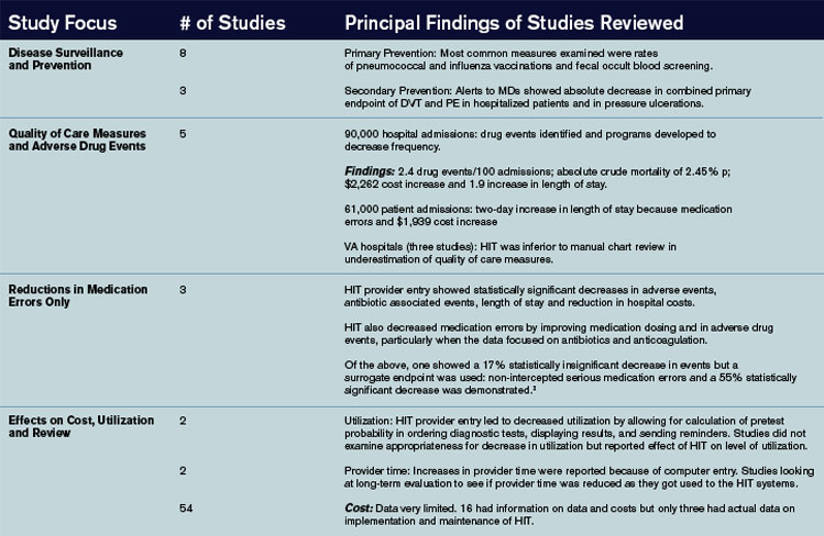
The authors of the article summarized 22 other studies in Appendix 22 from other institutions. The benefits noted were very similar to those found in the studies from the benchmark institutions; however, one major difference was the use of pre/post time-series designs that did not have a control or comparative group. Internally and commercially, HIT systems were evaluated in most of these studies with thirteen focusing on the former and nine on the later.
Summary and discussion: This study raises several questions worthy of further research and study:
- How are systems to interface with regard to the portability of health care information?
- Which are better: internally devised systems or commercially available systems?
- Who is going to be responsible for establishing standard guidelines for the implementation of HIT systems across the diverse healthcare systems in the United States? and
- How are the data to be used and analyzed by insurers and even public health entities?
HIT has shown many benefits with regard to quality of patient care issues and efficiency. Studies from these four benchmark institutions had one important limitation, however, and that is that the development of an internally developed HIT system took many years and is unlikely to be applicable to other institutions that have fewer resources and are struggling to implement the technology in a timely manner.
As the authors of the article acknowledge, it is not clear if HIT systems, though useful as tools, alter states of disease and health. More research looking at the direct impact of HIT information on morbidity and mortality is needed. As the authors state, this raises the question of a national HIT system, thereby making adoption of HIT a bipartisan initiative.
Providers need better evidence on information systems in order to make informed decisions about acquiring and implementing technology in community settings. The authors of this article made the first attempt to collate and appraise such evidence. Additional research is needed in academic, governmental, and community settings in both acute and nonacute facilities. As the authors point out, a uniform standard should be devised at the federal, state, and local level to report research on the implementation of HIT systems similar to the CONSORT and Quorum statement.
References
- Chaudry B, Wang J, Wu S, et al. Systematic review: impact of health information technology on quality, efficiency and costs of medical care. Ann Intern Med. 2006 May 16;144(10):742-752.
- Bates DW, Leape LL Cullen DJ. Effect of computerized physician order entry and a team intervention on prevention of serious medication errors. JAMA. 1998 Oct 21;280(15):1311-1316.
Gatifloxacin and Dysglycemia
Park-Wyllie LY, Juurlink DN, Kopp A, et al. Outpatient gatifloxacin therapy and dysglycemia in older adults. N Engl J Med. 2006 Mar;354(13):1352-1361.
Fluoroquinolones are the most commonly prescribed antibiotics in the United States. Side effects associated with some fluoroquinolones (temafloxacin, grepafloxacin, sparfloxacin, and trovafloxacin) have prompted their restriction or withdrawal. Until now, dysglycemia had been associated with gatifloxacin only in small studies or case reports.
This study sought to examine, by a nested case control design, the association between gatifloxacin and dysglycemia that required hospital care. The researchers reviewed all prescription records from the Ontario (Canada) Drug Benefit database, which houses complete prescription drug sales for all patients older than age 65 (1.4 million residents). They linked this with a national Canadian database of those same patients’ emergency room visits and hospital admissions. Cases were patients who received hospital care for hypoglycemia (or hyperglycemia) within 30 days of filling an antibiotic prescription (macrolide, cephalosporin, or fluoroquinolone). Controls were patients who had not received hospital care after an antibiotic prescription in the same time period. They were matched by age, gender, whether or not they had been diagnosed as having diabetes, diabetic drug use, and time of antibiotic prescription. Patients were excluded if they turned 65 within the year, had been hospitalized within 90 days, had subsequent hospitalizations for dysglycemia, or had more than one antibiotic in 30 days. Logistic regression was used to determine the odds ratio for the association between dysglycemia and recent antibiotic use. In multivariable analysis, they adjusted for liver disease, renal disease, alcohol use, hospitalizations, physician visits, diabetic and P-450 medications, socioeconomic status, and number of prescription drugs. They were also stratified by diabetes status.
The cases treated for hypoglycemia were four times more likely than controls to have been treated with gatifloxacin than with a macrolide (OR 4.3, CI 2.9-6.3). The association was slightly less with levofloxacin (OR 1.5, CI 1.2-2.0), and there was no association of hypoglycemia with moxifloxacin, ciprofloxacin, or cephalosporins. The cases treated for hyperglycemia were 17 times more likely than controls to have been treated with gatifloxacin than with a macrolide (OR 16.7, CI 10.4-26.8). There was no association of hyperglycemia with the other fluoroquinolones or cephalosporins. The analyses were similar when stratified by diabetes status. In total, 1.1% of all gatifloxacin treatments were associated with dysglycemia requiring hospital care within 30 days. As to what to do with gatifloxacin, as stated precisely in an accompanying editorial, “For every approved indication for gatifloxacin, there are safer, equally effective, and less costly alternatives.”
Physician Board Certification and Acute MI Quality Measures.
Chen J, Rathore SS, Wang Y, et al. Physician board certification and the care and outcomes of elderly patients with acute myocardial infarction. J Gen Intern Med. 2006 Mar;21(3):238-244.
Board certification may have implications for practicing physicians because there is evidence that both hiring organizations and patients prefer board-certified physicians over non-board certified physicians. However, it is unknown if such certification translates into better quality of patient care.
In this study researchers sought to examine the relationship between physician board certification and quality of care in patients with acute myocardial infarction (AMI). Medical records were abstracted from the Cooperative Cardiovascular Project, a cohort of Medicare beneficiaries hospitalized with AMI. Board certification was obtained from the American Medical Association (AMA) Physician Masterfile, which is reportedly 94% accurate. Quality of care measures included ASA and beta-blocker use at the time of admission and discharge. Researchers also evaluated 30-day and one-year mortality.
They evaluated these quality measures on 101,251 patients and 36,668 physicians, of which 80% were board certified. In multivariable analyses adjusting for patient, hospital, and physician characteristics, board-certified internal medicine and cardiology physicians (but not family practitioners) were more likely to prescribe ASA and beta-blockers at admission and discharge than non-board certified physicians (adjusted relative risk ranged from 1.04 to 1.20). There was no difference in 30-day and one-year mortality among any specialty after multivariable adjustment.
These results, albeit modest, suggest that physician board certification may be associated with superior quality of care in elderly patients with AMI. The authors offer that board-certified physicians may be more aware or familiar with guidelines or may be more likely to agree with clinical guidelines. Although board-certified physicians have been shown to complete more hours of CME and spend more time reading journals, they caution that board certification should not be used as a surrogate marker of quality. Because 30% of U.S. practicing physicians are not currently board certified, these results certainly warrant further study.
PE in COPD
Tillie-Leblond I, Marquette CH, Perez T, et al. Pulmonary embolism in patients with unexplained exacerbation of chronic obstructive pulmonary disease: prevalence and risk factors. Ann Intern Med. 2006 Mar 21;144(6):390-396.
This study sought to evaluate the prevalence and risk factors for pulmonary embolus (PE) in patients with unexplained exacerbations of COPD. All patients with an unexplained COPD exacerbation requiring hospitalization (not ICU) were evaluated for PE by spiral chest CT and lower limb venous ultrasound (USG). The COPD flare was “unexplained” if there were no signs or symptoms of respiratory tract infection, no pneumothorax, or a discrepancy between the clinical/radiologic features and the degree of hypoxemia. Patients were considered to have a PE if either of the two tests (CT or USG) were positive. Patients were not considered to have a PE if both tests were negative, and there was no evidence of PE at three-month follow-up.
Of 211 referred patients, 14 were not included due to inconclusive results or iodine intolerance. Of 197 enrolled patients, 25% had PE. There was no difference between the PE and non-PE groups with respect to referral location, clinical symptoms (dyspnea, pleuritic pain, hemoptysis, tachycardia, lower limb edema), need for long term oxygen therapy, admission P02 or PC02, recent trauma, recent surgery, obesity, or immobilization for more than seven days. Factors associated with PE on bivariate analysis included a decrease in PC02 >5mmHg from baseline, previous thromboembolism, and malignancy.
Researchers then calculated these patient’s Geneva score (a PE predictor score based on age, history, pulse, ABG, and chest X-ray findings. Those with a low probability Geneva score had a 9% incidence of PE, those with an intermediate score had a 47% incidence, and those with a high Geneva score had a 100% incidence.
In conclusion, in this study population of non-ICU patients with unexplained exacerbations of COPD, one in four had evidence of thromboembolic disease. Significant risk ratios between groups were found for patients with a prior history of thromboembolism, malignancy, or a decrease in their baseline PC02. These factors should be weighted heavily in determining which patients with unexplained COPD exacerbations should undergo workup to exclude a PE. The Geneva score also correlated well with PE incidence in this population.
Telithromycin in Asthma Exacerbations
Johnston SL, Blasi FB, Black PN, et al. The effect of telithromycin in acute exacerbations of asthma. New Engl J Med. 2006 Apr 13;354(15):1589-1600.
The use of antibiotics for uncomplicated asthma exacerbations is not currently considered standard of care. This randomized placebo controlled trial was designed to determine if telithromycin (a ketolide, structurally similar to macrolides, with efficacy against mycoplasma and chlamydia pneumonia) improves symptoms and peak flow rates in patients with asthma exacerbations. Researchers enrolled 278 patients age 18 to 55 with a history of asthma (less than six months) with an exacerbation (peak flow <80% and symptoms of wheeze and dyspnea). They excluded those requiring ICU care, smokers, known infection/recent antibiotic use, or regular use of oral steroids. Baseline demographics, pulmonary function, and acute medication use (steroids and beta agonists) were similar between the groups.
Intervention patients received telithromycin 800 mg a day for 10 days. The primary outcome was change in their asthma symptom score (rated on a seven-point scale) and change in their peak flows (recorded every morning for the study duration). Secondary outcomes were clinic pulmonary function tests (PFTs).
The intervention group had a mean decrease in their symptom score of 1.3 versus 1.0 in the placebo group (40% versus 27% reduction, p=.004). There was no difference between the groups in home peak flow measurements. There were statistically significant differences between the groups in clinic PFTs (FEV1, FVC, FEF 25%-75%), although the P values were not adjusted for multiple outcomes. The intervention group had a higher percentage of symptom-free days (16% versus 8%, P=.006). A total of 61% of the patients had serologic evidence of chlamydia or mycoplasma infection. Nausea (seven versus 0) and LFT elevations (two versus 0) were more frequent in the intervention group.
In conclusion, this is the first trial to report a benefit of antibiotic use in uncomplicated asthma exacerbations. This conclusion must be tempered by the underwhelming clinical improvement (1.3 versus 1.0 improvement in symptom scores), lack of peak flow improvement, recent reports of serious hepatotoxicity associated with telithromycin, and the possibility for antibiotic over-prescribing in unrefined patient populations.
Pneumonia Quality and Outcomes by Volume of Care
Lindenauer PK, Behal R, Murray CK, et al. Volume, quality of care, and outcome in pneumonia. Ann Intern Med. 2006 Feb 21;144(4):262-269.
It is widely known that, for many medical conditions and surgical procedures, outcomes are better when delivered by high-volume providers. It is not known if this holds true for patients with community-acquired pneumonia. To determine the association between physician/hospital volume and patient quality/outcomes, researchers utilized data from the Medicare National Pneumonia Quality Improvement Project. They included patients with an ICD9 diagnosis of pneumonia (850 patients were randomly selected from each state) at acute care hospitals with general or family medicine attendings. They excluded patients with no documentation of pneumonia, a normal chest X-ray, age <65, death on day one, comfort measures only, and departure against medical advice or transfer. Each physician and hospital volume were calculated from Medicare Part A claims for the same calendar year and analyzed by quartiles of volume.
Of the 9,741 physicians analyzed (13,480 patients), the median annual number of pneumonia patients was four, nine, 15, and 29 (in quartiles 1-4). Physician volume of care had little or no effect on performance rates of the timing of antibiotics, appropriate initial antibiotic, use of blood cultures, or in-hospital/30-day mortality rates. Low volume providers were significantly more likely to provide pneumococcal and influenza screening.
Of the 3,243 hospitals analyzed, the median annual pneumonia caseload was 57, 142, 262, and 465 (in quartiles 1-4). Hospital volume of care had little or no effect on performance rates of the appropriate initial antibiotic, use of blood cultures, pneumococcal/influenza screening, or in-hospital/30-day mortality rates (after severity of illness adjustment). Low volume centers were significantly more likely to give the first antibiotic within four hours.
In conclusion, volume of care by individual providers and hospital systems does not necessarily translate into better quality outcomes in pneumonia care. This may be partially explained by the challenges that larger medical centers face regarding delivering timely care (in overcrowded emergency rooms) and influencing behavior change (such as with standardized order sets).
By Debra Anoff, MD, assistant professor of medicine, Lucy Guerra, MD, assistant professor of medicine, Bjorn Holmstrom, MD, assistant professor of medicine, Asha Ramsakal, DO, assistant professor of medicine and chief, Richard Gross, MD, FACP, professor of medicine and chief, Division of Internal and Hospital Medicine H. Lee Moffitt Cancer Center and Research Institute, University of South Florida College of Medicine
Risk Factors for Mortality in Neutropenic Cancer Patients
Kuderer, NM, Dale DC, Crawford J, et al. Mortality, morbidity, and cost associated with febrile neutropenia in adult cancer patients. Cancer. 2006 May 15;106(10):2258-2266.
Background: Neutropenic fever in a cancer patient is considered a medical emergency. Most episodes of febrile neutropenia result in hospital admission with prompt institution of broad spectrum antibiotics. The definition of neutropenia is usually regarded as an absolute neutrophil count (neutrophils plus bands) less than 500 cells/µL. The mortality rate of a cancer patient hospitalized with neutropenic fever ranges from 5 to 11%. There have been numerous small studies looking at risk factors associated with inpatient mortality. However, their small sample size and very select group of patients limit their generalizability to a standard hospitalist practice.
Methods: The authors used a longitudinal discharge database derived from 115 U.S. medical centers—both academic and community-based—in 39 states, looking retrospectively at all adult cancer patients hospitalized for febrile neutropenia between 1995 and 2000. The database included 55,276 hospitalizations and 41,779 patients and was analyzed for length of stay, cost per episode, mortality, and the clinical factors associated with mortality and prolonged hospitalization.
Results: The mortality rate for cancer patients with neutropenic fever averaged 9.5% per hospital. The average mortality rate was 14.3% for leukemia, 8.9% for lymphoma, and 8.0% for solid tumors. Lung cancer had the highest average mortality rate, 13.4%, followed by colorectal cancer at 8.8%, and breast cancer, which had the lowest mortality rate with 3.6%. Moreover, the highest mortality rate, 39.2%, was seen in those patients with a documented infection with invasive aspergillosis, while the rate of mortality for invasive candidiasis was 36.7%, followed by gram-negative bacteremia at 33.9%, pneumonia at 26.5%, and gram-positive bacteremia at 21.2%.
Patients without comorbidities, including common illnesses such as diabetes, congestive heart failure, and renal failure, had an average 2.6 % risk of dying. Those with one comorbidity had a 10.3% risk, and patients with more than one comorbidity had a mortality risk of 21.4%. Multivariate analysis revealed that the odds ratios for inpatient mortality were 3.94 for lung disease, 3.26 for cerebrovascular disease, 3.16 for renal disease, 2.89 for liver disease, 1.94 for pulmonary embolism, and 1.58 for heart disease.
The study also showed that patients hospitalized for longer than 10 days (comprising 35% of all patients) accounted for the majority of overall deaths (65%), overall cost (78%), and overall days spent in the hospital (74%).
Conclusions: The study provided an estimate of inpatient mortality associated with neutropenic fever in cancer patients and highlighted those subgroups as being at heightened risk for death. Important limitations include the retrospective nature of the study, the use of administrative data sets as the primary data source, and a failure to analyze for the duration of neutropenia prior to treatment. The authors’ conclusion that the identified factors may be useful in determining which patients warrant “more aggressive supportive care measures” is plausible, but does not find enough support in the study to warrant general implementation.
Long-Term Value of Electrocardiogram Before Noncardiac Surgery
Jeger RV, Probst C, Arsenic R, et al. Long-term prognostic value of the preoperative 12-lead electrocardiogram before major noncardiac surgery in coronary artery disease. Am Heart J. 2006 Feb;151(2):508-513.
Background: The 12-lead electrocardiogram is commonly utilized as part of the cardiovascular preoperative risk stratification. Certain ECG abnormalities, such as left bundle branch block, Q waves, ST-T abnormalities, arrhythmias, and left ventricular hypertrophy (LVH), have been associated with elevated risk in the perioperative period.1,2 This article prospectively evaluated whether an exhaustive analysis of preoperative ECGs in this population would have long-term predictive validity.
Methods: The study utilized a “predefined” analysis applied retrospectively on data obtained for another study.3,4
The study analyzed 172 patients, all of whom had documented coronary artery disease (CAD) as defined by previous myocardial infarction (MI), prior revascularization, significant stenoses on prior catheterization, ischemia (by dobutamine stress echocardiogram or adenosine thallium imaging), or who were high risk for CAD. High risk required at least two of the following: age over 70, hypertension, diabetes mellitus, prior stroke, exercise intolerance, or a “pathological resting ECG.” These patients were to undergo noncardiac surgery. The primary endpoint was all-cause mortality after two years, and the secondary endpoint was major adverse cardiac events (MACE) after two years. The MACE were defined as nonfatal MI, coronary revascularization, re-hospitalization due to recurrent ischemia, and/or cardiac death.
The ECGs were obtained one day prior to surgery and analyzed using the Minnesota classification. Telephone interviews were done at six, 12, and 24 months to evaluate outcome, and the Swiss Mortality Registry was used to determine cause of death. Analyzed data were adjusted for current beta-blocker use and perioperative ischemia using a sensitivity analysis-like correction that is described briefly in the report.
Results: ST depression (odds ratio [OR] 4.5) and faster heart rate (OR 1.6) were independent predictors of all-cause mortality. Faster heart rate (OR 1.7) was also an independent predictor of MACE.
Commentary: The results of this relatively small study raise awareness of resting tachycardia and ST depression as possible markers for poor perioperative and two-year postoperative outcomes. The latter is already included in risk stratification guidelines5, and the former is of uncertain validity due to possible confounding variables such as anxiety or pain, beta blocker dosing differences, and lack of intra- and postoperative beta blocker usage data which may affect postoperative cardiac morbidity and mortality long after the drugs are stopped.
For hospitalists and other physicians performing cardiac risk stratification for noncardiac surgery patients, the current study does not warrant any change in current practice as recommended in the American College of Cardiology/American Heart Association Guidelines 2002.5 As to whether resting tachycardia should be considered as a predictive ECG criterion in this context, further valid and applicable evidence is needed.
References
- De Bacquer D, De Backer G, Kornitzer M, et al. Prognostic value of ECG findings for total, cardiovascular disease, and coronary heart disease death in men and women. Heart. 1998 Dec;80(6):570-577.
- Tervahauta M, Pekkanen J, Punsar S, et al. Resting electrocardiographic abnormalities as predictors of coronary events and total mortality among elderly men. Am J Med. 1996 Jun;100(6):641-645.
- Filipovic M, Jeger R, Probst C, et al. Heart rate variability and cardiac troponin I are incremental and independent predictors of one-year all-cause mortality after major noncardiac surgery in patients at risk of coronary artery disease. J Am Coll Cardiol. 2003 Nov 19;42(10):1767-76.
- Filipovic M, Jeger RV, Girard T, et al. Predictors of long-term mortality and cardiac events in patients with known or suspected coronary artery disease who survive major noncardiac surgery. Anaesthesia. 2005 Jan;60(1):5-11.
- Eagle KA, Berger PB, Calkins H, et al. ACC/AHA guideline update for perioperative cardiovascular evaluation for noncardiac surgery—executive summary. Circulation. 2002 Mar 12;105(10):1257-1267.
Statins and Cardiovascular Disease
Hackam DG, Mamdani M, Li P, et al. Statins and sepsis in patients with cardiovascular disease: a population-based cohort analysis. Lancet. 2006 Feb 4;367(9508):413-418.
Background: The benefits of statins in secondary prevention of MI and ischemic CVA, as well as in reduction of all-cause mortality and their primary prevention in diabetics, are well documented in the literature.1 The concept of statins reducing subsequent sepsis in humans with atherosclerotic cardiovascular disease (CVD) is relatively new. One hypothesis holds that such an effect may be due to several common pathophysiologic mechanisms of sepsis and CVD: endothelial instability, immune dysregulation, inflammation, and thrombogenesis. Statins have been shown to provide improved outcomes from sepsis in animal studies, and observational studies in humans have raised similar hypotheses. This study attempts to analyze whether statin use in humans is associated with similar benefits regarding prevention of sepsis.
Methods: This was a population-based cohort analysis of 141,487 patients in Ontario, Canada, during the period of time from 1997-2002, who were older than age 65 and had been hospitalized for acute coronary syndrome or ischemic stroke, or had revascularization and survived three months post discharge. Using four reputable linked administrative databases, the authors found that 46,662 (33%) patients were prescribed a statin within three months of the discharge date (the index date), and 94,825 (67%) were not. About 19% of enrolled patients died within nine months and were excluded from the analysis. Of the eligible patients, propensity-based matching was used to identify 69,168 patients, of whom half were prescribed a statin and half were not. The patients were then analyzed through provincial administrative databases for the end points of hospital admission with an International Classification of Disease (ICD) code of sepsis, severe sepsis, fatal sepsis, death, or the end of the study.
Results: A total of 551 out of 34,584 patients were admitted for sepsis from the statin group, and 667 out of 34,584 were admitted from the control group during a mean follow-up of 2.2 years. The rate of sepsis in the statin group was significantly lower than in the control group (71.2 versus 88.0 per 10,000 person-years; p=0.0003). Statin use was also associated with fewer episodes of sepsis in an extended duration of 3.8 years, and with a lower risk of severe and fatal sepsis where sepsis was the admission diagnosis. This protection was evident at both high and low doses. The hazard ratio for sepsis was .81. There was a 19% relative reduction in the risk of sepsis in patients older than 65 with atherosclerosis. The absolute risk reduction for the occurrence of sepsis per 10,000 person-years was 16.8%, or .168% per person-year, with a corresponding number needed to treat (NNT) of 595 patients to prevent one episode of sepsis per year.
Comments: Though sophisticated and diverse statistical methods were used to assess the robustness of the analysis, this study is retrospective. Unidentified confounders, clinical sepsis risk stratification (the presence of obstructive uropathy or impairments leading to aspiration or skin breakdown, for example) are not accounted for in the analysis. Nonetheless, the hypothesis is of high relevance to hospitalists; it has biologic plausibility, and the results are intriguing if not definitive.
From a practical perspective, it seems prudent to ensure ongoing statin use for all patients with appropriate traditional indications. Initiating statins for the sole purpose of reducing future risk of sepsis is not yet sufficiently supported by the available evidence, and the NNT of almost 600 patients per year in the current study does little to alter that recommendation. In reply to a subsequent letter to the editor, the authors pointed out that the high NNT in part reflects the low-risk profile of the cohort and would be lower in higher risk patients; for example, patients with chronic renal failure, previous infections, or corticosteroid use would have NNTs of 166, 250, and 250, respectively.2 A randomized, controlled prospective trial would be a welcome addition to the evidence on this important topic, as would studies of the use of statins solely as an acute intervention upon suspicion of sepsis.
References
- MRC/BHF Heart Protection Study of cholesterol lowering with simvastatin in 20,536 high-risk individuals: a randomised placebo-controlled trial. Lancet. 2002 Jul 6;360(9326):7-22.
- Hackam DG, Redelmeier DA. Statins and sepsis—authors' reply. Lancet. 2006;367:1651.
Review: Impact of Health Information Technology
Advances in information technology have transformed the world of business and education and will undoubtedly change the delivery and implementation of medical services. What arguably sets the world of health information apart from other systems is, however, the extreme sensitivity of the data involved and the impact it has on human lives.
Methods and data sources: In the article, “Systemic Review: Impact of Health Information Technology on Quality, Efficiency, and Costs of Medical Care,” the authors reviewed four data sources and extracted information on system design, effects on quality and cost benefit analysis. Four data sources were utilized including a MEDLINE analysis of articles indexed as health information technology (HIT) from 1995 to January 2004 utilizing:
- Cochrane Center Registry of Controlled Trials;
- Cochrane Database of Abstracts of Reviews of Effects;
- Periodical Abstracts Database; and
- Studies singled out by experts up to April of 2005
Of the four data sources, 24% of all studies came from the following academic centers: the Regenstrief Institute (Indianapolis), Brigham and Women’s Hospital (Boston), Department of Veterans Affairs, and the Latter Day Saints Hospital/Intermountain Health Care.
Inclusion criteria: The HIT evaluation and efficacy information derived was from these four academic institutions, each with its own EHR (electronic health record) system. Of 4,582 initial articles derived from a title review, 867 were deemed suitable given their title matter, but only 257 met inclusion criteria.
A modified framework from the Institute of Medicine’s six aims for care and a framework to analyze costs developed by the authors themselves was used for analysis.1
Reasons for rejection: Articles that did not have HIT as the subject, others that did not report outcomes data, and those that did not examine barriers for outcomes analysis were rejected.
Information examined: Extracted information on system capabilities including components of the system such as:
- User friendliness;
- Types of systems such as EHR that allowed for medical decision making support and e-prescribing; and
- Functional capabilities such as order entry and clinical documentation.
Common characteristics shared by the HIT systems evaluated by the four academic institutions: they were all multifunctional and included medical decision making support, all were internally developed by research experts and all had capabilities that had been improved upon and added incrementally over time.
The data supported five primary goals:
- Increasing delivery of healthcare according to established protocols and guidelines;
- Enhancing capacity to perform surveillance and monitoring of disease conditions and care delivery;
- Reducing medication error rates;
- Decreasing utilization of care; and
- Producing a mixed effect on time utilization.
Major findings: (see Figure 1, below).
The authors of the article summarized 22 other studies in Appendix 22 from other institutions. The benefits noted were very similar to those found in the studies from the benchmark institutions; however, one major difference was the use of pre/post time-series designs that did not have a control or comparative group. Internally and commercially, HIT systems were evaluated in most of these studies with thirteen focusing on the former and nine on the later.
Summary and discussion: This study raises several questions worthy of further research and study:
- How are systems to interface with regard to the portability of health care information?
- Which are better: internally devised systems or commercially available systems?
- Who is going to be responsible for establishing standard guidelines for the implementation of HIT systems across the diverse healthcare systems in the United States? and
- How are the data to be used and analyzed by insurers and even public health entities?
HIT has shown many benefits with regard to quality of patient care issues and efficiency. Studies from these four benchmark institutions had one important limitation, however, and that is that the development of an internally developed HIT system took many years and is unlikely to be applicable to other institutions that have fewer resources and are struggling to implement the technology in a timely manner.
As the authors of the article acknowledge, it is not clear if HIT systems, though useful as tools, alter states of disease and health. More research looking at the direct impact of HIT information on morbidity and mortality is needed. As the authors state, this raises the question of a national HIT system, thereby making adoption of HIT a bipartisan initiative.
Providers need better evidence on information systems in order to make informed decisions about acquiring and implementing technology in community settings. The authors of this article made the first attempt to collate and appraise such evidence. Additional research is needed in academic, governmental, and community settings in both acute and nonacute facilities. As the authors point out, a uniform standard should be devised at the federal, state, and local level to report research on the implementation of HIT systems similar to the CONSORT and Quorum statement.
References
- Chaudry B, Wang J, Wu S, et al. Systematic review: impact of health information technology on quality, efficiency and costs of medical care. Ann Intern Med. 2006 May 16;144(10):742-752.
- Bates DW, Leape LL Cullen DJ. Effect of computerized physician order entry and a team intervention on prevention of serious medication errors. JAMA. 1998 Oct 21;280(15):1311-1316.
Conference Report: AHRQ
The Agency for Healthcare Research and Quality (AHRQ) held its annual Patient Safety and Health IT (internet technology) Conference June 4-7, 2006, at the Renaissance Hotel in Washington, D.C. This annual meeting provides grantees, quality improvement and risk assessment managers, and all members of the healthcare team the opportunity to learn how AHRQ-funded patient safety and health IT research contributes to improved healthcare quality.
This was AHRQ’s sixth national conference with a focus on patient safety, and it was the second time the conference had a combined focus on patient safety and health IT. To highlight the importance of these two focus areas working together, the conference was entitled “Strengthening the Connections.” The significance of this relationship was further emphasized by holding the conference during the inaugural “National Health IT Week” and closing the sessions with a handoff (on National Health IT Day) to a health IT Summit held by the Healthcare Information and Management Systems Society (HIMSS).
This year’s conference opened with remarks from AHRQ Director Carolyn Clancy, MD, and opening keynote speaker David M. Sundwall, MD, director of the Utah Department of Health.
Dr. Clancy set the focus of the week by emphasizing that the pursuit of quality must be the shared ambition of patient safety and health IT. Quality healthcare is “the right treatment, for the right patient, at the right time” and the objectives of patient safety and health IT are best aligned when these goals are kept in sight by both focus areas. Dr. Clancy noted that AHRQ has helped pursue quality in patient safety and health IT by “fund[ing] some 240 Patient Safety and Health IT projects, and award[ing] more than $400 million in grants and contracts for both programs [since 2001].” Ultimately, “AHRQ’s efforts,” Dr. Clancy said, “are helping to light the path and measure the benefits for the average practice and community hospital. … [And they are] helping [to] show these providers [that] how to make a successful transition to health IT practice is a keystone in successfully adopting health IT.”
To share the work of this year’s presenting grantees, the sessions were categorized by topic into nine different, but concurrent, tracks. This allowed individuals with particular interests to easily identify which sessions would be most valuable to them. These tracks included:
- Track 1: Patient Safety and Health IT across Settings and Populations: These sessions covered the use of health IT in special populations, in outpatient clinics and physician offices, in emergency care, for the elderly, and in distance/telemedicine.
- Track 2: Implementation Issues in Patient Safety and Health IT: The track’s two sessions discussed how health IT will move into the 21st century, the relationship between clinical redesign and improved safety, process approaches to improving clinical performance, and the establishment and implementation of standards for e-prescribing.
- Track 3: Improving the Health of Communities through Regional Health Information Exchange (HIE): Track three featured ways to get started in HIE by gaining stakeholder support, ways to design and implement HIE at the front lines, and the development of sustainable business models for HIE, and offered updates on the shaping of the health IT vision at the Office of the National Coordinator.
- Track 4: Assessing Value and Evaluating Project Impact: Track four presentations covered the impact of certain health IT tools, a brainstorming session for rural health IT initiatives, an evaluation of medication administration technologies, an assessment of clinical decision support, new tools for assessing value, and an evaluation of regional health information exchange systems.
- Track 5: Achieving and Sustaining Improvements: This series of sessions highlighted the frequency of adverse medical-device events, socio-technical approaches to planning and assessing redesign with health IT, clinical decision support and reporting systems to improve population health and minimize harm in patient handoff settings, and ways to recognize and manage unintended consequences of health IT.
- Track 6: Using Reporting Systems for Safety and Quality Improvement: Track six presentations covered efforts to improve the accuracy and utility of reporting systems, ways to track changes over time through reporting systems, methods to track adverse drug events through reporting systems, and innovative applications of reporting systems.
- Track 7: Working Conditions Challenges in Patient Safety: Track seven described how health IT impacts provider working conditions and performance, how working conditions, and culture can impact performance of providers and patient outcomes, and how programs can be used to promote patient safety cultures.
- Track 8: Patient and Family Centered Health IT and Safety: The sessions in track eight presented strategies for improving medication management across transitions in care, as well as perceptions and approaches to patient-centered health IT.
- Track 9: Emerging Approaches to Drive Change in Healthcare: The ninth track discussed examples of how IT solutions intended to reduce error can sometimes induce new error, current uses of and research involving AHRQ’s patient safety indicators, appropriate and inappropriate use of technology in developing high reliability hospital systems, an introduction to and overview of using positive deviance as a quality improvement methodology, and the use of lean methodology in healthcare.
Of particular note is the involvement of five hospitalists in AHRQ-funded research. Jeff Greenwald, MD, Brian Jack, MD, Greg Maynard, MD, MS, Tosha Wetterneck, MD, and Mark Williams, MD, are each involved with quality improvement and patient safety research through AHRQ grants. (See The Hospitalist, June issue, p. 35.)
Doctors Jack and Greenwald are partners in researching the re-engineering of the hospital discharge process—a process that they note is not standardized and is often marked with poor quality as observed by the frequency of post-discharge adverse events and 30 day re-hospitalization rates—to improve patient safety. They have grants through AHRQ’s Partnerships in Implementing Patient Safety (PIPS) and Safe Practices Risk Assessment Challenge Grants (SPRACGS) research initiatives and presented their work as a part of track two.
Dr. Maynard is also funded through AHRQ’s PIPS research initiative as his research pursues the elimination of hospital-acquired venous thromboembolism (VTE). Noting that easy and cost effective prophylactic treatment exists, but is underutilized, Greg’s project involves the creation of a toolkit that allows institutions to acquire optimal prevention of hospital-acquired VTE, ultimately reducing morbidity, mortality, and the associated but unnecessary costs.
Dr. Wetterneck presented her work on medication administration technologies as a part of track four. Her research has focused on the development and evaluation of Smart IV pump technology that has been designed with human factors in mind.
Dr. Williams presented, along with John Bajha, PhD, his research on Hospital Patient Safe-D(ischarge). This initiative is funded by a PIPS grant and was presented as a part of track five. The project intends to first implement a “discharge bundle” that consists of medication reconciliation, patient-centered hospital discharge education, and a post-discharge continuity check by a clinician. Once the discharge bundle is established, its level of adoption and acceptance will be evaluated before the researchers develop an “implementation toolkit” for dissemination of the bundle. The bundle will be disseminated with the intended result of safer discharges.
The Patient Safety and Health IT Conference concluded on National Health IT Day and the start of the HIMSS Summit. Speakers included Dr. Clancy; Newt Gingrich, former speaker of the House and founder of the Center for Health Transformation; David Brailer, national coordinator for Health Information Technology; and Mark McClellan, MD, PhD, administrator of Centers for Medicare and Medicaid Services.
Additional panels throughout the day included leaders from outside of healthcare: David Hom, vice president, human resources strategic initiatives, Pitney Bowes; Bruce Bradley, director, health plan strategy and public policy, General Motors; and Dale Whitney, corporate health care manager, United Parcel Service; as well as a number of leaders from within the healthcare field who are actively involved with health IT implementation.
Ultimately, the day’s topics highlighted the importance of leveraging health IT in the pursuit of improved patient safety from the perspectives of policymakers, business leaders, and healthcare leaders and successfully served as a transition to the HIMSS Summit. TH
Resources
- For more information about the conference visit: http://healthit.ahrq.gov/PatientSafetyandHIT2006Conference.
- Webcasts and transcripts of the last day of the conference have been provided by the Kaiser Family Foundation: www.kaisernetwork.org/health_cast/hcast_index.cfm?display=detail&hc=1731
Bedside Arts
Hospital healthcare providers have supplemented clinical care with creative arts since the mid-20th century. For example, art and dance therapy have played a supporting role in hospital patient care since the 1930s. Using music to soothe cancer patients during treatment was pioneered at the University of Chicago Hospital as early as 1948. Music was also piped into some hospitals’ surgical suites to calm patients under various forms of spinal, local, or regional anesthesia.1
In recent years, there has been a groundswell of interest in both art therapy and the expressive arts in healthcare, resulting in the proliferation of bedside programs involving not only the visual arts and music, but also dance and creative writing.
According to the Art Therapy Credentials Board, “[A]rt therapy is a human service profession, which utilizes art media, images, the creative art process, and patient/client responses to the created art productions as reflections of an individual’s development, abilities, personality, concerns, and conflicts.”2 Anecdotal evidence has long supported the efficacy of art therapy in treating the chronically ill. But only recently have clinical studies proved that making art and the creative process it involves helps hospitalized patients heal in a quantifiable way.
One such study, conducted among adult cancer inpatients at Chicago’s Northwestern Memorial Hospital, was published in the February Journal of Pain and Symptom Management. That study determined that a series of one-hour art making sessions with a therapist yielded statistically significant decreases in a broad spectrum of symptoms, including pain, fatigue, depression, anxiety, lack of appetite, and shortness of breath. It also helped reduce apprehension, tension, nervousness, and worry. In addition to the quantifiable positive effects of art making, “subjects made numerous anecdotal comments that the art therapy had energized them.”3
But art therapy, administered by a credentialed practitioner with the specific goal of treating emotional and psychological issues associated with illness as a clinical practice, is not the only type of bedside artistic production happening in hospitals. Expressive art making, which falls under the umbrella of the arts in healthcare movement, has gained a significant foothold. In 2002 the National Endowment for the Arts (NEA), which funds arts in healthcare research, issued a call for creative artists not specifically trained as art therapists to play a larger role in patient care.
According to Elizabeth A. Curry, MA, coordinator of the Mayo Clinic Center for Humanities in Medicine, expressive bedside art-making has a different goal than therapy.
“The art therapist is part of the care plan team,” says Curry. “She writes in the charts.” As guided by a creative artist rather than by a therapist, the very experience of making art—rather than the information a finished work of art may furnish the care plan team—is central to the undertaking. It is the artist’s experience with the patient and the patient’s experience with the media that are important—not the end result. Of this model of bedside art making, Curry says, “it has no therapeutic goal other than to relieve stress.” The scope of expressive bedside art creation administered by an arts in healthcare program is potentially much broader than one that is therapy-based and has the potential to reach more patients.
Among studies of a number of arts-in-healthcare programs, the NEA cites the success of Healing Icons, an art-support program for young adult cancer patients age 16 and older.
According to the NEA, “In the program, patients create a three-dimensional mixed-media art piece to convey a unique personal perspective on receiving a diagnosis of cancer and then experiencing treatments.”
The mixed-media piece “provides a way for unstructured expression of feelings and thoughts.”4 The NEA also points to an article published in The Lancet (May 2001) that discussed the creative output of several expressive arts programs implemented in the United Kingdom. In one, comic artists held a series of workshops with young patients at the Great Ormond Street Children’s Hospital. Those workshops resulted in a comic book, HospiTales. Not only did this undertaking produce “interesting therapeutic and creative results” for the participants, the finished HospiTales promoted “a positive view of being in hospital, which makes it seem a less scary place for young patients.”5
Not surprisingly, the number of new arts in healthcare initiatives has continued to grow. For two months, beginning in August, for example, the Mayo Clinic Center for Humanities in Medicine began a pilot program in conjunction with the Rochester Arts Center in Minnesota to bring bedside art making to the Hematology Department. The Hematology Department, in particular, appears to be the ideal place to conduct such a pilot program. “People are stuck in the hospital with a lot of uncertainty, stress, and discomfort,” says Mayo’s Curry. “Making art can give people back a sense of control and relieve some anxiety.”
At the outset, Education Coordinator Michele Heidel of the Rochester Art Center will work with the nursing staff to identify 20 patients who might be interested in participating in what have been termed “art interventions.” Patients will paint and draw with professional artist-educators, who will offer participants a variety of media such as oil pastels, chalk, charcoal, and watercolors in which to work. These materials are chosen not only for their ease of use, but because they are safe, nontoxic, clean, and conform to ASTM Standard D-4236 Practice for Labeling Art Materials for Chronic Health Hazards. They also carry No-Odor labeling.
Prior to engaging with patients, artist-educators receive training in infection control, including OSHA Bloodborne Pathogen training and instruction in disinfecting equipment, art supplies, and work surfaces. They are also briefed on HIPAA compliance. Attendance at the Arts in Healthcare Summer Intensive Training at the University of Florida (Gainesville) and a site visit to the Mayo Clinic Jacksonville Arts at the Bedside program completes their orientation. Though they are not officially part of the care plan team, artist-educators also attend hematology inpatient rounds.
Each of the 20 patients chosen to participate in the pilot study will be assessed both before and after working with the educator-artist by means of questionnaires, as well as by Visual Analogue Scales to see how a single art intervention affects anxiety, discomfort, and stress. Ultimately, the purpose of this benefactor-funded pilot program is to provide quantifiable evidence for the efficacy of bedside art making.
According to Curry, the Center for Humanities in Medicine would like to grow the program significantly, eventually offering patients a menu of choices of creative arts in which to participate. This menu would also include music, dance, and creative writing. “It’s a big goal for the future,” says Curry.
For patients participating in Mayo’s pilot study, talent or artistic ability is not an issue. According to Curry, the program is process oriented rather than project oriented. Unlike the HospiTales project, the pilot study focuses on the relationship among the patient, the artist, and the media rather than on creating a finished piece. The Mayo Clinic’s Center for Humanities in Medicine has no specific plans either to exhibit or publish any of the artistic productions created by study participants.
“There will be a lot of amazing art and amazing writing,” says Curry. But the legal technicalities involved in publishing or mounting an exhibition of art work, including the necessity of having patients give permission and sign release forms, may simply be too daunting for those involved. Curry does not, however, rule out an exhibit or a book of patient work in the future.
In conjunction with Arizona State University, the Mayo Clinic’s Scottsdale center has also introduced several arts programs, including music at the bedside in Palliative Care, and a bedside creative writing program. During sessions that last about 45 minutes and center around the one-on-one interaction between the artist and the patient, patients narrate their personal stories, from which participating writers generate original works on hand-made paper. The finished pieces are then returned to the patient-narrators. These works have proved extremely meaningful not only to the people whose stories they tell, but to the storytellers’ families as well.
Based on its own successful programs, which include bedside art making, the Integrative Medicine department at Hartford Hospital in Connecticut has published an on-line Program Development Manual, “Building Bridges,” which provides “a blueprint for spanning the not-yet-connected terrain of Conventional Medicine and Complementary and Alternative Medicine,”6 Indeed, in addition to sections dealing such practices as massage therapy, acupuncture, Reiki, and Tai Chi, as well as guided imagery, “a mind-body intervention that focuses the imagination and the five senses to create soothing and relaxing images,”7 “Building Bridges” includes a chapter on “Creating an Art for Healing Program,” written by Diana S. Boehnert, artist-in-residence and coordinator of the Art for Healing Program.
According to Boehnert, art making as part of a larger Integrative Medicine program “creates a better quality of life for people with chronic illness.” Hartford Hospital’s program, which she administers, employs both clinically trained art therapy interns and volunteers, whose work follows the expressive bedside art making model. As such, the Art for Healing section of “Building Bridges” deals extensively with the training and preparation of artists. According to the manual, candidates without previous experience working in a hospital setting benefit from partnering with a clinical staff member as part of the training process. In addition to the requisite “orientation to patient care area with review of patient care environment, equipment, safety issues, and the needs of the specific patent population,” “Building Bridges” suggests that trainees also engage in “mock art sessions with a preceptor or mentor.”8 While it is also recommended that candidates have some background in the expressive arts, formal art training is not an absolute requirement. In reality, says Boehnert, “It doesn’t matter how much [formal art] training they have, the patient does the work.”
Unlike the Mayo Clinic’s pilot study, Hartford Hospital’s program is project oriented. “The project is the impetus that gets the patient going,” says Boehnert. “Adults aren’t willing to play without a purpose. They just want a little direction.”
For the most part, individual projects are small. They range from mandalas (circular designs generally associated with Buddhist and Hindu practice) to cards for family members. “Intuition,” explains Boehnert, “tells the volunteers what will work best with a patient.”
Hartford Hospital’s Arts for Healing is not limited to patients in a single department. Boehnert, whose previous experience with arts in healthcare included plaster cast mask-making with survivors of domestic violence, began working with rehab patients and extended the program to include dialysis patients. It’s now available in various departments throughout the hospital. Some of the work created by Arts for Healing participants in the Art for Healing program is on display in a small gallery in the hospital.
According to Boehnert, patient response to arts initiatives like the ones advocated in “Building Bridges” has been overwhelmingly positive. As an example, she cites a heart transplant patient who was introduced to the expressive arts during his six-week stay at Hartford Hospital. Before he was discharged, he created his own little gallery in his room. A patient being treated for leukemia also created an impressive body of work, giving pieces away to cheer up fellow patients who were not having good days. Staff, too, says Boehnert, benefit from Art for Healing: “My volunteers also go home better than when they came.”
Since 1991, the Society for the Arts in Healthcare (SAH) has provided support for programs such as the ones at Hartford Hospital and the Mayo Clinic, as well as others like the Artists in Residence program at Florida’s Shands HealthCare hospitals, which offers patients a variety of bedside art making activities. Examples include Art Infusion, a multi-media program for adults on chemotherapy, creative arts for pediatric inpatients, and (like Mayo Scottsdale) an oral history program which seeks to transcribe patients’ personal stories.
“In a lot of places, funding is a struggle,” explains Curry. To help secure funding for arts in healthcare programs, the SAH provides grant opportunities, like the SAH/Johnson & Johnson Partnership to Promote Arts and Healing and SAH Consulting Grants, as well as several awards.
In April, the SAH hosted its 15th international arts in healthcare conference in Chicago, the topic of which was “Vision + Voice—Charting the Course of Arts, Health and Medicine.” The conference urged attendees to “focus (their) vision for the future.”9 Given ever-increasing interest in integrating the arts into healthcare—especially inpatient care—be it by means of the clinical practice of art therapy or by expressive, creative arts programs, the future of such programs seems bright. As Dana Gioia, chair of the NEA, says: “The arts have an extraordinary ability to enhance our lives, to help us heal, and to bring us comfort in times of great stress. We must reconnect the arts with the actual human existence that Americans lead, the journeys we take in life, which lead us through hospitals, to hospices, to the end of life.”10 TH
Roberta Newman is based in Brooklyn, N.Y.
References
- NEA News Room: Arts in Healthcare Research. Available at: http://arts.endow.gov/news/news03/AIHResearch.html. Last accessed June 16, 2006.
- Art Therapy Credentials Board, “What is Art Therapy?” Available at: www.atcb.org. Last accessed June 16, 2006.
- Nainis N, Paice J, Ratner J. Relieving symptoms in cancer: innovative use of art therapy. J Pain Symptom Manage. 2006 Feb;31(2):162-169.
- Arts in Healthcare Research. National Endowment For the Arts New Room. Available at: http://arts.endow.gov/news/news03/AIHResearch.html. Last accessed June 12, 2006.
- Foster H. Medical settings foster the creation of art. Lancet. 2001;357(9268):1627.
- Foreword. “Building Bridges.” Available at: www.harthosp.org/IntMed/manual/foreword.asp. Last accessed June 18, 2006.
- Guided Imagery. “Building Bridges.” Available at www.harthosp.org/IntMed/manual/guidedimagery.asp. Last accessed June 18, 2006.
- Creating an Art for Healing Program: Training. “Building Bridges.” Available at: www.harthosp.org/IntMed/manual/art.asp. Last accessed June 18, 2006.
- Vision +Voice—Charting the Course of Arts, Health and Medicine Society for the Arts in Healthcare 15th Annual International Conference Program. Available at: www.thesah.org/doc/FINAL%20program.pdf. Last accessed June 18, 2006.
- Society for the Arts in Healthcare Fact Sheet. Available at: www.thesah.org. Last accessed June 19, 2006.
Things You Can Do To Save Lives
In April 2005, the American Hospital Association’s magazine, Hospital and Health Networks (H&HN), published the article “25 Things You Can Do to Save Lives Now.”1 In it, experts from the Joint Commission on Accreditation of Healthcare Organizations (JCAHO), the Institute for Healthcare Improvement (IHI), the National Quality Forum (NQF), and the Centers for Medicare and Medicaid Services (CMS), commented on an action plan to advance hospitals’ patient safety activities.
Now The Hospitalist has researched hospitalists’ views on these same 25 items. Those views are presented below.
A number of these items “are already highly ensconced in the JCAHO and CMS criteria,” says Dennis Manning, MD, FACP, FACC, director of quality in the Department of Medicine and an assistant professor at the Mayo Clinic College of Medicine, Rochester, Minn. “In terms of power of the things on the list for potentially saving lives, what we sometimes look at are the things that have the potential for the most prevention.”
Brian Alverson, MD, pediatric hospitalist at Hasbro Children’s Hospital in Providence, R.I., adds his thoughts on the 25 items: “We have to hold in our minds a healthy nervousness about patients being hospitalized, in that there is an inherent danger to that phenomenon. No matter how hard we strive for perfection in patient care, to err is human.”
Shortening hospital length of stay to within a safe range, he believes, is one of the best ways to reduce those daily dangers.
Some of the 25 items pose more challenges for hospitalists than others, and the contrary is true as well. Some were judged to be of lesser concern due to guidelines or imperatives imposed on hospitals by regulatory organizations. Other items fall outside hospitalists’ accountabilities, such as incorrect labeling on X-rays or CT scans, overly long working hours, medical mishaps (such as wrong-site, wrong-person, and wrong-implant surgeries), and ventilator-associated pneumonia. A few items were those that hospitalists found challenging, but for which they had few suggestions for solutions. In some, there were obstacles standing in the way of their making headway toward conquering the menace. These included:
1. Improper Patient Identification
“Until we set up a system that improves that, such as an automated system,” says one hospitalist, “I’ll be honest with you, I think we can remind ourselves ’till we’re blue in the face and we’re still going to make mistakes.”
2. Flu Shots
“Flu shots are probably more important in the pediatrics group than in any [other] except the geriatric group,” says Dr. Alverson, who strongly believes that pediatricians should be able to administer flu shots in the inpatient setting, “because we can catch these kids with chronic lung disease—many of [whom] are admitted multiple times.”
3. Fall Prevention
This item is one of the National Patient Safety goals, and one that every institution is trying to address. In pediatrics, says Dr. Alverson, the greater problem “is getting people to raise the rails of cribs. Kids often fall out of cribs because people forget to raise the rail afterwards, or don’t raise it high enough for a particularly athletic or acrobatic toddler.”
The other items on the list of 25 are below, including a section for medication-related items and the sidebar on a venous thromboembolism (VTE) prevention program.
4. Wash Hands
Provider hand-washing has been well studied, says one hospitalist, and “the data are so depressing that no one wants to deal with it.” Another says, “We just nag the hell out of people.”
One of the hospitalists interviewed for this story read the H&HN article and responds, “We do all these things.” But a lack of self-perception regarding this issue—as well as others—is also well-documented: Physicians who are queried will say they always wash their hands when, in fact, they do so less than 50% of the time.2-5
Despite the value of hand sanitizers—whether they are available at unit entrances, along the floors, at individual rooms, or carried in tiny dispensers that can be attached to a stethoscope—some pathogens, such as the now-epidemic Clostridium difficile, are not vulnerable to the antisepsis in those mechanisms.
“C. dif is a set of spores that are less effectively cleaned by the topical hand sanitizers,” says Dr. Alverson, who is also an assistant professor of pediatrics at Brown University in Providence, R.I. “In those cases, soap and water is what you need.”
Peter Angood, MD, FRCS(C), FACS, FCCM, vice president and chief patient safety officer of JCAHO, Oakbrook, Ill., says provider hand-washing is a huge patient safety issue and, in general, a multi-factorial problem that is more complicated than it would seem on the surface.
“We can rationalize and cut [providers] all kinds of slack, but at the bottom line is human behavior and their willingness to comply or not comply,” he says. “It’s like everything else: Why do some people speed when they know the speed limit is 55?”
Addressing the solution must be multi-factorial as well, but all hospitalists can serve as role models for their colleagues and students, including remaining open to reminders from patients and families.
5. Remain on Kidney Alert
Contrast media in radiologic procedures can cause allergic reactions that lead to kidney failure. This is a particularly vexing problem for elderly patients at the end stages of renal dysfunction and patients who have vascular disease, says Dr. Manning. Although the effects are not generally fatal, the medium can be organ-damaging. “This is a hazard that’s known, and it has some mitigating strategies,” he says, “but often it can’t be entirely eliminated.”
Measures that reduce the chance of injury, say Dr. Manning, include ensuring that the contrast medium is required; confirming that the procedure is correct for the patient, with the right diagnosis, with a regulated creatinine, and well coordinated with the radiology department; “and then getting true informed consent.” But at a minimum, he emphasizes, is the importance of hydration. “There is some evidence that hydration with particular types of intravenous fluids can help reduce the incidence of the kidney revolting.” And, he says, “there are a number of things that we have to do to make sure this is standardized.”
6. Use Rapid Response Teams
Use of “[r]apid response teams [RRTs] is one of the most powerful items on the list,” says Dr. Manning, who serves on SHM’s committee on Hospital Quality and Patient Safety as well as the committee helping to design the Ideal Discharge for the Elderly Patient checklist. “Whereas every hospital has a plan for response,” he says, RRTs are “really a backup plan.”
In 2003, Dr. Manning served as faculty for an IHI program in which a collaborative aimed at reducing overall hospital mortality. The formation and application of RRTs at six hospitals in the United States and two in the United Kingdom was the most promising of the several interventions, with impact on a variety of patients whose conditions were deteriorating in non-ICU care areas.
The advantage of RRTs with children, says Daniel Rauch, MD, FAAP, director of the Pediatric Hospitalist Program at NYU Medical Center, New York City, is that it is often difficult for providers to know what may be wrong with a child who is exhibiting symptoms. “Is the kid grunting because they’re constipated, because that’s the developmental stage they’re in, they’re in pain, or are they really cramping on you?” he asks.
7. Check for Pressure Ulcers
Checking for pressure ulcers is the task of nurses and physicians, say hospitalists, and they agree that it has to be done at admission. “The patient’s entire skin needs to be checked,” says Dr. Manning, “and often it takes both the nurse and doctor to roll the patient and get a good look at their bottom or their back … especially if the patient might have come from a nursing home and has a chronic serious illness.”
Also important, he says, is to fully assess the type of decubitus skin situation or any skin problem and then to monitor the patient to prevent advancement. “Multidisciplinary rounds can help,” he says, “and collaborative communication is key.”

8. Give the Patient and Family a Voice
“We fully embrace the involvement of the patient in the process of their care,” says Dr. Angood, who is also the co-director of the Joint Commission International Center for Patient Safety, for which patient and family involvement is a priority.
Giving patients and family members a voice is a fine idea, say our hospitalists, especially with children: No one knows a patient like their parents. As the H&HN article points out, anecdotal evidence is largely responsible for the belief that patient and family involvement helps reduce the likelihood for errors, and patient and family participation on safety committees can be a boon to advancing safety as well as satisfaction. But, says Dr. Alverson, “one has to keep in mind that parents have a perspective and not the only perspective on patient safety. I think a broad group of people has to sit down to address these issues.”
In the post-surgical setting, says Dr. Rauch, hospitalists make an invaluable contribution. “If surgeons don’t even come by to listen to the parents or see the child, it’s helpful to have that co-management of someone who’s used to listening to parents, who credits the parents for knowing their kids, and who will do the appropriate thing.” Dr. Angood, who is a past president of the Society of Critical Care Medicine, believes “that that patient-physician relationship is still going to be the driver for the majority of healthcare for some time yet.”
9. Reduce Catheter-Related Bloodstream Infections
“If it’s not required, we want every foreign body out,” says Dr. Manning. “We have to ask ourselves every day whether they are still required.”
The geriatric service at the Mayo Clinic (Rochester, Minn.) developed a daily mnemonic of A-B-C-D-E where B stands for binders. This, he says, “is a way for us to remind ourselves that any therapeutic foreign objects that are tethering the patient—and many of them are catheters—are of concern. We need to push the question [Is this still required?] to ourselves and then act on it.”
Dr. Alverson says, “There are certain infections [for which] we’re starting to move away from PICC [peripherally inserted central catheters] line management, and one way to mitigate that is to be on top of when you can actually discontinue the catheter.” For example, “in pediatrics, there are emerging data that with osteomyelitis you can have a shortened course of IV antibiotics and then switch to oral antibiotics. … That can reduce by half your PICC line duration. Being savvy about this is important.”
10. Reduce Heart Attack Death Rates
“There are about eight interventions for heart attacks that have increased survival,” says Dr. Manning. “So every hospital is working with these. We are using the all-or-none criteria, meaning that there are assurances [in place] that every patient will get all of them.”
Re-engineering systems has been particularly meaningful in preventing and treating heart attacks, says Dr. Manning, who represented SHM at a meeting of the Alliance for Cardiac Care Excellence (ACE), a CMS-based coalition that includes leaders from more than 30 healthcare organizations, and is working to ensure that all hospitalized cardiac patients regularly receive care consistent with nationally accepted standards.
11. Institute Multidisciplinary Rounds
Time constraints mean rounding with 10 people will necessarily be slower, says Dr. Alverson. In academic institutions where the hospitalist has the dual responsibility of teaching, this is especially time-consuming. Although there is an increasing emphasis that providers should participate at bedside rounds, and this is “clearly better from the patient’s perspective and, I would argue, better from the educational perspective,” says Dr. Alverson, it is “fairly bad from the getting-things-done-in-a-timely-fashion perspective. So it’s tough, and to a certain degree, in a practical world you have to pick and choose.”
When a nurse representative is there to respond to the question, “ ‘Why didn’t the kid get his formula? [and says] because he didn’t like the taste,’ that’s something that we might not pick up on,” says Dr. Alverson.
At NYU Medical Center, where Dr. Rauch works, formal rounds take place at least once a week (sometimes more), depending on volume, and they informally take place twice a day, every day.
“It works pretty well,” he says. “The nurses are a critically important part of teams; everybody recognizes that, and they are included in decisions.” Physicians put out the welcome mat for nurses even in casual circumstances. “Sometimes I am discussing things with the house staff [and] a nurse will pull up a chair and become part of the conversation. It’s a part of our culture.”
Although it is unusual to get a pharmacist to round with his team, says Dr. Alverson, a nearby pharmacy school sends students to join rounds, providing what might otherwise be a missing element of education.
12. Avoid Miscommunication
A number of the hospitalists interviewed were asked what they considered to be the top two or three communication points for hospitalists. Verbal orders, clarifying with read-backs, clear handwriting, and order sets were named frequently. In academic settings, says one hospitalist, instructors should be careful to make sure that residents, interns, and medical students understand what you’re saying and why you’re saying it. Good communication with the family was also cited as crucial.
“The most challenging issue is communicating at all,” says Dr. Rauch, who is also an associate professor of pediatrics at the NYU School of Medicine. Although he was the only one to phrase it this way, it is probably not a unique view. “In a large, old, academic medical institution, there are a lot of hierarchical issues that [impede] rapidly responding to [patients’] needs.” Unfortunately, it may mean communicating up one authorization pathway and down another. “And you can see the layers of time and the game of telephone as the concerns go around,” he says. “We’ve tried to break that down so the people who are on site can speak to someone who can make a decision.”
Along with that, he says, it is important from the outset to make it very clear who makes the decision. “For example, when the patient is a child getting neurosurgery because they have a seizure disorder and they also are developmentally delayed and they have medical issues, you now have at least three services involved with managing the child,” says Dr. Rauch. When three people are making decisions, he points out, no decision gets made. “You really have to decide when that child comes in who is going to call the shots for what issue. It’s usually the hospitalist who brings it up, and when it works, it works well.”

13. Empower Nurses and Other Clinicians
Nursing staff should have the power to halt unsafe practices. To Tahl Humes, MD, hospitalist at Exempla St. Joseph’s Hospital in Denver, halting unsafe practices depends, once again, on good lines of communication, and recognizing that patient care is a joint responsibility. For example, she says, “instead of just going to see the patient, writing the note, writing the order, and putting the chart away,” the hospitalists “talk with nurses daily and tell them what they’re planning to do,” so there is more opportunity to catch what might be unsafe practices.
14. Reduce Wound Infections
Although reducing wound infections is something in which their surgical colleagues take the lead, says Dr. Manning, “in our perioperative consultation care, we often work with surgery and anesthesiology in the pre-op evaluations of the patients. So in the surgical care improvement projects, we are often partners.”
Hospitalists are also frequently members on quality committees that help to brainstorm solutions to serious problems. One such project is the Surgical Care Improvement Program (SCIP), spearheaded by David Hunt, MD, with the Office of Clinical Standards and Quality, CMS. SCIP is an effort to transform the prevention of postoperative complications. Its goal is to reduce surgical complications by 25% in the United States by the year 2010 in four target areas: surgical site infections, and cardiac, respiratory, and venous thromboembolic complications. (See Figures 1 and 2, p. 33.)
This includes those patients who are already on beta-blockers. “From the hospitalist’s standpoint,” says Dr. Manning, “we have a real role in … [ensuring] that their beta blockade is maintained.”
Dr. Humes says that at her institution, a wound care nurse can have that responsibility. If a provider is concerned about any patient in this regard, he or she can order that the patient be seen by a wound care nurse and, depending on what’s needed, by a physical therapist.
Now we move on to address those issues that are medication-related:
15. Know Risky Meds
Pediatric hospitalists are involved with postoperative patients at Dr. Rauch’s institution. All patients’ orders are double-checked, he says, and computer order entry also helps providers calculate pediatric dosage norms or dosages calculated by weight.
The hospitalist has the opportunity to be involved in the pharmacy’s selection of drugs for the formulary, says Erin Stucky, MD, pediatric hospitalist at University of California, San Diego, and to help decide the drug choices within a certain class and limit the numbers of things that are used most frequently that are visually different in appearance. “And although that’s the pharmacist’s purview,” she says, “the hospitalist has a vested interest in being on the Pharmacy and Therapeutics Committee to review and restate to pharmacists what they’re using based on clinical need and to find a way for that drug to be safely stored in pharmacy if, indeed, there are a couple of drugs from one class that are truly useful.”
A drug’s generic name, brand name, dose strength, frequency of administration, place of use, indications, and contraindications are all important factors to determine the potential risks of drugs. But “you can’t say a list of risky medications at one institution is the same as it should be elsewhere,” says Dr. Stucky. Risky medications will depend on the setting in which the physician works. Hospitalists need to think logically about the drugs that are the most used or are new, including any new drug that has a different method by which it is administered or a different interaction capability with standard drugs.
“If there’s a new antibiotic that’s known to be processed through the liver and you have multiple patients with heart failure medications who have a medication basis that could be at conflict with that new drug, that’s a potentially risky medication,” she points out. “It may be easier in some ways for the pharmacist to be the rate-limiting factor for how they’re dispensed and for which patients they recheck [against] that incompatibility list.” But in large part, the avoidance of those risky-medication errors must be a commitment of the pharmacist and a bedside nurse.

16. Beware of Sound-Alike and Look-Alike Drugs
Dr. Stucky believes a majority of physicians don’t know the color or size of the pills they’re prescribing. “I would challenge all hospitalists to take every opportunity at the bedside,” she says, “to watch the process happening and know what those drugs and pills need to look like.”
Another opportunity is to educate family members “to remind them that [the patient is] going to be getting these medicines, these are the names of the medicines, and please ask the nurse about these medicines when you get them,” she says. If the hospitalist gives a new drug to the patient, the family can be another safeguard.
Dr. Stucky points out that you can tell the patient and family, “I’m going to tell the nurse that you’re going to be asking about this because … you are the best guide to help us make sure that these medicines are administered safely.” She also emphasizes that assigning this responsibility to the patient is important “because when people leave the hospital, we suddenly expect them to know how to take 18 pills.”
If, on a given unit, you have to handle cases with multiple diagnoses, says Dr. Stucky, it may be difficult to physically isolate the look-alike drugs. “At our institution we found that we actually had to pull the machines out,” she says, referring to the PIXUS units. “You can’t have them on the same wall even in different locations. You have to choose one or the other [similar looking pills].”
The sound-alike drugs are most ripe for errors with verbal orders. “Hospitalists can set a precedent in their institutions that any verbal orders should have the reason for that order given,” she explains. If you order clonazepam, after you finish giving the order verbally to the nurse, you should state, “This is for seizures.”
“When the nurse is writing it down, she may or may not be the one to know that that drug name is indeed in that drug class, but the pharmacist will know,” explains Dr. Stucky.

17. Reconcile Medications
“It is important for people to do verbal sign-out, certainly among attendings,” says Dr. Alverson, “to explain [in better depth] what’s going on with the patient and to maintain those avenues of communication in case something goes wrong. Hospitals get in trouble when physicians aren’t able to communicate or speak with each other readily.”
The biggest challenge for the pediatric hospitalists at NYU Hospital, says Dr. Rauch, is assessing the most up-to-date list of medications. “For instance,” he says, “we had a child yesterday as part of post-op care. I hadn’t met them pre-op. The father said, ‘I think my daughter’s on an experimental protocol with this additional medication.’ It wasn’t something we were used to so we called Mom: Can you bring in the protocol? She said, ‘Oh, she hasn’t been on that drug in a long time.’”
In fact, whether the patient is a child or adult, the majority of cases assigned to hospitalists are unplanned admissions and this is something with which all hospitalists struggle. But regarding transferring patients from unit to unit, says Dr. Stucky, “this is a whole different ballgame. That’s where we have a huge opportunity to make an impact.”
She suggests that matching medications to patients can be ameliorated by computer-based systems in which at each new place the hospitalist can fill in a printout regarding whether they’re continuing a drug order, changing it, or discontinuing it, and this system also works effectively on discharge. “In a perfect world,” says Dr. Stucky, … “the hospitalist would be the implementer of this kind of medication reconciliation in their institution.”
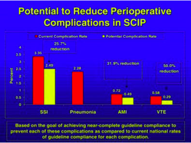
18. Avoid Unacceptable Drug Abbreviations
Some medications have abbreviations that can be misinterpreted. The classic ones, say several hospitalists, are magnesium and morphine. Others pertain to miswritten units of administration.
Read-backs on verbal medication orders was one of the elements most cited by our hospitalists as priority communication practices. Eliminating confusing abbreviations is one of JCAHO’s National Patient Safety Goals and “hospitals are aggressively rolling out ways to remind physicians not to use them,” says Dr. Alverson.
At Dr. Manning’s institution they use the Safest in America criteria, a collaboration of 10 Twin Cities and the hospital systems in Rochester, Minn., as well as the Institute for Clinical Systems Improvement. At Mayo, they call it “Write It Right.” An accentuated campaign to reduce ambiguities in medication communications, he says, has resulted in “profound improvement” in standardizing medication prescribing and following the read-back rules.
Dr. Stucky suggests that hospitalists take on mini-projects where they review the past six months of order-writing errors in their institutions, noticing any trends and, particularly, any unit-specific trends (such as the misunderstanding of the abbreviation cc). If you notice the errors are unit-specific, you can also analyze whether they are treatment-specific. In that way, “order sets can be pre-typed and all the providers have to do is fill in the numbers,” she says, adding that hospitalists can perform these analyses outside their own patient area.
19. Improper Drug Labeling, Packaging, and Storage
Drug names, labels, and packaging contribute significantly to medication errors. The risk for errors is determined by both the product and the environment in which it is used. Most hospitalists say they are continually developing new protocols and checking information multiple times. Sometimes, small changes go a long way. “Our patient safety officer has a favorite phrase: ‘How can I facilitate you to do something different next Tuesday?’ Within your own hospital, that means look at your system and pick something you know you can change,” says Dr. Stucky. “You can’t buy IT tomorrow; you can’t do physician order entries [because] your computer system doesn’t allow it—but what can you do?”

Conclusion
Dr. Angood encourages hospitalists to continue learning how to interact with other disciplines that are also evolving into hospital-based practices and to learn how to manage the specific details-of-change topics such as this list of 25—not just to gloss over them, but to understand them, and to encourage patient involvement and nurture the physician-patient relationship to help change the culture within health care.
“We can pick these kinds of topics and can dissect them all down, but each time, in the end, it is a matter of people and their behaviors as a culture inside a system,” he says. “The system can be changed a little bit, but still it is ultimately about the culture of people.” TH
Andrea Sattinger writes regularly for The Hospitalist.
References
- Runy LA. 25 things you can do to save lives now. Hosp Health Netw. 2005 Apr;79(4):27-28.
- Meengs MR, Giles BK, Chisholm CD, et al. Hand washing frequency in an emergency department. Ann Emerg Med. 1994 Jun;23(6):1307-1312.
- McGuckina M, Watermana R, Storrb J, et al. Evaluation of a patient-empowering hand hygiene programme in the UK. J Hosp Infect. 2001 Jul;48(3):222-227.
- Whitby M, McLaws ML, Ross MW. Why healthcare workers don't wash their hands: a behavioral explanation. Infect Control Hosp Epidemiol. 2006 May;27(5):484-492.
- Lipsett PA, Swoboda SM. Handwashing compliance depends on professional status. Surg Infect. 2001 Fall;2(3):241-245.
In April 2005, the American Hospital Association’s magazine, Hospital and Health Networks (H&HN), published the article “25 Things You Can Do to Save Lives Now.”1 In it, experts from the Joint Commission on Accreditation of Healthcare Organizations (JCAHO), the Institute for Healthcare Improvement (IHI), the National Quality Forum (NQF), and the Centers for Medicare and Medicaid Services (CMS), commented on an action plan to advance hospitals’ patient safety activities.
Now The Hospitalist has researched hospitalists’ views on these same 25 items. Those views are presented below.
A number of these items “are already highly ensconced in the JCAHO and CMS criteria,” says Dennis Manning, MD, FACP, FACC, director of quality in the Department of Medicine and an assistant professor at the Mayo Clinic College of Medicine, Rochester, Minn. “In terms of power of the things on the list for potentially saving lives, what we sometimes look at are the things that have the potential for the most prevention.”
Brian Alverson, MD, pediatric hospitalist at Hasbro Children’s Hospital in Providence, R.I., adds his thoughts on the 25 items: “We have to hold in our minds a healthy nervousness about patients being hospitalized, in that there is an inherent danger to that phenomenon. No matter how hard we strive for perfection in patient care, to err is human.”
Shortening hospital length of stay to within a safe range, he believes, is one of the best ways to reduce those daily dangers.
Some of the 25 items pose more challenges for hospitalists than others, and the contrary is true as well. Some were judged to be of lesser concern due to guidelines or imperatives imposed on hospitals by regulatory organizations. Other items fall outside hospitalists’ accountabilities, such as incorrect labeling on X-rays or CT scans, overly long working hours, medical mishaps (such as wrong-site, wrong-person, and wrong-implant surgeries), and ventilator-associated pneumonia. A few items were those that hospitalists found challenging, but for which they had few suggestions for solutions. In some, there were obstacles standing in the way of their making headway toward conquering the menace. These included:
1. Improper Patient Identification
“Until we set up a system that improves that, such as an automated system,” says one hospitalist, “I’ll be honest with you, I think we can remind ourselves ’till we’re blue in the face and we’re still going to make mistakes.”
2. Flu Shots
“Flu shots are probably more important in the pediatrics group than in any [other] except the geriatric group,” says Dr. Alverson, who strongly believes that pediatricians should be able to administer flu shots in the inpatient setting, “because we can catch these kids with chronic lung disease—many of [whom] are admitted multiple times.”
3. Fall Prevention
This item is one of the National Patient Safety goals, and one that every institution is trying to address. In pediatrics, says Dr. Alverson, the greater problem “is getting people to raise the rails of cribs. Kids often fall out of cribs because people forget to raise the rail afterwards, or don’t raise it high enough for a particularly athletic or acrobatic toddler.”
The other items on the list of 25 are below, including a section for medication-related items and the sidebar on a venous thromboembolism (VTE) prevention program.
4. Wash Hands
Provider hand-washing has been well studied, says one hospitalist, and “the data are so depressing that no one wants to deal with it.” Another says, “We just nag the hell out of people.”
One of the hospitalists interviewed for this story read the H&HN article and responds, “We do all these things.” But a lack of self-perception regarding this issue—as well as others—is also well-documented: Physicians who are queried will say they always wash their hands when, in fact, they do so less than 50% of the time.2-5
Despite the value of hand sanitizers—whether they are available at unit entrances, along the floors, at individual rooms, or carried in tiny dispensers that can be attached to a stethoscope—some pathogens, such as the now-epidemic Clostridium difficile, are not vulnerable to the antisepsis in those mechanisms.
“C. dif is a set of spores that are less effectively cleaned by the topical hand sanitizers,” says Dr. Alverson, who is also an assistant professor of pediatrics at Brown University in Providence, R.I. “In those cases, soap and water is what you need.”
Peter Angood, MD, FRCS(C), FACS, FCCM, vice president and chief patient safety officer of JCAHO, Oakbrook, Ill., says provider hand-washing is a huge patient safety issue and, in general, a multi-factorial problem that is more complicated than it would seem on the surface.
“We can rationalize and cut [providers] all kinds of slack, but at the bottom line is human behavior and their willingness to comply or not comply,” he says. “It’s like everything else: Why do some people speed when they know the speed limit is 55?”
Addressing the solution must be multi-factorial as well, but all hospitalists can serve as role models for their colleagues and students, including remaining open to reminders from patients and families.
5. Remain on Kidney Alert
Contrast media in radiologic procedures can cause allergic reactions that lead to kidney failure. This is a particularly vexing problem for elderly patients at the end stages of renal dysfunction and patients who have vascular disease, says Dr. Manning. Although the effects are not generally fatal, the medium can be organ-damaging. “This is a hazard that’s known, and it has some mitigating strategies,” he says, “but often it can’t be entirely eliminated.”
Measures that reduce the chance of injury, say Dr. Manning, include ensuring that the contrast medium is required; confirming that the procedure is correct for the patient, with the right diagnosis, with a regulated creatinine, and well coordinated with the radiology department; “and then getting true informed consent.” But at a minimum, he emphasizes, is the importance of hydration. “There is some evidence that hydration with particular types of intravenous fluids can help reduce the incidence of the kidney revolting.” And, he says, “there are a number of things that we have to do to make sure this is standardized.”
6. Use Rapid Response Teams
Use of “[r]apid response teams [RRTs] is one of the most powerful items on the list,” says Dr. Manning, who serves on SHM’s committee on Hospital Quality and Patient Safety as well as the committee helping to design the Ideal Discharge for the Elderly Patient checklist. “Whereas every hospital has a plan for response,” he says, RRTs are “really a backup plan.”
In 2003, Dr. Manning served as faculty for an IHI program in which a collaborative aimed at reducing overall hospital mortality. The formation and application of RRTs at six hospitals in the United States and two in the United Kingdom was the most promising of the several interventions, with impact on a variety of patients whose conditions were deteriorating in non-ICU care areas.
The advantage of RRTs with children, says Daniel Rauch, MD, FAAP, director of the Pediatric Hospitalist Program at NYU Medical Center, New York City, is that it is often difficult for providers to know what may be wrong with a child who is exhibiting symptoms. “Is the kid grunting because they’re constipated, because that’s the developmental stage they’re in, they’re in pain, or are they really cramping on you?” he asks.
7. Check for Pressure Ulcers
Checking for pressure ulcers is the task of nurses and physicians, say hospitalists, and they agree that it has to be done at admission. “The patient’s entire skin needs to be checked,” says Dr. Manning, “and often it takes both the nurse and doctor to roll the patient and get a good look at their bottom or their back … especially if the patient might have come from a nursing home and has a chronic serious illness.”
Also important, he says, is to fully assess the type of decubitus skin situation or any skin problem and then to monitor the patient to prevent advancement. “Multidisciplinary rounds can help,” he says, “and collaborative communication is key.”
8. Give the Patient and Family a Voice
“We fully embrace the involvement of the patient in the process of their care,” says Dr. Angood, who is also the co-director of the Joint Commission International Center for Patient Safety, for which patient and family involvement is a priority.
Giving patients and family members a voice is a fine idea, say our hospitalists, especially with children: No one knows a patient like their parents. As the H&HN article points out, anecdotal evidence is largely responsible for the belief that patient and family involvement helps reduce the likelihood for errors, and patient and family participation on safety committees can be a boon to advancing safety as well as satisfaction. But, says Dr. Alverson, “one has to keep in mind that parents have a perspective and not the only perspective on patient safety. I think a broad group of people has to sit down to address these issues.”
In the post-surgical setting, says Dr. Rauch, hospitalists make an invaluable contribution. “If surgeons don’t even come by to listen to the parents or see the child, it’s helpful to have that co-management of someone who’s used to listening to parents, who credits the parents for knowing their kids, and who will do the appropriate thing.” Dr. Angood, who is a past president of the Society of Critical Care Medicine, believes “that that patient-physician relationship is still going to be the driver for the majority of healthcare for some time yet.”
9. Reduce Catheter-Related Bloodstream Infections
“If it’s not required, we want every foreign body out,” says Dr. Manning. “We have to ask ourselves every day whether they are still required.”
The geriatric service at the Mayo Clinic (Rochester, Minn.) developed a daily mnemonic of A-B-C-D-E where B stands for binders. This, he says, “is a way for us to remind ourselves that any therapeutic foreign objects that are tethering the patient—and many of them are catheters—are of concern. We need to push the question [Is this still required?] to ourselves and then act on it.”
Dr. Alverson says, “There are certain infections [for which] we’re starting to move away from PICC [peripherally inserted central catheters] line management, and one way to mitigate that is to be on top of when you can actually discontinue the catheter.” For example, “in pediatrics, there are emerging data that with osteomyelitis you can have a shortened course of IV antibiotics and then switch to oral antibiotics. … That can reduce by half your PICC line duration. Being savvy about this is important.”
10. Reduce Heart Attack Death Rates
“There are about eight interventions for heart attacks that have increased survival,” says Dr. Manning. “So every hospital is working with these. We are using the all-or-none criteria, meaning that there are assurances [in place] that every patient will get all of them.”
Re-engineering systems has been particularly meaningful in preventing and treating heart attacks, says Dr. Manning, who represented SHM at a meeting of the Alliance for Cardiac Care Excellence (ACE), a CMS-based coalition that includes leaders from more than 30 healthcare organizations, and is working to ensure that all hospitalized cardiac patients regularly receive care consistent with nationally accepted standards.
11. Institute Multidisciplinary Rounds
Time constraints mean rounding with 10 people will necessarily be slower, says Dr. Alverson. In academic institutions where the hospitalist has the dual responsibility of teaching, this is especially time-consuming. Although there is an increasing emphasis that providers should participate at bedside rounds, and this is “clearly better from the patient’s perspective and, I would argue, better from the educational perspective,” says Dr. Alverson, it is “fairly bad from the getting-things-done-in-a-timely-fashion perspective. So it’s tough, and to a certain degree, in a practical world you have to pick and choose.”
When a nurse representative is there to respond to the question, “ ‘Why didn’t the kid get his formula? [and says] because he didn’t like the taste,’ that’s something that we might not pick up on,” says Dr. Alverson.
At NYU Medical Center, where Dr. Rauch works, formal rounds take place at least once a week (sometimes more), depending on volume, and they informally take place twice a day, every day.
“It works pretty well,” he says. “The nurses are a critically important part of teams; everybody recognizes that, and they are included in decisions.” Physicians put out the welcome mat for nurses even in casual circumstances. “Sometimes I am discussing things with the house staff [and] a nurse will pull up a chair and become part of the conversation. It’s a part of our culture.”
Although it is unusual to get a pharmacist to round with his team, says Dr. Alverson, a nearby pharmacy school sends students to join rounds, providing what might otherwise be a missing element of education.
12. Avoid Miscommunication
A number of the hospitalists interviewed were asked what they considered to be the top two or three communication points for hospitalists. Verbal orders, clarifying with read-backs, clear handwriting, and order sets were named frequently. In academic settings, says one hospitalist, instructors should be careful to make sure that residents, interns, and medical students understand what you’re saying and why you’re saying it. Good communication with the family was also cited as crucial.
“The most challenging issue is communicating at all,” says Dr. Rauch, who is also an associate professor of pediatrics at the NYU School of Medicine. Although he was the only one to phrase it this way, it is probably not a unique view. “In a large, old, academic medical institution, there are a lot of hierarchical issues that [impede] rapidly responding to [patients’] needs.” Unfortunately, it may mean communicating up one authorization pathway and down another. “And you can see the layers of time and the game of telephone as the concerns go around,” he says. “We’ve tried to break that down so the people who are on site can speak to someone who can make a decision.”
Along with that, he says, it is important from the outset to make it very clear who makes the decision. “For example, when the patient is a child getting neurosurgery because they have a seizure disorder and they also are developmentally delayed and they have medical issues, you now have at least three services involved with managing the child,” says Dr. Rauch. When three people are making decisions, he points out, no decision gets made. “You really have to decide when that child comes in who is going to call the shots for what issue. It’s usually the hospitalist who brings it up, and when it works, it works well.”
13. Empower Nurses and Other Clinicians
Nursing staff should have the power to halt unsafe practices. To Tahl Humes, MD, hospitalist at Exempla St. Joseph’s Hospital in Denver, halting unsafe practices depends, once again, on good lines of communication, and recognizing that patient care is a joint responsibility. For example, she says, “instead of just going to see the patient, writing the note, writing the order, and putting the chart away,” the hospitalists “talk with nurses daily and tell them what they’re planning to do,” so there is more opportunity to catch what might be unsafe practices.
14. Reduce Wound Infections
Although reducing wound infections is something in which their surgical colleagues take the lead, says Dr. Manning, “in our perioperative consultation care, we often work with surgery and anesthesiology in the pre-op evaluations of the patients. So in the surgical care improvement projects, we are often partners.”
Hospitalists are also frequently members on quality committees that help to brainstorm solutions to serious problems. One such project is the Surgical Care Improvement Program (SCIP), spearheaded by David Hunt, MD, with the Office of Clinical Standards and Quality, CMS. SCIP is an effort to transform the prevention of postoperative complications. Its goal is to reduce surgical complications by 25% in the United States by the year 2010 in four target areas: surgical site infections, and cardiac, respiratory, and venous thromboembolic complications. (See Figures 1 and 2, p. 33.)
This includes those patients who are already on beta-blockers. “From the hospitalist’s standpoint,” says Dr. Manning, “we have a real role in … [ensuring] that their beta blockade is maintained.”
Dr. Humes says that at her institution, a wound care nurse can have that responsibility. If a provider is concerned about any patient in this regard, he or she can order that the patient be seen by a wound care nurse and, depending on what’s needed, by a physical therapist.
Now we move on to address those issues that are medication-related:
15. Know Risky Meds
Pediatric hospitalists are involved with postoperative patients at Dr. Rauch’s institution. All patients’ orders are double-checked, he says, and computer order entry also helps providers calculate pediatric dosage norms or dosages calculated by weight.
The hospitalist has the opportunity to be involved in the pharmacy’s selection of drugs for the formulary, says Erin Stucky, MD, pediatric hospitalist at University of California, San Diego, and to help decide the drug choices within a certain class and limit the numbers of things that are used most frequently that are visually different in appearance. “And although that’s the pharmacist’s purview,” she says, “the hospitalist has a vested interest in being on the Pharmacy and Therapeutics Committee to review and restate to pharmacists what they’re using based on clinical need and to find a way for that drug to be safely stored in pharmacy if, indeed, there are a couple of drugs from one class that are truly useful.”
A drug’s generic name, brand name, dose strength, frequency of administration, place of use, indications, and contraindications are all important factors to determine the potential risks of drugs. But “you can’t say a list of risky medications at one institution is the same as it should be elsewhere,” says Dr. Stucky. Risky medications will depend on the setting in which the physician works. Hospitalists need to think logically about the drugs that are the most used or are new, including any new drug that has a different method by which it is administered or a different interaction capability with standard drugs.
“If there’s a new antibiotic that’s known to be processed through the liver and you have multiple patients with heart failure medications who have a medication basis that could be at conflict with that new drug, that’s a potentially risky medication,” she points out. “It may be easier in some ways for the pharmacist to be the rate-limiting factor for how they’re dispensed and for which patients they recheck [against] that incompatibility list.” But in large part, the avoidance of those risky-medication errors must be a commitment of the pharmacist and a bedside nurse.
16. Beware of Sound-Alike and Look-Alike Drugs
Dr. Stucky believes a majority of physicians don’t know the color or size of the pills they’re prescribing. “I would challenge all hospitalists to take every opportunity at the bedside,” she says, “to watch the process happening and know what those drugs and pills need to look like.”
Another opportunity is to educate family members “to remind them that [the patient is] going to be getting these medicines, these are the names of the medicines, and please ask the nurse about these medicines when you get them,” she says. If the hospitalist gives a new drug to the patient, the family can be another safeguard.
Dr. Stucky points out that you can tell the patient and family, “I’m going to tell the nurse that you’re going to be asking about this because … you are the best guide to help us make sure that these medicines are administered safely.” She also emphasizes that assigning this responsibility to the patient is important “because when people leave the hospital, we suddenly expect them to know how to take 18 pills.”
If, on a given unit, you have to handle cases with multiple diagnoses, says Dr. Stucky, it may be difficult to physically isolate the look-alike drugs. “At our institution we found that we actually had to pull the machines out,” she says, referring to the PIXUS units. “You can’t have them on the same wall even in different locations. You have to choose one or the other [similar looking pills].”
The sound-alike drugs are most ripe for errors with verbal orders. “Hospitalists can set a precedent in their institutions that any verbal orders should have the reason for that order given,” she explains. If you order clonazepam, after you finish giving the order verbally to the nurse, you should state, “This is for seizures.”
“When the nurse is writing it down, she may or may not be the one to know that that drug name is indeed in that drug class, but the pharmacist will know,” explains Dr. Stucky.
17. Reconcile Medications
“It is important for people to do verbal sign-out, certainly among attendings,” says Dr. Alverson, “to explain [in better depth] what’s going on with the patient and to maintain those avenues of communication in case something goes wrong. Hospitals get in trouble when physicians aren’t able to communicate or speak with each other readily.”
The biggest challenge for the pediatric hospitalists at NYU Hospital, says Dr. Rauch, is assessing the most up-to-date list of medications. “For instance,” he says, “we had a child yesterday as part of post-op care. I hadn’t met them pre-op. The father said, ‘I think my daughter’s on an experimental protocol with this additional medication.’ It wasn’t something we were used to so we called Mom: Can you bring in the protocol? She said, ‘Oh, she hasn’t been on that drug in a long time.’”
In fact, whether the patient is a child or adult, the majority of cases assigned to hospitalists are unplanned admissions and this is something with which all hospitalists struggle. But regarding transferring patients from unit to unit, says Dr. Stucky, “this is a whole different ballgame. That’s where we have a huge opportunity to make an impact.”
She suggests that matching medications to patients can be ameliorated by computer-based systems in which at each new place the hospitalist can fill in a printout regarding whether they’re continuing a drug order, changing it, or discontinuing it, and this system also works effectively on discharge. “In a perfect world,” says Dr. Stucky, … “the hospitalist would be the implementer of this kind of medication reconciliation in their institution.”
18. Avoid Unacceptable Drug Abbreviations
Some medications have abbreviations that can be misinterpreted. The classic ones, say several hospitalists, are magnesium and morphine. Others pertain to miswritten units of administration.
Read-backs on verbal medication orders was one of the elements most cited by our hospitalists as priority communication practices. Eliminating confusing abbreviations is one of JCAHO’s National Patient Safety Goals and “hospitals are aggressively rolling out ways to remind physicians not to use them,” says Dr. Alverson.
At Dr. Manning’s institution they use the Safest in America criteria, a collaboration of 10 Twin Cities and the hospital systems in Rochester, Minn., as well as the Institute for Clinical Systems Improvement. At Mayo, they call it “Write It Right.” An accentuated campaign to reduce ambiguities in medication communications, he says, has resulted in “profound improvement” in standardizing medication prescribing and following the read-back rules.
Dr. Stucky suggests that hospitalists take on mini-projects where they review the past six months of order-writing errors in their institutions, noticing any trends and, particularly, any unit-specific trends (such as the misunderstanding of the abbreviation cc). If you notice the errors are unit-specific, you can also analyze whether they are treatment-specific. In that way, “order sets can be pre-typed and all the providers have to do is fill in the numbers,” she says, adding that hospitalists can perform these analyses outside their own patient area.
19. Improper Drug Labeling, Packaging, and Storage
Drug names, labels, and packaging contribute significantly to medication errors. The risk for errors is determined by both the product and the environment in which it is used. Most hospitalists say they are continually developing new protocols and checking information multiple times. Sometimes, small changes go a long way. “Our patient safety officer has a favorite phrase: ‘How can I facilitate you to do something different next Tuesday?’ Within your own hospital, that means look at your system and pick something you know you can change,” says Dr. Stucky. “You can’t buy IT tomorrow; you can’t do physician order entries [because] your computer system doesn’t allow it—but what can you do?”
Conclusion
Dr. Angood encourages hospitalists to continue learning how to interact with other disciplines that are also evolving into hospital-based practices and to learn how to manage the specific details-of-change topics such as this list of 25—not just to gloss over them, but to understand them, and to encourage patient involvement and nurture the physician-patient relationship to help change the culture within health care.
“We can pick these kinds of topics and can dissect them all down, but each time, in the end, it is a matter of people and their behaviors as a culture inside a system,” he says. “The system can be changed a little bit, but still it is ultimately about the culture of people.” TH
Andrea Sattinger writes regularly for The Hospitalist.
References
- Runy LA. 25 things you can do to save lives now. Hosp Health Netw. 2005 Apr;79(4):27-28.
- Meengs MR, Giles BK, Chisholm CD, et al. Hand washing frequency in an emergency department. Ann Emerg Med. 1994 Jun;23(6):1307-1312.
- McGuckina M, Watermana R, Storrb J, et al. Evaluation of a patient-empowering hand hygiene programme in the UK. J Hosp Infect. 2001 Jul;48(3):222-227.
- Whitby M, McLaws ML, Ross MW. Why healthcare workers don't wash their hands: a behavioral explanation. Infect Control Hosp Epidemiol. 2006 May;27(5):484-492.
- Lipsett PA, Swoboda SM. Handwashing compliance depends on professional status. Surg Infect. 2001 Fall;2(3):241-245.
A New Hospitalist in the House
Effective spiritual care interventions require the right person with the right training who understands, for example, that spiritual care is more than intercessory prayer alone.1 No one on the healthcare team knows this better than the clinically trained, board-certified professional chaplain.2 When it comes to understanding the unique role of the professional chaplain on the healthcare team, no metaphor communicates this better to physicians and other healthcare providers than that of “hospitalist.” The “chaplain as hospitalist” differentiates the in-house chaplain’s care from that of other spiritual care providers and thereby sheds light on the problematic nexus of managing the provision of spiritual care in healthcare today.3-6
“Hospitalist” as a metaphor for chaplains communicates well with other healthcare professionals whose preconceived notions of chaplains may be based on parish-based models for pastoral care, or on chaplains as harbingers of death, or on the notion that chaplains only hold hands and pray with patients. As a metaphor, the hospitalist compares with the primary care physician as the chaplain compares to local clergy. That is, among all spiritual care providers, including local parish-based clergy and other healthcare professionals (such as physicians, nurses, and social workers) who might consider spirituality as part of their care, the professional chaplain is the “hospitalist.”
Like the hospitalist, the clinically trained and board-certified [hospital-based] chaplain is thoroughly and specifically trained in hospital-based interventions. These interventions range from the more traditional listening, prayer, and religious rituals to those interventions that incorporate an awareness of multi-cultural concerns, including ethical decision-making on end-of-life issues, and those that utilize current psychosocial and behavioral models as they relate to illness responses.7
Unlike many local pastors who come from the surrounding communities to visit hospitalized parishioners, the in-house chaplain is available 24/7 for emergent care and for consultations and provides a continuum of spiritual care from admission through discharge. In-house chaplains also understand and can respond to the variety of spiritual stresses that patients and families experience as a result of being hospitalized.
Whereas local faith representatives from the surrounding communities have little to no input in the patient’s overall plan of care, chaplains—as clinical members of the healthcare team—can advocate effectively for the patients’ spiritual, pastoral, and religious needs through interdisciplinary rounds and charting. In addition, as a valued member of the healthcare team, the in-house chaplain provides spiritual support for staff who are affected by the stresses of working in a healthcare environment.8
Like hospitalists, chaplains provide teaching, research, and leadership within the hospital. Finally, many healthcare providers understand spiritual support as a part of their overall care, but do not see themselves as ultimately responsible for such care.9,10 The “chaplain as hospitalist” can communicate to healthcare providers that the chaplain has the skills, abilities, and responsibility to assess and manage this particular aspect of their clinical care.3-6 When it comes to providing overall spiritual care for inpatients within the hospital context, chaplains are exceptional.
Problems encountered by the hospitalist movement are informative for professional chaplaincy. For example, upon admission and discharge, medical care is transferred between the primary care physician and the hospitalist. Patients often dislike this transfer, and the continuum of care may be disrupted.11 Educating patients and families concerning the benefits of hospitalists helps facilitate this transfer of care, and good communication between hospitalists and primary care physicians ensures a continuum of professional care.12
Complete transfer of spiritual care from local pastor to in-house chaplain will most likely never become standard practice, though it already occurs in special circumstances, as when local clergy are unavailable or in an emerging crisis. Yet, the hospitalist metaphor invites us to examine intentionally how spiritual care is shared among hospital-based chaplains and community-based local clergy. Formalizing and communicating a confidential and effective transfer or sharing of spiritual care will be difficult, but the hospitalist movement provides models that will help.
In summary, using “hospitalist” as a metaphor to describe professional chaplains has advantages and disadvantages. Within the context and culture of healthcare it is advantageous in providing a clinically based metaphor readily accessible to healthcare providers that communicates well the chaplain’s unique role and work. Within the context and culture of faith communities, however, its disadvantage rests in suggesting a transfer of spiritual care between providers.
Regardless of this disadvantage, the “chaplain as hospitalist” metaphor highlights a problematic nexus where authorities, responsibilities, and accountabilities overlap in the provision of spiritual care. It also suggests a possible solution: the chaplain as manager, but not sole provider, of spiritual care. Naming and examining this nexus can only enhance the overall provision of spiritual care. TH
Rev. Dr. Mark LaRocca-Pitts is a board-certified staff chaplain at Athens (Ga.) Regional Medical Center and is endorsed by the United Methodist (UM) Church. He also teaches as an adjunct professor in the Religion Department at the University of Georgia and pastors three small rural UM churches. Rev. Dr. LaRocca-Pitts received his MDiv from Harvard Divinity School and his PhD from Harvard University in Near Eastern Languages and Civilizations. He completed his clinical training in chaplaincy at Emory University and Duke University.
References
- Benson H, Dusek JA, Sherwood JB, et al. Study of the therapeutic effects of intercessory prayer (STEP) in cardiac bypass patients: a multicenter randomized trial of uncertainty and certainty of receiving intercessory prayer. Am Heart J. 2006 Apr;151(4):934-942.
- VandeCreek L, Burton L, eds. Professional chaplaincy: its role and importance in healthcare. J Pastoral Care. 2001 Spring;55(1):81-97.
- Handzo G, Koenig HG. Spiritual care: whose job is it anyway? South Med J. 2004 Dec;97(12):1242-1244.
- Post SG, Puchalski CM, Larson DB. Physicians and patient spirituality: professional boundaries, competency, and ethics. Ann Intern Med. 2000 Apr 4;132(7):578-583.
- Bessinger D, Kuhne T. Medical spirituality: defining domains and boundaries. South Med J. 2002 Dec;95:1385-1388.
- Sloan RP, Bagiella E, VandeCreek L, et al. Should physicians prescribe religious activity? N Engl J Med. 2000 Jun;342(25):1913-1916.7.
- LaRocca-Pitts M. Walking the wards as a spiritual specialist. Harvard Divinity Bulletin. 2004;32,3:20,29.
- LaRocca-Pitts M. The chaplain’s response to moral distress. Chaplaincy Today. 2004;20,2:23-29
- Kristeller JL, Zumbrun CS, Schilling RF. “I would if I could”: how oncologists and oncology nurses address spiritual distress in cancer patients. Psychooncology. 1999 Sep-Oct;8(5):451-458.
- Meyer CL. How effective are nurse educators preparing students to provide spiritual care? Nurse Educ. 2003;28:185-190
- Pantilat SZ, Alpers A, Wachter RM. A new doctor in the house: ethical issues in hospitalist systems. JAMA. 1999 Jul 14; 282(2):171-174.
- Wachter RM, Goldman L. The hospitalist movement 5 years later. JAMA. 2002 Jan 23-30;287(4):487-494.
Resource
For further information on board-certified chaplains, see the Association of Professional Chaplains at www.professionalchaplains.org.
No Shortcuts
Despite repeated warnings for more than 25 years by the Institute for Safe Medication Practices (ISMP) and other organizations, one of the major causes of medication errors is the ongoing use of potentially dangerous abbreviations and dose expressions. Root cause analyses information contained in the Joint Commission Sentinel Event Database shows that the underlying factors contributing to many of these medication errors are illegible or confusing handwriting by clinicians and the failure of healthcare providers to communicate clearly with one another.
Symbols and abbreviations are frequently used to save time and effort when writing prescriptions and documenting in patient charts; however, some symbols and abbreviations have the potential for misinterpretation or confusion. Examples of especially problematic abbreviations include “U” for “units” and “µg” for “micrograms.” When “U” is handwritten, it can often look like a zero. There are numerous case reports where the root cause of sentinel events related to insulin dosage has been the interpretation of a “U” as a zero. Using the abbreviation “µg” instead of “mcg” has also been the source of errors because when handwritten, the symbol “µ” can look like an “m.” The use of trailing zeros (e.g., 2.0 versus 2) or use of a leading decimal point without a leading zero (e.g. .2 instead of 0.2) are other dangerous order-writing practices. The decimal point is sometimes not seen when orders are handwritten using trailing zeros or no leading zeros. Misinterpretation of such orders could lead to a 10-fold dosing error.
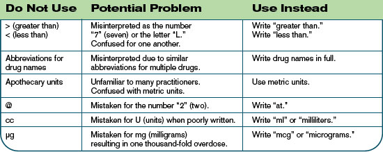
A New Approach
As part of efforts to improve patient safety, the Joint Commission has long worked with hospitals to develop practical, cost-effective strategies that can be implemented at organizations regardless of unique characteristics, such as ownership, size, or location. One such Joint Commission initiative is a National Patient Safety Goal to improve communication. This goal and one of its requirements specifically addresses the role that abbreviations, acronyms, symbols, and dose designations play in medication errors.
The Joint Commission began establishing National Patient Safety Goals in 2002 as a means to target critical areas where patient safety can be improved through specific action in healthcare organizations. The resulting National Patient Safety Goals are designed to give focus to evidence-based or expert consensus-based, well-defined, practical, and cost-effective actions that have potential for significant improvement in the safety of individuals receiving care. New Goals are recommended annually by the Sentinel Event Advisory Group, a Joint Commission-appointed, multidisciplinary group of patient safety experts.
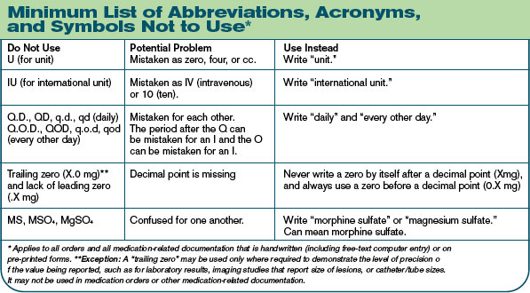
JCAHO Expectations
In order to comply with the National Patient Safety Goal related to abbreviations, an organization must conduct a thorough review of its approved abbreviation list and develop a list of unacceptable abbreviations and symbols with the involvement of physicians. In addition, organizations must do the following to meet this goal:
- The list of prohibited abbreviations, acronyms, symbols, and dose designations must be implemented for all handwritten, patient-specific communications, not just medication orders;
- These requirements apply to printed or electronic communications;
- This goal requires organizations to achieve 100% compliance with a reasonably comprehensive list of prohibited dangerous abbreviations, acronyms, symbols, and dose designations. This list need not be as extensive as some published lists, but must, at a minimum, include a set of Joint Commission-specified dangerous abbreviations, acro-nyms, symbols, and dose designations (see “Minimum List of Abbreviations, Acronyms, and Symbols Not to Use,” top right) and
- An abbreviation on the “do not use” list should not be used in any of its forms—uppercase or lowercase, with or without periods.
In addition to this minimum list, each organization should consider which abbreviations, acronyms, symbols, and dose designations it commonly uses; examine the risks associated with usage; and develop strategies to reduce usage. Hospitals also may wish to look to expert resources such as the Institute of Safe Medication Practices (ISMP)—available at www.ismp.org/Tools/abbreviationslist.pdf—and the National Coordinating Council for Medication Error Reporting and Prevention (NCC MERP)—available at www.nccmerp.org/dangerousAbbrev.html—to develop a list of prohibited abbreviations. Finally, organizations may wish to consider the Joint Commission list (see table below) of abbreviations, symbols, and acronyms for future possible inclusion in the official “do not use” list.
Risk Reduction Strategies
To comply with this National Patient Safety Goal Requirement, hospitals may wish to consider implementing the following risk reduction strategies:
- Examine medication error data in the organization. By identifying and selecting an organization-specific set of prohibited abbreviations, it will be much easier to gain support to eliminate certain abbreviations that have been found to be problematic at that organization.
- Provide simplified alternative abbreviations. For example, some staff members may resist writing out “international units” in place of “IU.” A simpler alternative such as “Intl Units” may be a solution.
- Make the list visible. Print the list on brightly colored paper or stickers and place it in patient charts.
- Provide staff with pocket-sized cards with the “do not use” list.
- Print the list in the margin or bottom of the physician order sheets and/or progress notes.
- Attach laminated copies of the list to the back of the physician order divider in the patient chart.
- Send monthly reminders to staff.
- Delete prohibited abbreviations from preprinted order sheets and other forms.
- Work with software vendors to ensure changes are made to be consistent with the list.
- Take a digital picture or scan the document containing the prohibited abbreviation and send it via e-mail directly to the offending prescriber to call attention to the issue.
- Direct the pharmacy not to accept any of the prohibited abbreviations. Orders with dangerous abbreviations or illegible handwriting must be corrected before being dispensed.
- Conduct a mock survey to test staff knowledge.
- At every staff meeting give patient safety updates, including information about the prohibited abbreviations.
- Ask all staff to sign a statement that he or she has received the list and agrees not to use the abbreviations.
- Promote a “do-not-use abbreviation of the month” policy.
- Develop and implement a policy to ensure that staff refer to the list and take steps to ensure compliance. Consider including a policy that states if an unacceptable abbreviation is used, the prescriber verifies the prescription order before it is filled.
- Monitor staff compliance with the list and offer additional education and training, as appropriate.
Conclusion
During the past decade, healthcare providers have been searching for more effective ways to reduce the risk of systems breakdowns that result in serious harm to patients. The Joint Commission is committed to working with organizations through the accreditation process on ways to anticipate and prevent errors. National Patient Safety Goals, such as the one associated with prohibited abbreviations, are one such method to promote specific improvements in patient safety. By using the principals of sound system design, organizations and providers further strengthen foundations that support safe, high-quality care. TH
Dr. Jacott is special advisor for professional relations for the Joint Commission on Accreditation of Healthcare Organizations.
Despite repeated warnings for more than 25 years by the Institute for Safe Medication Practices (ISMP) and other organizations, one of the major causes of medication errors is the ongoing use of potentially dangerous abbreviations and dose expressions. Root cause analyses information contained in the Joint Commission Sentinel Event Database shows that the underlying factors contributing to many of these medication errors are illegible or confusing handwriting by clinicians and the failure of healthcare providers to communicate clearly with one another.
Symbols and abbreviations are frequently used to save time and effort when writing prescriptions and documenting in patient charts; however, some symbols and abbreviations have the potential for misinterpretation or confusion. Examples of especially problematic abbreviations include “U” for “units” and “µg” for “micrograms.” When “U” is handwritten, it can often look like a zero. There are numerous case reports where the root cause of sentinel events related to insulin dosage has been the interpretation of a “U” as a zero. Using the abbreviation “µg” instead of “mcg” has also been the source of errors because when handwritten, the symbol “µ” can look like an “m.” The use of trailing zeros (e.g., 2.0 versus 2) or use of a leading decimal point without a leading zero (e.g. .2 instead of 0.2) are other dangerous order-writing practices. The decimal point is sometimes not seen when orders are handwritten using trailing zeros or no leading zeros. Misinterpretation of such orders could lead to a 10-fold dosing error.
A New Approach
As part of efforts to improve patient safety, the Joint Commission has long worked with hospitals to develop practical, cost-effective strategies that can be implemented at organizations regardless of unique characteristics, such as ownership, size, or location. One such Joint Commission initiative is a National Patient Safety Goal to improve communication. This goal and one of its requirements specifically addresses the role that abbreviations, acronyms, symbols, and dose designations play in medication errors.
The Joint Commission began establishing National Patient Safety Goals in 2002 as a means to target critical areas where patient safety can be improved through specific action in healthcare organizations. The resulting National Patient Safety Goals are designed to give focus to evidence-based or expert consensus-based, well-defined, practical, and cost-effective actions that have potential for significant improvement in the safety of individuals receiving care. New Goals are recommended annually by the Sentinel Event Advisory Group, a Joint Commission-appointed, multidisciplinary group of patient safety experts.
JCAHO Expectations
In order to comply with the National Patient Safety Goal related to abbreviations, an organization must conduct a thorough review of its approved abbreviation list and develop a list of unacceptable abbreviations and symbols with the involvement of physicians. In addition, organizations must do the following to meet this goal:
- The list of prohibited abbreviations, acronyms, symbols, and dose designations must be implemented for all handwritten, patient-specific communications, not just medication orders;
- These requirements apply to printed or electronic communications;
- This goal requires organizations to achieve 100% compliance with a reasonably comprehensive list of prohibited dangerous abbreviations, acronyms, symbols, and dose designations. This list need not be as extensive as some published lists, but must, at a minimum, include a set of Joint Commission-specified dangerous abbreviations, acro-nyms, symbols, and dose designations (see “Minimum List of Abbreviations, Acronyms, and Symbols Not to Use,” top right) and
- An abbreviation on the “do not use” list should not be used in any of its forms—uppercase or lowercase, with or without periods.
In addition to this minimum list, each organization should consider which abbreviations, acronyms, symbols, and dose designations it commonly uses; examine the risks associated with usage; and develop strategies to reduce usage. Hospitals also may wish to look to expert resources such as the Institute of Safe Medication Practices (ISMP)—available at www.ismp.org/Tools/abbreviationslist.pdf—and the National Coordinating Council for Medication Error Reporting and Prevention (NCC MERP)—available at www.nccmerp.org/dangerousAbbrev.html—to develop a list of prohibited abbreviations. Finally, organizations may wish to consider the Joint Commission list (see table below) of abbreviations, symbols, and acronyms for future possible inclusion in the official “do not use” list.
Risk Reduction Strategies
To comply with this National Patient Safety Goal Requirement, hospitals may wish to consider implementing the following risk reduction strategies:
- Examine medication error data in the organization. By identifying and selecting an organization-specific set of prohibited abbreviations, it will be much easier to gain support to eliminate certain abbreviations that have been found to be problematic at that organization.
- Provide simplified alternative abbreviations. For example, some staff members may resist writing out “international units” in place of “IU.” A simpler alternative such as “Intl Units” may be a solution.
- Make the list visible. Print the list on brightly colored paper or stickers and place it in patient charts.
- Provide staff with pocket-sized cards with the “do not use” list.
- Print the list in the margin or bottom of the physician order sheets and/or progress notes.
- Attach laminated copies of the list to the back of the physician order divider in the patient chart.
- Send monthly reminders to staff.
- Delete prohibited abbreviations from preprinted order sheets and other forms.
- Work with software vendors to ensure changes are made to be consistent with the list.
- Take a digital picture or scan the document containing the prohibited abbreviation and send it via e-mail directly to the offending prescriber to call attention to the issue.
- Direct the pharmacy not to accept any of the prohibited abbreviations. Orders with dangerous abbreviations or illegible handwriting must be corrected before being dispensed.
- Conduct a mock survey to test staff knowledge.
- At every staff meeting give patient safety updates, including information about the prohibited abbreviations.
- Ask all staff to sign a statement that he or she has received the list and agrees not to use the abbreviations.
- Promote a “do-not-use abbreviation of the month” policy.
- Develop and implement a policy to ensure that staff refer to the list and take steps to ensure compliance. Consider including a policy that states if an unacceptable abbreviation is used, the prescriber verifies the prescription order before it is filled.
- Monitor staff compliance with the list and offer additional education and training, as appropriate.
Conclusion
During the past decade, healthcare providers have been searching for more effective ways to reduce the risk of systems breakdowns that result in serious harm to patients. The Joint Commission is committed to working with organizations through the accreditation process on ways to anticipate and prevent errors. National Patient Safety Goals, such as the one associated with prohibited abbreviations, are one such method to promote specific improvements in patient safety. By using the principals of sound system design, organizations and providers further strengthen foundations that support safe, high-quality care. TH
Dr. Jacott is special advisor for professional relations for the Joint Commission on Accreditation of Healthcare Organizations.