User login
Two new drugs
Two new drugs have arrived to challenge our prescription pad or electronic record, depending on which you use. Empagliflozin (Jardiance), a drug used to modify glucose metabolism in type 2 diabetes in patients with preexisting cardiovascular disease, demonstrated a decrease in cardiovascular mortality. The other drug, Entresto, appears to provide an added benefit in heart failure therapy and was compared with a standard ACE inhibitor.
The results of EMPA-REG OUTCOME, reported at the European Association for the Study of Diabetes meeting in Stockholm, showed empagliflozin to be the first drug to decrease the mortality and morbidity of cardiovascular disease in diabetes. It is one of a group of new sodium-glucose cotransporter 2 (SGLT-2) blockers being tested in type 2 diabetes with established cardiovascular disease. Patients were randomized to placebo or empagliflozin while receiving standard medical and cardiovascular medications. After 3 years of follow-up, patients receiving the drug experienced a lower cardiovascular mortality rate, compared with placebo patients (3.7% vs. 5.9%, a 38% reduction) in addition to a decrease in hospitalization for heart failure and death from any cause. No effect was observed on the incidence of myocardial infarction or stroke. In addition, the drug also lowered blood glucose and blood pressure and led to some significant weight loss. The drug also was shown to decrease vascular resistance and albuminuria (N Engl J Med. 2015. 373:2117-2).

Furthermore, an analysis of EMPA-REG OUTCOME presented in November at the American Society of Nephrology meeting in San Diego, showed a profound benefit on the new onset and progression of chronic renal disease in diabetes patients. The importance of these results needs emphasis. Up until recently, the Food and Drug Administration has given a pass to diabetes drugs in regard to cardiovascular endpoints; approval has been based on their primary effect on lowering blood glucose. Some drugs in the past, such as rosiglitazone, actually have shown an increase in mortality in some diabetes patients. At long last, FDA approval for diabetes drugs hinges on acceptable outcomes in cardiovascular endpoints. The addition of a drug that can actually affect cardiovascular mortality and morbidity, the major risk factor of diabetes, provides an important addition to therapy.
The other drug that provides a choice of drugs for the treatment of heart failure is Entresto, a combination of sacubitril, a neprilysin inhibitor, and the angiotensin receptor inhibitor valsartan, approved in July 2015. In PARADIGM-HF, the compound was compared to enalapril in the treatment of patients with class II, III, and IV heart failure who were also receiving beta-blockers. Entresto-treated patients reported a 21.8% incidence of the primary outcome measure, cardiovascular death and hospitalization for heart failure, compared with the enalapril alone incidence of 26.5% (P less than .001) (N Engl J Med. 2014;371:993-1004). Investigators initially excluded 11.4% of the recruited patients from the study who could not tolerate Entresto or enalapril therapy. The drugs were well tolerated without any adverse reactions during therapy. Entresto was more effective than enalapril in regards to the occurrence of heart failure and death from any cause over a 27-month average follow-up.
The observations in this study emphasize how much the mortality of heart failure has decreased over the last decade. Cardiovascular deaths have decreased to roughly 7% per year and rehospitalization occurs in about 8% in the first year. Both drugs provide an important incremental benefit in heart failure patients.
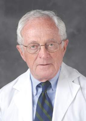
Dr. Goldstein, medical editor of Cardiology News, is professor of medicine at Wayne State University and division head emeritus of cardiovascular medicine at Henry Ford Hospital, both in Detroit. He is on data safety monitoring committees for the National Institutes of Health and several pharmaceutical companies.
Two new drugs have arrived to challenge our prescription pad or electronic record, depending on which you use. Empagliflozin (Jardiance), a drug used to modify glucose metabolism in type 2 diabetes in patients with preexisting cardiovascular disease, demonstrated a decrease in cardiovascular mortality. The other drug, Entresto, appears to provide an added benefit in heart failure therapy and was compared with a standard ACE inhibitor.
The results of EMPA-REG OUTCOME, reported at the European Association for the Study of Diabetes meeting in Stockholm, showed empagliflozin to be the first drug to decrease the mortality and morbidity of cardiovascular disease in diabetes. It is one of a group of new sodium-glucose cotransporter 2 (SGLT-2) blockers being tested in type 2 diabetes with established cardiovascular disease. Patients were randomized to placebo or empagliflozin while receiving standard medical and cardiovascular medications. After 3 years of follow-up, patients receiving the drug experienced a lower cardiovascular mortality rate, compared with placebo patients (3.7% vs. 5.9%, a 38% reduction) in addition to a decrease in hospitalization for heart failure and death from any cause. No effect was observed on the incidence of myocardial infarction or stroke. In addition, the drug also lowered blood glucose and blood pressure and led to some significant weight loss. The drug also was shown to decrease vascular resistance and albuminuria (N Engl J Med. 2015. 373:2117-2).
Furthermore, an analysis of EMPA-REG OUTCOME presented in November at the American Society of Nephrology meeting in San Diego, showed a profound benefit on the new onset and progression of chronic renal disease in diabetes patients. The importance of these results needs emphasis. Up until recently, the Food and Drug Administration has given a pass to diabetes drugs in regard to cardiovascular endpoints; approval has been based on their primary effect on lowering blood glucose. Some drugs in the past, such as rosiglitazone, actually have shown an increase in mortality in some diabetes patients. At long last, FDA approval for diabetes drugs hinges on acceptable outcomes in cardiovascular endpoints. The addition of a drug that can actually affect cardiovascular mortality and morbidity, the major risk factor of diabetes, provides an important addition to therapy.
The other drug that provides a choice of drugs for the treatment of heart failure is Entresto, a combination of sacubitril, a neprilysin inhibitor, and the angiotensin receptor inhibitor valsartan, approved in July 2015. In PARADIGM-HF, the compound was compared to enalapril in the treatment of patients with class II, III, and IV heart failure who were also receiving beta-blockers. Entresto-treated patients reported a 21.8% incidence of the primary outcome measure, cardiovascular death and hospitalization for heart failure, compared with the enalapril alone incidence of 26.5% (P less than .001) (N Engl J Med. 2014;371:993-1004). Investigators initially excluded 11.4% of the recruited patients from the study who could not tolerate Entresto or enalapril therapy. The drugs were well tolerated without any adverse reactions during therapy. Entresto was more effective than enalapril in regards to the occurrence of heart failure and death from any cause over a 27-month average follow-up.
The observations in this study emphasize how much the mortality of heart failure has decreased over the last decade. Cardiovascular deaths have decreased to roughly 7% per year and rehospitalization occurs in about 8% in the first year. Both drugs provide an important incremental benefit in heart failure patients.
Dr. Goldstein, medical editor of Cardiology News, is professor of medicine at Wayne State University and division head emeritus of cardiovascular medicine at Henry Ford Hospital, both in Detroit. He is on data safety monitoring committees for the National Institutes of Health and several pharmaceutical companies.
Q.N.S.
In the early 1970’s, the three letters that a pediatric house officer hated to see on a slip returning from the lab were Q.N.S. Quality Not Sufficient meant that the minutes, which seemed like hours, you had invested torturing some poor sick child to obtain just a few cc’s of blood had been wasted. It also meant returning to the patient’s crib or bedside to explain to the child and her parents that the torture you had promised was over for the day was in fact not over.
Tourniquets were fished out of lapel buttonholes, and the search for a decent vein had to begin all over again. If the child was chubby or bloated with retained fluid, those veins were invisible. If the child had been ill for weeks – particularly if the patient had been on chemotherapy – all of the good veins had been blown or had clotted days ago.

Many of the patients were saintly and eerily cooperative despite your fumbling attempts at venipuncture, but most were not. Some parents were so supportive of your efforts that you wanted to hug them when the ordeal was over (and you did). A few parents amped up the tension at the bedside so much that you wanted to ask them to leave (but you didn’t). If a parent was understandably incapable of effectively restraining the child, you needed to find an experienced nurse to help. A few of the best nurses were so good that the house officer merely needed to hold the needle still, and the child was repositioned in just the right orientation so that the puncture occurred miraculously.
There were some last ditch efforts at phlebotomy that were so ghastly that you had to ask the parents to leave. I don’t know if the infamous internal jugular stick is still used, but it wasn’t pretty. And it was almost as frightening for the physician holding the needle as it was for the patient. Even in the big teaching hospitals, dedicated phlebotomists hadn’t been invented yet. A few nurses had earned reputations as good vein finders, but for the most part it was on-the-job training for the house officers.
It was not until 1973 that Dr. John Broviac’s central line catheters became available in some hospitals and 1979 until Dr. Robert Hickman’s version appeared. It took a few more years before techniques were perfected for safely drawing specimens from these lines that had been originally intended for infusion. But for me and my cohort of house officers and our unfortunate patients, it was years too late. I am sure that caring for hospitalized pediatric cancer patients today continues to be dominated by challenges. But for those of us tasked with drawing blood from patients without the benefit of central line catheters, it was gut wrenching.
Those battles for a few cc’s of blood left their scars. I have seldom ordered any blood test without asking myself whether there wasn’t a bloodless way of assessing the patient’s condition. Or couldn’t we just do the test on a drop or two of blood? Of course, as I as finishing my training, more tests were downsized so that they could be done “micro.” But as you know, getting enough blood from a heel stick or finger prick isn’t always as easy as it sounds. If the child is shocky or cold, a good blood flow is hard to obtain. Warming helps but squeezing doesn’t because tissue juices can dilute the sample, and the trauma of squeezing can contaminate the sample.
A study published in the American Journal of Clinical Pathology raises the question of how accurately even a single drop of blood reflects what is going on in the patient’s total blood pool (“Drop by drop variation in the cellular components of fingerprick blood: Implications for point-of-care diagnostic development” [Am J Clin Pathol. 2015 Dec;144(6):885-94]). Two bioengineers from Rice University discovered that six successive drops of blood from a single finger prick varied by a significant amount when analyzed for a variety of cellular components. For example, the drop-to-drop variability for hemoglobin was five times that of a sample collected by venipuncture.
You and I may dream of the day when just a drop will do it and we can put our needles away for good. Unfortunately, for now, the answer is that a single drop of blood is a Q.N.S.
Dr. Wilkoff practiced primary care pediatrics in Brunswick, Maine, for nearly 40 years. He has authored several books on behavioral pediatrics including “How to Say No to Your Toddler.”
In the early 1970’s, the three letters that a pediatric house officer hated to see on a slip returning from the lab were Q.N.S. Quality Not Sufficient meant that the minutes, which seemed like hours, you had invested torturing some poor sick child to obtain just a few cc’s of blood had been wasted. It also meant returning to the patient’s crib or bedside to explain to the child and her parents that the torture you had promised was over for the day was in fact not over.
Tourniquets were fished out of lapel buttonholes, and the search for a decent vein had to begin all over again. If the child was chubby or bloated with retained fluid, those veins were invisible. If the child had been ill for weeks – particularly if the patient had been on chemotherapy – all of the good veins had been blown or had clotted days ago.
Many of the patients were saintly and eerily cooperative despite your fumbling attempts at venipuncture, but most were not. Some parents were so supportive of your efforts that you wanted to hug them when the ordeal was over (and you did). A few parents amped up the tension at the bedside so much that you wanted to ask them to leave (but you didn’t). If a parent was understandably incapable of effectively restraining the child, you needed to find an experienced nurse to help. A few of the best nurses were so good that the house officer merely needed to hold the needle still, and the child was repositioned in just the right orientation so that the puncture occurred miraculously.
There were some last ditch efforts at phlebotomy that were so ghastly that you had to ask the parents to leave. I don’t know if the infamous internal jugular stick is still used, but it wasn’t pretty. And it was almost as frightening for the physician holding the needle as it was for the patient. Even in the big teaching hospitals, dedicated phlebotomists hadn’t been invented yet. A few nurses had earned reputations as good vein finders, but for the most part it was on-the-job training for the house officers.
It was not until 1973 that Dr. John Broviac’s central line catheters became available in some hospitals and 1979 until Dr. Robert Hickman’s version appeared. It took a few more years before techniques were perfected for safely drawing specimens from these lines that had been originally intended for infusion. But for me and my cohort of house officers and our unfortunate patients, it was years too late. I am sure that caring for hospitalized pediatric cancer patients today continues to be dominated by challenges. But for those of us tasked with drawing blood from patients without the benefit of central line catheters, it was gut wrenching.
Those battles for a few cc’s of blood left their scars. I have seldom ordered any blood test without asking myself whether there wasn’t a bloodless way of assessing the patient’s condition. Or couldn’t we just do the test on a drop or two of blood? Of course, as I as finishing my training, more tests were downsized so that they could be done “micro.” But as you know, getting enough blood from a heel stick or finger prick isn’t always as easy as it sounds. If the child is shocky or cold, a good blood flow is hard to obtain. Warming helps but squeezing doesn’t because tissue juices can dilute the sample, and the trauma of squeezing can contaminate the sample.
A study published in the American Journal of Clinical Pathology raises the question of how accurately even a single drop of blood reflects what is going on in the patient’s total blood pool (“Drop by drop variation in the cellular components of fingerprick blood: Implications for point-of-care diagnostic development” [Am J Clin Pathol. 2015 Dec;144(6):885-94]). Two bioengineers from Rice University discovered that six successive drops of blood from a single finger prick varied by a significant amount when analyzed for a variety of cellular components. For example, the drop-to-drop variability for hemoglobin was five times that of a sample collected by venipuncture.
You and I may dream of the day when just a drop will do it and we can put our needles away for good. Unfortunately, for now, the answer is that a single drop of blood is a Q.N.S.
Dr. Wilkoff practiced primary care pediatrics in Brunswick, Maine, for nearly 40 years. He has authored several books on behavioral pediatrics including “How to Say No to Your Toddler.”

A Perfect Storm: Patterns of care
Editor’s Note: This is the third installment of a five-part monthly series that will discuss the pathologic, genomic, and health system factors that contribute to the racial survival disparity in breast cancer. The series, which is adapted from an article that originally appeared in CA: A Cancer Journal for Clinicians1, a journal of the American Cancer Society, will also review exciting and innovative interventions to close the survival gap. This month’s column reviews patterns of care – the second element in the perfect storm.
Mammography
Despite advances in breast cancer imaging technology, the mainstay of breast cancer screening has remained mammography. Chu et al.2 found that African American women have less early-stage disease in every age group for each hormone receptor status, and this raises the concern that mammography screening might be inadequate in this population. Although historically, African American women used mammography less than did white women, this difference has fortunately disappeared with time.3 According to results from the 2010 National Health Interview Survey, among women who were 40 years or older, 50.6% of non-Hispanic African Americans and 51.5% of non-Hispanic whites reported having had a mammogram within the past year.4
Although mammography uptake may be similar between these groups, there are still differences both in quality and in follow-up of abnormal imaging results. A study of mammography capacity and quality in a large urban setting found that the facilities that served predominantly minority women were more likely to be public institutions (31% vs. 0%) and less likely to be academic (27% vs. 71%), less likely to have digital mammography (18% vs. 71%), and less likely to have dedicated breast imaging specialists reading the films (23% vs. 87%). The authors concluded that the mammography process was broken, with quality differences in the manner in which the centers provided care and reported results.5

The accompanying graphic illustrates the disparities seen in breast cancer mammography and care for women in underserved communities on Chicago’s South Side. As the figure demonstrates, there are fewer mammography centers on the city’s South Side, with the concentration of breast cancer imaging and treatment resources localized in the more affluent communities of central and northern Chicago. A total of 300,000 women who were eligible for screening went unscreened because of improper management of resources.
Highlighting the importance of location in breast cancer care, Gehlert et al.6 asserted that ensuring that inner-city health facilities have up-to-date, well-maintained equipment and that mammographers have access to continuing training and opportunities for consultation should help reduce breast cancer mortality in African Americans.
With respect to follow-up of abnormal imaging results, a large retrospective cohort study of 6,722 women with abnormal mammogram results seen at a New York academic medical center from January 2002 through December 2002 found longer times to diagnostic follow-up for African American versus white women. The median number of days to diagnostic follow-up was 20 for African American patients versus 14 for white patients. In addition, racial disparities remained significant after the researchers controlled for age, Breast Imaging Reporting and Data System (BI-RADS) category, insurance status, provider practice location, and median household income. More important, in women with a BI-RADS classification of 4 or 5 – signifying a lesion seen on mammography that is either suspicious for or highly suggestive of malignancy, respectively – the median number of days to follow-up among those without same-day additional imaging was 26 for African Americans and 14 for whites (P < .05).7
Delays in treatment
A cascade of delays also has been documented in breast cancer care for African American women. Silber et al.8 investigated factors associated with differences in breast cancer outcomes in a large population-based study using Surveillance, Epidemiology, and End Results (SEER)-Medicare data. The mean time from diagnosis to treatment was 29.2 days for African Americans versus 22.5 days for whites (P < .001). The authors also found that African Americans were more likely to have very-long treatment delays. At least 6% of African Americans did not initiate treatment within the first 3 months of diagnosis, whereas only 3% of whites failed to start treatment (P < .001). Gwyn et al.9 also found potentially clinically significant treatment delays more often for African American women than for white women. The time from medical consultation to the initiation of treatment was longer than 3 months for 22.4% of African American women versus 14.3% of white women. Three months was chosen as a clinically significant time period, because Richards et al.10 demonstrated that a delay ≥ to 3 months affects survival. Thus, delays in the diagnosis and treatment of African American women are factors that worsen the survival gap.
Misuse of treatment
Once treatment is initiated, African Americans often receive inappropriate therapy, studies have demonstrated. In a prospective analysis of 957 patients in 101 oncology practices, Griggs et al.11 found more frequent use of non–guideline concordant adjuvant chemotherapy regimens in African American women. In a univariate analysis, African American patients were more likely than were whites to receive a nonstandard regimen (19% vs. 11%; P = .047). Although we will discuss further in this column whether guidelines based on clinical trials are appropriate for African American patients, the study demonstrates that these women are not uniformly receiving standard-of-care treatment.
Underuse of treatment
In addition to misuse of treatment, studies also have examined undertreatment of African American patients with breast cancer. One study investigated chemotherapy administration among African American patients with stage I-III breast cancer at 10 different treatment sites. Compared with white patients, African Americans received a lower dose proportion (actual vs. expected dose) and lower relative dose intensity.
The authors found that between-group differences in biological and medical characteristics, such as tolerance of therapy, comorbidities, and leukocyte counts, did not explain these variations in treatment. In fact, despite the association between lower leukocyte counts and African American ethnicity, there was no evidence that white blood cell levels accounted for the difference in dose proportion or relative dose intensity. Significantly, the authors discovered that more African Americans had chemotherapy dose reductions in the first cycle of treatment, perhaps indicating physician assumptions regarding African American patients’ ability to tolerate chemotherapy.12
Silber et al.8 also examined differences in the administration of chemotherapy between white and African American breast cancer patients. The authors found that 3.7% of African Americans received both an anthracycline and a taxane; that figure rose to 5.0% among whites who were matched to African Americans at presentation.
Bickell et al.13 explored further racial disparity in the underuse of adjuvant breast cancer treatment. The researchers examined the medical records of 677 women treated surgically for stage I or II breast cancer. The study defined underuse as omissions of radiotherapy after breast-conserving surgery, adjuvant chemotherapy after resection of hormone receptor–negative tumors ≥ 1 cm, or hormonal therapy for receptor-positive tumors ≥ 1 cm. Underuse of appropriate adjuvant treatment was found in 34% of African American patients versus 16% of white patients (P less than .001). There were racial disparities present in all three adjuvant therapies assessed.
Hormonal therapy has been shown effective in clinical trials for preventing breast cancer recurrence and death in women with early-stage breast cancer.14 The study by Bickell et al.13 documented underuse of this treatment in African American patients. Partridge et al.15 conducted the largest study of oral antineoplastic use outside of a clinical trial setting. Their study consisted of 2,378 primary breast cancer patients enrolled in New Jersey’s Medicaid or pharmaceutical assistance program; the main outcome was the number of days covered by filled tamoxifen prescriptions in the first year of therapy. The study found that nonwhite patients had significantly lower adherence rates than did whites. Although further investigation is needed to determine the drivers of this nonadherence in African American patients, medication cost has been proposed as a significant factor leading to underuse of these agents. Streeter et al.16 analyzed a nationally representative pharmacy claims database for oral antineoplastics and calculated abandonment rates for the initial claim. Not surprisingly, high cost sharing and low incomes were associated with a higher abandonment rate (P < .05). Despite being an important component of health equity research, treatment adherence has been identified by the Association of American Medical Colleges as a critically underrepresented area of disparities-focused health services research.17 More attention to this area is needed to understand the underuse of hormonal therapies in African American breast cancer patients.
The treatment strategies that have been shown to be delayed, underused, or misused in African American patients in the aforementioned studies have improved disease-free and overall survival in large randomized trials. Furthermore, diminished total dose and dose intensity of adjuvant chemotherapy both have been associated with lower breast cancer survival rates.18,19 These quality-of-care failures in breast cancer treatment for minority patients are thought to partially explain the survival disparity between African Americans and whites. It has been proposed that patients in both groups derive a similar benefit from systemic therapy when it is administered in accordance with their clinical and pathologic presentation,20 but that assumption becomes more nuanced when the clinical trial experience is reviewed.
Clinical trial experience
Dignam20 examined survival by race in several National Surgical Adjuvant Breast and Bowel Project trials. He found that the benefit from systemic adjuvant therapy for reductions in disease recurrence and mortality was comparable between African American and white patients. His survey of trials consistently indicated equivalent disease-free survival, but a mortality deficit for African Americans also was found consistently. Among African Americans, the excess risk of mortality was 21% for those who were lymph node–negative and 17% for those who were lymph node–positive. The excess mortality risk was thought to be attributable to greater mortality from noncancer causes among African American patients rather than a failure of African Americans to respond to breast cancer treatment.
In contrast to Dignam’s findings20, Hershman et al.21 assessed the association between race and treatment discontinuation/delay, white blood cell counts, and survival in women enrolled in the Southwest Oncology Group adjuvant breast cancer trials. The study found that African American women were significantly more likely to experience treatment discontinuation/delay than were white women (87% vs. 81%, respectively; P = .04). These delays were not accounted for by toxicities, which were experienced in similar proportions by race. African American women also were more likely to miss appointments (19% vs. 9%; P = .0002); perhaps, as Hassett and Griggs22 speculated, this finding speaks to economic barriers, including the inability to arrange alternate child care, miss work, or afford transportation to the clinic. Despite these barriers to care for African American patients, they still received the same mean relative dose intensity (87% vs. 86%).
In their survival analysis, Hershman et al.21 controlled for treatment-related factors such as dose reductions and delays, body surface area, baseline white blood cell counts, and other predictors of survival and still found that African Americans had worse disease-free and overall survival than did white women. The authors concluded that the study was “unable to demonstrate that any factor related to treatment quality or delivery contributed to racial differences in survival between the groups.”21 The study thus established two important findings related to the disparity gap. First, even in the controlled setting of a clinical trial, African American patients faced barriers to optimal treatment,22 and second, despite attempts to control for treatment quality and delivery, African American women still had worse outcomes. These findings suggest that tumor biology and genomics remain important.
In next month’s installment, we will discuss interventions aimed at closing the racial survival disparity in breast cancer. Eliminating racial disparities in cancer mortality through effective interventions has become an increasingly important imperative in federal, state, and community health care programs.
Other installments of this column can be found in the Related Content box.
1. Daly B, Olopade OI. A perfect storm: How tumor biology, genomics, and health care delivery patterns collide to create a racial survival disparity in breast cancer and proposed interventions for change. CA Cancer J Clin. 2015 May-Jun;65(3):221-38.
2. Chu KC, Lamar CA, Freeman HP. Racial disparities in breast carcinoma survival rates: Separating factors that affect diagnosis from factors that affect treatment. Cancer. 2003 Jun;97(11):2853-60.
3. DeLancey JO, Thun MJ, Jemal A, Ward EM. Recent trends in black-white disparities in cancer mortality. Cancer Epidemiol Biomarkers Prev. 2008 Nov;17(11):2908-12.
4. DeSantis C, Naishadham D, Jemal A. Cancer statistics for African Americans, 2013. CA Cancer J Clin. 2013 Nov;63(3):151-66.
5. Ansell D, Grabler P, Whitman S, et al. A community effort to reduce the black/white breast cancer mortality disparity in Chicago. Cancer Causes Control. 2009 Nov;20(9):1681-8.
6. Gehlert S, Sohmer D, Sacks T, Mininger C, McClintock M, Olopade O. Targeting health disparities: a model linking upstream determinants to downstream interventions. Health Aff (Millwood). 2008 Mar-Apr;27(2):339-49.
7. Press R, Carrasquillo O, Sciacca RR, Giardina EG. Racial/ethnic disparities in time to follow-up after an abnormal mammogram. J Womens Health (Larchmt). 2008 Jul;17(6):923-30.
8. Silber JH, Rosenbaum PR, Clark AS, et al. Characteristics associated with differences in survival among black and white women with breast cancer. JAMA. 2013 Jul;310(4):389-397.
9. Gwyn K, Bondy ML, Cohen DS, et al. Racial differences in diagnosis, treatment, and clinical delays in a population-based study of patients with newly diagnosed breast carcinoma. Cancer. 2004 Apr;100(8):1595-604.
10. Richards MA, Westcombe AM, Love SB, Littlejohns P, Ramirez AJ. Influence of delay on survival in patients with breast cancer: a systematic review. Lancet. 1999 Apr 3;353(9159):1119-26.
11. Griggs JJ, Culakova E, Sorbero ME, et al. Social and racial differences in selection of breast cancer adjuvant chemotherapy regimens. J Clin Oncol. 2007 Jun 20;25(18):2522-7.
12. Griggs JJ, Sorbero ME, Stark AT, Heininger SE, Dick AW. Racial disparity in the dose and dose intensity of breast cancer adjuvant chemotherapy. Breast Cancer Res Treat. 2003 Sep;81(1):21-31.
13. Bickell NA, Wang JJ, Oluwole S, et al. Missed opportunities: racial disparities in adjuvant breast cancer treatment. J Clin Oncol. 2006 Mar 20;24(9):1357-62. 14. Fisher B, Costantino J, Redmond C, et al. A randomized clinical trial evaluating tamoxifen in the treatment of patients with node-negative breast cancer who have estrogen-receptor-positive tumors. N Engl J Med. 1989 Feb 23;320(8):479-84.
15. Partridge AH, Wang PS, Winer EP, Avorn J. Nonadherence to adjuvant tamoxifen therapy in women with primary breast cancer. J Clin Oncol. 2003 Feb 15;21(4):602-6.
16. Streeter SB, Schwartzberg L, Husain N, Johnsrud M. Patient and plan characteristics affecting abandonment of oral oncolytic prescriptions. J Oncol Pract. 2011 Jul;7(3 Suppl):46s-51s.
17. Alberti PM KN, Sutton K, Johnson BH, Holve E. The state of health equity research: closing knowledge gaps to address inequities. ©2014 Association of American Medical Colleges. May not be reproduced or distributed without prior permission.
18. Wood WC, Budman DR, Korzun AH, et al. Dose and dose intensity of adjuvant chemotherapy for stage II, node-positive breast carcinoma. N Engl J Med. 1994 May 5;330(18):1253-9.
19. Budman DR, Berry DA, Cirrincione CT, et al. Dose and dose intensity as determinants of outcome in the adjuvant treatment of breast cancer. The Cancer and Leukemia Group B. J Natl Cancer Inst. 1998 Aug 19;90(16):1205-11.
20. Dignam JJ. Efficacy of systemic adjuvant therapy for breast cancer in African-American and Caucasian women. J Natl Cancer Inst Monogr. 2001(30):36-43.
21. Hershman DL, Unger JM, Barlow WE, et al. Treatment quality and outcomes of African American versus white breast cancer patients: retrospective analysis of Southwest Oncology studies S8814/S8897. J Clin Oncol. 2009 May;27(13):2157-62.
22. Hassett MJ, Griggs JJ. Disparities in breast cancer adjuvant chemotherapy: moving beyond yes or no. J Clin Oncol. 2009 May 1;27(13):2120-1.
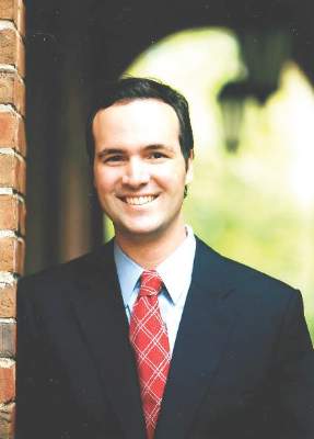
Bobby Daly, MD, MBA, is the chief fellow in the section of hematology/oncology at the University of Chicago Medicine. His clinical focus is breast and thoracic oncology, and his research focus is health services. Specifically, Dr. Daly researches disparities in oncology care delivery, oncology health care utilization, aggressive end-of-life oncology care, and oncology payment models. He received his MD and MBA from Harvard Medical School and Harvard Business School, both in Boston, and a BA in Economics and History from Stanford (Calif.) University. He was the recipient of the Dean’s Award at Harvard Medical and Business Schools.

Olufunmilayo Olopade, MD, FACP, OON, is the Walter L. Palmer Distinguished Service Professor of Medicine and Human Genetics, and director, Center for Global Health at the University of Chicago. She is adopting emerging high throughput genomic and informatics strategies to identify genetic and nongenetic risk factors for breast cancer in order to implement precision health care in diverse populations. This innovative approach has the potential to improve the quality of care and reduce costs while saving more lives.
Disclosures: Dr. Olopade serves on the Medical Advisory Board for CancerIQ. Dr. Daly serves as a director of Quadrant Holdings Corporation and receives compensation from this entity. Frontline Medical Communications is a subsidiary of Quadrant Holdings Corporation.
Published in conjunction with Susan G. Komen®.
Editor’s Note: This is the third installment of a five-part monthly series that will discuss the pathologic, genomic, and health system factors that contribute to the racial survival disparity in breast cancer. The series, which is adapted from an article that originally appeared in CA: A Cancer Journal for Clinicians1, a journal of the American Cancer Society, will also review exciting and innovative interventions to close the survival gap. This month’s column reviews patterns of care – the second element in the perfect storm.
Mammography
Despite advances in breast cancer imaging technology, the mainstay of breast cancer screening has remained mammography. Chu et al.2 found that African American women have less early-stage disease in every age group for each hormone receptor status, and this raises the concern that mammography screening might be inadequate in this population. Although historically, African American women used mammography less than did white women, this difference has fortunately disappeared with time.3 According to results from the 2010 National Health Interview Survey, among women who were 40 years or older, 50.6% of non-Hispanic African Americans and 51.5% of non-Hispanic whites reported having had a mammogram within the past year.4
Although mammography uptake may be similar between these groups, there are still differences both in quality and in follow-up of abnormal imaging results. A study of mammography capacity and quality in a large urban setting found that the facilities that served predominantly minority women were more likely to be public institutions (31% vs. 0%) and less likely to be academic (27% vs. 71%), less likely to have digital mammography (18% vs. 71%), and less likely to have dedicated breast imaging specialists reading the films (23% vs. 87%). The authors concluded that the mammography process was broken, with quality differences in the manner in which the centers provided care and reported results.5
The accompanying graphic illustrates the disparities seen in breast cancer mammography and care for women in underserved communities on Chicago’s South Side. As the figure demonstrates, there are fewer mammography centers on the city’s South Side, with the concentration of breast cancer imaging and treatment resources localized in the more affluent communities of central and northern Chicago. A total of 300,000 women who were eligible for screening went unscreened because of improper management of resources.
Highlighting the importance of location in breast cancer care, Gehlert et al.6 asserted that ensuring that inner-city health facilities have up-to-date, well-maintained equipment and that mammographers have access to continuing training and opportunities for consultation should help reduce breast cancer mortality in African Americans.
With respect to follow-up of abnormal imaging results, a large retrospective cohort study of 6,722 women with abnormal mammogram results seen at a New York academic medical center from January 2002 through December 2002 found longer times to diagnostic follow-up for African American versus white women. The median number of days to diagnostic follow-up was 20 for African American patients versus 14 for white patients. In addition, racial disparities remained significant after the researchers controlled for age, Breast Imaging Reporting and Data System (BI-RADS) category, insurance status, provider practice location, and median household income. More important, in women with a BI-RADS classification of 4 or 5 – signifying a lesion seen on mammography that is either suspicious for or highly suggestive of malignancy, respectively – the median number of days to follow-up among those without same-day additional imaging was 26 for African Americans and 14 for whites (P < .05).7
Delays in treatment
A cascade of delays also has been documented in breast cancer care for African American women. Silber et al.8 investigated factors associated with differences in breast cancer outcomes in a large population-based study using Surveillance, Epidemiology, and End Results (SEER)-Medicare data. The mean time from diagnosis to treatment was 29.2 days for African Americans versus 22.5 days for whites (P < .001). The authors also found that African Americans were more likely to have very-long treatment delays. At least 6% of African Americans did not initiate treatment within the first 3 months of diagnosis, whereas only 3% of whites failed to start treatment (P < .001). Gwyn et al.9 also found potentially clinically significant treatment delays more often for African American women than for white women. The time from medical consultation to the initiation of treatment was longer than 3 months for 22.4% of African American women versus 14.3% of white women. Three months was chosen as a clinically significant time period, because Richards et al.10 demonstrated that a delay ≥ to 3 months affects survival. Thus, delays in the diagnosis and treatment of African American women are factors that worsen the survival gap.
Misuse of treatment
Once treatment is initiated, African Americans often receive inappropriate therapy, studies have demonstrated. In a prospective analysis of 957 patients in 101 oncology practices, Griggs et al.11 found more frequent use of non–guideline concordant adjuvant chemotherapy regimens in African American women. In a univariate analysis, African American patients were more likely than were whites to receive a nonstandard regimen (19% vs. 11%; P = .047). Although we will discuss further in this column whether guidelines based on clinical trials are appropriate for African American patients, the study demonstrates that these women are not uniformly receiving standard-of-care treatment.
Underuse of treatment
In addition to misuse of treatment, studies also have examined undertreatment of African American patients with breast cancer. One study investigated chemotherapy administration among African American patients with stage I-III breast cancer at 10 different treatment sites. Compared with white patients, African Americans received a lower dose proportion (actual vs. expected dose) and lower relative dose intensity.
The authors found that between-group differences in biological and medical characteristics, such as tolerance of therapy, comorbidities, and leukocyte counts, did not explain these variations in treatment. In fact, despite the association between lower leukocyte counts and African American ethnicity, there was no evidence that white blood cell levels accounted for the difference in dose proportion or relative dose intensity. Significantly, the authors discovered that more African Americans had chemotherapy dose reductions in the first cycle of treatment, perhaps indicating physician assumptions regarding African American patients’ ability to tolerate chemotherapy.12
Silber et al.8 also examined differences in the administration of chemotherapy between white and African American breast cancer patients. The authors found that 3.7% of African Americans received both an anthracycline and a taxane; that figure rose to 5.0% among whites who were matched to African Americans at presentation.
Bickell et al.13 explored further racial disparity in the underuse of adjuvant breast cancer treatment. The researchers examined the medical records of 677 women treated surgically for stage I or II breast cancer. The study defined underuse as omissions of radiotherapy after breast-conserving surgery, adjuvant chemotherapy after resection of hormone receptor–negative tumors ≥ 1 cm, or hormonal therapy for receptor-positive tumors ≥ 1 cm. Underuse of appropriate adjuvant treatment was found in 34% of African American patients versus 16% of white patients (P less than .001). There were racial disparities present in all three adjuvant therapies assessed.
Hormonal therapy has been shown effective in clinical trials for preventing breast cancer recurrence and death in women with early-stage breast cancer.14 The study by Bickell et al.13 documented underuse of this treatment in African American patients. Partridge et al.15 conducted the largest study of oral antineoplastic use outside of a clinical trial setting. Their study consisted of 2,378 primary breast cancer patients enrolled in New Jersey’s Medicaid or pharmaceutical assistance program; the main outcome was the number of days covered by filled tamoxifen prescriptions in the first year of therapy. The study found that nonwhite patients had significantly lower adherence rates than did whites. Although further investigation is needed to determine the drivers of this nonadherence in African American patients, medication cost has been proposed as a significant factor leading to underuse of these agents. Streeter et al.16 analyzed a nationally representative pharmacy claims database for oral antineoplastics and calculated abandonment rates for the initial claim. Not surprisingly, high cost sharing and low incomes were associated with a higher abandonment rate (P < .05). Despite being an important component of health equity research, treatment adherence has been identified by the Association of American Medical Colleges as a critically underrepresented area of disparities-focused health services research.17 More attention to this area is needed to understand the underuse of hormonal therapies in African American breast cancer patients.
The treatment strategies that have been shown to be delayed, underused, or misused in African American patients in the aforementioned studies have improved disease-free and overall survival in large randomized trials. Furthermore, diminished total dose and dose intensity of adjuvant chemotherapy both have been associated with lower breast cancer survival rates.18,19 These quality-of-care failures in breast cancer treatment for minority patients are thought to partially explain the survival disparity between African Americans and whites. It has been proposed that patients in both groups derive a similar benefit from systemic therapy when it is administered in accordance with their clinical and pathologic presentation,20 but that assumption becomes more nuanced when the clinical trial experience is reviewed.
Clinical trial experience
Dignam20 examined survival by race in several National Surgical Adjuvant Breast and Bowel Project trials. He found that the benefit from systemic adjuvant therapy for reductions in disease recurrence and mortality was comparable between African American and white patients. His survey of trials consistently indicated equivalent disease-free survival, but a mortality deficit for African Americans also was found consistently. Among African Americans, the excess risk of mortality was 21% for those who were lymph node–negative and 17% for those who were lymph node–positive. The excess mortality risk was thought to be attributable to greater mortality from noncancer causes among African American patients rather than a failure of African Americans to respond to breast cancer treatment.
In contrast to Dignam’s findings20, Hershman et al.21 assessed the association between race and treatment discontinuation/delay, white blood cell counts, and survival in women enrolled in the Southwest Oncology Group adjuvant breast cancer trials. The study found that African American women were significantly more likely to experience treatment discontinuation/delay than were white women (87% vs. 81%, respectively; P = .04). These delays were not accounted for by toxicities, which were experienced in similar proportions by race. African American women also were more likely to miss appointments (19% vs. 9%; P = .0002); perhaps, as Hassett and Griggs22 speculated, this finding speaks to economic barriers, including the inability to arrange alternate child care, miss work, or afford transportation to the clinic. Despite these barriers to care for African American patients, they still received the same mean relative dose intensity (87% vs. 86%).
In their survival analysis, Hershman et al.21 controlled for treatment-related factors such as dose reductions and delays, body surface area, baseline white blood cell counts, and other predictors of survival and still found that African Americans had worse disease-free and overall survival than did white women. The authors concluded that the study was “unable to demonstrate that any factor related to treatment quality or delivery contributed to racial differences in survival between the groups.”21 The study thus established two important findings related to the disparity gap. First, even in the controlled setting of a clinical trial, African American patients faced barriers to optimal treatment,22 and second, despite attempts to control for treatment quality and delivery, African American women still had worse outcomes. These findings suggest that tumor biology and genomics remain important.
In next month’s installment, we will discuss interventions aimed at closing the racial survival disparity in breast cancer. Eliminating racial disparities in cancer mortality through effective interventions has become an increasingly important imperative in federal, state, and community health care programs.
Other installments of this column can be found in the Related Content box.
1. Daly B, Olopade OI. A perfect storm: How tumor biology, genomics, and health care delivery patterns collide to create a racial survival disparity in breast cancer and proposed interventions for change. CA Cancer J Clin. 2015 May-Jun;65(3):221-38.
2. Chu KC, Lamar CA, Freeman HP. Racial disparities in breast carcinoma survival rates: Separating factors that affect diagnosis from factors that affect treatment. Cancer. 2003 Jun;97(11):2853-60.
3. DeLancey JO, Thun MJ, Jemal A, Ward EM. Recent trends in black-white disparities in cancer mortality. Cancer Epidemiol Biomarkers Prev. 2008 Nov;17(11):2908-12.
4. DeSantis C, Naishadham D, Jemal A. Cancer statistics for African Americans, 2013. CA Cancer J Clin. 2013 Nov;63(3):151-66.
5. Ansell D, Grabler P, Whitman S, et al. A community effort to reduce the black/white breast cancer mortality disparity in Chicago. Cancer Causes Control. 2009 Nov;20(9):1681-8.
6. Gehlert S, Sohmer D, Sacks T, Mininger C, McClintock M, Olopade O. Targeting health disparities: a model linking upstream determinants to downstream interventions. Health Aff (Millwood). 2008 Mar-Apr;27(2):339-49.
7. Press R, Carrasquillo O, Sciacca RR, Giardina EG. Racial/ethnic disparities in time to follow-up after an abnormal mammogram. J Womens Health (Larchmt). 2008 Jul;17(6):923-30.
8. Silber JH, Rosenbaum PR, Clark AS, et al. Characteristics associated with differences in survival among black and white women with breast cancer. JAMA. 2013 Jul;310(4):389-397.
9. Gwyn K, Bondy ML, Cohen DS, et al. Racial differences in diagnosis, treatment, and clinical delays in a population-based study of patients with newly diagnosed breast carcinoma. Cancer. 2004 Apr;100(8):1595-604.
10. Richards MA, Westcombe AM, Love SB, Littlejohns P, Ramirez AJ. Influence of delay on survival in patients with breast cancer: a systematic review. Lancet. 1999 Apr 3;353(9159):1119-26.
11. Griggs JJ, Culakova E, Sorbero ME, et al. Social and racial differences in selection of breast cancer adjuvant chemotherapy regimens. J Clin Oncol. 2007 Jun 20;25(18):2522-7.
12. Griggs JJ, Sorbero ME, Stark AT, Heininger SE, Dick AW. Racial disparity in the dose and dose intensity of breast cancer adjuvant chemotherapy. Breast Cancer Res Treat. 2003 Sep;81(1):21-31.
13. Bickell NA, Wang JJ, Oluwole S, et al. Missed opportunities: racial disparities in adjuvant breast cancer treatment. J Clin Oncol. 2006 Mar 20;24(9):1357-62. 14. Fisher B, Costantino J, Redmond C, et al. A randomized clinical trial evaluating tamoxifen in the treatment of patients with node-negative breast cancer who have estrogen-receptor-positive tumors. N Engl J Med. 1989 Feb 23;320(8):479-84.
15. Partridge AH, Wang PS, Winer EP, Avorn J. Nonadherence to adjuvant tamoxifen therapy in women with primary breast cancer. J Clin Oncol. 2003 Feb 15;21(4):602-6.
16. Streeter SB, Schwartzberg L, Husain N, Johnsrud M. Patient and plan characteristics affecting abandonment of oral oncolytic prescriptions. J Oncol Pract. 2011 Jul;7(3 Suppl):46s-51s.
17. Alberti PM KN, Sutton K, Johnson BH, Holve E. The state of health equity research: closing knowledge gaps to address inequities. ©2014 Association of American Medical Colleges. May not be reproduced or distributed without prior permission.
18. Wood WC, Budman DR, Korzun AH, et al. Dose and dose intensity of adjuvant chemotherapy for stage II, node-positive breast carcinoma. N Engl J Med. 1994 May 5;330(18):1253-9.
19. Budman DR, Berry DA, Cirrincione CT, et al. Dose and dose intensity as determinants of outcome in the adjuvant treatment of breast cancer. The Cancer and Leukemia Group B. J Natl Cancer Inst. 1998 Aug 19;90(16):1205-11.
20. Dignam JJ. Efficacy of systemic adjuvant therapy for breast cancer in African-American and Caucasian women. J Natl Cancer Inst Monogr. 2001(30):36-43.
21. Hershman DL, Unger JM, Barlow WE, et al. Treatment quality and outcomes of African American versus white breast cancer patients: retrospective analysis of Southwest Oncology studies S8814/S8897. J Clin Oncol. 2009 May;27(13):2157-62.
22. Hassett MJ, Griggs JJ. Disparities in breast cancer adjuvant chemotherapy: moving beyond yes or no. J Clin Oncol. 2009 May 1;27(13):2120-1.
Bobby Daly, MD, MBA, is the chief fellow in the section of hematology/oncology at the University of Chicago Medicine. His clinical focus is breast and thoracic oncology, and his research focus is health services. Specifically, Dr. Daly researches disparities in oncology care delivery, oncology health care utilization, aggressive end-of-life oncology care, and oncology payment models. He received his MD and MBA from Harvard Medical School and Harvard Business School, both in Boston, and a BA in Economics and History from Stanford (Calif.) University. He was the recipient of the Dean’s Award at Harvard Medical and Business Schools.
Olufunmilayo Olopade, MD, FACP, OON, is the Walter L. Palmer Distinguished Service Professor of Medicine and Human Genetics, and director, Center for Global Health at the University of Chicago. She is adopting emerging high throughput genomic and informatics strategies to identify genetic and nongenetic risk factors for breast cancer in order to implement precision health care in diverse populations. This innovative approach has the potential to improve the quality of care and reduce costs while saving more lives.
Disclosures: Dr. Olopade serves on the Medical Advisory Board for CancerIQ. Dr. Daly serves as a director of Quadrant Holdings Corporation and receives compensation from this entity. Frontline Medical Communications is a subsidiary of Quadrant Holdings Corporation.
Published in conjunction with Susan G. Komen®.
What Matters: What’s the magic behind successful bariatric patients?
A fair number of my patients have had or are undergoing bariatric surgery. Disconcertingly, a not insignificant number of them are regaining the weight after surgery. Weight regain will occur in 20% of patients undergoing bariatric surgery after initial weight loss.
When this occurs, not only do we have a patient with an altered gut putting them at risk for nutritional deficiencies if we are not fastidious in our follow-up, but they are discouraged and overweight again.

Add this to the concern that bariatric surgery has been associated with an increase in suicides (2.33-3.63 per 1000 patient-years), and we may have some cause for alarm.
So, what predicts success – and can we facilitate it?
Several factors have been shown to predict successful weight loss after bariatric surgery. An “active coping style” (that is, planning vs. denial) and adherence to follow-up after bariatric surgery have both been shown to be associated with a higher percentage of excess weight loss. Interestingly, psychological burden and motivation have not been associated with weight loss.
In a recent article, Lori Liebl, Ph.D., and her colleagues conducted a qualitative study of the experiences of adults who successfully maintained weight loss after bariatric surgery (J Clin Nurs. 2016 Feb 23. doi: 10.1111/jocn.13129). Success was defined as 50% or more of the excessive weight loss 24 months after bariatric surgery.
The voice of the successful bariatric patient is an interesting and important one. Several themes were identified: 1) taking life back (“I did it for myself”); 2) a new lease on life (“There are things I can do now that I am not exhausted”); 3) the importance of social support; 4) avoiding the negative (terminating unhealthy relationships in which “food is love”); 5) the void (food addiction and sense of loss); 6) fighting food demons; 7) finding the happy weight; and 8) a ripple effect (that is, if you don’t eat it, the rest of family doesn’t, either).
I was left wondering how I can best help my patients using this information.
First, I think the themes can mature our empathy for the struggles that these patients face, and perhaps help us combat bias. Second, I think this knowledge can inform early discussions around what sorts of things need to be lined up for after the procedure, such as social support.
Finally, I think the themes can be universalized and help us counsel patients who may be struggling with weight, but who are otherwise not candidates for bariatric surgery.
Dr. Ebbert is professor of medicine, a general internist at the Mayo Clinic in Rochester, Minn., and a diplomate of the American Board of Addiction Medicine. The opinions expressed are those of the author and do not necessarily represent the views and opinions of the Mayo Clinic. The opinions expressed in this article should not be used to diagnose or treat any medical condition nor should they be used as a substitute for medical advice from a qualified, board-certified practicing clinician. Dr. Ebbert has no relevant financial disclosures about this article.
A fair number of my patients have had or are undergoing bariatric surgery. Disconcertingly, a not insignificant number of them are regaining the weight after surgery. Weight regain will occur in 20% of patients undergoing bariatric surgery after initial weight loss.
When this occurs, not only do we have a patient with an altered gut putting them at risk for nutritional deficiencies if we are not fastidious in our follow-up, but they are discouraged and overweight again.
Add this to the concern that bariatric surgery has been associated with an increase in suicides (2.33-3.63 per 1000 patient-years), and we may have some cause for alarm.
So, what predicts success – and can we facilitate it?
Several factors have been shown to predict successful weight loss after bariatric surgery. An “active coping style” (that is, planning vs. denial) and adherence to follow-up after bariatric surgery have both been shown to be associated with a higher percentage of excess weight loss. Interestingly, psychological burden and motivation have not been associated with weight loss.
In a recent article, Lori Liebl, Ph.D., and her colleagues conducted a qualitative study of the experiences of adults who successfully maintained weight loss after bariatric surgery (J Clin Nurs. 2016 Feb 23. doi: 10.1111/jocn.13129). Success was defined as 50% or more of the excessive weight loss 24 months after bariatric surgery.
The voice of the successful bariatric patient is an interesting and important one. Several themes were identified: 1) taking life back (“I did it for myself”); 2) a new lease on life (“There are things I can do now that I am not exhausted”); 3) the importance of social support; 4) avoiding the negative (terminating unhealthy relationships in which “food is love”); 5) the void (food addiction and sense of loss); 6) fighting food demons; 7) finding the happy weight; and 8) a ripple effect (that is, if you don’t eat it, the rest of family doesn’t, either).
I was left wondering how I can best help my patients using this information.
First, I think the themes can mature our empathy for the struggles that these patients face, and perhaps help us combat bias. Second, I think this knowledge can inform early discussions around what sorts of things need to be lined up for after the procedure, such as social support.
Finally, I think the themes can be universalized and help us counsel patients who may be struggling with weight, but who are otherwise not candidates for bariatric surgery.
Dr. Ebbert is professor of medicine, a general internist at the Mayo Clinic in Rochester, Minn., and a diplomate of the American Board of Addiction Medicine. The opinions expressed are those of the author and do not necessarily represent the views and opinions of the Mayo Clinic. The opinions expressed in this article should not be used to diagnose or treat any medical condition nor should they be used as a substitute for medical advice from a qualified, board-certified practicing clinician. Dr. Ebbert has no relevant financial disclosures about this article.

WHO’s psychosocial guidelines for Zika are a useful tool
As the Zika virus spreads across 31 countries in the Americas, bringing with it the threat of microcephaly seen in Brazil, local and international public health experts are scrambling to assess the extent of the threat. Systems for epidemiologic surveillance are emerging, as are guidelines for pregnant women and those of childbearing age.
Amid all of this is the World Health Organization’s recently released guidelines, “Psychosocial Support for Pregnant Women and for Families With Microcephaly and Other Neurological Complications in the Context of Zika Virus” (http://who.int/csr/resources/publications/zika/psychosocial-support/en). These guidelines, an adaptation of previous interventions used in disasters, are a helpful resource for physicians.

The guidelines emphasize eight areas: having accurate information, what information is conveyed, how that information is conveyed, understanding common distress reactions, providing basic support, strengthening social support, teaching stress reduction, and educating mothers about parenting children with microcephaly. Readers familiar with psychological first aid (PFA), used in disaster response to provide basic psychological support, will recognize these elements as distillations of PFA specifically for Zika.
PFA focuses on addressing peoples’ basic physiologic, safety, and social needs as a means of addressing their overall psychological needs. It has been promulgated as a best practice by the National Institute of Mental Health since the Sept. 11, 2001, terrorist attacks.
Another useful aspect of the guidelines is that they are aimed at health professionals in general, rather than mental health professionals in particular. This approach makes sense, because women concerned about potential infection with Zika, of course, are not going to go to a mental health professional to address their anxiety but to an internist, family physician, ob.gyn., or possibly a pediatrician. They are understandably focused on the distressing problem rather than on the distress itself.
Health care professionals now on the frontlines of the Zika public health response may naturally be following many of the principles in the psychosocial guidelines. Nevertheless, they probably would benefit from reviewing them in order to lend some more structure to the psychosocial soundness of their practice. In addition, becoming aware of the guidelines might help those health professionals deal with their own risk for burnout.
In discussing common distress reactions, the guidelines note that severely distressed individuals should be referred for “specialized care,” which means a mental health professional when psychosocial care is discussed in low-resource settings around the world. The overwhelming majority of countries in the Americas where Zika transmission has been reported are low- and middle-income countries, according to the World Bank’s ranking system. As such, they are surely the places where governments are most likely to devote the least amount of their health care budgets to mental health services.
And, even in high-income countries among the list of Zika-affected countries, it is not clear from consulting the World Health Organization’s 2014 Mental Health Atlas that even those countries fund anything more than inpatient psychiatric care (for example, information from Barbados lists only inpatient psychiatric resources). My point? The ranks of mental health professionals in Zika-affected countries who might benefit from the WHO’s Zika guidelines probably are few. Those who are there probably are overwhelmed tending to the preexisting (mostly inpatient) psychiatric needs of their countries.
Fortunately, the WHO’s Mental Health Atlas shows Brazil to be an exception with a comparatively robust outpatient as well as inpatient public mental health system. This is fortuitous given that Brazil for the moment is the center of the surge in microcephaly. Hopefully, the Northeastern region of Brazil, where that surge is highest, has a proportionate share of Brazil’s mental health resources. For mental health professionals there and potentially elsewhere in Zika-affected countries, the guidelines for psychosocial support can prove to be an essential tool.
To the extent that mental health professionals can and should provide support to their Zika-affected communities, these guidelines will help them to “stand down” from a traditional psychiatric model of care to a more normalizing one, where diagnoses and treatment are not the focus of attention. This was certainly the case in the comparable setting of post-Ebola Liberia, where mental health clinicians trained by the Carter Center found PFA central to what they could offer their devastated communities and to gaining unprecedented acceptance from those communities (unpublished observations).
As of this writing, the Zika virus has not spared the continental United States. Florida has been hit the hardest, followed by Texas, according to data from the Centers for Disease Control and Prevention. Those of us looking to help patients deal with the possibility of coming in contact with the Zika virus should remember the WHO psychosocial guidelines. They can help health professionals integrate mental health into their practices, and help mental health professionals transfer their skills and knowledge to their communities.
Dr. Katz is associate clinical professor of psychiatry and medical education, and director of the program in global mental health, at the Icahn School of Medicine at Mount Sinai, New York.
As the Zika virus spreads across 31 countries in the Americas, bringing with it the threat of microcephaly seen in Brazil, local and international public health experts are scrambling to assess the extent of the threat. Systems for epidemiologic surveillance are emerging, as are guidelines for pregnant women and those of childbearing age.
Amid all of this is the World Health Organization’s recently released guidelines, “Psychosocial Support for Pregnant Women and for Families With Microcephaly and Other Neurological Complications in the Context of Zika Virus” (http://who.int/csr/resources/publications/zika/psychosocial-support/en). These guidelines, an adaptation of previous interventions used in disasters, are a helpful resource for physicians.
The guidelines emphasize eight areas: having accurate information, what information is conveyed, how that information is conveyed, understanding common distress reactions, providing basic support, strengthening social support, teaching stress reduction, and educating mothers about parenting children with microcephaly. Readers familiar with psychological first aid (PFA), used in disaster response to provide basic psychological support, will recognize these elements as distillations of PFA specifically for Zika.
PFA focuses on addressing peoples’ basic physiologic, safety, and social needs as a means of addressing their overall psychological needs. It has been promulgated as a best practice by the National Institute of Mental Health since the Sept. 11, 2001, terrorist attacks.
Another useful aspect of the guidelines is that they are aimed at health professionals in general, rather than mental health professionals in particular. This approach makes sense, because women concerned about potential infection with Zika, of course, are not going to go to a mental health professional to address their anxiety but to an internist, family physician, ob.gyn., or possibly a pediatrician. They are understandably focused on the distressing problem rather than on the distress itself.
Health care professionals now on the frontlines of the Zika public health response may naturally be following many of the principles in the psychosocial guidelines. Nevertheless, they probably would benefit from reviewing them in order to lend some more structure to the psychosocial soundness of their practice. In addition, becoming aware of the guidelines might help those health professionals deal with their own risk for burnout.
In discussing common distress reactions, the guidelines note that severely distressed individuals should be referred for “specialized care,” which means a mental health professional when psychosocial care is discussed in low-resource settings around the world. The overwhelming majority of countries in the Americas where Zika transmission has been reported are low- and middle-income countries, according to the World Bank’s ranking system. As such, they are surely the places where governments are most likely to devote the least amount of their health care budgets to mental health services.
And, even in high-income countries among the list of Zika-affected countries, it is not clear from consulting the World Health Organization’s 2014 Mental Health Atlas that even those countries fund anything more than inpatient psychiatric care (for example, information from Barbados lists only inpatient psychiatric resources). My point? The ranks of mental health professionals in Zika-affected countries who might benefit from the WHO’s Zika guidelines probably are few. Those who are there probably are overwhelmed tending to the preexisting (mostly inpatient) psychiatric needs of their countries.
Fortunately, the WHO’s Mental Health Atlas shows Brazil to be an exception with a comparatively robust outpatient as well as inpatient public mental health system. This is fortuitous given that Brazil for the moment is the center of the surge in microcephaly. Hopefully, the Northeastern region of Brazil, where that surge is highest, has a proportionate share of Brazil’s mental health resources. For mental health professionals there and potentially elsewhere in Zika-affected countries, the guidelines for psychosocial support can prove to be an essential tool.
To the extent that mental health professionals can and should provide support to their Zika-affected communities, these guidelines will help them to “stand down” from a traditional psychiatric model of care to a more normalizing one, where diagnoses and treatment are not the focus of attention. This was certainly the case in the comparable setting of post-Ebola Liberia, where mental health clinicians trained by the Carter Center found PFA central to what they could offer their devastated communities and to gaining unprecedented acceptance from those communities (unpublished observations).
As of this writing, the Zika virus has not spared the continental United States. Florida has been hit the hardest, followed by Texas, according to data from the Centers for Disease Control and Prevention. Those of us looking to help patients deal with the possibility of coming in contact with the Zika virus should remember the WHO psychosocial guidelines. They can help health professionals integrate mental health into their practices, and help mental health professionals transfer their skills and knowledge to their communities.
Dr. Katz is associate clinical professor of psychiatry and medical education, and director of the program in global mental health, at the Icahn School of Medicine at Mount Sinai, New York.
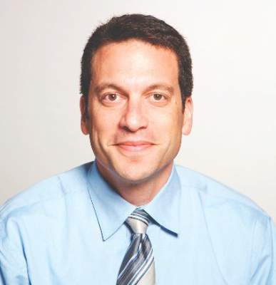
Vascular Surgery Chronicles: Charles Lindbergh and Alexis Carrel: Strange Bedfellows
How does one of the smartest and most well-known men of his time become almost forgotten in history? Dr. Alexis Carrel’s contributions to medicine brought him to the height of fame in the worlds of surgery and science. By designing a curved needle coated in Vaseline, Carrel developed a new method of blood-vessel anastomosis that created a new standard for vascular surgery. This development earned him the Nobel Prize in Medicine or Physiology in 1912, making Carrel the second surgeon and youngest scientist at that time to earn this recognition. The ability to repair, reconnect, or attach blood vessels to one another opened the door for open heart surgery, coronary artery bypass grafts, transplantation, and countless other procedures. He further gained respect while working with Henry Drysdale Dakin in the French Army Medical Corps by revolutionizing the treatment of major wounds with wound antisepsis in the form of Carrel-Dakin fluid. This contribution alone earned him the Cross of the Legion of Honor.
However, by the time of the 52nd Vascular Annual Meeting in 1998, Dr. William Abbott in his SVS Presidential Address would focus on Carrel as an example of a surgeon with vast achievements who had come to be underrecognized. Despite Carrel’s amazing accomplishments throughout his life, the choices he ultimately made later significantly affected his legacy. Dr. Abbott attributes this to Carrel’s “unfortunate leadership decisions, in both boldness and judgment.”
Similar issues affected the legacy of Carrel’s close friend and colleague, Charles Lindbergh. The relationship between these two legendary men demonstrates the serendipity of history, the power of partnerships, and the importance of one’s choices, as well as the fleeting nature of fame. Both men reached the heights of praise and public admiration, then tumbled in a downward spiral of public condemnation.
Lindbergh, America’s golden boy aviator, had won the hearts of the world after he became the first to fly solo from New York to Paris in 1927. On Nov. 28, 1930, the American hero met the pioneering scientist Carrel through the auspices of Dr. Charles Flagg, a caretaker for Elisabeth Morrow, Lindbergh’s chronically ill sister-in-law. He and Carrel met at the Rockefeller Institute for Medical Research and formed a quick bond of mutual respect and admiration. Lindbergh was interested in questioning Carrel on potential treatments for his sister-in-law’s diseased heart valves: “Why could not a part of the body be kept alive indefinitely if a mechanical heart was attached to it – an arm, or even a head?… Why would not a mechanical heart be valuable for certain surgical operations?”
For his part, Carrel, a firm believer in “physiognomy,” the assessment of a person’s character from their outer appearance, and eugenics, the science of improving a population through controlled breeding, viewed Lindbergh as the perfect human specimen.
He interpreted the tall and handsome American hero as one of the elite selected by nature to play a role in society by promoting the production of the fit.
It was after this meeting that Lindbergh was invited to work in Carrel’s lab at the Rockefeller Institute. Lindbergh was enthralled with Carrel’s intellect, stating that his friend’s “mind flashed with the speed of light in space between the logical world of science and the mystical world of God.” Through Carrel’s tutelage and supervision, Lindbergh became focused on research on organ preservation.
During the mid-1930s, Lindbergh’s contribution to Carrel’s laboratory culminated in the design and production of the first efficient perfusion pump. This pump was intended to perfuse organs with pulsatile flow while maintaining a sterile environment free from contamination. The two men coauthored a book, “The Culture of Organs,” which detailed the process and theories for allowing living organs to exist outside the body during surgery. Their combined work is said to have been a crucial step in the later development of open heart surgery and organ transplantation, as well as to have laid the groundwork for the development of the artificial heart. Their collaboration raised their combined fame to the point where both men appeared on the cover of Time magazine on June 13, 1938, highlighting their heart perfusion work.
However, the very ideas that bonded the two famed men in mutual admiration would come to be unpopular, if not reviled, and led to their mutual downfall. Carrel’s views stating the superiority of evolution, survival of the fittest, and thoughts of eugenics paralleled Lindbergh’s thoughts of heredity and evolution. Lindbergh grew up on a farm and knew about breeding livestock and was comfortable with Carrel’s philosophy of racial superiority.
Therefore, despite the incredible accomplishments of both men, these jointly held views and their later affiliation with Nazi Germany and its principles tarnished their legacy.
Lindbergh, one of the few men with his level of fame who had lived among people of all skin colors in many cultures, was constantly being charged with racism and antisemitism. And even though he had previously stated, “I can’t feel inferior or superior to another man because of race, or in any way antagonistic to him. I judge the individual not by his race, and have always done so,” he constantly spoke of the value of genetics in promoting individual importance. And this talk of race betterment was a concept synonymous with the growing Nazi movement in Germany.
To make matters worse, Lindbergh had openly admired the Third Reich after having received the German Medal of Honor in 1938, bestowed by Herman Goering. This combined with Lindbergh’s past appreciation of Germany and his well-known views on eugenics caused many to view him as a Nazi sympathizer. It didn’t help that Lindbergh was also a great isolationist during World War II and acted as a spokesperson for the “America First” committee, which believed that the United States should not intervene. The once great man was denounced within his own country in a manner that would parallel what would happen to Carrel.
During the same period, Carrel returned to France to display his patriotism. In support of the war effort, Carrel volunteered his time toward supporting and designing mobile military hospitals and combating malnutrition. However, in the early 1940s, Germany conquered France and set up a puppet French government at Vichy. The new government offered Carrel the opportunity to continue his research at his own “Institute of Man.” Because of his past sentiments and this relationship formed with the Nazi-supported Vichy government, Carrel would come to be seen as a Nazi collaborator as well.
After the liberation of France in 1944, Carrel was dismissed from the institute and placed under surveillance to investigate his collaboration with the Nazis. Although no conclusions were ever reached, Carrel’s reputation was further destroyed by the press; this left him depressed and ruined. He died later that year on Nov. 5 (J Vasc Surg. 1999;29[1]:1-7).
Through their similar political views, Lindbergh and Carrel became despised in their own countries. Lindbergh would later regain his stature as an American hero and icon after advising the Army and Navy in World War II and continuing his work in the aeronautics industry. But his reputation remained forever tarnished as a Nazi sympathizer, and he died with his legacy disgraced in the eyes of many.
Lindbergh and Carrel’s contributions, despite their personal choices and judgments throughout life, have not been forgotten. There are many who still appreciate and remember the advances that both brought to the fields of medicine and science. Their legacies remain linked through the Lindbergh-Carrel Prize, established at the Medical University of South Carolina in Charleston. The award celebrates their contributions to the “development of perfusion and bioreactor technologies for organ preservation and growth.”
Lindbergh and Carrel exemplify the idea that one must consider the legacy that individuals leave behind in the context of their overall interactions and influences on the society in which they lived. Both men had significant individual failings and made choices that tarnished their public image and affected their legacies. With regard to Carrel, his opinions regarding the superiority of the white man and his proclamation of his mystical views alienated him from the public and the scientific community. Lindbergh’s alleged racism and antisemitism tarnished his image as a true American hero. Whatever their personal failings, however, medicine was forever changed by the impact of the great surgeon and the pilot.
Sources:
Berg AS. Lindbergh, Putman Adult Press, 1998.
Friedman DM. The Immortalists: Charles Lindbergh, Dr. Alexis Carrel, and Their Daring Quest to Live Forever. Ecco Publishing, 2007.
Presidential address: Legend, leadership, legacy. Abbott WM. J Vasc Surg. 1999;29:1-7.
Chaudhuri J, Al-Rubeai M. Bioreactors for Tissue Engineering: Principles, Design and Operation. Springer Publishing, 2005.
Dr. Phair is at the Department of Cardiovascular and Thoracic Surgery, Division of Vascular and Endovascular Surgery, Montefiore Medical Center, Bronx, N.Y.

Elections?
The SVS is currently soliciting nominations for election to the positions of VP, Treasurer, and Secretary. Because this is an election year in the USA, I wonder how the current crop of Republican and Democratic candidates would campaign if they were vascular surgeons running for an Executive position in the SVS. As a corollary, I also question how they would be elected President of the nation if we use the format the SVS uses to elect its Executive. The following examples of what I think their platforms would be implies no resemblance to any current or past executive member. Rather, I use these candidates’ personalities to identify issues that may be of concern to the SVS and its members.

Dr. Bernie Sanders is horrified that most Medicare payments go to “the top 1%” of doctors, most of whom are doing unnecessary procedures like venous ablations for spider veins or angioplasties for 15-block claudication. He would support a move to a single payer “Medicare for all” to make sure that all vascular surgeons were compensated equitably for what they do, regardless of their form of employment. As someone who wants to limit wars, he would not get involved in any turf battles with cardiologists or interventional radiologists.
While Dr. Hilary Clinton has held many high office positions in the SVS, she would emphasize that there has been only one prior female President, Dr. Julie Freischlag, and so she would claim that it is certainly time for another woman to lead the organization. As a pragmatist, she would try to follow the leadership principles of past President Perler. However, Dr. Clinton has been accused of taking money from the Super PACS, such as the device manufacturers, and so would be indebted to Industry.
Dr. Martin O’Malley, a busy community practice vascular surgeon would have to drop out of the race. Unfortunately, because none of the voters had any idea what a vascular surgeon does, he would get very few votes. Most of the voters in his constituency think that cardiologists take care of vascular conditions so they would cast their vote for anyone other than a vascular surgeon.
Dr. Donald Trump, on the other hand, is a very rich private practice vascular surgeon who had his own medical show on national television. Accordingly, while he had achieved notoriety, he would probably be envied by academic vascular surgeons. Having accumulated so much money investing in angiosuites and medical real estate he even had his own plane to fly between hospitals (which he also owned!). His penchant for hiring only the most beautiful nurses is common knowledge. A drawback is that he had never served on any committee of the SVS exposing his lack of understanding of the organization’s function. But this shortcoming would not bother him as he would lead “by hiring really smart people.” He proclaimed, without substantive explanation, that he would make the SVS “great again.” His popularity would be based on his promise to “build a wall” to keep other specialists from being able to treat vascular patients.
Dr. Jeb Bush was well regarded by the established Executive, some of whom considered him “part of the family.” However, he would not acknowledge any relationship with these “relatives.” He would use his success as President of the Florida Vascular Society to support his candidacy. However, Dr. Marco Rubio, who had trained under him at Florida State Medical School in Tallahassee, would suggest that he had not done such a good job after all.
Dr. Marco Rubio, even as a young resident, had his sights set on the Presidency. However, although he succeeded in getting elected to many positions, even the Board, he had never really achieved anything substantial. He had not authored any major research or excelled in clinical practice. His campaign would consist of repeating derogatory remarks about the current President and Dr. Jeb Bush.
Dr. Ted Cruz had served on the Board but was not well liked. His views were very conservative. He was adamantly against a separate Board of Vascular Surgery. Therefore, he would solicit the votes of the “Evangelicals” consisting of older surgeons, many of whom still did General Surgery. He was also the most academic candidate having completed his fellowship at Harvard. He would insist that only academics with a strong conservative viewpoint deserve to be elected.
Dr. Chris Christie, on the other hand, favors splitting vascular surgery from general surgery. He will belligerently state that if a bridge is built to maintain the relationship he would make sure his officers make it impassable.
Dr. Carly Fiorina believes her prior experience running a major academic institution makes her the most qualified. However, the other candidates would be quick to point out that she had been fired! Like some other academics, she had been forced to take many positions around the country and so she did not have a local constituency to support her. On the other hand, having lectured all over the world she would have many international contacts including “knowing Vladimir Putin.” Of course, no one would know what that would offer the SVS.
Dr. Rand Paul, a Vascular Surgeon with a strong interest in Amaurosis Fugax, wants the SVS to save its money and not do anything. In fact, he thinks it should not be involved in any aspect of vascular surgery, leaving everything up to the individual practitioner or other specialties. His views are also held to a large degree by Dr. Ben Carson. As a retired pediatric neurosurgeon from Johns Hopkins, he would try to follow in the highly respected footsteps of presidents Perler and Freischlag, also from Hopkins. Since he believes brain surgeons are the smartest, he would claim that he could be the President of the SVS even with his eyes closed. To prove this, he would give most of his speeches with closed eyes.
Dr. Kasich was the President of the Ohio Vascular Society. He would argue that if voters only paid attention to what he has accomplished in Ohio, he would get everyone’s vote. But he would be concerned that since he had never published his results in any national journal, no one would respect him.
Well, that’s perhaps how the candidates would do their electioneering. But what if our national election were to be held in the manner we elect our SVS Executives? Rather than allowing the public to hear all the candidates express their viewpoints over and over in a multitude of televised debates, a small group would determine who should be nominated. According to the bylaws of the SVS, they would be the last two surviving ex-presidents (Bush and Clinton) as well one Governor elected by all the Governors, one Senator elected by the Senate and one person elected by the nation. This nominating committee would then invite the entire voting public to a luncheon on the Washington Mall, where the attendees would accept their recommendations voting by acclamation. Of course, most voters would not attend since it would be too inconvenient and expensive to make the trip to this luncheon.
After all, they would have to take time away from their jobs. As a result the vast majority of the “voting” public would in essence be excluded from the electoral process. Not very democratic – but certainly efficient. Further, who better to assess the candidates’ potential than these esteemed members of the nominating committee? But is it efficiency that we really need? Should we not rather have a fully open election in which potential candidates can express their goals for the SVS? This could be achieved by publishing their platforms in this Newspaper. This would enfranchise all of our members to participate in the election of candidates that represent their own goals for the Society.
To tell the truth, I am ambivalent as to which method is preferable, but it is at least worth discussion. Why don’t you use the “Letters to the Editor” section of this, our official newspaper, to make your viewpoint heard? If that is not possible in your schedule, then take a few minutes and click this link to take our poll and place your vote for the option you prefer.
Dr. Russell H. Samson is a physician in the practice of Samson, Showalter, Lepore, and Nair, and clinical professor of surgery, Florida State University, Tallahassee. He is also the medical editor of Vascular Specialist.
The SVS is currently soliciting nominations for election to the positions of VP, Treasurer, and Secretary. Because this is an election year in the USA, I wonder how the current crop of Republican and Democratic candidates would campaign if they were vascular surgeons running for an Executive position in the SVS. As a corollary, I also question how they would be elected President of the nation if we use the format the SVS uses to elect its Executive. The following examples of what I think their platforms would be implies no resemblance to any current or past executive member. Rather, I use these candidates’ personalities to identify issues that may be of concern to the SVS and its members.
Dr. Bernie Sanders is horrified that most Medicare payments go to “the top 1%” of doctors, most of whom are doing unnecessary procedures like venous ablations for spider veins or angioplasties for 15-block claudication. He would support a move to a single payer “Medicare for all” to make sure that all vascular surgeons were compensated equitably for what they do, regardless of their form of employment. As someone who wants to limit wars, he would not get involved in any turf battles with cardiologists or interventional radiologists.
While Dr. Hilary Clinton has held many high office positions in the SVS, she would emphasize that there has been only one prior female President, Dr. Julie Freischlag, and so she would claim that it is certainly time for another woman to lead the organization. As a pragmatist, she would try to follow the leadership principles of past President Perler. However, Dr. Clinton has been accused of taking money from the Super PACS, such as the device manufacturers, and so would be indebted to Industry.
Dr. Martin O’Malley, a busy community practice vascular surgeon would have to drop out of the race. Unfortunately, because none of the voters had any idea what a vascular surgeon does, he would get very few votes. Most of the voters in his constituency think that cardiologists take care of vascular conditions so they would cast their vote for anyone other than a vascular surgeon.
Dr. Donald Trump, on the other hand, is a very rich private practice vascular surgeon who had his own medical show on national television. Accordingly, while he had achieved notoriety, he would probably be envied by academic vascular surgeons. Having accumulated so much money investing in angiosuites and medical real estate he even had his own plane to fly between hospitals (which he also owned!). His penchant for hiring only the most beautiful nurses is common knowledge. A drawback is that he had never served on any committee of the SVS exposing his lack of understanding of the organization’s function. But this shortcoming would not bother him as he would lead “by hiring really smart people.” He proclaimed, without substantive explanation, that he would make the SVS “great again.” His popularity would be based on his promise to “build a wall” to keep other specialists from being able to treat vascular patients.
Dr. Jeb Bush was well regarded by the established Executive, some of whom considered him “part of the family.” However, he would not acknowledge any relationship with these “relatives.” He would use his success as President of the Florida Vascular Society to support his candidacy. However, Dr. Marco Rubio, who had trained under him at Florida State Medical School in Tallahassee, would suggest that he had not done such a good job after all.
Dr. Marco Rubio, even as a young resident, had his sights set on the Presidency. However, although he succeeded in getting elected to many positions, even the Board, he had never really achieved anything substantial. He had not authored any major research or excelled in clinical practice. His campaign would consist of repeating derogatory remarks about the current President and Dr. Jeb Bush.
Dr. Ted Cruz had served on the Board but was not well liked. His views were very conservative. He was adamantly against a separate Board of Vascular Surgery. Therefore, he would solicit the votes of the “Evangelicals” consisting of older surgeons, many of whom still did General Surgery. He was also the most academic candidate having completed his fellowship at Harvard. He would insist that only academics with a strong conservative viewpoint deserve to be elected.
Dr. Chris Christie, on the other hand, favors splitting vascular surgery from general surgery. He will belligerently state that if a bridge is built to maintain the relationship he would make sure his officers make it impassable.
Dr. Carly Fiorina believes her prior experience running a major academic institution makes her the most qualified. However, the other candidates would be quick to point out that she had been fired! Like some other academics, she had been forced to take many positions around the country and so she did not have a local constituency to support her. On the other hand, having lectured all over the world she would have many international contacts including “knowing Vladimir Putin.” Of course, no one would know what that would offer the SVS.
Dr. Rand Paul, a Vascular Surgeon with a strong interest in Amaurosis Fugax, wants the SVS to save its money and not do anything. In fact, he thinks it should not be involved in any aspect of vascular surgery, leaving everything up to the individual practitioner or other specialties. His views are also held to a large degree by Dr. Ben Carson. As a retired pediatric neurosurgeon from Johns Hopkins, he would try to follow in the highly respected footsteps of presidents Perler and Freischlag, also from Hopkins. Since he believes brain surgeons are the smartest, he would claim that he could be the President of the SVS even with his eyes closed. To prove this, he would give most of his speeches with closed eyes.
Dr. Kasich was the President of the Ohio Vascular Society. He would argue that if voters only paid attention to what he has accomplished in Ohio, he would get everyone’s vote. But he would be concerned that since he had never published his results in any national journal, no one would respect him.
Well, that’s perhaps how the candidates would do their electioneering. But what if our national election were to be held in the manner we elect our SVS Executives? Rather than allowing the public to hear all the candidates express their viewpoints over and over in a multitude of televised debates, a small group would determine who should be nominated. According to the bylaws of the SVS, they would be the last two surviving ex-presidents (Bush and Clinton) as well one Governor elected by all the Governors, one Senator elected by the Senate and one person elected by the nation. This nominating committee would then invite the entire voting public to a luncheon on the Washington Mall, where the attendees would accept their recommendations voting by acclamation. Of course, most voters would not attend since it would be too inconvenient and expensive to make the trip to this luncheon.
After all, they would have to take time away from their jobs. As a result the vast majority of the “voting” public would in essence be excluded from the electoral process. Not very democratic – but certainly efficient. Further, who better to assess the candidates’ potential than these esteemed members of the nominating committee? But is it efficiency that we really need? Should we not rather have a fully open election in which potential candidates can express their goals for the SVS? This could be achieved by publishing their platforms in this Newspaper. This would enfranchise all of our members to participate in the election of candidates that represent their own goals for the Society.
To tell the truth, I am ambivalent as to which method is preferable, but it is at least worth discussion. Why don’t you use the “Letters to the Editor” section of this, our official newspaper, to make your viewpoint heard? If that is not possible in your schedule, then take a few minutes and click this link to take our poll and place your vote for the option you prefer.
Dr. Russell H. Samson is a physician in the practice of Samson, Showalter, Lepore, and Nair, and clinical professor of surgery, Florida State University, Tallahassee. He is also the medical editor of Vascular Specialist.

Chestnut extract
Known as sweet chestnut, Castanea sativa is a member of the Fagaceae family, and is found in abundance in Southern and Southeastern Europe and Asia.1 In traditional medicine, chestnut tree flower preparations have been used for various indications.2 Chestnut has been used in French folk medicine as a tea to treat severe cough, colds, and bronchitis as well as diarrhea.2-6 In modern times, C. sativa leaf extract has been described as having the capacity to scavenge various free radicals associated with oxidative stress induced by ultraviolet exposure.7
Traditional uses
A 2014 study of the therapeutic and traditional uses of the plants native to the Western Italian Alps revealed that C. sativa has long been important in the region, typically for food and wood.8 But medical uses have been uncovered in that region as well. In fact, ancient Romans found C. sativa to exhibit antibacterial, astringent, antitoxic, and tonic qualities, with chestnut honey used then to dress chronic wounds, burns, and skin ulcers.9 A 2014 study by Carocho et al. of the phytochemical profile and antioxidant activity of C. sativa flowers is noteworthy for buttressing the reported health benefits of the use of chestnut flower infusions and decoctions in traditional medicine.2
Antioxidant activity
In 2005, Calliste et al. investigated the antioxidant potential of C. sativa leaf to act against the stable free radical 2,2-diphenyl-1-pycrylhydrazyl, superoxide anion, and hydroxyl radical. Using electronic spin resonance, the investigators showed that C. sativa exhibited high antioxidant potential equivalent to reference antioxidants quercetin and vitamin E.3

Three years later, Almeida et al. conducted an in vitro assessment of an ethanol/water (7:3) extract from C. sativa leaves and an ethanol/water (2:3) extract from Quercus robur (English oak) leaves, finding that both plants demonstrated a high potency to scavenge various reactive oxygen and nitrogen species. The researchers concluded that these findings supported the burgeoning interest in these extracts for use in topical antioxidant formulations.4 An in vivo investigation using an ethanol/water (7:3) extract from C. sativa conducted by the same team later in the year yielded similar results, with the researchers concluding that chestnut extract has the potential to confer benefits against photoaging and other oxidative stress–mediated conditions when included in an appropriately formulated topical antioxidant preparation.6 Subsequently, Barreira et al. demonstrated that chestnut skin and leaves exhibited sufficient antioxidant potency to warrant use in novel antioxidant formulations.10
In 2015, Almeida et al. characterized an antioxidant semisolid surfactant-free topical formulation featuring C. sativa leaf extract. In the process of ascertaining the physical, functional, and microbiologic stability of the antioxidant formulation, the investigators identified a hydrating effect and good skin tolerance, which they concluded suggested a capacity to prevent or treat cutaneous conditions in which oxidative stress plays a role.11
Photoprotective potential
In 2010, Sapkota et al. evaluated the antioxidant and antimelanogenic characteristics of several prebloom and full-bloom chestnut flower extracts, finding that a prebloom methanol extract and an ethanol extract evinced the greatest levels of phenolic and flavonoid compounds. These extracts also displayed the best radical scavenging and mushroom tyrosinase–inhibiting activities. Notably, the prebloom extract was effective in protecting the skin from the deleterious impact of UV radiation. The investigators also observed that all of the tested extracts lowered the tyrosinase activity and melanin formation of SK-MEL-2 cells similarly to arbutin. They ascribed the antimelanogenic effects of chestnut flower extracts to their antioxidant-mediated inhibitory effects on tyrosinase. They concluded that chestnut flower extracts have considerable potential as cosmetic agents.12

Recently, Almeida et al. studied the protective effects in a human keratinocyte cell line of C. sativa extract at various concentrations (0.001-, 0.01-, 0.05-, and 0.1-mcg/mL) against UV-induced DNA damage. They found that the chestnut extract concentration dependently protected against UV-mediated DNA damage, with the 0.1-mcg/mL concentration affording maximum protection (66.4%). This result was considered to be a direct antioxidant effect attributed to various phenolic antioxidants present in C. sativa. In addition, the investigators observed no phototoxic or genotoxic effects on HaCaT cells incubated with up to 0.1 mcg/mL of chestnut leaf extract. They concluded that C. sativa leaf extract has the potential to prevent or mitigate UV-induced harm to the skin.7
Other benefits and bioactivity
Assessments of C. sativa by-products have shown a favorable profile of bioactive constituents that demonstrate antioxidant, anticarcinogenic, and cardioprotective activity. Braga et al. conducted a 2015 review that concluded these compounds, as part of agro-industrial waste, offer value to the pharmaceutical, cosmetics, and food industries, with the potential to lower pollution costs and raise profits while enhancing social, economic, and environmental sustainability in growing regions.1
A related chestnut species also has been linked to dermatologic uses. In East Asia, a skin firming/antiwrinkle formulation features the inner shell of Castanea crenata as an active ingredient.13 In 2002, Chi et al. showed that the chestnut inner shell extract improved cell-associated expression of the adhesion molecules fibronectin and vitronectin. They also found that scoparone (6,7-dimethoxycoumarin) isolated from the chestnut extract exhibited comparable qualities. The investigators concluded that the enhanced expression of adhesion molecules imparted by the chestnut inner shell extract may account for the prevention of cell detachment and the manifestation of antiaging effects.13
Allergy
It is worth noting that chestnut is one of the many allergens associated with the latex-fruit syndrome.14 However, in a patch test investigation of the skin irritation potential of C. sativa leaf extract in 20 volunteers, Almeida et al. identified five phenolic compounds in the extract (chlorogenic acid, ellagic acid, rutin, isoquercitrin, and hyperoside) and found it safe for topical application.6 Chestnut is considered to pose a low to moderate risk of inducing allergic reactions.9
Conclusion
Recent research appears to suggest the in vitro antioxidant activity of sweet chestnut and potential for use in topical formulations. There remains a paucity of in vivo evidence, however. While much more research is necessary to determine whether it has a place in the dermatologic armamentarium, current data are intriguing.
References
1. Nat Prod Res. 2015;29(1):1-18
2. Biomed Res Int. 2014;2014:232956
3. J Agric Food Chem. 2005 Jan 26;53(2):282-8
4. J Photochem Photobiol B. 2008 May 29;91(2-3):87-95
5. A Modern Herbal (vol. I). New York: Dover Publications, 1971, p. 195
6. Basic Clin Pharmacol Toxicol. 2008 Nov;103(5):461-7
7. J Photochem Photobiol B. 2015 Mar;144C:28-34
8. J Ethnopharmacol. 2014 Aug 8;155(1):463-84
9. J Sci Food Agric. 2010 Aug 15;90(10):1578-89
10. Food Sci Technol Int. 2010 June;16(3):209-16
11. Drug Dev Ind Pharm. 2015 Jan;41(1):148-55
12. Biosci Biotechnol Biochem. 2010;74(8):1527-33
13. Arch Pharm Res. 2002 Aug;25(4):469-74
14. Allergy. 2007 Nov;62(11):1277-81
Dr. Baumann is chief executive officer of the Baumann Cosmetic & Research Institute in the Design District in Miami. She founded the Cosmetic Dermatology Center at the University of Miami in 1997. Dr. Baumann wrote the textbook “Cosmetic Dermatology: Principles and Practice” (New York: McGraw-Hill, 2002), and a book for consumers, “The Skin Type Solution” (New York: Bantam Dell, 2006). Her latest book, “Cosmeceuticals and Cosmetic Ingredients,” was published in November 2014. Dr. Baumann has received funding for clinical grants from Allergan, Aveeno, Avon Products, Evolus, Galderma, GlaxoSmithKline, Kythera Biopharmaceuticals, Mary Kay, Medicis Pharmaceuticals, Neutrogena, Philosophy, Topix Pharmaceuticals, and Unilever.
Known as sweet chestnut, Castanea sativa is a member of the Fagaceae family, and is found in abundance in Southern and Southeastern Europe and Asia.1 In traditional medicine, chestnut tree flower preparations have been used for various indications.2 Chestnut has been used in French folk medicine as a tea to treat severe cough, colds, and bronchitis as well as diarrhea.2-6 In modern times, C. sativa leaf extract has been described as having the capacity to scavenge various free radicals associated with oxidative stress induced by ultraviolet exposure.7
Traditional uses
A 2014 study of the therapeutic and traditional uses of the plants native to the Western Italian Alps revealed that C. sativa has long been important in the region, typically for food and wood.8 But medical uses have been uncovered in that region as well. In fact, ancient Romans found C. sativa to exhibit antibacterial, astringent, antitoxic, and tonic qualities, with chestnut honey used then to dress chronic wounds, burns, and skin ulcers.9 A 2014 study by Carocho et al. of the phytochemical profile and antioxidant activity of C. sativa flowers is noteworthy for buttressing the reported health benefits of the use of chestnut flower infusions and decoctions in traditional medicine.2
Antioxidant activity
In 2005, Calliste et al. investigated the antioxidant potential of C. sativa leaf to act against the stable free radical 2,2-diphenyl-1-pycrylhydrazyl, superoxide anion, and hydroxyl radical. Using electronic spin resonance, the investigators showed that C. sativa exhibited high antioxidant potential equivalent to reference antioxidants quercetin and vitamin E.3
Three years later, Almeida et al. conducted an in vitro assessment of an ethanol/water (7:3) extract from C. sativa leaves and an ethanol/water (2:3) extract from Quercus robur (English oak) leaves, finding that both plants demonstrated a high potency to scavenge various reactive oxygen and nitrogen species. The researchers concluded that these findings supported the burgeoning interest in these extracts for use in topical antioxidant formulations.4 An in vivo investigation using an ethanol/water (7:3) extract from C. sativa conducted by the same team later in the year yielded similar results, with the researchers concluding that chestnut extract has the potential to confer benefits against photoaging and other oxidative stress–mediated conditions when included in an appropriately formulated topical antioxidant preparation.6 Subsequently, Barreira et al. demonstrated that chestnut skin and leaves exhibited sufficient antioxidant potency to warrant use in novel antioxidant formulations.10
In 2015, Almeida et al. characterized an antioxidant semisolid surfactant-free topical formulation featuring C. sativa leaf extract. In the process of ascertaining the physical, functional, and microbiologic stability of the antioxidant formulation, the investigators identified a hydrating effect and good skin tolerance, which they concluded suggested a capacity to prevent or treat cutaneous conditions in which oxidative stress plays a role.11
Photoprotective potential
In 2010, Sapkota et al. evaluated the antioxidant and antimelanogenic characteristics of several prebloom and full-bloom chestnut flower extracts, finding that a prebloom methanol extract and an ethanol extract evinced the greatest levels of phenolic and flavonoid compounds. These extracts also displayed the best radical scavenging and mushroom tyrosinase–inhibiting activities. Notably, the prebloom extract was effective in protecting the skin from the deleterious impact of UV radiation. The investigators also observed that all of the tested extracts lowered the tyrosinase activity and melanin formation of SK-MEL-2 cells similarly to arbutin. They ascribed the antimelanogenic effects of chestnut flower extracts to their antioxidant-mediated inhibitory effects on tyrosinase. They concluded that chestnut flower extracts have considerable potential as cosmetic agents.12
Recently, Almeida et al. studied the protective effects in a human keratinocyte cell line of C. sativa extract at various concentrations (0.001-, 0.01-, 0.05-, and 0.1-mcg/mL) against UV-induced DNA damage. They found that the chestnut extract concentration dependently protected against UV-mediated DNA damage, with the 0.1-mcg/mL concentration affording maximum protection (66.4%). This result was considered to be a direct antioxidant effect attributed to various phenolic antioxidants present in C. sativa. In addition, the investigators observed no phototoxic or genotoxic effects on HaCaT cells incubated with up to 0.1 mcg/mL of chestnut leaf extract. They concluded that C. sativa leaf extract has the potential to prevent or mitigate UV-induced harm to the skin.7
Other benefits and bioactivity
Assessments of C. sativa by-products have shown a favorable profile of bioactive constituents that demonstrate antioxidant, anticarcinogenic, and cardioprotective activity. Braga et al. conducted a 2015 review that concluded these compounds, as part of agro-industrial waste, offer value to the pharmaceutical, cosmetics, and food industries, with the potential to lower pollution costs and raise profits while enhancing social, economic, and environmental sustainability in growing regions.1
A related chestnut species also has been linked to dermatologic uses. In East Asia, a skin firming/antiwrinkle formulation features the inner shell of Castanea crenata as an active ingredient.13 In 2002, Chi et al. showed that the chestnut inner shell extract improved cell-associated expression of the adhesion molecules fibronectin and vitronectin. They also found that scoparone (6,7-dimethoxycoumarin) isolated from the chestnut extract exhibited comparable qualities. The investigators concluded that the enhanced expression of adhesion molecules imparted by the chestnut inner shell extract may account for the prevention of cell detachment and the manifestation of antiaging effects.13
Allergy
It is worth noting that chestnut is one of the many allergens associated with the latex-fruit syndrome.14 However, in a patch test investigation of the skin irritation potential of C. sativa leaf extract in 20 volunteers, Almeida et al. identified five phenolic compounds in the extract (chlorogenic acid, ellagic acid, rutin, isoquercitrin, and hyperoside) and found it safe for topical application.6 Chestnut is considered to pose a low to moderate risk of inducing allergic reactions.9
Conclusion
Recent research appears to suggest the in vitro antioxidant activity of sweet chestnut and potential for use in topical formulations. There remains a paucity of in vivo evidence, however. While much more research is necessary to determine whether it has a place in the dermatologic armamentarium, current data are intriguing.
References
1. Nat Prod Res. 2015;29(1):1-18
2. Biomed Res Int. 2014;2014:232956
3. J Agric Food Chem. 2005 Jan 26;53(2):282-8
4. J Photochem Photobiol B. 2008 May 29;91(2-3):87-95
5. A Modern Herbal (vol. I). New York: Dover Publications, 1971, p. 195
6. Basic Clin Pharmacol Toxicol. 2008 Nov;103(5):461-7
7. J Photochem Photobiol B. 2015 Mar;144C:28-34
8. J Ethnopharmacol. 2014 Aug 8;155(1):463-84
9. J Sci Food Agric. 2010 Aug 15;90(10):1578-89
10. Food Sci Technol Int. 2010 June;16(3):209-16
11. Drug Dev Ind Pharm. 2015 Jan;41(1):148-55
12. Biosci Biotechnol Biochem. 2010;74(8):1527-33
13. Arch Pharm Res. 2002 Aug;25(4):469-74
14. Allergy. 2007 Nov;62(11):1277-81
Dr. Baumann is chief executive officer of the Baumann Cosmetic & Research Institute in the Design District in Miami. She founded the Cosmetic Dermatology Center at the University of Miami in 1997. Dr. Baumann wrote the textbook “Cosmetic Dermatology: Principles and Practice” (New York: McGraw-Hill, 2002), and a book for consumers, “The Skin Type Solution” (New York: Bantam Dell, 2006). Her latest book, “Cosmeceuticals and Cosmetic Ingredients,” was published in November 2014. Dr. Baumann has received funding for clinical grants from Allergan, Aveeno, Avon Products, Evolus, Galderma, GlaxoSmithKline, Kythera Biopharmaceuticals, Mary Kay, Medicis Pharmaceuticals, Neutrogena, Philosophy, Topix Pharmaceuticals, and Unilever.
Readers weigh in on vaginal cleansing prior to cesarean delivery

“SHOULD YOU ADOPT THE PRACTICE OF VAGINAL CLEANSING WITH POVIDONE-IODINE PRIOR TO CESAREAN DELIVERY?”
ROBERT L. BARBIERI, MD (EDITORIAL; JANUARY 2016)
In his January 2016 Editorial, Editor in Chief Robert L. Barbieri, MD, presented evidence supporting the practice of vaginal cleansing with povidone-iodine prior to cesarean delivery (CD) to prevent postoperative endometritis. He then asked readers if they would consider adopting such a practice. More than 250 readers weighed in through the Quick Poll at obgmanagement.com, and many readers sent in letters with follow-up questions and comments on controlling bacterial contamination, vaginal seeding, etc. Here are some of the letters, along with Dr. Barbieri’s response and the Quick Poll results.
A contradiction in definitions?
There seems to be a contradiction in definitions. The second sentence of the article defines endometritis as the presence of fever plus low abdominal tenderness. However, the studies presented state that vaginal cleansing pre-CD decreased endometritis but did not decrease postpartum fever. Is this not a discrepancy?
Nancy Kerr, MD, MPH
Albuquerque, New Mexico
A question about povidone-iodine
Have any studies been done on newborn iodine levels after vaginal cleansing with povidone-iodine prior to CD?
G. Millard Simmons Jr, MD
Hilton Head, Bluffton, South Carolina
Additional tips for controlling bacterial contamination
Dr. Barbieri’s editorial on vaginal cleansing prior to CD is eye opening. I have a few additional suggestions to control bacterial contamination.
First, I examine my patients in labor as few times as necessary, and I ask the nurses (RNs) not to place their fingers in the patient’s vagina while she is pushing. I remove the Foley catheter when I feel progress (descent of fetal head) is being achieved. In addition, physicians as well as RNs should consider changing their scrubs between deliveries, as I believe that bacterial contamination is splattered all over the place, especially into the birth canal. These methods have worked for me in my over-20 years of practice.
I also firmly remind the RN circulator to perform a generous vaginal cleanse with povidone-iodine, in addition to the usual intravenous prophylaxis, before hysterectomy.
Luis Leyva Jr, MD
Miami, Florida
Mixed feelings
My first reaction to this Editorial was: Is this a solution in search of a problem? That is to say, how much of a clinical problem is endometritis after CD? Are we really treating the proposed problem, and does treatment affect long-term outcomes?
Upon reflection, I have concluded that vaginal cleansing pre-CD does intuitively make sense. What sways me in this direction is that the practice is simple, easy, and inexpensive. Since we typically have the patient positioned for Foley catheter insertion, performing vaginal cleansing as we put in the Foley would be easy. If vaginal cleansing were to be done, I definitely would be in favor of doing such practice liberally—for all CDs to make vaginal cleansing part of the “routine.”
Keep in mind that we are still chasing a problem of little clinical significance.
The biggest accomplishment has been to get everyone to give antibiotics preoperatively rather than after cutting the umbilical cord. We knew that this was best practice as early as the late 1980s/early 1990s, and I have been fighting this battle ever since. Believe it or not, there are still a few holdouts.
George H. Davis, DO
Johnson City, Tennessee
Would vaginal cleansing benefit all women in labor?
Vaginal cleansing before CD reminds me of my residency days when all women having hysterectomies were admitted early and given povidone-iodine (Betadine) douches the evening before surgery (unless an iodine allergy was present).
While reading your Editorial, I had several thoughts and questions. 1) Since vaginal cleansing seems to benefit CD patients, might it not benefit all laboring patients? 2) Is the timing of vaginal cleansing critical? 3) Should we do vaginal cleansing on all laboring patients if timing is not critical?
I plan to bring up the topic of vaginal cleansing for CD with my colleagues at our next department meeting, since it seems like such a simple, logical, inexpensive, and beneficial thing to do.
Douglas G. Tolley, MD
Yuba City, California
An early study on using povidone-iodine gel before CD
When I was a chief resident at Kings County Hospital in 1973, we had a very high rate of post-CD endometritis. I conducted a small study on the use of povidone-iodine gel in the last month of pregnancy. Before commencing, we confirmed that the gel did not interfere with diagnosing ruptured membranes.
Obstetric service patients were randomly divided into “A” and “B” groups. The A patients were asked to use povidone-iodine gel at night for the last 2 weeks before their estimated due date. When admitted in labor, they were asked to confirm its use. When a resident diagnosed post-CD endometritis, we kept track of which group the patient was in and whether or not that patient had used povidone-iodine. Approximately 100 infected patients were evaluated from each group.
As it turned out, there were about 3 times the number of infections among the patients who did not use povidone-iodine than among those who said they used it. It did not seem to matter how many times povidone-iodine was used. The “As” who did not use povidone-iodine had results similar to the “Bs.”
It was many years ago, and the study design was crude. However, it does seem to support the suggestion for vaginal cleansing.
Steve Ross, MD
Port Jefferson, New York
Two different ideas about the vaginal biome
This Editorial is timely in that Dr. Dominguez-Bello and colleagues recently published an article in Nature Medicine titled, “Partial restoration of the microbiota of cesarean-born infants via vaginal microbial transfer.”1 Dr. Dominguez-Bello is one of the founders of the idea of “vaginal seeding,” or using the natural biome of the vagina on a newborn immediately after CD by swabbing the baby with the bacteria from the vagina.
I find it interesting that there are two very different ideas about the biome at this time. Vaginal seeding is a new trend that a few patients have asked about during prenatal care. The jury is still out on seeding, but a larger study is currently underway at New York University. Of course, infection is one of the risks of seeding. I appreciate hearing both sides of the issue.
Deborah Herchelroath, DO
Harrisburg, Pennsylvania
Reference
- Dominguez-Bello MG, De Jesus-Labor KM, Shen N, et al. Partial restoration of the microbiota of cesarean-born infants via vaginal microbial transfer [published online ahead of print February 1, 2016]. Nat Med. doi:10.1038/nm.4039.

Dr. Barbieri responds
I would like to thank our readers for taking the time from their busy schedules to write about their clinical experiences and current practices for reducing infectious complications following CD.
Dr. Kerr raises the important issue of the apparent contradictory finding of the beneficial impact of vaginal cleansing on endometritis without a beneficial effect on the overall rate of fever. In the trial reported by Starr,1 fever was defined as a temperature above 38˚C at any time after CD and endometritis was defined as a temperature above 38.4˚C PLUS uterine tenderness occurring more than 24 hours after CD. Given these 2 definitions one can understand the differential effect of vaginal cleansing on fever versus endometritis.
Dr. Simmons raises the intriguing question of the impact of an iodine-containing surgical preparation on newborn thyroid function. There are few studies addressing this issue. One study reports a transient increase in thyroid-stimulating hormone (TSH) levels in a small percentage of newborns whose mothers received an iodine preparation.2 Another study reports no effect of an iodine surgical preparation on newborn thyroid function indices.3
I agree with the guidance of Drs. Leyva and Davis that we can help prevent postcesarean endometritis by minimizing the number of cervical examinations, changing scrubs between deliveries, and by ensuring that an intravenous anti‑ biotic is given before skin incision.
Dr. Tolley wonders if all women should receive vaginal cleansing, regardless of delivery route. It is possible that such an approach would be effective and it deserves study. Given the lower rate of endometritis following vaginal delivery compared with CD, many more women having a vaginal delivery would need to be treated to prevent one case of endometritis. Dr. Ross mentions his experience with the benefit of outpatient vaginal cleansing in the 2 weeks prior to delivery. Many general surgeons are recommending that their patients shower with chlorhexidine the day before surgery in order to reduce the rate of postoperative infection. Short-term and long-term outpatient vaginal cleansing prior to delivery deserves additional study.
Dr. Herchelroath raises the possibility that vaginal cleansing will decrease the ability of the newborn to develop a normal microbiome because it may not be exposed to sufficient vaginal bacteria. This possibility certainly deserves additional study.
The questions and guidance of our readers were incredibly helpful and stimulating. Thank you for sharing your perspective.
References
- Starr RV, Zurawski J, Ismail M. Preoperative vaginal preparation with povidone-iodine and the risk of postcesarean endometritis. Obstet Gynecol. 2005;105(5 pt 1):1024–1029.
- Nili F, Hantoushzadeh S, Alimohamadi A, et al. Iodine-containing disinfectants in preparation for cesarean section: impact on thyroid profile in cord blood. Postgrad Med J. 2015;91(1082):681–684.
- Ordookhani A, Pearce EN, Mirmiran P, Azizi F, Braverman LE. The effect of type of delivery and povidone-iodine application at delivery on cord dried-blood-specimen thyrotropin level and the rate of hyperthyrotropinemia in mature and normal-birth-weight neonates residing in an iodine-replete area. Thyroid. 2007;17(11):1097–1102.

“CELL-FREE DNA SCREENING FOR WOMEN AT LOW RISK FOR FETAL ANEUPLOIDY” MARY E. NORTON, MD (JANUARY 2016)
The price of cfDNA screening is dropping
I found Dr. Norton’s article on cell-free DNA (cfDNA) screening for women at low risk for fetal abnormalities to be enlightening and educational. The section addressing cost-effectiveness, however, was somewhat obsolete. The referenced study by Cuckle and colleagues,1 which estimated the cost of cfDNA per case of Down syndrome in low-risk patients at $3.6 million, was published in 2013. With 4 major companies in the market, the cost/benefit ratio has been changing rapidly. At least one company has dropped the cost of the cfDNA test nearly 80% from 2015 to 2016, making the above reference irrelevant. Recently, Ariosa dropped the price of their Harmony cfDNA test to just $119 in our area, regardless of a patient’s insurance or poverty level. This is significantly less than the cost of performing an early screen and is being welcomed by my patients even after substantial counseling on the test’s limitations in the low-risk population. Natera, another laboratory with a similar test, offers a low-cost option. However, patients must provide proof that their income is below a specified level.
Guidelines from the American College of Obstetricians and Gynecologists (ACOG) and the Society for Maternal-Fetal Medicine (SMFM) likely will have a hard time keeping up with the cost-effectiveness of noninvasive prenatal testing, as the price continues to be dynamic.
Samuel Wolf, DO
Panama City, Florida
Reference
- Cuckle H, Benn P, Pergament E. Maternal cfDNA screening for Down syndrome—a cost sensitivity analysis. Prenat Diagn. 2013;33(7):636–642.
“DOES THE DISCONTINUATION OF MENOPAUSAL HORMONE THERAPY AFFECT A WOMAN’S CARDIOVASCULAR RISK?”
ANDREW M. KAUNITZ, MD; JOANN E. MANSON, MD, DRPH; AND CYNTHIA A. STUENKEL, MD(EXAMINING THE EVIDENCE; DECEMBER 2015)
Disagrees with conclusion
In their expert commentary, Drs. Kaunitz, Manson, and Stuenkel state:
They support this claim by citing Heiss 2008.1 In fact, however, the Women’s Health Initiative (WHI) data show opposite to their statement: In the WHI, all-cause mortality was increased among the women who were assigned to estrogen-progestin therapy (EPT) relative to those who were assigned to placebo within the 3 years of EPT cessation (hazard ratio [HR], 1.15; 95% confidence interval [CI], 0.95–1.39). More importantly, mortality was significantly increased among women who were originally assigned to EPT relative to those who were assigned to placebo and were at least 80% adherent with intervention (HR, 1.53; 95% CI, 1.04–2.24). Thus, the statement by Drs. Kaunitz, Manson, and Stuenkel is incorrect.
In addition to the WHI studies, data are available from at least 2 other randomized controlled trials addressing the issue of HT withdrawal. In the Heart and Estrogen/progestin Replacement Study (HERS) II,2 the unblinded 2.7-year follow-up to the HERS trial, women originally assigned to EPT had a 3.3-fold higher rate of ventricular arrhythmia requiring resuscitation than women assigned to placebo (HR, 3.30; 95% CI, 1.08–10.10). During the first 6 months of posttrial follow-up of the Women’s Estrogen for Stroke Trial (WEST),3 there were 3 fatal strokes and 18 nonfatal strokes among the women originally randomized to estradiol therapy; there were 9 strokes (1 fatal and 8 nonfatal) among the women originally assigned to placebo (HR, 2.3; 95% CI, 1.1–5.0; P = .03).
In our study we detected that women who stopped HT, compared with women who continued HT, had a 2.3-fold (95% CI, 2.12–2.50) greater risk of cardiac death within the first post-HT year and a 1.3-fold (95% CI, 1.21–1.31) greater risk of cardiac death more than 1 year after stopping HT.4 In addition, women who stopped HT, compared with women who continuedHT, had a 2.5-fold (95% CI, 2.28–2.77) greater risk of dying from stroke within the first post-HT year and a 1.3-fold (95% CI, 1.19–1.31) greater risk of dying from stroke more than 1 year after stopping HT. We believe that these data substantially further our understanding of the posttrial data from WHI, as well as HERS and WEST. Thus, cumulative data support that HT withdrawal potentially has detrimental implications for women. In total, the data are highly informative when counseling women regarding use or discontinuation of HT.
Tomi Mikkola, MD
Helsinki, Finland
References
- Heiss G, Wallace R, Anderson GL, et al; WHI investigators. Health risks and benefits 3 years after stopping randomized treatment with estrogen and progestin. JAMA. 2008;299(9):1036–1045.
- Grady D, Herrington D, Bittner V, et al; HERS Research Group. Cardiovascular disease outcomes during 6.8 years of hormone therapy: Heart and Estrogen/progestin Replacement Study follow-up (HERS II) [published correction appears in JAMA. 2002;288(9):1064]. JAMA. 2002;288(1):49–57.
- Viscoli CM, Brass LM, Kernan WN, Sarrel PM, Suissa S, Horwitz RI. A clinical trial of estrogen-replacement therapy after ischemic stroke. N Engl J Med. 2001;345(17):1243–1249.
- Mikkola TS, Tuomikoski P, Lyytinen H, et al. Increased cardiovascular mortality risk in women discontinuing postmenopausal hormone therapy. J Clin Endocrinol Metab. 2015;100(12):4588–4594.
Drs. Kaunitz, Manson, and Stuenkel respond
We thank Dr. Mikkola for his response to our commentary, but we do not agree with his interpretation of the WHI reports or our conclusions. As we originally stated, the WHI trial of estrogen-only therapy (ET) and EPT provides an opportunity to observe outcomes in the largest randomized controlled trial of HT in healthy postmenopausal women. Our commentary was based on the most recent, 13-year follow-up of the WHI trials,1 and we are confident in the accuracy of our presentation of the results.
As the debate apparently focuses on the safety of stopping HT, we wish to reiterate, for those who may not be familiar with the data, that, in the ET trial, all-cause mortality declined (although not significantly) after stopping ET, as summarized here:
HR (95% CI) | |
Intervention phase | 1.03 (0.88–1.21) |
Postintervention phase (after stopping study medication) | 0.96 (0.84–1.10) |
Cumulative 13 years of follow-up | 0.99 (0.90–1.10) |
Similarly, in the EPT trial, as the following findings indicate, stopping HT did not increase all-cause mortality:
HR (95% CI) | |
Intervention phase | 0.97 (0.81–1.16) |
Postintervention phase (afterstopping study medication) | 1.01 (0.91–1.11) |
Cumulative 13 years of follow-up | 0.99 (0.91–1.08) |
Again, these findings from the largest randomized trial of HT in healthy postmenopausal women are adequate for us to conclude that stopping HT does not elevate risk of mortality. Among all women participating in the WHI HT trials, HRs for coronary heart disease, pulmonary embolism, stroke, and cardiovascular disease mortality likewise were lower (better) after stopping treatment than during the intervention phase. The results for these outcomes in younger women followed similar patterns but, due to smaller numbers of events, could not be tested formally for differences in time trends.
Moreover, the data Dr. Mikkola cites from analyses conducted 3 years postcessation2 reflected a borderline increased risk of cancer mortality that emerged in the EPT trial after stopping treatment. This clearly was related to the prolonged effects of EPT on breast cancer and other cancers, given the known latency period for cancer, and was not observed in the ET trial postcessation. The risk elevation in the EPT trial became attenuated with longer follow-up and, as of 13 years, the HRs for cancer mortality were 1.07 (0.93–1.23) in the EPT trial and 0.95 (0.81–1.13) in the ET trial.
It is interesting that Dr. Mikkola now inculcates his interpretation of his findings3 with those from secondary prevention trials such as the Heart and Estrogen/progestin Replacement Study and the Women’s Estrogen for Stroke Trial, neither of which was included as corroborative evidence in the discussion section of his originally published manuscript, and neither of which is considered applicable to healthy postmenopausal women taking HT for treatment of menopausal symptoms. Based on these findings, we do not recommend that clinicians counsel women that stopping HT increases their risk of cardiovascular or overall mortality. Thank you for the opportunity to clarify the evidence and our position.
References
- Manson JE, Chlebowski RT, Stefanick ML, et al. Menopausal hormone therapy and health outcomes during the intervention and extended poststopping phases of the Women’s Health Initiative randomized trials. JAMA. 2013;310(13):1353–1368.
- Heiss G, Wallace R, Anderson GL, et al; WHI investigators. Health risks and benefits 3 years after stopping randomized treatment with estrogen and progestin. JAMA. 2008;299(9):1036–1045.
“SHOULD YOU ADOPT THE PRACTICE OF VAGINAL CLEANSING WITH POVIDONE-IODINE PRIOR TO CESAREAN DELIVERY?”
ROBERT L. BARBIERI, MD (EDITORIAL; JANUARY 2016)
In his January 2016 Editorial, Editor in Chief Robert L. Barbieri, MD, presented evidence supporting the practice of vaginal cleansing with povidone-iodine prior to cesarean delivery (CD) to prevent postoperative endometritis. He then asked readers if they would consider adopting such a practice. More than 250 readers weighed in through the Quick Poll at obgmanagement.com, and many readers sent in letters with follow-up questions and comments on controlling bacterial contamination, vaginal seeding, etc. Here are some of the letters, along with Dr. Barbieri’s response and the Quick Poll results.
A contradiction in definitions?
There seems to be a contradiction in definitions. The second sentence of the article defines endometritis as the presence of fever plus low abdominal tenderness. However, the studies presented state that vaginal cleansing pre-CD decreased endometritis but did not decrease postpartum fever. Is this not a discrepancy?
Nancy Kerr, MD, MPH
Albuquerque, New Mexico
A question about povidone-iodine
Have any studies been done on newborn iodine levels after vaginal cleansing with povidone-iodine prior to CD?
G. Millard Simmons Jr, MD
Hilton Head, Bluffton, South Carolina
Additional tips for controlling bacterial contamination
Dr. Barbieri’s editorial on vaginal cleansing prior to CD is eye opening. I have a few additional suggestions to control bacterial contamination.
First, I examine my patients in labor as few times as necessary, and I ask the nurses (RNs) not to place their fingers in the patient’s vagina while she is pushing. I remove the Foley catheter when I feel progress (descent of fetal head) is being achieved. In addition, physicians as well as RNs should consider changing their scrubs between deliveries, as I believe that bacterial contamination is splattered all over the place, especially into the birth canal. These methods have worked for me in my over-20 years of practice.
I also firmly remind the RN circulator to perform a generous vaginal cleanse with povidone-iodine, in addition to the usual intravenous prophylaxis, before hysterectomy.
Luis Leyva Jr, MD
Miami, Florida
Mixed feelings
My first reaction to this Editorial was: Is this a solution in search of a problem? That is to say, how much of a clinical problem is endometritis after CD? Are we really treating the proposed problem, and does treatment affect long-term outcomes?
Upon reflection, I have concluded that vaginal cleansing pre-CD does intuitively make sense. What sways me in this direction is that the practice is simple, easy, and inexpensive. Since we typically have the patient positioned for Foley catheter insertion, performing vaginal cleansing as we put in the Foley would be easy. If vaginal cleansing were to be done, I definitely would be in favor of doing such practice liberally—for all CDs to make vaginal cleansing part of the “routine.”
Keep in mind that we are still chasing a problem of little clinical significance.
The biggest accomplishment has been to get everyone to give antibiotics preoperatively rather than after cutting the umbilical cord. We knew that this was best practice as early as the late 1980s/early 1990s, and I have been fighting this battle ever since. Believe it or not, there are still a few holdouts.
George H. Davis, DO
Johnson City, Tennessee
Would vaginal cleansing benefit all women in labor?
Vaginal cleansing before CD reminds me of my residency days when all women having hysterectomies were admitted early and given povidone-iodine (Betadine) douches the evening before surgery (unless an iodine allergy was present).
While reading your Editorial, I had several thoughts and questions. 1) Since vaginal cleansing seems to benefit CD patients, might it not benefit all laboring patients? 2) Is the timing of vaginal cleansing critical? 3) Should we do vaginal cleansing on all laboring patients if timing is not critical?
I plan to bring up the topic of vaginal cleansing for CD with my colleagues at our next department meeting, since it seems like such a simple, logical, inexpensive, and beneficial thing to do.
Douglas G. Tolley, MD
Yuba City, California
An early study on using povidone-iodine gel before CD
When I was a chief resident at Kings County Hospital in 1973, we had a very high rate of post-CD endometritis. I conducted a small study on the use of povidone-iodine gel in the last month of pregnancy. Before commencing, we confirmed that the gel did not interfere with diagnosing ruptured membranes.
Obstetric service patients were randomly divided into “A” and “B” groups. The A patients were asked to use povidone-iodine gel at night for the last 2 weeks before their estimated due date. When admitted in labor, they were asked to confirm its use. When a resident diagnosed post-CD endometritis, we kept track of which group the patient was in and whether or not that patient had used povidone-iodine. Approximately 100 infected patients were evaluated from each group.
As it turned out, there were about 3 times the number of infections among the patients who did not use povidone-iodine than among those who said they used it. It did not seem to matter how many times povidone-iodine was used. The “As” who did not use povidone-iodine had results similar to the “Bs.”
It was many years ago, and the study design was crude. However, it does seem to support the suggestion for vaginal cleansing.
Steve Ross, MD
Port Jefferson, New York
Two different ideas about the vaginal biome
This Editorial is timely in that Dr. Dominguez-Bello and colleagues recently published an article in Nature Medicine titled, “Partial restoration of the microbiota of cesarean-born infants via vaginal microbial transfer.”1 Dr. Dominguez-Bello is one of the founders of the idea of “vaginal seeding,” or using the natural biome of the vagina on a newborn immediately after CD by swabbing the baby with the bacteria from the vagina.
I find it interesting that there are two very different ideas about the biome at this time. Vaginal seeding is a new trend that a few patients have asked about during prenatal care. The jury is still out on seeding, but a larger study is currently underway at New York University. Of course, infection is one of the risks of seeding. I appreciate hearing both sides of the issue.
Deborah Herchelroath, DO
Harrisburg, Pennsylvania
Reference
- Dominguez-Bello MG, De Jesus-Labor KM, Shen N, et al. Partial restoration of the microbiota of cesarean-born infants via vaginal microbial transfer [published online ahead of print February 1, 2016]. Nat Med. doi:10.1038/nm.4039.
Dr. Barbieri responds
I would like to thank our readers for taking the time from their busy schedules to write about their clinical experiences and current practices for reducing infectious complications following CD.
Dr. Kerr raises the important issue of the apparent contradictory finding of the beneficial impact of vaginal cleansing on endometritis without a beneficial effect on the overall rate of fever. In the trial reported by Starr,1 fever was defined as a temperature above 38˚C at any time after CD and endometritis was defined as a temperature above 38.4˚C PLUS uterine tenderness occurring more than 24 hours after CD. Given these 2 definitions one can understand the differential effect of vaginal cleansing on fever versus endometritis.
Dr. Simmons raises the intriguing question of the impact of an iodine-containing surgical preparation on newborn thyroid function. There are few studies addressing this issue. One study reports a transient increase in thyroid-stimulating hormone (TSH) levels in a small percentage of newborns whose mothers received an iodine preparation.2 Another study reports no effect of an iodine surgical preparation on newborn thyroid function indices.3
I agree with the guidance of Drs. Leyva and Davis that we can help prevent postcesarean endometritis by minimizing the number of cervical examinations, changing scrubs between deliveries, and by ensuring that an intravenous anti‑ biotic is given before skin incision.
Dr. Tolley wonders if all women should receive vaginal cleansing, regardless of delivery route. It is possible that such an approach would be effective and it deserves study. Given the lower rate of endometritis following vaginal delivery compared with CD, many more women having a vaginal delivery would need to be treated to prevent one case of endometritis. Dr. Ross mentions his experience with the benefit of outpatient vaginal cleansing in the 2 weeks prior to delivery. Many general surgeons are recommending that their patients shower with chlorhexidine the day before surgery in order to reduce the rate of postoperative infection. Short-term and long-term outpatient vaginal cleansing prior to delivery deserves additional study.
Dr. Herchelroath raises the possibility that vaginal cleansing will decrease the ability of the newborn to develop a normal microbiome because it may not be exposed to sufficient vaginal bacteria. This possibility certainly deserves additional study.
The questions and guidance of our readers were incredibly helpful and stimulating. Thank you for sharing your perspective.
References
- Starr RV, Zurawski J, Ismail M. Preoperative vaginal preparation with povidone-iodine and the risk of postcesarean endometritis. Obstet Gynecol. 2005;105(5 pt 1):1024–1029.
- Nili F, Hantoushzadeh S, Alimohamadi A, et al. Iodine-containing disinfectants in preparation for cesarean section: impact on thyroid profile in cord blood. Postgrad Med J. 2015;91(1082):681–684.
- Ordookhani A, Pearce EN, Mirmiran P, Azizi F, Braverman LE. The effect of type of delivery and povidone-iodine application at delivery on cord dried-blood-specimen thyrotropin level and the rate of hyperthyrotropinemia in mature and normal-birth-weight neonates residing in an iodine-replete area. Thyroid. 2007;17(11):1097–1102.
“CELL-FREE DNA SCREENING FOR WOMEN AT LOW RISK FOR FETAL ANEUPLOIDY” MARY E. NORTON, MD (JANUARY 2016)
The price of cfDNA screening is dropping
I found Dr. Norton’s article on cell-free DNA (cfDNA) screening for women at low risk for fetal abnormalities to be enlightening and educational. The section addressing cost-effectiveness, however, was somewhat obsolete. The referenced study by Cuckle and colleagues,1 which estimated the cost of cfDNA per case of Down syndrome in low-risk patients at $3.6 million, was published in 2013. With 4 major companies in the market, the cost/benefit ratio has been changing rapidly. At least one company has dropped the cost of the cfDNA test nearly 80% from 2015 to 2016, making the above reference irrelevant. Recently, Ariosa dropped the price of their Harmony cfDNA test to just $119 in our area, regardless of a patient’s insurance or poverty level. This is significantly less than the cost of performing an early screen and is being welcomed by my patients even after substantial counseling on the test’s limitations in the low-risk population. Natera, another laboratory with a similar test, offers a low-cost option. However, patients must provide proof that their income is below a specified level.
Guidelines from the American College of Obstetricians and Gynecologists (ACOG) and the Society for Maternal-Fetal Medicine (SMFM) likely will have a hard time keeping up with the cost-effectiveness of noninvasive prenatal testing, as the price continues to be dynamic.
Samuel Wolf, DO
Panama City, Florida
Reference
- Cuckle H, Benn P, Pergament E. Maternal cfDNA screening for Down syndrome—a cost sensitivity analysis. Prenat Diagn. 2013;33(7):636–642.
“DOES THE DISCONTINUATION OF MENOPAUSAL HORMONE THERAPY AFFECT A WOMAN’S CARDIOVASCULAR RISK?”
ANDREW M. KAUNITZ, MD; JOANN E. MANSON, MD, DRPH; AND CYNTHIA A. STUENKEL, MD(EXAMINING THE EVIDENCE; DECEMBER 2015)
Disagrees with conclusion
In their expert commentary, Drs. Kaunitz, Manson, and Stuenkel state:
They support this claim by citing Heiss 2008.1 In fact, however, the Women’s Health Initiative (WHI) data show opposite to their statement: In the WHI, all-cause mortality was increased among the women who were assigned to estrogen-progestin therapy (EPT) relative to those who were assigned to placebo within the 3 years of EPT cessation (hazard ratio [HR], 1.15; 95% confidence interval [CI], 0.95–1.39). More importantly, mortality was significantly increased among women who were originally assigned to EPT relative to those who were assigned to placebo and were at least 80% adherent with intervention (HR, 1.53; 95% CI, 1.04–2.24). Thus, the statement by Drs. Kaunitz, Manson, and Stuenkel is incorrect.
In addition to the WHI studies, data are available from at least 2 other randomized controlled trials addressing the issue of HT withdrawal. In the Heart and Estrogen/progestin Replacement Study (HERS) II,2 the unblinded 2.7-year follow-up to the HERS trial, women originally assigned to EPT had a 3.3-fold higher rate of ventricular arrhythmia requiring resuscitation than women assigned to placebo (HR, 3.30; 95% CI, 1.08–10.10). During the first 6 months of posttrial follow-up of the Women’s Estrogen for Stroke Trial (WEST),3 there were 3 fatal strokes and 18 nonfatal strokes among the women originally randomized to estradiol therapy; there were 9 strokes (1 fatal and 8 nonfatal) among the women originally assigned to placebo (HR, 2.3; 95% CI, 1.1–5.0; P = .03).
In our study we detected that women who stopped HT, compared with women who continued HT, had a 2.3-fold (95% CI, 2.12–2.50) greater risk of cardiac death within the first post-HT year and a 1.3-fold (95% CI, 1.21–1.31) greater risk of cardiac death more than 1 year after stopping HT.4 In addition, women who stopped HT, compared with women who continuedHT, had a 2.5-fold (95% CI, 2.28–2.77) greater risk of dying from stroke within the first post-HT year and a 1.3-fold (95% CI, 1.19–1.31) greater risk of dying from stroke more than 1 year after stopping HT. We believe that these data substantially further our understanding of the posttrial data from WHI, as well as HERS and WEST. Thus, cumulative data support that HT withdrawal potentially has detrimental implications for women. In total, the data are highly informative when counseling women regarding use or discontinuation of HT.
Tomi Mikkola, MD
Helsinki, Finland
References
- Heiss G, Wallace R, Anderson GL, et al; WHI investigators. Health risks and benefits 3 years after stopping randomized treatment with estrogen and progestin. JAMA. 2008;299(9):1036–1045.
- Grady D, Herrington D, Bittner V, et al; HERS Research Group. Cardiovascular disease outcomes during 6.8 years of hormone therapy: Heart and Estrogen/progestin Replacement Study follow-up (HERS II) [published correction appears in JAMA. 2002;288(9):1064]. JAMA. 2002;288(1):49–57.
- Viscoli CM, Brass LM, Kernan WN, Sarrel PM, Suissa S, Horwitz RI. A clinical trial of estrogen-replacement therapy after ischemic stroke. N Engl J Med. 2001;345(17):1243–1249.
- Mikkola TS, Tuomikoski P, Lyytinen H, et al. Increased cardiovascular mortality risk in women discontinuing postmenopausal hormone therapy. J Clin Endocrinol Metab. 2015;100(12):4588–4594.
Drs. Kaunitz, Manson, and Stuenkel respond
We thank Dr. Mikkola for his response to our commentary, but we do not agree with his interpretation of the WHI reports or our conclusions. As we originally stated, the WHI trial of estrogen-only therapy (ET) and EPT provides an opportunity to observe outcomes in the largest randomized controlled trial of HT in healthy postmenopausal women. Our commentary was based on the most recent, 13-year follow-up of the WHI trials,1 and we are confident in the accuracy of our presentation of the results.
As the debate apparently focuses on the safety of stopping HT, we wish to reiterate, for those who may not be familiar with the data, that, in the ET trial, all-cause mortality declined (although not significantly) after stopping ET, as summarized here:
HR (95% CI) | |
Intervention phase | 1.03 (0.88–1.21) |
Postintervention phase (after stopping study medication) | 0.96 (0.84–1.10) |
Cumulative 13 years of follow-up | 0.99 (0.90–1.10) |
Similarly, in the EPT trial, as the following findings indicate, stopping HT did not increase all-cause mortality:
HR (95% CI) | |
Intervention phase | 0.97 (0.81–1.16) |
Postintervention phase (afterstopping study medication) | 1.01 (0.91–1.11) |
Cumulative 13 years of follow-up | 0.99 (0.91–1.08) |
Again, these findings from the largest randomized trial of HT in healthy postmenopausal women are adequate for us to conclude that stopping HT does not elevate risk of mortality. Among all women participating in the WHI HT trials, HRs for coronary heart disease, pulmonary embolism, stroke, and cardiovascular disease mortality likewise were lower (better) after stopping treatment than during the intervention phase. The results for these outcomes in younger women followed similar patterns but, due to smaller numbers of events, could not be tested formally for differences in time trends.
Moreover, the data Dr. Mikkola cites from analyses conducted 3 years postcessation2 reflected a borderline increased risk of cancer mortality that emerged in the EPT trial after stopping treatment. This clearly was related to the prolonged effects of EPT on breast cancer and other cancers, given the known latency period for cancer, and was not observed in the ET trial postcessation. The risk elevation in the EPT trial became attenuated with longer follow-up and, as of 13 years, the HRs for cancer mortality were 1.07 (0.93–1.23) in the EPT trial and 0.95 (0.81–1.13) in the ET trial.
It is interesting that Dr. Mikkola now inculcates his interpretation of his findings3 with those from secondary prevention trials such as the Heart and Estrogen/progestin Replacement Study and the Women’s Estrogen for Stroke Trial, neither of which was included as corroborative evidence in the discussion section of his originally published manuscript, and neither of which is considered applicable to healthy postmenopausal women taking HT for treatment of menopausal symptoms. Based on these findings, we do not recommend that clinicians counsel women that stopping HT increases their risk of cardiovascular or overall mortality. Thank you for the opportunity to clarify the evidence and our position.
References
- Manson JE, Chlebowski RT, Stefanick ML, et al. Menopausal hormone therapy and health outcomes during the intervention and extended poststopping phases of the Women’s Health Initiative randomized trials. JAMA. 2013;310(13):1353–1368.
- Heiss G, Wallace R, Anderson GL, et al; WHI investigators. Health risks and benefits 3 years after stopping randomized treatment with estrogen and progestin. JAMA. 2008;299(9):1036–1045.

African American suicide is rare, but research still needed
The recent suicide of a 23-year-old black man who was a leading member of Ohio’s Black Lives Matter movement raises concerns about African American suicidal behavior.
Anyone delving into the issue of African American suicide finds a great deal of confusion and ambiguity. On the one hand, the national statistics reveal that African American women have the lowest rates of suicide of anyone in the United States at 2/100,000 – about half that of white women. Similarly, African American males historically have had half the suicide rates of white men. As to the question of what accounts for this disparity – no one knows.
Some have argued that spirituality protects African Americans from such self-destruction, but the solid scientific evidence for this is watery. When I have asked black women about the reason for their low rates of suicide, their responses have been “I don’t have time to kill myself,” and “I have too many people to take care of to kill myself,” but these reactions are anecdotal at best.
Another consideration is the fact that suicide is the third leading cause of death in young people, according to data from the Centers for Disease Control and Prevention. However, the actual overall rates of suicide are 11/100,000, and in young adults, it may be as high as 20/100,000, making completed suicide a very rare event. How can someone identify 20 people in a population of 100,000? And when we look at homicide rates, we find that they have never exceeded suicide rates.
These two considerations have made the scientific study of suicide prevention extraordinarily difficult. Of course, any study of suicide in the African American community would have to factor in depression, schizophrenia, traumatic brain injuries, and posttraumatic stress disorders, as these disorders are most certainly associated with a higher risk of suicide. The same holds true for alcohol and drug addiction, as well as anxiety disorders, but even here, the rates are fairly low.
To further complicate matters, there is the association of adverse childhood experiences with suicide attempts. However, again, suicides are a very rare phenomenon, making drawing any statistical conclusions about what causes or prevents suicide impossible. It has been suggested that the never-ending microinsults and discrimination that African Americans experience slowly but surely erode vital self-esteem, resulting in a fatalistic demoralization and ending in self-destruction.
Someone taking his own life is a very complex conundrum, and when that lens gets focused on African American suicides, the challenge is all the more difficult because of the dearth of research on African Americans. Perhaps if there were more research, by now someone would have answered the question of why African American women have the lowest rates of suicide, compared with other demographic groups in our nation.
The long and short of it is, whenever there is a suicide that is highly publicized, everyone comes out of the woodwork with various theories and explanations, but we cannot afford to let the media shape our science. We need serious inquiry into mental health issues of people of color. Behavior is multidetermined. With serious investigation, we may be able to identify some factors that have a 30%-40% influence on a behavioral outcome, and research may be able to understand how to influence those factors and change them so their impact is reduced in shaping behavioral outcomes. But for now, we simply do not know enough about the rare event of suicide in African Americans to speculate about why individuals took their lives or how to prevent such tragedies. Clearly, whenever we hear of a suicide of a gifted African American youth like MarShawn M. McCarrel II, who did so much for so many, we wonder what his motivation may have been. He certainly had so much more to contribute to life.
Dr. Bell is a retired professor of psychiatry and public health at the University of Illinois at Chicago and staff psychiatrist at Jackson Park Hospital’s Outpatient Family Practice Clinic in Chicago. Dr. Bell is the former president and CEO of the Community Mental Health Council and former director of the Institute for Juvenile Research (birthplace of child psychiatry) at the university.
